A LESS INVASIVE APPROACH TO RESTORE VALVE FUNCTION...A LESS INVASIVE APPROACH TO RESTORE VALVE...
7
A LESS INVASIVE APPROACH TO RESTORE VALVE FUNCTION Melody TPV therapy is a non-surgical option to restore pulmonary valve function in children and adults with post-operative right ventricular outflow tract (RVOT) conduit or surgical bioprosthetic pulmonary valve dysfunction. Melody ™ Transcatheter Pulmonary Valve (TPV) Therapy
Transcript of A LESS INVASIVE APPROACH TO RESTORE VALVE FUNCTION...A LESS INVASIVE APPROACH TO RESTORE VALVE...

A LESS INVASIVE APPROACH TO RESTORE VALVE FUNCTION
Melody TPV therapy is a non-surgical option to restore pulmonary valve function in children and adults with post-operative right ventricular outflow tract (RVOT) conduit or surgical bioprosthetic pulmonary valve dysfunction.
Melody™
Transcatheter Pulmonary Valve (TPV) Therapy

A VALVE DESIGNED SPECIFICALLY FOR A PULMONIC INDICATIONThe Melody TPV was the first transcatheter valve commercially approved. Since 2006 it has benefited over 11,000 patients globally. It has been proven to relieve conduit and surgical valve obstruction, restore valve function, and delay the patient’s next surgical intervention.
OPTIMAL HEMODYNAMICS FOR THE RVOTThe Melody Valve is specifically designed to treat RVOT valve dysfunction. Comprised of a bovine jugular vein (BJV) valve sutured within a platinum iridium frame.
· Natural thin leaflets open and close under minimal pressure for optimal hemodynamics in the low pressure RVOT
· Deep coaptation of leaflets provides valve competency across a range of landing zone sizes and geometries, including non-circular environments
· Consistent outcomes with excellent performance at more than 7 years of patient follow-up
EXCEPTIONAL DELIVERABILITY AND EASE OF USEThe Ensemble™ II Transcatheter Delivery System is designed for controlled, stepwise deployment of the valve with balloon-in-balloon technology.
· Simple hand crimping and loading
· Balloon marker bands aid in visualization of valve position on the balloons prior to unsheathing and during deployment
· Integrated sheath eliminates need for additional sheath and protects valve during delivery
· Flexible 16 Fr shaft with true 22 Fr outer diameter profile

0 1 Year
Free
dom
Fro
m R
e-In
terv
enti
on
Years Post-Implant2 Year 3 Year 4 Year 5 Year 6 Year 7 Year 8 Year
100%
90%
80%
70%
60%
50%
40%
30%
20%
10%
0%
149 143 133 123 114 99 74 45 9 99 96 90 74 47 16 62 55 50 48 47 36
97.3% 87.7% 81.0% 78.9%100% 96.6% 90.3%91.8% 85.0% 81.0%
IDEPASPMSS
The Melody valve is the longest studied TPV, with the largest body of clinical evidence. Accumulated data have consistently demonstrated excellent clinical results, including high rates of freedom from surgical reoperation, confirming the Melody TPV safely and effectively delays the need for surgical conduit or surgical valve exchange.
UNMATCHED CLINICAL EVIDENCE
DELAYS PATIENT’S NEXT SURGICAL INTERVENTIONLow rates of surgical conduit reoperation out to 8 years.
FREEDOM FROM CATHETER RE-INTERVENTIONFreedom from catheter- based re-intervention on the TPV was greater than 78% out to 8 years.
0 1 Year
Free
dom
Fro
m C
ond
uit R
eop
erat
ion
Years Post-Implant2 Year 3 Year 4 Year 5 Year 6 Year 7 Year 8 Year
100%
90%
80%
70%
60%
50%
40%
30%
20%
10%
0%
149 146 145 138 133 121 94 58 14 99 96 90 76 50 17 62 60 58 56 55 40
99.3% 96.6% 92.2% 81.5%98.0% 89.4% 81.9%98.4% 96.7% 93.2%
IDEPASPMSS
Study # of Centers # of Patients First Implant Last Implant Mean Length of Follow-up
US IDE 5 150 2007 2010 6.1 ± 1.7 years
US PAS 10 100 2010 2012 3.6 ± 1.2 years
EU/CA PMSS 7 63 2007 2009 4.7 ± 1.1 years
US Investigational Device Exemption Study (IDE) | US Post Approval Study (PAS) | EU/CA Post-Market Surveillance Study (PMSS)
LOW RVOT GRADIENTSFollowing Melody TPV implant the mean RVOT gradients decreased and remained consistent throughout follow-up in all 3 studies.
Mean RVOT Gradient By Time Interval Baseline 1 Year 3 Year 5 Year 7 Year
US IDE (N = 149) 32.1 ± 13.9 18.7 ± 9.1 17.6 ± 7.9 17.5 ± 8.4 17.9 ± 9.8
US PAS (N = 99) 33.4 ± 14.1 15.1 ± 7.1 16.7 ± 10.8 14.4 ± 12.6 --
EU/CA PMSS (N = 62) 37.7 ± 12.1 17.9 ± 9.2 17.3 ± 8.4 17.3 ± 9.7 --Pulmonary Regurgitation During Follow-up
100%
90%
80%
70%
60%
50%
40%
30%
20%
10%
0%
Baseline
Pulm
ona
ry R
egur
gita
tio
n R
ates
1 Year 3 Years 5 years 7 years
IDE
PAS
PMS
S
IDE
PAS
PMS
S
IDE
PAS
PMS
S
IDE
IDE
PAS
PMS
S
Severe
Moderate
Mild
None/Trace
IDE (N=149)
PAS (N=99)
PMSS (N=62)
MINIMAL REGURGITATIONThe majority of subjects in all 3 studies had a moderate or severe pulmonary regurgitation at baseline. Throughout follow-up, the majority of subjects had no more than trace pulmonary regurgitation.
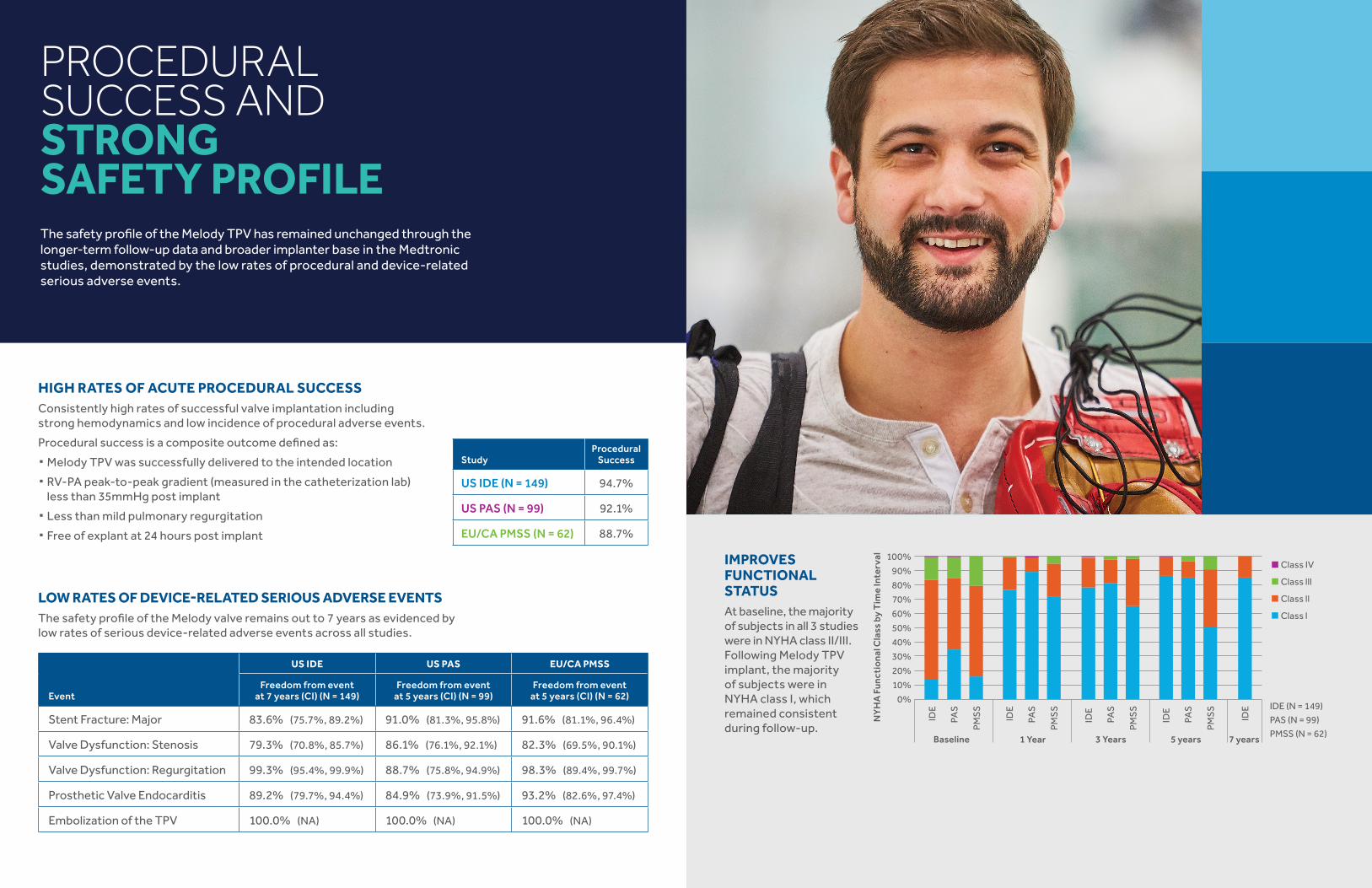
StudyProcedural
Success
US IDE (N = 149) 94.7%
US PAS (N = 99) 92.1%
EU/CA PMSS (N = 62) 88.7%
US IDE US PAS EU/CA PMSS
EventFreedom from event
at 7 years (CI) (N = 149)Freedom from event
at 5 years (CI) (N = 99)Freedom from event
at 5 years (CI) (N = 62)
Stent Fracture: Major 83.6% (75.7%, 89.2%) 91.0% (81.3%, 95.8%) 91.6% (81.1%, 96.4%)
Valve Dysfunction: Stenosis 79.3% (70.8%, 85.7%) 86.1% (76.1%, 92.1%) 82.3% (69.5%, 90.1%)
Valve Dysfunction: Regurgitation 99.3% (95.4%, 99.9%) 88.7% (75.8%, 94.9%) 98.3% (89.4%, 99.7%)
Prosthetic Valve Endocarditis 89.2% (79.7%, 94.4%) 84.9% (73.9%, 91.5%) 93.2% (82.6%, 97.4%)
Embolization of the TPV 100.0% (NA) 100.0% (NA) 100.0% (NA)
LOW RATES OF DEVICE-RELATED SERIOUS ADVERSE EVENTSThe safety profile of the Melody valve remains out to 7 years as evidenced by low rates of serious device-related adverse events across all studies.
HIGH RATES OF ACUTE PROCEDURAL SUCCESS Consistently high rates of successful valve implantation including strong hemodynamics and low incidence of procedural adverse events.
Procedural success is a composite outcome defined as:
· Melody TPV was successfully delivered to the intended location
· RV-PA peak-to-peak gradient (measured in the catheterization lab) less than 35mmHg post implant
· Less than mild pulmonary regurgitation
· Free of explant at 24 hours post implant
PROCEDURAL SUCCESS AND STRONG SAFETY PROFILEThe safety profile of the Melody TPV has remained unchanged through the longer-term follow-up data and broader implanter base in the Medtronic studies, demonstrated by the low rates of procedural and device-related serious adverse events.
100%
90%
80%
70%
60%
50%
40%
30%
20%
10%
0%
Baseline
NY
HA
Fun
ctio
nal C
lass
by
Tim
e In
terv
al
1 Year 3 Years 5 years 7 years
IDE
PAS
PMS
S
IDE
PAS
PMS
S
IDE
PAS
PMS
S
IDE
IDE
PAS
PMS
S
Class IV
Class lll
Class ll
Class l
IDE (N = 149)
PAS (N = 99)
PMSS (N = 62)
IMPROVES FUNCTIONAL STATUSAt baseline, the majority of subjects in all 3 studies were in NYHA class II/III. Following Melody TPV implant, the majority of subjects were in NYHA class I, which remained consistent during follow-up.

APPROVED FOR USE IN DYSFUNCTIONAL SURGICAL BIOPROSTHETIC PULMONARY VALVES
Data pooled from two U.S. prospective studies that included both failed conduits and BPVs and one retrospective study assessing Melody in dysfunctional BPVs only, demonstrated safety and effectiveness in restoring pulmonary valve function without open heart surgery.
The following outcomes demonstrate the safety and effectiveness of the Melody Transcatheter Pulmonary Valve (TPV) implanted in a bioprosthetic pulmonary valve (BPV) restoring pulmonary valve competency while delaying the need for surgical intervention.
Variable
Bioprosthesis (n = 125) RVOT Conduit (n = 225)
Number of Subjects in the Analysis
Endpoint Rate (95% CI)
Number of Subjects in the Analysis
Endpoint Rate (95% CI)
Procedural success 117 88.9% (82.9%, 93.3%)
225 93.8% (90.4%, 96.2%)
Procedure-related serious AE at 1 year
125 4.0% (2.6%, 10.1%)
225 12.4% (12.0%, 20.0%)
Device-related serious AE at 1 year
125 2.4% (0.6%, 6.0%)
225 16.0% (16.7%, 25.6%)
The confidence intervals are exact (Clopper-Pearson) confidence intervals for the binomial proportion.
Variable
Bioprosthesis (n = 125) RVOT Conduit (n = 225)
Number of Subjects in the Analysis
1-year Freedom Rate (95% CI)
Number of Subjects in the Analysis
1-year Freedom Rate (95% CI)
TPV Dysfunction 125 97.4% (90.0%, 99.4%)
223 94.1% (90.1%, 96.6%)
Reoperation 125 100.0% (NA)
223 98.6% (95.9%, 99.6%)
Reintervention 125 100.0% (NA)
223 98.2% (95.2%, 99.3%)
All-cause Mortality 125 100.0% (NA)
223 99.6% (96.8%, 99.9%)
Major Stent Fracture 125 100.0% (NA)
223 97.7% (94.6%, 99.1%)
Endocarditis 125 100.0% (NA)
223 97.3% (94.0%, 98.8%)
The cumulative probability of event-free estimate is based on the Kaplan-Meier method.The 95% confidence interval is the loglog transformed 95% Confidence Interval (CI) using the Peto standard error.

Sizing Information
Delivery System Size Inner Balloon/Outer Balloon
Inner Balloon Maximum Applied
Pressure (RBP)
Outer Balloon Applied Pressure
(RBP)
Corresponding Valve Outside
Diameter (Balloon Inflated)
Deployed Length (After Balloon
Deflated)
atm kPa atm kPa mm mm
Size 18 mm (9 mm x 3.5 cm/18 mm x 4 cm)
5 506 4 405 20.1 26
Size 20 mm (10 mm x 3.5 cm/20 mm x 4 cm)
5 506 4 405 22.4 24
Size 22 mm (11 mm x 3.5 cm/22 mm x 4 cm)
4.5 456 3 304 24.1 21
BJV = Bovine Jugular Vein | RBP = Rated Burst Pressure = Maximum Applied Pressure | atm = atmosphere | kPa = kilopascal
DEPLOYMENT SPECIFICATIONS
Melody TPV 20 (PB1016) is not designed to be dilated greater than 20 mm. Choose delivery system and valve size based on prepared conduit or surgical valve inside diameter and intended final implant size. Valve performance for both sizes is comparable, per bench testing data.1
1 Medtronic bench testing data on file.
Melody Transcatheter Pulmonary Valve
Product Order Number
Description A bovine jugular vein (BJV) valve sutured within a platinum iridium frame
PB1016 · Melody TPV 20· 16 mm BJV valve· Acceptable deployment: up to 20 mm
PB1018 · Melody TPV 22· 18 mm BJV valve· Acceptable deployment: up to 22 mm
Torque Wrench
Product Order Number
Description
01-0055Reusable jar opener
PRODUCT ORDERING INFORMATION
Note: To facilitate manufacturing (sewing of the tissue onto the TPV frame), the initial out-of-the-jar lengths of the two valves will differ slightly (30 mm length for PB1016 and 28 mm length for PB1018). Once crimped on the delivery system, the length of both TPV sizes will be the same and will remain as such during deployment to any size.
Ensemble and Ensemble ll Transcatheter Delivery System
Ensemble Product Order Number
Ensemble II Product Order Number Balloon Size French Size Overall Length
NU1018 ENS1018 18 mm 22 100 cm
NU1020 ENS1020 20 mm 22 100 cm
NU1022 ENS1022 22 mm 22 100 cm
· Balloon-in-balloon catheter delivery system with a retractable polytetrafluoroethylene (PTFE) sheath covering.· Nylon inner and outer balloons available in three sizes: 18 mm, 20 mm, and 22 mm. At inflation, the inner balloon
is half the diameter of the outer balloon.· Sheath with side port for flushing the system and a hemostatic sleeve to minimize bleeding at the insertion site.

©2016-2017 Medtronic. All rights reserved. Medtronic, Medtronic logo and Further, Together are trademarks of Medtronic. All other brands are trademarks of a Medtronic company.
710 Medtronic Parkway Minneapolis, MN 55432-5604 USA Tel: (763) 514-4000 Fax: (763) 514-4879 Toll-free: (800) 328-2518
LifeLine CardioVascular Technical Support Tel: (877) 526-7890 Tel: (763) 526-7890 Fax: (763) 526-7888 [email protected]
medtronic.comUC201703014a EN 11/2017
Melody™ Transcatheter Pulmonary Valve | Ensemble™ II Transcatheter Valve Delivery System
Important Labeling Information for the United StatesIndications: The Melody TPV is indicated for use in the management of pediatric and adult patients who have a clinical indication for intervention on a dysfunctional right ventricular outflow tract (RVOT) conduit or surgical bioprosthetic pulmonary valve that has ≥ moderate regurgitation, and/or a mean RVOT gradient ≥ 35 mm Hg.
Contraindications: None known.
Warnings/Precautions/Side Effects
· DO NOT implant in the aortic or mitral position. Pre-clinical bench testing of the Melody valve suggests that valve function and durability will be extremely limited when used in these locations.
· DO NOT use if patient’s anatomy precludes introduction of the valve, if the venous anatomy cannot accommodate a 22 Fr size introducer, or if there is significant obstruction of the central veins.
· DO NOT use if there are clinical or biological signs of infection including active endocarditis. Standard medical and surgical care should be strongly considered in these circumstances.
· Assessment of the coronary artery anatomy for the risk of coronary artery compression should be performed in all patients prior to deployment of the TPV.
· To minimize the risk of conduit rupture, do not use a balloon with a diameter greater than 110% of the nominal diameter (original implant size) of the conduit for pre-dilation of the intended site of deployment, or for deployment of the TPV.
· The potential for stent fracture should be considered in all patients who undergo TPV placement. Radiographic assessment of the stent with chest radiography or fluoroscopy should be included in the routine postoperative evaluation of patients who receive a TPV.
· If a stent fracture is detected, continued monitoring of the stent should be performed in conjunction with clinically appropriate hemodynamic assessment. In patients with stent fracture and significant associated RVOT obstruction or regurgitation, reintervention should be considered in accordance with usual clinical practice.
Potential procedural complications that may result from implantation of the Melody device include the following: rupture of the RVOT conduit, compression of a coronary artery, perforation of a major blood vessel, embolization or migration of the device, perforation of a heart chamber, arrhythmias, allergic reaction to contrast media, cerebrovascular events (TIA, CVA), infection/sepsis, fever, hematoma, radiation-induced erythema, blistering, or peeling of skin, pain, swelling, or bruising at the catheterization site.
Potential device-related adverse events that may occur following device implantation include the following: stent fracture,* stent fracture resulting in recurrent obstruction, endocarditis, embolization or migration of the device, valvular dysfunction (stenosis or regurgitation), paravalvular leak, valvular thrombosis, pulmonary thromboembolism, hemolysis.
* The term “stent fracture” refers to the fracturing of the Melody TPV. However, in subjects with multiple stents in the RVOT it is difficult to definitively attribute stent fractures to the Melody frame versus another stent.
For additional information, please refer to the Instructions for Use provided with the product or available on http://manuals.medtronic.com.
CAUTION: Federal law (USA) restricts this device to sale by or on the order of a physician.
Important Labeling Information for Geographies Outside of the United States
Indications: The Melody™ TPV is indicated for use in patients with the following clinical conditions:
· Patients with regurgitant prosthetic right ventricular outflow tract (RVOT) conduits or bioprostheses with a clinical indication for invasive or surgical intervention, OR
· Patients with stenotic prosthetic RVOT conduits or bioprostheses where the risk of worsening regurgitation is a relative contraindication to balloon dilatation or stenting
Contraindications
· Venous anatomy unable to accommodate a 22 Fr size introducer sheath
· Implantation of the TPV in the left heart
· RVOT unfavorable for good stent anchorage
· Severe RVOT obstruction, which cannot be dilated by balloon
· Obstruction of the central veins
· Clinical or biological signs of infection
· Active endocarditis
· Known allergy to aspirin or heparin
· Pregnancy
Potential Complications/Adverse Events: Potential procedural complications that may result from implantation of the Melody device include the following: rupture of the RVOT conduit, compression of a coronary artery, perforation of a major blood vessel, embolization or migration of the device, perforation of a heart chamber, arrhythmias, allergic reaction to contrast media, cerebrovascular events (TIA, CVA), infection/sepsis, fever, hematoma, radiation-induced erythema, pain, swelling or bruising at the catheterization site.
Potential device-related adverse events that may occur following device implantation
include the following: stent fracture,* stent fracture resulting in recurrent obstruction, endocarditis, embolization or migration of the device, valvular dysfunction (stenosis or regurgitation), paravalvular leak, valvular thrombosis, pulmonary thromboembolism, hemolysis.
* The term “stent fracture” refers to the fracturing of the Melody TPV. However, in subjects with multiple stents in the RVOT it is difficult to definitively attribute stent fractures to the Melody frame versus another stent.
For additional information, please refer to the Instructions for Use provided with the product or available on http://manuals.medtronic.com.
The Melody Transcatheter Pulmonary Valve and Ensemble II Transcatheter Delivery System has received CE Mark approval and is available for distribution in Europe.
Magnetic Resonance Imaging (MRI) Safety InformationNonclinical testing and modeling has demonstrated that the Melody™ TPV is MR Conditional. A patient with this device can be safely scanned in an MR system meeting the following conditions:
· Static magnetic field of 1.5 T and 3 T
· Maximum spatial gradient magnetic field of 2500 gauss/cm (25 T/m)
· Maximum MR system reported, whole body averaged specific absorption rate (SAR) of 2.0 W/kg for 15 minutes of scanning (Normal Operating Mode)
Based on nonclinical testing and modeling, under the scan conditions defined above, the Melody™ TPV is expected to produce a maximum in vivo temperature rise of less than 2.1°C after 15 minutes of continuous scanning.
MR image quality may be compromised if the area of interest is in the same area, or relatively close to the position of the device. In nonclinical testing, the image artifact caused by the device extends approximately 3 mm from the Melody™ TPV when imaged with a spin echo pulse sequence and 6 mm when imaged with a gradient echo pulse sequence and a 3 T MRI System. The lumen of the device was obscured.
Scanning under the conditions defined above may be performed after implantation.
The presence of other implants or medical circumstances of the patient may require lower limits on some or all of the above parameters.
MR
MR Conditional