A Late Jurassic salamander (Amphibia: Caudata) from the ... · Zoological Journal of the Linnean...
18
Zoological Journal of the Linnean Society , 2005, 143 , 599–616. With 8 figures © 2005 The Linnean Society of London, Zoological Journal of the Linnean Society, 2005, 143 , 599–616 599 Blackwell Science, Ltd Oxford, UK ZOJZoological Journal of the Linnean Society 0024-4082The Lin- nean Society of London, 2005? 2005 143 ? 599616 Original Article JURASSIC SALAMANDER FROM NORTH AMERICAS. E. EVANS ET AL. *Corresponding author. E-mail: [email protected] A Late Jurassic salamander (Amphibia: Caudata) from the Morrison Formation of North America S. E. EVANS FLS 1 *, C. LALLY 1 , D. C. CHURE 2 , A. ELDER 2 and J. A. MAISANO 3 1 Department of Anatomy and Developmental Biology, University College London, Gower Street, London, WC1E 6BT, UK 2 United States Parks Service, Dinosaur National Monument, Jensen, UT 84035, USA 3 Department of Geological Sciences, University of Texas at Austin, Austin, TX 78712–1100, USA Received February 2004; accepted for publication November 2004 Despite some remarkable recent discoveries, the Mesozoic fossil record of salamanders remains limited, particularly for the Jurassic. Here we describe the first articulated salamander skeleton from the Jurassic of Euramerica, recov- ered from Upper Jurassic deposits of the Morrison Formation, Dinosaur National Monument, USA. The specimen was studied using both conventional methods and high-resolution computed tomography. It shows a combination of primitive and derived character states that distinguish it from all known Mesozoic salamanders and which permit the erection of a new genus and species, Iridotriton hechti . The derived states (including the presence of spinal nerve foramina in the tail) suggest a position on the stem of the Salamandroidea. Together with microvertebrate material from Britain, Portugal, and North America, this specimen confirms the presence of both stem- and crown- group salamanders in Euramerica from the Middle Jurassic (Bathonian) onwards, paralleling their evolution in Cen- tral and eastern Asia. This, in turn, provides qualified support for the current vicariance model of salamander evolution whereby basal caudates on an undivided Laurasian plate became separated into two populations by the incursion of the Turgai Sea in the Middle Jurassic, yielding Cryptobranchoidea in Asia and Salamandroidea in Euramerica. © 2005 The Linnean Society of London , Zoological Journal of the Linnean Society , 2005, 143 , 599 - 616. ADDITIONAL KEYWORDS: biogeography – computed tomography – evolution – Lissamphibia – phylogeny – skeleton – Urodela. INTRODUCTION Salamanders and their immediate relatives (Caudata) are traditionally grouped with other living amphibi- ans (frogs, caecilians) in the Lissamphibia (e.g. Milner, 1988; McGowan & Evans, 1995; Ruta, Coates & Quicke, 2003) although the monophyly of the latter clade has been questioned (e.g. Carroll & Holmes, 1980; Laurin & Reisz, 1997). Patterns of relationship within and between component clades are also strongly debated, with morphology generally placing frogs and caudates as sister taxa (e.g. Milner, 1988; Laurin & Reisz, 1997; Ruta et al ., 2003; but see Bolt, 1991), whereas molecular analysis supports a rela- tionship between caudates and caecilians (e.g. Larson, 1991; Hedges & Maxson, 1993; Hay et al ., 1995; Feller & Hedges, 1998). Clearly, more information is needed about the early fossil record of each clade. Stem-frogs are known from the Early Triassic of Madagascar (e.g. Rage & Ro ek, 1989) and Poland (Evans & Borsuk- Bia ynicka, 1998), but caecilians are not recorded before the Early Jurassic of North America (Jenkins & Walsh, 1993), and caudates are first described from the Middle Jurassic of Central Asia (Nessov, 1981, 1988; Nessov et al ., 1996), Britain (Evans, Milner & Mussett, 1988; Evans, 1992; Evans & Milner, 1994; Evans & Waldman, 1996), and, putatively, China (Gao & Shubin, 2003; but see Discussion). Triassurus (Ivachnenko, 1978) is a possible salamander from the Upper Triassic of Uzbekistan, but the specimen is apparently larval and of uncertain systematic position (Estes, 1981). Caudata are essentially a Laurasian group with limited extensions into Gondwana. Milner (1983) argued that a pan-Laurasian ancestral group of stem- caudates became subdivided when an incursion of the c ˇ l ¢
Transcript of A Late Jurassic salamander (Amphibia: Caudata) from the ... · Zoological Journal of the Linnean...

Zoological Journal of the Linnean Society
, 2005,
143
, 599–616. With 8 figures
© 2005 The Linnean Society of London,
Zoological Journal of the Linnean Society,
2005,
143
, 599–616
599
Blackwell Science, Ltd
Oxford, UK
ZOJZoological Journal of the Linnean Society
0024-4082The Lin-nean Society of London, 2005? 2005
143
?
599616Original Article
JURASSIC SALAMANDER FROM NORTH AMERICAS. E. EVANS ET AL.
*Corresponding author. E-mail: [email protected]
A Late Jurassic salamander (Amphibia: Caudata) from the Morrison Formation of North America
S. E. EVANS
FLS
1
*, C. LALLY
1
, D. C. CHURE
2
, A. ELDER
2
and J. A. MAISANO
3
1
Department of Anatomy and Developmental Biology, University College London, Gower Street, London, WC1E 6BT, UK
2
United States Parks Service, Dinosaur National Monument, Jensen, UT 84035, USA
3
Department of Geological Sciences, University of Texas at Austin, Austin, TX 78712–1100, USA
Received February 2004; accepted for publication November 2004
Despite some remarkable recent discoveries, the Mesozoic fossil record of salamanders remains limited, particularlyfor the Jurassic. Here we describe the first articulated salamander skeleton from the Jurassic of Euramerica, recov-ered from Upper Jurassic deposits of the Morrison Formation, Dinosaur National Monument, USA. The specimenwas studied using both conventional methods and high-resolution computed tomography. It shows a combination ofprimitive and derived character states that distinguish it from all known Mesozoic salamanders and which permitthe erection of a new genus and species,
Iridotriton hechti
. The derived states (including the presence of spinalnerve foramina in the tail) suggest a position on the stem of the Salamandroidea. Together with microvertebratematerial from Britain, Portugal, and North America, this specimen confirms the presence of both stem- and crown-group salamanders in Euramerica from the Middle Jurassic (Bathonian) onwards, paralleling their evolution in Cen-tral and eastern Asia. This, in turn, provides qualified support for the current vicariance model of salamanderevolution whereby basal caudates on an undivided Laurasian plate became separated into two populations by theincursion of the Turgai Sea in the Middle Jurassic, yielding Cryptobranchoidea in Asia and Salamandroidea inEuramerica. © 2005 The Linnean Society of London
, Zoological Journal of the Linnean Society
, 2005,
143
, 599
-
616.
ADDITIONAL KEYWORDS: biogeography – computed tomography – evolution – Lissamphibia – phylogeny –
skeleton – Urodela.
INTRODUCTION
Salamanders and their immediate relatives (Caudata)are traditionally grouped with other living amphibi-ans (frogs, caecilians) in the Lissamphibia (e.g. Milner,1988; McGowan & Evans, 1995; Ruta, Coates &Quicke, 2003) although the monophyly of the latterclade has been questioned (e.g. Carroll & Holmes,1980; Laurin & Reisz, 1997). Patterns of relationshipwithin and between component clades are alsostrongly debated, with morphology generally placingfrogs and caudates as sister taxa (e.g. Milner, 1988;Laurin & Reisz, 1997; Ruta
et al
., 2003; but see Bolt,1991), whereas molecular analysis supports a rela-tionship between caudates and caecilians (e.g. Larson,1991; Hedges & Maxson, 1993; Hay
et al
., 1995; Feller& Hedges, 1998). Clearly, more information is needed
about the early fossil record of each clade. Stem-frogsare known from the Early Triassic of Madagascar (e.g.Rage & Ro ek, 1989) and Poland (Evans & Borsuk-Bia ynicka, 1998), but caecilians are not recordedbefore the Early Jurassic of North America (Jenkins &Walsh, 1993), and caudates are first described fromthe Middle Jurassic of Central Asia (Nessov, 1981,1988; Nessov
et al
., 1996), Britain (Evans, Milner &Mussett, 1988; Evans, 1992; Evans & Milner, 1994;Evans & Waldman, 1996), and, putatively, China (Gao& Shubin, 2003; but see Discussion).
Triassurus
(Ivachnenko, 1978) is a possible salamander from theUpper Triassic of Uzbekistan, but the specimen isapparently larval and of uncertain systematic position(Estes, 1981).
Caudata are essentially a Laurasian group withlimited extensions into Gondwana. Milner (1983)argued that a pan-Laurasian ancestral group of stem-caudates became subdivided when an incursion of the
cl¢

600
S. E. EVANS
ET AL.
© 2005 The Linnean Society of London,
Zoological Journal of the Linnean Society,
2005,
143
, 599–616
Turgai Sea separated Asia and Euramerica in theMiddle Jurassic. Each population then gave rise to oneof the two major caudate clades, Cryptobranchoidea(in Asia) and Salamandroidea (in Euramerica).
To date, the first attributable salamandroids are ofEarly Cretaceous age:
Apricosiren
(Berriasian,England; Evans & McGowan 2002); indet. taxa(Berriasian, England; Ensom, Evans & Milner,1991; Evans & McGowan, 2002);
Hylaeobatrachus
(Hauterivian, Belgium; Dollo 1884); indet. taxa(Hauterivian-Valanginian, England; Milner & Evans1998);
Valdotriton
(Barremian, Spain; Evans &Milner, 1996);
Galverpeton
(Barremian-Aptian, Spain;Estes & Sanchíz, 1982), an undescribed perennibran-chiate form (Barremian, Spain; Evans
et al
., 1995),and
Prosiren
(Aptian/Albian, USA; Estes, 1969. Note,however, that Estes incorrectly combined the realsalamander vertebrae of
Prosiren
with the jaws of thenoncaudate
Albanerpeton
; Fox & Naylor, 1982). Ofthese Euramerican taxa, the most completely knownis
Valdotriton
, a stem-salamandroid (Evans & Milner,1996). Fragmentary remains from the Middle Jurassicof Britain (‘Salamander B’; Evans & Milner, 1994) andthe Late Jurassic of North America (Evans & Milner,1993) have suggested that derived salamanders werepresent in Euramerica before the Cretaceous, but dis-cussion has been constrained by the limitations of thematerial (isolated vertebrae, pieces of jaw).
In recent years, the Rainbow Park microsite atDinosaur National Monument, Utah, has yielded twoblocks with articulated salamander skeletons, bothfrom the Upper Jurassic Brushy Basin Member of theMorrison Formation. The two blocks carry specimensrepresenting two distinct taxa. One taxon (repre-sented by several entwined partial skeletons) is cur-rently being studied by Bruce Naylor and JamesGardner at the Royal Tyrrell Museum, Drumheller,Canada. The second taxon is described here.
Institutional acronym used in this paper: DINO, USNational Parks Service, Dinosaur National Monument.
GEOLOGY AND MATERIALS
The Brushy Basin Member of the Morrison Formationat Dinosaur National Park, Utah, is best known for itsmacrovertebrate (dinosaur) remains but has begun toyield a valuable assemblage of small vertebratesincluding frogs (Henrici, 1998), lizards (Evans &Chure, 1998a, b), sphenodontians (Fraser & Wu,1998), mammals (Engelmann & Callison, 1998), andsalamanders. Fortunately the Morrison Formation inthis region contains volcanic ash layers that haveyielded isotopic ages (
40
Ar/
39
Ar). These consistentlyplace the Brushy Basin Member between 150.3
±
0.3 Myr (base) and 148.1
±
0.5 Myr (top) ( Kowallis
et al
., 1998). It is thus Kimmeridgian or early Titho-
nian in age (according to the timescale of Gradstein
et al
., 1995).Although fragmentary salamander material (a
femur,
Comonecturoides marshi
Hecht & Estes, 1960;partial vertebrae and other elements; Estes, 1981;Evans & Milner, 1993) has already been describedfrom the Morrison Formation (Quarry 9, Como Bluff),the specimens are difficult to classify. The new speci-mens from Dinosaur National Monument currentlyrepresent the earliest known articulated materialfrom Euramerica. The specimen described here, DINO16453 (Fig. 1), is in two parts: 16453a (Fig. 2) carriesthe bulk of the skeleton, preserved in dorsal view,whereas 16453b (Fig. 3) is a partial counterpart bear-ing vertebrae, girdle and hind limb elements in ven-tral view.
METHODOLOGY
The specimen was prepared mechanically as far aspracticable, but cracks in the undersurface as well asthe compression and superimposition of many bones(e.g. in the skull, forelimbs) preclude full exposure. Tocompensate, the main block (DINO 16453a) wasscanned at the High-Resolution X-ray ComputedTomography Facility at The University of Texas (Aus-tin), Geological Sciences, and then digitally recon-structed (Figs 4, 5, http://digimorph.org/specimens/Iridotriton_hechti). The resulting data set consists of378 slices taken along the long axis of the specimen,each slice 62
m
m thick, with an interslice spacing of62
m
m and an in-plane resolution of 38
m
m per pixel.Visualizations were generated using VGStudioMax1.1 (Volume Graphics, Heidelberg). This imaging tech-nique has provided new information, particularly withrespect to the limb skeleton, pectoral girdle, and theunderside of the skull. However, not all details couldbe fully resolved because DINO 16453a consists of asmall, delicate skeleton in a relatively large block ofmatrix that could not be further trimmed because ofdeep internal cracks (as revealed by the scan). Thepresence of the broad matrix rim surrounding thespecimen limits the resolution of very small structures(e.g. delicate teeth) and of superimposed or adjacentbones where there is little or no matrix between them.
SYSTEMATIC PALAEONTOLOGY
L
ISSAMPHIBIA
H
AECKEL
, 1866 C
AUDATA
S
COPOLI
, 1777 U
RODELA
D
UMÉRIL
, 1806 N
EOCAUDATA
C
ANNATELLA
& H
ILLIS
, 1993 F
AMILY
I
NDET
.
I
RIDOTRITON
GEN
.
NOV
.
Generic diagnosis:
as for
Iridotriton hechti
, the onlyspecies.

JURASSIC SALAMANDER FROM NORTH AMERICA
601
© 2005 The Linnean Society of London,
Zoological Journal of the Linnean Society,
2005,
143
, 599–616
Figure 1.
Iridotriton hechti
gen.
et
sp. nov.
, holotype. Main figure (left) DINO 16453a; adjoining figure (right), DINO16453b. Scale bar
=
1 mm.
Figure 2.
Iridotriton hechti
gen.
et
sp. nov.
, DINO 16453a.
Abbreviations:
At, atlas; cr.V, crista ventralis humeri; d.hd,distal head of humerus; L.H, left humerus; L.Ra, left radius; L.ScC, left scapulocoracoid; Ph
+
Mc, phalanges and metac-arpals; Ps, presacral vertebra; Rb, rib; Rb.b, rib-bearer; R.D, right dentary; R.H, right humerus; R.Oc, right otic capsule;R.ScC, right scapulocoracoid; Ul-l, fused ulnare and intermedium; Un, ungual phalanx. Scale bar
=
1 mm.

602
S. E. EVANS
ET AL.
© 2005 The Linnean Society of London,
Zoological Journal of the Linnean Society,
2005,
143
, 599–616
Type species: Iridotriton hechti
gen. et sp. nov.
Derivation of generic name:
from the Greek
Iris
,meaning rainbow, an allusion to the Rainbow Parkmicrosite, and
Triton
, a newt.
I
RIDOTRITON
HECHTI
SP
.
NOV
.
Derivation of specific name:
in honour of the late MaxHecht, one of the first authors to describe salamandermaterial from the Morrison Formation.
Holotype:
DINO 16453a, b, parts of a single skeletonmissing only small skull bones, digits and the distaltail.
Locality:
Dinosaur National Monument, RainbowPark microsite (Dinosaur National Monument no. 96),Utah, USA (detailed locality data is held in the recordsat the Monument).
Horizon:
Brushy Basin Member of the Morrison For-mation, Upper Jurassic (
c.
150–148 Myr; Kowallis
et al
., 1998: Kimmeridgian or early Tithonian).
Specific diagnosis:
a small (snout
-
sacrum length
c
.55 mm) fully metamorphosed salamander distin-guished by the following combination of characters:thin unsculptured skull bones; a fully open Meckelianfossa in the dentary; premaxilla with wide, short alaryprocess having angled lateral edge; prootic, opisthoticand exoccipital form a single unit, although suturallines separate the opisthotic from the other bones;stapes free; parasphenoid without internal carotidforamina, narrower anteriorly than posteriorly; anestimated 16 postatlantal presacral vertebrae; simpleectochordal vertebral centra, with small anteriorbasapophyses but no ventromedian keel; atlas shorterthan succeeding vertebrae; spinal nerve foramina inatlas and in tail vertebrae; co-ossified scapula andcoracoid, with narrow, waisted scapula and large,heavily ossified coracoid plate perforated by supraco-racoid foramen; strongly built forelimbs (relativelymassive humerus with deep crista ventralis humeriand expanded distal head); radius with expanded dis-tal head; well-ossified tarsus and carpus includingfusion of ulnare and intermedium; rib-bearers withconjoined heads throughout the column.
Remarks: Iridotriton
differs from the stem-caudates
Karaurus
,
Kokartus
and
Marmorerpeton
in lackingany trace of sculpture on the skull bones, and in hav-ing spinal nerve foramina in both the atlas and caudalvertebrae. It resembles the Chinese salamanders
Chunerpeton
(Gao & Shubin, 2003),
Jeholotriton
(Wang, 2000a),
Laccotriton
(Gao & Shubin, 2001),
Liaoxitriton
(Dong & Wang, 1998; Wang, 2004) and
Sinerpeton
(Gao & Shubin, 2001) in having conjoinedsurfaces on the rib-bearers and retaining a separate
angular in the jaw, but differs from
Sinerpeton
and
Laccotriton
in lacking a separate coronoid, and fromall five Chinese taxa in having a much more massivehumerus.
Iridotriton
further differs from
Chunerpeton
in prefrontal shape (shorter and squarer in
Chunerpe-ton
), squamosal shape (waisted below dorsal head in
Chunerpeton), and the ossification of the tarsals andcarpals (unossified in Chunerpeton). It differs from theCretaceous Valdotriton (Evans & Milner, 1996) in hav-ing a broad rather than spike-like alary process of thepremaxilla, a dentary with an open rather than ante-riorly closed Meckelian groove, and conjoined ratherthan double-headed rib-bearers; and differs from Val-dotriton, Prosiren, and Apricosiren (S. E. Evans, pers.observ.) in lacking ventromedian keels on the presac-ral centra. The Cretaceous Hylaeobatrachus is peren-nibranchiate (Estes, 1981) whereas Iridotriton ismetamorphosed. The Cretaceous Spanish Galverpeton(Estes & Sanchíz, 1982) is based on a single trunk ver-tebra distinguished by the presence of a spinal nerveforamen and strong lateral crests, both of which areabsent in the Morrison form. Ramonellus from theEarly Cretaceous of Israel (Nevo, 1964; Nevo & Estes,1969) differs in being very long-bodied (at least 34 pre-sacrals) and in having a long retroarticular process onthe lower jaw. Generic distinction for Iridotriton istherefore defensible.
Comonecturoides marshi Hecht & Estes, 1960, wasdescribed from the Morrison Formation at Quarry 9,Como Bluff, on the basis of a single isolated femur and,though clearly caudate, is a nomen dubium since it isrestricted to an indeterminate type (Evans & Milner,1993). The holotype femur is slightly smaller thanthat of Iridotriton, has a less projecting trochanter,and a less compressed proximal head.
DescriptionThe specimen (DINO 16453a, b) is preserved in artic-ulation and includes much of the skull, the completepresacral axial skeleton, the sacrum, a small set ofpostsacrals, and parts of the girdles and the limbs. Theskull, forelimbs, and anterior presacral series (DINO16453a) are preserved in dorsal view, but the posteriorpresacral region and left hind limb are on a smallblock (DINO 16453b) detached during collection andprepared in ventral view. The specimen is generallywell preserved in three dimensions, with the vertebraefully articulated, but there has been some disarticula-tion of the limbs and girdles and of parts from theright side of the skull roof and jaws. It is not possibleto get an accurate measurement of the snout-sacrumlength, but comparison of humeral and femorallengths with those of similarly proportioned modernanalogues suggests a snout-sacrum length of 50–60 mm, and a total length (with tail) of between 80

JURASSIC SALAMANDER FROM NORTH AMERICA 603
© 2005 The Linnean Society of London, Zoological Journal of the Linnean Society, 2005, 143, 599–616
and 100 mm overall. Despite this small size, the spec-imen appears to represent a metamorphosed individ-ual (dermal roofing bones ossified and in position;squamosal full size, all bones of lower jaw ossified, dor-sal process of maxilla ossified, vomer fully formed, oticcapsule complete and stapes ossified: Rose, 2003).
Figures 1–3 show the specimen as preserved on theblocks, but the description that follows also relies onthe digital reconstructions from computed tomogra-phy (Figs 4, 5).
The skullThe posterior part of the skull aligns with the verte-bral column, but the more anterior half, including thejaws, has rotated to the right (Fig. 6). Despite this, thebones of the left side are roughly in situ (but tele-scoped) whereas those of the right have been displacedout to the side of the specimen. A majority of the skullelements can be identified but some of the small bones(lacrimals and septomaxillae, if present) cannot berecognized.
Premaxilla: both bones are preserved, the left in situand the other displaced and rotated to the right of thespecimen. They show an elongated maxillary processthat either abuts or underlaps the maxilla. The alaryprocess (processus dorsalis) of the left premaxilla isdamaged but the right is complete. It is short, broad,and asymmetric, with a strong lateral angle. A pre-maxillary tooth count is not possible.
Maxilla: the left bone is also in situ and essentiallycomplete except for the medial edge of its dorsal(facial) process. The bone has an elongate premaxil-lary process, a short dorsal process, and a slender pos-terior process. The right bone is adjacent to the rightpremaxilla but has been rotated so that its lingual sur-face is exposed. The teeth are damaged and no toothcount is possible.
Nasal: a probable right nasal lies adjacent to the dor-sal process of the right premaxilla. It appears to bedivided into two parts by a deep cleft (though thiscould be an artefact of breakage). Division wouldimply paired nasal anlagen that are in contact poste-riorly (as in the Cretaceous Valdotriton; Evans & Mil-ner, 1996). There is a bone of similar size behind theleft premaxilla, but the details are obscured.
Prefrontal: a single slender element lies adjacent tothe dorsal process of the left maxilla. The identifica-tion of this bone as a prefrontal relies on its position toone side of the midline and its posteriorly taperingshape. It is closely similar to the same element in theextant Cryptobranchus and the hynobiid Onychodac-tylus (S. E. Evans, pers. observ.), and to the recon-structed shape of the Cretaceous Valdotriton (Evans &Milner, 1996). The bone is damaged in the midsectionbut the intervening impression suggests that this is asingle bone and there is no trace of any groove or fora-men for the lacrimal duct. No lacrimal has been rec-ognized in Iridotriton, but given the telescoping ofindividual elements, and the various small unidenti-fied elements within the skull mass, we cannot deter-mine whether a lacrimal was present or absent.
Frontals: the left bone is represented by a thin platedeep to the left prefrontal and overlying the left pari-etal. It is long and relatively narrow, but shows no
Figure 3. Iridotriton hechti gen. et sp. nov., DINO16453b. Abbreviations: CaS.V, caudosacral vertebra; Ca.V,caudal vertebra; L.Fe, left femur; L.Fi, left fibula; L.Il, leftilium; Pe, elements of pes; Ps, presacral vertebra; Rb.b, rib-bearer; Sa.rb, sacral rib; Sa.V, sacral vertebra; sp.f, spinalnerve foramen; Ta, tarsal; L.Ti, left tibia; tr, trochanter.Scale bar = 1 mm.

604 S. E. EVANS ET AL.
© 2005 The Linnean Society of London, Zoological Journal of the Linnean Society, 2005, 143, 599–616
Figure 4. Iridotriton hechti gen. et sp. nov., DINO 16453a, digital reconstruction of dorsal surface based on high-resolution computed tomography. Abbreviations: At, atlas; C, C1-2, carpals; L.An, left angular; L.D, left dentary; L.H, lefthumerus; L.Mx, left maxilla; L.Oc, left otic capsule; L.Pmx, left premaxilla; L.Pra, left prearticular; L.Prf, left prefrontal;L.Ra, left radius; L.ScC, left scapulocoracoid; L.Sq, left squamosal; Ph, phalanx; Ps, parasphenoid; Q, quadrate; R.D, rightdentary; R.H, right humerus; R.Mx, right maxilla; R.Oc, right otic capsule (small arrow points to foramen for endolym-phatic duct); R.P, right parietal; R.Pmx, right premaxilla; R.Pt, right pterygoid; R.Ra, right radius; R.ScC, right scapulo-coracoid; R.Sq, right squamosal; Ul.I, fused ulnare and intermedium. Scale bar = 1 mm.

JURASSIC SALAMANDER FROM NORTH AMERICA 605
© 2005 The Linnean Society of London, Zoological Journal of the Linnean Society, 2005, 143, 599–616
trace of sculpture and provides no detail of articularsurfaces for adjacent bones. The right bone has notbeen identified with confidence. It may be a partiallyobscured flat bone, the edge of which is exposedbetween the left margin of the parasphenoid and theleft prefrontal. The frontals appear to have beenslightly narrower than the parietals, but of similarlength.
Parietals: the left parietal partially underlies the leftfrontal and prefrontal. The right has been carried outwith other bones of this side and lies between theright premaxilla, maxilla, and dentary, extendingunder the last of these bones for a short distance. Thebone is rectangular with at least one (anterior or pos-terior) straight margin. It is certainly not acutelytapered at either end and therefore probably did notextend far forward under the frontal. Neither parietalis sculptured.
Squamosal: the left bone is in situ, overlapping thebraincase medially and the pterygoid distolaterally. Itis roughly triangular, broad dorsally and tapers at itsventral tip. The posterior margin is curved. As pre-served, the squamosal contacts only the braincase andnot the parietal. The right element has not been iden-tified with certainly, but it may be represented by acurved flange underlying the right pterygoid (?R.Sq,Fig. 6).
Vomer: this lies beneath the left jaw symphysis (visi-ble only on the digital reconstructions; V in Fig. 5). Ifit is the left element, then it is in situ relative to theleft dentary, but since the skeleton has partially dis-articulated, the two bones have rotated to the left ofthe frame. The anterior margin [now directed to theleft of Fig. 5 ( = right side of body)] has two short sur-faces meeting at a slight angle; presumably these metthe premaxilla and maxilla. The lateral margin (nowat the top) is embayed by the choana and then flaresout lateral to it to form a postchoanal flange. Themedial margin (now at the bottom) is strongly obliquesuggesting the presence of an anterior palatinefontanelle.
Behind it, the posteromedial border (facing to theright of the figure) is relatively long and straight.Teeth are not visible, but this is probably an artefact ofthe resolution of the high resolution X-ray scanner,because dentary teeth are visible on the specimen butnot on the scans (CT slices and reconstructions). Analternative explanation would be that this is the rightbone, either turned 180 ∞ on its long axis so that thedorsal surface is exposed, or reversed so that the wideredge, now posterior, met the jaw margin, with the bonenarrowing posteriorly (a better match for that of pri-mitive living salamanders like cryptobranchids andhynobiids, S. E. Evans, pers. observ.). Conceivably, it
could be the left element, rotated both anteroposteri-orly and dorsoventrally. No other bone shows such aradical displacement, but the vomers could have beenseriously disrupted when the jaws were disarticulatedand rotated outwards.
Pterygoid: the left pterygoid is largely obscured by theoverlying squamosal in dorsal view, and by parts of thebraincase ventrally. A distinct blade, presumably theposterolateral pterygoid process, extends ventrallybeyond the squamosal and quadrate towards thelower jaw, whereas a more fragmentary process isdirected anteriorly. From the underside of the speci-men (digital reconstructions, Fig. 5), the bone appearsmore complex, and the medial surface was probablyconcave, but it is neither large nor strongly expanded.The right pterygoid may be represented by the irreg-ular bone mass adjacent to the right otic elements. Itis clearly bent in more than one plane, and curvesaround the edge of the mandible. Overlying it on theunderside is a small, unidentified bony plate that alsooverlaps the jaw.
Parasphenoid: this has a long parallel-sided anteriorrostrum that is overlapped ventrally by the vomer.Posteriorly, at the level of the braincase, the boneexpands slightly to overlap the otic capsules, althoughthere are no strong alae and no visible perforations forthe internal carotid arteries. Dorsally, the parasphe-noid forms the right boundary of the skull mass, andits dorsal surface is concave.
Quadrate: visible on the digital reconstructions as asmall dense mass of bone displaced to the left side ofthe skull.
Braincase: in salamanders, this has two principalcomponents: the sphenethmoid (= orbitosphenoid;Trueb, 1993) that underlies the frontal and attaches tothe dorsal margin of the parasphenoid rostrum, andthe otic capsule made up of the prootic, opisthotic, andexoccipital, or some combination of these three ele-ments (Trueb, 1993). The sphenethmoid seems to berepresented in Iridotriton by a long narrow elementwedged between the skull roof (left frontal) and theparasphenoid rostrum on the left side (Fig. 5). It isperforated by a small foramen (perhaps for a branch ofthe ophthalmic division of the trigeminal nerve; Fran-cis, 1934) and has a larger posterior notch for the opticnerve (these structures lie to the left and right of ‘Sp’,respectively, in Fig. 5, and are seen most clearly if thedigital reconstructions are rotated).
The otic capsule is preserved on both sides of theskull. Its components are fused into a relatively large,rounded structure that extended beyond the confinesof the parietal table. On the left side, the exoccipital isroughly in situ against the atlantal cotyle, although ithas rotated laterally so that the exoccipital condyle

606 S. E. EVANS ET AL.
© 2005 The Linnean Society of London, Zoological Journal of the Linnean Society, 2005, 143, 599–616
has disarticulated from the atlas. The otic capsule con-tinues forward as a single unit into the prootic, withthe squamosal overlapping the lateral and dorsolat-eral surfaces but not reaching the level of the parietal.
Seen in ventral view (Fig. 5), the otic capsulesurrounds a rather bulbous vestibular cavity. Thefenestra ovalis opens ventrolaterally and containsfragments of bone that probably pertain to the stapes.On the right side of the skull, the otic capsule is dis-placed and has rotated slightly. The opisthotic compo-nent is united with the exoccipital and prootic, but isdelimited by sutures (although these must be at leastpartially closed as there is no displacement). Dorsally,the opisthotic (right otic capsule, Fig. 4) exposes asmall distinct foramen (small arrow, Fig. 4) that opensfrom the inside of the vestibular cavity. This foramenis presumably for the endolymphatic duct (Francis,1934).
MandibleThe left mandible is in articulation with, and largelyobscured by, the maxilla and premaxilla of that side.The right mandible is displaced and exposed in lingualview to the side of the specimen.
The dentary has a narrow but relatively deep ter-minal symphysis and a long, but very narrow, Mecke-lian sulcus that is open throughout its length. Thealveolar margin is separated from the subdental ridgeby a deep groove, so that the tooth-bearing part of thejaw forms a rather shallow margin along the dorsaledge of the dentary. It bears a row of around 40 slen-der pedicellate teeth, but the tooth crowns are notclearly preserved in any position. On both sides, thedentary extends to the posterior end of the mandible,bracing the accessory bones from the labial side.
Between the rear of the dentary and the maxilla onthe left side there are two distinct anteroposteriorlydirected structures (Figs 4, 6). The more laterallyplaced of these is a narrow lamina with a thickenedmargin. It corresponds in structure and position to theangular in the living Cryptobranchus and hynobiids.The more medial structure has a dorsally expandedanterior coronoid process and a posterior edge thatcurves medially. It then continues ventrolaterally intoa flange-like blade that lies parallel to the posteriorpterygoid lamina. This is the prearticular-coronoid.On the right mandible, the angular is in situ at therear of the bone, whereas the slender anterior tip ofthe prearticular is visible within the posterior half ofthe Meckelian canal (Fig. 5).
Axial skeletonThe axial skeleton is preserved in two parts. In 16453a(Figs 2, 4, 5), the atlas is preserved in situ. Following
it are eight complete vertebrae, fragments of a ninth(Ps 10, Fig. 2), and then impressions and fragments ofa further four. Specimen 16453b (Fig. 3) preserveseight presacral vertebrae followed by a sacral, at leastthree caudosacrals, and one further isolated caudal. Itis difficult to be certain of the relationship of the twoblocks, but it seems likely that the first vertebrae pre-served on 16453b (Ps10, Fig. 3) is part of the last ver-tebra on block 16543a, with the vertebrae following on16453b responsible for the impressions on 16453a.Under this interpretation, there were a total of 17 pre-sacrals. There may have been more, but there cannothave been fewer.
Atlas: this is preserved in dorsal and left lateral viewsbut is otherwise obscured by the bones around it. Thusthe presence of the interglenoid tuberosity cannot beconfirmed, although, judging from the other featuresof the skeleton, it is likely to have been present in acaudate of this grade. The atlas is slightly shorterthan the vertebra following it, and had a low neuralarch with a midline crest but no spine, and a convexposterior margin. Anteriorly, the left cotyle remains inarticulation with the exoccipital condyle. In lateralview, however, crushing obscures the detail. There iscertainly at least a notch in the anterior margin of theatlas for the first spinal nerve (between the cotyle andthe anterodorsal margin of the arch, Fig. 6), butwhether it was a fully enclosed foramen or not isimpossible to judge.
Postatlantal presacral vertebrae: the first eight post-atlantal vertebrae have low neural arches with amiddorsal keel and short horizontal spines that weredirected posteriorly and were probably completed incartilage (judging by the pitted distal tips). Ventrally,the centrum is rounded with no midline keel andonly small anterior basapophyses. The centra formweak amphicoelous cylinders that probably devel-oped ectochordally. There is no evidence of spinalnerve foramina in trunk vertebrae. The zygapophysesare strong and horizontal, whereas the rib-bearersare long and directed posterolaterally. On each, thedorsal and ventral rib facets have coalesced to form asingle head, although there is a slight waisting of thesurface in more posterior vertebrae. The ribs them-selves are certainly single-headed and relatively long(equal or nearly equal to the length of the centrum).Most are very gracile, but the second and third post-atlantal vertebrae bear more robust ribs for thesupport of the pectoral girdle. The centra of the pos-terior presacrals (16453b, Fig. 3) are also simple andspool-like, with neither median keels nor prominentbasapophyses. At most, there is a slight bilateralthickening of the surface in the region of the basapo-physes. The rib-bearers become weaker towards thesacrum.

JURASSIC SALAMANDER FROM NORTH AMERICA 607
© 2005 The Linnean Society of London, Zoological Journal of the Linnean Society, 2005, 143, 599–616
Figure 5. Iridotriton hechti gen. et sp. nov., DINO 16453a, digital reconstruction of ventral surface of specimen basedon high-resolution computed tomography. Abbreviations: C, C2-3, carpals; Fr, frontal; L.D, left dentary; L.H, left humerus;L.Pt, parts of left pterygoid; L.Ra, left radius; L.ScC, left scapulocoracoid; Ph, phalanges; Ps, parasphenoid; Q, quadrate;R.An, right angular; R.D, right dentary; R.H, right humerus; R.Mx, right maxilla; R.Oc, right otic capsule; R.P, rightparietal; R.Pmx, right premaxilla; R.Ra, right radius; R.ScC, scapulocoracoid (small arrow points to supracoracoidforamen); R.Ul, right ulna; Sp, sphenethmoid; Ul.I, fused ulnare and intermedium; V, vomer;? unidentified fragment.Scale bar = 1 mm.
608 S. E. EVANS ET AL.
© 2005 The Linnean Society of London, Zoological Journal of the Linnean Society, 2005, 143, 599–616
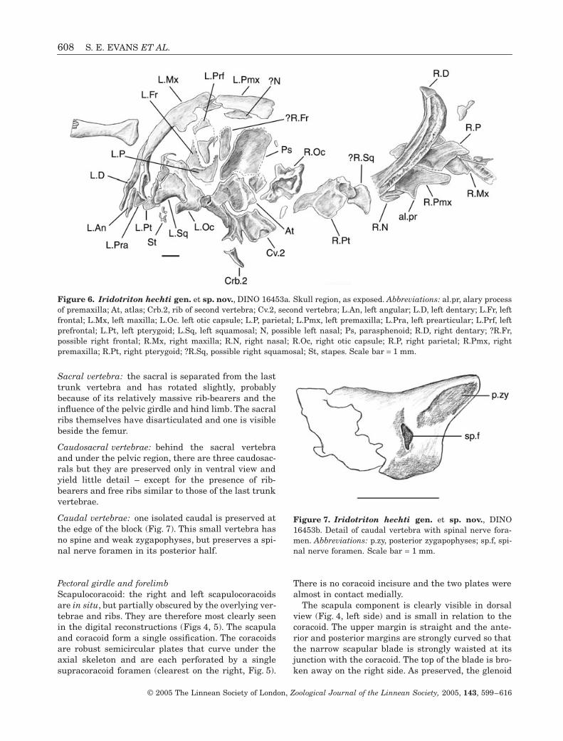
Sacral vertebra: the sacral is separated from the lasttrunk vertebra and has rotated slightly, probablybecause of its relatively massive rib-bearers and theinfluence of the pelvic girdle and hind limb. The sacralribs themselves have disarticulated and one is visiblebeside the femur.
Caudosacral vertebrae: behind the sacral vertebraand under the pelvic region, there are three caudosac-rals but they are preserved only in ventral view andyield little detail – except for the presence of rib-bearers and free ribs similar to those of the last trunkvertebrae.
Caudal vertebrae: one isolated caudal is preserved atthe edge of the block (Fig. 7). This small vertebra hasno spine and weak zygapophyses, but preserves a spi-nal nerve foramen in its posterior half.
Pectoral girdle and forelimbScapulocoracoid: the right and left scapulocoracoidsare in situ, but partially obscured by the overlying ver-tebrae and ribs. They are therefore most clearly seenin the digital reconstructions (Figs 4, 5). The scapulaand coracoid form a single ossification. The coracoidsare robust semicircular plates that curve under theaxial skeleton and are each perforated by a singlesupracoracoid foramen (clearest on the right, Fig. 5).
There is no coracoid incisure and the two plates werealmost in contact medially.
The scapula component is clearly visible in dorsalview (Fig. 4, left side) and is small in relation to thecoracoid. The upper margin is straight and the ante-rior and posterior margins are strongly curved so thatthe narrow scapular blade is strongly waisted at itsjunction with the coracoid. The top of the blade is bro-ken away on the right side. As preserved, the glenoid
Figure 6. Iridotriton hechti gen. et sp. nov., DINO 16453a. Skull region, as exposed. Abbreviations: al.pr, alary processof premaxilla; At, atlas; Crb.2, rib of second vertebra; Cv.2, second vertebra; L.An, left angular; L.D, left dentary; L.Fr, leftfrontal; L.Mx, left maxilla; L.Oc. left otic capsule; L.P, parietal; L.Pmx, left premaxilla; L.Pra, left prearticular; L.Prf, leftprefrontal; L.Pt, left pterygoid; L.Sq, left squamosal; N, possible left nasal; Ps, parasphenoid; R.D, right dentary; ?R.Fr,possible right frontal; R.Mx, right maxilla; R.N, right nasal; R.Oc, right otic capsule; R.P, right parietal; R.Pmx, rightpremaxilla; R.Pt, right pterygoid; ?R.Sq, possible right squamosal; St, stapes. Scale bar = 1 mm.
Figure 7. Iridotriton hechti gen. et sp. nov., DINO16453b. Detail of caudal vertebra with spinal nerve fora-men. Abbreviations: p.zy, posterior zygapophyses; sp.f, spi-nal nerve foramen. Scale bar = 1 mm.

JURASSIC SALAMANDER FROM NORTH AMERICA 609
© 2005 The Linnean Society of London, Zoological Journal of the Linnean Society, 2005, 143, 599–616
is posterolateral in position, and deep, with its longaxis orientated dorsoventrally. This suggests a degreeof dorsoventral movement for the large humerus, asoccurs in some modern salamanders in slow-gaitterrestrial locomotion (Evans, 1946). Among modernsalamander scapulocoracoids examined, the morphol-ogy of Iridotriton most closely resembles that of theterrestrial Ambystoma (S. E. Evans, pers. observ.).
Humerus: both humeri are preserved. The proximaland distal heads are at roughly 90 ∞ to one another,and the distal head is relatively massive compared tothe slender humeral shaft. The proximal end bears anexpanded crista ventralis humeri but no crista dorsa-lis. At the distal end, the condyles for articulation withthe radius and ulna are not ossified, and it is clearfrom the embayed shape of the distal end that it borea large cartilaginous joint surface.
Radius and ulna: the left radius lies adjacent to theleft maxilla (Figs 2, 4, 5), whereas the right radius andulna are visible only on the digital reconstructions(Figs 4, 5). Both bones have relatively narrow shaftsand expanded ends, although the ulna is the moregracile element. They are roughly half the length ofthe humerus (R/H = 53%).
Carpus and manus: wrist and manus elements arepreserved on both sides, but are disarticulated. Thereis one isolated carpal, probably from the left side, justbehind the left dentary (Fig. 5). On the right side, threecarpal elements are clearly preserved (digital recon-structions, Figs 4, 5), three (C1-3, Figs 4, 5) lie clus-tered around the distal ends of the ulna and radius,whereas a single, slightly larger element (Ul.I) is posi-tioned further distally, close to a phalanx. This largerounded element has a small central perforation andmatches the fused ulnare + intermedium of living sala-manders (Francis, 1934; S. E. Evans, pers. observ.).
According to Francis (1934) the short canal marksthe line of fusion between the two bones and conveysthe perforans carpi artery (a second similar elementlies in front of the left scapulocoracoid, Fig. 5). Thethree smaller carpals cannot be identified with anyconfidence. In the extant Salamandra (Francis, 1934),there are four ossified carpals in addition to theulnare-intermedium: a centrale, a basale commune(representing a fusion of the first two distal carpals),and then a basale 3 and a basale 4, although more ele-ments occur in some taxa.
Of the three distal elements in Iridotriton, C1 is thesmallest and may be a basale; C2 is strongly concavealong its long axis and could be a centrale. C3 is morecylindrical, with a small constriction around the mid-point. It is either another basale, or possibly a basalecommune. If this latter is correct, then the carpalstructure of Iridotriton (fused ulnare-intermedium,
large basale commune, small number of well ossifieddistal carpals/centrale) would be relatively derived.The phalangeal formula cannot be reconstructed: noneof the digits is complete on the right side, and the pha-langes of the left manus are scattered amongst thebones of the skull. Overall, however, the forelimb isrobust and strongly ossified.
Pelvic girdle and hindlimbParts of the left hind limb and girdle are preserved butdisarticulated.
Ilium: the left ilium is seen in medial view thusobscuring the structure and size of the acetabulum.The bone is small in comparison to the femur(although this is exaggerated in Fig. 3 by the edge-onview) and quite gracile. There is no trace of eitherischiadic plate and these are presumably deep in thematrix (although this second block was not scanned).
Femur: this is of similar length to the humerus, butless robust. The proximal and distal heads are some-what compressed and there is a distinct projectingproximal trochanter.
Tibia and fibula: as in the forelimb, the epipodialsare short and stout, with the tibia the more robust ofthe two elements.
Tarsus and pes: a small number of scattered bones ofthe foot (metatarsals and short phalanges) are alsopreserved. The elements of the pes are larger andlonger than those of the manus but the phalangeal for-mula cannot be reconstructed. There is one elementbeside the caudal vertebra that may be a tarsal.
Figure 8. Phylogenetic tree showing suggested relation-ship of Iridotriton within Caudata. Node 1: Caudata; Node2: Urodela (minimally ch. 9–12 in Iridotriton, see text);Node 3: Cryptobranchoidea; Node 4: stem-salamandroids(minimally ch. 15 in Iridotriton); Node 5: unnamed clade(Evans & Milner, 1996; ch. 1, 3–4); Node 6: Salamandroidea(minimally ch. 8).

610 S. E. EVANS ET AL.
© 2005 The Linnean Society of London, Zoological Journal of the Linnean Society, 2005, 143, 599–616
DISCUSSION
PHYLOGENETIC POSITION
Relationships amongst extant salamandersUnder traditional classifications (e.g. Estes, 1981;Duellman & Trueb, 1986; Milner, 1988) nine or tenclades of living salamanders are recognized: Crypto-branchidae, Hynobiidae, Dicamptodon, Rhyacotri-tonidae, Sirenidae, Amphiumidae, Proteidae,Salamandridae, Ambystomatidae, and Plethodon-tidae. There is a general agreement that Cryptobran-chidae and Hynobiidae (if monophyletic) are groupedwithin the Cryptobranchoidea (Larson & Dimmick,1993; Cryptobranchiformes of Milner, 2000) and thatmost or all of the remainder fall within a second super-group, the Salamandroidea (Larson & Dimmick, 1993;Salamandriformes of Milner, 2000).
Sirenidae are problematic and there is disagree-ment as to whether these taxa should be regarded asaberrant, but derived, salamandroids (e.g. Estes,1981) or as members of a primitive clade that is thesister group to Cryptobranchoidea + Salamandroidea(Neocaudata; Larson & Dimmick, 1993). Milner (1983,1988, 2000) supported the outgroup position ofsirenids, as did Duellman & Trueb (1986, albeit withsome reservation), Larson & Dimmick (1993), Hedges& Maxson (1993), and Hay et al. (1995).
Trueb (1993: fig. 6.9) presented a tree derived fromunpublished work by R. Cloutier in which sirenids arenested well within salamandroids as the sister groupof plethodontids (with salamandrids, ambystomatids,and proteids + amphiumids as successive outgroups).Most recently, the analysis of Gao & Shubin (2001)also nested sirenids within salamandroids, as a sistergroup to proteids and, in one tree, to amphiumids.However, the paedomorphic specializations of each ofthese three families may be distorting the result.
On the relationships of the remaining groups, thereis little consensus. Dicamptodon and rhyacotritonidshave been variously considered as primitive (Duell-man & Trueb, 1986) or derived, with Dicamptodon fre-quently classified with ambystomatids (e.g. Estes,1965; Larson & Dimmick, 1993; Milner, 2000; Gao &Shubin, 2001; but see Hedges & Maxson, 1993), andrhyacotritonids with plethodontids and amphiumids(e.g. Milner, 1983, 2000; Hillis, 1991; Hedges & Max-son, 1993; Gao & Shubin, 2001; combined consensus).
Proteids are variously placed as primitive (Milner,1983; Duellman & Trueb, 1986; Hillis, 1991); in a cladewith salamandrids, ambystomatids and Dicamptodon(Larson & Dimmick, 1993); or with Dicamptodon alone(Hay et al., 1995); whereas salamandrids andambystomatids are sometimes linked (e.g. Milner,1983, 2000; Larson & Dimmick, 1993; Gao & Shubin,2001) and sometimes not (Hay et al., 1995). Milner(1983) and Hillis (1991) had ambystomatids as the sis-
ter group of salamandrids, whereas Duellman & Trueb(1986) placed ambystomatids as the sister group ofplethodontids, and the molecular analysis of Hay et al.(1995) found no link between any of the three.
For the purposes of this discussion, however, theintricacies of crown-group relationships are not impor-tant, except with respect to Dicamptodon, since thelatter shows a number of primitive characters (sepa-rate angular and prearticular, spinal nerve foraminaonly in atlas and tail) whose significance and polarityare affected by its position in the phylogeny. The sameapplies to sirenids.
The position of IridotritonIridotriton shows a combination of primitive andderived characters that permit some discussion of itsphylogenetic position:
1. Premaxillae separate, broad with strong but shortalary process that did not make a major contribu-tion to the dorsal roof. This is the condition inkaraurids and in some primitive members of bothcryptobranchoids and salamandroids (hynobiids,cryptobranchids, Dicamptodon). Most salaman-droids show a more derived condition in which thealary process is longer and meets the frontals.
2. Maxilla with relatively short dorsal process. Thisis a primitive character found in karaurids, cryp-tobranchoids and Dicamptodon (Trueb, 1993).
3. Premaxilla with alary process that must haveabutted the nasal rather than overlain it, as inkaraurids. Many cryptobranchoids also have ashort dorsal process, as does Dicamptodonamongst salamandroids. This is undoubtedly aprimitive trait. In derived caudates, the long dor-sal process separates the right and left nasals inthe midline. In the Early Cretaceous Valdotriton(Evans & Milner, 1996), a long dorsal processpartly divides the two nasal anlagen of each side.
4. Separate prearticular and angular bones in thelower jaw, but no free coronoid. The possession ofseparate prearticular and angular bones is aprimitive character found in karaurids (Estes,1981), basal Chinese salamanders (Gao & Shubin,2001), and cryptobranchoids. In salamandroidsand stem-salamandroids (Valdotriton), the bonesare fused into a single compound prearticular ele-ment. A separate coronoid bone is found insirenids (Estes, 1965; Trueb, 1993) and also inproteids, as well as some fossil salamanders(Sinerpeton, Laccotriton) considered either basal(Gao & Shubin, 2001) or cryptobranchoid (Wang,2000b).
5. Free ribs on at least the anteriormost threecaudals. Their presence on anterior caudal verte-

JURASSIC SALAMANDER FROM NORTH AMERICA 611
© 2005 The Linnean Society of London, Zoological Journal of the Linnean Society, 2005, 143, 599–616
brae is a primitive trait, although Gao & Shubin(2003) cite the reduction in number of these ribsto 2–4? as a derived cryptobranchid character.In Iridotriton, only three anterior caudals arepreserved.
6. Prefrontal present, slender and elongate. Sincethe dorsal process of the maxilla is short, it islikely to have contacted only the prefrontal andnot the frontal. This is the primitive condition(Gao & Shubin, 2001; supplementary data, char-acter 33) found in karaurids and many extantsalamanders. Exceptions include Cryptobranchus,in which the frontals extend forward betweenthe prefrontals to meet the maxilla, and someplethodontids where the prefrontal is lost.
7. Vomer large, with choanal notch and evidence ofmedian palatine fenestra. A short broad vomer isa primitive feature found in karaurids, primitiveChinese salamanders (Sinerpeton, Laccotriton;Gao & Shubin, 2001) and some members of bothmajor extant lineages (e.g. cryptobranchids,Dicamptodon). In more derived taxa, there is atendency toward posterior elongation of the vomerover the parasphenoid (Trueb, 1993). In conjunc-tion with the latter condition, there is a change inthe pattern of the vomerine tooth row from essen-tially transverse (either immediately behind thepremaxillary row, e.g. Cryptobranchus; or midwaythrough the vomer, e.g. Valdotriton) to longitudi-nal. The left vomer of Valdotriton is visible in thedigital reconstructions at the front of the palate. Itis short but the tooth row cannot be seen, presum-ably because the teeth were small and below theresolution of the scan (individual tooth positionsare also not visible on the dentary in the scans,but are visible under the microscope).
8. ?Paired nasal anlagen. Living salamanders varyas to whether they possess one or two nasalanlagen on each side of the skull midline. Twoanlagen are generally considered to be primitive(e.g. Larson, 1991; Larson & Dimmick, 1993;Trueb, 1993). In some hynobiids (e.g. Ranodon,Salamandrella), these anlagen lie medial and lat-eral to the alary process of the premaxilla (Rose,2003). This appears also to be the condition incryptobranchids, but Rose (2003) argues that thelateral anlage has actually expanded into theregion medial to the alary process in this group.According to Larson (1991) and Larson & Dim-mick (1993), some salamander taxa retain onlythe medial anlage (e.g. sirenids), some the lateralanlage (most salamandroids), and some (e.g. Rhy-acotriton, Necturus) lose the nasal completely.Valdotriton retains paired anlagen, conjoined pos-teriorly, and the same condition may have beenpresent in Iridotriton.
9. Gracile skull bones without sculpture. Fullymetamorphosed karaurid salamanders likeKaraurus (Ivachnenko, 1978) and Marmorerpeton(Evans & Waldman, 1996) have thick, heavilysculptured skull roofing bones, not unlike those ofancestral temnospondyls. In most derived cau-dates, as in Iridotriton, the skull bones are thinwith little, if any, sculpture.
10. Triangular squamosal, wider dorsally than ven-trally, meets braincase but not parietal and isessentially mediolateral in orientation. The artic-ulation with the braincase leaves a gap betweenthe squamosal and parietal that permits theadductor muscles to pass back across the brain-case, lengthening their action (Estes, 1981). Thesquamosal of karaurids shows the primitive con-dition. It has a thick, geometric dorsal portionthat abuts the parietal, leaving no space for themuscles (adductor mandibulae internus, pseudot-emporalis superficialis portion; Carroll & Holmes,1980).
11. No evidence of a quadratojugal in the cheekregion. Karaurids primitively retain this element(Ivachnenko, 1978; Estes, 1981) but it is lost incrown-group salamanders.
12. Notochordal ectochordal vertebrae. This is aprimitive character within postkaraurid sala-manders where the vertebrae are formed frommembrane bone. Karaurids have heavily ossifiedvertebrae that appear to have an endochondralcomponent (S. E. Evans, pers. observ.).
13. Scapulocoracoid a single ossification, small lowscapula and large coracoid plate. This is a prob-lematic character. Sirenids have separate scapulaand coracoid ossifications, and this has beenregarded as a primitive character (Duellman &Trueb, 1986) supporting the placement of sirenidsoutside the cryptobrachoid-salamandroid node(e.g. Milner, 1988, 2000; Larson & Dimmick,1993). However, as stem-frogs (Triadobatrachus,Czatkobatrachus; Borsuk-Bia ynicka & Evans2000) and stem-salamanders (Marmorerpeton, S.E. Evans, pers. observ.) have a single scapulocora-coid plate, the sirenid condition is likely to besecondarily derived, not primitive.
14. Single-headed or conjoined ribs on most vertebrae.Karaurid salamanders have two headed ribs andthis primitive condition is retained in salaman-droids. In most cryptobranchoids (but see Milner,2000) the two heads have coalesced into a singlerib facet and this is often regarded as a derivedcharacter state of the group (Duellman & Trueb,1986; Larson & Dimmick, 1993). The distributionpattern, however, may be more complex than thatbecause rib-bearers with conjoined surfaces arepresent in both Iridotriton and a small sala-
l¢

612 S. E. EVANS ET AL.
© 2005 The Linnean Society of London, Zoological Journal of the Linnean Society, 2005, 143, 599–616
mander (‘salamander B’) from the Middle Jurassiclocality of Kirtlington in Oxfordshire, England(Evans et al., 1988; Evans & Milner, 1994), nei-ther of which shows other cryptobranchoid char-acters (other than primitive states). Most of therib-bearers also have conjoined heads in a secondKirtlington caudate, ‘salamander A’, that appearsto be a paedomorphic karaurid. Further work isneeded on the developmental history of this trait,but the evidence suggests that it is not uniquelycryptobranchoid.
15. At least one caudal vertebra with spinal nerveforamina. This is a character formulated byEdwards (1976), and discussed by several laterauthors (e.g. Larson & Dimmick, 1993). In theprimitive tetrapod condition, the spinal nervesemerge intervertebrally, but some salamandershave the spinal nerves emerging through the bodyof the vertebra itself (intravertebral). Karauridsshow the primitive condition. In cryptobran-choids, the only nerve emerging intravertebrallyis that of the atlas (see above), making thepossession of this atlantal foramen a definingcharacter of crown-group urodeles. Within sala-mandroids, Dicamptodon and Rhyacotriton havespinal nerve foramina in the tail, but there arenotches in the sacral, and larger notches in cau-dosacrals suggesting that the spinal nerve hasalready moved from its intervertebral position.The Cretaceous Valdotriton also has spinal nerveforamina in at least the anterior caudal vertebrae,but not in the postatlantal presacrals. In morederived salamandroids (ambystomatids, pleth-odontids, salamandrids), spinal nerve foraminaalso exist in the trunk vertebrae – creating anapparent trend towards greater numbers of theseforamina (Duellman & Trueb, 1986). However,proteids lack any spinal nerve foramina behindthe atlas (primitive or derived?), whereas sirenidshave them almost throughout the body. If sirenidsare genuinely basal, then this implies either thatforamina evolved independently within the group,or that trunk foramina are primitive rather thanderived, with varying patterns of loss. Under thetraditional view (e.g. Larson & Dimmick, 1993;Milner, 2000), the presence of spinal nerve foram-ina in the tail of Iridotriton, but not the trunk,places it in a similar position (with respect to thischaracter) as Valdotriton. However, if the charac-ter is unstable, this feature may be phylogeneti-cally uninformative.
16. Parietals broad, midline contact, short. The pari-etals are not fully exposed, but they are relativelysimple rectangular bones with a midline contact.It is unlikely that they were strongly overlappedby the frontals (as, for example, in cryptobran-
chids). Short parietals are found in most extantsalamanders. The cryptobranchid condition isderived.
17. Otic capsule fully ossified with exoccipital andprootic forming strong mass; opisthotic appearsconjoined but with a visible suture. The bones ofthe otic capsule are separate in cryptobranchoidsand sirenids, but also in some living salaman-droids, including Dicamptodon, Rhyacotriton, andamphiumids (Trueb, 1993). In proteids, as in Iri-dotriton, the opisthotic is discrete from the com-bined prootic/exoccipital (although conjoined inIridotriton) whereas all three bones are fused inambystomatids, salamandrids, plethodontids and,apparently, the Early Cretaceous Valdotriton.Whether or not this is a phylogenetically usefulcharacter remains to be seen; it may simply varywith levels of ossification in different caudatelineages.
18. Parasphenoid without internal carotid foramina.Internal carotid foramina are present (the primi-tive condition) in the lateral alae of the paras-phenoid in karaurids, cryptobranchoids, andDicamptodon; they are absent in sirenids, Valdo-triton and most salamandroids (plethodontids,salamandrids, some ambystomatids) (Trueb,1993).
19. Carpals and tarsals ossified, with a compoundulnare + intermedium. The carpus of Iridotritonmay have been relatively derived with a smallnumber of large robust elements. The ulnare andintermedium had certainly fused. This compoundulnare + intermedium appears to be a derivedcharacter of salamandroids (Shubin, Wake &Crawford, 1995).
20. Unfused margins of the Meckelian fossa behindthe symphysis. In most salamanders, the Mecke-lian fossa is closed for at least some distancebehind the symphysis, but the polarity of the char-acter is not clear. This may be an autapomorphy ofIridotriton.
Recent hypotheses of caudate relationships (e.g.Duellman & Trueb, 1986; Milner, 1988; Evans & Mil-ner, 1996; Gao & Shubin, 2001, 2003) provide a basisfor discussion of the position of Iridotriton, using thecharacters listed above.
Iridotriton shares a number of primitive featureswith basal caudates, including karaurids, somecryptobranchoids, and some salamandroids (ch. 1–7).However, known karaurids differ from urodeles(= crown-group caudates) in several important fea-tures, notably: heavy cranial sculpture; the absence ofan adductor groove on the squamosal or between itand the parietal; the retention of a quadratojugal;heavily built vertebrae, with a possible endochondral

JURASSIC SALAMANDER FROM NORTH AMERICA 613
© 2005 The Linnean Society of London, Zoological Journal of the Linnean Society, 2005, 143, 599–616
component; and the absence of a spinal nerve notch orforamen in the atlas (Marmorerpeton, Evans et al.,1988; condition uncertain in Karaurus and Kokartus).A tuberculum interglenoideum is absent in Marmorer-peton, but apparently present in Kokartus.
Iridotriton resembles urodeles in all of these char-acters for which it can be coded (ch. 9–12). It has a spi-nal nerve foramen or notch in the atlas, but thepolarity of the latter character is somewhat problem-atic. Although stem-salamanders lack this feature, aforamen is present in basal members of two other lis-samphibian groups (caecilians; Evans & Sigogneau-Russell, 2001. Albanerpetontids; Estes, 1981) andthere is a notch for the spinal nerve in the atlas ofthe stem-frog Czatkobatrachus (Evans & Borsuk-Bia ynicka, 1998). This suggests the absence of a fora-men in karaurids might be a peculiarity of that group,rather than a primitive character state. Nonetheless,the combination of other features (ch. 9–13) allowsplacement of Iridotriton on at least the urodelan stem.
As outlined above, the position of sirenid sala-manders (stem or crown) is problematic, and thisaffects the polarity of characters such as the co-ossification of the scapulocoracoid (ch. 13: Milner,1983; Larson & Dimmick, 1993) and the presence anddistribution of spinal nerve foramina (ch. 15:Edwards, 1976). This makes it difficult to assess therelative positions of sirenids and Iridotriton.
Most current classifications (e.g. Milner, 1988, 2000;Larson & Dimmick, 1993) separate crown-groupurodeles into two major clades: the Cryptobranchoidea(cryptobranchids and hynobiids) and the Salaman-droidea (all other salamanders). There is some debateas to the monophyly of Cryptobranchoidea and ofHynobiidae (e.g. Trueb, 1993), although recent workby Gao & Shubin (2001, 2003) found support for theclade.
Cryptobranchoid salamanders are mostly character-ized by the retention of primitive characters, includ-ing: low alary processes of the premaxillae and pairednasal anlagen; retention of a separate angular andprearticular in the lower jaw; and absence of a spinalnerve foramen in any postatlantal vertebra. The onefrequently cited derived skeletal character is the coa-lescence of the rib-bearers into a single-head (ch. 14),but this is problematic, and one hynobiid (Onychodac-tylus; Okajima, 1908 in Milner, 2000) reportedly hasdouble-headed rib-bearers.
Salamandroids are characterized by the fusion ofthe prearticular and angular into a single element, thepresence of a single nasal anlage, and of spinal nerveforamina in at least some of the caudal vertebrae(extending to the trunk in derived clades, but lost sec-ondarily in proteids, Edwards, 1976; Good & Wake,1992). The presence of spinal nerve foramina in thetail of Iridotriton (ch. 15) supports its placement
l¢
within salamandroids, as do the imperforate paras-phenoid (ch. 18) and the fused ulnare + intermedium(ch. 19).
The most completely known early Euramericansalamander, in terms of well-preserved specimens, isthe Early Cretaceous Valdotriton (Evans & Milner,1996) from the Barremian locality of Las Hoyas,Spain. Valdotriton has caudal spinal nerve foraminaand a single prearticular-angular bone, although thenasal anlagen are not fully fused. For this reason, itwas placed on the salamandroid stem (Evans &Milner, 1996). It thus provides a reference point forIridotriton. Like Valdotriton, Iridotriton may havehad paired nasal anlagen connected posteriorly (ch. 8),but unlike Valdotriton, the alary process of thepremaxilla is short and the prearticular and angularwere separate. The balance of characters thereforesuggests a position for Iridotriton within salaman-droids, but on the stem below Valdotriton.
BIOGEOGRAPHY
According to the vicariance model proposed by Milner(1983), caudates arose within a united Laurasianlandmass. Subdivision of that landmass left two stem-caudate populations, providing the ancestral stock ofcryptobranchoids in Asia and of salamandroids inEuramerica. At the time Milner’s paper was written,the Jurassic and Early Cretaceous record of caudateswas extremely poor. It has improved over the last twodecades but, with the notable exception of the LateJurassic Karaurus (Kazakhstan; Ivachnenko, 1978),all demonstrably Jurassic salamander material re-covered has been fragmentary (Hecht & Estes, 1960;Evans et al., 1988; Evans, 1992; Evans & Milner, 1994;Evans & Waldman, 1996). In addition, most Middleand Late Jurassic specimens described to date arereferable to the stem-caudate group Karauridae(Karaurus [Ivachnenko, 1978], Kazakhstan. Kokartus[Nessov, 1981, Nessov et al., 1996], Kirghizia. Mar-morerpeton [Evans & Waldman, 1996; Evans et al.,1988], UK, Portugal. Kirtlington ‘salamander A’[Evans, 1992; Evans & Milner, 1994; Evans & Wald-man, 1996; indet. material, Estes, 1981], USA). UnderMilner’s (1983) vicariance model, this suggests arather slow diversification within the new Asian andEuramerican landmasses.
Gao & Shubin (2001) offered an alternative biogeo-graphical hypothesis on the basis of two new Chinesefossil salamanders, Laccotriton and Sinerpeton,reportedly of Late Jurassic age. Preliminary analysesplaced both taxa on the caudate stem above karaurids(but see Wang, 2000b). Arguing that all known Juras-sic taxa (i.e. the new genera and the karaurids Karau-rus and Kokartus) were Asian (and primitive), Gao &Shubin (2001) proposed an Asian origin for Caudata.

614 S. E. EVANS ET AL.
© 2005 The Linnean Society of London, Zoological Journal of the Linnean Society, 2005, 143, 599–616
This implied that caudates reached Euramerica fromAsia only after the reestablishment of a land route inthe late Early Cretaceous, as manifested by theappearance of genera like Valdotriton in the Barre-mian of Spain (Evans & Milner, 1996).
However, the Asian origin hypothesis set aside theMiddle and Late Jurassic record of caudates inEuramerica (Hecht & Estes, 1960; Estes, 1981; Evanset al., 1988; Evans, 1992; Evans & Milner, 1994; Evans& Waldman, 1996). Furthermore, the reported LateJurassic age of Laccotriton and Sinerpeton is based onan Early Tithonian date for the underlying Zhangjia-kou Formation (Gao & Shubin, 2001). More detailedgeological work (Davis et al., 2001) has given an EarlyCretaceous age to these beds, making Sinerpeton andLaccotriton roughly contemporaneous with the Hau-terivian-Barremian (Zhou, Barrett, & Hilton, 2003;Wang, 2004) salamanders of the Jehol biota (?crypto-branchoids Liaoxitriton; Dong & Wang, 1998. Jeholot-riton; Wang, 2000a), and with European salamandersof the same age (Dollo, 1884; Evans et al., 1995; Evans& Milner, 1996; Milner & Evans, 1998).
More recently, Gao & Shubin (2003) described asuperb collection of articulated salamanders (Chuner-peton tianyiensis) from the Jiulongshan Formation ofInner Mongolia, China, which they date as MiddleJurassic (Bathonian) in age. They place Chunerpetonfirmly within crown-group Cryptobranchidae, a posi-tion requiring an early divergence of cryptobranchidsand hynobiids (if the latter are monophyletic; Trueb,1993), perhaps before the initial separation ofEuramerica and Asia. However, there are again con-cerns with respect to the age of the Jiulongshan For-mation (Zhonghe Zhou & Yuan Wang, pers. comm. toEvans, September 2003), and the beds may be consid-erably younger than Middle Jurassic. Thus the earlystages of cryptobranchoid evolution are still obscure,but there is no evidence that the caudate record ofAsia predates that of Euramerica.
Fragmentary crown-group salamander material(Kiyatriton) has also recently been described from theAptian-Albian of Western Siberia (Averianov &Voronkevich, 2002), but it is not assignable beyondUrodela indet.
If we are correct in our attribution of Iridotriton tothe salamandroid stem, then it provides qualified sup-port for Milner’s (1983) vicariance model because itextends the history of the clade in Euramerica backinto the Late Jurassic. However, additional materialand further analyses are required to establish the rateand pattern of diversification more fully, and moredetailed studies of the Chinese salamanders (e.g. Dong& Wang, 1998; Wang, 2000a, b, 2004) are needed todetermine their affinities.
In fact, Iridotriton may not be the earliest stem-salamandroid. Isolated jaws, atlantes, and postatlan-
tal trunk vertebrae of another small salamander areknown from the Middle Jurassic British locality ofKirtlington, although they have been discussed in theliterature only as ‘Salamander B’ (Evans & Milner,1994; Evans & Waldman, 1996). The bones of thissmall salamander are very gracile. There is a fullydeveloped tuberculum interglenoideum on the atlas,although this element is notched for the spinal nerve,rather than perforate. The presacral vertebrae resem-ble those of Iridotriton in having conjoined rib-bearers, but the caudals are unknown.
LIFESTYLE
Iridotriton is surprisingly robust for its size, with fullossification of the limb elements including the carpalsand tarsals (often remaining unossified in living andfossil taxa), but not the joint surfaces of the longbones. The pectoral girdle is extensively ossified withstrong coracoid plates that probably approached oneanother, or met, in the ventral midline. The humerusis relatively massive, with a greatly expanded distalhead and strong proximal crests. The trunk is shortand the tail lacks any development of tall neural orhaemal spines. All these features argue for a pre-dominantly terrestrial salamander with powerfullimbs and a wide, shallow head. The teeth are numer-ous and very small, indicating a microphagous diet.
ACKNOWLEDGEMENTS
We owe a debt of gratitude to the High Resolution X-ray Computed Tomography Facility at UT Austin forscanning the specimen (especially Matt Colbert andTim Rowe), and to the NSF who funded the scan.Thanks also to Scott Madsen (Dinosaur National Mon-ument) for the initial mechanical preparation of thespecimen; to Dr Jim Gardner (Royal Tyrrell Museum,Canada) for information about the second Morrisontaxon; to Drs Barry Clarke (Zoology Department, TheNatural History Museum) and Helen Chatterjee(Grant Museum, UCL) for the loan of comparativesalamander material; to Drs Zhonghe Zhou and YuanWang (Institute of Vertebrate Palaeontology andPalaeoanthropology, Beijing) for information on thedating of Chinese deposits; and to Jane Pendjiky(Anatomy, UCL) for help in the preparation and label-ling of electronic images. Drs Jim Gardner andAndrew Milner reviewed an earlier version of themanuscript and we thank them for their helpfulcomments.
REFERENCES
Averianov AO, Voronkevich AV. 2002. A new crown-groupsalamander from the Early Cretaceous of Western Siberia.Russian Journal of Herpetology 9: 209–214.

JURASSIC SALAMANDER FROM NORTH AMERICA 615
© 2005 The Linnean Society of London, Zoological Journal of the Linnean Society, 2005, 143, 599–616
Bolt JR. 1991. Lissamphibian origins. In: Schulze H-P, TruebL, eds. Origins of the higher groups of tetrapods. Ithaca, NY:Comstock Publishing, 194–222.
Borsuk-Bia ynicka M, Evans SE. 2002. The scapulocora-coid of an Early Triassic stem-frog from Poland. Acta Palae-ontologia Polonica 47: 79–96
Cannatella DC, Hillis DM. 1993. Amphibian relationships:phylogenetic analysis of morphology and molecules. In:Cannatella DC, Hillis DM, eds. Amphibian relationships.Phylogenetic analysis of morphology and molecules. Herpe-tological Monographs 7: 1–7.
Carroll RL, Holmes R. 1980. The skull and jaw musculatureas guides to the ancestry of salamanders. Zoological Journalof the Linnean Society 68: 1–40.
Davis GA, Zheng Y, Wang C, Darby BJ, Zhang C, GeorgeG. 2001. Mesozoic tectonic evolution of the Yanshan fold andthrust belt, with emphasis on Hebei and Liaoning provinces,northern China. Geological Society of America, Memoir 194:171–197.
Dollo L. 1884. Note sur le batracien de Bernissart. Bulletin duMusée Royal d’Histoire Naturelle de Belgique 3: 85–96.
Dong Z, Wang Y. 1998. A new urodele (Liaoxitriton zhongjianigen. et sp. nov.) from the Early Cretaceous of Western Lia-oning Province, China. Vertebrata PalAsiatica 36: 159–172[In Chinese with an English summary].
Duellman WE, Trueb L. 1986. Biology of amphibians. NewYork: McGraw-Hill.
Edwards JL. 1976. Spinal nerves and their bearing on sala-mander phylogeny. Journal of Morphology 148: 305–328.
Engelmann GF, Callison G. 1998. Mammalian faunas of theMorrison Formation. In: Carpenter K, Chure DJ, KirklandJI, eds. The Upper Jurassic Morrison Formation: an inter-disciplinary study. Part 1. Modern Geology 23: 343–380.
Ensom PC, Evans SE, Milner AR. 1991. Amphibians andreptiles from the Purbeck Limestone Formation (UpperJurassic) of Dorset. Contributions to the PaleontologicalMuseum, University of Oslo 364: 19–20.
Estes R. 1965. Fossil salamanders and salamander origins.American Zoologist 5: 319–334.
Estes R. 1969. Prosirenidae, a new family of fossil sala-manders. Nature 224: 87–88.
Estes R. 1981. Gymnophiona, Caudata. In: Wellnhofer P,ed. Handbuch der Paläoherpetologie 2. Stuttgart: GustavFischer.
Estes R, Sanchíz B. 1982. Early Cretaceous lower verte-brates from Galve (Teruel), Spain. Journal of VertebratePaleontology 2: 21–39.
Evans FG. 1946. The anatomy and function of the forelimb insalamander locomotion. Anatomical Record 95: 257–281.
Evans SE. 1992. Small reptiles and amphibians from the For-est Marble (Middle Jurassic) of Dorset. Proceedings of theDorset Natural History and Archaeological Society 113: 201–202.
Evans SE, Borsuk-Bia ynicka M. 1998. A stem-frog fromthe Early Triassic of Poland. Acta Paleontologia Polonica 43:573–580.
Evans SE, Chure DJ. 1998a. Morrison lizards: structure,relationships and biogeography. In: Carpenter K, Chure DJ,
l¢
l¢
Kirkland JI, eds. The Upper Jurassic Morrison Formation:an interdisciplinary study. Part 1. Modern Geology 23: 35–48.
Evans SE, Chure DJ. 1998b. Paramacellodid lizards fromthe Jurassic Morrison Formation at Dinosaur NationalMonument, Utah. Journal of Vertebrate Paleontology 18: 99–114.
Evans SE, McGowan G. 2002. An amphibian assemblagefrom the Purbeck Limestone Group. Special Papers in Palae-ontology 68: 103–119.
Evans SE, McGowan G, Milner AR, Sanchìz B. 1995.Amphibians. In: Melendez MN, ed. Las Hoyas. A lacustrineKonservat-Lägerstatte, Cuenca, Spain. Madrid: EdicionesUniversidad Complutense de Madrid, 51–53.
Evans SE, Milner AR. 1993. Frogs and salamanders from theUpper Jurassic Morrison Formation (Quarry Nine, ComoBluff) of North America. Journal of Vertebrate Paleontology13: 24–30.
Evans SE, Milner AR. 1994. Middle Jurassic microvertebrateassemblages from the British Isles. In: Fraser NC, Sues H-D,eds. In: The shadow of the dinosaurs: Early Mesozoic tetra-pods. Cambridge: Cambridge University Press, 303–321.
Evans SE, Milner AR. 1996. A metamorphosed salamanderfrom the Early Cretaceous of Las Hoyas, Spain. Philosophi-cal Transactions of the Royal Society of London B 351: 627–646.
Evans SE, Milner AR, Mussett F. 1988. The earliest knownsalamanders (Amphibia, Caudata): a record from the MiddleJurassic of England. Geobios 21: 539–552.
Evans SE, Sigogneau-Russell D. 2001. A stem-group caecil-ian (Amphibia: Lissamphibia) from the Lower Cretaceous ofMorocco. Palaeontology 44: 259–273.
Evans SE, Waldman M. 1996. Small reptiles and amphibiansfrom the Middle Jurassic of Skye, Scotland. In: Morales M,ed. The Continental Jurassic. Museum of Northern ArizonaBulletin 60: 219–226.
Feller AE, Hedges SB. 1998. Molecular evidence for the earlyhistory of living amphibians. Molecular Phylogenetics andEvolution 9: 509–516.
Fox RC, Naylor BG. 1982. A reconsideration of the relation-ships of the fossil amphibian Albanerpeton. Canadian Jour-nal of Earth Sciences 19: 118–128.
Francis ETB. 1934. The anatomy of the salamander. Oxford:Clarendon Press.
Fraser NC, Wu X-C. 1998. Sphenodontians from the BrushyBasin member of the Morrison Formation in DinosaurNational Monument. In: Carpenter K, Chure DJ, KirklandJI, eds. The Upper Jurassic Morrison Formation: an inter-disciplinary study. Part 1. Modern Geology 23: 17–34.
Gao KQ, Cheng ZW, Xu X. 1998. First report of a Mesozoicurodele from China. Chinese Geology 1998 (1): 40–41 [InChinese].
Gao KQ, Shubin NH. 2001. Late Jurassic salamanders fromnorthern China. Nature 410: 574–577.
Gao K, Shubin NH. 2003. Earliest known crown-group sala-manders. Nature 422: 424–428.
Good DA, Wake DB. 1992. Geographic variation and specia-tion in the torrent salamanders of the genus Rhyacotriton

616 S. E. EVANS ET AL.
© 2005 The Linnean Society of London, Zoological Journal of the Linnean Society, 2005, 143, 599–616
(Caudata: Rhyacotritonidae). University Of California Pub-lications in Zoology 126: 1–91.
Gradstein FW, Agterberg FP, Ogg JJ, Hardenbol J, VanVeen P, Thierry J, Huang Z. 1995. A Triassic, Jurassicand Cretaceous time scale. In: Berggren WA, Kent DV,Aubry MP, Hardenbol J, eds. Geochronology time scales andglobal stratigraphic correlation. Society for SedimentaryGeology Special Publication 54, 95–126.
Hay JM, Ruvinsky I, Hedges SB, Maxson LR. 1995. Phy-logenetic relationships of amphibian families inferred fromDNA sequences of mitochondrial 12S and 16S ribosomalRNA genes. Molecular Biology and Evolution 12: 928–937.
Hecht MK, Estes R. 1960. Fossil amphibians from QuarryNine. Postilla 46: 1–19.
Hedges SB, Maxson IR. 1993. A molecular perspective on lis-samphibian phylogeny. In: Cannatella DC, Hillis DM, eds.Amphibian relationships. Phylogenetic analysis of morphol-ogy and molecules. Herpetological Monographs 7: 27–42.
Henrici AC. 1998. New anurans from the Rainbow Parkmicrosite, Dinosaur National Monument, Utah. In: Carpen-ter K, Chure DJ, Kirkland JI, eds. The Upper Jurassic Mor-rison Formation: an interdisciplinary study. Part 1. ModernGeology 23: 1–16.
Hillis DM. 1991. The phylogeny of amphibians: currentknowledge and the role of cytogenetics. In: Green DM,Sessions SK, eds. Amphibian cytogenetics and evolution.London: Academic Press, 7–32.
Ivachnenko M. 1978. Urodeles from the Triassic and Jurassicof Soviet Central Asia. Palaeontologicheski Zhurnal 1978:84–89 [In Russian].
Jenkins FE, Walsh DM. 1993. An early Jurassic caecilianwith limbs. Nature 365: 246–250.
Kowallis BJ, Christiansen EH, Deino AL, Peterson F,Turner CE, Kunk MJ, Obradovich JD. 1998. The age ofthe Morrison Formation. In: Carpenter K, Chure DJ,Kirkland JI, eds. The Upper Jurassic Morrison Formation:an interdisciplinary study. Part 1. Modern Geology 22: 235–260.
Larson A. 1991. A molecular perspective on the evolutionaryrelationships of salamanders. Evolutionary Biology 25: 211–277.
Larson A, Dimmick WW. 1993. Phylogenetic relationships ofthe salamander families: an analysis of congruence amongmorphological and molecular characters. In: Cannatella, DC,Hillis, DM, eds. Amphibian relationships. Phylogenetic anal-ysis of morphology and molecules. Herpetological Mono-graphs 7: 77–93.
Laurin M, Reisz RR. 1997. A new perspective on tetrapodphylogeny. In: Sumida SS, Martin KLM, eds. Amniote ori-gins. Completing the transition to land. San Diego: AcademicPress, 9–59.
McGowan G, Evans SE. 1995. Albanerpetontid amphibiansfrom the Cretaceous of Spain. Nature 373: 143–145.
Milner AC, Evans SE. 1998. First report of lizards andamphibians from the Wealden (Lower Cretaceous ofEngland). In: Lucas SG, Kirkland JI, Estep JW, eds. Lowerand Middle Cretaceous terrestrial ecosystems. New MexicoMuseum of Natural History and Science 14: 173–176.
Milner AR. 1983. The biogeography of salamanders in theMesozoic and early Caenozoic: a cladistic-vicariance model.In: Sims RW, Price JH, Whalley PES, eds. Evolution, timeand space: the emergence of the biosphere. London: AcademicPress, 431–468.
Milner AR. 1988. The relationships and origin of livingamphibians. In: Benton MJ, ed. The phylogeny and classifi-cation of the tetrapods, vol. 1. Amphibians, reptiles, birds.Systematics Association Special Volume 35a. Oxford: Clar-endon Press, 59–102.
Milner AR. 2000. Mesozoic and Tertiary Caudata andAlbanerpetontidae. In: Heatwole H, Carroll RL, eds.Amphibian Biology, Vol. 4. Palaeontology. Chipping Norton,New South Wales: Surrey Beaty, 1413–1444.
Nessov LA. 1981. Cretaceous salamanders and frogs of Kizyl-Kum Desert. Academy of Sciences of the U.S.S.R. ZoologicalInstitute Proceedings 101: 57–88 [In Russian].
Nessov LA. 1988. Late Mesozoic amphibians and lizards ofSoviet Middle Asia. Acta Zoologica Cracovienska 31: 475–486.
Nessov LA, Fedorov PV, Potanov DO, Golovyeva LS.1996. The structure of the skulls of caudate amphibians col-lected from the Jurassic of Kirgizstan and the Cretaceous ofUzbekistan. Vestnik Sankt-Petersburgiskogo Universiteta,Geologia 7: 3–11 [In Russian].
Nevo E. 1964. Fossil urodeles in early Lower Cretaceousdeposits of Makhtesh Ramon, Israel. Nature 201: 415–416.
Nevo E, Estes R. 1969. Ramonellus longispinus, an early Cre-taceous salamander from Israel. Copeia 1969: 91–123.
Rage J-C, Ro ek Z. 1989. Redescription of Triadobatrachusmassinoti (Piveteau, 1936) an anuran amphibian from theEarly Triassic. Palaeontographica A206: 1–16.
Rose CS. 2003. The developmental morphology of salamanderskulls. In: Heatwole H, Davies M, eds. Amphibian biology,Vol. 5. Osteology. Chipping Norton, New South Wales: SurreyBeatty, 1686–1783.
Ruta M, Coates MI, Quicke DLJ. 2003. Early tetrapod rela-tionships revisited. Biological Reviews 78: 251–345.
Shubin N, Wake DB, Crawford AJ. 1995. Morphologicalvariation in the limbs of Taricha granulosa (Caudata: Sala-mandridae): evolutionary and phylogenetic implications.Evolution 49: 874–884.
Trueb L. 1993. Patterns of cranial diversity among the Lissa-mphibia. In: Hanken J, Hall BK, eds. The skull, Vol. 2. Pat-terns of structural and systematic diversity. Chicago: TheUniversity of Chicago Press, 255–343.
Wang Y. 2000a. A new salamander (Amphibia: Caudata) fromthe Early Cretaceous Jehol Biota. Vertebrata Palasiatica 38:100–103.
Wang Y. 2000b. Amphibians in the Jehol Biota from North-eastern China. Vertebrata Palasiatica 38 (Suppl.): 32–33.
Wang Y. 2004. A new Mesozoic caudate (Liaoxitriton dao-hugoensis sp. nov.) from Inner Mongolia, China. Chinese Sci-ence Bulletin 49: 858–860.
Zhou Z, Barrett PM, Hilton J. 2003. An exceptionallypreserved Lower Cretaceous ecosystem. Nature 421: 807–814.
c