A lady with prickly nodules on both lower limbs
21
41 MJD 2012 July Vol 28 Malaysian Journal of Derm a t o l og y CLINICOPATHOLOGIC CHALLENGE A lady with prickly nodules on both lower limbs Choon SE, FRCP Correspondence Choon Siew Eng FRCP Department of Dermatology, Hospital Sultanah Aminah, Johor Bahru Email: [email protected] 49 year-old lady referred by nephrologist for increasing number of skin nodules on both legs for past 2 years. Systemic lupus erythematosus with lupus nephritis was diagnosed 10 years ago when she presented with Raynauld’s phenomenon, hair loss and nephrotic syndrome. Patient was treated initially with monthly pulses of IV cyclophosphamide and oral prednisolone ranging between 30-10mg. Her renal function is preserved with instituted treatment. On presentation, she was still on prednisolone 7.5mg, mycophenolate mofetil 500mg bd and telmisartan for hypertension which she developed 5 years ago. Physical examination revealed non-tender, woody-hard induration of both legs with underlying firm, sharply angulated papules and nodules (Fig 1). Figure 1 A & B show induration of both legs with smooth shiny, hide-bound skin. B A
Transcript of A lady with prickly nodules on both lower limbs

41MJD 2012 July Vol 28
M a l aysian Journal of D e rm a t o l og y
C L I N I C O PATHOLOGIC CHALLENGE
A lady with prickly nodules on both lower limbsChoon SE, FRCP
CorrespondenceChoon Siew Eng FRCPDepartment of Dermatology,Hospital Sultanah Aminah, Johor BahruEmail: [email protected]
49 year-old lady referred by nephrologist forincreasing number of skin nodules on both legsfor past 2 years. Systemic lupus erythematosuswith lupus nephritis was diagnosed 10 years agowhen she presented with Ray n a u l d ’sphenomenon, hair loss and nephrotic syndrome.Patient was treated initially with monthly pulsesof IV cyclophosphamide and oral prednisoloneranging between 30-10mg. Her renal function is
p r e s e rved with instituted treatment. Onpresentation, she was still on prednisolone 7.5mg,mycophenolate mofetil 500mg bd and telmisartanfor hypertension which she developed 5 yearsago. Physical examination revealed non-tender,woody-hard induration of both legs withunderlying firm, sharply angulated papules andnodules (Fig 1).
Figure 1 A & B show induration of both legs with smooth shiny,hide-bound skin.
B
A
42 MJD 2012 July Vol 28
M a l aysian Journal of D e rm a t o l og y
Fig. 2 shows epidermal atrophy with loss of rete pegs, atrophic eccrine glands, haphazardly arrangedthickened collagen, scanty inflammatory infiltrates and calcium deposits in dermis and subcutis. 2Ashows close-up of calcinosis cutis, 2B shows abnormal thick, homogenous and haphazardly-arrangedcollagen fibre.
What is your diagnosis?
Histologic features are shown Fig 2.
A B

43MJD 2012 July Vol 28
M a l aysian Journal of D e rm a t o l og y
Contact and Occupational Derm a t i t i sfor Beginners
Continuous Professional Development - CME
Organizers Dermatology, Selayang Hospital
Venue Auditorium, Selayang Hospital
Date 10-11 May 2012
Email [email protected]
Phototherapy Course
Organizers Kuala Lumpur Hospital
Venue Auditorium Kuala Lumpur Hospital
Date 24-25 May 2012
Email [email protected]
Paediatrics Dermatology Update 2012
Organizers Dermatology, Sarawak General Hospital
Venue Pullman Hotel, Kuching
Date 26 May 2012
Email [email protected]
Ms Doroty Tan

44 MJD 2012 July Vol 28
M a l aysian Journal of D e rm a t o l og y
D e rmatology Update 2012
Continuous Professional Development - CME
Organizers National Skin Centre
Venue Mandarin Hotel, Singapore
Date 11-13 May 2012
Program website www.nsc.gov.sg/dermupdate2012
37th Annual General Meeting & Malaysian Dermatology Congre s s
Organizers Dermatological Society of Malaysia
Theme Allergy and Occupational Dermatoses
Venue Holiday Inn, Malacca
Date 14-17 September 2012
Program website www.dermatology.org.my

45MJD 2012 July Vol 28
M a l aysian Journal of D e rm a t o l og y
Advance Masters in Dermatology Graduates and Thesis
GRADUATES (DR.)
ADAWIYAH BINTI JAMIL
AZURA AFANDI
CHANG CHOONG CHOR
CHONG YEW THONG
FELIX YAP BOON BIN
KARTINI FARAH ABD.RAHIM
LEE CHEW KEK
LEE YIN YIN
MAZLIN BASERI
NG TING GUAN
THESIS
The effect of smoking cessation on severity of psoriasis
Development of a computerized objective assessment of areaand erythema for PASI scoring of severity of psoriasis andcomparing with the conventional visual assessment of PASI bydermatologist
Human leukocyte antigen (HLA) in toxic epidermal necrolysis(TEN) and Stevens Johnson Syndrome
Comparison of two dosing regimens for administering oralmethotraxate in patients with moderate to severe plaquepsoriasis (the Co-tromp study)
Acne vulgaris: Quality of life and cost of illness ingovernment dermatology clinics in Sarawak
Narrowband UVB phototherapy: Comparison of two startingdoses using 50%MED and skin phototype
Fingerprint biometrics changes in hand dermatitis
Assessment of skin phototype, skin colour and minimalerythema dose (MED) to ultravioliet B (UVB) radiation in themultiethnic Malaysian population
Impact of systemic glucocorticoids onbone mineral density in patients with pemphigus
The efficacy and safety of tacrolimus ointment in patientswith moderate to severe atopic eczema

46 MJD 2012 July Vol 28
M a l aysian Journal of D e rm a t o l og y
GRADUATES (DR.)
NORASHIKIN SHAMSUDIN
NOORLAILY MOHD NOOR
NOOR ZALMY AZIZAN
PENNY LIM POH LU
PRIYA GILL
TANG JYH JONG
TANG MIN MOON
TARITA BINTI TAIB
WONG SU-MING
THESIS
Efficacy anf safety of tacrolimus ointment in vitiligo usingboth an objective and subjective method for the evaluation ofrepigmentation progression
Comparison of transepidermal water loss (TEWL) betweennormal and erythrodermic patients of various aetiology
Significance of toe web microbiology in the aetiology ofrecurrent cellulitis of the lower leg - a pilot study
Comparative study of the efficacy of benzyl benzoate andpermethrin in the treatment of scabies
Cardiac abnormalities in Psoriasis
Antibiotic sensitivity of propionibacterium acnes isolated frompatients with acne vulgaris in Kuala Lumpur Hospital,Malaysia
Quality of life and cost of illness in patients with psoriasis inMalaysia: A multicentre study
Assessment of cutaneous and systemic manifestations ofpatients with lupus erythematosus (LE) using clinical scoringindices
Efficacy and safety of sodium hypochlorite (bleach) baths inpatients with moderate to severe atopic dermatitis

47MJD 2012 July Vol 28
M a l aysian Journal of D e rm a t o l og y
Aesthetic Medical Practice Guidelines for Medical Specialists
1. PREREQUISITES FOR MEDICAL SPECIALISTS PERFORMING AESTHETIC MEDICAL PROCEDURES
1.1 A medical practitioner who wishes to perform aesthetic/cosmetic medical procedures must be fullyregistered with the Malaysian Medical Council.
1.2 He/she must possess a current and valid Annual Practicing Certificate.
1.3 He/she is required to possess a higher qualification in dermatology with full dermatological training; oralternatively, is registered on the National Specialist Register in a medical related field in order to beregarded as medical Specialist.
1.4 He/she must possess experience through recognised practical training courses conducted by bona-fide professional bodies specialising in aesthetic medical practice.
1.5 He/she must exercise strict patient selection criteria, must communicate to the potential client/patient therisks involved, the possible outcome, obtain valid consent for the aesthetic medical procedure planned, andgenerally observe all aspects of the Code of Professional Conduct of the Malaysian Medical Council.
1.6 He/she must place client/patient safety as the primary concern and should provide aesthetic medicalservices in a healthcare facility licensed or registered under the Private Healthcare Facilities and ServicesAct 1998 and regulations 2006.
1.7 He/she is required to obtain a Letter of Credentialing and Privileging (LCP) for the aesthetic/cosmeticmedical procedure(s) which he/she intends to perform. The LCP shall be issued by the CosmeticDermatology and Laser Medicine (CDLM) Board under the Dermatological Society, Malaysia, Academyof Medicine, Malaysia.
1.8 With the LCP, he/she is eligible for registration with the Registry of Aesthetic Medical Practice which shallbe maintained by the Medical Practice Division, Ministry of Health (MOH), Malaysia.
2. SCOPE OF PRACTICE
The basic considerations for the scope of practice in aesthetic medical practice by medical specialists arewhether they are core medical specialists or non-core medical specialists (refer Table 1).
A. Core Medical Specialists This consists of dermatologists performing aesthetic/cosmetic surgery within their core curriculum andcore competency.
The core specialist society will submit a list of their specialists to the CDLM Board for inclusion in theNational Registry of Aesthetic Medical Practice.
Specialists may also apply directly to the CDLM Board.
B. Non-Core Medical Specialists This refers to medical specialists whose routine areas of practice are completely unrelated to dermatology e.g.anaesthetists, pathologists, radiologists etc.
These specialists may be subjected to similar requirements for privileging of a general practitioner practisingaesthetic medical practice.
If possible they should be sanctioned by their own professional peers before application to the CDLM Board;alternatively they may apply directly to the Board with the necessary documentation.

48 MJD 2012 July Vol 28
M a l aysian Journal of D e rm a t o l og y
PROCEDURES
Chemical peels(superficial)
Microdermabrasion
Intense pulse light(IPL)
Chemical peel(medium)
Botulinum toxininjection
Filler injection
Sclerotherapy
Laser for treatingskin pigmentation
Laser for treatingskin tumours
Laser for skinrejuvenation(incl fractional)
Laser for hairremoval(e.g long-pulsedNd-YAG, Diode)
Laser for treatingvascular lesions
Chemical peels(Deep)
Ablative skinresurfacing lasers
Hair transplant
Mechanicaldermabrasion
B. MINIMALLY INVASIVE
CORESPECIALISTS
Dermatologists
Dermatologists
Dermatologists
Dermatologists
Dermatologists
Dermatologists
Dermatologists
Dermatologists
Dermatologists
Dermatologists
Dermatologists
Dermatologists
Dermatologists
Dermatologists
Dermatologists
Dermatologists
NON-CORESPECIALISTS
Case by case basis
Case by case basis
Case by case basis
Case by case basis
Case by case basis
Case by case basis
Case by case basis
Case by case basis
Case by case basis
Case by case basis
Case by case basis
NA**
NA
NA
NA
NA
APPROPRIATEPREMISESNEEDED
Clinic
Clinic
Clinic
Clinic
Clinic
Clinic
OT/ Clinic
OT/ Clinic
OT/ Clinic
Clinic
Clinic
OT/ Clinic
OT/Clinic
OT/ Clinic
OT
OT/ Clinic
PROCEDURESPERFORMED FOR
NON-CORE SPECIALISTS
25
20
40
25
25
25
20
20
20
20
20
A. NON INVASIVE
C. INVASIVE
Table 1 Scope of practice for Medical Specialists.

49MJD 2012 July Vol 28
M a l aysian Journal of D e rm a t o l og y
PROCEDURES
Phlebectomy
Photodynamictherapy
Radiofrequency
Ultrasound device
Tumescentliposuction
CORESPECIALISTS
Dermatologists
Dermatologists
Dermatologists
Dermatologists
Dermatologists
NON-CORESPECIALISTS
NA
NA
NA
NA
NA
APPROPRIATEPREMISESNEEDED
OT/Clinic
OT/ Clinic
OT/ Clinic
OT/Clinic
OT/ Clinic
PROCEDURESPERFORMED FOR
NON-CORE SPECIALISTS
Note:This list is subjected to be reviewed whenever there is new evidence-based treatment available.*OT = Operation theatre, **NA = Not applicable
3. PROCESS OF REGISTRATION
3.1 Medical specialists who intend to practise aesthetic medical practice are required to apply to the CosmeticDermatology and Laser Medicine (CDLM) Board of the Dermatological Society Malaysia.
3.2 The CDLM Board shall assess and/or examine medical specialists who intend to practiceaesthetic/cosmetic procedures.
3.3 The CDLM Board will issue a Letter of Credentialing and Privileging (LCP) to the successful candidate,specifying the core specialty, aesthetic/cosmetic procedure(s) approved, and duration of validity. The LCPis valid for 5 year and renewable upon endorsement by the CDLM Board.
3.4 With the LCP, the medical specialists’ names are eligible to be included in the National Registry ofAesthetic Medical Practice.
50 MJD 2012 July Vol 28
M a l aysian Journal of D e rm a t o l og y
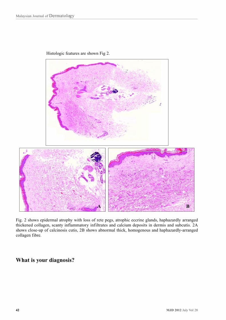
4. THE CREDENTIALING AND PRIVILEGING COMMITTEE
The Cosmetic Dermatology and Laser Medicine (CDLM) Board of the Dermatological Society Malaysia will bethe credentialing and privileging committee for the medical specialists.
5. THE SECRETARIAT FOR MEDICAL SPECIALISTS
Persatuan Dermatologi Malaysia (PDM)Rumah Dermatologi2-16-2, Block 2 (Remis) Pantai Panorama CondominiumJalan 112 H, off Jalan Kerinchi59200 Kuala Lumpur
Medical Specialists
National Registry of AestheticMedical Practice
Non-dermatologists
Apply to CDLM
Case by case basis
LCP issued by CDLM
Dermatologists
Apply to CDLM
LCP issued by CDLM
Figure 1 Process of registration for medical specialists.

51MJD 2012 July Vol 28
M a l aysian Journal of D e rm a t o l og y
B O O K R E V I E W
Textbook of Laser and Light Derm a t o l o g yin the Asian Skin
Edited by Yong-Kwang Tay and Yuin Chew ChanPublished by World Scientific Publishing Co Pte 2011. 140 pp.
I S B N - 1 3 : 9 7 8 - 9 8 1 4 3 3 8 8 6 8
It was a pleasure to review this well illustrated and informative textbook of laser and light dermatology.This is the latest textbook of laser and light dermatology for Asian skin just released in 2011. It is writtenand edited by Tay Yong Kwang and Chan Yuin Chew along with a further 10 other contributors. The bookhas 12 chapters and its contributors are all experienced dermatologists including Prof Goh Chee Leok,Joyce Lim, Chua SH and Melvin Ee from Singapore who gave practical pearls of wisdom and tipstreating darker skin phenotypes.
The book is divided into 12 chapters covering a comprehensive range of laser and light based devices.It's only 140 pages, full of color images and is very readable. One of the outstanding features of the textis that it has numerous actual case presentations and the author would include the laser settings to be usedin the treatment. Each chapter begins with an introduction giving the reader a general overview of thetopic followed by mechanism of action, patient selection and treatment procedure including the laserparameters, precautions and post operative care. In some chapters the chapter would end with clinicalcase histories. The book include chapters on laser tissue interactions, CO2 laser, vascular, pigment, hairremoval, ablative resurfacing, non ablative resurfacing, fractional laser, IPL and photodynamic therapy.
Of special interests are chapters on treatment of pigmented lesions, fractionated lasers, intense pulsedlight devices and photodynamic therapy in the Asian skin. Conditions unique to the Asian skin such asNevus of Ota, Hori’s nevus, melasma, and post inflammatory hyperpigmentation were well covered inthe text.
Currently there are very few reference textbook of laser and light devices on Asian skin. The Textbookof Laser and Light Dermatology in the Asian Skin will serve as a valuable reference textbook and anessential companion for all dermatologists and medical practitioners managing patients of Asian descentwith laser and light based devices. The clinical information and the high-quality photographs will makethis color textbook an enduring addition to any personal or institutional medical library.
Henry Foong Boon Bee FRCP (Edin)Ipoh, Malaysia

52 MJD 2012 July Vol 28
M a l aysian Journal of D e rm a t o l og y
Dr. Sorya A. Aziz hailed from Johor Bahru whereshe was the eldest in her fa m i ly. She completed herearly school years in Johor and completed her MCE at SekolahSeri Puteri, Kuala Lumpur. She found her calling in Medicine and went on to do herMD at the Universiti Kebangsaan Malaysia (UKM) and subsequently her Masters in Internal Medicineat the Universiti Sains Malaysia (USM). She served as a physician at Hospital Melaka for a few yearsbefore she pursued her dream to train in Dermatology. She joined the Department of Dermatology,Hospital Kuala Lumpur in 1998 and completed her fellowship training in 2001.
She developed an interest in Infectious Dermatology and went on to subspecialise in this field at theUniversity of Amsterdam in 2003. She was involved in research projects involving Multiplex PCR innon-tuberculous mycobacterium. She returned to Malaysia in 2004 and headed the Infectious DiseaseSpecialised Service in the Department of Dermatology, Hospital Kuala Lumpur.
Unfortunately, her health declined gradually and she was diagnosed with end stage renal failure in 2006and commenced on renal replacement therapy. Despite all this, Sorya remained in high spirits andcontinued to work full-time as a Dermatologist which was commendable. She was transferred as theHead, Department of Dermatology, Hospital Sultan Ismail, Johor Bahru in 2008 where she served untilher untimely demise in 2011.
Sorya will be remembered by all who knew her as exemplary and dedicated Dermatologist with immensepatience and courage. More importantly, she was a true friend and great listener who was with us throughthick and thin. In her unassuming ways, she was an astute diagnostician in our department. She was amentor not only to her juniors but also to her colleagues.
Sorya will also be remembered as a fun-loving person who was in high spirits until the end. She remainedoptimistic despite all the hurdles that came her way. We are sure that Sorya would like us to celebrate herlife as she lived it.
To a great Dermatologist, a superb teacher,a true friend and a wonderful person.
We miss you….Al- fatihah Semoga Allah mencucuri rahmat ke atas rohnya “May her soul rest in eternal peace”
Asmah Johar, Suganthi Thevarajah, Dawn Ambrose, Noor Zalmy Azizan
T R U E G R I TDR. SORYA A. AZIZ (1958-2011)

53MJD 2012 July Vol 28
M a l aysian Journal of D e rm a t o l og y
ANSWER TO CLINICOPATHOLOGIC CHALLENGE
49 year-old lady referred by nephrologist for increasing number of skin nodules on both legs for past 2years. Systemic lupus erythematosus with lupus nephritis was diagnosed 10 years ago when shepresented with Raynauld’s phenomenon, hair loss and nephrotic syndrome. Patient was treated initiallywith monthly pulses of IV cyclophosphamide and oral prednisolone ranging between 30-10mg. Her renalfunction is preserved with instituted treatment. On presentation, she was still on prednisolone 7.5mg,mycophenolate mofetil 500mg bd and telmisartan for hypertension which she developed 5 years ago. Onfurther questioning, patient did notice progressive hardening of both legs for 2 years but attributed it topast cellulitis in 2008. Hence, she only consulted her nephrologist when prickly lesions on thighs causeddiscomfort. She has no associated ulceration or discharge. She still has Raynauld’s phenomenon but didnot have difficulty in swallowing, thickening of facial skin or skin on her upper limbs. Fig 3 Biopsy ofa lesion on her right calf showed atrophy of skin with loss of rete pegs, thickened collagen in haphazardarrangement and subcutaneous calcinosis
Figure 3 shows calcinosis in the subcutaneous tissue.

54 MJD 2012 July Vol 28
M a l aysian Journal of D e rm a t o l og y
Figure 4 A & B show diffuse fluffy calcinosis affecting soft tissues of both lower limbs.

55MJD 2012 July Vol 28
M a l aysian Journal of D e rm a t o l og y
The detached subcutaneous tissue (Fig. 4) alsoshowed obvious calcification. Hence, patient’schief complaint was due to calcinosis cutis.Calcification of the skin and subcutaneous tissueis known to occur in a variety of disorders andm ay be classified as dystrophic, metastatic,idiopathic or iatrogenic calcification, andcalciphylaxis1. Dystrophic calcification appearsas a result of local tissue damage in patients withn o rmal serum calcium and phosphate leve l s .Metastatic calcification is characterized by anabnormal calcium and/or phosphate metabolism,leading to the precipitation of calcium incutaneous and subcutaneous tissues. Idiopathicc a l c i fication occurs without any underly i n gtissue damage or metabolic disorder. Iatrogeniccalcinosis cutis is a side effect of therapyreported after IV calcium gluconate infusion.C a l c i p hylaxis is defined as small-ve s s e lcalcification mainly affecting blood vessels of thed e rmis or subcutaneous fat. There may bedisturbed calcium and phosphate metabolism andhy p e rp a r a t hyroidism. This potentially fa t a lsyndrome predominantly occurs in patients withend-stage renal disease.
Blood investigations including full blood count,renal function tests, muscle enzymes, seru mcalcium and phosphate were unremarkabl e .Hence, patient has dystrophic calcinosis cutis.Widespread fluffy opacities affecting both legsand thighs were seen on radiography (Fig.5).Dystrophic calcinosis cutis is the most commontype of cutaneous calcification. T h eectopic calcified mass typically consistsof hy d r oxyapatite and amorp h o u scalciumphosphate1. The pathophysiology of thedisorder is still unclear although it has beensuggested that phosphate-bound denaturedproteins of necrotic cells serve as a nidus forectopic calcification, and that alterations incollagen, elastin and subcutaneous fat promotethe calcification process. Histopatholog i c a l ly,calcium deposits stain dark blue withhematoxyline and eosin stain and black with vonKossa stain. Fine granules of calcium are usuallyseen in the dermis while large, irregular calciummasses occur in the subcutaneous tissue. Aforeign body reaction with inflammation andfibrosis may be seen around larger calcifieddeposits.
Dystrophic calcification is associated with avariety of disorders, including connective tissuediseases, inherited disorders, cutaneousneoplasms and infections. Among the connectivetissue dieases, calcinosis is most commonly seenin juvenile dermatomyosistis. Calcinosis cutis insystemic lupus erythematosus (SLE) is rare andu s u a l ly asymptomatic. Calcinosis cutis is acommon finding in systemic sclerosis (SS),e s p e c i a l ly in the limited cutaneous form ofsystemic sclerosis (CREST syndrome)2 , 3.Subcutaneous or intracutaneous calcifi c a t i o noccurs in 25% to 40% of patients with limitedsystemic sclerosis (LcSS), typically 10 years ormore after disease onset. Clinically, nodules andplaques of calcium deposits occur at sites ofr e c u rrent microtrauma such as the forearm s ,elbows, or fingers. Ulceration of the overlyingskin and discharge of chalky material may occur.D i ffuse and tumoral calcinosis is rare bu treported in localised systemic sclerosis.
So does patient has SS, SLE or ovelap syndromewith calcinosis cutis? Closer examination ofpatient’s biopsy showed atrophy of epidermallayer with loss of rete pegs and atrophic eccrineglands (Fig.2), haphazardly arranged thickenedcollagen and scanty inflammatory infi l t r a t e s ,s u p p o rting a diagnosis of systemic sclerosis.SLE has much more inflammatory infiltrateswith immunog l o bulin deposition at thedermoepidermal junction and within small bloodvessels whereas direct immunoflourescencestudies are usually nega t ive in SS. Indirectimmunofloresecence test showed positive ANAwith titre of 1:640 and a homog e n o u s lydistributed speckled pattern in the nucleus whichis characteristic of anti-centromere antibody4,5.Enzyme-linked immunoassay was negative fordouble-stranded DNA, anti-Smith, anti-RNP, antiSSA, anti-SSB, antiJo1 and anti-topoisomerase(anti-Scl 70). Anti-topoisomerase and anti-centromere antibodies are the classic ANA foundin systemic sclerosis4 , 5. T h ey are intrinsicallyspecific for SSC and rarely found in healthyindividual or patients with other connective tissuedisease. Anti-topoisomerase antibody isassociated with diffuse SS and predicts earlyincidence of interstitial lung disease, renal andheart involvement4,5. Anti-centromere antibody inSS patient is associated with limited cutaneousdisease

56 MJD 2012 July Vol 28
M a l aysian Journal of D e rm a t o l og y
disease, peripheral vascular damage, calcinosisand later onset of pulmonary hypertension. Whenanticentromere antibody is found in patients withR ay n a u l d ’s phenomenon, it predicts futured evelopment of localised cutaneous systemicsclerosis. Final diagnosis is limited cutaneoussystemic sclerosis with dystrophic calcification.
There is no recommended standard treatment forcalcinosis cutis6,7. Success had been reported withuse of warfarin, diltiazem, aluminium hydroxide,m i n o cycline, cefuroxime, laser therapy andexcision6,7.
References
1. Reiter N, El-Shabrawi L, Leinweber B, et al. Calcinosiscutis: part I. Diagnostic pathway. J Am Acad Dermatol2011; 65(1):1-12.
2. Katsumoto TR, W h i t field ML, Conolly MK. T h epathogenesis of systemic sclerosis. Annu. Rev. Pathol.Mech. Dis 2011;6:509-37.
3. Derk CT, Jimenez SA. Systemic sclerosis: current viewof its pathogenesis. Autoimmunity Rev 2003;2:181-191.
4. Czompoly T, Simon D, Czirjak L, Nemeth P. Anti-topoisomerase 1 autoantibodies in systemic sclerosis.Autoimmunity Rev 2009;8:692-696.
R
5. Hamaguchi Y. Antibody profile in systemic sclerosis:Predictive value for clinical evaluation and prognosis. JDermatol 2010;37:42-53.
6. Reiter N, El-Shabrawi L, Leinweber B, et al. Calcinosiscutis: part II. Treatment options. J Am Acad Dermatol2011;65(1):15-22.
7. L P Robertson, R W Marshall, P Hickling Treatment ofcutaneous calcinosis in limited systemic sclerosis withminocyclineAnn Rheum Dis 2003;62:267-269.

57MJD 2012 July Vol 28
M a l aysian Journal of D e rm a t o l og y
N O T E S

58 MJD 2012 July Vol 28
M a l aysian Journal of D e rm a t o l og y
N O T E S

59MJD 2012 July Vol 28
M a l aysian Journal of D e rm a t o l og y
N O T E S

60 MJD 2012 July Vol 28
M a l aysian Journal of D e rm a t o l og y
N O T E S

ContentsGENERAL DERMATOLOGY
Original Article
1 Antibiotic sensitivity of propionibacterium acnes isolated from patients with acne vulgaris in Hospital Kuala Lumpur, Malaysia Tang JJ, Heng A, Chan LC et al
9 Comparison of the efficacy and safety of Sungai Buloh Augmented Multiple Drug Therapy (SBA-MDT) and the World Health Organisation Multiple Drug Therapy (WHO-MDT) in the Treatment of Leprosy in Malaysia Felix BB Yap, Chang CC, Asmah J et al
Case Report
18 Disseminated fusariosis in a patient with acute lymphoblastic leukaemia: A case report and literature review Tang Jyh Jong
22 Asymptomatic infant with high titre of immunoglobulin M Mycobacterium Leprae antibody whose mother has Morbus borderline lepromatous leprosy Wahyu Lestari, Sri Lestari, Qaira Anum et al
PAEDIATRIC DERMATOLOGY
Case Report
27 Cutis marmorata telangiectatica congenita Sabeera BKI. Mardziah A
30 Neonatal lupus erythematosus presenting as multiple photosensitive annular plaques with skin atrophy Sabeera BKI. Mardziah A
DERMATOLOGY THERAPEUTICS
Original Article
34 Effect of oral or pulse cyclosphosphamide in recalcitrant pemphigus: an audit of eighteen patients from Hospital Kuala Lumpur Tang MM, Priya G, Suganthi T
CLINICOPATHOLOGIC CHALLENGE
41 A lady with prickly nodules on both lower limbs Choon SE, FRCP
CONTINUOUS MEDICAL EDUCATION
43 Contact & Occupational Dermatitis Update
43 Phototherapy Course
43 Paediatric Dermatology
44 National Skin Centre Dermatology Update 2012
44 Malaysian Dermatology Congress & AGM
45 ADVANCE MASTERS IN DERMATOLOGY - THESIS
47 Aesthetic Medical Practice Guidelines for Medical Specialists
51 BOOK REVIEW
52 TRUE GRIT - Sorya
53 ANSWER TO CLINICOPATHOLOGIC CHALLENGE