A Laboratory Manual of Invertebrate Zoology 1920
248
9 * DltEW
-
Upload
pramestidwiputri -
Category
Documents
-
view
13 -
download
1
description
biologi
Transcript of A Laboratory Manual of Invertebrate Zoology 1920

9 *
DltEW


!
51_j|CD'
ma-D-Dao
ama


A LABORA NUAL OF
INVERTEBRATEZOOLOGY
BY
OILMAN A. DREW, PH.D.
ASSISTANT DIRECTOR OF THE MARINE BIOLOGICAL LABORATORY,WOODS HOLE, MASSACHUSETTS
WITH THE AID OF
FORMER AND PRESENT MEMBERS OF THEZOOLOGICAL STAFF OF INSTRUCTORS ATTHE MARINE BIOLOGICAL LABORATORY
WOODS HOLE, MASS.
THIRD EDITION, REVISED
PHILADELPHIA AND LONDON
W. B. SAUNDERS COMPANY1924

Copyright, 1907, by W. B. Sp.unders Company. Revised, reprinted,,
and recopyrighted July, 1913. Reprinted January, 1916, October,
1917, May, 1918, and September 1919. Revised, reprinted,
and recopyrighted September, 1920
Copyright, 1920, by W. B. Saunders Company
Reprinted June, 1924'
MADE IN U. S. A,
PRESS OFB. SAUNOERS COMPANY
PHIUADEL.PHI A

PREFACE.
THIS manual has for its basis a set of laboratory directions
prepared by the instructors in the Zoology Course given at the
Marine Biological Laboratory at Woods Hole, Massachusetts,for the use of students in that course. These manifolded out-
lines were first used in 1901. Associated with me in the prep-
aration of the first notes were Dr. Robert W. Hall, Dr. James
H. McGregor, Mr. Robert A. Budington, and Dr. Caswell Grave.
For several years the notes were modified and additions were
made before there was any thought of publication. Duringthis period other instructors became members of the staff and
added to the directions. These instructors were Dr. Winterton
C. Curtis, Dr. D. H. Tennent, Dr. Otto C. Glaser, Dr. Grant
Smith, Dr. John H. McClellen, and Dr. Lorande L. Woodruff.
Since publication, the instructors in this course have offered
many suggestions and criticisms that have aided greatly in
revisions. I am especially indebted to Dr. Lorande L. Woodruff,who has given much attention to the revision of the Protozoa,and to Dr. Winterton C. Curtis, Dr. Caswell Grave, and Dr.
W. C. Allee, who have successively been in charge of this course.
Probably few instructors will find it desirable for their stu-
dents to follow closely all that is given in this manual, but it has
seemed better to arrange the matter in a logical order, and in
some of the forms to call attention to only the important pointsof anatomy or adaptation, than to try to make the directions
for each form complete in themselves.
Since the first appearance of the manual in book form there
have been many suggestions that directions for other forms be
included, or that more complete directions be given for some of
the forms treated. These suggestions have been followed in
many cases. There is, however, danger that students will
iii

IV PREFACE
learn only to follow directions, while they should be encouragedto devise their own methods of getting at the facts. For the
comparative study of related forms complete directions are
not needed and should not be given. The method sometimes
used, evidently the favorite method of Agassiz, of giving astudent an animal without directions and letting him work out
his own salvation, is the true research method, and to this all
who continue with Zoology must come in time. It is, of course,laborious and time consuming and not adapted to course work,but there is danger that its great value will be overlooked.
It is so much easier for both instructor and student to follow
directions.
The type method of laboratory study has for many yearsbeen the prevailing method, but care needs to be exercised to
keep students from making everything conform to type, and in
leading them to see the wonderful adaptations that fit the dif-
ferent animals for their particular lives. The manual is not
intended to lead students to a knowledge of comparative
anatomy alone, but to an appreciation of adaptation as well.
There has been no attempt to make the literature list at
all complete, but it seems desirable to refer students to some of
the available papers, for by consulting them in connection with
their laboratory work they become acquainted with methods of
work and develop the spirit of research that is the beginning of
real understanding.Certain books that have not been mentioned under the
special heads, as they apply to practically all groups, should
be used freely for reference. Among these may be mentioned
Parker and Haswell, Text-book of Zoology, Macmillan; Lan-
kester, A Treatise on Zoology, Black; Harmer and Shipley,
The Cambridge Natural History, Macmillan; Lang, Lehrbuch
der Vergleichenden Anatomie, Fischer; or the English transla-
tion, Macmillan; Korschel't und Heider, Lehrbuch der Vergleich-
enden Entwicklungsgeschichte, Fischer; or the English trans-
lation, Macmillan; Delage et Herouard, Traite de Zoologie
Concrete, Schmidt; Pratt, A Manual of Common Invertebrate
Animals, McClurg & Co.; MacBride, Text-book of Embryology,
Vol. I, Macmillan; Verrill and Smith, Invertebrate Animals
of Vineyard Sound, Bui. U. S. F. C., 1871; and Sumner, Osborn;

PREFACE. V
Cole and Davis, A Biological Survey of the Waters of WoodsHole and Vicinity, Bui. U. S. Bur. Fish., 30, 1911.
It has been my part to put the original directions into bookform and to see that desirable changes were made in them,but much credit belongs to the men who have been associated
with me in the instruction work at the Marine Biological
Laboratory.THE AUTHOR.

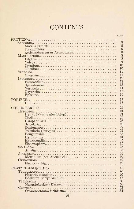
CONTENTSPAGE
PROTOZOA 1
SARCODINA 5
Amoeba proteus 5
Foraminifera 6
Actinosphserium or Actinophrys 6
MASTIGOPHORAEuglena 8
VolvoxCeratiumNoctiluca 10
SPOROZOAGregarina 11
INFUSORIAParameciumSpirostomumVorticeila
Oxytricha 15
Ephelota 16
PORIFERA 17
Grantia 18
CCELENTERATA 22
HYDROZOA 24
Hydra (Fresh-water Polyp) 24Obelia 26
Campanularia 28Sertularia 29Gonionemus 29Tubularia (Parypha)
BougainyilliaHydractinia 34
Hydrocorallina 35
Siphonophora 35SCYPHOZOA 35
Aurelia 35ACTINOZOA 40
Metridium (Sea-Anemone) 40CTENOPHORA 42
Pleurobrachia 42
PLATYHELMINTHES 45TURBELLARIA 46
Planaria maculata 46Bdelloura or Syncoelidium 47
TREMATODA 50Haematoloechus (Distomum) 50
CESTODA 52Crossobothrium lacmiatum 52
Vll

Vlll CONTENTS
PLATYHELMINTHES (Continued). PAGE
NEMERTINEA 55Tetrastemma 55
NEMATHELMINTHES 57Ascaris 57Trichina 58
TROCHELMINTHES 60ROTIFERA 60
Brachionus (A Rotifer) 60
MOLLUSCOIDA 62POLYZOA 62
Bugula 62Plumatella 64
BRACHIOPODA 64Terebratulina 64
ECHINODERMATA 66ASTEROIDEA 67
Asterias (Starfish) 67OPHIUROIDEA 73
Ophiura (Serpent-Star) 73ECHINOIDEA 74
Strongylocentrotus (Sea-Urchin) 74HOLOTHUROIDEA 80
Thyone (Sea-Cucumber) 80
ANNELIDA 84CaETOPODA 85
Nereis virens (Clam-Worm) 85
Autolytus cornutus 89
Polynce (Lepidonotus) squamatus 89
Diopatra cuprea 91
Chaetopterus 92
Amphitrite ornata 93Pectinaria (Cistenides) gouldii 94
Clymenella torquata 94Arenicola cristata 95Sabella microphthalma 95
Hydroides 96
Spirorbis borealis 96Lumbricus (Earthworm) 97Macrobdella (Leech) 102
GEPHYREA 106
Phascolosoma 106
MOLLUSCA 109
LAMELLIBRANCHIATA Ill
Venus mercenaria (Quahog) Ill
Yoldia limatula 119
Mytilus or Modiola (Mussels) 121
Pecten irradians (Scallop) 122
Ostrea virginiana (Oyster) 123
Solemya"
124
Mya arenaria (Long Clam) 124
Ensis directus (Razor-shell) Clam 125

CONTENTS IX
MOLLUSCA (Continued).AMPHINEURA ............................................. 127
Chsetopleura ...................................... .... 127GASTROPODA .............................................. 128
Busycon (Fulgur, Sycotypus) ........................... 128CEPHALOPODA ............................................ 137
Loligo pealeii (Squid) .................................. 137
ARTHROPODA .............................................. 148CRUSTACEA ............................................... 152
Homarus americanus (Lobster) ......................... 152Callinectes hastatus (Blue Crab) ........................ 159
Eupagurus (Hermit Crab) .............................. 163
Hippa (Sand Mole) .................................... 164
Squilla ............................................... 165
Mysis ................................................ 166Talorchestia (Beach-Flea) .............................. 167Porcellio or Oniscus (Sow-Bug) ......................... 168
Caprella ............................................. 168
Branchipus (Fairy Shrimp) ............................. 169
Daphnia ............................................. 169
Cyclops (Water-Flea) .................................. 170Argulus (Fish-Louse) .................................. 171
Lepas (Goose-Barnacle) ................................ 172ARACHNOIDEA ............................................. 173
Limulus (Horseshoe Crab) .............................. 173Buthus (Scorpion) ..................................... 175
Epeira (Round-Web Spider) ............................ 175Phoxichilidium ........................................ 177
MYRIAPODA .............................................. 178Lithobius (Centipede, Earwig) .......................... 178Julus (Thousand-legs) ................................. 179
INSECTA .................................................. 179Acridium (Grasshopper) ................................ 179
Apis mellifica (Honey-Bee) ............................. 185
CHORDATA ................................................. 190ENTEROPNEUSTA .......................................... 192
Dolichoglossus (Balanoglossus) Kowalevskii .............. 192
Molgula manhattensis ................................. 193
Perophora ............................................ 197
BotryUus ............................................. 198Amarcecium (Sea-Pork) ................................ 199
Salpa cordiformis ...................................... 201CEPHALOCHORDA .......................................... 202
Amphioxus lanceolatus ................................. 202
NOTES FOR GUIDANCE IN MAKING PERMANENT PREPA-RATIONS ................................................. 204
GLOSSARY .................................................. 209
INDEX. 223


INVERTEBRATE ZOOLOGY.
PROTOZOA.
Unicellular Animals.
CLASS 1. Sarcodina.
No permanent locomotor organs in "adult"
phase of the life history; the cells moving and
ingesting food by temporary pseudopodia."Young" phases may be amoeboid or flagellate.
(Minchin, pp. 178 and 234-237.)Subclass 1. Rhizopoda.
Forms with branched, root-like pseudopodia.Locomotion chiefly by creeping.
Order 1. Amcebcea.
Simple amoeboid forms, typically with lobose
pseudopodia; with or without a simple test.
(Amoeba, Arcella, Diffiugia.)Order 2. Foraminifera.
Chiefly marine forms with reticulose pseudo-podia and complex tests. (Lecythium, Globi-
gerina.)Subclass 2. Actinopoda.
Chiefly spherical floating forms with slender
unbranched radiating pseudopodia supportedby an internal axial filament.
Order 1. Heliozoa.
Fresh-water forms without a "central capsule"
separating ectoplasm and endoplasm. (Actin-
osphserium, Actinophrys, Clathrulina.)Order 2. Radiolaria.
Marine forms with a central capsule. (Thalas-
sicolla.)
CLASS 2. Mastigophora.Locomotor organs of adult phases consist of
one or more vibratile lash-like appendages or
flagella.
1 1

2 PROTOZOA.
Subclass 1. Flagellata.
Typically small forms with a definite anterior
end with one or more flagella. All types of
nutrition represented.Order 1. Pantostomina.
Nutrition holozoic. Cell more or less amoeboid.
One or several flagella. (Mastigamoeba ;Mul-
ticilia.)
Order 2. Protomonadina.Nutrition holozoic, saprophytic or parasitic.
One flagellum or a principal flagellum and one
or two accessory flagella. (Monas, Cercomonas,Bodo, Trypanosomes, Choanoflagellates.)
Order 3. Polymastigina.Three to eight flagella usually of nearly equal
length. Entozoic. (Trichomonas.)Order 4. Euglenpidina.
Typically large complex forms with one prin-
cipal flagellum, mouth aperture and vacuole
system. Frequently provided with stigma and
chlorophyll apparatus for holophytic nutrition.
(Euglena, Peranema, Phacus.)Order 5. Chromomonadina.
Small forms with one or two flagella, and usu-
ally one or two large chromatophores. Typic-
ally holophytic. (Dinobryon, Chilomonas,
Cryptomonas.)Order 6. Phytomonadina.
Holophytic forms enclosed in cellulose envelope.
Colony formation common. Usually two fla-
gella. (Sphserella, Gonium, Volvox.)Subclass 2. Dinoflagellata.
Two flagella, one of which encircles the cell.
Test of cellulose plates. Typically holophytic.Subclass 3. Cystoflagellata.
Large marine forms, with locomotion by a
typical flagellum and a "tentacle," or by rhyth-mic contractions of the body. (Noctiluca,
Craspedotella.)CLASS 3. Sporozoa.
Without flagella or cilia in the adult period of
the life-cycle. Reproduction is by spore-for-mation. All are endoparasites.

PROTOZOA. O
Subclass 1. Telosporidia.
Speculation phase of the life-cycle is distinct
from and follows the trophic phase. Sporozo-ites are gregarinulse.
Order 1. Gregarinoidea.The young stages are intracellular parasites,
while the adults are free and motile in cavities
of the body of the host. Sporulation occurs
within a cyst during the free period of the life-
cycle. (Gregarina.)Order 2. Coccidia.
Without a free and motile adult stage. Re-
production by both schizogony and sporogony,the latter occurring within a cyst during the
intracellular period of the life-cycle. (Eimeria.)
Order 3. Hsemosporidia.Living chiefly in the blood-corpuscles of verte-
brates. In many forms the entire sexual
period of the life-cycle takes place in an
intermediate host, as the mosquito. (Plas-
modium.)Subclass 2. Neosporidia.
Sporulation takes place during the "trophic"
phase of the life-cycle. Sporozoites are amce-
bulse.
Order 1. Myxosporidia.Typically tissue parasites. Principal trophic
phase is a multinucleate plasmodium. Spores
usually with two polar capsules. (Myxidium.)Order 2. Actinomyxidia.
Spore-forming phase is a binucleate amcebula.
Spore large with three polar capsules. (Sphse-
ractinomyxon.)Order 3. Microsporidia.
Amceboid trophozoites. Spores very minute
and with but one polar capsule. (Nosema.)Order 4. Sarcosporidia.
The initial stage of the life-cycle occurs in the
muscle cells of vertebrates. Spores with a
single polar capsule. (Sarcocystis.)
Order 5. Haplosporidia.
Spores are simple uninucleate cells without
polar capsules. (Haplosporidium.)

4 PROTOZOA.
CLASS 4. Infusoria.
With motile organs in the form of cilia duringall or part of the life cycle. Nucleus dimorphic(macronucleus and micronucleus). Reproduc-tion is by simple transverse division or by bud-
ding.Subclass 1. Ciliata.
With cilia throughout the life-history
Order 1. Holotrichida.
The cilia are of approximately equal length and
equally distributed over the body. Tricho-
cysts are frequently present. (Prorodon, Didi-
nium, Paramecium.)Order 2. Heterotrichida.
With a uniform covering of cilia, together with
an "adoral zone" formed of cilia fused into
membranelles. (Spirostomum, Stentor, Halte-
ria.)
Order 3. Hypotrichida.The cilia are limited to the ventral surface of a
dorso-ventrally flattened body. Cilia often
fused into cirri, membranelles, etc. (Oxy-tricha, Pleurotricha, Euplotes, Stylonychia.)
Order 4, Peritrichida.
More or less bell-shaped in form. Cilia usuallyreduced to those constituting the adoral zone.
(Vorticella, Zoothamnium, Lichnophora.)Subclass 2. Acinetaria.
Usually possessing cilia only during the embry-onic stages of the life-history. Tentacles
adapted for piercing and sucking are present.
(Podophrya, Ephelota, Acineta.)
Blochmann: Die Mikroscopische Tierwelt des Siisswassers. Abt. 1. Pro-
tozoa, 1895.
Biitschli: Protozoa. Bronn's Thierreich, 1889.
Calkins: Protozoa, 1901.: Protozoology, 1909.: Marine Protozoa of Woods Hole. Bui. U. S. Fish. Com., 1901.
Conn: Fresh Water Protozoa of Connecticut. Bui. State Nat. Hist.
Surv., 1905.Doflein: Lehrbuch der Protozoenkunde. 4 Auf., 1916.
Edmondson: Protozoa of Iowa. Davenport Acad. Sci., 1906.
Hartrnann: Praktikum der Protozoologie, 1910.
Jennings: Behavior of the Lower Organisms, 1906.
: Life and Death, Heredity, and Evolution in Unicellular Organisms,1920.

AMOEBA PROTEUS. 5
Kent: Manual of the Infusoria, 1881.
Lankester: Treatise on Zoology. 1. Protozoa.
Leidy: Fresh Water Rhizopods of North America, 1879.
Maupas: Studies on Infusoria, in Arch. Zool. Exp. et Gen., 1883, 1888, and1889.
Minchin: Protozoa, 1912.
Prowazek: Einfiihrung in die Physiologic der Einzellgen (Protozoen), 1910.
: Taschenbuch der Mikroskopischen der Protistenuntersuchen, 1907.
Stokes : Contribution Toward a History of the Fresh Water Infusoria of the
United States. Jour. Trenton Nat. Hist. Soc., 1, 1888.
Whipple: Microscopy of Drinking Water, 3d ed., 1914.
Woodruff: Observations on the Origin and Sequence of the Protozoan
Fauna of Hay Infusions. Jour. Exp. Zool., 12, 1912.
SARCODINA.
AMOEBA PROTEUS.
Amoebae are usually just discernible under the low power of
the microscope as irregular, semi-transparent, granular bodies.
Find a specimen in the material provided, which is known to con-
tain amcebse, and determine the following points:
1. With the high power observe the peculiar method of loco-
motion, the constant but slow change in the shape of the body
by means of projections, pseudopodia, or"false feet."
Make sketches at intervals of one or two minutes to show the
changes in the form of the body.
2. Observe the peripheral zone of hyaline protoplasm, the
ectoplasm, and compare this with the inner protoplasm, the endo-
plasm. Observe in detail the formation of a pseudopodium.Does the endoplasm extend into the pseudopodium? Can you
explain how the movement is caused ?
3. Find a clear space which appears and disappears at inter-
vals; this is the contractile vacuole. Determine the length of
time between successive contractions. Are the intervals regu-
lar? When the vacuole contracts what becomes of the con-
tents? What is its function?
4. Note the oval or rounded nucleus moving with the flowing
endoplasm. What is its structure?
5. Food materials in process of digestion are readily seen.
Of what do they consist? They are contained in gastric vacu-
oks. By careful watching, it is often possible to observe the man-
ner in which food is ingested and the manner in which the undi-
gested matter is egested.

6 PROTOZOA.
Make a careful drawing of an Amceba.
Amoebae of various kinds represent in many respects the
simplest type of protozoan, and are therefore placed in the first
class of these animals, the Sarcodina. The individuals of this
class all possess pseudopodia, but many are quite unlike those
of Amceba. Look over the figures of various Rhizopoda.If time and material permit, study Amoeba verrucosa, Arcella,
and Difflugia, and compare them with Amceba proteus. Do youunderstand how the shells of the last two genera are made,and of what service they are?
Drawings of these forms are desirable.
Calkins: Genera and Species of Amoeba. Trans. Fifteenth International
Congress on Hygiene, 1912. The Fertilization of A. proteus. Biol.
Bui., 13, 1907.
Bellinger : Locomotion of Amceba and Allied Forms. Jour. Ex. Zool., 3,1906.
Metcalf : Amceba Studies. Jour. Ex. Zool., 9, 1910.
Popoff: Ueber den Entwicklungs cyclus von A. minuta. Arch. f. Protis-
tenk., 22, 1911.
THE FORAMINIFERA.
With very few exceptions Foraminifera are marine and pro-
vided with shells. Empty shells from deep-sea dredgings or from
the sand beaches of such islands as the Bermudas may be had
for study. Examine them with a low power by reflected light.
1. Carefully examine various shells, compare them with each
other and with figures. Notice the great variety in size and
shape and determine how the chambers must have been added
during growth.
2. Observe a single opening in a shell, and determine whether
the general surface has any finer perforations. Be sure to under-
stand the relation of the live animal to the shell.
Make drawings of several types of shells.
Farmer: Foraminifera, pp. 133-139, Lankester's Treatise.
Flint: Recent Foraminifera. Rep. U. S. Nat. Mus., 1897.
ACTINOSPHAERIUM OR ACTINOPHRYS.
Find, as usual, with the low power, and increase the magni-
fication as occasion demands.
1. Note the many fine radiating pseudopodia. These are quite
ACTINOSPH^RIUM OR ACTINOPHRYS. 7
stiff compared with those of Amoeba and for a considerable time
show little change, not being pushed out and retracted constantly
as in Amoeba. Is the animal flat or spherical?
2. Both ectoplasm and endoplasm are so filled with vacuoles
that they present a frothy appearance characteristic of most
Heliozoa. The endoplasm of all Protozoa is alveolar in struc-
ture, but in Actinospha3rium the vacuoles are exceptionally
large, though not as large as those in the ectoplasm. In Ac-
tinophrys the endoplasm is not so sharply separated from the
ectoplasm.
3. The nucleus of Actinophrys is present in the center of the
organism, but it is somewhat difficult to demonstrate in the live
animal. In ActinosphaBrium there are many nuclei.
4. At some point near the periphery, the contractile vacuole
can usually be seen. When it is found notice its action, and
immediately after it has contracted look among the pseudopodiaof that region for indications of its extruded contents.
Draw a specimen, indicating all of the points observed.
5. When the contractile vacuole discharges, or when any
foreign body touches the ends of the pseudopodia, notice the
way in which this type of pseudopodium is moved. What does
this indicate in regard to its structure ? How far do the pseudo-
podia extend? They may be seen to contain minute granules
when studied with the high power and best light.
6. If possible, observe the process of catching food with the
tips of the pseudopodia and the manner in which it is drawn
toward the body. Note any motion on the surface of the bodyas the food is drawn closer, and also the manner in which the
food is finally ingested. Are there any indications that the
pseudopodia extend as still finer filaments beyond the point to
which it is possible to trace them with the highest magnifica-
tion at hand? If the capturing of food is observed, make* a
series of diagrams to illustrate the process. (Minchin, p. 50, and
Doflein, pp. 223, 707.)
If possible, observe a specimen undergoing division. Draw.It is desirable to examine Clathrulina, noting the stalk and
skeleton. Look over figures.
8 PROTOZOA.
R. Hertwig: Ueber die Kernteilung, Richtungskorperbildung und Befruch-
tung bei Actinosphserium. Abt. d. Math. Phys. Kl. d. Ak. d. Wiss.,
Miinchen, 19, 1898.
MASTIGOPHORA.
EUGLENA.
Understand its habitat and with what forms it is usually
associated.
1. Observe the free-swimming movements of the organism,and the euglenoid changes in the form of the body.
Make drawings showing the changes in the shape of a single in-
dividual.
2. Distinguish anterior and posterior ends. Is there anydorso-ventral differentiation? Note the motile organ, the flagel-
lum. Where is it attached? What relation does it bear to the
gullet? How is it directed during locomotion of the organism.
Does it serve any other purpose besides locomotion? (Minchin,
p. 52.)
3. The green color of Euglena is due to chlorophyl, and this
enables it to live in clear water, being nourished like a typical
green plant. (Minchin, p. 14.)
4. Note the absence of color near the anterior and posterior
ends of the organism. Near the anterior end also notice the red
pigment spot, or stigma. What is its probable function?
5. Stain a specimen with iodin and look for the nucleus.
It is somewhat obscured by the chlorophyl.
6. Observe specimens in the resting stage.
Make a drawing showing all of the points observed.
Look through the stock cultures for other forms of Masti-
gophora, such as Trachelomonas, Peranema, Phacus, etc.
It is desirable to make drawings of the different forms.
Klebs: Ueber die Organisation einiger Flagellatengruppen und ihre Be-
ziehungen zu Algen und Infusorien. Unt. Bot. Inst. Tubingen, 1, 1883.
-: Flagellatenstudien. Zeit. f. Wiss. Zool., 55, 1893.
Walton: Review of the Order Euglenoidina. Ohio State Univ., 1915,

VOLVOX. 9
VOLVOX.
Volvox globator is better for study than V. aurens. It maybe distinguished from the latter by the larger size of the colony,
the greater number of cells that compose it (about 15,000), the
angular shape of the individual cells, and the stout connecting
processes of protoplasm, into which chromatophores may enter.
Observe the movements of colonies in a watch-glass of water,
with the naked eye and with a low power of the microscope.1. Do the colonies tend to collect toward a particular side
of the dish? What reason is there for the reaction?
2. Place a number of colonies on a slide with enough water
to allow them to be covered without crushing them. Studyfirst with the low and then with the high power and determine
the species. Understand the relation of the individual cells to
the colony. (See Doflein, p. 240.)
Draw a figure showing several cells and their protoplasmic con-
nections.
3. Compare in detail an individual cell with Euglena.4. Observe, if possible, certain cells, called parthenogonidia,
which are specialized for asexual reproduction. These divide and
form the daughter colonies, which become detached and swimin the interior of the parent colony. They are finally liberated
by the rupture of the wall of the parent colony.
Make a figure of a parent colony that incloses several daughter
colonies of different sizes.
5. V. globator is monoecious. Look for macrogametes and
bundles of microgametes.
Figure them.
6. Be sure to recognize the significance of the fact that the
cells of Volvox are differentiated into somatic and germ cells,
and to understand the resulting physiological division of labor.
(Calkins, Protozoa, p. 232.)
7. Consider the reasons for and against regarding Volvox
and allied organisms as animals rather than plants.
Meyer: Ueber den Bau von V. aurens and V. globator. Bot. Cent., 63,1895,
10 PROTOZOA.*
,
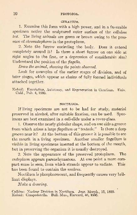
CERATIUM.
1. Examine this form with a high power, and in a favorable
specimen notice the sculptured outer surface of the cellulose
test. The living animals are green or brown owing to the pres-ence of chromatophores in the protoplasm.
2. Note the furrow encircling the body. Does it extend
completely around it? Is there a short furrow on one side at
right angles to the first, or a depression of considerable size?
Understand the position of the flagella.
Draw the animal, showing the points observed.
Look for examples of the earlier stages of division, and of
later stages, which appear as chains of fully formed individuals
attached together.
Kofoid: Exuviation, Autotomy, and Regeneration in Ceratiurn. Univ.Calif., Pub. 4, 1908.
NOCTILUCA.
If living specimens are not to be had for study, material
preserved in alcohol, after suitable fixation, can be used. Spec-
imens are best examined in a cell-slide under a cover-glass.
1. Observe the nearly globular shape, and on one side a groove
from which arises a large flagellum or"tentacle." Is there a deep
groove near it ? At the bottom of this groove it is possible to see
the mouth in a living specimen. Another smaller flagellum is
visible in living specimens inserted at the bottom of the mouth,
but in preserving the organism it is usually destroyed.
2. Note the appearance of the preserved protoplasm. The
endoplasm appears parenchymatous. At one point a more com-
pact mass is seen, from which strands appear to radiate. This
has been found to contain the nucleus.
Noctiluca is phosphorescent, and frequently causes very bril-
liant displays.
Make a drawing.
Calkins: Nuclear Division in Noctiluca. Jour. Morph., 15, 1899.
Kofoid: Craspedotella. Bull. Mus., Harvard, 46, 1905.

GREGARINA. 11
SPOROZOA.
GREGARINA.
Remove the head and posterior end of a larval or adult
meal beetle and pull out the digestive tract with a pair of for-
ceps. Place the digestive tract on a slide, split it open length-
wise with a sharp scalpel, and then spread it out, with the
inner wall exposed, and cover. The operation should be per-
formed rapidly to prevent the material from drying. If the
beetle is infected, numerous gregarines will be visible under the
microscope. Study with low and high powers.
1. Does the animal move? A great number of refractive
granules are present in the protoplasm. They are regarded as
reserve nourishment. They can be removed with acid.
2. Note that the body is covered with a membrane, and is
divided into a dense superficial layer, the ectoplasm, and a cen-
tral, more fluid mass, the endoplasm.
3. The endoplasm is separated into two parts by a portion
of the ectoplasm. The anterior part is termed the protomerite,
and the posterior part the deutomerite. In which is the nucleus
situated ?
4. Is it possible to distinguish a layer of myonemes just ex-
ternal to the endoplasm?5. Is there another section of the body just anterior to the
protomerite? If so, this is the epimerite.
6. Note that occasionally two (or more) individuals are
united. These aggregations are termed syzygies.
Before reproduction Gregarina throws off the epimerite,
leaves it in the cell-host, and falls into the lumen of the digestive
tract. It then encysts, and the protomerite and the deutome-
rite form one spore-producing individual. The attached stage
in the life-history of Gregarina is termed the cephalont, and the
detached stage, the sporont. (Calkins' Protozoa, Fig. 77, and
Minchin, Fig. 7.)
Make a drawing.
Berndt: Beitrag zur Kenntnis der im Darme der Larve von Tenebrio moli-
tor lebenden Gregarinen. Arch. f. Protistenk., 1, 1902.Minchin: Sporozoa, pp. 177-179, Lankester's Treatise.

12 PROTOZOA.
INFUSORIA. .
PARAMECIUM.
Place a drop of the culture on a slide, cover, and examine
with the low power.
1. In an animal not closely confined note the shape and
movements. Is it possible to distinguish an anterior and a
posterior end? A forward and backward movement? Is one
side of the animal kept constantly uppermost ? Is there a dorsal
and ventral surface? Do the animals change their shape either
permanently or temporarily? Individuals tend to collect about
air-bubbles and at the edge of the cover-glass. Why?Indicate by a sketch all the points which can be determined
with the low power.
2. Draw off all superfluous water by means of filter-paper,
add a trace of powdered carmine, and then find a specimen
which is narrowly confined and examine it with the high power.
The particles of carmine are taken into the body. Deter-
mine how and where. Note that the carmine collects in gastric
vacuoles. What do you think is probably the nature of the
|luid in the vacuoles? In watching them do you notice any
definite movement of the protoplasm? Try to see the undi-
gested material ejected.
3. Determine the arrangement of the cilia, and the nature
of their motion. Is there a reversal of the direction of the stroke,
etc.? 1
4. Observe the contractile vacuoles. How many are there?
Is their position constant? What is their action? In com-
pressed specimens the contractile vacuoles and their reservoirs
are usually conspicuous. Note the order of appearance and
disappearance of the vacuoles and reservoirs.
5. Focus carefully on the margin of the body and note a verythin outer cuticle. A thick layer, the ectoplasm, devoid of gran-ules but containing radially arranged, minute, oval bodies, the
trichocysts, is just internal to the cuticle. The inner mass of1 It is possible to decrease the rate of movement of the animals by placing
them in a solution of quince seed jelly. Specimens so treated remain alivefor some time.

SPIROSTOMUM. 13
protoplasm, containing the contractile and gastric vacuoles,
and small granules, is the endoplasm.
6. If possible distinguish the clear, centrally located nucleus
(macronucleus).
Make a sketch showing all of the above points.
7. Kill the animal by running a drop of methyl-green under the
cover-glass. What happens to the cilia? To the trichocysts?
Sketch the trichocysts with the threads protruded, and also note
and sketch the macronucleus and the micronucleus.
8. Observe, if possible, animals dividing and conjugating.
9. Study demonstrations of permanently stained specimens
for finer structure.
Calkins and Cull: Conjugation of P. caudatum. Arch. f. Protistenk.,
10, 1907.
Jennings: Effect of Conjugation in Paramecium. Jour. Exp. Zool., 14, 1913.
Metalinkow: Contributions a 1'eHude de la digestion. Arch d. Zool. Exp.et Gen., 9, 1912.
Woodruff: Paramecium aurelia and Paramecium caudatum. Jour.
Morph., 22, 1911.
Woodruff and Erdmann: A Normal, Periodic Reorganization Process
(Endomixis) Without Cell Fusion in Paramecium. Jour. Exp. Zool., 17,1914.
SPIROSTOMUM.
1. Compare Spirostomum with Paramecium, noting the
method of locomotion, the shape of the body, the ciliation, the
buccal groove and mouth, and the large excretory reservoir, fill-
ing the posterior end of the body and in communication with
the anterior end of the body by a canal.
2. Note the highly refractive, long, band-like (moniliform)macronucleus. In another species of Spirostomum the macro-nucleus is similar to that of Paramecium.
3. Note the sudden contractions of the body. When these
occur spiral lines appear on the surface. Can you distinguishthese lines when the animal is extended? These are primitivestructures (myonemes) functioning as muscles.
Make a drawing of the extended animal and a diagram show-
ing the Jorm when contracted. (Doflein, p. 1123.)

14 PROTOZOA.
VORTICELLA.
Place a number of individuals on a slide and cover loosely
to avoid crushing. As usual, study first with the low power and
then with the high.
1. Notice that the body of Vorticella has the general shapeof an inverted bell. The covering of the body is a very thin
transparent layer, the cuticle, underneath which is the periphe-
ral layer of ectoplasm enveloping the more fluid and granular
endoplasm.
2. The peristome is the rounded rim about the base of the bell.
3. The elevated and inclined area included within the peri-
stome, and ciliated around the edge, is the disk. It is some-
what convex.
4. The marked depression between the disk and the peri-
stome is the vestibule. It is also lined with cilia. The vestibule
defines the ventral surface of the animal.
5. The gullet, a slender canal, leads from the vestibule toward
the center of the body.6. The feces escape from the body by the side of the vestibule.
The opening is temporary.7. Within the endoplasm are situated the clear contractile
vacuole, several gastric vacuoles, the long U-shaped macronucleus,
and the small round micronucleus. The macronucleus may be
made more distinct by treating with methyl-green.
8. The stalk is composed of a sheath, which is continuous with
the cuticle of the body, and, within the sheath, the contractile
axis or myoneme, which is continuous with the body ectoplasm.
Notice that this myoneme is situated within the sheath in a
very loose spiral, and that the stalk quickly contracts into a
close spiral when the animal is stimulated. Observe also the
manner in which the peristome folds over simultaneously with
the contraction of the stalk. What purpose does the contrac-
tion of the stalk serve?
Vorticella is distinguished from its allied genera by its sim-
ple unbranched stalk and also by the spiral form assumed by the
contracted stalk. In which order of the Ciliata does the cilia-
tion of Vorticella place it? Compare with Zoothamnium.

OXYTRICHA. 15
Make a drawing of an expanded individual and a sketch to
show the condition when contracted. (Minchin, p. 434; Doflein,
p. 1144.)
9. Study, by means of finely powdered carmine, the vortex
currents set up by the cilia. Note how the particles are collected
in the gullet, and at intervals are forced in rounded masses into
the endoplasm to form gastric vacuoles. Is there a definite
circulation in the endoplasm ?
10. Endeavor to find several stages of reproduction by divi-
sion.
Large fresh-water species of Vorticella are preferable for
study, but marine species may be substituted when necessary.
If time and material permit, study Lichnophora, a marine peri-
trichous form parasitic on Crepidula. (See Calkins' Protozoa,
p. 203.)
Schroder: Beitrage zur Kenntnis von V. monilata. Arch. f. Protistenk., 7,
1906.OXYTRICHA.
Infusoria belonging to the genus Oxytricha, or the genera
Stylonychia, Pleurotricha, Euplotes, etc. (Doflein, p. 147),
may be used for the following study. These forms belongto the order Hypotrichida. Hypotrichous forms are amongthe most highly organized of the class Infusoria, as well as of
the entire phylum of Protozoa, and present a complexity of
structure and function which probably is not exceeded within
the limits of a single cell elsewhere in the animal series.
1. In an animal which is becoming quiet, note the mode of
locomotion, the shape of the body, the buccal groove, the con-
tractile vacuole, etc., as in other forms studied. Compare the
dilation with that of other forms. Refer to Calkins' Protozoa,
Fig. 98, and understand the relation of cirri, membranelles, etc.,
to cilia.
Draw, showing the structure in detail.
2. Run some methyl-green under the cover-glass. Whatis the shape of the macronucleusf The shape varies consider-
ably in the different genera. Is it possible to distinguish the
micronuclei?

16 PROTOZOA.
3. Prepare a fresh slide and observe in detail the character-
istic movements and manner of creeping over various objects.
As the animal turns sidewise, note the marked dorso-ventral
compression of the body.
Represent this diagrammatically beside the previous drawing.
It is desirable to examine permanently stained preparations
for division stages, finer details of the nuclei, etc.
Maier: Ueber den feineren Bau der Wimperapparate der Infusorien. Arch.f. Protistenk., 2, 1903.
Wallengren: Zur Kenntnis des Neubildungs und Resorptionsprocess bei
den Teilung der Hypotrichen Infusorien. Zool. Jahrb., 15, 1901.
EPHELOTA.
Mount a small piece of hydroid under a supported cover-
glass and with a low power observe the suctorians attached
by delicate stalks. Select a field where the animals are abun-
dant and study under a high power.
1. Note the general shape of the cell and the distribution
of the tentacles. Draw. Are all of the tentacles of one kind?
Observe the movements of the tentacles and their use. Is
there any morphological relation between tentacles and cilia?
(See Minchin's Protozoa, p. 458.)
2. Study the method of exogenous budding. What is the
relation of this type to simple division? Is the number of
buds in process of formation the same on all specimens?
3. Fix, stain, and mount in balsam a piece of hydroid with
many Ephelota attached. Under the high power note the
character of the macronucleus and its relation to the buds.
Are micronuclei visible?
4. Examine carefully the relation of the stalk to the cell
body. Compare with that of Vorticella.
If the material is available study Podophrya and allied
forms, with particular reference to the method of budding.
Collin: Etude monographique sur les Acine"tiens. Arch. Zool. Exp. et
Gen., 1911 and 1912.
Root: Reproduction and Reactions to Food in the Suctorian, PodophryaCollini, n. sp. Arch. f. Protistenk., 35, 1914.

PORIFERA.
Cells not differentiated to form definite organs. Water
admitted through surface pores and ejected through an osculum
or through oscula.
CLASS 1. Calcarea.
With a skeleton composed of calcareous spicules.Subclass 1. Homoccela.
With the gastreal layer continuous so the col-
lar cells line the whole gastreal cavity. (Leu-
cosolenia.)Subclass 2. Heteroccela.
Gastreal layer discontinuous. Collar cells restrict-
ed to the flagellated chambers. (Grantia.)CLASS 2. Hexactinellida.
With a skeleton composed of siliceous six-rayed
spicules.Order 1. Lyssacina.
Spicules separate or becoming united. (Eupleo-tella.)
Order 2. Dictyonina.
Spicules united from the first into a firm frame-work. (Eurete.)
CLASS 3. Demospongise.Great diversity of structure. Dominant formsof today.
Subclass 1. Tetraxonida.
Typically with four-rayed spicules. (Corticella.)Subclass 2. Monaxonida.
Simple, usually unbranched spicules. Sponginfrequently present. (Cliona, Suberites, Chalina,
Spongilla.)Subclass 3. Keratosa.
Skeleton of spongin fibers. No true spicules.
(Euspongia, Aplysina.)Subclass 4. Myxospongida.
Without skeleton. (Oscarella.)2 17

18 PORIFERA.
Lankester: A Treatise on Zoology, Porifera, and Coelenterata, Pt. 2, 1900.Moore: A Practical Method of Sponge Culture. Bui. U. S. Bur. Fish., 28,
1908.
: The Commercial Sponges and the Sponge Fisheries. Bui. U. S. Bur.
Fish., 1908.
Parker: The Reactions of Sponges, with a consideration of the Origin of theNervous System. Jour. Ex. Zool., 8, 1910.
H. V. Wilson: On Some Phenomena of Coalescence and Regeneration in
Sponges. Jour. Ex. Zool., 5, 1907.
: Development of Sponges from Dissociated Tissue Cells. Bui. U. S.
Bur. Fish., 30, 1910.
GRANTIA.
This form is quite common along the New England coast,
where it occurs attached to rocks, seaweeds, and submergedwoodwork from just below the lowest tide-mark to a number of
fathoms in depth. You should visit an old wharf where speci-
mens may be found, and study their relation to the forms with
which they are associated. Specimens will be found to vary
considerably in size. The largest sometimes reach an inch in
length.
1. Examine a dry specimen and notice its general shape,
manner of attachment, and osculum. The osculum is surrounded
by a funnel of rather long spicules. Distributed over the gen-
eral surface, more or less hidden by the numerous spicules, are
many small pores. Their presence may be demonstrated more
satisfactorily later.
2. Look for indications of budding. If your specimen does
not show this, examine others.
Make an enlarged drawing of a sponge.
With a razor or sharp scalpel cut a dry specimen into halves,
with a stroke from base to osculum, and notice:
3. The central cavity or cloaca.
4. Many apopyles, the inner openings of tubes that are em-
bedded in the walls of the sponge, will be seen opening into
the cloaca. Are the apopyles arranged in any order?
5. With the low power of your microscope (with the light
turned off) examine the cut wall and find that it is traversed by
parallel tubes. Determine that these tubes are of two kinds.

GRANTIA. 19
(a) Regular, nearly cylindrical tubes that open into the
cloaca through the apopyles and that bear tufts of spicules on
their closed ends, at the surface of the body. These are the
radial canals. It is frequently hard to see their openings into
the cloaca, as the apopyles are narrow, so the section only occa-
sionally passes through them.
(b) Smaller and less regular tubes that open on the outer
surface between the clusters of spicules, and do not open into
the cloaca. These are the incurrent canals. In life there are
small pores, prosopyles, that open from the incurrent canals
into the radial canals. These openings are very minute and are
apparently capable of being closed. They are never visible
in dried material.
6. Examine thin, transverse sections of a dry sponge and
determine the positions of radial and incurrent canals.
Make a drawing that will show the arrangement of the canals.
7. Examine the spicules and determine their positions as
regards canals. Boil a portion of a sponge in caustic potash
until only the spicules remain and examine the spicules. See if
more than one kind occurs.
Draw specimens of the spicules.
LIVING AND SECTIONED MATERIAL.
1. Place a living sponge in a watch-glass of sea-water, add a
little powdered carmine, and examine it with the low power of
your microscope for currents of water. See if particles are mov-
ing in a definite direction near the general surface and near the
osculum.
2. With a sharp razor cut tangential sections of the wall,
as thin as possible, mount in sea-water under a cover, and
examine with a low power. This will show both incurrent and
radial canals in cross-section. How can you distinguish one
from the other? In a favorable place look for moving flagella.
Are flagella in all of the canals ? In favorable situations it
can be easily seen that the cells that have flagella possess
collars also. (Collars may be withdrawn by cells so they pro-
20 PORIFERA.
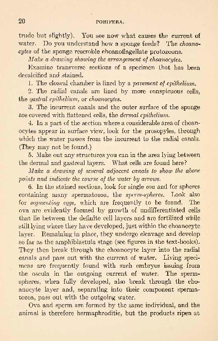
trude but slightly). You see now what causes the current of
water. Do you understand how a sponge feeds? The choano-
cytes of the sponge resemble choanoflagellate protozoons.Make a drawing showing the arrangement of choanocytes.
Examine transverse sections of a specimen that has been
decalcified and stained.
1. The cloacal chamber is lined by a pavement of epithelium.
2. The radial canals are lined by more conspicuous cells,
the gastral epithelium, or choanocytes.
3. The incurrent canals and the outer surface of the spongeare covered with flattened cells, the dermal epithelium.
4. In a part of the section where a considerable area of choan-
ocytes appear in surface view, look for the prosopyles, through
which the water passes from the incurrent to the radial canals.
(They may not be found.)
5. Make out any structures you can in the area lying between
the dermal and gastreal layers. What cells are found here?
Make a drawing of several adjacent canals to show the above
points and indicate the course of the water by arrows.
6. In the stained sections, look for single ova and for spheres
containing many spermatozoa, the sperm-spheres. Look also
for segmenting eggs, which are frequently to be found. The
ova are evidently formed by growth of undifferentiated cells
that lie between the definite cell layers and are fertilized while
still lying where they have developed, just within the choanocyte
layer. Remaining in place, they undergo cleavage and develop
so far as the amphiblastula stage (see figures in the text-books).
They then break through the choanocyte layer into the radial
canals and pass out with the current of water. Living speci-
mens are frequently found with such embryos issuing from
the oscula in the outgoing current of water. The sperm-
spheres, when fully developed, also break through the cho-
anocyte layer and, separating into their component sperma-
tozoa, pass out with the outgoing water.
Ova and sperm are formed by the same individual, and the
animal is therefore hermaphroditic, but the products ripen at

GRANTIA. 21
different periods and are seldom both present in an individual
at the same time.
// the time allows, draw ova, sperm-spheres, segmenting eggs,
and embryos.
It is desirable to examine specimens of Leucosolenia, a still
simpler sponge, and of some of the more complicated forms,
like commercial sponges, Spongilla, Cliona, and Chalina. Whyis more than a single osculum desirable in such forms? Under-
stand the relation of the internal structure of the complicated
forms to the more simple forms. What reason is there for the
complication?
The individual cells of sponges may be separated by squeez-
ing through fine silk bolting cloth. Such cells will come to-
gether in a dish of sea-water to form aggregates that will develop
into new sponges. (See Wilson, loc cit.)

COELENTERATA.
With a single continuous coelenteron or gastro-vascular cav-
ity. With the exception of the Ctenophora all have nettle cells.
There are two cellular layers and a mesoglea.
CLASS 1. Hydrozoa.Ccelenteron simple, without septa. Gonads usu-
ally ectodermal. Fully formed medusae have a
velum.
Order 1. Leptolinse.With a fixed zoophyte stage.
Suborder 1. Anthomedusse.Without hydrothecse or gonothecse. The medusabears gonads on the manubrium. (Bougain-villia, Clava, Hydra, Hydractinia, Tubularia.)
Suborder 2. Leptomedusse.With hydrothecse and gonothecse. The medusabears gonads on the radial canal. (Campanu-laria, Gonionemus, Obelia, Sertulfiria.)
Order 2. Trachylinse.Without fixed zoophyte stage.
Suborder 1. Trachymedusa?.Tentacles from the margin of the umbrella.
Gonads on the radial canals. (Petasus.)Suborder 2. Narcomedusse.
Tentacles from the exumbrella. Gonads on the
manubrium. (^Eginopsis.)Order 3. Hydrocorallina.
Massive calcareous exoskeleton. (Millepora.)
Order 4. Siphonophora.Pelagic. Colonial. Colony usually shows extreme
polymorphism of its zooids. (Physalia.)CLASS 2. Scyphozoa.
Body-wall of polyp thrown into four ridges
(tsenioles) which project into the ccelenteron.
Medusa generally without velum and with gas-
tric tentacles. Medusoid form predominating.22

CCELENTERATA. 23
Order 1. Stauromedusse.Conical or vase-shaped umbrella. No tentacu-
locysts. (Tessera.)Order 2. Peromedusse.
Conical umbrella with transverse constriction.
Four inter-radial tentaculocysts. (Pericolpa.)
Qrder 3. CubomedusaB.Four-sided umbrella. With per-radial tentacu-
locysts. Velum present. (Charybdea.)Order 4. Discomedusse.
Saucer-shaped umbrella. Per-radial and inter-
radial tentaculocysts. (Aurelia.)
CLASS 3. Actinozoa.
With a stomodaBum, and with mesenteries ex-
tending into the ccelenteron. Fixed forms.
Subclass 1. Zoantharia.
Mesenteries and tentacles usually very numerous.Order 1. Actiniaria.
Usually single. No skeleton. (Metridium. Sa-
gartia.)
Order 2. Madreporaria.
Usually form colonies and always have calcare-
ous exoskeleton. (Astrangia, Orbicella, Mean-drina.)
Order 3. Antipatharia.Tree-like. Mesenteries and tentacles compara-tively few. Chitinoid skeleton. (Cirripathes.)
Subclass 2. Alcyonaria.Mesenteries and tentacles eight in number. Ten-tacles branched.
Order 1. Alcyonacea.Skeleton in the form of small, irregular bodies,
frequently calcareous spicules. (Alcyonium,Tubipora.)
Order 2. Gorgonacea.Tree-like, with calcareous or horny exoskeleton.
No syphonoglyphes. (Gorgonia.)Order 3. Pennatulacea.
Colony with one end usually embedded in thesea-bottom. (Pennatula, Renilla.)
CLASS 4. Ctenophora.Single. Pelagic. Eight rows of meridional
swimming plates. No nettle cells.

24 CCELENTERATA.
Order 1. Cydippida.Nearly circular. Two tentacles, each of which
may be retracted into a sheath. (Pleurobra-
chia, Mnemiopsis.)Order 2. Lobata.
Compressed in the vertical plane. Two large
oral lobes. No tentacle-sheaths. (Deiopea.)Order 3. Cestida.
Ribbon-shaped. Two tentacles with sheaths, and
numerous other tentacles. (Cestus.)
Order 4. Beroida.
Laterally compressed. Without tentacles.
(Berce.)
Mayer: Mudusse of the World. Carnegie Inst., Wash., 1910.
Nutting: The Hydroids of the Woods Hole Region. Bui. U. S. Fish. Com.,19, 1899.
HYDROZOA.HYDRA. (Fresh-water Polyp.)
Hydra, the common fresh-water ccelenterate, is frequently
found in quiet pools or sluggish streams that contain lily-
pads, decaying leaves, and other vegetable matter. The ani-
mals may frequently be found by examining the surfaces of
submerged leaves, but it is usually better to allow such material
to stand in glass jars for a day or two, as the animals then
tend to collect on the lighter sides of the vessels. They are
easily kept in balanced aquaria.
Examine specimens in an aquarium and find what you can
about their mode of life. Do they form colonies?
Place a specimen in a watch-glass of water and examine it
with a lens.
1. What is its shape and color? Is it attached? If so, bywhat part of the body? Notice the circlet of tentacles. How
many are there? Compare notes with others and see if all have
the same number. How are they placed?
2. Does the Hydra move its body or tentacles? Is it sensi-
tive?
HYDRA. 25
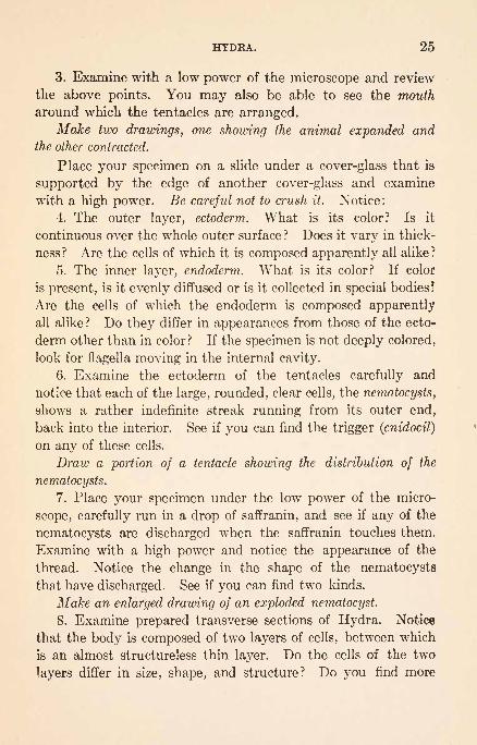
3. Examine with a low power of the microscope and review
the above points. You may also be able to see the mouth
around which the tentacles are arranged.
Make two drawings, one showing the animal expanded andthe other contracted.
Place your specimen on a slide under a cover-glass that is
supported by the edge of another cover-glass and examine
with a high power. Be careful not to crush it. Notice:
4. The outer layer, ectoderm. What is its color? Is it
continuous over the whole outer surface ? Does it vary in thick-
ness? Are the cells of which it is composed apparently all alike?
5. The inner layer, endoderm. What is its color? If color
is present, is it evenly diffused or is it collected in special bodies!
Are the cells of which the endoderm is composed apparently
all alike ? Do they differ in appearances from those of the ecto-
derm other than in color? If the specimen is not deeply colored,
look for flagella moving in the internal cavity.
6. Examine the ectoderm of the tentacles carefully and
notice that each of the large, rounded, clear cells, the nematocysts,
shows a rather indefinite streak running from its outer end,
back into the interior. See if you can find the trigger (cnidocil)
on any of these cells.
Draw a portion of a tentacle showing the distribution of the
nematocysts.
7. Place your specimen under the low power of the micro-
scope, carefully run in a drop of saffranin, and see if any of the
nematocysts are discharged when the saffranin touches them.
Examine with a high power and notice the appearance of the
thread. Notice the change in the shape of the nematocysts
that have discharged. See if you can find two kinds.
Make an enlarged drawing of an exploded nematocyst.
8. Examine prepared transverse sections of Hydra. Notice
that the body is composed of two layers of cells, between which
is an almost structureless thin layer. Do the cells of the two
layers differ in size, shape, and structure? Do you find more

26 COELENTERATA.
than one kind of cell in each or either of these layers? Where
are they? What are they?
Make a careful drawing of the section showing the arrangement
as you see it.
Examine longitudinal sections, for differences in the char-
acter of the ectoderm and endoderm in different parts of the
body.
9. Reproduction. Examine living specimens in a watch-
glass of water for bud formation and for sexual organs. Sperm-aries are just beneath the tentacles; ovaries, lower down;buds may be found at different levels. What cells are involved
in the formation of each of these?
Eggs are not formed at all seasons of the year and vary
greatly in appearance according to their stage of development.Make drawings of the stages of reproduction that you find.
Tannreuther: The Development of Hydra. Biol. Bui., 14, 1908.
Whitney: Artificial Removal of the Green Bodies from Hydra viridis.
Biol. Bui., 14, 1908.
OBELIA.
These small, colonial animals are common on submerged or
floating wood, stones, and seaweeds, where the water is rather free
from sediments. With the aid of a glass-bottomed pail they,
in company with many other forms, may usually be seen about
old wharfs.
Note the appearance of large colonies of this form that are
growing on stones or on pieces of board or kelp.
1. Notice the tree-like form of any single stem. Do the
branches have a definite size and arrangement ?
2. At the extremities of the branches are the single individuals,
hydranths or zooids. Each is similar to a single Hydra in cer-
tain ways, but is inclosed in a vase-like formation, the hydrotheca.
3. The latter is a continuation of a tough, membranous
sheath, the perisarc, which covers each part of the whole
colony.

OBELIA. 27
Do you notice any modifications of the perisarc below the
hydrotheca? Do the modifications serve any purpose?
4. Trace the stem to the creeping, stolon-like portion of
the colony, the hydrorhiza.
Make a drawing of a colony.
5. The fleshy continuation of the zooid down into the stalk
is termed the ccenosarc. Is it in close contact with the perisarc?
6. In an expanded hydranth, note the mouth, the arrange-
ment of the tentacles, and the number of tentacles. How is the
individual supported in the hydrotheca? Trace the coelenteric
cavity through branches and hydranths and determine whether
it is continuous.
7. Can you determine what keeps the fluid in the cavity in
motion?
8. Examine a hydranth with a high power and look for the
cell-layers characteristic of ccelenterates. Determine how its
tentacles differ from the tentacles of Hydra, and explode nemato-
cysts as in Hydra.Make a drawing of a hydranth.
9. Look for certain extremities which show neither tentacles
nor any opening in the outer covering. Such a condition sig-
nifies either an immature hydranth or a reproductive indi-
vidual. If the latter, it is considerably swollen and is termed
a gonosome. The central core of a gonosome, the blastostyle,
should be examined for gonophores, frequently called medusce
buds. This may require a high power. Determine how the
gonophores are arranged around the blastostyle. Are all in
equal stages of development? What relation has the end of the
blastostyle to the outer covering, the gonangium f
Make a drawing of a gonosome.
10. The free medusae are small, transparent, and easily
overlooked. During the breeding season they may usually
be found in abundance in dishes in which colonies have been
kept over night. Notice their movements and their positions
while at rest on the bottom. The number of tentacles and the
position of the sense organs is definite for the species. Two

28 CCELENTERATA.
species that differ in the number of tentacles are common at
Woods Hole. The inverted bell with the manubrium sticking
out from the convex surface of the resting specimen is char-
acteristic for this form. Notice the quick reversal when the
animal swims. The radial canals are easily seen, but the gonadsare not developed at the time of liberation. The velum is verysmall.
Gonionemus is a more favorable medusa to study. This
form is valuable for comparison.
CAMPANULARIA.
In structure and habits this form is so much like Obelia that
it is not easy to distinguish the two genera without studying the
gonosomes. Several species are found at Woods Hole, two of
which are usually abundant during the summer.
The gonosome of one species superficially looks like the
gonosome of Obelia, while the other has a notch on one side
near its extremity. In structure they are similar.
The blastostyle runs throughout the length of the gonangiumand gives rise to buds that develop into imperfect gonophores.The structure of these gonophores is difficult to make out in
fresh material. While they are comparable to medusse, theynever become detached, and organs usually present are largely
aborted.
The distinct manubrium of the male gonophore becomes
charged with sperm which, as they develop, press the ecto-
derm of the manubrium against the ectoderm of the sub-
umbrella. Ultimately the ectoderm of the manubrium rup-tures and the sperm escape through the sub-umbrellar cavity.
A female gonophore ripens usually one, sometimes two, eggs.
The mature egg before segmentation, which lies inside the
ectoderm of the manubrium, is flattened and molded between
the mass of the manubrium and sub-umbrellar wall. The
growth of the egg presses the manubrium to one side. Such
an egg appears as a brownish granular mass with a distinct
clear nucleus. The ectoderm of the manubrium ultimately
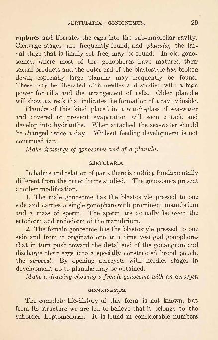
SERTULARIA GONIONEMUS. 29
ruptures and liberates the eggs into the sub-umbrellar cavity.
Cleavage stages are frequently found, and planulce, the lar-
val stage that is finally set free, may be found. In old gono-
somes, where most of the gonophores have matured their
sexual products and the outer end of the blastostyle has broken
down, especially large planulse may frequently be found.
These may be liberated with needles and studied with a high
power for cilia and the arrangement of cells. Older planulse
will show a streak that indicates the formation of a cavity inside.
Planulse of this kind placed in a watch-glass of sea-water
and covered to prevent evaporation will soon attach and
develop into hydranths. When attached the sea-water should
be changed twice a day. Without feeding development is not
continued far.
Make drawings of gpnosomes and of a planula.
SERTULARIA.
In habits and relation of parts there is nothing fundamentally
different from the other forms studied. The gonosomes present
another modification.
1. The male gonosome has the blastostyle pressed to one
side and carries a single gonophore with prominent manubriumand a mass of sperm. The sperm are actually between the
ectoderm and endoderm of the manubrium.
2. The female gonosome has the blastostyle pressed to one
side and from it originate one at a time vestigial gonophoresthat in turn push toward the distal end of the gonangium and
discharge their eggs into a specially constructed brood pouch,
the acrocyst. By opening acrocysts with needles stages in
development up to planulse may be obtained.
Make a drawing showing a female gonosome with an acrocyst.
GONIONEMUS.
The complete life-history of this form is not known, but
from its structure we are led to believe that it belongs to the
suborder Leptomedusse. It is found in considerable numbers

30 CCELENTERATA.
throughout the summer in the border of eel-grass in the Eel
Pond at Woods Hole, where it may be obtained with a dip-net.
It is more satisfactory to study than the medusa of Obelia, as
it is much larger and its movements and organs are more easily
observed. In plan of structure the two are quite similar.
Put a living specimen in a jar containing sea-water, or in
a finger-bowl, with a black tile beneath, and notice :
1. Its method of locomotion. To the contraction of what
part of the bell is movement due? How large is the jet of water
that is delivered from the bell? Why is the jet made narrow?
Does the jet necessarily leave at the center or may it be thrown
from one side? Should it be thrown from one side, what would
be the result?
2. Its position in the water when quiet. Why is this position
more desirable than the opposite? With a needle-point provethat various parts of the body are sensitive.
With either fresh or preserved material notice:
1. Its flattened dome-shape. The convex face is called the
ex-umbrella (aboral), while the concave portion is termed the
sub-umbrella (oral).
2. The velum is the perforated diaphragm that partly closes
in the sub-umbrella. All medusse possessing this structure are
classed as Craspedota. Do you understand its use?
3. In the center of the sub-umbrella is seen the large pen-
dent manubrium, at the extremity of which is a wide-lipped
mouth. If the medusa is alive, feed it with small bits of clam
meat.
4. From the capacious sac at the base of the cavity of the
manubrium, the stomach, the four radial canals, lead to the
periphery of the disk, where they open into the very delicate
circular canal. The four radii marked out by these canals
are called the per-radii. Do you understand the use of
these canals?
5. The gonads hang from beneath the radial canals into the
sub-umbrellar space. They are lobulated in structure, and
more or less prominent according to maturity and the breeding

GONIONEMUS. 31
season. The eggs or spermatozoa, as the case may be, are de-
hisced from these into the water directly.
During the breeding season specimens placed in the dark in
the latter part of the afternoon and left for two or three hours
will shed eggs and sperm. The fertilized egg undergoes cleav-
age, a planula is formed that finally attaches at one end and
develops, into the hydra stage. Eggs are normally laid about
8 P. M.
6. The tentacles. Is their arrangement a radially symmetrical
one? How are the nematocysts arranged on them? Look for
adhesive organs on them. Of what use are such organs?
Turn your specimen with the velum side toward you and
study the edge of the medusa with a low-power objective for
the sense organs. These are of two kinds :
(a) The larger, round bodies at the bases of the tentacles
communicate with the circular canal (which may possibly be
seen along the edge of the bell). They are filled with a layer
of strongly pigmented endoderm cells and are probably light-
percipient organs.
(b) Other small sessile and transparent outgrowths, situated
between the bases of the tentacles, are the so-called otocysts,
which are probably static organs.
All of the tentacles are abundantly supplied with tactile,
sensory cells. There is a well-established circumvelar nerve
ring (not easily determined in living material) derived from the
ectoderm, also scattering nerve cells beneath the ectoderm in
connection with the muscular tissue. Ex-umbrellar and sub-
umbrellar layers of muscle fibers are also present.
Make a drawing from the side, slightly tipped, to show the
velum, and another as seen from the oral surface.
Brooks: Life History of Hydromedusse. Mem. Bost. Soc. Nat. Hist., 3,1886.
Murbach: The Static Function in Gonionemus. Am. Jour. Physiol., 10,1903.
Perkins: The Development of Gonionema murbachii. Proc. Acad. Nat.
Sci., Phila., 1902.Yerkes: A Study of the Reaction Time of the Medusa Gonionema murba-. chii to Photic Stimuli. Am. Jour. Physiol., 9, 1903.

32 CCELENTERATA.
TUBULARIA. (Parypha.)
This form is frequently abundant on the piles of old wharfs
and on rocks, where the colored colonies form conspicuous masses
just below low-water mark.
Examine the general form of a colony and note, either with
a hand lens or with the naked eye, the stem, or hydrocaulus, as
it arises from the branching, matted hydrorhizal portion of the
colony. The parts of the colony will be seen to differ from the
Leptomedusan (Campanularian) form studied, especially in
branching, rigidity, hydrotheca3, and gonosomes.Make a drawing to show the formation of the colony.
1. How does a hydranth differ from the hydranth of Obelia
in the matter of tentacles? Is a hydrotheca present?
2. The mouth is terminal and is situated at the end of a
proboscis.
3. The short but rather large body of the hydranth passes
back to the perisarc as the fleshy axis, ccenosarc.
4. Notice the gonosomes between the rows of tentacles.
What is their origin and arrangement? This is a form in which
the medusae are not set free, but remain vestigial. They show
neither radiating nor circular canals. The gonads ripen on the
partially developed manubrium of the medusa. The sexes are
separate.
Make a drawing of a hydranth.
5. The male gonophores when nearly mature are rounded or
elongated with the space apparently between the manubrium
and sub-umbrellar surface filled with sperm. In fact, the spermare enclosed between the ectoderm and endoderm of the
manubrium, but the ectoderm is pressed over against the ecto-
derm of the sub-umbrella so this space is practically obliterated.
These sperm become active when liberated in sea-water.
6. The female gonophore when mature is more elongated,
shows indications of tentacles at the free extremity, and there is
an actual sub-umbrellar space. The eggs are formed in the
ectoderm of the manubrium, are shed into the sub-umbrellar
cavity, and develop into actinulce. With needles open a female
BOUGAINVILLIA. 33
gonophore and examine the developmental stages, (a) Some-
what irregular disc-shaped embryos with a variable number of
projections around the margin, the forming tentacles (6).
Older stages with the tentacles more developed and with disc-
or lens-shaped bodies in which the coelenteric cavity can be
easily seen, (c) Actinula stage. Essentially a small polyp.
Notice the number of tentacles, the position of the mouth, and
the method of locomotion.
Actinulse kept in a covered watch-glass of sea-water will
attach and form the basis of new colonies.
Make drawings of gonosomes, gonangia, and developmental
stages.
7. The arrangement of the attached medusae is best seen in
sections.
Sections show the same body layers as Hydra, and the
derivation of the medusa as an outpocketing of the wall of the
hydranth is evident.
Hargitt: The Early Development of Penneria tiarella. Arch f. Entwick-
lungsmech., 18, 1904.
Pearse: Reactions of Tubularia crocea. Am. Nat., 40, 1906.
Torrey: Biological Studies on Corymorpha. I. Jour. Exp. Zool., 1, 1904;II. Univ. Calif. Pub. Zool., 3, 1907.
^BOUGAINVILLIA.
This form is not always obtainable during the summer
months. It occurs in fair abundance at Woods Hole earlier in
the season, attached to piles and floating timbers.
1. Examine the colony for arrangement of branches, and
determine the relation of perisarc and ccenosarc.
2. How do the hydranths differ from those of Obelia? Is
the number of tentacles constant? Is the hydranth as contrac-
tile as it is in Obelia?
3. Look for gonosomes. The gonophores are borne singly
or in clusters on the main stem and branches. By examininga number of buds the general method of medusa formation can
be determined. If possible, find (a) A young bud slightly
swollen showing the thin perisarc with the cellular layers inside
3

34 CCELENTERATA.
and a somewhat enlarged ccelenteron. (b) A bud showing a
thickening of the ectoderm at the distal end, in which a cavity
appears, the sub-umbrellar cavity, (c) A bud showing the
formation of the manubrium as a projection into this cavity.
The manubrium involves both layers, as the sub-umbrellar
cavity is wholly ectodermal. The ectodermal distal covering
of the sub-umbrellar cavity will later perforate and form the
velum, (d) A bud showing the perforated velum and the
tentacles. The tentacles are at first directed through the open-
ing of the velum into the sub-umbrellar cavity.
4. Find medusce that have become detached. Notice the
arrangement and number of tentacles, the eye spots at the
bases of the tentacles, the radial and circular canals, and the
mouth appendages. Gonads are not developed at the time of
liberation. Study the swimming movements.
Make drawings to illustrate development and adult structure
of medusce.HYDRACTINIA.
This form is particularly abundant at Woods Hole on the
shells of gastropods inhabited by hermit crabs, but at certain
seasons is abundant on rocks or pebbles and sometimes on piles.
1. Examine a shell covered with a colony, and notice the
distribution and size of the individuals.
2. Notice the hard secretion that sticks up as prominent
points and ridges between the individuals.
3. Break a shell and place the fragments incrusted side upin a watch-glass of sea-water and examine with a low power.
Three kinds of individuals will be apparent : (a) Large individuals
with long tentacles. These are the feeding hydranths. Theydiffer somewhat in appearance in the male and female colony.
The male individual has a large proboscis, while the female in-
dividual has only a slightly arched disc with the mouth in the
center. (6) Reproductive individuals with knob-like tenta-
cles, a proboscis that is usually retracted, a mouth, and with
gonophores along its sides. In female gonophores the manu-
brium and a number of eggs may be seen. These gonophores

AURELIA. 35
never become detached and never show further medusoid
structure, (c) Elongated individuals, especially near the out-
skirts of the colony, that have rounded tentacles, proboscis,
and mouth like those of the reproductive individuals. These
sometimes branch and have a habit of bending the head toward
the base or even twisting the body into a spiral. They are not
distinguishable from the reproductive individual except by
shape and the fact they have no gonophores.
4. Notice that the individuals are connected at the bases
by a fleshy layer which is responsible for the deposit already
mentioned.
Make a drawing of each kind of individual.
HYDROCORALLDMA.
To this group belong forms that have heavy calcareous exo-
skeletons. While material is generally not at hand to study
the polyps, it is desirable to study and sketch the characteristic
forms of colonies such as Millepora and Stylaster, and to note
the difference in the distribution of pores. Later you will see
how decidedly these differ from the ordinary stony corals.
SIPHONOPHORA.
Examine living or preserved specimens of Physalia, and
sketch the type with reference to showing, if possible, the follow-
ing structures: (a) pneumatophore, (6) dadylozooids, (c) gastro-
zooids, (d) gonodendrons, (e) tentacles. It will be well to refer
to a text-book to find the positions and functions of each of
these.
Bigelow: The Siphonophorae. Mem. Mus. Comp. Zool., Harvard, 38, 1911.
SCYPHOZOA.AURELIA.
This form is one of the common jelly-fishes, and is found
floating freely in the water. It is frequently washed up on
shore. To be appreciated these medusae should be seen as theyoccur at the surface of the sea, before they have been handled
36 COELENTERATA.
or injured. Frequently vast numbers may be seen together, all
gently pulsating and thus keeping near the surface. The move-
ment is very different from that of most hydrozoan medusae,
being very deliberate and graceful.
If living material is offered, study the method of locomotion
and compare it with the locomotion of Gonionemus. Like the
latter, the discoid animal presents ex-umbrellar (aboral) and sub-
wnbrellar (oral) surfaces, but the edges of the disk are indented,
fringed with very numerous short tentacles, and a velum is
wanting. What difference does the velum make in locomotion?
The ex-umbrellar surface presents little of interest. In the
live specimens, however, prove that the animal is sensitive over
this area as elsewhere.
Preserved and hardened material is better than living for the
study of the rest of the anatomy of this form. With a specimenin water in a finger-bowl, with a black tile for the background,find the following from the sub-umbrellar surface :
1. The shape of the animal. Is the margin perfectly circular
or regularly indented? Are all of the marginal portions similar?
2. Four large, fringed oral arms or lips hang from the corners
of the nearly square mouth, which is located in the center. No-
tice how each arm is similar to a long, narrow leaf, with the sides
folded, especially along their margins. Examine the arms for
nematocysts. Do you understand how the animal gets its food?
If the arm edges appear to be covered with dark specks and
granules, examine to see if embryos may not be entangled.
3. The mouth is found to lead by a short gullet into a rather
spacious stomach, which is produced in the region between each
two corners of the mouth to form a gastric pouch. In each of
the pouches are a number of gastric filaments. Determine the
shape of the stomach.
4. The remaining parts of the digestive (and also in this
case circulatory) system include the numerous radial canals
and the single circular canal.
(a) Directly beneath each oral arm a per-radial canal is given
off, which, at a short distance from the stomach, gives off a

AURELIA. 37
branch on either side. One portion of the per-radial canal con-
tinues straight to the margin and joins the circular canal, without
further subdivision, but the two side branches in turn subdivide
several times.
(b) From the peripheral wall of each gastric pouch three
canals pass toward the margin; the middle one (inter-radial
canal) branches somewhat after the manner of the per-radial
canals, but the other two (ad-radial canals) continue to the cir-
cular canal without further branching.1
5. The position of the gastric pouches is made clearly mani-
fest by the gonads, which lie on the floor of the pouches, as frill-
like structures, horseshoe-shaped, with their open sides toward
the mouth. The ova or spermatozoa are shed into the stomach
and pass out of the mouth. Embryos in various stages of
development may frequently be found adhering to the oral
aims. The sexes are separate. On the sub-umbrellar surface,
opposite each gonad, is a little pocket, the sub-genital pit, which
opens freely to the outside. Whatever purpose this may serve,
it does not function to conduct the genital products to the
outside.
6. Parallel with the inner or concave border of each gonadis a row of delicate gastric filaments. These are supplied with
nematocysts, and they may aid in killing live food taken into
the stomach. These structures are not present in the Hydro-medusa.
7. At the marginal extremity of each per-radial and inter-
radial canal there is an incision on the edge of the animal, in
which there are sensory organs. In each incision find:
(a) A tentaculocyst in the form of a short, club-like struc-
ture containing a prolongation of the radial canal. At its
outer extremity are calcareous concretions or lithites, and a pig-
ment-spot or ocellus. Each tentaculocyst is covered aborally
by a hood-like projection, and on the sides by marginal lappets.
1 In most cases the foregoing canals are very evident, but if they are
not, they may be injected with water in which powdered carmine is mixed,by inserting a large-mouthed pipet into the stomach.

38 CCELENTERATA.
(6) Two depressions, one above and the other below the
tentaculocyst. These have been assigned olfactory functions,
and are called the olfactory pits. Evidence of function is lack-
ing.
Make a drawing showing the profile of the entire animal, and
show the structure of at least one quadrant, as seen from the oral
surface.
8. If time permits, study developmental stages.
The eggs are shed through the mouth and frequently be-
come entangled in the oral arms, where they may develop into
planulse. Most of the eggs are set free in the water, where they
develop.
The planula after swimming some time attaches by one end,
acquires a ccelenteron, mouth, and tentacles. Longitudinal
ridges called ta3niola3 are formed in the coelenteron, septal
funnels are formed between the tentacles and mouth, and
from the septal funnels ectoderm cells are budded off that form
the four longitudinal septal muscles. This larva is called a
scyphistoma.
The scyphistoma grows, acquires more tentacles, may bud
to form other scyphistoma, and usually acquires stolons, which
grow out from the body wall just above the base. From the
stolons new scyphistoma arise. Subgenital pits make their ap-
pearance in the position formally occupied by the septal fun-
nels, and an ostium appears in each ta3Riola near the oral surface.
In this way a ring sinus is formed. Gastric filaments are formed
on the edges of the tseniolae.
From the oral side of the first eight tentacles sense organs
bud out. Eight lobes make their appearance opposite these
sense organs, each lobe divides into two lappets, between which
the sense organ lies. While these changes are taking place
constrictions running around the body appear and deepen so
the body is divided into a series of plates, each of which has
eight lobes, eight sense organs, and sixteen marginal lappets.
The disc at the free extremity is the oldest and most differen-
tiated.

AURELIA. 39
This stage is frequently called the strobila, but there is no
definite dividing line between scyphistoma and strobila. Thenumber of discs formed by a strobila seems to be dependent
upon conditions, probably largely food supply.
Before the discs are ready to be detached as ephyrce the
tentacles disappear. Ephyra? are detached one at a time from
the free end as they mature.
Up to this point students will be able to determine only part
of the points mentioned unless an abundance of material and
sections are provided. The remaining points are easily de-
termined.
Examine a free ephyra. If it is alive, watch it swim. Find
the mouth, stomach, marginal lobes, marginal lappets, and
sense organs. Use these as landmarks to determine the rela-
tion of parts to the adult. Are there any outgrowths from the
stomach? Do the sense organs have any relation to branches
from the stomach? Can you find gastric filaments?
From the shape of the mouth determine which of the lobes
are per-radial and which inter-radial. What part of the adult
is represented by the notches between the lobes?
Study a somewhat older ephyra and find the starting of the
ad-radial canals and the beginning of the formation of ad-
radial cushions. Examine a series of older stages and find howthe ad-radial cushions expand, how the canals branch, and howthe circular canal is formed.
Make drawings of the stages.
By way of comparison, examine demonstrations of Cyanea,
Dactylometra, Liicernarw, or other forms belonging to this group.
Hargitt: Variations among Scyphomedusae. Jour. Exp. Zool., 11, 1905.
Hargitt, C. W. and G. T.: Studies in the Development of Scyphomedusae.Jour. Morph., 21,. 1910.
Mayer: Rhythmical Pulsation in Scyphomedusse. Carnegie Inst. of Wash-ington, 1906.

40 CCELENTERATA.
ACTINOZOA.
MKTRIDIUM. (Sea-Anemone.)
Specimens are quite common on piles, as well as on rocky
bottoms, and may be easily observed by means of a glass-
bottomed pail. Most of the observations can be made muchbetter on specimens in aquaria, but it is desirable to see their
natural surroundings.
1. Notice the shape and attachment of expanded, living speci-
mens in an aquarium, or in a deep finger-bowl. The free end,
called the disk or peristome, is fringed with tentacles, and the
elongated mouth is located in the middle of this area. At one
or both angles of the mouth the lips are thickened into what is
called a siphonoglyph.
Make a drawing of the animal.
2. Feed a specimen with bits of mashed clam to ascertain
its manner of taking in food. Drop bits on the tentacles at one
time, and disk at another.
Endeavor also to determine whether there are currents
constantly passing in or out of the mouth that are due to ciliary
action.
3. Irritate the animal and observe its manner of contraction.
When fully contracted, if the irritation is continued, thread-
like structures, acontia, are thrust out through minute pores,
cinclides, in the body-wall.
Make a drawing of the contracted animal.
Internal Anatomy. Using preserved material, place the edge
of a razor across the peristomial area, at right angles to the
mouth-slit, and divide the animal from disk to base into halves.
1. Note the extent of the esophagus and siphonoglyphes;
they lead into the ccelenteric chamber. Find the extent of this
chamber, and the method of its subdivision by delicate parti-
tions, the mesenteries, or septa. Are all of the mesenteries alike?
2. Forming the free edges of the mesenteries, below the
esophagus, are the convoluted mesenteric filaments, which are

METRIDIUM. 41
secretory organs that are probably equivalent to the gastric
filaments of the Scyphozoa.3. Quite near the bases of the mesenteries are the attachments
of the acontia. What relation have they to the mesenteric fila-
ments? Mount living acontia under a cover-slip in sea-water
and notice the central muscle strand, nematocysts, and cilia.
4. Also located on the mesenteries, and arranged parallel
to the filaments, but back from the edge a bit, are the repro-
ductive organs or gonads. Are they found on all of the mesen-
teries? The ova or spermatozoa are shed into the coelenteric
chamber and pass out through the mouth.
Cut one of the halves of your specimen transversely in the
region of the esophagus, and study the arrangements of the
mesenteries, their attachments, etc.
5. How many pairs of primary mesenteries, i. e., those attached
both to the outer body-wall and to the esophagus, are there?
The directive septa are those at the angles of the esophageal tube.
The portion of the coelenteric cavity between any two pairs
of mesenteries is termed an inter-radial chamber. The space
between the two mesenteries of each pair is called an intra-
radial chamber.
6. Carefully determine the disposition of the longitudinal
retractor muscles on the mesenteries. Do they occupy similar
positions on all of the mesenteries?
7. Examine the upper parts of the mesenteries for openings,
septal stomata, that put the chambers in communication
8. Are the tentacles solid or hollow?
Make a drawing of a longitudinal section and another of a
cross-section. Put into these all of the points of the anatomy youhave seen.
If time and opportunity permit, it is very desirable that this
form should be compared with specimens of the order Madre-
poraria, and later with the Alcyonaria. Such a form as Astran-
gia may easily be obtained either alive or properly preserved,
and will serve to show the relation of the hard parts of the coral

42 CCELENTERATA.
to the polyp. You should understand the relation of the septa
and the mesenteries, and of the polyps to each other. If speci-
mens are at hand, compare such forms as Orbicella, Favia, and
Meandrina, or any forms that show gradations from separate
calices to fused groups, and understand the positions of mouths,
the arrangement of the coeleriteric chambers, and the way in
which the colony has come to its present form. You should
also examine large branching colonies and determine why branches
are formed and how they arise.
Examine the structure of an Alcyonarian colony and see
how the polyps are placed. The structure of the expanded
polyps is nicely shown by Renilla. The spicules of such forms
as Gorgonia may be obtained by boiling a portion of a colony
in caustic potash. What purpose do such spicules serve?
Parker: The Reactions of Metridium to Food and Other SubstancesBui. Mus. Comp. Zool. Harvard, 29, 1896.
: The Mesenteries and Siphonoglyphes in Metridium marginatum.Bui. Mus. Comp. Zool., Harvard, 30, 1897.
: Longitudinal Fission in Metridium marginatum. Bui. Mus. Comp.Zool., Harvard, 35, 1899.
: The Reversal of the Effective Stroke of the Labial Cilia of Sea-
Anemones by Organic Substances. Am. Jour. Physiol., 14, 1905.
: The Origin and Significance of the Primitive Nervous System.Bui. Mus. Comp. Zool., 50, 1911.
CTENOPHORA.PLEUROBRACHIA.
This form belongs to the group of animals popularly called"comb-jellies," and occurs along the coast in irregular abun-
dance during the summer months. Specimens are very phos-
phorescent when disturbed, so, when they are abundant, the
display caused by them while rowing at night is sometimes bril-
liant. They may frequently be seen during the daytime and can
often be satisfactorily observed in the shade of a wharf when the
water is calm.
Unmutilated, living material can be studied to best advan-
tage, but preserved material may be had that is quite satisfac-
tory for anatomic study.

PLEUROBRACHIA. 43
1. In general appearance a specimen resembles a hydrozoan
medusa, with its aboral surface elongated until, as a whole, it
approaches the shape of a fowl's egg.
2. The broader or oral end bears two small lip-like lobes,
between which is the slit-like mouth. We may consider the
elongation of the mouth to be in the antero-posterior plane.
Bilateral symmetry is thus evident.
3. At the aboral pole is the "sensory body."
4. Leading away from this and extending as meridional
lines toward the oral pole are eight ctenophoral rows of swim-
ming plates. Examine the plates with a hand-lens and deter-
mine their structure and function. Determine the positions
of the rows with respect to the antero-posterior plane.
5. By the sides of the stomodaBum are a pair of yellowish or
orange tentacles that may be retracted wholly into the tentacle
sheath or extended through pores near the aboral pole. Whenextended the tentacles are seen to be branched. They are very
sensitive and contractile.
Digestive System. With a pipet inject a solution of carmine
into the mouth opening.
1. You can then more plainly see the long ribbon-like
stomoda3um which extends two-thirds of the distance to the
sensory body, where it joins the infundibulum.2. From the stomodaum are given off the canals, which in a
successful injection will be seen to be as follows:
(a) The axial funnel tube extending to the sensory body.
(6) Two paragastric canals, one on each side, passing down
along the stomodseum.
(c) Two tentacular canals, one on each side, passing to the
tentacular structures.
(d) Two per-radial canals, one on each side, each of which
bifurcates to form the inter-radial canals (four in all), each of
which again bifurcates to form the ad-radial canals (eight in
all), which are continued orally and aborally just beneath the
swimming plates as the meridional canals. These canals end
blindly without intercommunication.

44 CCELENTERATA.
Reproductive System. The ctenophore is hermaphroditic
and ova and spermatozoa are proliferated from the walls of the
meridional /vessels.
A portion of a ctenophoral row should be cut off, and exam-
ined under a microscope, to ascertain the arrangement and rela-
tion of plates and cilia.
Make a drawing of a side view.
Make a diagram that will show the appearance of a merid-
ional cross-section.
Abbott: The Morphology of Cceloplana. Zool. Jahrb., 24, 1907.
A. Agassiz: Embryology of the Ctenophorse. Am. Acad. Arts and Sci., 10,
1874.
Mayer: Ctenophores of the Atlantic Coast of North America. CarnegieInst. of Washington, 1912.
Parker: The Movements of the Swimming-plates in Ctenophores, with
Reference to the Theories of Ciliary Metachronism. Jour. Exp. Zool., 2,
1905.

PLATYHELMINTHES.
Body elongated, flattened and unsegmented. Anus gener-
ally absent.
CLASS 1. Turbellaria.
Outer surface ciliated. Free living.
Order 1. Polycladida.Intestine complexly branched. No separatevitellaria. (Planocera, Leptoplana, Stylochus.)
Order 2. Tricladida.
Intestine with anterior median, and two posteriorlateral limbs. Vitellaria numerous. (Planaria,
Bdelloura, Syncoelidium.)Order 3. Rhabdocoelida.
Simple, sac-like intestine. Body usually elon-
gated. (Polychcerus, Microstomum.)CLASS 2. Trematoda.
Parasitic. Generally with sucking disks. Well-
developed digestive system.Order 1. Monogenetica.
Ectoparasitic. Direct development. Three or
more suckers. (Polystomum.)Order 2. Digenetica.
Endoparasitic. Complicated development. Nevermore than two suckers. (Distomum.)
CLASS 3. Cestoda.
Endoparasitic. Without digestive cavity. Usu-
ally having a scolex, bearing clinging organs
(suckers or hooks).Order 1. Monozoa.
Body not divided into proglottids. (Caryophyl-
laeus.)
Order 2. Polyzoa.
Body consisting of scolex and proglottids. (Tse-
nia, Crossobothrium.)45

46 PLATYHELMINTHES.
CLASS 4. Nemertinea. '
Elongated, ciliated, with eversible proboscis not
directly connected with the alimentary canal.
Intestine usually with lateral diverticula. Anus
present. (Tetrastemma, Cerebratulus.)
TURBELLARIA.
PLANARIA MACULATA.
This form is very common in fresh-water ponds throughout
the United States. It is found during the day on the lower or
shaded surfaces of stones and other submerged objects, a fact
which suggests that it is nocturnal in its habits. Most fresh-
water planarians have very opaque bodies and their internal
organization cannot be studied in the fresh specimens.
1. Notice the general shape of the body.
2. The methods of locomotion. Look for cilia.
3. The pharynx and mouth near the middle of the ventral
surface.
4. The eye-spot's on the anterior dorsal surface.
5. Try feeding specimens by crushing a live pond-snail and
putting the fragments in the dish with them. If any of the
worms are at rest, set them in motion by lifting one end of each
with a bit of wood or some blunt instrument. Observe the ani-
mals at intervals of a few minutes and see if any of them begin
to feed. If so, by turning them over quickly with a blunt instru-
ment, try to see how the pharynx is used. If not successful,
try turning a specimen ventral side up, and placing a small bit
of snail meat on its body in the region of the pharynx.
6. Look among the specimens in the dishes on the prepara-
tion table for animals that show marks of normal fission.
7. Clean a heavy watch-glass thoroughly and pour it about
two-thirds full of clean pond-water from the jar on the prepa-
ration table. Transfer all of tfye specimens to this dish, lifting
each carefully with a bit of wood. With a scalpel mutilate
them in various ways; cut one transversely, another longitu-
dinally, another into several pieces of various shapes. Make

BDELLOURA OR 'SYNCCELIDIUM. 47
memoranda, if necessary, of the -shapes of the various pieces.
Carefully cover the dish and set it away. Examine the pieces
with a hand-lens every twenty-four hours for the next week or
ten days. If the water in the dish begins to show signs of becom-
ing foul, transfer the pieces to a clean dish of fresh pond-water.
Do not use water from the tap.
Curtis: The Life History, the Normal Fission, and the Reproductive Organsof Planaria maculata. Proc. Bost. Soc. Nat. Hist., 30, 1902.
Morgan: Experimental Studies of the Regeneration of Planaria maculata.Arch. f. Entwickelungsmechv 7, 1898.
Parker and Burnett: The Reactions of Planarians With and Without Eyesto Light. Am. Jour. Physiol., 4, 1900.
BDELLOURA OR SYNCOELIDIUM.
Most triclads are free-living, but a few live on the external
surfaces of other animals. The above-mentioned forms are
found upon the proximal joints of the walking legs and in the
gill-books of Limulus. Owing to the absence of pigment, theyare very favorable for the study of internal structure, and maybe used to demonstrate the structures, not observed in Planaria
maculata.
1. Observe the movements of the living worms in a watch-
glass of sea-water; then place a specimen on a slide, dorsal side
uppermost, and cover with a slip.
If any of the points of structure mentioned for Planaria have
not been observed, try to find them on this form.
2. Notice that the gut with its three main branches (triclad
type) and many secondary diverticula is easily recognizable.
The mouth can sometimes be made out as a small circular open-
ing leading ventrally from the posterior end of the pharyngeal
sheath.
Compress the specimen as much as possible by drawing off
the water with filter-paper and look for:
3. The cerebral ganglia, a bilobed structure beneath the eye-
spots, that appears as a slightly lighter area.
4. From the cerebral ganglia two longitudinal nerve cords
pass backward, and several smaller nerves pass off in front. Ex-
amine the specimen by reflected light, looking particularly at

48 PLATYHELMINTHES.
the nervous system and pharynx. What relation have the nerve
cords behind?
5. With the high power and good light, look for the water-
vascular tubules. These tubules are more easily seen in speci-
mens that have been under the cover-slip some time. The
region anterior to the cerebral ganglia is a favorable place.
They form a clear, branching tracery, a little lighter than
the surrounding tissue. The flicker of the flame cells can
usually be seen, but they may be more easily seen in Crosso-
bothrium. Examine chart and test-book figures of the water-
vascular system.Make a good-sized drawing of a worm, showing the above
points.
Reproductive Organs. Turbellarian worms are hermaphro-ditic. In this form the various organs are so crowded together
that it will be best to follow each system separately. Com-
press a specimen under the slip and find the male organs as fol-
lows:
(a) The testes are the numerous rounded masses between
the lateral branches of the gut. They are connected by means
of fine tubes which cannot be seen in fresh specimens.
(b) The vasa deferentia, two large tubes, one on either side
of the pharynx, that unite posteriorly near the base of the penis.
(c) The genital atrium, within which the penis lies with-
drawn, is situated behind the pharynx. The penis and atrium
may be considered as a replica, in miniature, of the pharynxand its sheath.
If the above structures cannot be satisfactorily seen, try
preserved, stained, and mounted^ specimens.1
1
Specimens may be readily killed by compressing under a slip, beingcareful to draw the excess of fluid out on one side so that the animalcannot contract, and running in killing fluid. (Sublimate acetic is good.)As soon as they become opaque white, put on enough killing fluid to float
the slip off and transfer the specimens to a dish of the fixative for five
minutes, then 50 percent alcohol a few minutes, 70 percent several hours,stain with borax carmine or Delafield's hematoxylin; dehydrate, clear andmount in balsam. (See directions in the appendix for making permanentpreparations.)
BDELLOURA OR SYNCGELIDIUM. 49
Draw the male reproductive system. Refer to charts and
text-books for anything that is obscure.
The female organs are as follows:
(a) Opening into the genital atrium are the two large sacs,
the so-called uteri, which lie near the margins, just posterior to
the end of the pharynx. Each has a separate opening on the
ventral surface of the body, but has no direct connection with
any other part of the reproductive system. These may not
be homologous with the single uterus found in most triclads.
(See Wheeler.)
(6) Place a worm ventral side up and look carefuly be-
tween the second and third or the third and fourth anterior gut
diverticula on either side of the main anterior ramus for the
two ovaries.
(c) The oviducts pass backward from the ovaries, parallel
to the vasa deferent ia, and unite posterior to the penis. The
common duct thus formed enters the posterior part of the geni-
tal atrium. The oviduct is difficult to demonstrate and it maybe necessary to try both fresh and stained material.
(d) Along the margins of the animal, between the divertic-
ula of the gut, are rounded bodies, the vitellaria. These dis-
charge their products into the oviducts. Do you know what
they are for?
Draw the female reproductive system.
Study stained and mounted specimens for any points
which have not been found, and particularly examine the ner-
vous system. Look for the marginal nerve running along the
edge of the body, and for numerous transverse commissural
nerves. How many of these are there? How regular is their
arrangement?1
Wheeler: Synccelidium pellucidum, a new Marine Triclad. Jour. Morph.,9, 1894.
*
1 A Polyclad, Planocera, can be obtained without difficulty from the
mantle chamber of Busycon. If Busycon is allowed to remain out of
water for some hours the Planocera usually crawl out. The form is fairly
satisfactory for study.4

PLATYHELMINTHES.
TREMATODA.
Trematodes are flat worms which lead a wholly parasitic
life, but which have retained, to a greater or less degree, those
organs that characterize free-living animals. Some Trematodes
are parasitic upon the outside (or ectoderm) of other animals,
and are hence called ectoparasites.
HAEMATOLOECHUS (DISTOMUM).
This form is found as a parasite in the lungs of frogs. In
some localities a large proportion of the frogs are infested and
several specimens are frequently found in one frog. The host
of the asexual generation of this species is not known, but in a
closely allied species the asexual generation lives in the pond-snail. The living worm is cylindrical and pointed at both an-
terior and posterior ends. With a low-power objective note:
1. The anterior sucker, surrounding the mouth.
2. The ventral sucker, near the middle.
3. Do you find eyes ?
4. The alimentary canal,
(a) Mouth.
(6) The muscular pharynx.
(c) Soon after leaving the pharynx the intestine divides
into two equal branches, which pass, one on the left and one on
the right side, to near the end of the body. These intestinal
branches do not send out lateral branches as they do in Bdel-
loura.
The Water-vascular System. A small opening will be found
at the posterior end of the body from which a duct passes forward
in a median position to a point a little posterior to the median
sucker. Here it divides and sends a branch on either side of
the worm to near the anterior end.
The Nervous System. This is difficult to see, but on either
side of the pharynx a small, deeply stained massvthfc cerebral
ganglia, may be visible. Three pairs of longitudinal nerves pass
back to near the posterior end of the body.

BUEMATOLCECHUS (DISTOMUM). 51
Make a drawing showing the above structures as far as you
have seen them.
The Reproductive Organs. Male: Two large bodies, the
testes, very definite in outline, occupy the posterior end of the
animal. A duct from each, the vas deferens, passes forward,and the two unite just posterior to the point where the intestine
branches. By means of a median, common duct, they opento the exterior through the male genital opening. This is situ-
ated on the ventral surface, just below the point where the
intestine branches.
Female: Some of the ducts are difficult to see, and in manycases they cannot be followed, but some of the organs can be
found in most of the specimens.
The ovary is a lobed organ lying a little to one side of the
middle of the animal, and just anterior to the testes. Lying
against it is the sac-like ootype, into which the ovary opens.
From the posterior end of the ootype the long, coiled, duct-
like uterus passes backward to near the posterior end of the ani-
mal, turns and passes forward, and finally opens at a point on the
ventral surface near the male opening. The uterus of an adult
usually contains embryos and fills the body, so as to obscure
the other parts.
The vitellaria consist of numerous small, rounded masses that
lie near the margins of the animal. The products of these organsare emptied into the ootype through a short common duct, just
ventral to the ootype. Do you know what they are for? Laurer's
canal is a short duct which leads from the ootype to the exterior.
Its function is doubtful.
Goto: Studies on the Ectoparasitic Trematodes of Japan. Jour. Col. Sci.
Imp. Univ. Tokyo, 8, 1894.
Linton: The Process of Egg Making in the Trematode. Biol. Bui., 14,1908.
Leuckart: Die Blasenwtirmer und ihre Entwicklung. 1856.
: Die Parasiten des Menschen.Schauinsland : Beitrag zur Kenntnis der Embryonalentwicklung der Tre-
matoden. Jen. Zeit. f. Naturwiss. Neue Folge, 9, 1883.
Thomas: Development of the Liver Fluke. Quart. Jour. Mic. Sci., 23, 1883.

52 PLATYHELMINTHES.
CESTODA.
The Cestoda are endoparasites which possess very few of
those organs that are characteristic of free-living animals. Theyhave no alimentary canal, no organs of special sense, and,
except in the head, the nervous system is feebly developed.
On the other hand, the organs needed for the reproduction of
the species are enormously developed, so that in the more mature
portions of the animal, the ovaries, testes, and accessory organs
occupy nearly the whole space. Can you explain why this is
true?
CROSSOBOTHRIUM LACINIATUM.
This form passes its adult life in the intestine (spiral valve)
of the sand-shark. Cestode larvae which may be the young of
this species are abundant in the cystic duct of the squeteague.
How the developing eggs and embryos are conveyed from the
shark to the squeteague is not known. The transfer of the
larvae from the squeteague to the alimentary canal of the shark
can be easily understood.
Adult Stage. 1. Notice specimens that are attached to the
wall of the intestine of the shark.
'2. Observe movements of specimens in a dish of sea-water.
Do the swkers have independent movements?
3. With a low power of the compound microscope, or with
a hand-lens, note that the worm is made up of a head portion,
the scolex, and of numerous segments, the proglottids. What
is the relative size of the proglottids in the different regions of
any specimen? Where are new proglottids produced? (See
Curtis.) Are the proglottids attached to one another with
equal firmness in all parts of the body? Note their peculiar
shape, and how they are connected together. In the above ex-
amination, if living material is used it will often be desirable to
stretch portions of the animal very gently with your forceps.
4. Note the number and arrangement of the disk-like suck-
ers. How are they borne on the scolex ? Do you find each
sucker to be entirely simple ?
Draw the adult worm.
CROSSOBOTHRIUM LACINIATUM. 53
5. Cut from the head-end of a living specimen a piece con-
sisting of a scolex and not more than one or two proglottids
Place this on a slide, cover, being careful not to compress too
much at first, and examine the scolex carefully again to makesure you understand its structure.
6. Look for transparent tubes coiling about in the scolex
and ks suckers. Compress the specimen by drawing off as muchwater as possible with filter-paper, and look again for the trans-
parent tubes. These are portions of the water-vascular system.
Recall the description of this system given in the lecture or in
text-books. The finer branches which lead from the main trunks
are difficult to identify with certainty, but byusing the high powerof your microscope, and focusing just below the surface in the
more transparent portions of the scolex, the flame cells may easily
be seen. The ' '
flame"appears like a short, thick whip lost in con-
tinual vibration. Find such flames and watch them carefully. If
not found at once, let the preparation stand and examine in about
half an hour. In the older preparation they are frequently easier
to find.
7. In both scolex and proglottids of fresh specimens manyclear, transparent, thread-like muscle fibers may be seen. There
will also be found an abundance of clear, rounded granules of
lime.
8. Watch the movements of the large, detached proglottids.
Pull proglottids from the posterior end of the specimen to see how
easily they may be detached. Very many tape-worms have these"motile proglottids," which in some cases remain alive for so long
after being detached as to seem almost like independent animals.
Ripe proglottids, taken from the intestinal fluid of the host and
placed in sea-water, begin within a few minutes to extrude eggs.
Extrusion is accompanied by peculiar and extensive muscular
contractions.
Mount stained specimens of proglottids in balsam and studythe reproductive organs.
1
1Specimens may be killed in the manner described for Bdelloura.
Enough pressure should be used to flatten the proglottids decidedly.

54 PLATYHELMINTHES.
1. On one side of the proglottid the lateral genital aperture
will be seen. The penis is a long, slender organ, found pro-
truding, or lying in its sheath near the lateral aperture. Thevas deferens, a long, convoluted, tube, extends from the penis
to the testes, which form many rounded, deeply stained struc-
tures that lie about the oval outline of the uterus. On leaving
the penis the vas deferens extends toward the pointed end of
the proglottid, along the side of the uterus, until it reaches a
point anterior to it, where it may sometimes be seen sending
branches to the testes, but is frequently lost. Throughout its
length it is greatly convoluted and is generally filled with sper-
matozoa.
2. At the base of the penis, in the lateral genital aperture,
is the external opening of the female organs. From this point
a small tube, the vagina, leads to a point below the sac-like
uterus, which is sometimes very large and sometimes collapsed
and small. The vagina ends in a small pouch, the ootype, from
which a short canal (sometimes visible, but more often obscured
by the vagina, which lies above or below it) leads to the uterus.
3. The ovary consists of a large many-fingered mass in a
median position, near the posterior end of the proglottid. It
surrounds, more or less completely, the end of the vagina and
ootype.
4. The vitelline glands occupy the posterior corners of the
proglottid and may extend anteriorly along its margins, by the
sides of the testes, nearly to its anterior extremity. The ducts
from the vitelline glands unite and join the ootype.
5. The shell gland is a small median mass that is situated
between the lobes of the ovary around the ootype.Understand the relation of the ducts of these glands and
the vagina to the ootype and uterus, how and where the eggs
are fertilized, and how they are finally lodged in the uterus.
Why should hermaphroditism occur in this form?
Draw a figure of the proglottid showing all of the parts youhave seen.
Larval Stage. Examine and draw a specimen of the larva
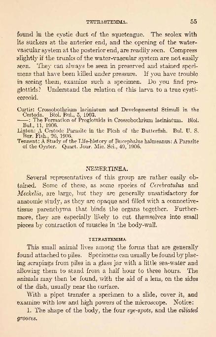
TETRASTEMMA. 55
found in the cystic duct of the squeteague. The scolex with
its suckers at the anterior end, and the opening of the water-
vascular system at the posterior end, are readily seen. Compress
slightly if the trunks of the water-vascular system are not easily
seen. They can always be seen in preserved and stained speci-
mens that have been killed under pressure. If you have trouble
in seeing them, examine such a specimen. Do you find pro-
glottids ? Understand the relation of this larva to a true cysti-
cercoid.
Curtis: Crossobothrium laciniatum and Developmental Stimuli in theCestoda. Biol. Bui., 5, 1903.
: The Formation of Proglottids in Crossobothrium laciniatum. Biol.
Bui., 11, 1906.Linton: A Cestode Parasite in the Flesh of the Butterfish. Bui. U. S.
Bur. Fish., 26, 1906.
Tennent : A Study of the Life-history of Bucephalus haimeanus : A Parasiteof the Oyster. Quart. Jour. Mic. Sci., 49, 1906.
NEMERTINEA.
Several representatives of this group are rather easily ob-
tained. Some of these, as some species of Cerebratulus and
Meckelia, are large, but they are generally unsatisfactory for
anatomic study, as they are opaque and filled with a connective-
tissue parenchyma that binds the organs together. Further-
more, they are especially likely to cut themselves into small
pieces by contraction of muscles in the body-wall.
TETRASTEMMA-
This small animal lives among the forms that are generally
found attached to piles. Specimens can usually be found by plac-
ing scrapings from piles in a glass jar with a little sea-water and
allowing them to stand from a half hour to three hours. The
animals may then be found, with the aid of a lens, on the sides
of the dish, usually near the .surface.
With a pipet transfer a specimen to a slide, cover it, and
examine with low and high powers of the microscope. Notice:
1. The shape of the body, the four eye-spots, and the ciliated
grooves.

56 PLATYHELMINTHES.
2. The straight alimentary canal. The diverticula of the
intestine and the terminal anus.
3. The enormous proboscis, consisting of a large anterior
eversible portion, and a smaller posterior portion that is not
eversible. When the proboscis is retracted it is bent uponitself. Stylets are present in the eversible portion, near its
inner end. Can you determine how the proboscis is protrudedand retracted? Does the proboscis have anything to do with
the digestive system?4. Beneath the posterior eye-spots are the cerebral ganglia,
from which lateral nerve cords extend posteriorly.
5. If the specimen happens to contain eggs, they will lie
between the diverticula of the intestine. They are compara-
tively very large.
Coe: Development of the Pilidium of Certain Nemerteans. Trans. Conn.Acad., 10, 1899.
: On the Anatomy of a Species of Nemertean (Cerebratulus lacteus).Trans. Conn. Acad., 10, 1890.
Verrill: The Marine Nemerteans of New England and Adjacent Waters.Trans. Conn. Acad. Sci., 8, 1892.
C. B. Wilson: Habits and Early Development of Cerebratulus lacteus.
Quart. Jour. Mic. Sci., 43, 1900.

NEMATHELMINTHES.
Body elongated, cylindrical, and not segmented. Theyhave a very general distribution and a great diversity of forms.
Many are parasitic. Anus usually present. Ccelom not filled
with parenchyma. The classes may not be genetically related.
CLASS 1. Nematoda.
Many are internal parasites, but others are found
in fresh and salt water and in damp earth. Bodypointed at both ends. Mouth terminal, anus ven-
tral. (Ascaris, Trichina, Gordius.)CLASS 2. Acanthocephala.
Formidable intestinal parasites. Proboscis bearinghooks. No alimentary canal. (Echinorhynchus.)
CLASS 3. Chatognatha.Marine, and all but one species pelagic. With caudal
and lateral fins and bristle-like jaws. (Sagitta.)
ASCARIS.
Animals belonging to this genus are common in the intestine
of the horse and pig, and are not uncommon in man. Examine
specimens and see if they have any organs that would aid them in
clinging to the intestinal wall. How can they retain their positions ?
1. Can you determine which is anterior and which is posterior?
Is there any indication of segmentation? Can the ventral
side be told from the dorsal ?
*
2. Find the mouth and see that it is bounded by three lips.
Notice how these are placed and find the papillae on the ventral
ones. Find the anus and note its position. This serves also as
a reproductive aperture for the male. In the female the repro-
ductive aperture is situated about one-third back from the
anterior end. It can be seen only in favorable specimens.57

58 NEMATHELMINTHES.
3. Open a well preserved or fresh specimen along the dor-
sal line and notice the definite ccelom, and the straight alimentary
canal. If the specimen is a female, find the Y-shaped genital
organs, the free, ovarian ends of which are slender and some-
what tangled. The position of the external genital opening has
already been noted. In the male there is a single, tangled, tnread-
like testis, which joins the enlarged seminal vesicle that extends
to the cloaca. The nervous system consists of a 'circum-esopha-
geal ring, six longitudinal nerves, the dorsal and ventral of
which are larger than the others, and anterior nerves. It is
not easily seen.
A drawing is desirable.
Montgomery: The Adult Organization of Paragordius varius. Zool. Jahrb.,
18, 1903.
TRICHINA.
Encysted specimens may frequently be found by examiningthin pieces of pig muscle obtained from the meat market. Pigs
fattened in small pens and fed on table waste, or in slaughter-
yards and fed on the offal of butchered animals, are muchmore likely to be infected than others. Scavenger rats and
cats are frequently infected.
1. Flatten a piece of muscle containing trichinae between
two slides in a little glycerin and notice the relation of the ani-
mal to the muscle fibers. Notice the cyst that surrounds it
and see if you can determine whether this was formed by the
host or the parasite. There are frequently fat cells at the ends
of the cyst. Just after the parasites are encysted, the cysts are
surrounded by capillaries that may be injected by injecting the
vessels of the host. These may be found only at a definite stage
after encystment. Why are they formed? Do they indicate
how the cysts were formed? If the trichinae are abundant see
if you can find more than one in a cyst.
2. Notice the shape that is assumed by the parasite. Is
the coiling always the same? If your material is fresh, mount
some of t"he muscle between slides without glycerin, warm the
slide, and see if the encysted animals will move.

TRICHINA. 59
3. Are the anterior and posterior ends alike? Is there anyindication of a mouth f The large cells that form the intestine
can frequently be seen. It should be borne in mind that the
encysted specimen is not fully adult and that the animal growsafter reaching the alimentary canal of the next host.
Make a drawing of an encysted animal.
Glazier: Report on Trichinae and Trichinosis. U. S. Treas. Dept. Doc.No. 84, Marine Hospital, 1881.
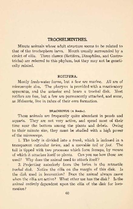
TROCHELMINTHES.
Minute animals whose adult structure seems to be related to
that of the trochophore larva. Mouth usually surrounded by a
circlet of cilia. Three classes (Rotifera, Dinophilea, and Gastro-
tricha) are referred to this phylum, but they may not be geneti-
cally related.
ROTIFERA.
Mostly fresh-water forms, but a few are marine. All are of
microscopic size. The pharynx is provided with a masticatory
apparatus, and the anterior end bears a trochal disk. Most
rotifers are free, but a few are permanently attached, and some,
as Melocerta, live in tubes of their own formation.
BRACHIONUS (A Rotifer).
These animals are frequently quite abundant in ponds and
aquaria. They are not very active, and spend most of their
time near the bottom among the plants and debris. Owingto their minute size, they must be studied with a high powerof the microscope.
1. The body is divided into a trunk, which is inclosed in a
transparent cuticular lorica, and a movable tail or foot. The
tail is tipped with two processes which form forceps, by means
of which it attaches itself to plants. Can you see how these are
used? Why does the animal need to attach itself?
2. Projecting anteriorly from the lorica is the retractile
trochal disk. Notice the cilia on the margin of this disk. Is
the disk used in locomotion? Does the animal always move
when the cilia are active ? What other use has the disk ? Is the
animal entirely dependent upon the cilia of the disk for loco-
motion?60
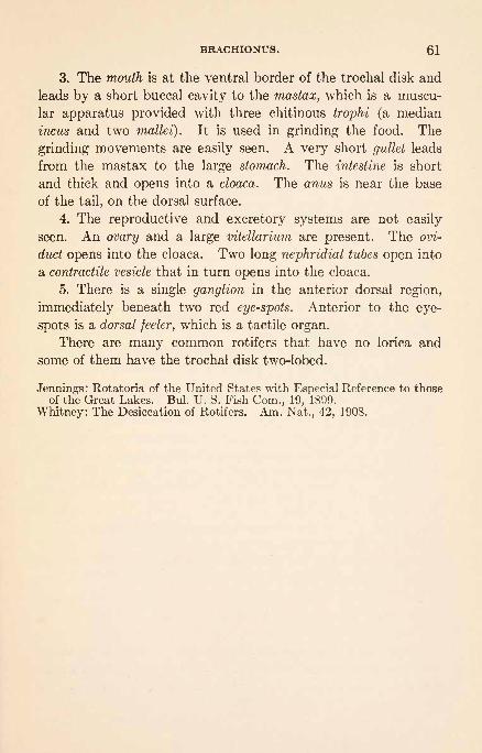
BRACHIONUS. 61
3. The mouth is at the ventral border of the trochal disk and
leads by a short buccal cavity to the mastax, which is a muscu-
lar apparatus provided with three chitinous trophi (a median
incus and two mallei). It is used in grinding the food. The
grinding movements are easily seen. A very short gullet leads
from the mastax to the large stomach. The intestine is short
and thick and opens into a cloaca. The anus is near the base
of the tail, on the dorsal surface.
4. The reproductive and excretory systems are not easily
seen. An ovary and a large vitellarium are present. The ovi-
duct opens into the cloaca. Two long nephridial tubes open into
a contractile vesicle that in turn opens into the cloaca.
5. There is a single ganglion in the anterior dorsal region,
immediately beneath two red eye-spots. Anterior to the eye-
spots is a dorsal feeler, which is a tactile organ.
There are many common rotifers that have no lorica and
some of them have the trochal disk two-lobed.
Jennings: Rotatoria of the United States with Especial Reference to thoseof the Great Lakes. Bui. U. S. Fish Com., 19, 1899.
Whitney: The Desiccation of Rotifers. Am. Nat., 42, 1908.

MOLLUSCOIDA.
Lophophore present. Mouth and anus closely approximated.
Coelom usually present.
CLASS 1. Polyzoa.
Usually colonial. Zooids of small size and pro-tected by a firm cuticle.
Subclass 1. Ectoprocta.Anus outside of the lophophore.
Order 1. Gymnolaemata.Marine. Circular lophophore. No epistomepresent. (Bugula, Membranipora.)
Order 2. Phylactolsemata.Fresh -water. Horseshoe -shaped lophophore.
Epistome present. (Plumatella, Pectinatella.)Subclass 2. Endoprocta.
Colonial or solitary. Anus and mouth both in-
side of the lophophore. (Loxosoma, Pedicellina.^
CLASS 2. Brachiopoda.Marine. Solitary. Bivalve shell. Usually at-
tached by a peduncle.Order 1. Inarticulata.
Valves not united by a hinge. (Lingula.)Order 2. Articulata.
Valves hinged. Usually with a shelly loop to
support the lophophore. (Terebratulina.)
POLYZOA. ,
BUGULA.
The colonies are very common in shallow water along shore,
attached to rocks and piles. They may be examined with the
aid of a glass-bottomed pail in the positions they occupy on the
62

BUGULA. 63
sides of the piles of almost any old wharf. What must be the
source of their food? What part of the colony is likely to be
best nourished? Collect specimens by scraping the piles and
see what forms are associated with them.
1. Examine a colony in a dish of water and see how it
branches. Does it present any regularity?
Make a drawing of a colony.
2. Remove one of the flat branches, place it in a watch-glass
of water, and examine it with a low power. What more can be
observed regarding the branches? How are the cups arranged?
Are the cups on the two sides of a twig placed in definite rela-
tions to each other ? Where are the empty cups found ? Why ?
Can you find connections between the cups of the two sides?
Make a drawing showing the arrangement of the cups.
3. Allow a living branch to remain undisturbed for a few
moments and with a microscope see how the thin outer mar-
gins of the, cups are unfolded as the zooids protrude.
4. Mount a specimen on a slide, cover, and compare the
tentacles of an expanded zooid with those of the hydroids that
you studied. How do they differ? How must the animal feed?
5. How are the tentacles arranged around the distal end of
the body? How many tentacles are there ? Look for the mouth.
6. Can you see the parts of the alimentary canal? Is there
food in the stomach ? How does the zooid pull itself back into
its cup?
7. Look for avicularia and observe their movements and
structure. Where is the jaw hinged? Where are the muscles
that open it? Where are the muscles that close it? Of these
muscles, which are largest? Why? See if"sense hairs" can
be found between the jaws. What is their probable use?
Draw an avicularium.
8. Ooecia with embryos will be found in some specimens.
Where are they placed?
9. Put powdered carmine in the water with a living branch
and see if the zooids will eat it.
10. Put a small living branch in a drop of sea-water under
a supported cover-glass and see if any of the zooids will expand.
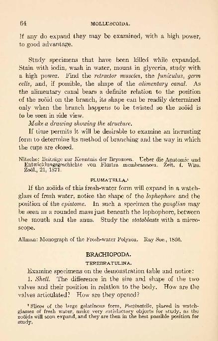
64 MOLLUSCOIDA.
If any do expand they may be examined, with a high power,
to good advantage.
Study specimens that have been killed while expanded.Stain with iodin, wash in water, mount in glycerin, study with
a high power. Find the retractor muscles, the juniculus, germ
cells, and, if possible, the shape of the alimentary canal. As
the alimentary canal bears a definite relation to the position
of the zooid on the branch, its shape can be readily determined
only when the branch happens to be twisted so the zooid is
to be seen in side view.
Make a drawing showing the structure.
If time permits it will be desirable to examine an incrusting
form to determine its method of branching and the way in which
the cups are closed.
Nitsche: Beitrage zur Kenntnis der Bryozoen. Ueber die Anatomic undEntwicklungsgeschichte von Flustra membranacea. Zeit. f. Wiss.
Zool., 21, 1871.
PLUMATELLA.1
If the zooids of this fresh-water form will expand in a watch-
glass of fresh water, notice the shape of the lophophore and the
position of the epistome. In such a specimen the ganglion maybe seen as a rounded mass just beneath the lophophore, between
the mouth and the anus. Study the statoblasts with a micro-
scope.
Allman: Monograph of the Fresh-water Polyzoa. Ray Soc., 1856.
BRACHIOPODA.
TEREBRATULINA.
Examine specimens on the demonstration table and notice:
1. Shell. The difference in the size and shape of the two
valves and their position in relation to the body. How are the
valves articulated ? How are they opened ?
1 Slices of the large gelatinous form, PectinateUa, placed in watch-
glasses of fresh water, make very satisfactory objects for study, as the
zooids will soon expand, and they are then in the best possible position for
study.

TEREBRATULINA. 65
2. Peduncle. Its position. What is its use?
3. Muscles. Those used in opening and closing the shell.
4. Lophophore. Consisting of two elongated arms with a
double row of tentacles on each.
5. Mouth. Notice its relation to the grooves running be-
tween the rows of tentacles on each of the arms of the lophophore.
Brooks: Development of Lingula. Ches. Zool. Lab. Sci. Results, 1878.
Conklin: The Embryology of a Brachiopod, Terebratulina septentrionalis.Proc. Am. Phil. Soc., 41, 1902.
Hancock: On the Organization of Brachiopoda. Trans. Roy. Soc., London,148, 1858.
Morse: Observations on Living Brachiopoda. Mem. Bost. Soc. Nat. Hist.,
5, 1902.

ECHINODERMATA.*
Radially symmetrical animals, with calcareous plates in the
integument. Water-vascular system always present.
CLASS 1. Asteroidea.
With radiating arms not sharply defined fromthe central disk. Ambulacral feet in grooveson the oral side.
Order 1. Phanerozonia.
With large marginal ossicles. (Astropecten.)Order 2. Cryptozonia.
Marginal ossicles inconspicuous. (Asterias.)CLASS 2. Ophiuroidea.
With slender radiating arms sharply defined
from the central disk. No ambulacral grooves.Order 1. Ophiurida.
Arms not branched. (Ophiura.)Order 2. Euryalida.
Arms branched. (Astrophyton.)CLASS 3. Echinoidea.
Globular, or somewhat disk-shaped, spiny bodies.
Shell or test composed of close-fitting plates.Order 1. Regularia.
Nearly globular test. Spines rather large.
Mouth and anus polar. Jaws present. (Ar-
bacia, Strongylocentrotus.)Order 2. Clypeastroidea.
More or less flattened test. Spines very small.
Anus not polar. Jaws present. (Echinarach-
nius.)
Order 3. Spatangoidea.Somewhat flattened and elongated. Spines verysmall. Neither mouth nor anus polar.
CLASS 4. Holothuroidea.
Bodies soft, elongated and cylindrical. Mouthand anus polar, the former surrounded by a cir-
clet of large oral tentacles.
Order 1. -Elasipoda.Well-marked bilateral symmetry. Tube feet onventral and papillae on dorsal surface. Deepsea only.
66

ASTERIAS. 67
Order 2. Pedata.Ambulacral feet in rows or scattered. (Thyone,Cucumaria.)
Order 3. Apoda.Without tube feet. Worm-like. (Synaptula.)
CLASS 5. Crinoidea.
Temporarily or permanently attached by astalk. With five branching arms radiatingfrom a small disk.
Order 1. Neo-Crinoidea.Characters as above. (Antedon, Pentacrinus.)
Berry: Metamorphosis of Echinoderms. Quart. Jour. Mic. Sci., 38, 1905.Coe: Echinoderms of Connecticut. State Geol. and Nat. Hist. Sun., 19,
1912.
Grave: Occurrence among Echinoderms of Larva? with Cilia Arranged in
Transverse Rings. Biol. Bui., 5, 1903.Tennent: Echinoderm Hybridization. Carnegie Inst., 132, 1912.
ASTEROIDEA.
ASTERIAS. (Starfish.)
Starfishes are rather common along most coasts and are
among the worst enemies of oysters, muscles, clams, and bar-
nacles. They occasionally capture fish in aquaria. They can
generally be most satisfactorily examined on shallow-water
mussel-beds or on rocks covered with barnacles. Places where
starfish occur should be visited, and the conditions under which
they live examined. Determine:
1. How they feed.
2. What their enemies must be.
3. How their arms are repaired when injured. Do you find
specimens that are growing new tips to injured arms or are such
arms apparently replaced? When an arm is injured how must
the animal proceed to repair it?
4. Do specimens ever conceal themselves? See if specimenscan be found with pieces of grass and weeds covering them.
Try picking these pieces off to see if they adhere.
5. Do the animals have other means of protection?
Examine a specimen and notice:
1. That the surface by which the animal clings, the oral

68 ECHINODERMATA.
surface, is different from the other, aboral surface, and that
both surfaces are covered with short spines. What is the use
of the spines?
2. It consists of radiating arms and a central disk.
3. On the aboral surface of the disk, near the junction of the
two arms, a small, frequently conspicuously colored, circular body,
the madreporic plate. The two arms adjacent to this plate are
sometimes referred to as the bivium, and the remaining three as
the trivium. The radial symmetry of the animal is disturbed ex-
ternally only by the madreporic plate. Examine this plate with
a lens and determine its structure.
4. On the oral surface, the mouth. Note its size and see if
.Jt is provided with jaws of any kind. Would you expect jaws?
Why?5. Radiating from the mouth are the ambulacral grooves,
one on each arm. In these grooves are the ambulacral or tube
feet. Do they have a definite arrangement? Along the sides
of the grooves are slender spines that differ from the general
body-spines in being movable.
6. Scrape the tube feet from a portion of an ambulacral groove
of a dried specimen and notice the pores through which the feet
are attached to organs inside the arm. Notice also the exposed
ambulacral plates and determine their relation to the pores.
Draw figures of the aboral and oral surfaces of a starfish, and a
diagram to show the relation of the ambulacral plates and pores.
Place a living starfish in a dish of sea-water.
1. Study its method of locomotion. How are the ambu-
lacral feet used? How far can they be protruded?2. Tear the starfish quickly from the bottom. Do any of
the feet remain behind? Understand how they are attached.
3. Place the starfish on its aboral surface and watch it turn
over.
4. Find the thread-like dermal branchice projecting through the
body integument. Are they withdrawn when touched? What
is their function?
5. Stroke the starfish with a cameFs-hair brush and notice4
how the hairs are caught. Can you determine by what and
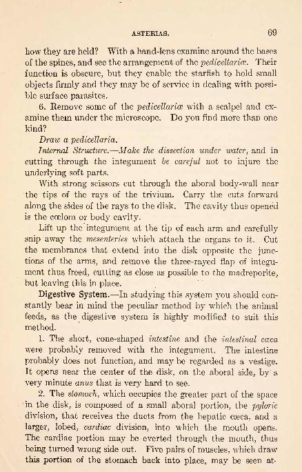
ASTERIAS. 69
how they are held? With a hand-lens examine around the bases
of the spines, and see the arrangement of the pedicellarice. Their
function is obscure, but they enable the starfish to hold small
objects firmly and they may be of service in dealing with possi-
ble surface parasites.
6. Remove some of the pedicellarice with a scalpel and ex-
amine them under the microscope. Do you find more than one
kind?
Draw a pedicellaria.
Internal Structure. Make the dissection under water, and in
cutting through the integument be careful not to injure the
underlying soft parts.
With strong scissors cut through the aboral body-wall near
the tips of the rays of the trivium. Carry the cuts forward
along the sides of the rays to the disk. The cavity thus openedis the coelom or body cavity.
Lift up the integument at the tip of each arm and carefully
snip away the mesenteries which attach the organs to it. Cut
the membranes that extend into the disk opposite the junc-tions of the arms, and remove the three-rayed flap of integu-
ment thus freed, cutting as close as possible to the madreporite,but leaving this in place.
Digestive System. In studying this system you should con-
stantly bear in mind the peculiar method by which the animal
feeds, as the digestive system is highly modified to suit this
method.
1. The short, cone-shaped intestine and the intestinal cceca
were probably removed with the integument. The intestine
probably does not function, and may be regarded as a vestige.
It opens near the center of the disk, on the aboral side, by a
very minute anus that is very hard to see.
2. The stomach, which occupies the greater part of the spacein the disk, is composed of a small aboral portion, the pyloric
division, that receives the ducts from the hepatic caeca, and a
larger, lobed, cardiac division, into which the mouth opens.The cardiac portion may be everted through the mouth, thus
being turned wrong side out. Five pairs of muscles, which drawthis portion of the stomach back into place, may be seen at'

70 ECHINODERMATA.
tached to the ridges formed by the ambulacral plates in each arm.
How is it possible for the stomach to be everted? What reason
is there for two divisions?
3. In each arm is a pair of long, glandular organs, the hepatic
caeca. The ducts of each pair unite and join the pyloric divi-
sion of the stomach by a common duct. These are digestive
glands. What reason is there for having ten enormous digestive
glands? Does this have anything to do with the method of feed
ing?
Make a drawing of the digestive system of the disk and one
arm.
Reproductive System. Turn the hepatic caeca to one side and
notice the ovaries or testes. The sexes are separate, but the
organs have the same general appearance in both sexes. They
vary in size according to the season of the year, sometimes
being so small that they are not easily found, and again being
nearly or quite as large as the hepatic cseca. With a pair of
forceps lift up one of these organs and see where it is attached.
It is at this point that the reproductive cells reach the exterior.
How many gonads are there?
Draw the gonads into another arm of your figure.
Water-vascular System.1
1. Carefully remove the side of the
stomach next to the bivium, being very careful not to disturb
the stone canal, which runs from the madreporic plate to the
margin of the membrane around the mouth. By the side of
the stone canal is a thin band of tissue formerly supposed to be a
heart. It is now known to be connected with the reproductive
system, and is frequently referred to as the axial organ or
genital stolon. It has nothing to do with the system now under
consideration.
2. The circular canal, which is joined by the stone canal at
the outer margin of the peristomial membrane, follows the mar-
gin of the membrane and so encircles the mouth. Originatingfrom it at points very near the ampullae of the first tube feet
1 This may be injected in fresh specimens, either with gelatin or fine
starch-mass, by picking up one of the radial canals with a hypodermicsyringe and injecting toward the disk.

ASTERIAS. 71
are nine small vesicles, Tiedemann bodies. They are smaller
than the ampullae and project in toward the mouth. The posi-
tion where the tenth Tiedemann body might be expected, is
taken by the stone canal.
3. Leaving the circular canal are five radial water tubes,
one for each arm. These tubes lie along the oral surfaces of
the ambulacral plates, and are accordingly not visible on the
inside of the animal. The position of the tube can best be
understood by making a transverse section of an arm. It will
then be seen either in injected or uninjected specimens, lying
immediately below the ambulacral plates. In injected speci-
mens it may be followed by dissecting from the oral side, from
the circular canal to the extremity of the arm, where it ends
in a small tentacle.
4. Along the sides of the ambulacral ridges, within the body-
cavity, are rows of little bag-like ampullae. Determine their re-
lation to the ambulacral pores. If the specimen is fresh, press a
few ampull and see if the corresponding tube feet are affected.
Can you determine their function? In a dissection it is hard to*
find the connecting tubes that join the radial tubes to the tube
feet, but they can sometimes be seen in sections of arms of
injected specimens. They can readily be .seen in microscopic
preparations.
The water-vascular system is very distinctive for the Echi-
nodermata, and you should understand perfectly:
(a) How the tube feet are extended.
(6) What causes them to adhere.
(c) The connection between tube feet, ampullae, connecting
canals, radial water tubes, circular canal, stone canal, and mad-
reporic plate.
(d) How it is possible to extend one foot without extend-
ing others.
Make a drawing showing the arrangement of the water-vascu-
lar system.
Nervous System. This is not easily studied by dissection.
It consists of a nerve ring which encircles the mouth and lies
just ventral to the circular water canal, and five radial nerves

72 ECHINODERMATA.
that extend down the arms just beneath the radial water tubes,
to end at the tips of the arms in pigment spots, the eye-
spots. The whole central nervous system is superficial and forms
a portion of the outer covering of the body. The radial
nerves can be seen by separating the rows of ambulacral feet,
but it is much more satisfactory to study them in preparedsections.
Muscular System. Examine the walls of the starfish for
its muscular system. If time permits, it will be desirable to
macerate a portion of an arm to see the skeleton to which these
muscles are attached.
Study prepared sections of the arm of a small starfish and
determine the relation of organs.
1. The hepatic csca. How are they supported? Whatis their structure ?
2. The radial canal, connecting tubes, tube feet, and ampullae.
3. The thickened, deeply stained, radial nerve between the
tube feet and below the radial water tube.
4. The perihsemal canal, divided by a thin partition, that lies
between the radial water tube and the radial nerve.
Make a drawing of a section of an arm that will show these
points.
Understand how a starfish can open an oyster or a mussel
and how it digests it when open. How can it digest a barnacle
or a small snail ? How does it respire ?
Cole: Experiments on Coordination and Righting in the Starfish. Biol.
Bui., 24, 1913.-: Direction of Locomotion in Starfish (Asterias forbesi). Jour. Exp.
Zool., 14, 1913.
Field: Larva of Asterias yulgaris. Quart. Jour. Mic. Sci., 34, 1892.
Goto: The Metamorphosis of Asterias pallida, with Special Reference to the
Fate of the Body Cavities. Jour. Col. Sci. Imp. Univ., Tokyo, 10, 1898.
Harvey: Methods of Artificial Parthenogenesis. Biol. Bui., 18, 1910.
Jennings: Behavior of the Starfish Asterias forreri. Univ. Calif. Pub.
Zool., 4, 1907.
MacBride: Development of Asterias gibbosa. Quart. Jour. Mic. Sci., 38,
1896.
Mead: The Natural History of the Starfish. Bui. U. S. Fish Com., 1899.
Tennent and Hogue: Studies on the Development of the Starfish Egg.Jour. Exp. Zool., 3, 1906.
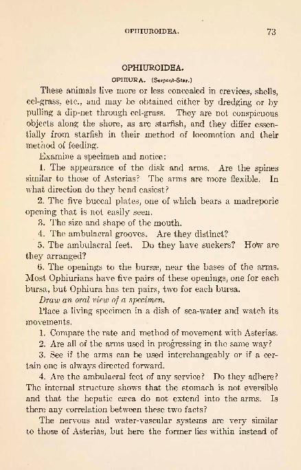
OPHIUROIDEA. 73
OPHIUROIDEA.
OPHIURA. (Srpent-Star.)
These animals live more or less concealed in crevices, shells,
eel-grass, etc., and may be obtained either by dredging or bypulling a dip-net through eel-grass. They are not conspicuous
objects along the shore, as are starfish, and they differ essen-
tially from starfish in their method of locomotion and their
method of feeding.
Examine a specimen and notice:
1. The appearance of the disk and arms. Are the spinessimilar to those of Asterias? The arms are more flexible. In
what direction do they bend easiest ?
2. The five buccal plates, one of which bears a madreporic
opening that is not easily seen.
3. The size and shape of the mouth.
4. The ambulacral grooves. Are they distinct?
5. The ambulacral feet. Do they have suckers? How are
they arranged?6. The openings to the bursse, near the bases of the arms.
Most Ophiurians have five pairs of these openings, one for each
bursa, but Ophiura has ten pairs, two for each bursa.
Draw an oral view of a specimen.
Place a living specimen in a dish of sea-water and watch its
movements.
1. Compare the rate and method of movement with Asterias.
2. Are all of the arms used in progressing in the same way?3. See if the arms can be used interchangeably or if a cer-
tain one is always directed forward.
4. Are the ambulacral feet of any service? Do they adhere?
The internal structure shows that the stomach is not eversible
and that the hepatic caBca do not extend into the arms. Is
there any correlation between these two facts?
The nervous and water-vascular systems are very similar
to those of Asterias, but here the former lies within instead of

74 ECHINODERMATA.
on the surface of the arm, the entire arm being encased with four
or more rows of shields. They can be studied best in sections.
Grave: Ophiura brevispina I. Mem. Biol. Lab. Johns Hopkins Univ., 4,
1900. Mem. Nat. Acad., 8, 1899.
: Ophiura brevispina II. An Embryological Contribution and Studyof the Effect of Yolk Substance upon the Developmental Process. Jour.
Morph., 27, 1916.
ECHINOIDEA.
STRONGYLOCENTROTUS. 1 (Sea-Urchin.)
In some localities sea-urchins can be found in tide pools
or near low-tide mark, where they may be very abundant. In
other localities they can be obtained only by dredging. When
possible they should be observed in their native place's and the
conditions noted.
1. What apparently serves as food for the animal ? Can youdetermine how this is obtained?
2. Do you find attempts at concealment?
3. Are the animals able to climb?
Put a living sea-urchin in a dish of sea-water and study its
movements.
1. When placed on its back, how does it turn over?
2. What is the normal method of progression?
3. How are the spines arranged when the animal is creeping
on the bottom?
4. What difference do you note between the spines on the
lower and upper surfaces ?
5. How Long are the tube feetf Are they used with the
spines in moving or do both sets of organs act independently?
6. Grasp a spine with your forceps and see if neighboring
spines respond. Do they form defensive armor?
7. In what directions may a spine be moved? Remove a
spine from a preserved specimen and determine how it wasattached and how the muscles that moved it were attached to
the spine and to the test.
Make a diagram showing the arrangement.1 These directionf will serve for any of our common sea-urchins.
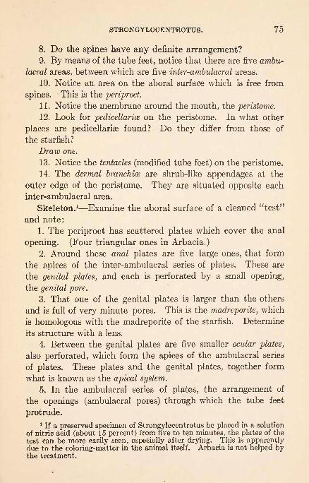
STRONGYLOCENTROTUS. 75
8. Do the spines have any definite arrangement?9. By means of the tube feet, notice that there are five ambu-
lacral areas, between which are five inter-ambulacral areas.
10. Notice an area on the aboral surface which is free from
spines. This is the periproct.
11. Notice the membrane around the mouth, the peristome.
12. Look for pedicellarice on the peristome. In what other
places are pedicellariae found? Do they differ from those of
the starfish?
Draw one.
13. Notice the tentacles (modified tube feet) on the peristome.
14. The dermal branchice are shrub-like appendages at the
outer edge of the peristome. They are situated opposite each
inter-ambulacral area.
Skeleton. 1 Examine the aboral surface of a cleaned "test"
and note:
1. The periproct has scattered plates which cover the anal
opening. (Four triangular ones in Arbacia.)
2. Around these anal plates are five large ones, that form
the apices of the inter-ambulacral series of plates. These are
the genital plates, and each is perforated by a small opening,
the genital pore.
3. That one of the genital plates is larger than the others
and is full of very minute pores. This is the madreporite, which
is homologous with the madreporite of the starfish. Determine
its structure with a lens.
4. Between the genital plates are five smaller ocular plates,
also perforated, which form the apices of the ambulacral series
of plates. These plates and the genital plates, together form
what is known as the apical system.
5. In the ambulacral series of plates, the arrangement of
the openings (ambulacral pores) through which the tube feet
protrude.
1 If a preserved specimen of Strongylocentrotus be placed in a solution
of nitric acid (about 15 percent) from five to ten minutes, the plates of the
test can be more easily seen, especially after drying. This is apparentlydue to the coloring-matter in the animal itself. Arbacia is not helped bythe treatment.

76 ECHINODERMATA.
6. Do all of the plates bear balls to which spines were artic-
ulated? Are the balls of equal size? Do they have a definite
arrangement ?
Can you homologize the positions of the ambulacral, inter-
ambulacral, ocular, and genital plates in the sea-urchin and
starfish? What portion of the starfish is represented by the
periproct of the sea-urchin?
Make a draining of the test, showing the ambulacral, inter-
ambulacral, and apical systems of plates.
7. Around the peristome, on the inside of the test, note the
five auricles forming arches or bridges over the bases of the
ambulacral areas. Their purpose will be seen later.
Cut around the equatorial region of an alcoholic specimen,
taking care to cut through the test only. Break the aboral portion
away bit by bit with forceps until near the genital plates, freeing
the fragments from the internal organs without disturbing their
positions.
Reproductive System. How were the gonads (their appear-ance is the same in both sexes) attached to the test? Howmany are there ? Opposite what areas of the test are they placed ?
Where do they open to the exterior? Without mutilating,
find the narrow strip of tissue that connects the gonads to
each other near their aboral ends. This is the genital rachis.
Connected with the genital rachis and lying alongside the stone
canal, which leads from the madreporite, is the genital stolon.
Digestive System. Remove the gonads from the three areas
farthest from the madreporic plate, lift the remaining aboral
portion of the test slightly, and examine the alimentary canal.
1. The large and conspicuous jaws, frequently called the
lantern. They will be studied later.
2. The esophagus, passing between the jaws, and bending
over to one side to join the intestine.
3. The intestine. Notice its size and its shape. Do its
loops have any relation to the positions of the gonads?4. The intestinal siphon, lying along the intestine and
attached to it at both ends.

STRONGYLOCENTROTUS. 77
5. The rectum, running from the end of the intestine to the
anus.
6. The mesenteries which hold the various organs in place.Make a drawing to show the reproductive and digestive organs.Water-vascular System. 1. The stone canal leads from the
madreporite to the circular canal, which encircles the esophagusat a point just above the lantern.
2. From the circular canal radial tubes pass over the top anddown the sides of the lantern, to pass through the auricles and
up the ambulacral tracts, to the ocular plates. They can easily
be seen along the sides of the test, but are difficult to see be-
fore they leave the lantern.
3. Along the course of each radial canal, the ampullce, which
supply the tube feet, are to be seen. The relations of the tube
feet and radial canals are practically the same as in the starfish
except that the removal of the radial tubes to the inner sides
of the ambulacral plates causes two perforations for each foot
here, while the starfish has only one. One of these perforationsis for the connection between the ampulla and the foot, the other
is for the connecting tube between the radial canal and the foot.
The connecting tube joins the foot outside of the plate (as in
the starfish), while it joins the radial canal inside of the plates
(different from the starfish).
Remove the intestine and study the lantern and its attach-
ments.
1. The whole lantern is inclosed in a delicate membrane,the peripharyngeal or lantern membrane which contains the
lantern coelom. This space communicates with the five radial
perihemal canals, which run along the ambulacral areas be-
tween the radial canals and radial nerves, and with the dermal
branchse. It is important in respiration.
2. The tip of the lantern is attached to the flexible peristoma,
and muscles extending from various parts of it are attached to
the hard parts of the surrounding test.
In shape the lantern is a five-sided radially symmetrical

78 ECHINODERMATA.
pyramid. Each of the sides consists of a massive calcareous
structure, the alveolus, which supports an elongated tooth the
tip of which projects through the peristome. The base of the
pyramid may be compared with a wheel, in which the ten
epiphyses,1 two of which are attached to each alveolus, are the
tire, and the five radially directed rotulce are the spokes. Eachrotula has a more slender bar, forked at the free extremity,
the compass or radius lying over it. Each of the five segments
represents a jaw that is articulated to its neighbors at its base,
near the esophagus. The points of the teeth can thus be sepa-
rated and closed, and the jaws protruded and retracted by means
of muscles.
3. Connecting adjacent alveoli from top to bottom are the
comminator muscles, that by their combined action close the
jaws.
4. To each of the arms of the radius fork a muscle is
attached. Where is it attached at the other end?
5. A pair of protractor muscles pass down from each epi-
physis. To what are they attached? They are used in pro-
troding the jaws.
6. A pair of retractor muscles is attached to the tip of each
alveolus. They can be used in opening the jaws or in retracting
the jaws. Do you see how?
7. There are also internal and external rotula muscles that
connect the epiphyses with the rotulse. Their contraction
moves these plates upon one another and thus causes a rocking
motion of the jaws.
Understand how the jaws may be protruded, opened, closed,
and retracted by means of these muscles.
8. The compasses are attached one to the other by the
elevator muscles. Their contraction elevates all of the compassesand thus enlarges the lantern ccelom.
9. Attached to the forked end of each compass is a pair of
1 In Arbacia the epiphyses form small hooks that do not unite across
the base of an alveolus.
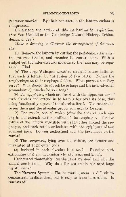
STRONGYLOCENTROTUS. 79
depressor muscles. By their contraction the lantern coelom is
compressed.
Understand the action of this mechanism in respiration.
(See Von Uexhiill or the Cambridge Natural History, Echino-
derms, p. 527.)
Make a drawing to illustrate the arrangement of the mus-
cles.
10. Remove the lantern by cutting the peristome, clear awaythe external tissues, and examine its construction. With a
scalpel cut the inter-alveolar muscles so the jaws may be sepa-
rated. Find:
(a) The large V-shaped alveoli (a straight suture indicates
that each is formed by the fusion of two parts). Notice the
roughenings on their esophageal sides. What purpose can theyserve? Why should the alveoli be so large and the inter-alveolar
(comminator) muscles be so strong?
(6) The epiphyses, which are fused with the upper corners of
each alveolus and extend in to form a bar over its base, thus
being functionally a part of the alveolus itself. The sutures be-
tween them and the alveolus proper can usually be seen.
(c) The rotulcc, one of which joins the ends of each epi-
physis and extends to the position of the esophagus. The five
rotulae of the lantern articulate with each other around the eso-
phagus, and each rotula articulates with the epiphyses of two
adjacent jaws. Do you understand how the jaws move on the
rotulae?
(d) The compasses, lying over the rotulse, are slender and
bifurcated at their outer ends.
(e) Inclosed in each alveolus is a tooth. Examine both
extremities of it and determine why the inner end is soft.
Understand thoroughly how the jaws are used and why the
animal needs them. Why does the sea-urchin not need large
hepatic caeca?
The Nervous System. The nervous system is difficult to
demonstrate in dissections, but is easy to trace in sections. It
consists of:

80 ECHINODERMATA.
1. A nerve ring that encircles the esophagus at a point just
above the mouth.
2. Five radial nerves that pass from the ring, along the in-
side of the ambulacral areas of the test, to the ocular plates.
The radial water tubes will be found in sections adjacent
to the radial nerves. The two are separated only by a narrow
space, the pseudohsemal canal. Between the radial nerves
and the tissue of the test there is another narrow cavity, the
epineural sinus.
If time permits, students will find a dissection of the sand-
dollar, Echinarachnius, valuable for purposes of comparison.
Special notes will not be necessary. Its shape and restricted
ambulacral areas should be studied in the light of its habits
and food-supply. How does the animal move?
MacBride: Cambridge Natural History, Echinodermata.Tennent: Variation in Echinoid Plutei. Jour. Exp. Zool., 9, 1910.
Von Uexhiill: Die Physiologie des Deeigelstachels. Zeit. f. Biol., 39.
: Ueber die Function der Polischen Blasen am Kauapparat der regu-laren Seeigel. Mitth. Zool. Stat. NeapeL, 12, 1897.
HOLOTHUROIDEA.THYONE. (Sea-Cacumber.)
These animals may be found in protected and usually muddyplaces, concealed in eel-grass. They are generally so effectually
concealed that they cannot be satisfactorily studied in their
native places. It is desirable to visit places where they occur
and find specimens by feeling for them near the bottom. It
is then possible to realize the life for which they are adapted.
Examine a living expanded specimen in an aquarium (taking
care not to disturb it) and note:
1. How the tentacles are used. What kind of food would
it get by this means? Compare the method of food-getting
with the starfish and sea-urchin.
2. The respiratory movements of the body. Notice the
strength of the current of water ejected.
3. The general shape of the body when expanded. Does
it seem to rest on a particular side?

THYONE. 81
4. The number and arrangement of the tentacles. To whatdo they probably correspond in the sea-urchin?
Kill the specimen by catching it with strong forceps behind
the mouth, when the tentacles are expanded, and holding it in
hot water. 1 Note that:
1. The body is covered with papilliform ambulacra! feet.
It is possible in some cases to see that they are arranged in five
broad, longitudinal bands.
2. The suckers are less abundant on the dorsal (upper)
surface than on the ventral.
3. A small papilla is to be found on the dorsal surface, be-
tween the tentacles. On it is the genital opening. This will be
referred to again.
Make a drawing of the animal as seen from the side, indicat-
ing all of the points of structure that have been seen.
With a pair of scissors, open the animal longitudinally along
the middle of the ventral (lower) surface.
Digestive System. 1. Note the delicate perforated mesen-
tery, which attaches it to the walls of the body.
2. The esophagus, leading from the mouth through a
calcareous structure, which recalls the lantern of the sea-urchin.
Examine and see if the arrangement is similar to that of the
sea-urchin lantern. The muscles for the retraction of the
lantern are frequently torn from their attachments at one end.
3. The thin-walled and enlarged stomach.
4. The coiled intestine, which leads to the cloaca.
Draw the alimentary canal in position.
Cut the alimentary canal just in front of the stomach, and
close to the cloaca, and as you remove it notice the blood-vessel
that runs along the intestine.
Respiratory and Excretory System. Arising laterally from
either side of the cloaca are the two respiratory trees. They are
*. Specimens that do not expand may be injected with a saturated
solution of chlorotone (saturated by heating). After the animal relaxes
the tentacles may be pushed out. Then kill in hot water or dissect im-
mediately.6

82 ECHINODERMATA.
branched and proj ect far forward into the body-cavity. Can youdetermine how they are filled with water and how the water is ex-
pelled? With a pipet inject them with starch-mass. The strong
jets of water ejected by the living specimen were thrown from
these tubes. Can you understand how they serve for respira-
tion? The walls of the tubes composing the trees are glandular
and may thus serve to excrete wastes. Notice the muscles that
radiate from the walls of the cloaca to the body-wall. Whatis their function?
Make a drawing of the cloaca and rspiratory trees.
Reproductive System. The single gonad (ovary or testis)
occupies a median dorsal position in the anterior part of the
body-cavity. It is composed of a multitude of filaments, which
join to make a brush. This brush projects backward into the
body-cavity. The duct of the organ lies along the dorsal mid-
line, between the right and left dorsal muscle bands, and leads
to the opening upon the small papilla near the mouth that has
already been noticed.
Water-vascular System. 1. The circular canal can be found
in favorable specimens, surrounding the deeper portions of the
esophagus. It gives rise to one or two Polian vesicles, which
are very large and hang down into the body-cavity.2. The five radial canals (homologous with the radial canals
of the starfish and sea-urchin) originate from the water-ring,
pass forward and then backward, and end near the cloaca. Thecourse of each radial canal is easily followed by means of the
numerous small, elongated ampullae which supply the tube feet.
3. Ten forwardly directed canals, the tentacular canals,
leave the radial canals near the water-ring and pass into the
tentacles, which may be homologized with tube feet.
4. The stone canal and madreporite are much reduced in
holothurians. The madreporite, except in larvae and very
young specimens, is not found on the outer surface. Thestone canal leads obliquely backward from the water-ring,
toward the dorsal body-wall, to join a small calcareous body, the
madreporite, which lies in the body-cavity and is not perfor-

THYONE. 83
ated. Does this give you a reason for the presence of large
Polian vesicles? The liquid in the water-vascular system is
not sea-water. Notice its color.
Make a diagram of the water-vascular system.
Muscular System. Beside the special muscles radiating
from the cloaca which have been referred to in connection with
the respiratory system, and the muscles of the lantern, there are
five strong longitudinal bands, really pairs. In which areas
do they lie? What function do they perform? Look for smaller
circular bands. Are there many of them? What is their
function? Can you explain the varied worm-like motions of
the body by the action of these muscles?
Nervous System. This cannot be satisfactorily studied in
dissections. There are five radial nerves and a circular ring.
The nerves are embedded in the body-wall and are hard to find.
The classes of the Echinodermata show exceptionally well
how a general type of structure may be retained and still modi-
fied in certain regards for special habits. Compare, for instance,
the feeding habits of the starfish, sea-urchin, and sea-cucumber.

ANNELIDA.
Body elongated, generally divided into somites. Coelom
usually extensive. Appendages when present form parapodia.
CLASS 1. Archi-annelida.
Without setae or parapodia. Nervous system not
separate from the epidermis. (Polygordius.)CLASS 2. Chgetopoda.
With numerous, distinct somites that are pro-vided with setae.
Subclass 1. Archi-chsetopoda.Setae retractile. Nervous system not separatefrom the epidermis. (Saccocirrus.)
Subclass 2. Polychaeta.With numerous setae. With a great variety of
structure. (Amphitrite, Arenicola, Autolytus,
Chaetopterus, Clymenella, Diapatra, Hydroides,Nereis, Pectinaria, Polynoe, Sabella, Spirorbis.)
Subclass 3. Myzostomida.Disk-shaped. Without external segmentation.Parasites on Echinodermata. (Myzostoma.)
Subclass 4. Oligochaeta.Without parapodia. Setae few and simple.
(Tubifex, Lumbricus.)CLASS 3. Gephyrea.
No segmentation. With or without setae. Withintrovert or proboscis.
Order 1. Inermia.
With introvert. Anus dorsal. No setae. (Phas-
colosoma.)Order 2. Armata.
With proboscis. Anus posterior. Setae few.
(Echiurus.)CLASS 4. Hirudinea.
Somites constant in number, with more external
annuli than there are somites. With suckingmouth and posterior sucker.

NEREIS VIRENS. 85
Order 1. Rhynchobdellida.Anterior end of body forming a proboscis or in-
trovert. No jaws. (Glossiphonia, Macrob-
della, Clepsine.)Order 2. Gnathobdellida.
No proboscis or introvert. Mouth usually with
three teeth. (Hirudo.)
Hatschek: Studien liber Entwicklungsgeschichte der Anneliden. Arb.Zool. Inst. Wien, 1, 1878.
Norman: Durfen wir aus den Reactionen neiderer Thiere auf des Vorhan-densein von Schmerzempfindungen Schliessen? Arch. ges. Physiol., 67,1897.
CH-ETOPODA.
NEREIS VIRENS. (Clam-Worm.)
These animals may be found inhabiting mud-flats from which
;he water flows at low tide. Occasionally they may be seen with
their head ends protruding from their burrows, but generally
specimens will have to be dug. Notice the conditions under
which the animals live and the forms with which they are asso-
ciated. It should also be understood that many of their worst
enemies are present only when the water covers their burrows.
External Structure. 1. Examine a living worm in a dish
of sea-water, noting the motions of the body and of the parapodiaor swimming feet.
Make a drawing of the animal.
2. Hold it down against the bottom of the dish or place in
fresh water for a few minutes to induce it to protrude the
proboscis, the protrusible anterior portion of the alimentarycanal. This is lined with chitin and armed with numerousdenticles and a pair of lateral jaws.
3. Is the general surface clean or slimy? Compare with the
earthworm in this respect and explain the basis for the difference.
4. Determine the direction of the peristaltic waves in the
dorsal blood-vessel.
5. Is the median ventral nerve cord visible through the body-wall?

86 ANNELIDA.
6. In a freshly killed or preserved worm, count the segments or
metameres and compare it with your neighbor's to ascertain
whether the number is constant.. What segments, if any, are
devoid of parapodia? Why?7. In the head distinguish the prostomium, which bears the
four eyes and a pair of short terminal tentacles. At each side of the
prostomium is a thick palp. Determine whether the worm has
the sense of vision, and whether its sense of touch is delicate.
8. Also in the head find the peristomium, the segment which
surrounds the mouth and bears four pairs of lateral tentacles or
cirri. Stretch the mouth with forceps.
Make an enlarged drawing of the head.
9. Find the small terminal anus and a pair of caudal cirri on
the last segment.
10. With scissors cut off a parapodium close to the body and
observe that it has a dorsal blade and a ventral blade (notopodium
and neuropodium). Each of these contains a bundle of bristles
or setw. What use can you ascribe to the setae ? In each bundle
is one very thick seta, the aciculum, which extends into the bodyand is attached to muscles. Of what use is the aciculum? Ex-
amine a few of the small setae microscopically. What is their
structure? Why is it desirable to have so many of the small
setaa? Why does this animal need more than an earthworm
needs?
Observe that each parapodium has a small dorsal and a small
ventral cirrus, and that the main portion of both notopodiumand neuropodium has the form of a flattened blade, somewhatdivided into lobes. The largest lobe of the notopodium is verythin and vascular. What function can you ascribe to it?
Draw a parapodium.11. Look for the nephridiopores, minute apertures which are
segmentally placed on the ventral surface near the neuropodial
cirri.
Internal Structure. For dissection use a specimen that has
been killed and fasten it down by a pin through the head and one
through the posterior part. With scissors cut through the body-
wall, longitudinally, near the mid-dorsal line.
NEREIS VIRENS. 87~f
A preserved specimen can easily be sectioned transversely
with a sharp razor at the somatic boundaries. These sections
are valuable for comparison during dissection.
Find the septa which divide the coelom, or body-cavity, into
metameric chambers. Cut through the septa with scissors and
pin the edges of the body-waft' apart, progressing toward the
head.
Circulatory System. The dorsal blood-vessel lies along the
dorsal surface of the alimentary canal and gives off branches in
each segment, which ramify through the body-wall and viscera
and connect with the longitudinal, ventral blood-vessel. The
blood-plasma contains red coloring-matter in solution.
Digestive System. The mouth-cavity leads into a muscu-
lar pharynx, a portion of which is protrusible as the proboscis.
Examine carefully the muscles of the pharynx, protractors and
retractors, and ascertain their attachments. Posterior to the
pharynx find a small dilation and a narrow esophagus with
a digestive gland at each side. Where does the duct of the
gland open? In the very long stomach-intestine, which follows the
crop, note the constrictions and their relations to the dissepi-
ments. Can you demonstrate dorsal or ventral mesenteries?
Cut open the pharynx and the anterior end of the stomach-
intestine and note the character of their walls.
Make a drawing of the digestive system.
Muscular System. How many distinct bands of longitu-
dinal muscles can be distinguished? Examine with a hand-lens
the parapodial muscles attached to the base of the acicula. Can
you make out a layer of circular muscles? Of what layers does
the body-wall consist?
Excretory System. The nephridia are not nearly as easily
found or studied as they are in the earthworm. Near or just
beneath the lateral edges of the ventral muscle-bands find the
minute pear-shaped nephridia. Determine their distribution
in the body. Each nephridium consists of a tortuous canal in
a multi-nucleate mass of protoplasm. The external opening is
the nephridiopore above mentioned. The inner end perforates
the septum anterior to the body of the nephridium and opens

88 ANNELIDA.
into the coelomic cavity of the segment next in front, by a
ciliated funnel, the nephrostome. With a hand-lens try to
find the nephrostome. Remove a nephridium by means of fine
forceps and examine it -with a microscope.
Reproductive System. The sexes are separate, but no per-
manent gonads are present. At the breeding season the ova
or spermatozoa are proliferated from the ccelomic epithelium
of a large number of segments and escape by rupture of the
body-wall.
Nervous System.1 On lifting the alimentary canal you will
see the ventral ganglionated nerve cord. Note the nerves pass-
ing off laterally from the ganglia. How many pairs of nerves
per segment are there, and how are they placed? Are the gan-
glia metameric? Is there any indication that the nerve cord is
double? At the anterior extremity of the cord note the infra-
esophageal ganglia and, extending from them and encircling the
anterior end of the alimentary canal, the circum-esophageal con-
nectives which unite above in the bilobed brain or supra-eso-
phageal ganglia. Sensory nerves connect the brain with the eyes,
tentacles, and palps.
Make a drawing of the nervous system.
Just: An Experimental Analysis of Fertilization in Platynereis megalops.Biol. Bui., 28, 1915.
: Breeding Habits of the Heteronereis Form of Heteronereis megalopsat Woods Hole. Biol. Bui., 27, 1914.
Lillie: Studies of Fertilization in Nereis. I. and II. Jour. Morph., 22, 1911.
III. and IV. Jour. Exp. Zool., 12, 1912. V. Jour. Exp. Zool., 14, 1913.
Lillie and Just : Breeding Habits of the Heteronereis Form of Nereis lim-
bata at Woods Hole, Mass. Biol. Bui., 24, 1913.
Mayer: The Annual Breeding-swarm of the Atlantic Palolo. CarnegieInst. Pub., 102, 1908.
E. B. Wilson: The Cell-Lineage of Nereis. A Contribution to the Cytologyof the Annelid Body. Jour. Morph., 6, 1892.
Woodworth: The Palolo Worm, Eunice viridis. Bui. Mus. Comp. Zool.,
Harvard, 51, 1907.
1 The nervous system can be most readily studied by tearing it out withneedles in a specimen which has been macerated in 20 percent nitric acidfor twenty-four hours. Sensory cells and their neurites can be identified
in the parapodia by placing them in a 1 percent solution of ammoniumpicrate after having let vigorous worms crawl around for three or fourhours in a small amount of 1 percent solution of methylen-blue. Mountsof the parapodia should be made in a mixture of glycerin and ammoniumpicrate solution.
POLYNCE SQUAMATUS. 89
AUTOLYTUS CORNUTUS.
This polychsete lives in cylindrical tubes of its own con-
struction that it attaches to seaweeds and hydroids, and is espe-
cially interesting because of its method of reproduction, by bud-
ding.
Study live and preserved specimens with the naked eye and
with the hand-lens, in order to form a correct idea of its natural
color, size, and movements, and then study stained specimens
with the low power.
1. Observe two individuals attached end to end. The ante-
rior one is a non-sexual zooid (or original"stock") and is giving
rise to a new sexual zooid by budding. Counting the peristomium
as one somite, on what somite does the bud begin and what
does it represent?
2. Study the head of the anterior, non-sexual zooid. Find three
prostomial tentacles. How are they arranged? Find the eyes.
How many pairs are there? Do you find palps? On the peri-
stomium find the two tentacles and a tentacular cirrus.
3. On the succeeding somites study the parapodia. Observe
the large dorsal cirri and the knob-like notopodium with the short
unjointed setae. There is no neuropodium.
4. Identify the pharynx, gizzard, and intestine.
5. Compare the sexual bud with the non-sexual individual.
The adult male and female differ. The outer prostomial tenta-
cles of the male are forked. Is this bud to be a male or a female?
In an older sexual individual make out a so-called thoracic region
in which the setae are short, and an abdominal region in which
the setae are long. Look for evidences of germ cells in the body-
cavity, between the intestine and body-wall. There is a ventral
brood-pouch on the adult female and the young partly develop
in it. Find the anal cirri.
A drawing illustrating the method of reproduction should be
made.POLYNCE (LEPIDONOTUS) SQUAMATUS.
The family Polynoidae, to which this belongs, can be distin-
guished from all others by the presence of peculiar plates (elytra)

90 ANNELIDA.
on the dorsal surface. They lead sluggish lives under stones
and are carnivorous. Note the size, color, and shape of the
worm.
1. The elytra. How are they arranged? What purpose
do they serve? How many are there? With a hand-lens ob-
serve the fringed condition of the outer edge and the small
tubercles covering the surface. Note the color of the elytra
and the notches in the inner edges of the posterior pair.
Remove with forceps all of the elytra on one side of the
specimen and the first two or three on the other side. Note the
stumps to which the elytra were joined.
2. Examine the dorsal aspect of the head, and note the small
prostomium, with two pairs of eyes, three slender tentacles,
and a pair of fleshy palps. Outside the palps are two pairs of
cirri arising from the peristomium. The significance of these
will be understood later.
3. Find the mouth, placed ventrally in the first or peris-
tomial somite. The mouth leads into a buccal region, which is
eversible and fringed at the end with papillae, each having a
dark spot at the base. If the pharynx is retracted, expose the
buccal cavity by a median ventral incision. In the anterior
end of the pharynx are four black chitinous jaws. Do youinfer that this species is carnivorous or herbivorous? The ever-
sible buccal region and the protrusible pharynx form the pro-
boscis.
4. The anus is dorsally placed, and can be found beneath the
notches in the last pair of elytra.
5. Examine the lateral appendage or parapodium of the
third or any subsequent somite. Note that it consists of a
stout ventral or neuropodial division, and a less prominent
dorsal or notopodial division, each supported internally by a
chitinous rod or aciculum, and bearing externally a tuft of
setae. If there is time, compare the form of the notopodial
and neuropodial setae. The typical parapodial structure is
completed by a soft neuropodial cirrus ventrally, and a noto-

DIOPATRA CUPREA 91
podial cirrus dorsally. Can you find any evidence that the
elytra are modified notopodial cirri?
6. Make a careful study of the appendages of the first or
peristomial and the last or anal segment. Cut off close to the
body, mount in glycerin, and examine with low power of the
microscope. Determine the homology of the parts observed.
Draw the dorsal aspect of the head, to show the appendagesand the proboscis, if exposed. Diagram the structure of the para-
podium as seen in a transverse section of the body.
Unlike most other worms, many of the Aphroditidse have a
fixed number of somites. Count the number in your specimen,
including in the enumeration the peristomial and anal segments.How many pairs of elytra? The number and position of the
elytra are also characteristic of various genera, and may be
conveniently represented by an elytral formula consisting of
the numbers of the somites on which elytra are borne, e. g.,
2, 4, 5, etc. Determine the elytral formula of your specimen.Draw one of the elytra, noting its form, surface, and border
markings, etc. These points are of importance in defining
species. The last pair of elytra are notched on the medianside over the anus, which in this form opens dorsally instead of
terminally.
DIOPATRA CUPREA.
This worm belongs to the family Eunicidse. Specimenslive on mud- and sand-flats, sometimes above low-tide mark,but usually where the burrows are covered by water. This
form is especially interesting because of its feeding and tube-
building habits, parapodial gills, and complex jaw-apparatus.
Study the preserved specimens for the structure and specimensin an aquarium for the habits. Notice the construction of the
tube and determine how it is formed.
1. Notice the size of the body, also its gradual attenuation
posteriorly. Account for this condition. Observe how degen-
erate the parapodia are posteriorly from the same cause.
2. The prostomium. Identify the tentacles. What is their

92 ANNELIDA.
number and arrangement? Find a pair of eyes dorsally placed
behind the tentacles, also a pair of palps in front of them. Note
a second, larger pair of palps which serve as an upper lip.
3. The peristomium. What appendages does it carry ? Note
the lower lip formed from the ventral edge of the peristomium.
4. The position of the jaw-apparatus can be identified as being
in a pouch ventral to the buccal region. Find both by means
of a probe. What kind of food are such jaws fitted for?
5. The parapodia vary greatly, depending upon their posi-
tion on the body. Notice that the notopodia are vestigial,
being represented only by the dorsal cirri and, toward the ante-
rior end, branchial cirri or gills. Acicula can be seen projecting
into the base of the dorsal cirrus. The neuropodium shows two
kinds of setse: (a) stiff and unjointed, (6) crochets. It also
bears an accessory cirrus and the ventral cirri, which are curiously
modified in most cases as glands for use in tube-building. Makeout all these modifications and where they occur.
CHAETOPTERUS.
This is one of the most aberrant of our Polychsetse. It lives
on mud-flats below low tide in a U-shaped, parchment-like tube
both ends of which protrude above the mud. In the bodythree regions can be distinguished. Examine a tube and see
the size of its outer openings. Specimens may be made to live
in tubes of glass, bent to correspond to their tubes, and their
normal movements may thus be studied in aquaria. Whatmust be the source of the animal's food.
1. The anterior region. Identify ten modified parapodia,the fourth of which is supplied with a group of much stouter
seta?. Observe that the tunnel-like mouth is placed dorsally
and surrounded ventrally and laterally with flaring peristomial
lips. Find the pair of peristomial cirri. The region between
these cirri represents the prostomium.2. The middle region consists of five somites. The first,
the eleventh segment, is marked by the great pair of wingswhich are used to bring food to the mouth. Their dorsal sur-

AMPHITRITE ORNATA. 93
faces are grooved and supplied with cilia, as is the median dorsal
line. Hence a current of water passes continually toward the
mouth. The twelfth somite is marked by a dorsal and a ven-
tral sucker, which are modified parapodia. Somites thirteen,
fourteen, and fifteen carry notopodial folds or fans, for keeping
up a stream of water through the tube. Their neuropodia are
mere knobs.
3. The posterior region is less highly modified. Of how
many segments does it consist? Notice their gradual diminu-
tion in size. Homologize the parts of their appendages.
4. The living Chsetopterus contains a green coloring-matter
and is very phosphorescent. A commensal polynoid often lives
in its tube.
5. The eggs are orange yellow and the sperm milky white.
Determine their location. The sexes are separate.
A drawing is desirable.
Lillie: Observations and Experiments Concerning the Elementary Phe-
nomena of Embryonic Development in Chsetopterus. Jour. Exp. Zool.,
3, 1906.
AMPHITRITE ORNATA.
This belongs to the family Terebellidse and lives under stones,
or in mud or sand, along shore in stout muddy tubes.
1. Find the prostomium, which forms an upper lip and bears
a transverse group of long, retractile tentacles.
2. The peristomium forms the under lip, but bears no appen-
dages.
3. Find three pairs of racemose gills. These are modifica-
tions of the dorsal cirri. (Terebella has three pairs, but they are
of unequal size.)
4. Notice again the feeble development of the parapodia
and the absence of ventral cirri and neuropodial setae. Setae
are not found posteriorly. On what somite do they begin?
5. Find the ventral shield glands which are concerned in
building the tube. How many are there?
6. The live worm is of a bright pinkish color, due to its red
blood. There is only one internal septum and it forms a so-
94 ANNELIDA.
called diaphragm. Anterior to the diaphragm the nephridiaare large and excretory in function. Posterior to the diaphragmthe nephridia serve as genital tubes.
A drawing is desirable.
PECTINARIA (CISTENIDES) GOULDIL
This very aberrant worm belongs to the family Amphictenidse.
1. Study the beautiful tube of sand and the manner in which
the grains are fitted together. It is said that the worms can
carry the tubes about.
2. See how the peristomium and the large golden setce close the
shell. The set are said to belong to the second somite. Notice
the ends of the tentacles protruding from the tube.
3. Find the tentacles, two pairs of gills, and the parapodia.
Notice how the latter diminish in size posteriorly and how each
typically consists of a ridge-like notopodium without setse and a
reduced neuropodium with long golden seta?. If the specimen is
complete you can see a much degenerated portion (the scapha)
at the posterior end, which serves to close the small end of the
tube.
A drawing is desirable.
CLYMENELLA TORQUATA.
This worm belongs to the family Maldanidae. It makes
tubes of sand and generally lives in sheltered places on sandy or
muddy shores.
1. Study the structure of the tube; observe how the animal
protrudes at either end of the tube.
2. Observe the diameter and length of the worm, the small
number of somites, their great length as compared to somites
of Nereis, and the reduced parapodia provided with simple seta3.
Notice the characteristic cottar on the fifth somite, and the
funnel at the posterior end, with the anus within it. The mouth
is more or less ventral and is overhung by a narrow prostomiumsurrounded by a peristomial rim.
A drawing is desirable.

ARENICOLA CRISTATA. SABELLA MICROPHTHALMA. 95
ARENICOLA CRISTATA.
This remarkable worm, called the "lug-worm" by fisher-
men, belongs to the family Arenicolidae.
1. Notice the color, and the gradual diminution in size pos-
teriorly. Also notice the false annulations between the appen-
dages, the arborescent gills representing modifications of certain
notopodia, the reduced parapodia, and the character of the setae.
2. If the buccal region is everted, observe the papilla? which
cover it. The prostomium is an inconspicuous dorsal knob and
it is fused with the peristomium. At the sides of the prostomiumis the ciliated nuchal groove.
3. On what somites can you find indications of neuropodia?of gills? of seta?? Notice the cirriform papilla? of the "tail."
Find large nephridiopores on certain somites about an eighth
of an inch below each notopodium. What is the distribution of
the pores?A drawing is desirable.
Gamble and Ashworth: The Anatomy and Classification of the Areni-
colidse with Some Observations on their Post-larval Stages. Quart.Jour. Mic. Sci., 43, 1900.
SABELLA MICROPHTHALMA.
This worm belongs to the family Sabellidae. It builds leath-
ery, muddy tubes on piles, among tunicates, algae, etc.
1. In addition to the general size, form, and color of the
worm, observe the reduced condition of the parapodia, and the
arrangement and general structure of the brachice or gills. These
structures are modifications of the palps and not of the parapo-
dia, as in the other species which have been studied. Observe
the two irregular rows of small ocelli or eye-spots. Account
for the presence of eyes in their position. A pair of short tenta-
cles can be seen by spreading the branchiae aside.
2. Find a collar which is used in smoothing the orifice of the
tube. This is a peristomial structure and is so extensively
developed in some species as to hide the prostomium entirely.
3. Identify eight setigerous somites anteriorly, in which

96 ANNELIDA.
the capillary setce are in the notopodium and the uncini are in
the neuropodium. With the peristomium they form a "thorax"
of nine somites. In the somites which follow, the "abdo-
men," observe that the uncini and the capillary setae stand in
the reverse order.
4. Find the ventral shield-glands. A furrow (sulcus or
faecal groove) divides them into pairs toward the posterior end
of the worm.
A drawing is desirable.
HYDROIDES.
This is a member of the family Serpulidae. Study living
specimens and their heavy calcareous tubes. Notice the banded
branchice (modified palps) and the dorsally placed operculum, a
modified gill filament. Look for "eyes" on the gill filaments.
When eggs and sperm are mature these animals will shed
them immediately upon being removed from their tubes and
placed in sea-water. The larvae are typical trochophores.
A drawing is desirable.
Hatschek: Entwicklung der Trochophora von Eupomatus uncinatus,
Philippi. (Serpula uncinata.) Arb. Zool. Ins., Wien, 6, 1886.
Shearer: On the Development and Structure of the Trochophore of Hy-droides uncinatus (Eupomatus). Quart. Jour. Mic. Sci., 56, 1911.
SPIRORBIS BOREALIS.
This animal is also a member of the family Serpulidse.
Specimens are very abundant along the shore, attached to Fucus.
1. Study the tube and notice the way in which it "parallels"
the form of a small snail-shell.
2. Remove a live specimen from the Fucus on which it grows
and crack the tube away with a needle. Study the animal in
a watch-glass with a low power. Identify the gills, the opercu-
lum (which serves as a "brood-pouch"), the setce, and the collar.
Are there any "eyes" on the gills?
3. Study the egg-strings which are lodged in the tube, and
the young embryos which are to be found in the brood-pouch.
A drawing is desirable.

LUMBRICUS. 97
LUMBRICUS. (Earthworm.)
Earthworms feed mostly at night. What reason is there
for this habit? You should look for earthworms with a lantern
some mild, calm summer evening when the ground is quite
moist. See if they leave their burrows entirely. How muchof the body is generally protruded? Can you determine what
the worms are doing? Are they disturbed by walking near
them? Are they ever disturbed by flashing the light suddenly
upon them? Of what service to them is the ability to distin-
guish light? Look for castings near the burrows. During day-
light look for castings and thus determine the relative abundance
of worms in lawns, gardens, etc. (As the worms come to the
surface only when it is moist, castings will be abundant onlyat such times.) Do the castings indicate anything about the
feeding habits.
Place a living specimen upon moist filter-paper and observe
the direction and method of movement. How can it reverse its
direction? Gently touch different parts of the body to see
which are the most sensitive.
Observe the movement of the blood in the dorsal vessel. In
what direction does it move? Does the vessel change in shape?
Place a preserved specimen in a dish with a little water and
notice :
1. The difference in shape of the two ends of the body. The
mouth is at the anterior end, below the protruding lobe of the
prostomium. The anus is a vertical slit at the end of the last
somite.
2. The dorsal and ventral sides. How do they differ?
3. The right and left sides are symmetrical. Count the somites
of the body, compare with others, and record the result.
4. On the anterior third of the body certain somites are
swollen and form the ctitellum. What somites are swollen?
The clitellum is not present in young individuals. It is used in
making egg-cases and providing food for developing embryos.Understand how this is accomplished.
5. Small swollen areas on the ventral side of the fifteenth
somite, where the vasa deferentia open.7
98 ANNELIDA.
6. Setce. project slightly from the surface of each somite.
These light colored spines are easily felt with the fingers. See
if you can determine the number and position of the rows bystroking gently. How are they used?
Draw a ventral view of the anterior end, including the ditellum,
and another view of the posterior end.
Taking care not to cut deep, with fine scissors cut throughthe dorsal wall of the body of a preserved specimen, and extend
the cut the whole length of the body. Carefully spread and
pin the animal open. In doing this you must tear or cut the
septa, but be careful not to tear or break the organs that per-
forate them.
Alimentary Canal. This consists of a straight tube that
runs the length of the body.
1. Immediately behind the mouth is a muscular, white organ,
the pharynx. Through how many somites does this extend ? It is
connected with the body-wall by numerous, radiating muscle
fibers. What function do these fibers perform?
2. Behind the pharynx is the narrow and long esophagus.
This runs posteriorly between lobed, light colored organs, the
seminal vesicles, that will be studied in connection with the
reproductive organs. Press these aside and notice the small
calciferous glands.
3. The esophagus leads to the crop, which lies just anterior
to and in contact with the gizzard. In what somites are
these organs placed? What is their shape ? Do you understand
the function of each?
4. Leaving the gizzard is the stomach-intestine, which runs
through the remainder of the body, giving off lateral diverticula
in each somite. Do you know its function?
Notice the relation of the septa to the alimentary canal.
Circulatory System. 1. Lying dorsal to the alimentary
canal is the blood-vessel that could be seen pulsating in the
living specimen. In most cases this vessel is full of blood and
appears brown.
2. Near the anterior end of the body large side branches,

LTJMBRICUS. 99
the aortic arches, are given off on either side and pass downaround the esophagus. How many aortic arches do you find?
In what somites are they placed?
3. Examine with a lens and see whether you find other vessels
connected with the dorsal aorta. If you do determine, how are
they placed? Do they appear like the aortic arches?
Make a drawing of the anterior end of the body, showing the
points you have seen.
4. Gently press the stomach-intestine to one side and see if
you find a blood-vessel beneath it. Do the aortic arches join
this? Other connections between blood-vessels are too small to
be studied in dissections, but you should understand from text-
books or lectures what they are, and the probable course of cir-
culation.
Excretory System. 1. A pair of nephridia occurs in each
somite, one nephridium on either side of the alimentary canal.
(The first three or four somites are not provided with nephridia.)
Each nephridium is a coiled tube, appearing to the unaided
eye as a fluffy mass, that opens externally between the groupsof seise, in the position already observed, and internally by a
small opening, the funnel. The inner opening is not in the
somite in which the most of the tube lies, but in the somite
anterior to it. That is, the nephridium that occupies the space
in somite twenty, opens externally on somite twenty, but in-
ternally perforates the septum directly anterior and opens into
somite nineteen.
2. Remove a nephridium with your forceps and examine it
with your microscope. Notice that it consists of a coiled tube
of varying diameter. The funnel is not easy to find and is hard
to remove. It may be found by removing the portion of the
septum through which the nephridium passes and examiningit with a microscope.
Draw the nephridia into your previous figure.
Cut the stomach-intestine behind the gizzard and pull it
forward, carefully separating the tissue from it as it is drawn for-

100 ANNELIDA.
ward, so underlying organs will not be disturbed. In this wayfree the alimentary canal to the position of the pharynx.
You can now see the extent of the nephridia, and possiblysee where they perforate the septa.
Reproductive System. 1. The seminal vesicles are large
white bodies, united in the median line. They send three lobes
on either side, that normally overlap the posterior part of the
esophagus. In what somites do the lobes occur?
2. Carefully open the seminal vesicles near the median dor-
sal line and examine their contents microscopically.
3. With a pipet wash out the contents and notice the two
pairs of convoluted funnels, the inner openings of the vasa defe-
rentia. The testes are hard to find, as they are the same color
as the coagulated mass that filled the seminal vesicles. Thev
are attached to the septa just anterior to the funnels. The
narrow tubes of the vasa deferentia may sometimes be seen leav-
ing the seminal vesicles. They open externally on somite fifteen.
4. The ovaries are a pair of very small organs attached to the
posterior surface of the septum that separates the twelfth from
the thirteenth somite, near the mid-ventral line. They maysometimes be found with a lens, but are not usually visible other-
wise. If possible, remove an ovary and examine it with a micro-
scope to see its shape, and to find which portion has the most
mature eggs. The oviducts open into the cavity of the thirteenth
somite and externally through the ventral wall of the fourteenth
somite, in line with the nephridia. They can seldom be seen in
dissections.
5. Between the ninth and tenth and the tenth and eleventh
somites, on the ventral side, are two pairs of white, rounded
pouches, the seminal receptacles, that open externally but not
internally. Understand their function. Make a drawing of
the reproductive system.
Nervous System. 1. On the dorsal surface of the pharynx,near its anterior end, are the two cerebral ganglia. They lie on
either side of the median line and are connected by a stout com-
missure. In what somite do they lie ?

LUMBRICUS. 101
2. The remainder of the ganglia lie ventral to the alimentary
canal. The first ventral ganglia are connected with the cerebral
ganglia by connectives that pass around the sides of the pharjTix.
Adjacent ganglia of the ventral chain are united by connec-
tives. The ganglia of each somite, and the cords that connect
those of adjacent somites, are fused so that the original paired
condition is not very apparent. How far does the ventral chain
of ganglia extend? Where do nerves leave it?
Draw the nervous system into the -figure that shows the repro-
ductive system.
Notice the sacs that inclose the setse and indicate them in
the above figure.
Examine prepared serial microscopic sections. 1
1. The cuticle will probably be absent in most sections, in
which case the outer covering will be the cellular hypodermis or
skin. How many cells thick is this layer? Look for the gland
cells that keep the living worm moist. Do you know how the
cuticle is formed ?
2. Beneath the hypodermis is the circular muscle layer, which
is followed by the longitudinal muscle layer. The fibers of the
latter are arranged in conspicuous bundles. Lining the body-
wall is the thin peritoneal layer. Do you understand the func-
tion of each of these layers? How is the body elongated?
3. Find the setce and determine where they are placed, how
many are in each group, how many groups there are, how they
pierce the body-wall, and what muscles are attached to them.
Why are set not in every section ?
4. The alimentary canal consists of a lining epithelium,
followed by connective tissue and muscle, and, on its outer wall,
1 Small worms should be kept in a dish and fed on clean moistened
filter-paper, which they will eat readily, until the alimentary canal is free
from grit, before they are preserved for sectioning. It is well to narcotize
them by placing them in a small quantity of water and adding a little
alcohol from time to time (never enough to make the worms squirm vio-
lently) until they cease to move. They may then be killed with subli-
mate acetic or other killing agent and treated in the usual manner.

102 ANNELIDA.
peritoneal cells, which in the region of the stomach-intestine
are large, very numerous, and are known as the chloragog cells.
5. Lying in the mid-ventral line, beneath the alimentarycanal and close to the body-wall, is the ventral nerve cord.
Examine its structure. See if any of the sections show nerves
leaving it.
6. Dorsal to the alimentary canal is the dorsal blood-vessel, on
its ventral side is the ventral blood-vessel, and ventral to the nerve
cord the sub-neural vessel.
7. Find sections of the nephridia. Where are they placed?
How do the sections appear? Why?Other organs will appear in most of the sections. See if you
can identify them.
Draw an enlarged cross-section.
Darwin: The Formation of Vegetable Mold through the Action of Worms.Appleton and Co., 1888.
Harrington: The Calciferous Glands of the Earthworm, with Appendix onthe Circulation. Jour. Morph., 15, 1899.
Parker and Arkin: The Directive Influence of Light on the Earthworm,Allolobophora foetida. Am. Jour. Physiol., 4, 1901.
Sedgewiek and Wilson : General Biology.Wilson: the Embryology of the Earthworm. Jour. Morph., 3, 1889.
MACROBDELLA. (Leech.)
If you have living specimens notice their methods of loco-
motion both in crawling around the dish and in swimming.A considerable volume of water is usually necessary to get the
animals to swim.
Specimens may be killed with chloroform, narcotizing ma-
terials, or killing agents, such as weak chromic acid.
Notice:
1. The shape of the body. Which is the anterior end?
2. Do the dorsal and ventral surfaces differ in shape and
color?
3. The rings which encircle the body. Determine their
number. There is good evidence that these do not represent
somites. The somites are fewer in number and each is composed
.of from one to five of these rings.

MACROBDELLA. 103
On the dorsal surface notice :
1. Near the anterior end a series of ten small black spots
arranged in the form of a horseshoe with the arched end for-
ward. These are the eyes. They are arranged in pairs on the
first, second, third, fifth, and eighth rings. These are believed
to be on the first five somites. The first and second somites
comprise a single ring each; the third includes the third and
fourth rings; the fourth, the fifth, sixth, and seventh rings;
the fifth, the eighth, ninth, and tenth rings.
2. Near the lateral edges notice the black pigment spots.
The larger spots are situated mostly on a single ring, but may be
extended on to others. Smaller pigment spots may occur on
other rings along the same line. There is evidence that these
larger spots mark the anterior rings of each somite wherever theyoccur. How many rings are commonly included in a somite?
3. On the midline between these pigment spots are white
spots.
4. If the specimen is favorable you may find with a lens a
series of segmental sense organs on the first ring of each somite.
They are of unknown function, the eyes are supposed to be
developed from certain of these organs, and they are landmarks
in determining the morphological boundaries of somites.
5. On the median line in the groove that separates the most
posterior ring from the sucker find the anus.
Make a drawing of the dorsal surface.
On the ventral surface notice:
1. The mouth, at the anterior end of the body, bounded bythe proboscis dorsally and anteriorly and by the fourth ring ven-
trally . Determine its shape. The mouth with the regionaround it forms a sucker.
2. The male reproductive aperture on the median ventral
portion of the thirtieth ring. This is surrounded by a thicken-
ing.
3. The female reproductive aperture on the median line at
the posterior margin of the thirty-fifth ring. This is not marked
by a distinct thickening.

104 ANNELIDA.
4. Four apertures with thickened margins arranged in the
form of a square between the thirty-ninth and fortieth and
fortieth and forty-first rings. These are the apertures of the
mucous glands.
5. The external apertures of the nephridia about half-waybetween the median line and the margins, on the posterior edgeof the last ring of a somite. The spots are light colored and
elliptical. These are important landmarks in determiningthe boundaries of somites. Some of the anterior and posterior
somites do not bear them.
6. The posterior sucker. Its size, shape, and structure.
Preserved specimens if very hard should be placed in water
some time before dissection. Cut through the body wall alongthe mid-dorsal line, being careful not to cut underlying organs.
Lift up the flap of integument and cut the connective tissue
loose so it may be turned and pinned back under water. Workforward and backward from the middle of the back.
1. The digestive tract consists of a buccal pouch, pharynx,
stomach, and intestine.
2. The pharynx is thick walled, elongated, and bound to the
body wall by radiating muscle-fibers. What is their function?
The pharynx is muscular and is provided with bands of longi-
tudinal and with circular muscles. What function is performed
by these fibers?
3. The stomach joins the pharynx, is large, and has divertic-
ula that nearly fill the body cavity. It occupies a considerable
portion of the length of the body. How many pouches has it?
Do the pouches bear any relation to somites? The posterior
end of the stomach narrows and projects into the intestine.
4. The intestine is enlarged a little at its anterior end and
tapers to a slight dilatation, just behind it, that is sometimes
called the colon. From this a short rectum runs to the anus.
Make a sketch of the digestive tract.
5. Open the digestive tract, wash it out, and examine with a
lens. Especially study the pharynx and see how the sucking

MACROBDELLA. 105
action is produced, and how the blood is forced into the stomach.
The cavity of the mouth will be studied later.
Cut the digestive tract at the rectum and at about the middle
of the pharynx and carefully dissect it loose.
Beneath it notice :
1. The nerve cord. Do the ganglia have any relation to
somites? Find the lateral nerves leaving them. Trace the con-
nectives between them. This will receive more attention later.
2. The ventral blood-vessel and the lateral branches.
3. The nephridia, of which there are eighteen pairs.
4. The male reproductive organs. On the mid-ventral line
ventral to the nerve cord, and opposite the external opening
already observed, is the globular muscular penis. Just anterior
and laterally are a pair of white seminal vesicles. A short broad
duct leads from each vesicle to the penis and a long narrow duct
leads posteriorly to connect with the nine testes on the sameside. The anterior pair of testes are four somites behind the
penis and the eight others are in the succeeding somites. Thetestes are rounded white organs near the nerve cord.
Draw.
5. The female reproductive organs lie behind the penis.
The vagina is a muscular sac on the median line opposite the
opening already described.
From the anterior dorsal side the oviducts run to the two small
white ovaries, which are near the vagina, but a little anterior
and lateral to it.
6. Behind the vagina opposite the openings on the ventral
side are the four mucous glands.
Draw.
7. Nervous System. The location of the ventral cord has
already been noticed. Find
(a) The number of ganglia.
(6) Whether the ganglia are of equal size? Do they all give
off the same number of lateral nerves?
(c) The supra-esophageal ganglia, above the pharynx.

106 ANNELIDA.
(d) The connectives that join the supra-esophageal ganglia
to the first pair of the ventral chain, the infra-esophageal ganglia.
(e) The nerves that run from the supra-esophageal ganglia
to the eyes.
(/) There are three stomo-gastric ganglia, one on each side
of the muscular lobes of the buccal pouch and one on the median
line. These are joined to the supra-esophageal ganglia.
Draw the nervous system.
8. Open the buccal cavity by cutting along one side and
notice the three large buccal muscles, one dorsal and two
lateral. These bear many minute denticles that may be seen,
in the right position, with a compound microscope. It is bymeans of these that the leech makes its wound.
GEPHYREA.PHASCOLOSOMA.
This form is commonly found buried in sand between tide-
marks. Specimens sometimes occur on the same flats with
Nereis, but they are generally more abundant where the mud is
of a slightly different, more sticky character.
1. Handle a living specimen and see how turgid it is. If
you touch a specimen that has been allowed to expand in a dish
of sea-water you will find it is rather soft, but becomes turgid
immediately upon being touched. How is this accomplished?
2. Examine a living animal in a dish of sea-water. The
anterior portion of the body, the introvert, is drawn in, but may
occasionally be extended, when it will be seen to bear at the
anterior extremity a crescentric crown of tentades, which partly
surrounds the mouth.
3. Compare with a preserved specimen which has been killed
with the introvert extended.
Make drawings showing the animal with the introvert pro-
truded and with the introvert concealed.
4. The anus is located on a dorsal papilla, anterior to the
middle of the body. Near the anus a pair of lateral papillae
mark the position of the nephridiopores. The coiled intestine

PHASCOLOSOMA. 107
and brown nephridial tubes can probably be seen through the
body-wall. Note carefully the character of the skin. Is there
any indication of spines, appendages, or eye-spots?
For dissection use both fresh and preserved specimens.
With scissors open the worm from end to end near the mid-
dorsal line, and pin the body-wall out flat.
5. In opening the fresh worm, note the pinkish cwlomic
fluid which fills the ccelom. Examine a drop under the micro-
scope. What functions has this fluid to perform?Alimentary Canal. Trace the alimentary canal (stomach-
intestine) from mouth to anus. Do any digestive glands openinto it at any point? Note the mesenterial thread which runs
through the axis of the intestine spiral. Where is it attached?
Does it seem to be contractile in the fresh worm ?
Muscular System. Note the silvery-white longitudinal
muscles composing the inner layer of the body-wall. Are they
arranged in distinct bands or in a continuous sheet? Removesome of these muscles carefully to expose the layer of circular
muscles. How many retractor muscles of the introvert are there?
How is the mechanism of protrusion of the introvert to be ex-
plained ?
Circulatory System. This system is very difficult to ob-
serve. Dorsal and ventral blood-sinuses are present, and com-
municate anteriorly by a circular sinus. A blood-sinus, purplish
red in living specimens, occurs, as an irregular tube, along the
anterior portion of the esophagus and intestine.
Excretory System. Find a pair of brown nephridia, an
inch or more in length. Cut off a nephridium (from the fresh
worm) as close as possible to the body-wall, and examine it
under a microscope. Near the cut (the attached) end find the
ccelomic opening or nephrostome. Is it ciliated?
Reproductive System. The sexes are separate. Oogoniaand spermatogonia are detached from the ccelomic epithelium,
at the points where the ventral retractor muscles are attached
to the body-wall. These cells become detached and mature

108 ANNELIDA.
while floating in the coelomic fluid. They pass out through
the nephridia, which function as gonoduds.
Nervous System. Does the ventral nerve-core seem to be
double? Is it ganglionated? Does it give off lateral nerve
branches? Trace the circum-esophageal connectives to the supra-
esophageal ganglion. The ganglion is small and situated behind
the crown of tentacles, to which sensory nerves extend. Does
any system of organs show segmentation?
Make a drawing to show the internal anatomy.
Gerould: The Development of Phascolosoma. Zool. Jahrb., 23, 1906.
C. B. Wilson: Our North American Echiurids. Biol. Bui., 1, 1900.

MOLLUSCA.
Unsegmented. Usually provided with a calcareous pro-
tecting shell and a ventral foot.
CLASS 1. Lamellibranchiata.
Bivalve shell. Gills adapted for gathering food
as well as for respiration. Foot usually adaptedfor burrowing. No hard mouth parts.
Order 1. Protobranchia.
Gills composed of a series of transverse plates.
Foot apparently split at the end. Two adductor
muscles, posterior frequently the smaller. (Nu-cula, Yoldia.)
Order 2. Filibranchia.
Gills lamelliform. Filaments united by modified
cilia. Anterior adductor muscle frequently
greatly reduced. (Mytilus, Modiola.)Order 3. Pseudo-lamellibranchia.
Gills lamelliform. Inter-filamentar junctions
usually not very extensive, may be either ciliary
or vascular. Only one adductor muscle. (Pec-
ten, Ostrea.)Order 4. Eulamellibranchia.
Gills lamelliform. Inter-filamentar junctionsextensive and vascular. Adductor muscles of
nearly equal size. (Venus, Unio, Mya.)Order 5. Septibranchia.
Gills reduced to a horizontal partition. Two ad-
ductor muscles. Deep sea forms. (Silenia,
Cuspidaria.)CLASS 2. Amphineura.
Bilaterally symmetrical, elongated. Nervous
system not concentrated. Radula sometimes
present. Shell, when present, composed of
eight transverse pieces.
Order 1. Placophora.Dorsal shell, composed of eight transverse pieces.
109

110 MOLLUSCA.
Foot broad. Gills simple, lateral. (Chiton,
Chsetopleura, Trachydermon.)Order 2. Aplacophora.
Body elongated, covered by a mantle. Adultwithout shell but with spicules. No true foot.
Gills posterior. (Neomenia, Dondersia.)CLASS 3. Gastropoda.
Body unsymmetrical, usually covered by a spiralshell. Foct usually flattened and adapted for
creeping. Radula usually present.Subclass 1. Streptoneura.
Nervous system twisted into the form of a figure8. Sexes separate.
Order 1. Aspidobranchia.Nervous system not concentrated. Gills usually
present and paired. Auricles paired. (Acmsea,Patella, Haliotus.)
Order 2. Pectinibranchia.
Nervous system somewhat concentrated. Single
gill. Single auricle. (Buccinum, Busycon,Crepidula.)
Subclass 2. Euthyneura.Nervous system not twisted into the form of a
figure 8. Hermaphroditic.Order 1. Opisthobranchia.
Aquatic respiration. Shell when present rather
delicate. (Bulla, ^Eolis.)
Order 2. Pulmonata.Air-breathers. Live on land or in fresh water.
Aperture to mantle cavity narrow and contrac-
tile. (Limax, Limnsea, Helix.)
CLASS 4. Scaphopoda.Bilaterally symmetrical. Shell tubular, elon-
gated dorso-ventrally and open at both ends.
Foot conical. |Dentalium.)CLASS 5. Cephalopoda.
Bilaterally symmetrical. Shell chambered or re-
duced and internal. Distinct head with arms
bearing suckers.
Subclass 1. Dibranchiata.
Arms forming a circlet around the mouth. Fun-
nel a complete tube. Shell usually internal.
Two gills.

VENUS MERCENARIA. Ill
Order 1. Decapoda.Ten arms, two of which are elongated, suckers
on stalks. (Loligo, Sepia, Spirula.)
Order 2. Octopoda.Eight arms, suckers sessile. (Octopus, Argo-nauta.)
Subclass 2. Tetrabranchiata.
Tentacles numerous. External chambered shell.
Funnel open along one side. Only one living
genus. (Nautilus.)
Brooks : The Origin of the Oldest Fossils and the Discovery of the Bottomof the Ocean. Smithsonian Kept., 1894.
Kellogg: Contribution to our Knowledge of the Morphology of Lamelli-branchiate Mollusks. Bui. U. S. Fish Com., 1S90.
: Shell-fish Industries. Henry Holt and Co., 1910.
: Ciliary Mechanisms of Lamelli branches, with Description of Anat-
omy. Jour. Morph., 26, 1915.
Pelseneer: Contribution a L'Etude des Lamellibranches. Arch. d. Biol.,
11, 1891.
: Recherches Morphologiques et Phyloge'ne'tiques sur les MollusquesArchaiques. Acad. roy. d. Sci. d. letters et d. beaux-arts d. Belgique,
v.
1899;
: Etude sur des Gastropodes Pulmone's. Mem. Acad. roy. d. Sci. d.
letters et d. beaux-arts de Belgique, 1901.
Ridewood: On the Structure of the Gills of Lamellibranchs. Phil. Trans.
Roy. Soc., London, B, 195, 1903.
Stenta: Zur Kenntnis der Stromungen im Mantelraume der Lamellibran-chiaten. Arb. Zool. Inst. Univ. Wien., 14, 1902.
LAMELLIBRANCHIATA.
VENUS MERCENARIA. (Quahog.) 1
Animals of this species wander around over muddy bottoms
in rather shallow water, keeping the siphon end, at least, above
the surface of the mud. If possible, you should find specimens
in their native places and watch their movements. Specimens
placed in water and left undisturbed for some hours are likely
to protrude the siphons, and the foot may be protruded in some
cases.2 Allow powdered carmine to slowly settle past the open-
1 Points in which the fresh-water mussel differ have been noted, so the
directions may be used for that form.2 Other species of lamellibranchs are more satisfactory than Venus for
studying movements, as they expand quickly after being disturbed. Amongthe common ones that may be mentioned are Ensis, Cumingia, Yoldia,and Mytilus.

112 MOLLUSCA.
ings of the siphons and determine the direction of the current
of water for each. Touch portions of the animal and find what
parts are most sensitive.
Shell. Note its general shape, and that it is composed of
two symmetrical parts, the valves. For each valve notice:
1. The outline.
2. A swelling, the umbo, ending in a point, the beak, from
which growth has proceeded.
3. The lines of growth. Were the valve cut off along one of
these lines, the shape would not be changed. Why are the lines
arranged in this manner? How were they formed?
The two valves are joined by the ligament. The margin
bearing the ligament is dorsal, and that toward which the beaks
point is anterior. Which valve is right and which is left?
Draw a valve, showing the points observed.
Pry the two valves apart and insert a knife-blade between
the mantle and one valve of the shell. Notice that the lobes
of the mantle are loosely attached to the shell along their mar-
gins, and more firmly attached a half inch or more from the
margins.
Separate the mantle from one valve, and cut the adductors
where they are attached to this valve. Why do the valves gapenow? Press them together, and notice that they stay closed
only while held. Remove a valve and study its interior.
1. Find the large scars where the anterior and posterior ad-
ductor muscles were attached.
2. Find smaller scars where the anterior and posterior foot
muscles were attached. The anterior scar is dorsal and a little
posterior to the corresponding adductor muscle scar. (Not the
position for Unio.) The posterior scar connects with the dorsal
portion of the corresponding adductor muscle scar.
3. The ventral borders of the adductor muscle scars are con-
nected by a distinct line, the pallial line. What forms it ? The
posterior end of this line is indented to form the pallial sinus.
(Not true for Unio.) What is the meaning of this sinus?
4. Along the dorsal margin of the valve notice prominences,
the teeth. There are two kinds of teeth. The anterior, cardi-

VENUS MERCENARIA. 113
nal, consist of short elevations. The posterior, lateral, are not
very prominent, but are comparatively long and extend along
the dorsal margin. Notice that the teeth on the two valves
interlock. What is their function?
Draw a valve as seen from the inside.
5. By examining the inside of a shell of Unio or mytilus near
its margin, the typical three layers of which it is composed can
be seen. How is it possible for all three layers to be secreted
by the mantle, which lines the inside of the shell? Can you find
any reason for more than one layer?
Mantle.- -This consists of a dorsal covering and two lateral
lobes (one of which is applied to the inner surface of each valve
of the shell).
1. The free border of each lobe is thickened, and contains
muscles that were attached to the shell along the pallial line.
What function do these muscles perform ?
2. The posterior portions of the lobes of the mantle are
thickened and united to each other so as to form two tubes (in
Unio the ventral tube is formed by contact only), the siphons,
through which water passes into and out of the shell.
3. See how the muscles of the siphons are arranged and at-
tached. Does the attachment bear any relation to the pallial
sinus in Venus?
Visceral Mass and Foot. These portions form the large
median mass. The viscera are contained in the dorsal portion.
1. The ventral portion is hard and muscular, and forms the
foot.
2. Besides the crossing muscle fibers of which the foot is
largely composed, it is supplied with two pairs of muscles that are
attached to the shell. The cut ends of these muscles, the an-
terior and the posterior foot muscles, may be seen protruding
through the lobe of the mantle. 1 They correspond in position
to the scars on the shell.
1 The anterior foot muscles are sometimes called protractors, and the
posterior foot muscles retractors. Both are actually used to retract the foot.
The greatly retracted foot may be pulled slightly forward by the anterior
muscles, but the mechanism of protruding the foot is very different.
8

114 MOLLUSCA.
Do you understand by what means the foot is protruded?Gills. These consist of two pairs of thin, striated, some-
what brownish organs, a pair lying on each side of the visceral
mass, between it and the lobes of the mantle.
1. Each gill extends from the wall that separates the two
siphons, anteriorly and dorsally to a point nearly opposite the
beaks of the shell, and is attached by its dorsal margin only.
2. Each outer gill is attached along its dorsal border to the
corresponding mantle lobe on the outer side. The inner gills,
besides being attached to the dorsal margins of the outer gills,
are on their inner sides attached to each other and to the vis-
ceral mass. (For some distance the inner side of the inner gill
lies against the visceral mass, but is not attached to it.)
By this arrangement the space between the lobes of the
mantle, which is known as the mantle chamber, is divided into a
ventral and a dorsal portion. The ventral portion is muchthe larger, communicates with the ventral siphon, and because
the gills hang into it, it is known as the branchial chamber. The
dorsal chamber is known as the cloacal chamber. The siphons
are frequently referred to by names corresponding to the cham-
bers with which they communicate. The minute structure of
the gills will be studied later.
3. Place a little powdered carmine on the gill of a specimenthat is submerged in sea-water and see what becomes of it.
Labial Palps. These consist of a pair of rather small tri-
angular flaps on each side of the visceral mass.
1. The two outer palps are united above the mouth, which
is situated just posterior to the dorsal border of the anterior
adductor muscle, and form a small fold that corresponds in
position to an upper lip.
2. The two inner palps likewise unite to form a fold corre-
sponding in position to an under lip.
Make a drawing showing the arrangement of the soft parts.
Structure of a Gill. Cut off a piece of the edge of a gill, putit on a slide with a drop of sea-water, and examine with a low
power of the microscope. Notice:
1. The cilia on the edge and surface of the gill.

VENUS MERCENARIA. 115
2. The surface is marked by a series of parallel ridges, the
filaments, with grooves between them. 1
The filaments are joined together laterally by series of bridges
(you will see them later), the inter-filamentar junctions, with
the pores, inhalant ostia, between them. Each side of the gill
is thus composed of a single layer of united parallel filaments,
which together form what is known as a lamella. Each gill is
composed of two such lamellae, one on each side. These lamellae
are united at intervals by bridges that run the whole width of
the gill (dorsal to ventral), parallel to the filaments, and at
right angles to the inter-filamentar junctions. These are called
the inter-lamellar junctions. By means of the inter-lamellar
junctions, the space between the two lamellae is divided into a
series of water tubes. The openings of these tubes into the
cloacal chamber may easily be seen after the cloacal chamber
has been cut open.
3. Separate a small piece of one lamella from the other.
This can most readily be done by catching the free dorsal bor-
der of the inner lamella of an inner gill with the forceps, and
either tearing off a piece or freeing it by cutting with scissors
while it is being pulled with the forceps. Mount this piece,
with the outer surface up, under a cover-glass in a drop of sea-
water and observe with a low power:
(a) Filaments, that run the width of the gill.
(6) Inter-filamentar junctions, which form bridges connect-
ing the filaments.
(c) Inhalant ostia. The opening bounded by filaments and
inter-filamentar junctions.
(d) The position of the torn inter-lamellar junctions, appear-
ing as indefinite dark stripes running in the same direction as
the filaments.
With a high power observe :
(a) The chitinous rods that lie inside of, and stiffen the
filaments.
1 The general surface features are especially easily seen in Pecten,where the inter-filamentar junctions are small and well marked, and theinhalant ostia are correspondingly large and distinct.

116 MOLLUSCA.
(6) The cilia on the sides of the filaments. These are of
two kinds: (1) Surface cilia that form currents of water along
the filaments. These will be seen waving back and forth, or
if still moving rapidly, apparently moving along the sides of
the filaments. (2) Deeper cilia that are down between the
filaments and can be seen by changing the focus. These move
at right angles to the others, and apparently become longer and
shorter. Why ?
Draw a surface view of a piece of a lamella.
Examine a piece of the gill of Mytilus for the above struc-
tures. In this form the inter-filamentar junctions are small
and Composed of modified cilia only, and the inhalant ostia
are correspondingly large. By pressing the gill the inter-fila-
mentar junctions can be pulled apart.
Study prepared sections of the gill of Venus and notice:
1. Lamellse.
2. Inter-lamellar junctions.
3. Water tubes.
4. Filaments.
5. Inter-filamentar junctions.
6. Cilia.
7. Inhalant ostia.
8. Blood spaces.
9. Chitinous rods.
Draw.
Understand the direction taken by water in passing from the
branchial to the cloacal siphon. What makes the water move?
Labial Palps. The positions of these organs have already
been noted.
1. Examine a piece of the palp with a microscope, and notice
that the side turned toward the adjacent palp is thrown into
ridges and grooves, and is densely ciliated.
2. The space between each outer and inner palp is con-
tinuous with the "corners" of the mouth. The free margins
come close to the borders of the gills and normally inclose them.
Understand how food is gathered and carried to the mouth.
Circulatory System. The pericardium, in which the heart

VENUS MERCENARIA. 117
lies, is a somewhat triangular space that appears clear, throughthe mantle. It lies just anterior to the posterior adductor
muscle. Open the pericardium, and notice the beating of the
heart. The heart consists of three parts:
1. A central portion, the ventricle, that surrounds the intes-
tine and gives rise to a blood-vessel at each end.
2. Two triangular portions, the auricles, that receive blood
from the gills and open into the sides of the ventricle.
Notice the sequence and power of the contractions.
Just posterior to the pericardium is an enlarged portion of
the alimentary canal. This has no relation to the heart, for
which it is sometimes mistaken.
Excretory and Genital Systems. The excretory system con-
sists of a pair of dark colored glandular organs that lie beneath
the pericardium. Each communicates with the pericardium
by a small opening that is not easy to demonstrate in dissections,
and with the cloacal chamber by another small opening.
By turning the two gills (of Venus) dorsally a very small
papilla may be seen, just beneath the free border of the inner
gill, lying in the cloacal chamber. On the tip of this papilla
are two openings. The inner one is the opening of the excretory
organ. The outer one is the opening of the genital duct.
The genital glands are light colored organs that, during the
breeding season, extend through the principal part of the vis-
ceral mass. Neither the genital nor the excretory systems can
be profitably studied in a general dissection of this form. In
Unio the excretory organs are more satisfactory for study. Do
you understand the supposed significance of their connection
with the pericardium?
Nervous System. 1. Carefully remove the body-wall bythe side of the esophagus and notice the cerebral ganglion of the
corresponding side. This is a rounded, slightly yellow organ,
about the size of a pin-head, lying just posterior to the dorsal
border of the anterior adductor muscle. (In Unio it is more
ventral in position.) The cerebral ganglia of the two sides are
united by a commissure that passes anterior to the esophagus.
Two connectives leave each cerebral ganglion. One passes

118 MOLLUSCA.
posteriorly to join the visceral ganglion of the corresponding
side. The other passes into the foot to join the pedal ganglion
of the corresponding side.
2. Cut the united lamellae of the inner gills ventral to the
posterior adductor muscle. This will expose the visceral ganglia.
They are pear-shaped bodies lying just beneath the posterior
adductor muscle, connected with each other by a short com-
missure, and connected with the cerebral ganglia by connectives
that may be traced a short distance forward without dissection.
A large nerve leaves the posterior end of each ganglion and
supplies the posterior end of the corresponding lobe of the mantle.
Smaller nerves go to the posterior adductor muscle and gills.
3. With a razor or sharp scalpel make a median sagittal
section of the foot, extending it some distance into the visceral
mass. This will expose the pedal ganglia, that lie just anterior
to a loop of the intestine, and dorsal to the muscular portion
of the foot. The pedal ganglia are connected with each other
by a broad commissure and with the cerebral ganglia by connec-
tives.
By careful dissection it is possible to trace the connectives
and many of the nerves. The razor clam, Ensis, is especially
favorable for dissections of the nervous system, as the ganglia,
connectives, and many important nerves lie very near the
surface and can be seen without cutting the tissues above them.
Make a drawing, indicating the position of the ganglia.
Digestive System. This may be traced by following a guarded
bristle that has been inserted into the mouth of a specimen that
has been killed in hot water (not boiling), or by very carefully
picking off the tissue from one side. The intestine where it
penetrates the heart has already been seen, and may easily be
followed to the anus.
The general arrangement of the alimentary canal is well
shown by a median sagittal section of a preserved specimen.
The brownish digestive gland, commonly called the"liver,"
will be seen surrounding a portion of the stomach.
The enlargement on the intestine in the posterior portion of
the pericardium is of unknown function. In some forms a special
YOLDIA LIMATULA. 119
diverticulum from the stomach bears a transparent cylindrical
rod, the crystalline style. This can easily be found in Mya.Probably all lamellibranchs have similar structures more or less
well developed, but many do not have special pouches for their
formation.
Draw the alimentary canal. (This may be included with
your sketch of the nervous system.)
Cut a preserved specimen into transverse sections about a
quarter of an inch thick, and place the sections in their properorder and position. (They should be placed in a dissecting panin a very little water.)
Study these sections for the arrangement of organs. Therelation of the gills to the branchial and the cloacal chambers
should be understood.
Make drawings of sections that pass through the heart and
through the posterior adductor muscle.
Belding: A Report upon the Quahog and Oyster Fisheries of Massachusetts.Fish and Game Com., Mass., 1912.
Lefevre and Curtis: Studies on the Reproduction and Artificial Propagationof Fresh-water Mussels. Bui. U. S. Bur. Fish., 30, 1910.
Smith: The Mussel Fishery and Pearl-button Industry of the MississippiRiver. Bui. U. S. Fish Com., 1898.
If time permits, it will be desirable to become acquainted with
some of the structures of theoretic importance and with some
of the adaptations of lamellibranchs for the lives they live. For
this purpose a few forms have been selected, and directions for
the study of the particular parts in question are given.
YOLDIA LIMATULA.
This form belongs to the order Protobranchia, and is supposed
to be one of the most primitive of living lamellibranchs. It
lives in soft mud, such as is found in quiet coves and bays.
(It is abundant in the Eel Pond at Woods Hole.) Although it
burrows in the mud, it lives near the surface, and frequently
has the posterior end above the mud.
1. Place a specimen in a dish of sea-water, and notice the
movements and shape of the foot. See if the movements are

120 MOLLUSCA.
always alike. What would happen if such movements were
made by a specimen lying on soft mud? Place a specimen on
mud and watch the results.
2. Leave a specimen in an aquarium in which two inches of
bottom mud has been placed, and see if it is feeding in the morn-
ing.
3. Place a young, transparent specimen in a watch-glass of
sea-water and study the parts. The foot has already been
observed. Its motions will probably be seen again here. It
has been considered a creeping organ. Do you find evidence
that confirms or opposes the view? With a low power of the
microscope notice:
4. The palps. These are very large. The outer palp on
each side is provided with a long appendage that may be pro-
truded from between the valves of the shell. This is the feeding
appendage.5. The gills. These are quite small and are composed of
parallel plates. They are situated behind the palps, are attached
dorsally by muscular membranes to the body-wall, and pos-
teriorly to the wall that separates the siphons. They illustrate
what is supposed to be the most primitive type of lamelli-
branch gill. Watch their movements and see if you can deter-
mine how they cause the jets of water to leave the cloacal
siphon. What reason is there for forming such strong jets of
water?
6. The heart and ganglia are nicely shown in such a speci-
men.
7. Remove one of the shell valves of an adult specimen and
examine the organs. An elongated sense tentacle occurs on one
or the other side of the base of the branchial siphon, between the
wall of the siphon and the corresponding mantle lobe.
A drawing of the organs will prove profitable.
Drew: The Anatomy, Habits, and Embryology of Yoldia limatula. Mem.Biol. Lab. Johns Hopkins Univ., 4, 1899.
: The Life-History of Nucula delphinodonta. Quart. Jour. Mic. Sci.,
44, 1901.
Mitsukuri: On the Structure and Significance of some Aberrant Forms of
Lamellibranchiate Gills. Quart. Jour. Mic. Sci., 21, 1881.

MYTILUS OR MODIOLA. 121
MYTILUS OR MODIOLA. (Mussels.)
These animals belong to the order Filibranchia, and show
comparatively simple gills, as well as interesting modifications
for their manner of living. They live attached to stones, shells,
piles, or even to sand grains, sometimes in moderately deep
water, but frequently between low- and high-tide marks. The
two forms may easily be distinguished by the positions of their
beaks. The beaks of Mytilus form the anterior end of the shell.
Those of Modiola are placed a short distance posteriorly. Youshould visit "mussel beds," and see where and how they are
attached and on what they must depend for food.
1. Place young specimens in dishes of sea-water and see if
they will attach themselves by their byssal threads. (They will
generally require some hours.) If you can get them to attach
to slides, the attachment may be microscopically examined.
2. Test the strength of the byssal threads of a rather old
specimen. Are they elastic? How would elasticity aid the
animal in remaining attached ?
3. Leave specimens in sea-water for some hours, and see if
they change their positions.
4. Notice the margins of the mantle. Are they fused? Whyare siphons not necessary? See if you can find where water
passes in and out.
5. Wedge the valves of a specimen apart, cut the adductor
muscles (take note of their relative size), and examine the ar-
rangement of organs.
6. Find where the byssal threads are attached.
7. Notice the relatively small foot, and compare it with the
powerful foot muscles. Why are such powerful foot muscles
necessary?
8. See how the gills are attached to the body. The filaments
of the gills of this form are very loosely attached to each other bymodified clumps of cilia, that represent the inter-filamentar
junctions. Cut off a piece of a gill, mount it in sea-water under
a cover, and examine with low and high powers. Find places
where filaments are attached by the bunches of cilia. Find

122 MOLLUSCA.
places where the cilia have pulled apart. Notice the size and
shape of the ostia and find the two kinds of movable cilia.
9. This form usually shows the way food is gathered espe-
cially well. Place powdered carmine on the surface of a gill and
see what becomes of it.
10. Notice the thickened condition of the mantle. The gonadsextend into them, and the thickening is due to sexual products.
Drawings of the arrangement of the organs, and especially of
the microscopic structure of the gill, will prove profitable.
Meisenheimer : Entwicklungsgeschichte von Dreissensia polymorpha.Zeit. f. Wiss. Zool., 69, 1900.
PECTEN IRRADIANS. (Scallop.)
This species belongs in the order Pseudo-lamellibranchia
and lives on muddy or sandy bottoms, generally where the water
is from a few inches to several fathoms deep. It has the powerof swimming pretty well developed. At rest on the bottom it
always lies on the right valve of the shell.
1. Do the valves of the shell differ in color or shape?2. On each side of the beak of each valve is a flattened pro-
jection frequently called an "ear" or "wing," the posterior
of which slopes backward, while the anterior, especially the one
on the right valve, is somewhat separated from the body of the
shell by a notch.
Place specimens in dishes of sea-water, and when they have
opened their shells notice:
3. The mantle. See if it is sensitive. How far can it be drawn
back into the shell? What muscles are used in withdrawing it?
Why is it necessary to withdraw it? What is peculiar about the
shape of the margin? What reason is there for this structure?
4. The bright specks, the pallial eyes, along the margins of
the mantle. Are they placed in any order?
5. The arrangement of the tentacles on the margins of the
mantle. Why should sense organs be placed in this position?
6. Specimens in aquaria will often swim. If possible, notice
how this is accomplished.
Wedge the valves of a specimen apart and notice the single
large adductor muscle. What need has Pecten for such a large

OSTREA VIRGINIANA. 123
adductor? Notice the foot and compare it with the foot of
Venus,
How are the gills attached to the body? What would be
the effect on the gills if they were attached to the mantle and to
each other, as in most forms, when water is ejected in swimming.Examine the structure of the gill and notice how much
larger the inter-filamentar junctions are near the inter-lamellar
junctions than elsewhere. Near the margins of the gills the
junctions are frequently simple bunches of cilia, as in Mytilus.
Observe the muscular movements of the gills. The gills of this
form need to be muscular so they can be drawn together whenthe animal swims.
Drawings to show the arrangement of the organs and the struc-
ture of the gill are desirable.
Belding: The Scallop Fishery of Massachusetts. Mass. Fish and GameCom., 1910.
Drew : The Habits, Anatomy, and Embryology of the Giant Scallop, Pectentenuicostatus. Univ. of Maine Stud., No. 6, 1906.
OSTREA VIRGINIANA. (Oyste*.)
This also belongs to the order Pseudo-lamellibranchia. It
forms a good example of adaptations for a sedentary life. It
occurs, fastened to rocks and other shells, in positions where it is
much exposed to attacks of the enemies of lamellibranchs.
1. Notice the difference in the size and shape of the valves.
Why is this desirable ?
2. Notice the thickness of the valves and the completeness
with which they come in contact when the shell is closed.
Would such a heavy or tight-closing shell be satisfactory for the
scallop or the razor-shell clam?
3. Open the shell by breaking the edge, inserting a knife-
blade through the opening, and cutting the adductor muscle
away from the flattened left va\ve of the shell and notice the
single adductor, extensive gills, and the absence of a foot. The
larval oyster has a foot, but this is lost early in life.
Brooks: The Oyster.Grave: Maryland Shell-Fish Commission, 4, Rep., 1912.
Horst: On the Development of The European Oyster (Ostrea edulus, L.).
Quart. Jour. Mic. Sci., 22, 1882.

124 MOLLUSCA.
SOLEMYA.
This form, a member of the order Protobranchia, with muchthe same structure as Yoldia, shows an interesting method of
swimming that should be compared with Pecten, and with the
jets of water formed by Mya. Specimens may be dug at low
tide from mud or sandy mud, placed in a dish of sea-water,
and observed. Does the posterior opening in the mantle cham-
ber correspond to typical siphons? See if you can find how the
animal swims. Is the movement continuous or jerky? Does the
animal move forward or backward? Is the foot active? Are
jets of water thrown from the shell? Is the animal adapted to
forming jets of water?
Examine a specimen that has the valves closely drawn to-
gether and see how rounded the margins appear. Examine a
shell from which the animal has been removed by maceration and
see the relation of the shell cuticle and the calcareous portion of
the shell. What becomes of the marginal cuticle when the shell
is closed? Can this have anything to do with throwing jets of
water from the shell ?
Drew: Locomotion in Solenomya and its Relatives. Anat. Anz., 17, 1900.
Stempell: Zur Anatomie von Solemya togata. Zool. Jahrb., 13, 1899.
MYA ARENAK1A. (Long Clam.)
This animal belongs to the order Eulamellibranchia, as does
Venus, and is introduced because of adaptations for its manner
of living. It lives buried in the mud, in which as an adult it
remains stationary. You should find a "clam bed" along the
shore, and after noticing the pits in4the surface of the mud, and
the jets of water that are sometimes thrown from the pits, dig
down and see how the animals are placed. If the water is calm,
see if you cannot find the openings of the siphons at the surface
of the mud, of specimens that are still covered by water. You
will need to walk very carefully so as to disturb mud and water
as little as possible, as the siphons are otherwise closed and
withdrawn.

ENSIS DIRECTUS. 125
1. Why does this animal not need a shell that is as heavyand closes as tightly as that of Venus ? Does it show the same
points regarding the valves (umbos, beaks, lines of growth, and
ligament)? Later, when the shell is removed, the large carti-
lage pit on the left valve will be seen.
2. The ventral borders of the mantle lobes are united exceptnear the anterior end, where there is a space through which the
foot may be seen.
3. The siphons are large and muscular and may be retracted,
as in the specimen that you are handling, or may be greatly
extended, as may sometimes be seen in aquarium specimens.
Why does Mya need larger siphons than Venus does?
4. Pick up a specimen that has the siphons extended and
notice the powerful ejection of water. Is it ejected from one
or both openings ? How is this accomplished ? Of what service
can such jets be to the animal ? Why are powerful jets of this
nature of more service to Mya than to Venus ?
Notice the cartilage in the cartilage pit on the left valve.
What function does it perform? Why is there no need for a
large and powerful foot? It is much easier to trace the alimen-
tary canal and the ganglion connectives in this form than in Ve-
nus?
Belding: The Mollusk Fisheries of Massachusetts. Mass. Fish and GameCom., 1909.
Kellogg: Life-History of the Common Clam, Mya arenaria. Bui. U. S.
Fish Com., 1899.Mead and Barnes: Observations on the Soft-shell Clam. Rhode IslandCom. Inland Fish., 20 to 24, 1900 to 1904.
ENSIS DIRECTUS. (Razor-shell Clam.)
This species is another representative of the order Eulamel-
libranchia and is introduced because of its adaptation for a
burrowing habit, and because of the great ease with which its
nervous system can be studied. Individuals are not uncommon
on mud- or sand-flats from which the water flows at low tide.
They may sometimes be seen protruding above the surface of
the mud, but are hard to approach because of their great sen-

126 MOLLUSCA.
sitiveness. Upon being disturbed they quickly disappear be-
neath the surface of the mud. These animals are sometimes
used for food. They are frequently collected in Japan by plac-
ing a little common salt in the opening of the burrows. Within
a few seconds an animal so treated energetically backs out of its
burrow.
1. Notice the shape of the shell, the way it gapes at both
ends, and the way the lobes of the mantle are fused.
2. With a pencil-point or seeker stroke the tentacles around
the siphon openings, while the animal is being held anterior end,
downward. This will cause it to perform the burrowing move-
ments. Study the movements carefully and see what the effects
would be vrere they performed in mud. Thrust the anterior end
of the shell in mud and watch the result of the movements.
3. Water is ejected by the sides of the foot to aid in burrow-
ing or to enable the animal to swirn, but observations on its
method of ejecting it are not easily made, and are sure to take
much time. Notice the way the anterior margins of the lobes
of the mantle scrape mud from the foot when the foot is being
withdrawn.
4. With a scalpel separate the united Margins of the mantle
throughout their length. Slowly pry the valves apart, lift upthe free end of the foot and pull it posteriorly.
The cerebral ganglia are plainly visible without further cutting.
They lie just posterior to the anterior adductor muscle and in front
of the mouth, and are widely separated. They are connected bya narrow commissure, and each gives rise to a cerebro-visceral
and a cerebro-pedal connective and to a number of nerves. The
nerves that supply the anterior part of the mantle and the ante-
rior adductor muscle are especially easily seen.
5. If the specimen is one that is nearly or quite dead, it is,
by cutting, easy to follow the cerebro-pedal connectives to the
pedal ganglia, which are not far from the base of the foot and
not deeply embedded.
6. Allow the foot to return to its normal position and cut
along the line of union of the inner gills. Without further cut-
ting the Visceral ganglia may be studied. Their connectives,

CH.ETOPLETJRA. 127
which may be followed easily as far forward as the palps, and
the posterior pallial and the branchial nerves may be seen.
A drawing of the nervous system should be made.
Drew: The Habits and Movements of the Razor-shell Clam, Ensis direc-
tus. Biol. Bui., 12, 1907.: The Physiology of the Nervous System of the Razor-shell Clam,
Ensis directus. Jour. Exp. Zool., 5, 1908.
AMPHINEURA.CHAETOPLEURA.
It will be profitable to study only external features, unless
time is to be had for cutting and studying sections, as the species
is small and difficult to dissect. Its apparently generalized struc-
ture, and its adaptations, make it desirable for students to under-
stand from descriptions and figures the main features of its an-
atomy.1. Examine specimens that are attached to stones 'and shells
and see how nicely they adapt their shapes to the shapes of the
objects to which they are attached. How is this possible?
2. Remove a specimen and quickly transfer it to a clean
glass slide, applying its ventral side to the glass. Put your
finger in its back and prevent it from curling for a minute. It
will then generally remain attached to the slide and may be
studied from both sides.
3. How many plates are there? WJiat is the shape of each?
Do they apparently join edge to edge or do they overlap? Dothe plates extend clear to the margin of the animal? Whatreason is there for having plates instead of a solid dorsal shell ?
4. Notice the thickened margin of the animal, and see that
dorsal ly it bears spicules, while ventrally it is smooth and is
applied closely to the slide.
5. Notice the flattened elliptical foot. Do you understand
how the animal creeps and adheres?
6. In front of the foot is the head fold in which the mouth can
be seen.
7. In the furrow bordering the foot are the gills.
8. Remove the animal from the slide and see how it curls up
Try to unroll it.

128 MOLLUSCA.
9. If you care to see the radula, the organ that especially
indicates affinity to the Gastropoda, it can be pulled out by grasp-
ing just behind the mouth with pointed forceps and pulling
forward. When removed it may be mounted on a slide with
water and studied with the microscope.
Haller: Die Organisation der Chitonen der Adria. Arb. Zool. Inst. Wien.,4, 1882; 5, 1884.
Heath: The Development of Ischnochiton. Zool. Jahrb., 12 (Anat.), 1899.
GASTROPODA.
A majority of the Gastropoda have the body protected by a
spirally wound shell, and crawl around by means of a flattened
muscular foot that forms the ventral portion of the body.
Examine specimens of Tritia or any other active form
and notice:
1. The relation of the animal to its shell when retracted and
when extended.
2. Movements. Can you determine how are they performed ?
3. The movements of the tentacles and proboscis. What do
the movements accomplish?4. Touch a specimen and see what positions the parts take
when it retracts into the shell. If the animal has an operculumsee where it is borne and how it fits into the aperture of the
shell.
BUSYCON (FULGUR, SYCOTYPUS).
This large gastropod lives in comparatively shallow water
and depenote largely on other Mollusca for its food. Examine
a retracted specimen and see how the shell is closed by a horny
lid, the operculum. Examine expanded specimens in the aqua-
ria, and see where the operculum is placed. What position
must the animal assume in the shell to bring the operculum in
position?
Shell. A somewhat conical tube, spirally wound, somewhat
like a spiral stairway. Observe the following parts:
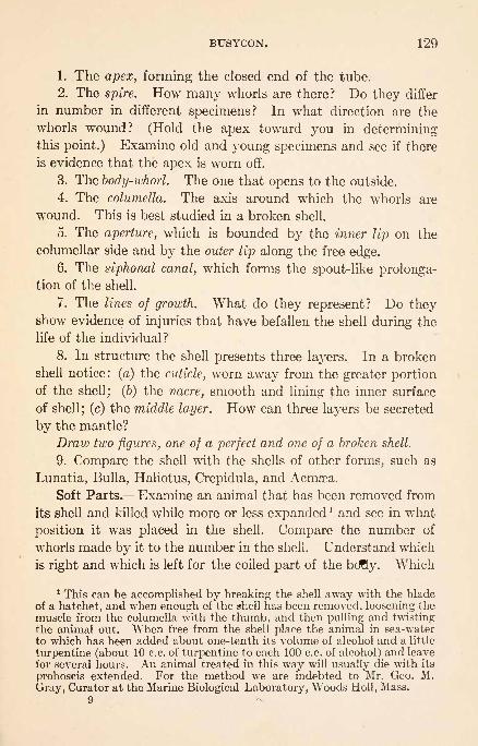
BUSYCON. 129
1. The apex, forming the closed end of the tube.
2. The spire. How many whorls are there? Do they differ
in number in different specimens? In what direction are the
whorls wound? (Hold the apex toward you in determiningthis point.) Examine old and young specimens and see if there
is evidence that the apex is worn off.
3. The body-whorl. The one that opens to the outside.
4. The columella. The axis around which the whorls are
wound. This is best studied in a broken shell.
5. The aperture, which is bounded by the inner lip on the
columellar side and by the outer lip along the free edge.
6. The siphonal canal, which forms the spout-like prolonga-tion of the shell.
7. The lines of growth. What do they represent? Do theyshow evidence of injuries that have befallen the shell during the
life of the individual ?
8. In structure the shell presents three layers. In a broken
shell notice: (a) the cuticle, worn away from the greater portionof the shell; (6) the nacre, smooth and lining the inner surface
of shell; (c) the middle layer. How can three layers be secreted
by the mantle?
Draw two figures, one of a perfect and one of a broken shell.
9. Compare the shell with the shells of other forms, such as
Lunatia, Bulla, Haliotus, Crepidula, and Acmsea.
Soft Parts. Examine an animal that has been removed from
its shell and killed while more or less expandedl and see in what
position it was placed in the shell. Compare the number of
whorls made by it to the number in the shell. Understand which
is right and which is left for the coiled part of the bofly. Which
1 This can be accomplished by breaking the shell away with the bladeof a hatchet, and when enough of the shell has been removed, loosening the
muscle from the columella with the thumb, and then pulling and twistingthe animal out. When free from the shell place the animal in sea-waterto which has been added about one-tenth its volume of alcohol and a little
turpentine (about 10 c.c. of turpentine to each 100 c.c. of alcohol) and leave
for several hours. An animal treated in this way will usually die with its
proboscis extended. For the method we are indebted to Mr. Geo. M.
Gray, Curator at the Marine Biological Laboratory, Woods Holl, Mass.
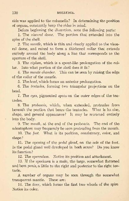
130 MOLLUSCA.
side was applied to the columella? In determining the position
of organs, constantly keep the sides in mind.
Before beginning the dissection, note the following parts:
1. The visceral dome. The portion that extended into the
spire of the shell.
2. The mantle, which is thin and closely applied to the visce-
ral dome, and raised to form a thickened collar that extends
entirely around the body along a line that corresponds to the
aperture of the shell.
3. The siphon, which is a spout-like prolongation of the col-
lar. Into what portion of the shell does it fit?
4. The mantle chamber. This can be seen by raising the edgeof the collar of the mantle.
5. The head, which forms an anterior prolongation.
6. The tentacles, forming two triangular projections on the
head.
7. The eyes, pigmented spots on the outer edges of the ten-
tacles.
8. The proboscis, which, when extended, protrudes from
beneath the portion that bears the tentacles. What is its size,
shape, and general appearance? It may be retracted entirely
into the body.
9. The mouth, at the end of the proboscis. The end of the
odontophore may frequently be seen protruding from the mouth.
10. The foot. What is its position, consistency, color, and
shape ?
11. The opening of the pedal gland, on the sole of the foot.
Is the pedal gland well developed in both sexes? Do you know
its function?
12. The operculum. Notice its position and attachment.
13. If the specimen is a male, the large, somewhat flattened
and bent penis, a little to the right and posterior to the right ten-
tacle.
A number of organs may be seen through the somewhat
transparent mantle. These are:
14. The liver, which forms the first two whorls of the spire
Notice its color.
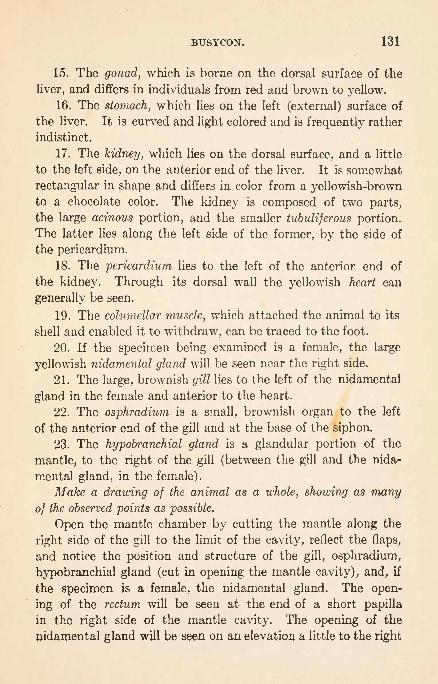
BTJSYCON. 131
15. The gonad, which is borne on the dorsal surface of the
liver, and differs in individuals from red and brown to yellow.
16. The stomach, which lies on the left (external) surface of
the liver. It is curved and light colored and is frequently rather
indistinct.
17. The kidney, which lies on the dorsal surface, and a little
to the left side, on the anterior end of the liver. It is somewhat
rectangular in shape and differs in color from a yellowish-brownto a chocolate color. The kidney is composed of two parts,
the large acinous portion, and the smaller tubuliferous portion.
The latter lies along the left side of the former, by the side of
the pericardium.
18. The pericardium lies to the left of the anterior end of
the kidney. Through its dorsal wall the yellowish heart can
generally be seen.
19. The columellar muscle, which attached the animal to its
shell and enabled it to withdraw, can be traced to the foot.
20. If the specimen being examined is a female, the large
yellowish nidamental gland will be seen near the right side.
21. The large, brownish gill lies to the left of the nidamental
gland in the female and anterior to the heart.
22. The osphradium is a small, brownish organ to the left
of the anterior end of the gill and at the base of the siphon.
23. The hypobranchial gland is a glandular portion of the
mantle, to the right of the gill (between the gill and the nida-
mental gland, in the female).
Make a drawing of the animal as a whole, showing as many
of the observed points as possible.
Open the mantle chamber by cutting the mantle along the
right side of the gill to the limit of the cavity, reflect the flaps,
and notice the position and structure of the gill, osphradium,
hypobranchial gland (cut in opening the mantle cavity), and, if
the specimen is a female, the nidamental gland. The open-
ing of the rectum will be seen at the end of a short papilla
in the right side of the mantle cavity. The opening of the
nidamental gland will be seen on an elevation a little to the right

132 MOLLUSCA.
and anterior to the anus. If possible, insert a guarded bristle
into this opening and see what becomes of it. Trace the oviduct
from the ovary along the columellar side of the liver. See what
becomes of it. Examine the inside of the nidamental gland
and see its relation to the oviduct.
If the specimen is a male, follow the vas deferens from the
testis to the base of the penis.
Circulatory System. Remove the thin membrane that forms
the roof of the pericardial chamber.
1. The heart consists of : (a) the large, rounded ventricle;
(b) the smaller, conical, thin-walled auricle.
2. The auricle receives blood by two vessels. One, return-
ing blood from the gill, runs along the left side of the gill to its
posterior end, where it bends abruptly to the right along the
margin of the pericardial cavity, and enters the auricle. The
other returns blood from the tubuliferous portion of the kidney
and follows the right side of the pericardium to the auricle.
3. The gill receives its blood through a vessel that borders
its right side. This vessel receives the blood from a portion of
the mantle, and from the large, acinous portion of the kidney.
4. The blood leaves the ventricle by a single vessel, the aorta,
that almost immediately gives rise to the visceral artery which
supplies the visceral hump. Trace its distribution.
The aorta makes an abrupt turn downward and forward and
enlarges to form the secondary heart which lies alongside the eso-
phagus. Follow the course of the aorta and its branches.
The course of general circulation is, beginning with the heart,
(a) system, (b) kidney, (c) gill, and (d) back again to the
heart. Why is such a course of circulation better than the
reverse?
Draw a figure showing the vascular system.
Excretory System. The two portions of the kidney have
already been noticed. Cut along their common line of union
and examine the inner surface of each part.
1. Notice the parallel lines of tubules that form the substance
of the tubuliferous portion, and the lobules that form the com-
paratively thick walls of the acinous portion.

BUSYCON. 133
2. Find the slit-like opening that leads from the kidney to
the mantle cavity. It is at a point between the two portions of
the kidney and is easily found from the mantle chamber. Asmall opening leads into the pericardium, but it is hard to find
it in dissections.
Digestive System. 1. Remove part of the integument at the
base of the proboscis and find the muscles that retract it. Howmany are there and how are they attached ? Do you understand
how the proboscis is extended?
2. With a pair of scissors open the extended proboscis along
the ventral line, pin it open, and notice that the exposed muscu-
lar mass, the buccal mass, is attached to the wall of the proboscis
in the region of the mouth, at its base, and by means of fibers,
along its sides.
3. Push the muscular mass slightly to one side and notice
the esophagus, which is closely applied to the dorsal wall of the
proboscis. Notice the muscle fibers that extend from it to the
proboscis. What is their function?
4. The odontophoral apparatus consists of a forked cartilage,
the odontophoral cartilage, that is surrounded by muscles and
cannot be seen until these are removed, a radula which is for
most of its length enclosed in a sac, the radular sac, and is ex-
posed only in the region of the mouth, and the muscles for mov-
ing the cartilage and the radula.
(a) The strands of muscles that run from its sides forward
to be inserted on the walls of the proboscis are attached to the
odontophoral cartilage. These are the cartilage protractors.
(b) Attached to the ends of the two horns of the cartilage
and running posteriorly to be attached to the walls of the pro-
boscis near its base are the flat cartilage retractors.
(c) Running lengthwise of the buccal mass, on its ventral
side, are three pairs of slender muscles, one pair median and the
others covering the horns of the odontophoral cartilage that has
just been observed. Find to what the muscles are attached
anteriorly and posteriorly. If the animal is fresh, pull on them

134 MOLLUSCA.
with the forceps and see what moves. These are the radula
protractors.
(d) Beneath the radula protractors observe the sheet of cross-
fibers that bind the horns of the odontophoral cartilage together.
Make a drawing showing the ventral side of the buccal mass.
(e) A portion of the radula is visible near the anterior end of
the proboscis. Introduce a bristle into the esophagus and deter-
mine its relation to the exposed radula.
(/) Loosen the anterior end of the buccal mass from the wall
of the proboscis, turn it back and see how the radula passes
around the odontophoral cartilage. With a hand-lens notice
the teeth on the open radula, ventral to the cartilage, and see
how the radula is folded as it passes over the dorsal side of the
cartilage so the teeth are turned in. What reason is there for
folding the radula in this manner?
(g) Cut the cartilage protractors and reflect the buccal mass.
It is attached to the wall of the proboscis at its posterior end by
strong muscles, the radula retractors. These may be studied
after cutting the sheath of cross-fibers that hold the mass to-
gether. Determine how they are attached to the sides of the
radula. Why do they need to be so powerful?
Make a drawing of the buccal mass as seen from the dorsal side.
(h) Pull away the muscles and examine the shape of the
odontophoral cartilage and its relation to the radula.
(i) Remove the radula, unfold it, and examine it microscop-
ically. Do the teeth differ in any way at the two ends? Whyis the radula so long?
Draw a portion.
The radula is the organ upon which most gastropods depend
for getting food. You should understand how:
1. The proboscis is protruded and retracted.
2. The odontophoral cartilage is protruded and retracted.
3. The radula is protracted and retracted.
4. The radula is folded by the cartilage and spread for action.
5. The food is torn off and taken into the mouth.
Near the base of the proboscis is a pair of large, yellow

BUSYCON. 135
salivary glands, the ducts from which extend on either side of
the esophagus to the mouth. Further back;on the right side
of the esophagus, is the delicate pancreas.
Trace the esophagus to the stomach and the intestine to the
anus.
Nervous System. Most of the ganglia are grouped around the
esophagus, about three-quarters of an inch posterior to the base
of the proboscis. They are all brown and accordingly conspic-
uous. Carefully cut around its base so the proboscis may be
turned back, and the ganglia on the ventral . surface of the
esophagus may be seen. Carefully pick away the tissue
that covers the ganglia and notice on the ventral side of the
esophagus:1. The small but conspicuous buccal ganglia. These are
united with each other and with the cerebral ganglia and send
nerves to the mouth apparatus.
2. The large pedal ganglia, fused together but distinctly
paired, lying posterior to the buccal ganglia and sending nerves
to the two sides of the foot. Each is united by connectives with
the corresponding cerebral and pleural ganglia.
From the dorsal side a number of ganglia may be seen, more
of which lie to the right than to the left of the median line.
1. On the left side there are two ganglia that are in rather
close union with each other. The most anterior, the left cerebral,
is the larger of the two. The left pleural joins it posteriorly and
ventrally and extends nearly to the ventral side of the esophagus.
2. On the right side four ganglia may be distinguished. The
right cerebral and right pleural are fused to form one mass, but
there is a marked constriction between them. Posteriorly and
dorsally the right pleural is connected by a connective with the
right visceral, which lies very close to it. The remaining ganglion,
the left visceral, which is almost in contact with the right pleural
and right pedal ganglia, lies ventrally and to the right of the
right visceral ganglion. It is connected with the left pleural
ganglion by a connective that runs behind the pedal ganglia.
There seems also to be a connection with the right pleural gan-

136 MOLLUSCA.v
glion, but this must be considered a secondary connection. Do
you understand why this ganglion occupies this position?3. Another ganglionic mass, the abdominal ganglion, possi-
bly formed by the fusion of two ganglia, lies just below the exter-
nal opening of the kidney, where it can be seen as a brown mass,
through the body-wall. It lies on the elongated commissure
that connects the two visceral ganglia. The commissure may be
followed by dissection.
The cerebral ganglia are the most centralized. Besides being
connected with each other by a commissure dorsal to the esopha-
gus, and being intimately connected with the pleural ganglia,
each cerebral ganglion is connected with the corresponding buc-
cal and pedal ganglion, and, through the pleural, with the visceral
ganglion. The visceral ganglia are connected with each other
by a long commissure on which the abdominal ganglion is placed.
Each pedal ganglion receives connectives from the cerebral and
from the pleural ganglion of the corresponding side.
Draw figures of the nervous system and compare them with
the clay model. 1
Colton: How Fulgur and Sycotypus Eat Oysters, Mussels, and Clams.Proc. Acad. Nat. Sci., Philadelphia, 1908. 4
Conklin : The Embryology of Fulgur : A Study of the Influence of Yolk on
Development. Proc. Acad. Nat. Sci., Philadelphia, 1907.: The Embryology of Crepidula. Jour. Morph., 13, 1897.
Copeland : The Olfactory Reactions and Organs of the Marine Snails, Alec-
tripn obsoleta and Busycon canaliculatum. Jour. Exp. Zool., 25, 1918.
Crozier: On the Use of the Foot in Some Mollusks. Jour. Exp. Zool., 27,1919. ..
Glaser: Uber den Kannibalismus bei Fasciolaria tulipa (var. distans) undderen larvale Excretionsorgane. Zeit. f. wiss. Zool., 80, 1905.
Herrick: Mechanism of the Odontophoral Apparatus in Sycotypus canalicu-
latus. Am. Nat., 40, 1906.
Olmstead: Notes on the Locomotion of Certain Bermudian Mollusks.Jour. Exp. Zool., 24, 1917.
Orton: An Account of the Natural History of the Slipper-Limpet (Crepi-dula fornicate). Jour. Marine Biol. Ass., 9, 1912.
Patten: The Embryology of Patella. Arb. Zool. Inst. Wien., 6, 1886.
1 Instructors will find that a model prepared by sticking lumps andstrands of modeling clay on a cylindrical graduate, to illustrate the positionsof the ganglia and connectives on the esophagus, will greatly aid thestudents.

LOLIGO PEALEII. 137
CEPHALOPODA.
LOLIGO PEALEII. (The Squid.)
Specimens of this or closely related species are rather common
along the Atlantic coast of the United States. They are active
swimmers, but may occasionally be seen in shallow, quiet water
near the shore. The movements and positions of adult speci-
mens in aquaria should be studied. Do you know what they
eat and how they capture their food ?
Study a small living specimen in a jar of sea-water and no-
tice:
1. Its general shape and distinct head.
2. Its position in the water. For convenience, the lower
surface may be referred to as ventral, but this is not to be con-
sidered as morphologically the same as the ventral surface of
other Mollusca. What parts are kept moving? Why is water
pumped when the animal is not swimming?3. In what direction it can swim best. Can it swim in the
other direction? How does it swim? Is the funnel movable?
How does it guide its movements?
4. Its color. Irritate it and see what happens. What pur-
pose does the change in color serve? What is the ink for?
5. What happens when the end of a finger is placed within
the circlet of tentacles of an animal about two inches long that
is being held firmly?
Using an adult specimen, observe:
6. The arrangement of the arms on the head. Are they ar-
ranged in any definite order ? Are they all alike ?
7. The suckers of the arms. Do they follow the same order
on all of the arms?
8. The structure of a sucker. Notice the peduncle, outer
thin margin, horny ring, and piston. Is the horny ring smooth?
What is its function? How does the sucker work? Split one
and draw the cut surface.
9. The mouth. Where is it placed? Notice the tips of the
horny beak. Which jaw is longest?
10. The eyes.

138 MOLLUSCA.
11. The fold of tissue behind each eye. These have been
called the olfactory organs, but there is no experimental evidence
of function.
12. The attachment of the head and the extent of the man-tle opening around the neck.
13. The funnel protruding from beneath the mantle on the
ventral surface. Notice the position and character of its opening.14. The median dorsal projection of the mantle.
\ 15. The tail-fin, its position and shape. What is its function?
Draw the animal as seen from the ventral side.
Carefully open a specimen by cutting through the mantle
a little to one side of the mid-ventral line.
Notice:
1. The thickness and character of the mantle and its relation
to the rest of the body. Why does it need to be so muscular?
2. The arrangement of the funnel. Why does it have a
thin posterior edge? How is it held in position against the
mantle. Does it have a valve? Is the funnel movable in the
living animal? Is there any provision for movement?3. The free edge of the mantle and its relation to the folds
beneath the eyes. Do you understand how the water gets into
and out of the mantle cavity?
4. The large retractor muscles of the funnel. How many are
there? How can the funnel be pointed in different directions?
What need is there for such a provision?
5. The retractor muscles of the head. How many are there?
Are they used in swimming in any way ?
"
6. The rectum, opening near the base of the funnel between
two small lateral flaps of tissue.
7. The ink-bag, dorsal to the rectum and opening into it
near the anus.
8. The gills, extending from a point about midway of the
body toward the free edge of the mantle. How many are there ?
How are they attached? Why does an animal that is not swim-
ming continually pump water through the mantle chamber?
9. The branchial hearts, at the bases of the gills, rounded,

LOLIGO PEALEII. 139
light colored organs that can be seen through the membrane
covering them.
10. The median ventral mesentery.
If the specimen is a male, notice:
1. The slender, tapering penis, to the left of the rectum.
2. The kidneys, white organs to be seen through the mem-branous covering, between the bases of the gills. From this
position they taper anteriorly for half an inch or more and send
small lobes posteriorly.
3. The openings of the kidneys near their anterior ends, on
small papilla.
4. The conical posterior portion of the viscera. This is com-
posed of a large visceral sac and portions of the sexual organs.
Draw the animal, showing the points observed.
If the animal is a female, notice:
1. The pair of large, white nidamental glands that cover a
portion of the rectum and the greater part of the ink-bag.
2. The openings of these glands at their anterior ends. Doyou know the function of these glands?
3. The small accessory nidamental glands just anterior to the
nidamental glands. These have large ventral openings. Just
before egg laying they become brilliantly red.
4. The opening of the oviduct dorsal to, and a little to the
left of, the left nidamental gland.
5. The rounded swelling, the oviducal gland on the oviduct.
6. The mass of eggs that fills the posterior portion of the
body. These are in the ovary and oviduct.
Draw the animal, showing the points observed.
Excretory System. If the animal is a female, remove the
nidamental glands, and the kidneys will be seen in the position
described for the male. The kidneys consist of:
1. The white, somewhat triangular, glandular portions al-
ready, noticed, extending from the region of each branchial heart
anteriorly, and forming a portion of the walls of the pre-cava?.
2. The cavities of the organs lying ventrally, and at the sides
of the glandular portions.

140 MOLLUSCA.
3. The external openings, at the ends of small papillae, on
either side of the rectum near the anterior ends of the kidneys.
Digestive System. Remove the funnel and its retractor
muscles and carefully lay the head open, along the ventral side.
Find :
1. The buccal mass. This is a rounded, muscular organ,
with a double ring of tissue, the buccal membranes, at its anterior
end, that surrounds the horny jaws. Examine the jaws and see
which is the larger.
2. Behind the buccal mass are the paired salivary glands.
3. Trace the narrow esophagus from the posterior end of the
buccal mass backward. At the base of the head it enters the
liver, a large, white organ that lies between the retractor muscles
of the head, and extends from the base of the head to a point dor-
sal to the external openings of the kidneys. Lying close to the
esophagus and covered by the anterior end of the liver is an elon-
gated median salivary gland, the duct from which follows the
esophagus into the head. The esophagus leaves the liver about
midway of its length, and follows along the ventral surface nearly
to the stomach. Before entering the stomach the esophagus
passes the pancreas, a white, lobed organ that lies just beneath
the glandular portion of the kidneys, and the systemic heart, a
roughly diamond-shaped organ that lies between the branchial
hearts.
The stomach proper is a rather small, thick-walled sac that
lies on the right side of the body, dorsal and posterior to the right
branchial heart. From the left side of the stomach a rather
large opening leads into a thin-walled blind sac, the visceral sac,
that when filled with partly digested food, as it frequently is,
extends posteriorly to the end of the body and occupies a consid-
erable part of the conical portion of the body. When empty,
it is quite small and inconspicuous.
The intestine leaves the stomach very near the point the eso-
phagus enters, and just anterior to the opening that leads into
the visceral sac. It passes ventrally, and becomes visible from
the surface, where its position has already been noted.
Draw a figure showing the digestive system.

LOLIGO PEALEII. 141
Cut a median sagittal section of the buccal mass and notice
the mouth cavity, the jaws, the muscles that move the jaws, the
tongue, and the position of the radula. Is the radula arranged
in the strap-over-pulley manner that it is in Sycotypus?
Draw a figure of the section.
Male Reproductive System. 1. The testis (morphologically
the left) is large, white, and flattened, and lies far back in the
pointed end of the body. It is enclosed in a sheath which
serves to collect the liberated sperm.
2. Just anterior to the testis is a small rounded vesicle, the
ampulla, which is the point of origin of the vas deferens.
3. The vas deferens is a plaited slender tube, which being
packed with sperm is opaque white. It extends from the am-
pulla along the right side of a large sac to be referred to later,
the spermatophoric sac, to and beneath (dorsal to) the spermato-
phoric organ, where it joins a portion of the organ on its left side
about one-third the length of the organ back from its anterior
end. Remove the left gill and branchial heart and strip awaythe thin tissue that covers the vas deferens and spermatophoric
organ, being careful not to injure either. Carefully lift the
right ventral side of the spermatophoric organ and see where
the vas deferens enters it. Drop the spermatophoric organinto position again.
4. The spermatophoric organ lies almost on the left side, but
a little ventrally. In it the spermatophores are formed. It
consists of a series of glands and mechanical arrangements that
secrete and wind materials into spermatophores. Briefly, the
parts are:
(a) On the right dorsal side, the part joined by the vas
deferens, the mucilaginous gland. This consists of two
parts.
(6) Ejaculatory apparatus gland, which extends from the
mucilaginous gland posteriorly and then ventrally. Both
ends of this gland are marked by constrictions.
(c) From this gland forward to a narrow duct is the middle
tunic gland.

142 MOLLUSCA.
(d) The narrow duct leads into a large blind pouch and is
the outer tunic gland.
(e) The large blind pouch extends posteriorly nearly the
length of the organ and is the hardening gland.
(/) A branch leaves the narrow duct just before it enters
the hardening gland that leads to the curved anterior extremity
of the gland. This is the finishing duct.
(g) The finishing duct leads to the curved gland mentioned,the finishing gland.
This completes the spermatophoric organ, although some
minor structures have not been mentioned. A section across
the ejaculatory apparatus gland will show a large ridge with a
groove along one side through which the spermatophore travels
and rotates while being formed.
5. From the last part of the spermatophoric organ, the
finishing gland, a straight duct leads posteriorly by the side of
the vas deferens to join the spermatophoric sac about the length
of a spermatophore from its posterior pointed end. This is the
spermatophoric duct.
6. The spermatophoric sac is somewhat spindle shaped,
usually filled with spermatophores, and joins the base of the
penis.
Make a drawing of the system.
Open the spermatophoric sac and remove some of the
spermatophores. If the specimen has not been preserved place
them immediately in 10 per cent, formalin or stronger to keep
them from ejaculating. Mount under a cover and examine. The
specimens may be stained with dilute Ehrlich's triacid stain and
mounted in glycerin jelly if preferred. Notice:
1. The spermatophore is covered by a transparent elastic
outer tunic which has a cap and cap thread at the smaller, oral,
end.
2. Inside this is the somewhat granular middle tunic, which
is much thicker at the aboral end.
3. The contents consist of the aboral sperm mass, the oral

LOLIGO PEALEII. 143
ejaculatory apparatus, and the cement body, which lies between
them.
4. The sperm mass may be seen to consist of a closely woundthread. It is actually covered by a very thin inner tunic which
is separated from the outer tunic by a space filled with liquid.
5. The cement body is attached to the sperm mass by a
narrow core and is covered by a continuation of the inner
tunic. It is flask shaped, with the narrow end pointing orally.
6. The neck of the cement body is covered by parts of the
ejaculatory apparatus and the bulge of the body is joined and
possibly covered by the outer membrane, which with the inner
tunic forms the outer covering of the ejaculatory apparatus that
appears as a double membrane. Inside the outer membrane is
the rather thick middle membrane, the aboral end of which
encloses the neck of the cement body and ends against the
bulge of the cement body, and the oral end of which is thrown
into bends and loops and is finally attached to the cap end of the
outer case. Inside the middle membrane is the thin inner
membrane and the spiral filament which enclose a narrow lumen.
7. In ejaculating the ejaculatory apparatus turns wrongside out and the cement body and sperm mass are crowded
down the tube thus formed by the elastic force of the outer
tunic and the elastic and osmotic action of the middle tunic.
The sperm mass is forced into a sac composed of the in-
verted inner tunic and outer membrane, which remain at-
tached to the bulge of the cement body, the cement body is
ruptured and the cement spread over the closed end of this sac.
The reservoir is now ready to stick in position.
Studying the method of ejaculation is time consuming.Fresh specimens placed in about one-fourth saturated solution
of magnesium chlorid for ten minutes or more will be slowed
in action so the process can be followed more readily. Removea specimen from this solution to sea-water, grasp the cap thread
with forceps, and shake the spermatophore. This should start
ejaculation. Ejaculation can be stopped promptly by squirting
full strength formaldehyd on it.

144 MOLLUSCA.
Make a drawing of a spermatophore.
Spermatophores are carried into position by the action of the
left ventral arm of the male. Examine its tip and notice the
modification of the suckers.
Female Reproductive System. The opening of the oviduct
has already been noticed. Observe:
1. The large, swollen portion, the oviducal gland, that lies
on the oviduct dorsal to the left branchial heart.
2. The long convoluted oviduct extending posteriorly from
the oviducal gland. It is frequently filled with eggs for the
greater part of its length.
3. The lighter colored, greatly lobulated ovary, also fre-
quently filled with eggs, lying dorsal to the oviduct and visceral
sac and extending from the region of the stomach to the end of
the body. The ovary is inclosed in a capsule from which the
oviduct leads.
4. The nidamental and accessory nidamental glands have
been studied and removed.
5. On the median line of the inner surface of the outer
buccal membrane of the female is the sperm receptacle. Duringthe summer this is usually filled with sperm, and is, accordingly,
white and conspicuous. Below the receptacle is a modified
area for the attachment of sperm reservoirs as they are de-
livered from the spermatophores.
Draw a figure of the female reproductive system.
Circulatory System. An injected specimen is desirable. Theblood that has been supplied to the body in general is collected
by veins and carried to the branchial hearts. The vessels that
collect the blood are:
1. The pre-cavce. A single vessel carries the blood from the
head to the anterior ends of the kidneys. Here the vessel divides
into right and left pre-cavse that are intimately connected with
the kidneys. The pre-cavae diverge near the posterior ends of
the kidneys and enter the corresponding branchial hearts.
2. The post-cavce. A pair of very large vessels that return
blood from the posterior end of the body. They join the corre-

LOLIGO PEALEII. 145
spending pre-cavse near the anterior borders of the branchial
hearts.
3. The mantle-veins. These return blood to the branchial
hearts from the anterior portion of the mantle.
The blood that is received by each branchial heart is sent
into the corresponding gill through a branchial artery that leaves
the heart near the opening of the mantle vein, and runs along
the side of the gill that is attached to the mantle.
The blood is collected from each gill by a large branchial vein
that runs along the ventral side of the gill, and enters the sys-
temic heart.
Draw a figure showing the vessels connected with the branchial
hearts.
Expose the systemic heart by carefully removing the superfi-
cial tissue between the branchial hearts, and notice that it
is not symmetrical. Its lateral angles receive the branchial
veins and it gives rise to an artery from each of the other two
angles.
1. The posterior aorta divides almost immediately into three
large vessels. These are:
(a) The median mantle artery which follows the edge of the
ventral mesentery to the mantle.
(6) A pair of lateral mantle arteries which diverge posteriorly
and supply the two sides of the mantle. Besides these large
vessels there is a small vessel that runs anteriorly over the ven-
tral surface of the heart and supplies the ink gland and rectum,
and another one that runs dorsally and posteriorly to supply part
of the reproductive system.
2. From the dorsal surface of the heart, near its anterior
end, a small vessel passes over the anterior and dorsal surfaces
of the stomach and finally passes into the gonad.
3. The anterior aorta is larger than the posterior aorta. Fromthe anterior angle of the heart, which is to the right of the median
line, it follows a straight course alongside the esophagus to the
head. A number of small vessels are given off along its course,
and it is finally distributed to the head and arms.
10

146 MOLLLUSCA.
Draw the vessels connected with the systemic heart, into the
figure you have just made.
Nervous System. The stellate ganglia may be seen throughthe transparent lining of the mantle, on either side of the neck,
where the body joins the mantle. They send nerves to the man-
tle and are joined to ganglia in the head (the infra-esophageal)
by connectives. Why does the mantle need such large, special
ganglia? Other small ganglia are situated in the body, but the
large and important ones are grouped in the head, where they are
supported and protected by cartilages.
With a razor make a median sagittal section of the head of
a squid and notice:
1. Dorsal to the esophagus a rounded mass, the supra-eso-
phageal ganglion, which is supposed to represent the fused cere-
bral ganglia.
2. Ventral to the esophagus the elongated infra-esophageal
ganglion, which is supposed to represent the fused pedal and
visceral ganglia and (together with the masses that connect the
supra- and infra-esophageal ganglia around the esophagus) the
pleural ganglia.
3. The anterior prolongation of the infra-esophageal gan-
glion to form the pro-pedal portion, which supplies nerves to the
arms.
4. The small supra-buccal ganglia, lying dorsal to the esoph-
agus, and a little further anterior than the ends of the pro-pedal
portion. These are joined by connectives with the supra-
esophageal ganglia.
5. The infra-buccal ganglia, about the same size as, and lying
ventral to, the supra-buccal ganglia, and joined with them byconnectives that run around the esophagus.
Draw a figure of a sagittal section of the head.
Two large ganglia, the optic ganglia, lie against the eyes
and will be seen in cross-sections of the head that will be studied
later. A dissection of one side of the head will show one.
Open the animal along the mid-dorsal line and find the pen
which is embedded in the mantle. After exposing it for its

LOLIGO PEALEII. 147
full length, turn the flaps aside and see that it lies in a pocket.
It probably represents a modified shell that has become entirely
inclosed by the mantle. What is its function?
Pull the pen out of the mantle and draw it.
With a razor make cross-sections of a squid, a quarter of an
inch thick, and arrange them in order, in a little water, as they
are made. Identify the parts you have found in dissection.
Make drawings of the sections that pass through the infra-eso-
phageal ganglion, through the eyes, through the liver, and through
the heart.
If time permits, study prepared sections that have pre-
viously been made. The structure of the eye and the positions
of the parts of the nervous system should receive special atten-
tion.
Specimens of other cephalopods, such as Octopus and Nau-
tilus, should be compared with the squid and the adaptations
that fit them for their particular lives noted.
Brooks: The Development of the Squid (Loligo Pealii). Mem. Bost.
Soc. Nat. Hist., 1880.: Handbook of Invertebrate Zoology.
Cowdry: Color Changes in Cephalopods. Univ. of Toronto Studies, 10,1911.
Drew: Sexual Activities of the Squid. I. Copulation, Egg-laying and Fer-tilization. Jour. Morph., 22, 1911.
: Sexual Activities of the Squid. II. The Spermatophore; Its Struc-
ture, Ejaculation and Formation. Jour. Morph., 32, 1919.
Griffin: The Anatomy of Nautilus pompilius. Mem. Nat. Acad. Sci., 9,
1900.
Vialleton: Recherches sur les primieres phases du developpment de la
Seiche (Sepia officinalis). Ann. Sci. Nat. (7) Zool., 6, 1888.
Willey: Contribution to the Natural History of the Pearly Nautilus.
Willey's Zool. Results. 4, Cambridge Univ. Press, 1902.
Williams: The Anatomy of the Common Squid, Loligo pealii. Amer.Mus. Nat. Hist.
: The Vascular System of the Common Squid. Am. Nat., 36, 1902.

ARTHROPODA.
With segmented bodies that are provided with segmented
appendages.
CLASS 1. Crustacea.
Usually aquatic. With a more or less hardenedouter covering and many thoracic appendages.
Subclass 1. Entomostraca.
Usually small. Appendages little differentiated.
The number of post-cephalic segments variable.
Order 1. Phyllopoda.
Appendages with leaf-like expansions. (Bran-
chipus, Daphnia.)Order 2. Ostracoda.
Free-swimming, with the body inclosed in a bi-
valve shell. Seven pairs of appendages. (Cy-
pris.)
Order 3. Copepoda.Body elongated and distinctly segmented (ex-
cept in parasitic forms). Four or five pairs of
biramous appendages. (Cyclops, Argulus.)Order 4. Cirripedia.
Comparatively large and usually attached. Usu-
ally with six pairs of biramous appendages.Forms that are not parasitic are covered by cal-
careous plates. (Lepas, Balanus.)Subclass 2. Malacostraca.
Usually of considerable size and generally highly
organized. Except in one order, thorax of eight
and abdomen of seven segments.Order 1. Phyllocarida.
Body inclosed in a large, bivalve cephalic cara-
pace. Abdomen of eight segments. Thoracic
segments free from the head. (Nebalia.)
Order 2. Schizopoda.Thoracic appendages all biramous. Shrimp-likein shape. (Mysis.)
148

ARTHKOPODA. 149
Order 3. Decapoda.Thoracic segments united with the head. Three
pairs of maxillipeds and five pairs of legs, of
which the first bear heavy pincers. (Homarus,Cambarus, Crangon, Eupagurus, Hippa, Calli-
nectes, Cancer, Uca.)Order 4. Stomatopoda.
Five anterior thoracic legs are maxillipeds. Eyesstalked. Gills borne on abdominal segments.(Squilla.)
Order 5. Cumacea.Two anterior thoracic legs are maxillipeds. Eyessessile. Small shrimp-like forms. (Diastylis.)
Order 6. Arthostraca.
First and sometimes second thoracic segmentsfused with the head. Eyes usually sessile. (Tal-
orchestia, Gammarus, Caprella, Porcellio.)CLASS 2. Arachnoidea.
Head and thorax fused and bearing six pairs of
appendages. Respiratory organs lamellate andabdominal or replaced by tracheae.
Subclass 1. Gigantostraca.With lamellate abdominal gills. Coxal joints of
legs used as jaws. Marine. (Limulus.)Subclass 2. Arachnida.
Respiration by lamellate lungs or tracheae. Four
pairs of walking legs. Mostly terrestrial.
Order 1. Scorpionida.Abdomen segmented, posterior portion slender
and very flexible, frequently ending in a sting.Four pairs of lung-books. Pedipalpi chelate.
(Buthus.)Order 2. Pseudoscorpionida.
Abdomen segmented, without slender posterior
portion or sting. Pedipalpi chelate. Respira-tion by tracheae. (Chelifer.)
Order 3. Pedipalpida.Abdomen flattened and segmented. Pedipalpi
simple or chelate. Two pairs of lung-books.
(Phrynus.)Order 4. Solpugida.
Body composed of three segments. Chelicerse
chelate. Pedipalpi elongate, simple. Respira-
tion by tracheae, (Galeodes.)
150 ARTHROPODA.
Order 5. Phalangida.
Body short and oval. Abdomen composed of
six segments. Chelicerae chelate. Pedipalpiand legs very long and slender. Respiration bytracheae. (Phalangium.)
Order 6. Araneida.
Abdomen unsegmented and usually distinct.
Chelicerae end in claws that are provided with
poison glands. Lung-books and sometimes tra-
cheae also are present. Spinnerets present on the
abdomen. (Epeira, Agalena.)Order 7. Acarida.
Body not divided into regions. Biting or pierc-
ing and sucking mouth-parts. Respiration bytracheae or through integument. (Sarcoptes,
Dermacentor.)
Supplementary to the Arachnoidea.
Pycnogonida.(Doubtfully referred to the group.) Body com-
posed of segmented cephalothorax and vestigialabdomen. Legs very long, angular, and con-
taining portions of the viscera. No special
respiratory organs. (Phoxichilidium.)CLASS 3. Onychophora.
Elongated bodies with some annelid-like charac-
ters. Appendages short, numerous, and creased
rather than jointed. Respiration by means of
tracheae. (Peripatus.)CLASS 4. Myriapoda.
Generally elongated bodies with numerous
jointed appendages. A distinct head bearing
ocelli, antennae, and jaws is present. Respira-tion by means of tracheae.
Order 1. Symphyla.With not more than twelve leg-bearing trunk
segments. A single pair of branching tracheae.
(Scolopendrella.)Order 2. Chilopoda.
With numerous trunk segments, each with a sin-
gle pair of legs. First pair of trunk appendagesforming poison jaws. Body dorso-ventrally com-
pressed. (Lithobius.)

ARTHROPODA. 151
Order 3. Diplopoda.With numerous trunk segments, each with two
pairs of legs. No poison jaws. Body not com-
pressed. (Julus.)
Order 4. Pauropoda.With ten trunk segments and nine pairs of legs.
(Pauropus.)CLASS 5. Insecta.
Body divided into head, thorax, and abdomen.Three pairs of thoracic legs and generally one or
two pairs of wings.Order 1. Thysanura.
No metamorphosis. No wings. Mouth-partsusually mandibulate. Some forms show vestigialabdominal appendages. (Lepisma, Sminthurus.)
Order 2. Orthoptera.
Metamorphosis direct. Two pairs of wings usu-
ally present, of which the anterior are usually
tough and protect the more delicate posteriorones. Mouth-parts mandibulate. (Acridium,
Gryllus, Periplaneta.)Order 3. Neuroptera.
Metamorphosis direct. Two pairs of netted
veined wings usually present. Mouth-parts man-dibulate. Prothorax free. (Libellula, Termes,Hexagenia.)
Order 4. Hemiptera.Metamorphosis direct. Two pairs of wingsusually present. Mouth-parts piercing andsuctorial. (Benacus, Cicada, Pediculus, Aphis.)
Order 5. Diptera.
Metamorphosis indirect. Wings, when present,one pair and membranous. Mouth-parts sucto-
rial. (Culex, Tabanus, Musca.)Order 6. Lepidoptera.
Metamorphosis indirect. Two pairs of scaly
wings. Mouth-parts suctorial. (Platysamia,Anosia, Philampelus.)
Order 7. Coleoptera.
Metamorphosis indirect. Membranous hind
wings folded and covered by modified fore
wings, the elytra. Mouth-parts mandibulate.
(Lachnosterna, Doryphora.)

152 ARTHROPODA.
Order 8. Hymenoptera.Metamorphosis indirect. Two pairs of membra-nous wings. Mouth-parts suctorial and man-dibulate. (Apis, Vespa.)
Exner: Die Physiologic der facettirten Augen von Krebsen und Insecten,1891.
Prentiss: The Otocyst of Decapod Crustacea: Its Structure, Development,and Functions. Bui; Mus. Comp. Zool. Harvard, 36, 1901.
Watase: On the .Morphology of the Compound Eyes of Arthropods.Stud. Biol. Lab. Johns Hopkins Univ., 4.
CRUSTACEA.
HOMARUS AMERIGANUS. (Lobster. 1)
These animals are not generally found where they can be
readily observed in nature, but many valuable observations can
be made on specimens confined in aquaria. If other animals
are present in the aquarium notice the position of defense that
is taken. In nature the animal probably spends considerable
time under rocks with the anterior end of the body turned to-
ward the opening. In this position both sense organs and
weapons are in the proper position for attack or defense. Notice
how the appendages are used . Are the sense organs moved fre-
quently? What is the advantage of having the eyes on stalks?
What appendages are used in walking? Are all of these ap-
pendages used in just the same way? Does the animal move
equally well in all directions? Perhaps you can make the animal
swim; if so, observe the method. Feed a specimen with por-
tions of a clam or fish, and see how food is torn to pieces and
transferred to the mouth, and determine, if possible, how the
mouth appendages are used.
Appendages may be missing. If any are, notice at what
point they are broken. Possibly small appendages may be
growing from the old stubs. Autotomy may be studied by
crushing a claw or a leg of the fiddler crab, Uca. Other forms
1 These directions may be used for the crayfish without much modifi-
cation. The smaller size of these animals will make it more difficult to
trace some of the nerves and blood-vessels.

HOMARTJS AMERICANUS. 153
will respond, but sometimes not promptly. What is the impor-tance of this reaction ?
External Anatomy. As in Nereis, the body is segmented.The metameres of the head and thorax, however, are immova-
bly fused to form a cephalo-thorax. This is covered dorsally bya single piece, the carapace.
1 . Note, on the carapace, the cervical groove between the head
and thorax, and the beak or rostrum forming an anterior spine.
The ventro-lateral edge of the carapace is not attached. A flat
object thrust between it and the body passes into the gill cham-
ber. This free plate of the carapace is called the gill-cover.
Do you know why the edge of the carapace is free? Notice the
hair-like spines along its free border. What purpose do these
serve ?
2. The abdomen is composed of seven movable segments,
each bearing a pair of jointed appendages except the last, which
is sometimes not considered a true segment and is called the
telson. Each abdominal segment consists of a dorsal piece, the
tergum, which is continued as a free plate laterally (the pleurori),
and of a ventral piece, the sternum. Move the abdominal seg-
ments and see where they are hinged. How are the terga and
sterna arranged to allow free movement? In the thorax the
sterna, though fused, can be distinguished. There are eight
segments in the thorax.
3. Appendages. Aside from the stalked eyes, whose homol-
ogy with true appendages is doubtful, there are nineteen pairs.
These are, counting from before backward: antennules, antennae,
six pairs of mouth appendages, five pairs of walking legs (pereio-
pods), of which the first are the claws or chelce, and six pairs of
swimmerets (pleopods). In the male, the first two pairs of
pleopods are modified to form copulatory organs. The first
pair is greatly modified and the second pair bears a special
portion.1
(a) Turn one of the fifth pair of pleopods forward and exam-
ine its posterior aspect. It consists of a basal piece, the proto-
1 The crayfish has the first two pairs, both greatly modified.

154 ARTHROPODA.
pod, a lateral branch, the exopod, and a median branch, the
endopod. This branched type of appendage is designated as
biramous. What is its use? Compare with this the modified
sixth pair of pleopods, called the uropods.
Make a drawing of one of the fifth pleopods.
(6) In front of the chelae will be seen the sixth pair of mouth
appendages, the third maxillipeds. Remove that of the right
side and compare it with the fifth pleopod. In addition to the
protopod, exopod, and endopod, it bears a long blade, the epipodt
which extended into the gill chamber. The protopod is com-
posed of two segments, coxopod and basipod; the endopod of five,
segments, ischipod, meropod, carpopod, propod, and dactylopod.
The exopod is composed of one long and many short segments.
How is the appendage modified to serve in feeding ?
Make a drawing of the third maxilliped.
(c) Remove the remaining five mouth appendages and com-
pare each with the third maxilliped. These are, beginning pos-
teriorly, the second maxilliped, first maxilliped, second maxilla
(with a broad paddle, the scaphognathite, the use of which
should be understood), first maxilla, and the mandible. Just
back of the mandibles are two small flaps, the paragnatha,which are not true appendages. Do you understand the use
of each of these appendages? Most of the appendages have
parts that may be compared with the typical biramous appen-
dage, but they are much modified to serve special functions, and
the exact homologies are not important. Between the man-dibles note the mouth, bounded in front by the labrum.
Drawings of these appendages may be made if time permits.
(d) The antennae are biramous. Notice on the ventral side of
the basal joint of an antenna the opening of the green gland or
nephridium.
(e) The antennules, though branched, are not considered to
be of the biramous type. Do you know why ? Remove one and
note on the dorsal surface of the basal joint a groove at whose
median extremity is a small hole, the opening into the otocyst.
Do you know the probable function of the antennules and of

HOMARUS AMERICANUS. 155
the otocysts? What reason is there for having both antennules
and antennae?
(/) Compare the pereiopods with the third maxilliped.
Which is lacking, endopod or exopod? Examine each of the
joints of one of these appendages and see in what directions
the appendage may be moved. Are there any ball-and-socket
joints? Compare the chelae with the other pereiopods and see
how they differ. To what part of a chela does the last segmentof the last pereiopod correspond. WT
hat reason is there for
having these appendages different? Do you think the arrange-
ment of the appendages would aid the lobster in climbing over
rough bottom?
Open one of the large chelae and determine how the muscles
are arranged to control its opening and closing. Which mus-
cles are strongest? Find how the muscles are attached to the
"thumb."
Find the openings of the sexual ducts on the basal joints of
the pereiopods; the fifth pair in the male, the fourth pair in
the female. 1 In the female there is an opening into a seminal
receptacle through a triangular elevation on the ventral side
of the thorax.
4. Gills. Remove the gill-cover of the left side, being care-
ful not to injure the gills. Extending up into the gill cavity
are seven epipods belonging to the three maxillipeds and the
four anterior pereiopods. They separate the gills into groups.
Each group will be seen to correspond to a segment. The gills
show three sorts of attachments: (a) to the appendages them-
selves (podobranchs) , (6) to .tfce articular membranes between
appendages and body-wall (arthrobranchs) ,and (c) to the body-
wall itself (pleurobranchs) . There are two arthrobranchs in
some segments, one behind and above the other. How is the
current of water forced through the gill-chamber? What is
the function of the epipods? WT
hat direction must the water
take through the gill chamber? Examine the structure of a gill.
1 In the crayfish the female opening is on the third pair.
156 ARTHROPODA.
Move one of the appendages to which a gill is attached and see
the effect on the gill.1
Internal Anatomy. Remove the carapace (beginning at the
middle of the posterior margin and cutting forward, holding the
cartilage knife parallel with the surface) as far laterally as the
upper limits of the gill chambers and anteriorly to the base of
the rostrum. What is the pigmented membrane for? Dissect
it off so underlying organs may be seen.
1. The chitinous stomach lies near the anterior end with the
ophthalmic artery running along its mid-dorsal line. Beside and
behind the stomach are two masses of muscle which you have
severed from the carapace. These are the mandibular muscles,
and each is divided into an anterior and a posterior bundle.
Lateral to these muscle masses are the yellow-green digestive
glands, commonly called liver. Between and in front of the
posterior mandibular bundle note the gonads, and follow one
forward by pressing aside the muscle mass. In the male the
testis is a slender, white, convoluted cord, which ends blindly
against the side of the stomach. The extent and position of the
far thicker yellow ovary is much the same (unless the animal
be mature, in which case it will be found greatly enlarged and
orange).
2. The heart extends through the posterior third of the tho-
rax. Remove the upper part of the delicate pericardium sur-
rounding it, cut its arterial and other connections, and place it
in water. Note the shape, the origin of the arteries, and the
three pairs of ostia. Do you understand how it receives blood?
3. Trace the gonads as far as the abdomen, noting the single
anastomosis between those of opposite sides just in front of the
heart. Beneath the heart the sexual ducts are given off vasa
deferentia in the male, oviducts in the female. Trace one out-
ward and downward to its opening by removing a portion of the
body-wall and of the basal joint of the proper leg.
4. Remove the posterior lateral body-wall forward to a posi-
tion opposite the anterior third of the stomach. Pull the an-
1 The crayfish differs slightly in gill arrangement.

HOMAKUS AMERICANUS. 157
terior lobe of the liver, which extends beneath the stomach,
outward and backward. The liver will be seen to be attached
to the pyloric end of the stomach (i. e., the smaller part, where
the stomach passes into the intestine). Cut this attachment
and note that it is really where the liver opens into the stomach.
Just back of this point the right and left lobes of the liver
are connected by a cross-branch passing beneath the intestine.
Remove one liver lobe back to the abdomen. After having the
circum-esophageal connectives pointed out, remove the stomach by
cutting the esophagus, the intestine, and the bands of muscles
attached to the stomach. Examine it in water, noting the cardiac
and pyloric parts, the chitinous grinding and straining apparatusin the interior, and the muscles and plates that cause the move-
ments of the grinding apparatus. Why does a lobster with
chelse and six pairs of mouth appendages need a gastric mill?
5. Between the circum-esophageal connectives medially and
the large antennary muscles laterally, note the oval excretory
organs, called the green glands. They are covered by a very
delicate membrane. Poke a small hole in one of the membranes
and with a blowpipe show that it is really a thin bladder. Its
opening on the antenna has already been seen.
6. Remove the dorsal wall of the abdomen and trace the
posterior portions of the gonads, liver lobes, and intestine. In
the sixth abdominal segment the intestine swells to form the
chitin-lined rectum and gives off the blind intestinal ccecum.
Circulatory and Nervous Systems.1 Remove the carapace of
an injected specimen as before, also the gill-cover and gills on
one side.
1. There can generally be seen, through the transparent
body-wall, efferent branchial veins, which return the blood from
the gills. These unite into six large ones which open into the
1 The circulatory system of a fresh specimen may be satisfactorily
injected with starch-mass by inserting the needle of a hypodermic syringeinto the pericardium from the posterior margin of the carapace. Theoperation is easily performed when the distance to the pericardium is
understood. The carapace may be cut away and the needle inserted
directly into the heart if preferred.

158 ARTHROPODA.
pericardium at the side. Find these openings if possible. Do
you understand how blood gets into the heart ?
2. Note, at the anterior end of the heart, the ophthalmic
artery and the two antennary arteries. Trace the former forward
to the rostrum, cut it on the stomach and turn it forward for
future study. Trace the antennary arteries to the mandibular
muscles and cut them near the heart. Press the front end of
the heart back and note the two small hepatic arteries. Each
branches immediately, one division passing between the gonads,
and the other laterally.
3. Remove the muscles on one side of the heart and examine
it from the side, noting the great sternal artery extending down-
ward, and the smaller dorsal abdominal artery running back above
the intestine. Follow the latter through the abdomen.
4. Cut all arteries and remove the heart. Trace the anten-
naries through the mandibular muscles, noting the branch to
the stomach.
5. Remove the thoracic viscera as before, follow the circum-
esophageal connectives forward and identify the cerebral ganglia
in order not to destroy them.
6. Follow one antennary artery to the green gland, antennary
muscle, eye muscle, etc.
7. Follow the distribution of the ophthalmic artery.
8. Remove the intestine and muscles of the abdomen, and find
and trace forward the ventral nerve chain. Notice the position
of the ganglia and the nerves that leave them and the connec-
tives. In the thorax the ventral nerve chain passes beneath a
system of chitinous plates (the endo-phragrnal skeleton) and lies
in a cavity, the ventral blood sinus. Note the enlarged sub-
esophageal ganglion, the cross commissure just back of the esoph-
agus, the nerves to the mouth appendages, nerves from the
cerebral ganglia, and nerves from the other ganglia. What indi-
cation is there that the sub-esophageal ganglia represent more
than a single pair?
Sketch the nervous system.
9. The sternal artery passes through the ventral nerve chain
CALLINECTES HASTATUS 159
and then extends backward and forward as the ventral longitu-
dinal artery. Remove the nervous system and follow this ar-
tery.
Draw a diagrammatic cross-section through the thorax, putting
in one drawing the circulation from the heart through the sternal
artery to the limbs and back through the gills to the heart.
Andrews: The Keeping and Rearing of Crayfish for Class Use. Nat.Stud. Rev., 2, 1906.
: The Young of the Crayfishes Astacus and Cambarus. SmithsonianCont. to Knowl., 35, 1907.
: Conjugation in the American Crayfish. Am. Nat. 29, 1895.
Bumpus: Movements of Certain Lobsters Liberated at Woods Hole. Bui.
U. S. Com. Fish, 1899.
: Embryology of the American Lobster. Jour. Morph., 5, 1891.Herrick: Natural History of the American Lobster. Bui. U. S. Bur. Fish,
29, 1909.
Huxley: The Crayfish. An Introduction to the Study of Zoology. 1884.
Mead: Habits and Growth of Young Lobsters. Rhode Island Com. In-
land Fisheries, 21, 1901.
Paul: Reflexes of Autotomy. Proc. Roy. Soc. Edinb., 35, 1915.
Pearl and Clawson: Variation and Correlation in the Crayfish. CarnegieInst. Pub., 64, 1907.
Pearse : Observations on Copulation Among Crawfishes with Special Refer-ence to Sex Recognition. Am. Nat., 43, 1909.
Steele: Regeneration of Crayfish Appendages. Univ. Missouri Studies, 2,
1904.
: Regeneration in Compound Eyes of Crustacea. Jour. Exp. Zool., 5,
1907.
Williams: The Stonlach of the Lobster and the Food of Larval Lobsters.An. Rep. Com. Inland Fish., Rhode Island, 37, 1907.
CALLINECTES HASTATUS. (Blue Crab.)
Crabs may be found in shallow water along shore, where they
may be easily observed on quiet days. In what direction does
the animal normally move? How are the legs used? What is
the attitude of defense? Determine how the blue crab swims.
What do crabs apparently use for food ? Do they conceal them-
selves, are they protectively colored, or do they depend entirely
upon their weapons for defense ?
In studying the anatomy of the crab, constant comparisonsshould be made with the lobster.
External Anatomy. 1. The body is composed of cephalb-
thorax and abdomen. Dorsally note the shape of the carapace
160 ARTHROPODA.
and the position of the abdomen. The size of the abdomendiffers in male and female. Why should it be larger in the
female ?
2. Note the antennae, antennules, and eyes, and see how theyare packed away in recesses in the carapace. In the living
animal see if any of these are frequently moved.
3. The third maxillipeds are flattened and cover the other
mouth appendages.4. Straighten the abdomen and note the anus. Compare the
abdomen of a male with that of a female and both with that of the
lobster. The dorsal side of each segment is covered by a tergum.
The covering between each pair of pleopods is the sternum, the
immovable flap lateral to them is the pleuron. Compare the ab-
dominal appendages or pleopods of a male and a female.
5. The ventral side of the cephalo-thorax is covered by the
sternal plastron. Note the eight sterna and six pairs of lateral
episterna, the anterior pair of which is very small.
6. In the female find the openings of the oviducts in the
sixth sternum.
Make a drawing of the ventral side.
7. Expose the gill chamber and compare the gill distribution
with that of the lobster.
8. Remove the left third maxilliped entire, and compare it
with the same appendage of a lobster. The protopod is com-
posed of two segments (coxopod and basipod). The endopodhas five pieces (ischipod, meropod, carpopod, propod, and dactyl-
opod). The exopod has two large and many small segments.
Attached to the coxopod laterally is an epipod which extended
into the gill chamber.
9. Remove the remaining mouth appendages on the left side
and compare them with the third maxilliped. They are: second
maxilliped bearing epipod and two small gills; first maxilliped
with an epipod; second maxilla with a flattened exopod, called
the scaphoghathite, which is made up of both exopod and epipod
and which has a special function that should be understood;
first maxilla, thin and leaf-like; mandible with two hard rods
for the attachment of muscles.

CALLINECTES HASTATUS. 161
10. Detach and examine one each of the eyes, antennules, andantennce. On the flattest side of the basal joint of the anten-
nule note a dark suture the scar of the former opening into the
otocyst. Do you understand what function is performed by the
otocysts? Near the base of the antenna find the opening of the
renal organ (green gland).
11. Compare each of the five walking legs (pereiopods) with
the third maxilliped. Which part, endopod or exopod, is lack-
ing? Which bear forceps or chela?? Why is this so? Note in
the male the openings of the sperm ducts on the coxopods of the
fifth pair.
Internal Anatomy. Remove the entire dorsal part of the
carapace.
1. Postero-laterally are two firm prominences, the flanks,
containing muscles. What are these muscles for? Anterior to
these are the gill chambers covered by a thin cuticle. Removethis and note the gills with their tips converging medially.
2. Between the gill chambers and flanks is the delicate peri-
cardium. Remove this and find the heart with its ostia. An-
teriorly it sends out an ophthalmic artery and two antennary
arteries. Just anterior to the heart are muscles which were
attached to the shell. What organs do they supply? The an-
tennary arteries pass through the heads of a pair of the muscles.
3. In front of the gill chambers-are the gonads. In the female
the orange ovary will be seen lying on the yellow liver. In the
male the slender, wavy, white cord, the tcstis, lies in approxi-
mately the same position.
4. The heart is attached to the pericardium by muscular
strands. Cut these, and the three anterior arteries, and remove
the heart, noting the two hepatic arteries beneath the antennary
arteries, the great sternal artery passing downward from the
under side, and the small abdominal artery just behind the last.
Draw dorsal and ventral views of the heart to show the ostia
and the origins of arteries.
5. Cut across a gill and notice its afferent and efferent vessels.
The latter is continuous with one of the sinuses which empty11

162 ARTHROPODA.
into the pericardial cavity. Can you determine how manysinuses there are? Do you understand how the heart receives
blood?
Reproductive System. Beginning antero-laterally, on one
side, dissect out the reproductive organs, noting at the same time
the distribution of arteries.
(a) Female reproductive organs.1 Each ovary passes inward
and backward, anastomoses with the one of the other side
behind the stomach, and extends back to the abdomen. On a
level with the posterior part of the stomach a branch passes down-
ward and outward and is continuous with a dense, white organ,
the seminal receptacle. Leave this receptacle in place, but re-
move the entire ovary.
(6) Mate reproductive organs. The usually slender testis
which is large during the season of activity passes inward and
backward, anastomoses with its fellow of the other side behind
the stomach, and is continued as a thick, much-coiled tube, the
vas deferens, to the median side of the flank. It then runs for-
ward nearly to the stomach, turns back again, and enters the
substance of the flank. By removing the top of the flank and
the upper side of the coxopod of the swimming leg, it can be fol-
lowed to its external opening.
Digestive System. 1. The liver is large and fills a large
part of the body cavity. Remove the portion of it that is in
the region of, and anterior to, the stomach, noting its connec-
tion with the alimentary tract.
2. The stomach is a chitinous box divided into a larger car-
diac and a smaller pyloric portion. On each side find the duct
from the liver, and a slender, white, coiled tube, the pyloric
c&cum.
3. Follow the delicate intestine back beneath the heart.
Between the posterior edges of the flank is a white mass com-
posed of a coiled tube, the intestinal ccecum. Remove the
terga of the abdominal segments, follow this ca3cum to its con-
nection with the intestine, and follow the latter to the anus,
noting its chitinous lining.
1 The specimen must be large and mature.

EUPAGURUS 163
4. Cut out the alimentary tract, open the stomach, and
examine the grinding and straining apparatus.
Make a drawing of the alimentary canal.
Excretory Organs. Examine the antennary gland (green
gland) on the inside of the carapace opposite the base of the
antenna. It consists of a thin bladder, and, anterior to this, a
mass composed of a coiled tube which opens at the base of the
antenna.
Nervous System. Find the ring of ganglia around the
ventral end of the sternal artery.1 Trace the nerves from this
to the appendages and to the small abdomen. Trace the circum-
esophageal connectives around the gullet (they anastomose just
behind it) to the cerebral ganglia. Along with the distribution
of the ophthalmic and antennary arteries, trace the nerves from
the cerebral ganglia to the eyes, antennae, antennules, etc. Whyshould the nervous system be more concentrated than it is in the
lobster?
Make a drawing of the nervous system.
Brooks: Hand-book of Invertebrate Zoology.Gurney: Metamorphosis of Corystes. Quart. Jour. Micro. Sci., 46, 1902.
Hay: Life History of the Blue Crab. Rep. U. S. Bur. Fish., 1912.
EUPAGURUS. (Hermit Crab.)
Examine a living specimen and see how it moves, and howthe aperture of the shell is closed by the two large claws whenthe animal withdraws.
With a hammer crack the shell away from the animal and
examine the twisted abdomen.
1. Has it lost its symmetry in appendages as well as in shape?2. How many of the appendages have been retained? What
is the function of these appendages?3. Remove several other specimens from their shells and
1 In a fresh specimen the ganglia can be more easily studied after
treating them with strong alcohol for a moment.

164 ARTHROPODA.
place them in a dish of sea-water together. Do they seem
disturbed? Compare their actions with those in shells.
4. Place an empty shell in the dish and see what happens.5. Put more empty shells in the dish, but be sure they are
not quite large enough for the crabs. Then add some larger
shells and watch the crabs test them to determine which will
serve best.
A drawing is desirable.
Thompson: The Metamorphoses of the Hermit Crab. Proc. Bost. Soc.Nat. Hist., 31, 1903.
HIPPA. (Sand Mole.)
On sand beaches, between low- and high-water mark, there
may frequently be seen the shallow depressions that mark the
places where these animals have burrowed. They may be dugout with a shovel, but they quickly disappear again.
1. Notice their shape and the ease and rapidity with which
they burrow.
2. Place specimens in a dish containing sand and a little sea-
water and try to determine just how the burrowing is done. This
may frequently be done by holding a specimen so it just touches
the sand. Which end goes into the sand first? Notice the posi-
tions in which the appendages are held. Does this have any-
thing to do with the direction in which it burrows? Does the
animal jump or crawl ? In what direction and how can it swim ?
3. Examine the body and see if it is divided into head, thorax,
and abdomen. Why has the telson such a peculiar shape ?
4. Examine the appendages.
(a) The stalked eyes.
(b) The biramous antennules and the exceedingly long, feath-
ery antennae. What is the usual position of the antennae ?
(c) The mouth appendages. Are strong, hard mandibles
present ? What must the character of the food be ?
(d) The thoracic appendages. How many are there? Are
they similar? Are there any chelae?
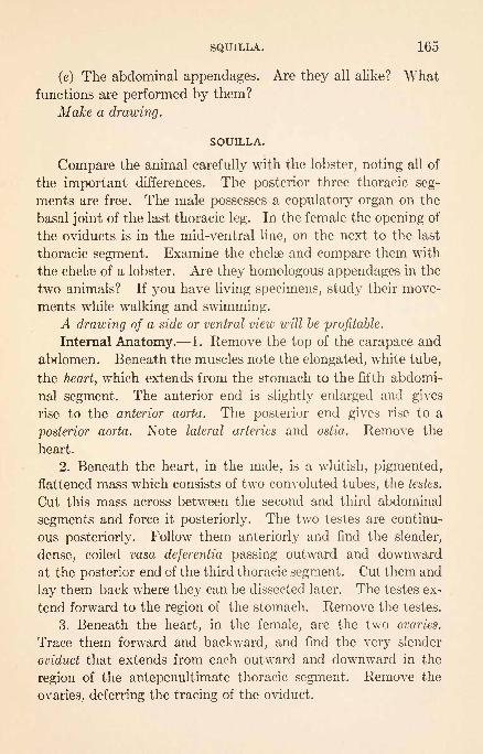
SQUILLA. 165
(e) The abdominal appendages. Are they all alike? Whatfunctions are performed by them?
Make a drawing.
SQUILLA.
Compare the animal carefully with the lobster, noting all of
the important differences. The posterior three thoracic seg-
ments are free. The male possesses a copulatory organ on the
basal joint of the last thoracic leg. In the female the opening of
the oviducts is in the mid-ventral line, on the next to the last
thoracic segment. Examine the chelae and compare them with
the chelse of a lobster. Are they homologous appendages in the
two animals? If you have living specimens, study their move-
ments while walking and swimming.A drawing of a side or ventral view will be profitable.
Internal Anatomy. 1. Remove the top of the carapace and
abdomen. Beneath the muscles note the elongated, white tube,
the heart, which extends from the stomach to the fifth abdomi-
nal segment. The anterior end is slightly enlarged and gives
rise to the anterior aorta. The posterior end gives rise to a
posterior aorta. Note lateral arteries and ostia. Remove the
heart.
2. Beneath the heart, in the male, is a whitish, pigmented,
flattened mass which consists of two convoluted tubes, the testes.
Cut this mass across between the second and third abdominal
segments and force it posteriorly. The two testes are continu-
ous posteriorly. Follow them anteriorly and find the slender,
dense, coiled vasa deferentia passing outward and downward
at the posterior end of the third thoracic segment. Cut them and
lay them back where they can be dissected later. The testes ex-
tend forward to the region of the stomach. Remove the testes.
3. Beneath the heart, in the female, are the two ovaries.
Trace them forward and backward, and find the very slender
oviduct that extends from each outward and downward in the
region of the antepenultimate thoracic segment. Remove the
ovaries, deferring the tracing of the oviduct.

166 ARTHROPODA.
4. Beneath the reproductive organs is the granular liver.
This consists of two lobes which extend from the stomach
to the end of the telson. They form saccular diverticula
between segments and in the telson. Where do they open into
the alimentary tract?
5. Free the intestine, which is between the lobes of the liver.
The rectum is in the sixth abdominal segment.
6. Pull back the anterior end of the stomach, identify the
circum-esopihageal connectives, in order not to destroy them,
and free the stomach by cutting the esophagus and intestine.
Examine the stomach under water.
7. Trace the nerve chain. What ventral ganglia are fused?
The cerebral ganglia are most easily exposed by slicing away,
very superficially, the dorsal surface of the rostrum and pressing
the eye muscles apart.
A drawing of the nervous system will be profitable.
8. Trace the genital ducts to their external openings.
MYSIS.
If living specimens are to be had, watch them swim, and de-
termine what parts are used in swimming. Does the animalswim in one direction or in both?
1. Compare the body with that of a lobster.
2. Are appendages present on each of the divisions of the
body? Compare them with the appendages of the lobster? Howdo the thoracic appendages differ?
3. Notice the otocysts in the tail fin.
4. The living animal is transparent, and many internal
organs, such as heart, gills, and portions of the alimentarycanal, can be seen.
// time permits, make a drawing.
Bergh: Beitrage zur Embryologie der Crustacean. Zool. Jahr. (Anat.),6, 1893.

TALOKCHESTIA 167
TALORCHESTIA. (Beach-Flea.)
These active little animals inhabit sand beaches, where they
burrow in the sand and hide in the decaying vegetable matter
that accumulates along such beaches near high-water mark.
Turn over some of this material and notice the activity of the
animals that are disturbed. Most of them probably belong to
another closely related genus, but their movements are much
the same. How far can a specimen leap? Are the leaps of an
individual continuously in one direction, so it may get awayfrom the point of danger ? Is each leap straight forward or does
the animal whirl in the air? What purpose may be served bythe leaping? Try to catch a specimen. Determine how the
leaping is accomplished. Determine how the specimens burrow.
If you will walk along a beach some quiet night with a lan-
tern you will probably see something of the night activities of
these animals.
1. Select a large specimen and count the number of segments.Is the body divisible into head, thorax, and abdomen?
2. The eyes are not stalked. Are they compound?3. The second antenna of the male are very large. Compare
them with the first antennae and with the antennas of a female.
4. Around the mouth are the labrum, forming an upper lip,
the first maxillipeds (fused), forming a lower lip, and between
them the mandibles, first maxillce, and second maxillae.
5. Examine the appendages behind the mouth. How manyare there? How many bear claws? Compare these claws with
those of a lobster, and see how they differ. Which appendagesare used in crawling? Why are some of the appendages arranged
so they can be twisted around by the sides of the animal ? Whatare the remaining appendages used for?
6. Spread the appendages apart and find the gills, which are
attached to the bases of the appendages.Make a drawing of the animal.
Smallwood: The Beach Flea: Talorchestia longicornis. Cold Spring Har-bor Monogr., 1, 1903.

168 ARTHROPODA
PORCELLIO OR ONISCUS. (Sow-Bug.)
These animals occur in damp places, such as under stones,
logs, etc., and in cellars. They live for the most part on decaying
vegetable matter. To what class of the Arthropoda do they be-
long?
1. Notice the shape. Is this an adaptation?
2. Is the body divisible into head, thorax, and abdomen?
Count the number of segments. Is there any evidence of fusion
at the posterior end of the body?3. Examine the appendages.
(a) Are the eyes stalked or sessile?
(6) Only one pair of antennae is present, the first pair being
rudimentary.
(c) The mouth appendages are small. They consist of man-
dibles, two pairs of maxillce, and one pair of maxillipeds.
(d) How many walking legs are there ? Are these all alike ?
(e) Notice the character and number of the abdominal appen-
dages. On the posterior surface of all but the last pair, which
are modified to form anal feelers, are gills. These are the only
respiratory organs. Why must these animals live in damp places ?
Make a drawing of the animal from the ventral side.
CAPRELLA.
These animals are very common on hydroids, but because of
their peculiar shape and slow motions are rather inconspicuous.
Watch the animals and see how they move. Is the body kept
at rest and moved by the action of the appendages, or how is
movement from place to place effected? Are the appendages
adapted for grasping? Why are they arranged at the two ends
of the body? Watch specimens and see if you can determine
on what they feed.
The form is of interest because of its extreme modification to
suit it to the needs of its life. There is some difference in the
structure of the male and female.
1. Count the segments of the body. Do they differ in num-
ber and shape in male and female? The first represents the head

DAPHNIA 169
with two fused thoracic segments. The abdomen forms a mi-
nute protuberance at the posterior end of the body.
2. At the anterior end of the body are the eyes, two pairs of
antenna?, a pair of maxillipeds, and a pair of legs.
3. At the hinder part of the body are three pairs of legs.
4. Near the middle of the body of the female, and near the
anterior end in the male, is another pair of legs.
5. On two of the segments which do not bear legs are gills.
If time permits, make a drawing.
BRANCHIPUS. (Fairy Shrimp. )
These animals may be found in pools of fresh water in the
early spring, just as the ice is leaving. Their method of swim-
ming by means of the large, expanded appendages should be
observed.
1. Into what parts does the body seem to be divided? Doall of these parts show segmentation?
2. Find the following organs,
(a) The stalked, prominent eyes.
(6) The antennce. In the female the first are slender and
the second vestigial. In the male the first are slender and the
second are enormously enlarged to form a clasping organ.
(c) The labrum forms an upper lip.
(d) The mandibles, beneath the labrum and by the sides of
the mouth. Do they have cutting-edges?
(e) Vestigial maxillce behind the mouth.
(/) Swimming appendages. How many are there? Notice
the fringe of hairs on each. What are these for? Remove one
and examine it with a microscope. The lobes have been des-
cribed as exopodite and endopodite, but their exact relation-
ship is not certain.
A drawing is desirable.
DAPHNIA.
This small fresh-water form frequently occurs in large num-
bers in small pools and brooks. Determine how it swims. Being

170 ARTHROPODA.
small and transparent, it may be satisfactorily studied with a
compound microscope.
1. Notice the shape and extent of the protective covering.
To what part of other crustaceans does this correspond? Are
the appendages and the abdomen capable of being thrust out?
Are there any signs of segmentation of the body?2. Determine what parts are used in keeping a current of
water passing through the shell. Why is such a current needed?
3. If the animal carries young, notice how they are kept in
the brood chamber by a spine that extends up from the dorsal
portion of the base of the abdomen.
4. Notice the beating of the heart.
5. Are the eyes stalked or sessile? They frequently show a
peculiar reaction to light. If the light is cut off from the micro-
scope, the eye will be seen to rotate on its axis. If the light is
admitted again, the eye rotates back to its original position.
6. The first antennce are very small and project ventrally.
What is the chief function of the second antennaB?
7. Several appendages will be seen inside of the shell, but
it is hard to determine their exact relation. The functions of
some of them may be apparent.
A drawing is desirable.
CYCLOPS. (Water-Flea.)
Most any free-swimming copepod, either fresh-water or marine,
will answer quite as well as the fresh-water Cyclops.
Cyclops may be found in almost any pool of fresh water and
the marine forms are among the most abundant of the animals
of the sea. Surface skimming of the sea, made with a net com-
posed of cheese-cloth or silk bolting-cloth, will yield an abun-
dance of material.
1. Watch the animals and see how they swim. With a
pipet try to catch a certain individual and see whether the jerky
movements probably aid these animals in escaping enemies.
Determine what organs are used in swimming.2. Examine specimens that have been confined under a cover-
glass with a microscope, and notice the shape of the body.
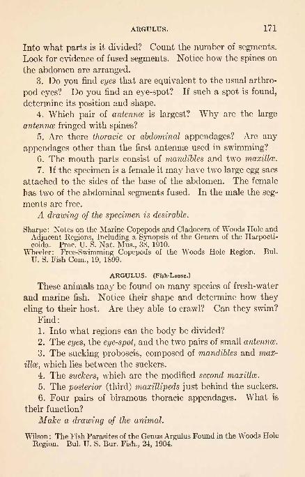
ARGULUS. 171
Into what parts is it divided? Count the number of segments.
Look for evidence of fused segments. Notice how the spines on
the abdomen are arranged.
3. Do you find eyes that are equivalent to the usual arthro-
pod eyes? Do you find an eye-spot? If such a spot is found,
determine its position and shape.
4. Which pair of antenna is largest? Why are the large
antennae fringed with spines?
5. Are there thoracic or abdominal appendages? Are any
appendages other than the first antennse used in swimming?6. The mouth parts consist of mandibles and two maxilla?.
7. If the specimen is a female it may have two large egg sacs
attached to the sides of the base of the abdomen. The female
has two of the abdominal segments fused. In the male the seg-
ments are free.
A drawing of the specimen is desirable.
Sharpe : Notes on the Marine Copepods and Cladocera of Woods Hole and
Adjacent Regions, Including a Synopsis of the Genera of the Harpocti-coida. Proc. U. S. Nat. Mus., 38, 1910.
Wheeler: Free-Swimming Copepods of the Woods Hole Region. Bui.
U. S. Fish Com., 19, 1899.
ARGULUS. (Fish-Louse.)
These animals may be found on many species of fresh-water
and marine fish. Notice their shape and determine how they
cling to their host. Are they able to crawl? Can they swim?
Find:
1. Into what regions can the body be divided?
2. The eyes, the eye-spot, and the two pairs of small antenna?.
3. The sucking proboscis, composed of mandibles and max-
illa?, which lies between the suckers.
4. The suckers, which are the modified second maxilla?.
5. The posterior (third) maxillipeds just behind the suckers.
6. Four pairs of biramous thoracic appendages. What is
their function?
Make a drawing of the animal.
Wilson : The Fish Parasites of the Genus Argulus Found in the Woods Hole
Region. Bui. U. S. Bur. Fish., 24, 1904.

172 AKTHROPODA.
LEPAS. (Goose-Barnacle.)
If possible, examine a cluster of specimens as they naturally
occur attached to floating timber.
1. Why are the peduncles much larger in some specimensthan in others? Are they contractile so the body may be movedinto different positions? Would such movements be of value?
2. Notice the thoracic appendages. Can they be thrust
from the shell? What is their character? What are their char-
acteristic movements? Drop a small piece of clam meat on
these appendages of a living specimen and see what happens.What kind of food would they naturally collect?
3. Examine the portions of the shell. The portion on the
closed margin is the carina, laterally and near the base of
the peduncle are the scuta, and near the extremity the terga.
Why are there so many pieces? Notice the lines of growth and
determine the direction of growth of each piece.
Draw the animal as seen from one side.
With a scalpel or razor cut a preserved specimen into right
and left halves, extending the cut through the peduncle.
4. The mouth will be seen at the end of a rather thick pro-
longation which extends to near the bases of the abdominal
appendages. On the margin of this prolongation are the small
scale-like mandibles, first maxillce, and second maxillce. The sto-
mach is rather large and the small intestine leads to the posterior
end of the abdomen, where it opens between the abdominal
appendages.5. The nervous system, consisting of a large pair of cerebral
ganglia and a short ventral chain of ganglia, should be seen in
such a section.
6. The animal is hermaphroditic. The testes lie dorsal to
the stomach and communicate with a conspicuous coiled vas
deferens that is continued to the elongated penis at the end of the
abdomen. What reason is there for such a long penis? The
ovary occupies the interior of the peduncle. The oviducts are
inconspicuous and hard to follow. They open near the bases
of the anterior thoracic appendages.

LIMTJLUS. 173
7. Examine the appendages carefully and be sure that you
understand the relation of parts. What part must the pedun-
cle represent? Understand the beautiful adaptation of the
animal for its life.
A drawing showing the organs is desirable.
Bigelow: Early Development of Lepas. Bui. Mus. Comp. Zool. Harvard,
40, 1902.
Delage: Evolution de la Sacculine. (Sacculina carcini.) Arch. Zool. Exp.et Gen., 2e
Series, 11, 1884.
ARACHNOIDEA.
LIMULUS. (Horseshoe Crab.)
Notice the way in which the animal crawls upon the bottom.
Is it well protected from enemies? Examine it carefully for
parasites and for animals that are attached to it. Disturb it
and see if it will swim. The animals are usually quite active
in the evening, and if you visit a car in which they are kept, at
this time of the day, you are likely to find them crawling up the
sides, falling over and swimming on their backs. In this posi-
tion it is easy to determine how they swim. The animals are
very hardy and will stand even complete removal from the water
for days at a time. During the spring and early summer, eggs
are deposited in the sand; the male holding to the edge of the
abdomen of the female with claws modified for the purpose, is
dragged after her. If possible, the method of egg deposition
and fertilization should be observed.
1. The animal consists of a hoof-shaped cephalothorax, an
abdomen, and a caudal spine. How are these joined? Is there
any indication of segmentation of any of them ?
2. Examine the eyes with a lens and see that they are com-
pound.3. On the lower side of the cephalothorax notice the appen-
dages. Are they all built on the same plan? Compare them in
male and female. Do you know what the modifications are
for? Compare the pincers with those of a lobster. The first
pair of appendages is called the chelicerce. Between the bases

174 ARTHROPODA.
of the last pair of walking legs are the chilaria. Behind the
chilaria is the broad flat operculum. Does this show evidence of
being modified appendages? What is its function?
4. Between the bases of the cephalothoracic appendages is
the mouth. Do the bases of the appendages show any modifi-
cations that may serve as teeth? Can the pincer-bearing ap-
pendages be so bent as to be used in feeding?
5. Along the sides of the abdomen notice the movable spines.
How many are there?
6. Under the operculum are the gills. How many groups
are there? Are they arranged in pairs? How are they attached
to the body? Are they movable? What reason is there for
moving them? Examine a bunch of gills, frequently called a
gill-book, and see how it is formed.
7. At the base of the caudal spine notice the anus.
Make a drawing of the ventral surface.
Internal Anatomy. This shows no very special adaptation
and can be pretty well understood by studying a longitudinal
section of a small preserved specimen.
In such a section the following organs may be found:
1. The dorsal extensor, the ventral flexor, and the leg muscles.
2. The elongated tubular heart just beneath the dorsal cover-
ing, in the posterior end of the cephalothorax and the anterior
end of the abdomen.
3. The alimentary canal, consisting' of the esophagus and the
anterior and posterior portions of the stomach, which extends
posteriorly without much change to the anus. The liver, which
surrounds the stomach and fills the greater portion of the
cephalothorax, sends its secretions to the stomach.
4. The cerebral ganglia, near the bases of the chelicerse, and
the ventral chain of ganglia should also be seen in satisfactory
sections.
A drawing is desirable.
Lankester: Limulus an Arachnid. Quart. Jour. Mic. Sci., 21, 1881.Packard: The Anatomy, Histology, and Embryology of Limulus poly-
phemus. Mem. Bost! Soc. Nat. Hist., 1880.Patten and Redenbaugh: Studies on Limulus, Jour. Morph., 16, 1899.

BUTHUS. EPEIRA. 175
BUTHUS. (Scorpion.)
Living specimens of these animals are not usually available
for laboratory study, They live for the most part concealed
during the day under old bark and in crevices and holes and are
active at night. Their food is largely spiders and insects which
are seized by the claws and killed with the abdominal sting.
1. Into what parts is the body divided? How many seg-
ments are recognizable? Which are the most freely movable?
2. Look for eyes. Do you find any besides the large pair?
3. Find four pairs of slit-like openings on the ventral side of
the pre-abdomen. These are the stigmata, the openings of the
lung-books.
4. Find the following appendages:
(a) The chelicerce. What is their structure and where are
they placed?
(b) The pedipalpi. Compare them with the chelicerae and
count their segments.
(c) Four pairs of walking legs. Count their segments and see
if they are armed with claws.
(d) The comb-shaped pectines. Are they on the thorax or
the abdomen? Their function is doubtful.
5. Examine the mouth. Are there any jaws? Is a labrum
present ?
6. Find the position of the anus. The terminal spine is pro-
vided with a poison gland and serves as a sting. In the living
animal, the post-abdomen is habitually carried over the back.
Make a drawing of the under side of a specimen.
EPEIRA. (Round-Web Spider.)
Examine the webs of different species of spiders and see how
they are constructed. Do all of the webs have places for the
concealment of the owners? Do all spiders seem to construct
definite webs for the capture of insects? How do spiders entan-
gle insects in their webs? Do different kinds use different
methods? What parts of insects are eaten?
By destroying webs that are occupied by spiders that are in

176 ARTHROPODA.
convenient places for observation, the construction of new webs
may be observed. Notice how the framework of a round web is
laid and then how the threads are attached to the famework.Are any of the legs used in handling the thread? Are spiders
equally active at all times of the day?
Spiders' webs may frequently be seen floating in the air,
especially in the late summer or autumn. By watching spidersthat are on fences and bushes the formation of these threads
may be observed. Watch such a spider and see if you can deter-
mine the use to which the thread is put.
Capture a spider and watch it descend by a thread. Whereis the thread formed? Does the spider hold to it with its legs?
Keep taking the thread up so that the spider cannot reach the
ground, and see if there is a limit to the amount that can be
formed. When the spider starts to climb the thread see howthis is done, and whether the thread is taken up as the animal
climbs or is allowed to float free.
Find where spiders lay their eggs. Some carry them. If
you can find a specimen with an egg-sac, see how it is carried
and whether it will drop its eggs when frightened. Remove the
egg-sac and see if the spider will accept it again. Open several
egg-sacs and see if the eggs all appear to be in the same stage
of development.
Study the movements of the animal and see how many of
the appendages are used in locomotion. Are any of the appen-
dages used sometimes for locomotion and sometimes for feel-
ing?
Examine the external structure of Epeira.
1. Into what parts is the body divided? Do both parts bear
appendages?2. Look for eyes on the anterior end of the body. How
many are there? Do they seem to be simple or compound?Determine whether a specimen can see.
3. The following appendages should be found:
(a) The chelicerce or mandibles. Notice their structure and
see that each ends in a sharp claw. The poison-gland discharges
at the tip of this claw.

PHOXICHILIDIUM. 177
(6) The pedipalpi or palpi. How many segments have
they? Examine their tips for claws. What are they appar-
ently used for?
(c) Four pairs of legs. Are they all alike? Count the seg-
ments and examine their tips for claws.
(d) On the abdomen, three pairs of spinnerets. Notice their
positions and see if they are segmented. Understand their func-
tion and whether they are all used at the same time. Theyare probably true abdominal appendages.
4. On the lower surface of the abdomen, near its anterior
end, are two slits, the openings into the lung-sacs or lung-books.
They are respiratory in function.
5. Just in front of the spinnerets is a minute median pore,
the spiracle, that is often very hard to find. It is the external
opening of a series of abdominal tracheae.
Make a drawing of a ventral view.
Montgomery: Studies on the Habits of Spiders, Particularly Those of the
Mating Period. Proc. Acad. Nat. Sci., Philadelphia, 1903.: On the Spinnerets, Cribellum, Colulus, Trachea? and Lung-books of
Araneads. Proc. Acad. Nat. Sci., Philadelphia, 1909.: The Development of Theridium, an Aranead, up to the Stage of
Reversion. Jour. Morph., 20, 1909.: The Significance of the Courtship and Secondary Sexual Characters
of Araneads. Am. Nat., 44, 1910.Peckham: Observations on Sexual Selection in Spiders of the Family At-
tidse. Occas. Papers Nat. Hist. Soc., Wisconsin, 1 and 2.
PHOXICHILIDIUM.
The exact affinities of the pygnogonids to other forms is
not known, but they have certain characters that have suggesteda possible relationship to the Arachnoidea. They are frequentlyfound in considerable abundance on the material that is attached
to piles. Notice their movements and see how they cling to the
material on which they are moving.1. The body is very slender and is composed of a number of
free segments that form the head and therax and a small, ves-
tigial abdomen. How many free segments are there? At the
anterior end is a rather prominent proboscis, with the mouth at
its end.12

178 ARTHROPODA.
2. The following appendages will be found :
(a) The chelicerce. What is their structure? Are theyarmed with pincers?
(6) Four pairs of long walking legs. How many segmentshave they? The viscera extends into the bases of these appen-
dages.
(c) The male is provided with a pair of ventral appendagescalled the ovigerous legs, by means of which the eggs are collected
as they are laid by the female. These appendages are not present
in the female.
Make a drawing of the under side of a specimen.
Cole: Pycnogonidia of the West Coast of North America. HarrimanAlaska Exped., 10, 1904.
MYRIAPODA.
LITHOBIUS. (Centipede, Earwig.)
These animals may frequently be found under stones, logs
or boards, or about rubbish or manure heaps. They live largely
on insects, larvae, and small worms, and are very active.
1. Notice the shape of the body and count the number of
segments. Is there a distinct head? Are the segments very
movable?
2. How many appendages does each segment possess? Are
all of the segments provided with appendages? Allow the ani-
mal to run and see how the legs are used. Do those of a side all
move in the same direction at the same time? Are all of the
legs alike? Notice the pair of appendages just behind the head
and see how they differ from the others. These appendages are
organs of prehension that are used in grasping the prey. Theyare provided with poison glands that open on their inner sides
near their free ends.
3. Examine the head and find the eyes, antennae, and mouth
parts. The latter consist of a labrum, a pair of mandibles, and
two pairs of maxillce, the last pair of which are united to form a
labium.

JULUS. ACRIDIUM. 179
4. Understand how the animal breathes. The stigmata are
situated near the bases of the legs, but are hard to see except in
favorable specimens.
Make a drawing of the animal.
JULUS. (Thousand-legs.)
These animals are frequently very abundant under the dead
bark of logs or stumps, in decaying wood, and in decaying heaps
of grass. In the autumn they frequently congregate under
boards and in corners. They feed largely on decaying vegetable
matter, but may become pests in gardens, destroying tomatoes
and fallen fruits and many vegetables.
1. Disturb a specimen and see how it rolls up. Can this
be protective? See if there is any odor when it is disturbed.
What purpose can such an odor serve ?
2. What is the shape of the body? Is it hard or soft ? How
many segments are there ?
3. How many appendages are borne on a segment? Do all
of the segments bear appendages? Does the animal move
rapidly? Why does it not need to move as rapidly as the pre-
ceding form? Do the first pair of appendages behind the head
show modifications for prehension? Does this animal need such
an organ?
4. Compare the organs of the head with those of the preced-
ing form.
Make a drawing of the under side of one segment.
Williams: Habits and Structure of Scutigerella immaculata. Proc. Bost.
Soc. Nat. Hist., 33, 1907.
INSECTA.
ACRIDIUM. (Grasshopper.)
Study grasshoppers as they occur in nature and determine
as far as possible the following points:
1. Do they see or hear? Are they equally sensitive to touch

180 ARTHROPODA.
on all parts of the body? Why should the animal be well pro-vided with sense organs?
2. What is their food? Are all plants eaten or are someavoided? See how the mouth parts are used in feeding.
3. What are the important enemies of grasshoppers? Howdo they escape their enemies? Do they hide? Are they pro-
tectively colored? How does jumping serve them better than
crawling? How many times its length can a grasshopper jump?Why are wings needed?
4. During late summer and autumn you may find individuals
depositing eggs. See if you can determine how the end of the
body is worked into the ground.
For study it is desirable to use a rather large, freshly killed
or alcoholic specimen.
The body is divided into three well-marked regions.
1. The Head. Is it movable? Does it need to be as mova-
ble as your own head ? It bears several organs.
(a) The compound eyes. Examine one with a lens or remove
its outer covering and examine it with a compound microscope.
You should understand the structure of the whole eye and howit gives a single visual image.
(b) The ocelli, three in number, one near the middle of the
front part of the head and the others placed near the bases of
the antennaa.
(c) The antennce. Why are they so flexible? Examine one
with a microscope and notice the spines. What are these for?
(d) Mouth parts. These should be studied later.
2. The Thorax. Why should it be large and comparativelyfirm? This portion is more or less distinctly divided into three
parts, each of which carries a pair of legs.
(a) Compare the three legs of one side. Do they have the
same number of segments? Do all of the joints of the leg move
in the same plane? The five divisions of a leg are, beginning
with the basal end: coxa, trochanter (immovably joined to the
coxa in the leaping legs), femur, tibia, and tarsus, which is com-
posed of four movable pieces. Why do the femurs of the leap-

ACRIDIUM. 181
ing legs differ from the femurs of the other legs? Determine
how the foot is arranged to hold to objects. Have you noticed
a grasshopper settle its feet preparatory to jumping? Examine
the joint between the femur and tibia.
(b) Examine the wings and notice their size, shape, places
of attachment, and general character. Do they apparently
have different functions to perform? Notice how the posterior
wings are folded so they may be covered by the anterior. Does
this seem to greatly reduce their strength?1
3. The Abdomen. Count the number of segments. Each
one is covered dorsally by a tergum and ventrally by a sternum.
Why should the abdomen be more movable than the other por-
tions? The posterior ends of the abdomens of male and female
differ. This portion of the female is modified to form the ovi-
positor, which consists of two large pairs of plates that inclose
a smaller pair of plates. It is between these plates that the
oviduct opens. Why do the larger plates possess hard tips?
Along the sides of the abdomen notice the stigmata, the external
openings of the respiratory system. Do you find stigmata on
other parts of the body?Draw an enlarged side view of a grasshopper, placing the appen-
dages in their proper positions.
Mouth Parts. It has already been noticed that the mouth
parts serve to cut off pieces of leaves, which are then passed
directly into the alimentary canal. For such a purpose there
should be holding as well as cutting parts.
1. Pass a needle under the labrum, which forms the upper
lip, and notice that it is hinged and that the end is lobed. It is
not supposed to be homologous with usual arthropod appendages.With fine scissors remove it and place it in a watch-glass contain-
ing water.
2. Immediately behind the labrum is a pair of hard, dark-
colored organs, the mandibles, that are used in cutting the food.
Their position should be carefully noted, but it will be better
1 You should examine the posterior wing of a beetle and see how it
is folded.

182 ARTHROPODA.
to leave them in position until the other mouth appendageshave been removed.
3. Situated by the side of the mouth and just behind the
mandibles are the maxillce. With a needle push one to one side
and notice that it consists of a somewhat flattened portion with
a jointed maxillary palp at one side. Carefully determine the
positions of the maxillae with relation to other parts. What
possible uses are served by the two parts? Remove them with
scissors and place them in the watch-glass with the labrum, in
approximately their relative positions and study carefully.
4. Pass a needle behind the remaining appendage, the labium,
and see that it is hinged and forms the lower lip. Remove it
with scissors and place it in position in the watch-glass. Youwill find that it bears a pair of labial palpi, and that there is a
deep cleft along the middle line. These are indications that the
appendage is the result of the fusion of a pair of appendages.5. Remove the mandibles and examine their cutting margins.
Place them in position in the watch-glass.
Make a drawing showing the structure of each of these appen-
dages. Arrange your figures as nearly as possible in the relative
positions of the parts.1
Internal Structure. Remove the wings, and before openingthe body notice the rather large, somewhat transparent tympa-num on each side of the first abdominal segment, very near the
base of the leaping leg. The structure of the auditory organ
may be easily studied by staining, clearing, and mounting in
balsam. (See Packard's " Text-Book of Entomology7;
or
Brooks's "Hand-book of Invertebrate Zoology.") Remove the
dorsal portion of the wall of the abdomen and thorax, and notice:
1. The heart, which will be found attached to the portion of
the wall of the abdomen that has been removed, by means of
numerous radiating muscle fibers. You probably will not be
1 The mouth parts of insects that depend on biting off portions of
plants for food are quite similar. Directions for the study of the mouthparts of the honey-bee are given further on, but the mouth parts of other
forms, such as the fly, butterfly, and bug, should be studied.

ACRIDIUM. 183
able to determine the structure of the heart in the dissection.
Read this up, and determine what the radiating muscle fibers
are for.
2. The space between the muscles and the viscera is filled
more or less completely by the fat-body and the trachece. Witha lens notice how the tracheae connect with the spiracles and
how they branch. Remove a portion of the tissue in which
you can see tracheae, mount it in water under a cover, and
examine it microscopically. Each tracheal tube is marked bystriations wound around it. Do you know what causes this
appearance and what the arrangement is for? Do you under-
stand how the tracheal system is arranged ? Why is it extended
all over the body and how is the air made to go in and out ?
3. Near the dorsal surface of the posterior part of the ab-
domen, surrounded by the tissues already mentioned, are the
gonads. These differ in size and shape according to the sex. In
the male the vasa deferentia may be seon leaving the lobulated
testes. In the female the oviducts pass around the sides of the
intestine. They may be followed later.
4. Loosen the anterior ends of the gonads and turn them
posteriorly to expose the hinder part of the alimentary canal. 1
(a) The esophagus, which bends backward from the mouth,
gradually enlarges as it enters the thorax.
(b) The crop, which is not sharply separated from the esoph-
agus, gradually narrows posteriorly.
(c) Following the constriction posterior to the crop is the
elongated stomach, frequently called the ventriculus. Surround-
ing the anterior end of this portion are a series of rather large
diverticula, the gastric caeca, that extend both anteriorly and
posteriorly from the points where they open into the stomach.
(d) Some distance behind the posterior ends of the hepatic
caeca, quite concealed by the mass of small uriniferous tubes, is a
slight constriction and hardening of the alimentary canal that
1 There is great diversity in the parts of the alimentary canals ofdifferent insects. The great differences in feeding habits render this
necessary.
184 AKTHROPODA.
marks the division between the stomach and intestine. It is at
this point that the uriniferous tubes join the alimentary canal.
(e) Behind the intestine the alimentary canal becomes muchsmaller and is known as the hind intestine or colon.
(/) Behind the colon, forming the hinder portion of the ali-
mentary canal, is the slightly enlarged rectum. The rectum
cannot be seen until the ovary is removed, which should be de-
ferred until the ducts have been seen.
Make a drawing showing the position of the parts of the alimen-
tary canal in side view.
Cut the intestine and turn the alimentary canal posteriorly
and anteriorly.
5. Notice the muscles:
(a) That move the abdominal segments.
(6) That move the legs (those that supply the wings have
been destroyed).
(c) That move the jaws.
Do you understand now why the thorax needs to be compar-
atively large and firm?
6. The nervous system is directly comparable to that of the
lobster, but the connectives between the ganglia will be found
to be distinctly double and the ganglia to be somewhat differently
arranged.1
The ventral chain will be found to consist of a pair of sub-
esophageal, three pairs of thoracic, and five pairs of abdominal
ganglia with the connectives between them. Which of these
are largest? Why is this the case? Trace the nerves from them
and see what organs they supply.
Trace the connectives forward from the sub-esophageal ganglia
and see that they pass around the esophagus, thus formingthe circum-esophageal connectives. Cut away the dorsal portion
of the head and expose the cerebral ganglia.
Add the nervous system to the figure that shows the alimentary
canal.
1 The arrangement of the ganglia in insects is very variable, showingmnny gradations in concentration.

APIS MELLIFICA. 185
7. Trace the oviducts down around the sides of the bodyand notice that they unite with each other ventral to the nervous
system, to form the vagina. This may be traced to its openingbetween the plates of the ovipositor. Dorsal to the vagina,
opening to the exterior very near it, is a small sac, the sperma-
theca, which serves to store the spermatozoa received from
the male until the eggs are laid.
The reproductive organs may also be added to your figure show-
ing internal anatomy.
Brooks: Hand-book of Invertebrate Zoology.
APIS MELLIFICA. (Honey-Bee.)
The life of this form is so different from that of the grasshopper
that, should time permit, a study of its complete anatomy would
be profitable, but attention will here be confined to a few of the
more general adaptations that fit it for its life.
Bees at work on flowers should be examined and the methods
of getting honey and pollen noticed.
1. Catch by the wings a bee that has been gorging itself
and bend the abdomen forward with your thumb-nail until the
bee disgorges. Notice where the fluid comes from and howmuch there is of it. When the abdomen is released watch the
bee as it swallows the drop it has disgorged.
2. Notice where the pollen is carried, and see if you can de-
termine how it is attached. Examine bees working on different
flowers, or watch them as they enter their hives, and see if the
pollen is always of the same color. Do you understand what
the pollen is and what the bees use it for?
3. You may find bees gathering pitch from buds, knots,
boards, or freshly varnished furniture, and fastening it on their
legs. Do you know what this is used for?
4. Watch the entrance of a bee-hive and see if those goingin are ever challenged. Perhaps you may see the method of
defense. If so, you will notice that the stranger simply tries

186 ARTHROPODA.
to get away. You may also see how dead bees and foreign ma-terials are removed.
5. It is desirable to see something of the activities in the hive.
This can be most satisfactorily done with an observatory hive,
by means of which comb-building, honey-storing, egg-laying,
brood-rearing, etc., can be very satisfactorily studied.
Directions for the study of the mouth parts and the sting are
all that seem necessary, but the wings should be examined
microscopically to see how those of a side are joined together,
and a hind-leg should be examined to see how the hairs on the
tibia form a pollen basket.
Mouth Parts. 1. With a lens notice the pair of hard
jaws, the mandibles, situated on the sides of the head at
the base of the tongue. These mandibles are directly homolo-
gous with the mandibles of the grasshopper. Between the bases
of the mandibles is a labrum, and extending from beneath the
end of the labrum is a small epipharynx.
2. With scissors remove the tongue, which is normally carried
against the lower surface of the thorax, and transfer it to a
watch-glass. It may now be dehydrated, passed into oil of
cloves, placed in position on a slide, and mounted in balsam,
when it can be studied best, or it may be immediately spread
under a cover or between slides in glycerin.
3. The central portion is the hairy, segmented labium (the
hypopharynx of some authors), bearing at its end a little padcalled the spoon. The labium is folded lengthwise so as to form
a pair of fine ducts which run from tip to base. The arrange-
ment is such that the bee may, through blood-pressure, unfold
the labium. This probably is an adaptation for cleaning it.
Attached to a median rod, the mentum, which forms the base
of the labium, is a pair of flattened appendages, the labial palps,
that are hinged so that they may be drawn together to inclose
the labium and thus form a rather large tube, which is made
more complete by means of the remaining pair of flattened
appendages, the maxillce. On the outer margin of each maxilla
is a small protuberance, the maxillary palp. When sipping
APIS MELLIFICA. 187
from an abundance of liquid the extemporized tube formed bythe labial palps and maxillae around the labium is used, the
liquid being drawn in by means of the sucking stomach. Whenthe liquid is in very small quantities it is apparently lapped up
by the spoon and transferred through the labium.
A figure of the mouth parts is desirable.
Sting. The sting is to be regarded as a modified ovipositor
that is no longer concerned in depositing eggs, but has become
a weapon of offense and defense. It is accordingly present only
in the female. The queen never uses her sting except on other
queens.Remove the dorsal integument of the abdomen of either a
fresh or preserved specimen, and find the dark brown shaft of
the sting, near the posterior end. Grasp the shaft with a pair
of fine forceps and forcibly remove it. A considerable mass of
tissue will be removed adhering to the base of the shaft, but this
consists for the most part of accessory organs that must be
understood. Spread the sting upon a slide, and either dehy-drate and mount in balsam, or mount in glycerin. The balsam
mount will prove more satisfactory, but the cover must be
clamped down until the balsam hardens.
1. The shaft consists of three parts:
(a) A heavy support, called the awl or sheath, pointed at its
extremity and sending a pair of arms or arches from its base,
which normally bend ventrally, but are here forced to the sides.
At its extremity each of these arches enlarges to form a rather
large flattened plate, the sheath plate, to which strong muscles
are attached.
(b) A pair of lancets which are fastened to the dorsal surface
of the sheath and the sheath arches by tongue and groove joints
(each tongue is enlarged along its inner margin so that it is held
firmly in the groove). Each lancet is pointed at its free extrem-
ity, and its sides near the point are set with barbs that point
toward the base of the sting. The arch of each lancet is con-
1 The comparative study of the mouth parts of a butterfly, horse-fly
house-fly, and mosquito will prove valuable.
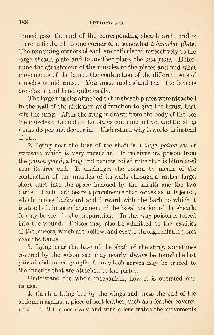
188 ARTHROPODA.
tinned past the end of the corresponding sheath arch, and is
there articulated to one corner of a somewhat triangular plate.
The remaining corners of each are articulated respectively to the
large sheath plate and to another plate, the oval plate. Deter-
mine the attachment of the muscles to the plates and find what
movements of the lancet the contraction of the different sets of
muscles would cause. You must understand that the lancets
are elastic and bend quite easily.
The large muscles attached to the sheath plates were attached
to the wall of the abdomen and function to give the thrust that
sets the sting. After the sting is drawn from the body of the bee
the muscles attached to the plates continue active, and the sting-
works deeper and deeper in. Understand why it works in instead
of out.
2. Lying near the base of the shaft is a large poison sac or
reservoir, which is very muscular. It receives its poison from
the poison gland, a long and narrow coiled tube that is bifurcated
near its free end. It discharges the poison by means of the
contraction of the muscles of its walls through a rather large,
short duct into the space inclosed by the sheath and the two
barbs. Each barb bears a prominence that serves as an injector,
which moves backward and forward with the barb to which it
is attached, in an enlargement of the basal portion of the sheath.
It may be seen in the preparation. In this way poison is forced
into the wound. Poison may also be admitted to the cavities
of the lancets, which are hollow, and escape through minute pores
near the barbs.
3. Lying near the base of the shaft of the sting, sometimes
covered by the poison sac, may nearly always be found the last
pair of abdominal ganglia, from which nerves may be traced to
the muscles that are attached to the plates.
Understand the whole mechanism, how it is operated and
its use.
4. Catch a living bee by the wings and press the end of the
abdomen against a piece of soft leather, such as a leather-covered
book. Pull the bee away and with a lens watch the movements

APIS MELLIFICA. 189
of the sting, which will remain caught in the leather. Observe
the spasmodic contractions of the poison sac. See how long
and how energetically the movements are continued and how
deep the sting is worked in. This should remind you that a
sting should be removed immediately, and that it should not be
pulled out, as grasping the poison sac will aid in injecting the
poison, but scraped off with a finger-nail or rubbed off.
A drawing showing the mechanism of the sting is desirable.
Field: A Study of an Ant. Proc. Acad. Nat. Sci., Philadelphia, 1901.
Philips: A Review of Parthenogenesis. Proc. Am. Phil. Soc., 42, 1903.Root: A, B, C and X, Y, Z of Bee Culture.

CHORDATA.
Bilaterally symmetrical ccelomate animals with a notochord,
dorsal and tubular central nervous system, and a pharynx per-
forated by branchial clefts (gill slits).
Sub-phylum 1. Hemichorda.The notochord is poorly developed and restricted
to the anterior end of the body.Order 1. Enteropneusta.
Worm-like, with numerous branchial clefts, a
straight intestine, and a terminal anus. Bodydivided into three regions proboscis, collar, andtrunk. Development usually with a metamor-
phosis, the larva being known as a tornaria.
(Balanoglossus, Dolichoglossus.)Order 2. Pterobranchia.
Tubicolous, with one pair of branchial clefts or
none, a U-shaped alimentary canal, and a dorsal
anus situated near the mouth. Proboscis flat-
tened ventrally into a large "buccal disc," its
base covered dorsally by the collar which is pro-duced into two or more tentaculiferous arms.
Trunk short, prolonged into a stalk. Reproduc-tion by budding occurs. (Cephalodiscus, Rhab-
dopleura.)Order 3. Phoronidea (doubtfully placed with the chordates).
Tubicolous, with gregarious habits. The bodyends in a plume of ciliated tentacles; the alimen-
tary canal is U shaped. There is a larva knownas actinotrocha. (Phoronis.)
Sub-phylum 2. Urochorda.The adult body is enclosed in a tunic or test, and
usually lacks a notochord; the central nervous
system is reduced to a simple ganglion. With anatrial cavity and a pharynx perforated by from
190

CHORDATA. 191
two to many gill clefts. There is usually a tad-
pole-shaped motile larva which possesses a tubu-
lar dorsal central nervous system and a noto-
chord restricted to the caudal region.
Order 1. Larvacea.Small pelagic tunicates swimming throughoutlife by means of a tail. With a persistent noto-
chord and a single pair of gill slits. (Appendic-
ularia, Oikopleura.)Order 2. Ascidiacea.
Mostly fixed, solitary or colonial tunicates, whichin the adult are never provided with a tail andhave no trace of a notochord. The test is well
developed, the pharynx large and perforated bymany gill slits. In most ascidians the sexually
produced embryo develops into a tailed larva;
many ascidians reproduce by budding to formcolonies. (Ciona, Molgula, Cynthia, Perophora,
Botryllus, Amarcecium, Leptoclinum.)Order 3. Thaliacea.
Pelagic tunicates which swim by forming cur-
rents in the water. The adult is never providedwith a tail or a notochord. The pharynx has
two or more gill slits. Alternation of genera-tions occurs, and may be complicated by poly-
morphism. (Salpa, Doliolum.)
Sub-phylum 3. Cephalochorda.The notochord extends the entire length of the
body including the head. The body is meta-
merically segmented. (Amphioxus.)
Sub-phylum 4. Vertebrata.
A brain is developed as an enlargement of the
anterior end of the central nervous system; the
notochord extends no further forward than the
middle of the brain, and a vertebral column andcranium are present. (Cyclostomes, fishes,
amphibians, reptiles, birds, mammals.)
Conklin: Organization and Cell Lineage of the Ascidian Egg. Jour. Acad.
Nat. Sci., Philadelphia, 2d Ser., 13, 1905.
: Does Half of an Ascidian Egg Give Rise to a Whole Larva? Arch.
f. Entwicklungsm. d. Org., 21, 1906.
Metcalf: Notes on the Morphology of the Tunicata. Zool. Jahr., 13,
1900.

192 CHORDATA.
ENTEROPNEUSTA.
DOLICHOGLOSSUS (BALANOGLOSSUS) KOWALEVSKII.
In the natural habitat, note the character of the bottom
where Dolichoglossus is found: Is the sand clean or is there an
admixture of organic material? Note the frail tube of sand
particles fastened together with mucus, and the numerous
"castings." The animal has a characteristic and unpleasantodor.
Note the division of the body into three general regions : (1)
A yellowish-white conical proboscis; (2) the collar, which is
brilliant orange-red, especially in males, with a white ring pos-
teriorly; and (3) the trunk, which is mainly orange-yellow,
shading to a greenish-yellow in the transparent posterior region,
which is often broken off when the animal is collected.
The trunk may be roughly divided into the following regions,
which overlap: (a) An anterior branchial region, bearing on
each side not far from the dorsal median line a row of transverse
gill slits; (6) a genital region, bearing on each side of the bodyan irregular and broken fold or ridge containing the reproductive
organs, which are gray in the female and yellow in the male;
(c) a posterior abdominal region, of much smaller diameter than
the rest of the body.The mouth is situated on the ventral side at the base of the
proboscis, and is concealed by the free anterior edge of the collar.
The animal is unable to close its mouth, and in burrowing a
continuous stream of sand passes through the alimentary canal,
forming the "castings" which are abundant in the natural
habitat of the animal. What must be the nature of its food?
Burrowing is effected partly by muscular contractions of the
body-wall, but mainly through the power of the proboscis and
collar to become turgid. In burrowing and feeding, of what
use to the animal is the collar?
Note the characteristic coiling of the genital region in this
species. The anterior end, including the branchial region, is
normally maintained in a vertical position. The posterior
end is also kept upright, and can be moved up and down in a

MOLGULA MANHATTENSIS. 193
vertical shaft opening on the surface, thus enabling the animal
to eject the residue of sand from the anus.
For the internal anatomy, the account in the CambridgeNatural History may be consulted. Important chordate
characters are the notochord, the dorsal central nervous system,
and the branchial arches.
Agassiz: The History of Balanoglossus and Tornaria. Amer. Acad. Artsand Sci., 9, 1873.
Bateson: The Early Stages of Balanoglossus. Quart. Jour. Mic. Sci., 24,1884.
: The Late Stages of the Development of Balanoglossus. Quart.Jour. Mic. Sci., 25, 1885.
Morgan: The Growth and Metamorphosis of Tornaria. Jour. Morph., 5,
1891.
: The Development of Balanoglossus. Jour. Morph., 9, 1894.
Ritter and Davis: Studies on the Ecology, Morphology and Speciology of
the Young of Some Enteropneusta of Western North America. Univ.Calif. Pub. Zool., 1, 1904.
MOLGULA MANHATTENSIS.
Specimens of this simple ascidian may be found attached to
old piles, associated with many other forms. In some localities
they may be so abundant as to practically incrust the piles, and
crowd each other out of shape. Examine such a mass and see
how different sized Individuals are associated. Pull them apart
and see if there is any tissue connection between them that
would indicate a definite relation between neighbors. Do youunderstand how the individuals get started in the places where
they are attached ? With a glass-bottomed pail you can see the
expanded individuals on the piles, but they can be more satisfac-
torily studied in small dishes of sea-water.
1. Observe the contraction and closure of the two siphons
when the animal is irritated.
2. Add a little powdered carmine to the water to determine
which is the incurrent or oral and which the excurrent or atrial
siphon.
3. Ascertain the number of lobes at the extremity of each
siphon. Are pigment-spots present on the siphonal lobes?13

194 CHORDATA.
Certain organs are distinguishable through the tough tunic
which incloses the body. The endostyle, a ciliated groove look-
ing like a white thread along the mid-ventral line of the pharynxor branchial basket, will serve as a guide in orienting the animal.
Determine dorsal, ventral, anterior, right, and left aspects.
Make a drawing of an expanded animal.
4. The tunic or test can be removed by cutting through it
with scissors, taking care not to injure the mantle or body-wall.
Enlarge the opening made in the tunic and strip it from the body.Where is the tunic most firmly attached? Examine a small
piece of the tunic microscopically. Are blood-vessels visible
in it? Does it contain any cells?
5. For further study use both fresh and preserved material
from which the tunic has been removed. Identify as manyorgans as possible through the mantle. In a living specimennote the beating of the heart (the heart is on the right side) and
the frequent reversal of the direction of the pulsations. The
endostyle, longitudinal pharyngeal folds, intestine, gonads, gono-
ducts, renal organ, and subneural gland are also visible throughthe mantle.
6. Note the muscle bands of the mantle which serve to con-
tract the body and especially the siphons. Where are the mus-
cles best developed? Is there any definite arrangement of the
muscle-bands ?
7. Fix a large specimen by pins through the siphons, and
with scissors and fine forceps remove a large section of the man-
tle from the left side, between the digestive tract and the siphons,
injuring the underlying pharynx as little as possible. The large
space between the pharynx and the mantle, laterally and dor-
sally, is the atrium, or peribranchial chamber, which is formed
as an ectodermal involution. Into this atrial cavity open the
intestine and the gonoducts, and also the numerous stigmata of
the pharynx. Ventrally the pharynx is fused with the mantle
in the region of the endostyle.
1. On each side of the upper part of the pharynx six longi-
tudinal pharyngeal folds will be seen.

MOLGULA MANHATTENSIS. 195
2. The endostyle is a ciliated groove along the mid-ventral
wall of the pharynx. In a very fresh specimen, cut out a large
piece of the ventral and left lateral wall of the pharynx, prefer-
ably near the siphon, mount it inside up in sea-water and ex-
amine with a microscope. Note the structure of the endostyle.
At some distance from the endostyle, on each side of it, note the
meshwork of bood-vessels, and the curved openings or stig-
mata lined with cilia. Of what use are the cilia?
3. Anteriorly the endostyle is continuous with the peri-
pharyngeal ciliated bands, which encircle the oral end of the
pharynx. From the point where they unite dorsally the dorsal
lamina extends backward along the mid-dorsal line of the phar-
ynx. At its posterior end will be seen the small opening into
the esophagus.Do you understand how the animal captures its food and
how the endostyle, peri-pharyngeal bands, and dorsal lamina are
used?
4. In front of the anterior end of the dorsal lamina note the
small, volute-shaped dorsal tubercle. This is the extremity of
the hypophysis, a tube connecting the subneural gland with the
oral cavity.
5. A ring of oral tentacles will be seen in the mouth, anterior
to the peri-pharyngeal bands. Of what use are tentacles in
the mouth? How many tentacles are there?
6. The very short esophagus opens into the stomach, which
will be recognized by the brown digestive glands that cover
it. From the stomach the intestine forms a loop on the left
side, and is easily traced to the anus, which opens dorsal to the
pharynx in the atrial chamber. A longitudinal fold, the typh-
losole, extends throughout the intestine. What is the use of
such a fold ?
Reproductive System. On each side of the body, adherent to
the inside of the mantle, is an elongate hermaphrodite gland.
Each gland consists of a lighter part, the testis, and a darker part,
the ovary. The gonoducts open on the outer wall of the atrial
cavity near the base of the atrial siphon. Each consists of two

196 CHORDATA.
ducts, oviduct and vas deferens. Microscopic examination of
the oviduct may show the presence of eggs.
Excretory System. The renal organ is a conspicuous, elon-
gated sac on the right side. It contains a brownish fluid and
usually some solid matter. It does not possess a duct.
Nervous System. The cerebral ganglion, which in Molgula is
almost completely surrounded by the subneural gland, lies close
to the mantle, between the two siphons, and is thus dorsal to
the mouth. Nerves can be seen passing from the ganglion to
the two siphons. The hypophysis, a tube leading from the sub-
neural gland, opens as the dorsal tubercle mentioned earlier.
Circulatory System. 1. The heart, which lies on the right
side between the hermaphrodite gland and the renal organ, is
inclosed within a pericardium which is a portion of the ccelom.
It should be studied in a living specimen, with the aid of a hand-
lens.
2. If a very small Molgula (one-eighth of an inch in length)
is studied alive in a watch-glass with the microscope, the course
of the circulation, and the frequent reversal of its direction, can
be observed.
3. From the dorsal end of the heart a cardio-visceral vessel
runs to the visceral mass, where it divides into smaller vessels.
These, reuniting, form the viscero-branchial vessel which extends
along the dorsal surface of the pharynx above the dorsal lamina.
Numerous small branchial vessels in the pharyngeal wall connect
this vessel with the branchio-cardiac, which lies ventral to the
endostyle and unites with the ventral end of the heart. The
frequent reversal of the current can be readily seen both in the
heart and in the vessels.
The relation of the parts will be more clearly understood if
a second large specimen is dissected as follows: With scissors
cut off the atrial siphon, thus exposing the atrium; then simi-
larly remove by a single cut the oral siphon, together with the
anterior end of the pharynx (the piece thus cut off should contain

PEROPHORA. 197
the ganglion, dorsal tubercle, peri-pharyngeal bands, oral tentacles,
anterior portion of the endostyle, dorsal lamina, etc.).
Make drawings that will show the structure.
Hunter: Notes on the Heart Action of Molgula manhattensis. Am. Jour.
PhysioL, 10, 1903.
Kingsley: Some Points in the Development of Molgula. Proc. Bost. Soc.Nat. Hist., 21, 1883.
Van Name: Simple Ascidians. Proc. Bost. Soc. Nat. Hist., 34, 1912.
PEROPHORA.
This ascidian occurs on piles and other submerged materials,
and is commonly attached by branching stolons to seaweeds,
simple tunicates, or other sessile animals. Material should be
quite fresh for satisfactory study, and should be carefully
handled to avoid crushing. Study in a watch-glass of sea-
water (or support the cover-glass) with a low power of the mi-
croscope.
1. Notice that the individuals are very much like miniature
Molgulas. Identify as many of the organs that were seen in
Molgula as possible, noting the differences.
2. The form illustrates the type (Clavelinidse) in which a
colony is formed by budding from a stolon, but in which the
individuals retain their identity to a great degree and have sepa-
rate tunics.
3. Study the stolon with its flattened epicardiac tube. This
tube is derived from the branchial sac and is accordingly endo-
dermic.
4. Study buds of various sizes and see how the inner vesicles
arise from the epicardiac tube.
5. Try to make out the entire course of the circulation of the
blood. Notice especially the heart, branchial vessels, vessels of
the mantle, and the circulation of the stolon. Watch the pulsa-
tions of the heart and see the reversal of the blood-current. Is
the heart-beat synchronous in different individuals? What part
of the blood is colored?
6. Study the action of the cilia in the gill clefts.
Drawings of a colony and of an individual are desirable.
Lefevre: Budding in Perophora. Jour. Morph., 14, 1898.

198 CHORDATA.
BOTRYLLUS.
The small, radially arranged colonies of this composite ascidian
are common on eel-grass, from which they may be separated bymeans of a knife, and studied alive in a watch-glass with a low
power of the microscope. The cleaner and more transparent
colonies should be selected.
1. Note the character which makes the form a "composite"ascidian the common tunic or test. Find the mouths and the
common cloacal cavity. Would it be correct to say that a commonatrium is present ?
2. Find the annular blood-vessel and its numerous ampullae.
Do you observe any striking facts regarding the circulation?
What function have the ampulla ?
3. With your knowledge of Molgula as a guide, identify as
many of the organs as possible. (This is sometimes difficult
because of pigment.)
4. Very young colonies, with only the first one or two genera-
tions of buds, may also be found on eel-grass, appearing as trans-
parent hemispherical lumps about a millimeter in diameter.
These should be fixed and stained on the eel-grass, and later
mounted (either still attached or removed) in balsam. These
will show very clearly the formation of buds of the"parietal"
or "peribranchial" type. (In this type the outer vesicle arises
from the integument, and the inner vesicle from the parietal
wall of the atrial cavity.) The inner vesicle may be seen partly
constricted into three divisions the pharynx and the two atrial
sacs. From which "germ layer" then are these parts in the
bud derived ?
5. Look for the tailed larvae or "tadpoles" near the sur-
face and on the side turned toward the light, in a dish in which
Botryllus has been kept for an hour or two. Is this positive
phototrophism advantageous? Examine a larva under a micro-
scope.
Drawings of the adult, the young colony, and the larva are de-
sirable.

AMARGECIUM. 199
AMAROECIUM. (Sea-Pork.)
Different species of this composite ascidian live at different
depths and show minor structural differences, especially in the
tests. Colonies may be found abundantly on piles and they are
frequently brought up with a dredge.
1. Compare the grouping of the individuals in the colony
with Botryllus. Is there any regularity in the number of a
group connected with a common cloacal cavity?
2. With a sharp knife, cut a section vertical to the surface of
the mass, and two or three millimeters thick, and study it with
a low power of the microscope. Other pieces should be squeezedin a finger-bowl half full of sea-water, the expressed material
(adult animals, fragments, embryos, etc.) allowed to settle,
and then rinsed with clean sea-water. A few entire adults maybe picked out with a pipet.
In the adult animal you may find:
(a) Oral and atrial openings.
(6) Pharynx, with the peri-pharyngeal bands and endostyle,
esophagus, the orange-brown corrugated stomach, and intestine.
(c) The cerebral ganglia.
(d) The long post-abdomen, with its hollow epicardium con-
nected with the pharynx. (The post-abdomen is really a stolon.
Recall Perophora.) If complete, the red-pigmented tip will be
seen.
(e) The slowly pulsating U-shaped heart, situated very near
the tip of the post-abdomen.3. In the atrium, which serves as a brood-pouch, embryos
in all stages may be found. How do the eggs compare in size
with those of Molgula ?
4. Look for buds formed by segmentation of the post-abdo-men (stolon). The "
inner vesicle" of these buds, which gives
rise to the alimentary canal and atrial sacs, comes from the
endodermic epicardium, as in Perophora. Compare this with
Botryllus.
5. If the material squeezed in the finger-bowl was quite fresh,

200 CHORDATA.
living embryos in all stages of development can be found.
Fresh specimens kept in a large jar of sea-water during the
summer will discharge larvae. These swim rapidly, and usually
swim away from the light. Does this correspond with Botryl-
lus? Is this negative phototrophism adaptive?
The tailed larvae may be picked up with a pipet while swim-
ming, dropped into fixing fluid, and finally stained and mounted.
Others may be transferred to watch-glasses and studied. If
the larvae are kept in watch-glasses of sea-water for some hours
some will attach. The dishes may then be kept in a cage under
a wharf submerged in sea-water, or in a dish where pure sea-
water can be conducted to it. Under these conditions they
will develop readily, but they must be kept clean from sediment
by washing them with a gentle current at least twice a day.
In larvae that have been previously stained and mounted
observe:
(a) The shape of the animal and its division into body and
tail.
(6) The thick test, and the oral and atrial openings.
(c) The adhesive organs. How many are there?
(d) The notochord. How far does it extend?
(e) The tail muscles.
(f) The pharynx, with as yet few gill clefts, the endostyk,
esophagus, stomach, intestine, and yolk-mass.
(g) The cerebral vesicle with the eye-spot and otolith.
If young individuals that have been attached but a short
time, but have lost their tails, are stained and mounted, they
will be found very instructive when compared with the larva.
The complete degeneration of the tail and the final rotation
into the position of the adult can be traced in a series of indi-
viduals.
Drawings of an adult individual, of a larva, and of a young
individual are desirable.
Van Name: Compound Ascidians of the Coasts of New England and Neigh-
boring Provinces. Proc. Bost. Soc. Nat. Hist., 34, 1910.

SALPA CORDIFORMIS. 201
SALPA CORDIFORMIS.
Examine a specimen in a bowl of water without dissecting.
Use a hand-lens.
Sexual form (occurring in chains) :
1. Note the transverse muscle bands. How many bands are
there? Are they complete or interrupted? Do you know what
they are for?
2. The oral aperture is dorsal and far forward. Are there
any muscles for opening and closing it?
3. What is the form and position of the cloacal aperture\
Is it provided with muscles?
4. Observe the processes of the tunic, one anterior, one mid-
ventral, and two posterior. These processes (except the dorsal
posterior) serve to unite the individuals of the chain.
5. Does the animal show perfect bilateral symmetry?6. Posterior to the mouth, the ganglion and the pigmented
eye-spot may be found. Immediately anterior to these is the
elongate hypophysis.
7. Note the endostyle in the floor of the pharynx, and the
dorsal lamina between the pharynx and atrial cavity. From the
anterior end of the dorsal lamina the peri-pharyngeal bands
extend to the anterior end of the endostyle.
8. The pharynx communicates laterally with the atrium bymeans of two very large stigmata. These are probably homol-
ogous with the numerous stigmata of Molgula.
9. The "nucleus," the large mass in the posterior end of the
body, contains the stomach and intestine.
The ova are fertilized by spermatozoa from individuals of
another chain, since in the same chain the spermatozoa
mature much later than the ova. The fertilized ova migrate to
a spot in the right wall of the atrium, where they develop into
the solitary non-sexual Salpa.
In this species as many as three or four embryos may be seen
attached by "placentae" to the cloacal wall on the right side.
The placental connection finally separates, and the embryo
passes out through the cloacal aperture.

202 CHORDATA.
Make an enlarged drawing (a latero-dorsal view is best).
Brooks: The Genus Salpa. Mem. Biol. Lab. Johns Hopkins Univ., 2, 1893.Grobben: Doliolum und sein Generationwecksel. Arb. Zool. Inst. Wien., 4,
1882.
Metcalf : The Salpidse: A Toxonomic Study. Bui. U. S. Nat. Mus., 100,1918.
CEPHALOCHORDA.
AMPHIOXUS LANCEOLATUS.
While living material is not easily provided for laboratory
work, it should be understood that this form spends most of its
time in the sand in rather shallow water and that it burrows
with great ease by movements of the body.1. In an alcoholic specimen note the dorsal, ventral, and
caudal regions of the median fin, metapleural folds, muscle plates
or myotomes, buccal cavity fringed with cirri, atriopore, and anus.
2. Using a specimen that has been macerated in 20 percent
nitric acid, remove the skin and myotomes from the right side
very carefully, by means of needles, exposing the notochord, nerve
cord, gonads, and the entire alimentary canal (pharynx, intestine,
and digestive diverticulum or"liver," which lies along the right
side of the pharynx).
3. Examine microscopically and notice:
(a) The nerve cord, cerebral vesicle, cerebral nerves, eye-spot,
and pigment cells. Note also the alternate metamerism of the
spinal nerves.
(6) The buccal skeleton.
(c) A large piece of the pharyngeal wall.
4. Examine an Amphionus one centimeter in length, stained
and mounted.
Identify as many as possible of the structures mentioned
above, and in addition note: the olfactory pit, oral velum with
velar tentacles, and "taste organ" in the buccal cavity.
A drawing showing the general structure is desirable.
5. Make thick free-hand sections of various regions and
study with a low power in a watch-glass, to supplement the study
of stained sections.

AMPHIOXUS LANCEOLATUS. 203
6. Prepared sections should be studied that show the follow-
ing five regions: (a) buccal cavity; (b) anterior part of pharynx;
(c) posterior part of pharynx with gonads and liver; (d) atrio-
pore; (e) anus.
The five sections should be studied with a low power and
drawn. In (b) (anterior part of pharynx), note especially the
limits of the coelom and atrium, the lymph-spaces in the meta-
pleural folds, the two dorsal aortce, the ventral aorta, the epibran-
chial groove, the endostyle, the sub-endostylar coelom, and the two
kinds of gill-bars, primary and tongue-bars.
With a high power study the nerve cord (best shown in re-
gion a) and the gill-bars and endostyle (best shown in region b).
Drawings of these regions are desirable.
Willey: Amphioxus and the Ancestry of Vertebrates. Columbia Univ.
Press.

NOTES FOR GUIDANCE IN MAKING PERMANENTPREPARATIONS.
Only very simple directions are here given, such as will
serve to aid students who have had no experience in preparing
objects for microscopic examination to make preparations whenthis is desirable for proper laboratory study. Those who desire
to prepare material for serial sections, or who wish to makewhole mounts of delicate material, are referred to Lee's Micro-
tomist's Vade Mecum. Short directions for preparing forms
like Paramsecium are given on p. 208.
The steps taken in preparing total mounts include:
1. Fixing, or killing.
2. Washing.3. Dehydrating and staining.
4. Clearing.
5. Mounting.
Fixing. This is necessary to keep the cells and tissues as
nearly as possible in their natural position, shape, and structure,
and in order that the protoplasm composing them may be keptin condition to stain satisfactorily.
In selecting a fixing agent remember:
1. If the material is highly irritable and contractile, it will
have to be killed practically instantly with hot solutions, or
be previously narcotized.
2. If there is much lime, an agent that contains much acid
should not be used, as the lime will be dissolved and the bubbles
of gas are likely to tear or distort tissues.
3. Where rapid fixation is desirable, as in expanded hydroids
and the like, sublimate-acetic (hot) is preferable. Where the
tissue, or the animal, is not specially muscular, or liable to contrac-
tion, any of the fluids can be used. The time objects should
be left in the killing solution varies, approximately, directly as
204
DEHYDRATING AND STAINING. 205
their size. Three minutes will suffice for killing hydroids in
sublimate-acetic.
Washing. All objects must be thoroughly washed, after using
most killing agents. This may be done with repeated changes
of fresh water or with alcohol, beginning with a low grade and
gradually working up to 70 percent. With most small objects
alcohol is preferable, but if the object is large this is too expen-sive.
In case a fixing agent is used which is an alcoholic solution,
wash out in the same grade of alcohol used in making the fixing
agent.
Dehydrating and Staining. From water, all objects should
be placed successively in 35 percent, 50 percent, and 70 percent
alcohol, five to fifteen minutes in each for small objects. In
subsequent changes from one grade to another allow about the
same time. All tissues killed in a corrosive sublimate mix-
ture should now be treated with a weak solution of iodin, to
dissolve the corrosive sublimate that still remains, and thus
prevent the later formation of crystals of that substance.
Such crystals would not appear immediately, but ever increas-
ingly, as the preparation is kept. Put a few drops of iodin
into the 70 percent alcohol containing the object, leave a few
minutes, and, if the yellow color caused by the iodin has disap-
peared, turn off the alcohol and use more 70 percent alcohol
with iodin, as before. The bleaching indicates that some cor-
rosive sublimate remains. Repeat until the yellow color does
not fade. Then transfer to clear 70 percent alcohol. At this
point either staining, or preparation for so doing, begins.
In case the stain you wish to use is a 70 percent alcoholic
solution, it may be used immediately. Otherwise, the object
must be run through the grades of alcohol, up or down as the
case may be, to that medium in which the stain to be used is
dissolved. If an aqueous stain such as alum-carmine is to be
used, pass through 50 percent and 35 percent alcohol to water.
If a 95 percent alcoholic stain is to be used, pass through 80 percent and 95 percent alcohol.

206 GUIDANCE IN MAKING PERMANENT PREPARATIONS.
The time an object should be treated with stain varies with
the stain and the size of the object. Alum-carmine should be
used from six to twenty hours, according to circumstances.
Borax-carmine should be used from five minutes to half an hour.
Aceto-carmine, used for killing and staining, acts very rapidly.
Delafield's hematoxylin (a dark wine-colored solution in water)
requires ten minutes to half an hour. In all these cases, exam-
ination of the objects themselves is the only means of deciding
when staining is sufficient. It is usually best to slightly over-
stain and then to bleach out, as certain parts of the protoplas-
mic structure will retain the stain better than others and thus
better differentiation will be secured. After staining, bring
the tissues gradually into 70 percent alcohol, and then treat
with acidulated alcohol to remove excess of stain. After this,
every trace of the acid must be removed by washing in clean
alcohol, or the tissues will continue to bleach after they are
mounted. The specimen is now ready for final dehydration.In damp climates, as at the seashore, your stronger alcohols
must be kept closely covered all of the time or they will take
water from the atmosphere and be unfit for the purpose. Run
through 80 percent, 95 percent, and 100 percent alcohol, thus
completing dehydration. Every trace of water must be removed
and then kept out.
Clearing and Mounting.1 From absolute alcohol, place ob-
jects in some clearing fluid (clove oil, cedar oil, or xylol) and leave
till they have a clear, translucent appearance, after which place
on a dean slide, with some Canada balsam or dammar, and cover
with a cover-glass.
If the object turns cloudy or milky when placed in the clean-
Ing fluid, it is evidence that all of the water has not been removed,
and it should be returned to absolute alcohol for complete dehy-
dration. Tissues left in the clove oil or xylol for any great
length of time will become hard and brittle. In case tissues
in the process of preparation must necessarily be left untreated
1Specimens may be successfully mounted in upural from 95 percent
alcohol. This avoids labor of dehydration and clearing and gives perma-nent mounts.
SECTIONED MATERIAL. 207
for several days, they should be left in a 70 percent or 80
percent alcoholic medium.
Sectioned Material. In a few cases sectioned material maybe distributed to the class. Be sure that the slide on which youintend mounting the sections is thoroughly dean. Remove any
greasy substance with 95 percent alcohol. On a cleaned slide,
smear a very little albumen fixative with your finger-tip and re-
move all except the thinnest film. Now place the sections on the
albumen over an area the size of the cover-glass to be used, and
press them down flat with the tip of a clean, dry finger.1 Warm
the slide over an alcohol lamp very carefully until the paraffin
in which the sections are embedded is just melted. While the
paraffin is still melted treat it with xylol (a jar containing xylol
for this purpose is desirable). This will dissolve the paraffin and
leave the sections alone adhering to the slide. When the paraf-
fin is completely dissolved (this will take but a few seconds),
drain off the xylol, apply a drop of balsam, and cover as in total
mounts. The preparation is now ready for use, and is per-
manent, but must be handled carefully while fresh.
Application of above directions in the case of a hydroid :
Hot corrosive, fifteen seconds.
Cold corrosive, five minutes.
Water or alcohol, four changes, three or four minutes
each.
Thirty-five percent, 50 percent, and 70 percent alcohol,
five minutes each.
Seventy percent alcohol plus iodin, as in directions above.
One-half of your material may now be placed in borax-carmine.
Leave the material in this till objects have taken on a good color.
(Ask an instructor about this.) When sufficiently stained, put
*If the sections are not needed for study for a day or more, they maybe floated out on water placed over the film of albumen. Heat the water
until the sections stretch out flat, buc do not melt the paraffin. In not
less than twelve hours after the slide is thoroughly dry, it may be treated
as directed in the other case. The value of this method is that it gives
perfectly flat sections.

208 GUIDANCE IN MAKING PERMANENT PREPARATIONS.
into acidulated alcohol till the color assumes a brilliant appear-
ance, but do not allow it to fade too far. Wash in 70 percent and
then run through 80 percent and 95 percent alcohol five min-
utes each and mount directly in upural.
If balsam is to be used, continue from the 95 percent alcohol
to 100 percent alcohol, five minutes, thence into clove oil, or
cedar oil, keeping all reagents carefully covered, and leave till
the object is thoroughly penetrated. This latter process maytake five to ten minutes.
If, on putting your objects into the clearing medium, the lat-
ter exhibits a milky-white appearance, the material is not suffi-
ciently dehydrated, and must be returned to 100 percent alcohol.
After clearing is completed, put the object on a clean slide
with a little balsam and cover.
The material not treated with borax-carmine may be run
back through 50 percent and 35 percent alcohols to water, to
which a few drops of hematoxylin have been added, or put from
water into alum-carmine. The former stain, if dense, should not
require over twenty to thirty minutes, but objects must be left
in alum-carmine ten to twenty hours. When a good color is
obtained, run the material through the grades of alcohol, from the
lowest to the highest (five minutes in each), and mount as in the
case of the borax-carmine objects.
Objects stained in alum-carmine will probably not overstain;
but excess of hematoxylin should be extracted with acidulated
alcohol when the 70 percent grade is reached, after which it is
very essential that all of the acid be removed by repeated changes
of 70 percent alcohol. Otherwise the objects will fade.
Protozoa Methods. A simple method for preparing such
forms as Paramaecium is as follows:
Kill in Sublimate Acetic (a small watch-glass or concave slide
is a good container), let settle, run to 70 percent alcohol, dropon a slide smeared with albumen fixative, let it remain a minute,
then thrust the slide into 70 percent alcohol. Stain, after
running to grade of alcohol or water in which stain is made
(haematoxylin or picro-carmine is good), dehydrate, clear, and
mount as usual.

GLOSSARY.
Abdomen. The posterior division of the body of an arthropod.Aboral. The surface away from the mouth.
Aciculum. A supporting rod in a parapodium of an annelid.
Acinous. Saccular or granular.
Acontium. One of the filaments that are attached to the mesenteries
of such forms as Metridium.
Actinula. A larval stage of a hydroid.
Adductor muscle. A closing or withdrawing muscle.
Adhesive organ. A sucker or sticky pad that will adhere.
Ad-radial canal. A canal in a medusa that lies between adjacent per-
and inter-radial canals.
Afferent. Coming toward, as a vessel that leads to an organ.
Alga. A simple plant.
Alimentary canal. Digestive tube.
Alternation of generation. Alternation of sexual and asexual genera-
tions in the life cycle of an organism.Alveolus. A little sac or cavity; also one of the plates that bears the
teeth in an echinoid.
Ambulacral area. The region bearing the tube feet of an echinoderm.
Ambulacral foot. A tube foot of an echinoderm.
Ambulacral groove. One of the depressions in which the tube feet of
a starfish are placed.
Ambulacral plate. One of the plates of an ambulacral area.
Ambulacral pore. The opening through which a tube foot projects.
Ambulacral ridge. The elevation in the ccelom of a starfish arm, caused
by the ambulacral plates.
Ambulacral sucker. The sucker at the end of a tube foot.
Amphiblastula. An embryonic stage of a sponge.
Ampulla. A reservoir connected with the tube foot of an echinoderm.
Anal plate. One of the plates in the periproct of an echinoid.
Analogous. Similar in function.
Anastomosis. The joining together, as of vessels and nerves.
Antenna. A sensory head appendage of an arthropod.Antennule. A sensory head appendage of an arthropod, placed just
anterior to the antenna when present.14
209
210 GLOSSARY.
Anterior. Front or head end.
Antero-posterior. Lengthwise of the body.Anus. The posterior opening of the alimentary canal.
Aorta. In invertebrates used to designate the chief blood-vessel.
Apical system. A group of plates surrounding the periproct of an
echinoid.
Apopyle. The opening of a radial canal of a sponge into the gastreal
cavity or cloaca.
Arthrobranch. A gill of a crustacean borne by the articular membraneat the base of an appendage.
Asexual. Reproduction by other than sexual methods, as by buddingor division.
Atriopore. External opening of the atrium of Amphioxus.Auricle. A division of the heart.
Avicularium. A structure shaped like a bird's head, to be found on
some of the Polyzoa.Axial organ. A structure near the stone canal of echinoderms that is
apparently connected with the genital organs.
Basipod. Second segment from the body of a crustacean limb.
Beak. Horny mouth parts; the point from which growth has proceededin a clam shell.
Bilateral symmetry. Right and left sides alike.
Biramous. Composed of two branches, as a typical crustacean appendage.Bivalve. Having two valves or pieces, as a clam shell.
Bivium. The two rays of a starfish that are nearest the madreporic
plate.
Blastostyle. The axial tissue of a reproductive polyp of certain Hydrozoa.
Body-cavity. Coelom; the cavity between the alimentary canal and body-wall.
Body-wall. The outer wall of the body.Brain. In invertebrates frequently applied to the cerebral ganglia.
Branchiae, Gills; organs adapted for aquatic respiration.
Branchial heart. An accessory heart placed at the base of the gill, as in
the squid.
Brood sac. A cavity or pouch in which developing embryos are carried.
Bud. An outgrowth of an animal that will become a new individual.
Byseal thread. One of the threads by which certain lamellibranchs attach
themselves.
Caecum. A sac-like appendage of the alimentary canal.
Calciferous glands. Esophageal glands of some annelids.
Carapace. Head and thoracic covering of some crustaceans.

GLOSSARY. 211
Cardiac division of stomach. Anterior or first division.
Carpopod. Fifth segment from the body of the leg of a crustacean.
Cellulose. The material that forms the walls of plant cells.
Cephalont. Attached stage in the life-history of Gregarina.
Cephalothorax. Fused head and thorax of many crustaceans.
Cervical groove. A groove that marks the boundary between the head
and thorax of some crustaceans.
Chela. Large claw of many crustaceans; also applied to pincer-like
claws on other appendages.Chelate. Bearing pincer-like claws.
Chelicera. One of the anterior pair of mouth appendages of Arachnoidea.
Chitin. The material that forms the outer covering of insects and other
animals.
Chlorogog. Excretory cells on the intestine of certain annelids.
Chlorophyl. The green coloring-matter of plants.
Choanocyte. A cell provided with a "collar."
Chromatophore. A body in which chlorophyl is lodged.
Cilia. Small vibrating appendages of cells.
Cinclides. Minute openings in the body-wall of crelenterates.
Circular canal. Marginal canal of a medusa; also applied to the water
canal that surrounds the mouth of an echinoderm.
Circumferential canal. Marginal canal of a medusa.
Cirrus. A soft tactile appendage.
Cleavage. The act of cell division.
Clitellum. The thickened glandular region on an earthworm that secretes
the egg case.
Cloaca. A space that receives the discharge from the alimentary canal
and kidneys, and frequently from other organs.
Cnidocil. The trigger of a nematocyst.Coslenteron. The internal space of a coelenterate.
Coelom. Body-cavity; the cavity between the alimentary canal and
body-wall.
Coenosarc. The fleshy continuation of a hydroid into the stalk.
Collar-cell. A cell provided with a collar; choanocyte.
Colon. Hinder part of the alimentary canal.
Columella. Axis around which the spire of a gastropod shell is wound.
Commensal. Organisms living together and usually partaking of the
same food.
Commissure. A nerve connecting two ganglia of a pair.
Compound eye. Eye of an arthropod that is composed of many similar
pieces, called omatidia.
Connecting canal. The canal that joins the tube foot to the radial canal
of an echinoderm.

212 GLOSSARY.
Connective. A nerve connecting two ganglia not of a pair.
Contractile vesicle. Contractile excretory organ of Protozoa.
Copulation. Sexual union.
Coxa. Basal segment of the leg of an insect.
Coxopod. Basal segment of a leg of a crustacean.
Crop. An enlargement of the alimentary canal used to store food.
Crystalline style. A transparent rod frequently found in the alimentarycanal of lamellibranchs.
Ctenophoral row. A row of swimming plates on a ctenophore.Cuticle. Outside protective covering.
Cyst. A sac or pouch.
Cystic duct. The duct that leads away from the gall-bladder
Cysticercus. A stage in the development of many tapeworms.
Dactylopod. Last segment, seventh, of a crustacean leg.
Dactylozooid. Elongated tentacle-like zooid of a siphonophore.Denticle. Small, tooth-like protuberance, as in the pharynx of some
annelids.
Dermal branchiae. Projections on the surface of the body that are used
for respiration. See starfish.
Development. The series of changes that lead from the fertilized egg to
the mature animal.
Digestive gland. Any gland that secretes a digestive fluid.
Dimorphism. Two distinct forms of individuals in the colony or species.
Dioecious. Sexes in two separate individuals.
Directive septa. Those placed opposite the syphonoglyphes of an
actinozoan.
Disk. The central portion of a starfish.
Dissepiment. A transverse partition in an annelid.
Distal. Remote from the point of origin or attachment.
Diverticulum. An out-pocketing from a tube.
Dorsal. Back.
Dorsal lamina. A ciliated ridge on the dorsal side of the pharynx of an
ascidian.
Dorso-ventrally. From the dorsal to the ventral position-
Ectoderm. The outer embryonic layer.
Ectoparasite. A parasite on the outside of the body.
Ectoplasm. Outer layer of Amoeba and of other Protozoa.
Efferent. Going away, as a vessel that leads away from an organ.
Elytra. The modified fore-wings of a beetle.
Embryo. An immature animal.
Encyst. To inclose in a cyst.

GLOSSARY. 213
Endoderm. The inner embryonic layer.
Endoparasite. A parasite inside of the body.
Endo-phragmal skeleton. Chitinous plates that cover the nerve-chain
and ventral blood-sinus in the thorax of certain crustaceans.
Endoplasm. Inner portion of an Amoeba and other Protozoa.
Endopod. The branch of a biramous appendage of an arthropod that is
nearest the mid-line of the body.Endoskeleton. An internal skeleton.
Endostyle. A ciliated groove in the ventral wall of the pharynx of an
ascidian.
Ephyra. An embryonic stage of Discomedusae.
Epicardium. A hollow process from the pharynx of some ascidians. See
Amaroecium.
Epipharynx. A projection from the roof of the mouth of some insects.
Epiphysis. A plate joined to the base of the alveolus in the mouth-partsof an echinoid.
Epipod. A membranous projection found on certain crustacean limbs,
that extends into the gill chamber.
Episternum. A lateral piece next to the sternum of arthropods.
Epistome. A projection above the mouth. See Pectinatella.
Esophagus. The portion of the alimentary canal that leads back from
the mouth or pharynx.
Euglenoid. Similar to Euglena, especially in movements.
Exopod. The branch of the biramous appendage of an arthropod that is
away from the mid-line of the body.Exoskeleton. An outer covering, as a shell.
Exumbrella. The convex side of a medusa.
Eye-spot. A pigment spot generally supposed to be associated with per-
ception of light.
Femur. The third segment from the body of the leg of an insect.
Fission. A method of asexual reproduction by division.
Flagellum. An elongated vibratory projection of a cell.
Flame-cell. The terminal cell of one of the excretory tubes of the Plathel-
minthes.
Foot. A locomotor organ. See Venus.
Funiculus. A strand of connective tissue that connects the stomach with
the body-wall in Polyzoa.Funnel. The tube through which water is expelled from the mantle
chamber by cephalopods.
Ganglion. A group of nerve cells.
Gastric filament. One of the filaments in the stomach of Scyphozoa.

214 GLOSSARY.
Gastro-vascular. Digestive and circulatory in function, as the gastro-vascular cavities of coelenterates.
Gastrozooid. Feeding individuals of hydroids.Genital atrium. A space receiving the genital ducts. See Bdelloura.
Genital gland. A gonad.Genital plate. One of the plates through which the gonads open in
echinoderms.
Genital pore. The opening in the genital plate or other external openingof a gonoduct.
Gill. Aquatic respiratory organ.Gizzard. A muscular triturating division of the alimentary canal.
Gonad. A generative tissue, a germ gland.
Gonangium. The transparent covering of gonophores.
Gonophore. The reproductive person of the colony that produces sexual
products among hydroids.
Gonosome. The assemblage of structures directly connected with sexual
reproduction in hydroids. The comprehensive term that includes
gonophores, Wastostyles, ovaries, gonangia, etc.
Green gland. One of the excretory glands of certain crustaceans.
Gullet. Esophagus; the tube leading back from the mouth or pharynx,Gut. Digestive tube.
Head. The anterior division of the body of higher animals
Hepatic caecum. Digestive gland of a starfish.
Hermaphrodite. With male and female sexual organs.
Holophytic. The nutrition characteristic of plants.
Holozoic. The nutrition characteristic of animals.
Homologous. Of similar structure.
Host. The animal that harbors a parasite.
Hyaline. Transparent, glassy.
Hydranth. An individual of a hydroid colony.
Hydrocaulus. The stem of a hydroid colony.
Hydrorhiza. The projections by which hydroid colonies are attached.
Hydrotheca. The outer secreted covering or cup of a hydranth.
Hypobranchial gland. A gland near the gill of some gastropods. Some
lamellibranchs have glands that bear the same name.
Hypodennis. A cellular layer that lies just beneath the cuticle of anne-
lids, arthropods and some other animals.
Hypopharynx. A projection borne on the lower side of the pharynx of
some insects.
Hypophysis. A ventral projection from the brain of Chordata.

GLOSSARY. 215
Incurrent canal. A canal that admits water to a sponge.
Integument. Skin; outer covering.
Inter-ambulacral area. One of the areas of an echinoderm that lies be-
tween adjacent ambulacral areas.
Inter-filamentar junction. A connection between adjacent filaments
in a lamellibranch gill.
Inter-lamellar junction. A connection between adjacent lamellae in a
lamellibranch gill.
Inter-radial canals. The canals of a medusa that lie midway between
the per-radial canals.
Intestine. One of the divisions of the alimentary canal.
Introvert. A portion that will turn inward, as the anterior end of
Phascolosoma.
Ischipod. The third segment of a typical crustacean leg.
Kidney. Frequently applied to the excretory organ of an invertebrate.
Labrum. The appendages that form the lower lip of insects and some
other arthropods.
Lamella. One of the two sides that form a lamellibranch gill; a flat
structure.
Lamelliform. Like a lamella; thin and flat.
Lamina. A thin plate or a scale.
Lancet. A sharp structure; a portion of the sting of a bee.
Larva. An embryo; a stage in the development of an animal.
Lateral. At or toward the side.
Ligament. The portion that unites the valves of a clam shell; an elastic
connection.
Lithite. One of the concretions in a tentaculocyst of a medusa.
Liver. Frequently applied to the largest digestive gland of an inverte-
brate.
Lophophore. The disk that surrounds the mouth and bears the tentacles
in the Molluscoida.
Lorica. The transparent covering of a rotifer.
Macronucleus. The larger of the two nuclei of certain Protozoa.
Madreporic plate. The perforated plate through which the water-vascular
system of an echinoderm is put in communication with the sea-water.
Mandible. One of a pair of mouth appendages of an arthropod.Mandibulate. Possessing mandibles.
Mantle. The outer fold of tissue of many animals; in many mollusks and
tunicates it secretes a protective covering.
216 GLOSSARY.
Manubrium. The projection at the end of which the mouth is situated in
medusae.
Marginal lappers. Small flaps of tissue near the sense organs of Dis-
comedusse.
Mastax. A division of the alimentary canal of a rotifer.
Maxilla. One of the mouth appendages of arthropods.
Medusa. Jelly-fish; the sexual stage of certain hydroids.
Membranells. Structures formed of fused cilia found in some Ciliata.
Meropod. The fourth segment from the body of a crustacean leg.
Mesenteric filament. The modified free edge of a mesentery of Actino-
zoa.
Mesentery. A membrane that supports the intestine; one of the par-
titions of Actinozoa.
Mesoglea. The jelly-like substance that separates the ectoderm and
endoderm of a coelenterate.
Metamere. One of the serial body-segments of an animal, as in annelids.
Metamorphosis. A change, especially in form, of an animal.
Metapleural fold. One of a pair of folds on the sides of Amphioxus.Micronucleus. The smaller of the two nuclei of some Protozoa.
Monilifonn. Resembling a string of beads.
Monoecious. Sexes united in one individual.
Mouth. The opening through which food is taken.
Myoneme. A contractile fiber. See Vorticella.
Nacre. The inner layer of a mollusk shell.
Nematocyst. A weapon of a coelenterate; nettle cell.
Nephridiopore. The external opening of a nephridium.
Nephrostome. The inner opening of a nephridium.Nerve commissure. A nerve connecting two ganglia of a pair.
Nerve connective. A nerve connecting two ganglia not of a pair.
Nettle cell. Nematocyst; a weapon of a ccelenterate.
Neuropodium. The ventral division of a parapodium of an annelid.
Nidamental gland. An accessory reproductive gland possessed by some
females, especially gastropods and cephalopods.Notochord. A supporting structure characteristic of Chordata.
Notopodium. The dorsal division of a parapodium of an annelid.
Nuchal groove. A groove in the neck.
Nucleus. An organ of a cell.
Odontophore. A special structure in the mouth of most mollusca except
lamellibranchs. The name is applied to the whole structure, cartilage,
radula and muscles. (It is used by some authors as the equivalentof radula.)

GLOSSARY. 217
Ocellus. A simple eye of an arthropod.
Ocular plate. A plate at the end of an ambulacral area of an echino-
derm.
Olfactory organ. An organ to distinguish odors.
Ocecium. A structure in Polyzoa in which the embryo develops.
OOtype. The space in flat-worms where the eggs are supplied with shells.
Qperculum. The horny lid that fits into the aperture of the shell of some
gastropods.
Oral. The mouth side.
Osculum. The opening of a sponge through which water escapes.
Osphradium. A supposed sense organ of Mollusca.
Ossicle. A small hard plate.
Ostium. A small pore; in lamellibranchs one of the pores in the gills
through which water is passed.
Otocyst. A sense organ, probably static in function.
Otolith. A hard body in an otocyst.
Ovary. A female sexual gland.
Oviducal gland. A glandular division of an oviduct. See squid.
Oviduct. A female sexual duct that leads from the ovary.
Ovipositor. A modified portion of some insects that is used in depositing
eggs.
Ovum. Female germ cell.
Pallial line. The depression in the shell of a lamellibranch that is caused
by the attachment of pallial muscles.
Pallial sinus. The indentation in the pallial line of some lamellibranchs,
caused by the insertion of the retractor muscles of the siphons.
Pancreas. A digestive gland.
Papilla. A small projection.
Paragnatha. Lamellae behind the mandibles of some Crustacea.
Parapodium. An appendage on a somite of an annelid.
Parenchyma. A soft tissue; that which fills the body-cavities of flat-
worms.
Pectine. One of a pair of appendages of scorpions.
Pedicellaria. A minute pincer-like organ that is present on some
echinoderms.
Pedipalpi. Appendages of Arachnoidea.
Peduncle. A short stalk.
Pelagic. Organisms that live at or near the surface of the water.
Pen. Vestigial shell of a cephalopod. See squid.
Penis. Male intromittent organ.
Pericardium. A membrane surrounding the heart.
Periopod. A walking-leg of a crustacean.
218 GLOSSARY.
Peri-pharyngeal bands. The ciliated bands that connect the endostyle
and dorsal lamina of an ascidian around the mouth.
Periproct. The region around the anus (especially applied to the echino-
derms).
Perisarc. The secreted outer covering of a hydroid.
Peristaltic. The motion caused by the successive contraction of the
muscle fibers in the walls of a tube.
Peristome. The region around the mouth (especially applied to echino-
derms).
Peristomium. The somite of an annelid that bears the mouth.
Peritoneum. The membrane that lines the coelom.
Per-radial canals. The canals of a medusa that lie opposite the corners
of the mouth.
Pharynx. An anterior division of the alimentary canal.
Planula. A young coelenterate embryo.
Pleopod. An abdominal appendage of a crustacean.
Pleurobrancji. A gill of a crustacean that is borne on the body-wall.Pleuron. One of the lateral pieces or processes of a somite of an arthropod.Podobranch. A gill of a crustacean that is borne on the basal joint of
an appendage.
Polymorphism. Many distinct forms of individuals.
Polyp. An individual of a hydroid stage of a coalenterate.
Pore. A small opening.
Post-cava. A blood-vessel that leads to the heart from the posterior
portion of the body. See squid.
Posterior. Hinder; anal end.
Pre-cava. A blood-vessel that leads to the heart from the anterior end
of the body. See squid.
Primary mesentery. One of the vertical muscular partitions that extends
from the body-wall to the esophagus of an actinozoan.
Proboscis. Applied to various tube-like organs around the head sometimes
capable of being everted or protruded.
Proglottid. One of the pieces that compose the body of a cestode.
Propod. The next to the last segment, sixth, of a typical crustacean
limb.
Prosopyle. One of the pores through which water passes from an incur-
rent to a radial canal of a sponge.
Prostomium. The anterior process that overhangs the mouth of an
annelid.
Prothorax. Anterior division of the thorax of an insect.
Protomerite. The anterior part of Gregarina.
Protoplasm. Cell substance; living matter.
Protopod. The first two segments of a crustacean limb (the protopodbears the exopod and endopod).
GLOSSARY. 219
Proximal. Toward the origin or attachment.
Pseudopodium. One of the changeable protoplasmic projections of the
Sarcodina.
Pyloric division of stomach. Posterior or second division.
Radial symmetry. With the parts symmetrically radiating from a com-
mon center.
Radius. One of the parts of the jaw apparatus of an echinoid; from
center to periphery.Radula. The flexible membrane of an odontophore that bears the teeth.
Ray. One of the arms of a starfish.
Rectum. The posterior division of the alimentary canal.
Renal organ. An organ that excretes nitrogenous wastes and other
materials.
Reservoir. A place where anything is stored; the poison sac of a bee.
Respiratory tree. The respiratory mechanism of some holothurians.
Retractor muscle. A muscle that withdraws an organ or portion.
Rostrum. The anterior spine of a lobster and of other crustaceans.
Rotula. One of the calcareous pieces of the jaw of an echinoid.
Rudimentary. When applied to adult animals means permanently unde-
veloped; vestigial.
Sagittal. In or parallel to the mesial plane.
Salivary gland. In invertebrates applied to any gland that opens into
the mouth cavity.
Scaphognathite. The epipod of the second maxilla of certain Crustacea.
that is used in baling water.
Schizopod. A biramus Arthropod appendage.Scolex. Anterior portion of the tapeworm.
Segment. One of a series of divisions of an animal's body or appendage.
Segmentation. Frequently applied to the cleavage of an embryo.Seminal receptacle. A sac in which spermatozoa are stored.
Seminal vesicles. The sacs that inclose the testes of an earthworm.
Septum. A plate that divides two spaces. See Nereis.
Sessile. Fixed; without the power of locomotion.
Seta. A small spine of an annelid that is usually of service in locomotion.
Setigerous gland. A gland that forms setae.
Sexual. Of or pertaining to sex or sexes.
Shell gland. A gland that secretes the shell; sometimes applied to ihe
kidneys of Entomostraca.
Siphon. Tubes for the transmission of water in Mollusca.
Somite. Metamere; one of the serial body-segments of an animal as an
annelid.
Sperm. Spermatozoon; male reproductive cell.

220 GLOSSARY.
Spermary. Male reproductive body.
Spermatheca. A seminal receptacle, used for storing spermatozoa in the
female.
Spermatophore. A specially formed package that contains sperm.
Spermatozoon. Male reproductive cell.
Sperm-sphere. A mass of spermatozoa in the earthworm.
Spicules. Minute skeletal bodies. See Grantia.
Spinneret. One of the organs by means of which a spider spins its thread.
Spiracle. Breathing pore; external opening of the tracheal system.
Spiral valve. The complicated posterior portion of the alimentary canal
of a shark.
Spongin. The material of which the fibers of the commercial spongesare composed.
Spore. A small reproductive body formed asexually.
Sporont. The detached stage of Gregarina.
Sporulation. The act of forming spores.
Stalk. A stem or a peduncle.
Statoblast. Asexual reproductive body of certain Polyzoa.
Sternum. The ventral covering of a segment of an arthropod.
Stigma. One of the external openings of the trachea; one of the aperturesin the pharynx of an ascidian.
Stolon. An extension of the body-wall from which buds are developed.Stomach. A division of the alimentary canal.
Stomach-intestine. A division of the alimentary canal that functions
as both stomach and intestine. See earthworm.
Stomodaeum. The anterior portion of the alimentary canal that is ecto-
dermal in origin.
Stone canal. The tube that leads from the madreporic plate to the cir-
cular water canal in echinoderms.
Stylet. A small sharp instrument.
Sub-genital pits. The pouches adjacent to the gonads of the Discome-
dusse, on the subumbrellar side.
Sub-neural gland. A glandular body in ascidians.
Sub-umbrella. The concave (oral) surface of a medusa.
Sulcus. A furrow or groove.
Suture. An immovable union between plates or ossicles.
Swimmeret. Pleopod; an abdominal appendage of a crustacean
Swimming plate. One of the swimming organs of a ctenophore.
Synchronous. Happening at the same time.
Syphonoglyphe. A ciliated groove in some of the actinozoa.
Systemic heart. A heart that sends blood to the system. See squid.
Tactile Capable of feeling.
Tarsus. The segmented foot of an insect

GLOSSARY. 221
Telson. Hinder division or segment of a crustacean.
Tentacle. An elongated, unsegmented tactile organ.
Tentaculocyst. A sense organ of certain medusae.
Tergum. The dorsal covering of a segment of an arthropod.
Test. Shell of an echinoid; tunic of an ascidian.
Testis. Male genital gland.
Thorax. The body division of arthropods posterior to the head.
Tibia. The segment of the leg of an insect that is between the femur and
tarsus.
Trachea. One of the respiratory tubes of certain arthropods.
Trichocyst. An infusorian defensive organ.
Trivium. The three rays of a starfish that are farthest from the madre-
poric plate.
Trochal disk. The ciliated disk of a rotifer.
Tro chanter. The second segment from the body, of the leg of an insect.
Trochophore. An embryo of certain forms, such as the Annelida and
Mollusca.
Tubercle. A small knob-like prominence.Tunic. The outer covering of an ascidian.
Tympanum. A membrane of an auditory organ.
Typhlosole. A longitudinal ridge in the intestine.
Umbo. The raised portion of the valve of a clam shell that ends in the
beak.
Umbrella. Applied to the arched portion of a medusa.
Uriniferous tube. One of the tubes of a kidney.
Uropod. One of the pair of abdominal appendages that, with the telson,
form the tail-fin of a crustacean.
Uterus. A female organ in which young develop.
Vacuole, contractile. An excretory organ of Protozoa.
Vacuole, food. A temporary space in Protozoa in which food is digested.
Vagina. The terminal division of the female reproductive duct.
Vas deferens. The duct that leads away from the testicle.
Vas efferens. Sometimes applied to one of the divisions of the male repro-ductive duct of the squid.
Velum. The circular muscular membrane of a medusa.
Ventral. Under surface; belly.
Ventricle. A division of the heart which forces blood to the body.Ventriculus. A division of the alimentary canal of an insect.
Vestibule. A depression near the mouth in certain Protozoa. See Vorti-
cella.

222 GLOSSARY.
Vestigial. An organ that remains undeveloped and has no function;
rudimentary as applied in anatomy.Viscera. Internal organs taken collectively.
Visceral mass. Applied to the portion of a mollusk that contains stom-
ach, intestine, liver, gonads, etc.
Vitellarium. A female reproductive gland that supplies cells to be used
as food for developing embryos. See Bdelloura.
Vitelline glands. Same as vitellarium.
Water tube. One of the tubes between the lamellae of a lamellibranch
gill.
Whorl. A turn of the shell of a gastropod.
Yolk-mass. A mass of food material for the nourishment of an embryo.
Zoophyte. An animal that is somewhat plant-like in appearance.Zooid. One of the individuals in a united colony of animals. See Obelia
and Bugula.

INDEX.
References to directions for the study of forms are indicated by the use of bold-face type for the page number.
ACANTHOCEPHALA, 57
Acarida, 150
Acineta, 4
Acinetaria, 4
Acmaea, 110
Acridium, 151, 179
Actiniaria, 23
Actinomyxidia, 3
Actinophrys, 1, 6
Actinopoda, 1
Actinosphserium, 1, 6
Actinozoa, 23, 40
^Eginopsis, 22
jEolis, 110
Agalena, 150
Alcyonacea, 23
Alcyonaria, 23, 41
Alcyonium, 23
Amaroecium, 191, 199
Amoeba, 1, 5
Amoebina, 1
Amphineura, 109, 127
Amphioxus, 191, 202
Amphitrite, 84, 93
Annelida, 84
Anosia, 151
Antedon, 67
Anthomedusae, 22
Antipatharia, 23
Aphis, 151
Apis, 152, 185
Aplacophora, 110
Aplysina, 17
Apoda, 67
Appendicularia, 191
Appendix, 204
Arachnida, 149
Arachnoidea, 149, 173
Araneida, 150
Arbacia, 66
Arcella, 1, 6
Archi-annelida, 84
Archi-chsetopoda, 84
Arenicola, 84, 95
Argonauta, 111
Argulus, 148, 171
Armata, 84
Arthostraca, 149
Articulata, 62
Ascaris, 57
Ascidiacea, 191
Ascidian, 193
Aspidobranchia, 110
Asterias, 66, 67
Asteroidea, 66, 67
Astrangia, 23, 41
Astropecten, 66
Astrophyton, 66
Aurelia, 23, 35
Autolytus, 84, 89
BALANOGLOSSUS, 190, 192
Balanus, 148
Barnacle, 172
223
224 INDEX.
Bdelloura, 45, 47
Beach-flea, 167
Bee, 185
Beetle, 181
Benacus, 151
Berce, 24
Beroida, 24
Blue crab, 159
Bodo, 2
Botryllus, 191, 198
Bougainvillia, 22, 31
Brachionus, 60
Brachiopoda, 62, 64
Branchipus, 148, 169
Buccinum, 110
Bug, 183
Bugula, 62
Bulla, 110
Busycon, 110, 128
Buthus, 149, 175
Butterfly, 183, 187
CALCAREA, 17
Callinectes, 149, 159
Cambarus, 149, 152
Campanularia, 22, 28
Cancer, 149
Caprella, 149, 168
Caryophyllseus, 45
Centipede, 178
Cephalochorda, 191, 202
Cephalopoda, 110, 137
Ceratium, 2, 10
Cercomonas, 2
Cerebratulus, 46, 55
Cestida, 24
Cestoda, 45, 52
Cestus, 24
Chsetognatha, 57
Chsetopleura, 110, 127
Chsetopoda, 84, 85
Chsetopterus, 84, 92
Chalina, 17, 21
Charybdea, 23
Chelifer, 149
Chilomonas, 2
Chilopoda, 150
Chiton, 110, 127
Choanoflagellates, 2
Chordata, 190
Chromomonadina, 2
Cicada, 151
Ciliata, 4
Cirripathes, 23
Cirripedia, 148
(Cistenides) Pectinaria, 84, 94
Clam, 111, 124, 125
Clam-worm, 85
Clathrulina, 1, 7
Clava, 22, 29
Clearing, 206
Clepsine, 85
Cliona, 17, 21
Clymenella, 84, 94
Clypeastroidea, 66
Coccidia, 3
Ccelenterata, 22
Coleoptera, 151
Copepoda, 148
Corticella, 17
Crab, 159, 163
horseshoe, 173
Crangon, 149
Craspedotella, 2
Crayfish, 152
Crepidula, 110
Crinoidea, 67
Crossobothrium, 45, 52
Crustacea, 148, 152
Cryptomonas, 2
Cryptozonia, 66
Ctenophora, 23, 42
Cubomedusse, 23
Culex, 151
Cumacea, 149
Cumingia, 111

INDEX. 225
Cuspidaria, 109
Cyclops, 148, 170
Cydippida, 24
Cypris, 148
Cystoflagellata, 2
DAPHNIA, 148, 169
Decapoda (Arthropoda), 149
(Mollusca), 111
Dehydrating, 205
Deiopea, 24
Demospongise, 17
Dentalium, 110
Dermacentor, 150
Diastylis, 149
Dibranchiata, 110
Dictyonina, 17
Didinium, 4
Difflugia, 1, 6
Digenetica, 45
Dinobryon, 2
Dinoflagellata, 2
Dinophilea, 60
Diopatra, 84, 91
Diplopoda, 151
Diptera, 151
Discomedusa?, 23
Distomum, 50
Dolichoglossus, 175, 190
Doliolum, 191
Dondersia, 110
Doryphora, 151
EARTHWORM, 97
Earwig, 178
Echinarachnius, 66
Echinodermata, 66
Echinoidea, 66, 74
Echinorhynchus, 57
Echiurus, 84
Ectoprocta, 62
Eimeria, 3
Elasipoda, 66
15
Endoprocta, 62
Ensis, 111, 125
Enteropenusta, 190, 192
Entomostrica, 148
Epeira, 150, 175
Ephelota, 4, 16
Euglena, 2, 8
Euglenoidina, 2
Eulamellibranchia, 109
Eupagurus, 149, 163
Euplectella, 17
Euplotes, 4, 15
Eurete, 17
Euryalida, 66
Euspongia, 17
Euthyneura, 110
FAIRY shrimp, 169
Favia, 42
Filibranchia, 109
Fish-louse, 171
Fixing, 204
Flagellata, 1
Fly, 183, 187
Foraminifera, 1, 6
Fresh-water mussel, 111
polyp, 24
Fulgur (Busycon), 110, 128
GALEODES, 149
Gammarus, 149
Gastropoda, 110, 128
Gastrotricha, 60
Gephyrea, 84, 106
Gigantostraca, 149
Globigerina, 1
Glossary, 209
Glossiphonia, 85
Gnathobdellida, 85
Gonionemus, 22, 29
Gonium, 2
Goose-barnacle, 172
Gordius, 57
226 INDEX.
Gorgonacea, 23
Gorgonia, 23, 42
Grantia, 17, 18
Grasshopper, 179
Gregarina, 3, 11
Gregarinoidea, 3
Gryllus, 151
Gymnolsemata, 62
H^MATOLCECHUS, 50
Haemosporidia, 3
Haliotus, 110
Halteria, 4
Haplosporidia, 3
Haplosporidium, 3
Heliozoa, 1
Helix, 110
Hemichorda, 190
Hemiptera, 151
Hermit crab, 163
Heterocoela, 17
Heterotrichida, 4
Hexactinellida, 17
Hexagenia, 151
Hippa, 149, 164
Hirudinea, 84
Hirudo, 85
Holothuroidea, 66, 80
Holotrichida, 4
Homarus, 149, 152
Homocoela, 17
Honey-bee, 185
Horseshoe crab, 173
Hydra, 22, 24
Hydractinia, 22, 34
Hydrocorallina, 22, 35
Hydroides, 84, 96
Hydrozoa, 22, 24
Hymenoptera, 152
Hypotrichida, 4
INARTICULATA, 62
Inermia, 84
Infusoria, 4, 12
Insecta, 151, 179
JULUS, 151, 179
KERATOSA, 17
LACHNOSTERNA, 151
Lamellibranchiata, 109, III
Larvacea, 191
Lecythium, 1
Leech, 102
Lepas, 148, 172
(Lepidonotus) Polynoe, 84, 89
Lepidoptera, 151
Lepisma, 151
Leptolinse, 22
Leptomedusse, 22
Leptoplana, 45
Leucosolenia, 17, 21
Libellula, 151
Lichnophora, 4, 15
Limax, 110
Limnsea, 110
Limulus, 149, 173
Lingula, 62
Lithobius, 150, 178
Lobata, 24
Lobster, 152
Loligo, 111, 137
Long clam, 124
Loxosoma, 62
Lumbricus, 84, 97
Lyssacina, 17
MACROBDELLA, 85, 102
Madreporaria, 23, 41
Malacostraca, 148
Mastigamoeba, 2
Mastigophora, 1, 8
Meandrina, 23, 42
Meckelia, 55
Melocerta, 60

INDEX. 22?
Membranipora, 62
Metridium, 23, 40
Microsporidia, 3
Microstomum, 45
Millepora, 22, 35
Mnemiopsis, 24
Modiola, 109, 121
Molgula, 191, 193
Mollusca, 109
Molluscoida, 62
Monas, 2
Monaxonida, 17
Monogenetica, 45
Monozoa, 45
Mosquito, 187
Mounting, 206
Multicilia, 2
Musca, 151
Mussel, 121
Mya, 109, 124
Myriapoda, 150, 178
Mysis, 148, 166
Mytilus, 109, 111, 121
Myxidium, 3
Myxospongida, 17
Myxosporidia, 3
Myzostoma, 84
Myzostomida, 84
NARCOMEDUSJE, 22
Natilus, 111, 147
Nebalia, 148
Nemathelminthes, 57
Nematoda, 57
Nemertinea, 46, 55
Neo-crinoidea, 67
Neomenia, 110
Neosporidia, 3
Nereis, 84, 85
Neuroptera, 151
Noctiluca, 2, 10
Nosema, 3
Nucula, 109
OBELIA, 22, 26
Octopoda, 111
Octopus, 111, 147
Oligochseta, 84
Oniscus, 168
Onychophora, 150
Ophiura, 66, 73
Ophiurida, 66
Ophiuroidea, 66, 73
Opisthobranchia, 110
Orbicella, 23, 42
Orthoptera, 151
Oscarella, 17
Ostracoda, 148
Ostrea, 109, 123
Oxytricha, 4, 15
Oyster, 123
PANTOSTOMINA, 2
Paramecium, 4, 12
Parypha (Tubularia), 22, 28
Patella, 110
Pauropoda, 151
Pauropus, 151
Pecten, 109, 115, 122
Pectinaria, 84, 94
Pectinatella, 62, 64
Pectinibranchia, 110
Pedata, 67
Pedicellina, 62
Pediculus, 151
Pedipalpida, 149
Pennatula, 23
Pennatulacea, 23
Pentacrinus, 67
Peranema, 2, 8
Pericolpa, 23
Peridinium, 2
Peripatus, 150
Periplaneta, 151
Peritrichida, 4
Peromedusae, 23
Perophora, 191, 197

228 INDEX.
Petasus, 22
Phacus, 2, 8
Phalangida, 150
Phalangium, 150
Phanerozonia, 66
Phascolosoma, 84, 106
Philampelus, 151
Phoronida, 174
Phoxichilidium, 150, 177
Phrynus, 149
PhylactolaBmata, 62
Phyllopoda, 148
Phyllocarida, 148
Phytomonadina, 2
Physalia, 22, 35
Placophora, 109
Planaria, 45, 46
Planocera, 45, 49
Plasmodium, 3
Platyhelminthes, 45
Platysamia, 151
Pleurobrachia, 24, 37
Pleurotricha, 4, 15
Plumatella, 62, 64
Podophrya, 4
Polychseta, 84
Polychoerus, 45
Polycladida, 45
Polymastigina, 2
Polygordius, 84
Polynce, 84, 89
Polystomum, 45
Polyzoa (Cestoda), 45
(Molluscoida), 62
Porcellio, 149, 168
Porifera, 17
Preparations, 204
Prorodon, 4
Proterospongia, 2
Protobranchia, 109
Protomonadina, 2
Protozoa, 1
method for preparing, 208
Pseudo-lamellibranchia, 109
Pseudo-scorpionida, 149
Pterobranchia, 190
Pulmonata, 110
Pycnogonidia, 150, 177
QUAHOG, 111
RADIOLARIA, 1
Razor-shell clam, 125
Regularia, 66
Renilla, 23, 42
Rhabdocoelida, 45
Rhizopoda, 1
Rhynchobdellida, 85
Rotifer, 60
Rotifera, 60
Round-web spider, 175
SABELLA, 84, 95
Saccocirrus, 84
Sagartia, 23
Sagitta, 57
Salpa, 191, 201
Sand mole, 164
Sarcocystis, 3
Sarcodina, 1, 5
Sarcoptes, 150
Sarcosporidia, 3
Scallop, 122
Scaphopoda, 110
Schizopoda, 148
Scolopendrella, 150
Scorpion, 175
Scorpionida, 149
Scyphozoa, 22, 35
Sea-anemone, 40
Sea-cucumber, 80
Sea-pork, 199
Sea-urchin, 74
Sections, 207
Sepia, 111
Septibranchia, 109

INDEX. 229
Serpent-star, 73
Sertularia, 22, 29
Shrimp (fairy), 169
Silenia, 109
Siphonophora, 22, 35
Sminthurus, 151
Solemya, 124
Solpugida, 149
Sow-bug, 168
Spatangoidea, 66
Sphaeractinomyxon, 3
Sphserella, 2
Spider, 175
Spirorbis, 84, 96
Spirostomum, 4, 13
Spirula, 111
Spongilla, 17, 21
Sporozoa, 2, 11
Squid, 137
Squilla, 149, 165
Staining, 205
Starfish, 67
Stauromedusae, 23
Stentor, 4
Stomatopoda, 149
Streptoneura, 110
Strongylocentrotus, 66, 74, 75
Stylaster, 35
Stylochus, 45
Stylonychia, 4, 15
Suberites, 17
Suctoria, 16
Sycotypus, 128
Symphyla, 150
Synaptula, 67
Synccelidium, 45, 47
Syzygies, 11
TABANUS, 151
Tsenia, 45
Talorchestia, 149, 167
Telosporidia, 3
Terebratulina, 62, 64
Termes, 151
Terobranchia, 190
Tessera, 23
Tetrabranchiata, 111
Tetrastemma, 46, 55
Tetraxonida, 17
Thaliacea, 191
Thalassicolla, 1
Thousand-legs, 179
Thyone, 67, 80
Thysanura, 151
Trachelomonas, 8
Trachydermon, 110
TrachylinsB, 22
Trematoda, 45, 50
Trichina, 57, 58
Trichomonas, 2
Tricladida, 45
Tritia, 128
Trochelminthes, 60
Trypanosomes, 2
Tubifex, 84
Tubipora, 23
Tubularia, 22, 28
Tunicate, 193
Turbellaria, 45, 46
UNIO, 109, 111
Urochorda (Enteropenusta), 190, 192
VENUS, 109, 111
Vertebrata, 191
Vespa, 152
Volvox, 2, 9
Vorticella, 4, 14
WASHING, 205
Water-flea, 170
YOLDIA, 109, 111, 119
ZOANTHARIA, 23
Zoothamnium, 4


V
43**