
A Hybrid Skin Detection Model from Multiple Color Spaces...
25
A Hybrid Skin Detection Model from Multiple Color Spaces Based on a Dual-Threshold Bayesian Algorithm Fujunku Chen * ,‡ , Zhigang Hu * , Keqin Li † and Wei Liu * * School of Software, Central South University Changsha, Hunan, P. R. China † Department of Computer Science, State University of New York New Paltz, NY 12561, USA ‡ [email protected] Received 10 December 2015 Accepted 12 February 2016 Published 29 April 2016 As a preliminary step of many applications, skin detection serves as an irreplaceable role in image processing applications, such as face recognition, gesture recognition, web image ¯ltering, and image retrieval systems. Combining information from multiple color spaces improves the recog- nition rate and reduces the error rate because the same color is represented di®erently in other color spaces. Consequently, a hybrid skin detection model from multiple color spaces based on a dual-threshold Bayesian algorithm (DTBA) has been proposed. In each color space, the pixels of images are divided into three categories, namely, skin, nonskin, and undetermined, when using the DTBA. Then, nearly all skin pixels are obtained by using a speci¯c rule that combines the recognition results from multiple color spaces. Furthermore, skin texture ¯ltering and morpho- logical ¯ltering are applied to the results by e®ectively reducing false identi¯ed pixels. In addition, the proposed skin model can overcome interference from a complex background. The method has been validated in a series of experiments using the Compaq and the high-resolution image datasets (HRIDs). The ¯ndings have demonstrated the proposed approach produced an improvement, the true positive rate (TPR) improves more than 6% and the false positive rate (FPR) reduces more than 11%, compared with the Bayesian classi¯er. We con¯rm that the method is competitive. Meanwhile, this model is robust against skin distribution, scaling, partial occlusions, and illu- mination variations. Keywords : Dual-threshold Bayesian algorithm; hybrid skin model; image process; multiple color spaces; skin detection; skin segmentation. 1. Introduction Skin detection is the process of selecting human skin regions from an image. Re- cently, skin detection has been widely applied, especially with the widespread use of ‡ Corresponding author. International Journal of Pattern Recognition and Arti¯cial Intelligence Vol. 30, No. 7 (2016) 1655018 (25 pages) # . c World Scienti¯c Publishing Company DOI: 10.1142/S0218001416550181 1655018-1
Transcript of A Hybrid Skin Detection Model from Multiple Color Spaces...

A Hybrid Skin Detection Model from Multiple Color Spaces
Based on a Dual-Threshold Bayesian Algorithm
Fujunku Chen*,‡, Zhigang Hu*, Keqin Li† and
Wei Liu*
*School of Software, Central South University
Changsha, Hunan, P. R. China†Department of Computer Science, State University of New York
New Paltz, NY 12561, USA‡[email protected]
Received 10 December 2015
Accepted 12 February 2016
Published 29 April 2016
As a preliminary step of many applications, skin detection serves as an irreplaceable role in image
processing applications, such as face recognition, gesture recognition, web image ¯ltering, and
image retrieval systems. Combining information from multiple color spaces improves the recog-
nition rate and reduces the error rate because the same color is represented di®erently in othercolor spaces. Consequently, a hybrid skin detection model from multiple color spaces based on a
dual-threshold Bayesian algorithm (DTBA) has been proposed. In each color space, the pixels of
images are divided into three categories, namely, skin, nonskin, andundetermined,whenusing the
DTBA. Then, nearly all skin pixels are obtained by using a speci¯c rule that combines therecognition results from multiple color spaces. Furthermore, skin texture ¯ltering and morpho-
logical ¯ltering are applied to the results by e®ectively reducing false identi¯ed pixels. In addition,
the proposed skin model can overcome interference from a complex background. The method hasbeen validated in a series of experiments using theCompaq and the high-resolution image datasets
(HRIDs). The ¯ndings have demonstrated the proposed approach produced an improvement, the
true positive rate (TPR) improves more than 6% and the false positive rate (FPR) reduces more
than 11%, compared with the Bayesian classi¯er. We con¯rm that the method is competitive.Meanwhile, this model is robust against skin distribution, scaling, partial occlusions, and illu-
mination variations.
Keywords : Dual-threshold Bayesian algorithm; hybrid skin model; image process; multiple color
spaces; skin detection; skin segmentation.
1. Introduction
Skin detection is the process of selecting human skin regions from an image. Re-
cently, skin detection has been widely applied, especially with the widespread use of
‡Corresponding author.
International Journal of Pattern Recognitionand Arti¯cial Intelligence
Vol. 30, No. 7 (2016) 1655018 (25 pages)
#.c World Scienti¯c Publishing Company
DOI: 10.1142/S0218001416550181
1655018-1

video capture devices and the popularity of smartphones. In short, skin detection
facilitates plenty of applications. In regard to biometric identi¯cation technology,
human biological characteristics are used for identity authentication of which the
representative examples include face recognition, ¯ngerprint recognition, and palm
print recognition. In an image retrieval system, skin detection is used for porno-
graphic image ¯ltering,34,35 photo beauti¯cation and the automatic classi¯cation of
images. Additionally, it has been successfully applied to the analysis of video sur-
veillance, cosmetic e®ects, human-computer interaction, and the analysis of the
polygraph based on behaviors. To be more speci¯c, Song has presented a new feature
descriptor named multi-scale histograms of principal oriented gradients by com-
bining multiple feature sets that describe the characteristics of eyes to determine
whether the eyes are closed.2,5 As the ¯rst step of face recognition, skin detection has
the ability to narrow the search area e®ectively and reduce the amount of compu-
tation greatly.2 Thanks to the utilization of skin detection, the accuracy of the face
recognition system has been improved.33 However, most of the studies are focused on
image recognition in the visible spectrum, which is a®ected by various factors in-
cluding light intensity, the characteristics of the camera, and the ethnicity of the
person. These factors cause di±culty when performing the research.14
Skin detection methods can be divided into two types: statistical methods and
physical process methods. Statistical methods consist of two processes, namely, color
space selection and skin modeling, which can be subdivided into three types: the
parametric model, the nonparametric model and the semi-parametric model. The
parametric model is expressed by explicit functions, while the nonparametric model
is not ¯xed. The semi-parametric model, which uses the same parameters by
adjusting to optimize the outcome, generally refers to a neural network. Neverthe-
less, through the combination of spectral characteristics and the skin color re°ection
model, methods based on a physical imaging process study the in°uence of light. In
general, as the skin detection algorithm is used di®usely, there are very successful
products in di®erent ¯elds.
Photo beauti¯cation applications prevail, especially for those applications that
include humans. People always focus on the face and skin when modifying a photo,
and the skin detection method matters. For the sake of more highly valued photos,
obtaining whole skin pixels with few misclassi¯ed pixels becomes more important.
In this paper, the hybrid skin detection model is proposed for detecting all skin pixels
under an intricate background with similar skin pixels. The main idea of the hybrid
skin detection model is using the joint probability to induce the error rate and
increase the recognition rate because the same color is represented di®erently in other
color spaces. At the same time, the double-threshold Bayesian algorithm contributes
to the precision in which the pixels are classi¯ed into three categories, namely, skin,
nonskin, and undetermined. First, the novel double-threshold Bayesian method is
used to separate pixels into three categories from each and every color space, where
the double thresholds are gained from the training set. Then, the pixels are extracted
F. Chen et al.
1655018-2

by combining the identi¯ed results from multiple color spaces using a discriminant.
Finally, the recognition results are re¯ned by a skin texture ¯lter and a morpho-
logical ¯lter.
The rest of this paper is organized as follows: Section 2 describes the achievements
in skin detection. Section 3 introduces the construction of the naïve Bayesian clas-
si¯er. The hybrid skin detection model we proposed is discussed in Sec. 4. The results
of the hybrid model are evaluated and compared in Sec. 5, where several experi-
mental images are shown. In Sec. 6, the conclusion is given.
2. Literature Review
In general, one of the most commonly used methods for skin detection is the provision
of skin color, which utilizes an expression to de¯ne the range of color. To illustrate, in
color space YCbCr, the CbCr plane is employed to determine the skin pixels re-
gardless of the lightness component (Y).6 After that, a variety of color spaces with
prescribed ranges have been proposed.
Admittedly, the Bayesian method is extensively applied in statistical models.
Proposed by Michael,13 the statistical color model considers not only the skin color
but also the nonskin color. Di®erent color spaces that use the Bayesian method have
come forth: YCbCr, HSV, and nRGB. Moreover, in the intuitive color space, which is
obtained by using the nonlinear change of RGB, is composed of hue, saturation, and
intensity. This color space was used by Garicia for skin detection.11 To achieve a
detection result rapidly in a real-time system, an approach without color space
transformation was proposed by Chen.7 Then, the regular three-dimensional (3D)
color spaces and two-dimensional (2D) chrominance planes were reviewed by
adopting the cohesiveness and separability of skin color that evaluated the perfor-
mance of the classi¯ers.24 The conclusion showed that RGB was the optimal 3D color
space. Meanwhile, wiping the luminance information would reduce the separability.
The existing skin detection methods were discussed by Shoyaib, including the color
spaces, image datasets, true positive rate (TPR), and false positive rate (FPR).23
Recently, studies on skin detection by Tsitsoulis combined a global detection tech-
nology with an appearance model that used the r, g, s, I, a, and Cr channels.28 For the
purpose of obtaining an accurate result, the machine learning method was proposed
to construct an optimal color space that can be partitioned by a simple rule.
The neural network, which is one of the most state-of-the-art skin detection
methods, is considered as the most suitable approach for skin detection in semi-
parametric.8 Based on skin color likelihood, Yuseok used the Boosting algorithm to
enhance skin color information and made a dent in the nonskin information at the
same time.1 Similarly, Michael used texture and spatial features to intensify
the separability of skin color. Duan proposed a neural network method based on
pulse couple to identify human skin areas.9 Moreover, by adopting a pixel-based
classi¯cation method, Bhoyar created a novel symmetry neural network classi¯er
in YCbCr.3 Chemical reaction optimization has showed signi¯cant performance in
Skin Detection Model Based on a DTBA
1655018-3

neural network training,17 A double molecular structure-based chemical reaction
optimization (DMSCRO) method is developed to improve the result,31 and it can be
applied to image processing. In RGB, the partial color invariance algorithm was
tested in a complex light environment. However, some similar skin colors were
classi¯ed incorrectly. The traditional neural network algorithms were complex and
computationally time consuming. An automatic segmentation approach based on
self-generating neural network (SGNN) and genetic algorithm (GA) was proposed. A
seed, which was selected by using GA, was used to initialize a neural tree. Then, the
neural forest was generated by SGNN.30 The clustering model, introduced by Sinan,
partitioned the skin by extending the marked seed pixels.18 Nevertheless, the
parametric model paid more attention to reach the target in the case of limited
training samples. For example, take the single gaussian model (SGM) and gaussian
mixture model (GMM). It works well by using interpolation with litter samples. In
addition, a small quantity of parameters and a storage space were needed.29 Com-
pared with the statistical probability model (SPM), it needs more training time.19 At
the same time, only the skin color distribution was considered, which is closely
dependent on the samples. In spite of the widely studied statistic models, it also has
some shortcomings when considering that the color information at the pixel lever is
incomplete, which has caused plenty of incorrect identi¯cations such as animal fur
and deserts.
Owing to the limitation of a single model, the hybrid models arose. Juan presented
an approach that two detectors were used to obtain a high and low probability for
the pixel area. Then, the threshold partition was taken. Finally, the results of the two
detectors were combined by morphological reconstruction.22 By taking into account
the skin texture features, the skin likely pixels were ¯ltered. Zaidan used a four-phase
anti-pornography based on Bayesian and neural methods, namely, preliminary study
phase, modeling phase, development phase, and evaluation phase.35 Pisharady
generated a saliency map by using of Bayesian model to detect the hand region, the
model also combined the shape, texture, and color features in hand postures recog-
nition.21 Du®ner used the shape-based upper-body detections to classify the soft
segmentation and fused both detection and segmentation score to improve the
precision.10 Naji made use of hue and saturation to establish a multi-layer clustering
model of the skin color, and the skin color was extracted by four clustering models. In
the end, a color correction algorithm was applied; thus, the experiment reached a
high accuracy and a low false recognition rate.18 Michael proposed a discriminative
skin-presence feature space to analyze skin pixels, the method extracts textural
features from the skin probability maps rather than the luminance channel. How-
ever, the accuracy is depending on the feature selection which is also time consum-
ing.16 Yogarajah presented a novel online learned dynamic thresholds to overcome
the drawbacks of ¯xed decision boundaries.32 Kawulok combined an adaptive
method with the spatial analysis of skin pixels, where a local dynamic skin model is
used to extract seeds for spatial analysis.15 In order to decrease the number of false
F. Chen et al.
1655018-4

alarms, the authors utilized the Harr-like and AdaBoost techniques to detect face or
hand.5 However, those models heavily depend on the performance of the eyes de-
tection, hand or face detection. In order to overcome the changes in skin color
because of tan level, races, genders, and illumination conditions, Bianco built an
ad hoc skin classi¯er for each person in an image, and that is computation con-
suming.4 In addition, the method adaptively chooses between pixel-based, face-
based, and both face-based and body-based skin classi¯ers. Furthermore, a skin
recognition method was performed using the Dempster–Shafer evidence theory in
literature.23 In addition, recognition speed is one of the most important aspects of
skin recognition. Sun run is a local skin model for improving the global skin model.26
However, several problems must be addressed in the above methods. To illustrate
these problems, there is no e®ective method that can distinguish between the skin
and similar colored pixels in complex surroundings exactly, especially in the single
color space methods. Some models require a long training time and a large number of
parameters for adjustment, such as the neural network method, which needs plenty
of arduous work.23 More importantly, no overall skin pixel segmentation method has
been proposed. From this respect, more attention and progress are needed. To gain
nearly all of the skin pixels of a photo, a more precious approach is needed. Although
there is research regarding skin detection from multiple color spaces, they only use a
classi¯er that divides pixels into two categories in each color space with a classify
border that always contains many misclassi¯ed pixels. The dual-threshold Bayesian
algorithm (DTBA) solves the problem perfectly by de¯ning a range to cover the
misclassi¯ed pixels, which is labeled as undetermined. Finally, the skin texture ¯lter
and morphological ¯lter re¯ne the detected results.
3. Related Concepts
Skin detection mainly includes two processes. One process is color space transfor-
mation, where the source image is converted to di®erent color spaces. The other
process is skin modeling, where a mathematical model is established to ¯t the color
distribution by analyzing the existing data. Afterward, the model is used to predict
or detect skin pixels. The selection of the color space a®ects the performance of color
recognition. To obtain the best color space, many analyses and comparisons were
performed by researchers. Nevertheless, no consistent agreement was reached. After
comparing the performance of two skin detection models in nine di®erent color
spaces, Terrillon27 came to the conclusion that the correlation of components are too
high in RGB and that converting to a color space whose luminance and chrominance
are separated is helpful. At the pixel level, the luminance contributed to the skin
detection. As for how to choose the best color space, there is no conclusion. However,
the combination of color spaces can optimize the recognition result.
Statistical probability model makes full use of a statistical histogram for skin
detection. Then, a probability lookup table for skin and nonskin is available through
SPM. P ðcolorjskinÞ and PðcolorjnonskinÞ are the prior probability that stand for the
Skin Detection Model Based on a DTBA
1655018-5

probability of a skin (or nonskin) pixel in the whole dataset. To classify images,
P ðskinjcolorÞ and P ðnonskinjcolorÞ are more accurate, namely, they are the proba-
bility of a speci¯ed color that will be skin (or nonskin). The probabilities can be
obtained according to Bayesian rule Eq. (1):
P ðskinjcolorÞ ¼ P ðcolorjskinÞP ðskinÞP ðcolorjskinÞP ðskinÞ þ P ðcolorjnonskinÞP ðnonskinÞ ; ð1Þ
where P ðcolorjskinÞ and P ðcolorjnonskinÞ can be achieved by SPM. At the same
time, P ðskinÞ and P ðnonskinÞ are ¯xed for the training set; hence, the rule is sim-
pli¯ed as Eq. (2):13
LðcolorÞ ¼ P ðcolorjskinÞP ðcolorjnonskinÞ ; ð2Þ
where LðcolorÞ indicates the ratio of the probability of a color appearing as skin and
nonskin. After that, LðcolorÞ is normalized. By selecting the proper classi¯cation
threshold � and comparing it with calculated LðcolorÞ, the skin areas are gained. The
naïve Bayesian method runs fast with an acceptable recognition rate, and it is robust
to the posture and position of the human body. Nevertheless, only one color space's
information is considered, and the change in illumination has a great in°uence on the
recognition result.
4. The Proposed Model
The skin model constructed in this paper is divided into two parts (Fig. 1): one part is
the DTBA based on multiple color spaces. The other part is the skin texture and the
morphological ¯lter. This paper mainly focuses on the former. In short, the hybrid
skin model we proposed takes the sample images labeled with skin, the original
images, the TPR and the FPR as input, while the model outputs original images with
the skin area labeled. In the training phase, the sample image is converted to di®erent
color spaces. Then, SPM is used to obtain the dual-threshold value. In the testing
phase, the original images are tested with the hybrid skin detection model.
4.1. DTBA based on multiple color spaces
In each color space, the recognition results are di®erent. The combination of the
recognition results of di®erent color spaces results in a higher recognition rate and a
lower false recognition rate.
4.1.1. Dual-threshold bayesian algorithm
The DTBA is based on multiple color spaces, whose main process is conducting skin
detection in each color space. Then, by combining the recognition results from each
color space with a certain rule, the synthesized skin area was achieved and the
misclassi¯ed pixels were excluded.
F. Chen et al.
1655018-6

In the RGB color space, the TPRs and FPRs of the skin detection model with a
variation in the thresholds changes. The threshold (�1Þ, when the recognition rate is
high, is taken as the lower bound. The threshold (�2Þ, when the recognition rate
is low, is taken as the upper bound for the classi¯cation. As a consequence, the
interval 0–1 is divided into three sections by the two thresholds, as shown in Eq. (3).
Speci¯cally, [0, �1Þ contains only a small amount of skin pixels. In (�2, 1], few nonskin
pixels are mistaken, while in [�1, �2], additional steps are executed to classify
the pixels:
g x; yð Þ ¼Skin; fðx; yÞ � �1
Undetermined; �1 < fðx; yÞ < �2
Nonskin; fðx; yÞ � �2
;
8><>:
ð3Þ
where ðx; yÞ is the coordinate of an image, which corresponds to a pixel. fðx; yÞstands for the ratio of the color's probability as skin and nonskin, while g x;yð Þ serves
Acquire dual-threshold
Obtain SPM by applying statisticals
Detect image using DTBA
Merge results from multiple color spaces
Use skin texture filter
Use morphology filter
Final results
Convert to different color spaces
…
…
…
Acquire dual-ll threshold
Obtain SPM by applying statisticals
Detect image using DTBA
Merge results from multiple color spaces
Convert to different color spaces
…
…
…
Dual-threshold Bayesian Algorithm
Acquire dual-threshold
Obtain SPM by applying statisticals
Detect image using DTBA
Datasets
Fig. 1. The main procedures of the proposed model.
Skin Detection Model Based on a DTBA
1655018-7

as the classi¯cation result of ðx; yÞ. �1 and �2 stand for the lower and upper bounds,
respectively.
4.1.2. The principle of using multiple color spaces
Previous studies show that the recognition rate and performance vary from one color
space to another. However, there are few investigations regarding the relationship of
the recognition results detected by the same skin model in di®erent color spaces. In
this paper, we proposed to use the Bayesian classi¯cation algorithm for skin detec-
tion in each color space. Then, we combine the result with intersection and union
operations. After that, the TPRs and FPRs of the color space combinations were
obtained, which are the metrics for the color space combinations. The test sample,
composed of Compaq and HRID datasets, has 1000 images as the training set, while
2000 images are used for the test set. Both skin images and nonskin images
were included. From the experimental results, it can be found that the recognition
results of the same skin detection model in di®erent color spaces exist for di®erent
recognized areas and share some uniform regions.
Sets A and B are the detection results in two color spaces, and the complementary
(CPL) of the two color spaces are de¯ned as follows: Complementary indicates the
diversity between the two color spaces. The range of CPL is [0, 1]. CPL can be
calculated from Eq. (4):
CPL ¼ ATP [BTPð Þ � ðAFP \BFPÞTskin
; ð4Þ
where ATP indicates the number of pixels that were detected as true positive (TP)
pixels in one color space, while BTP indicates the number of pixels that were detected
as TP pixels in the other space. Furthermore, AFP and BFP serve as the number of
pixels detected as false positive (FP) pixels, respectively. Tskin is the total amount of
pixels in the image. The larger the CPL value is, the better the complementarity
between the two color spaces is. Through the combination of recognition results from
two color spaces, better performance was achieved. On the contrary, when the CPL
value is smaller, this indicates that combining the recognition results of two color
spaces will cause a slight improvement.
Table 1 shows the complementarity between each color space from which we
can obtain a combination of RGB and HSV that works better, followed by the
combination of RGB and YUV. These experimental data provide strong support
for the use of a combination of multiple color spaces to improve the recognition
e®ect.
4.1.3. Obtaining dual-threshold
According to the statistical data of a large number of samples, the lower bound and
upper bound were selected. The color distribution probabilities of the samples to be
F. Chen et al.
1655018-8

measured were represented by the statistical samples. The approach is described as
follows:
(1) Count the labeled skin pixels in the training set using the statistical histogram
method. For each color, there is Eq. (5):
SðcolorÞ ¼X
colorskin; ð5Þwhere S(color) represents the total number of colors in the sample set that are
skin color. Colorskin stands for the number of colors marked as skin in each image.
Meanwhile, colornonskin stands for the number of colors is marked as nonskin in each
image. In the same way, the frequency of each color that does not serve as a skin color
can be acquired as Eq. (6):
NðcolorÞ ¼X
colornonskin: ð6ÞThe total number of skin pixels in the sample set is shown as Eq. (7):
Ts ¼X
SðcolorÞ: ð7ÞThe total number of the nonskin pixels in the sample set is shown as Eq. (8):
Tn ¼X
NðcolorÞ: ð8ÞAccording to the Bayesian algorithm, L(color) are obtained for every color. For each
color, the corresponding L(color), S(color) and N(color) are preserved.
(2) Sort L(color) values in descending order. The number of correctly detected
skin pixels Cs (Eq. (9)) and nonskin pixels Cn (Eq. (10)) needed to attain the
corresponding recognition rate and error rate are calculated based on the TPR and
FPR selected.
Cs ¼ TPR � Ts; ð9ÞCn ¼ FPR � Tn: ð10Þ
(3) Accumulate the number of colors detected as skin C 0s and nonskin C 0
n, re-
spectively. According to sorted L(color) and the dual-threshold (�1 and �2Þ, they are
recorded when the values are greater than or equal to Cs and Cn, respectively. The
larger number is treated as the upper bound, and the smaller numerical value is
treated as the lower bound.
Table 1. Complementary between each color spaces (Unit: %).
Color Space RGB HSV HSL YCbCr YUV XYZ LUV Lab
RGB 74.5 89.7 86.9 87.8 88.9 85.3 82.7 83.5HSV 70.6 78.0 88.3 87.1 77.5 80.1 79.1
HSL 69.9 85.4 85.3 77.5 81.4 76.3
YCbCr 70.7 78.9 78.5 81.1 77.1
YUV 72.8 80.6 80.0 79.3XYZ 64.5 75.3 71.6
LUV 66.6 70.7
Lab 65.0
Skin Detection Model Based on a DTBA
1655018-9

As showed in Fig. 2, in the DTBA, the computation of each color space is inde-
pendent as a consequence that parallel computing is feasible. Therefore, it can be
extended to distributed systems.
4.2. Skin texture characteristics and morphological ¯ltering
The approach above is not e®ective in identifying the pixels similar to the skin color,
such as animal's fur, carpet, and desert, because only the color information was taken
into account in the skin detection process. For the purpose of distinguishing the skin
likely pixels better, skin texture characteristics were introduced to ¯lter the recog-
nition result. The texture features mean the relationship between the pixel and the
∑
Algorithm: Dual-threshold Bayesian Algorithm
Input TPR FPR Image sets
Output Images with skin pixels labeled
FOR each color space Get and ( ) END FOR IF L
ELSE IF
ELSE IF L
END IF // is the number of color spaces labeled as skin IF number of ( )
// is the number of color spaces labeled as non-skin ELSE IF number of ( )
ELSE
in color space i ,
//M(color) indicates sum of multiple color spaces’ value.
IF
ELSE
END IF
END IF
Fig. 2. Pseudocode of the DTBA.
F. Chen et al.
1655018-10
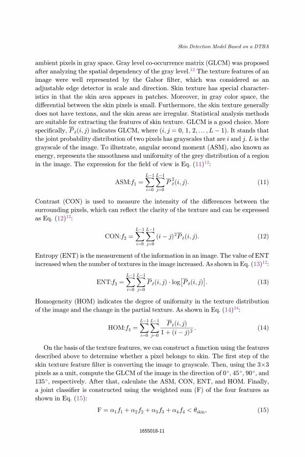
ambient pixels in gray space. Gray level co-occurrence matrix (GLCM) was proposed
after analyzing the spatial dependency of the gray level.12 The texture features of an
image were well represented by the Gabor ¯lter, which was considered as an
adjustable edge detector in scale and direction. Skin texture has special character-
istics in that the skin area appears in patches. Moreover, in gray color space, the
di®erential between the skin pixels is small. Furthermore, the skin texture generally
does not have textons, and the skin areas are irregular. Statistical analysis methods
are suitable for extracting the features of skin texture. GLCM is a good choice. More
speci¯cally, P �ði; jÞ indicates GLCM, where (i; j ¼ 0, 1, 2,… ,L� 1). It stands that
the joint probability distribution of two pixels has grayscales that are i and j. L is the
grayscale of the image. To illustrate, angular second moment (ASM), also known as
energy, represents the smoothness and uniformity of the grey distribution of a region
in the image. The expression for the ¯eld of view is Eq. (11)12:
ASM:f1 ¼XL�1
i¼0
XL�1
j¼0
P2�ði; jÞ: ð11Þ
Contrast (CON) is used to measure the intensity of the di®erences between the
surrounding pixels, which can re°ect the clarity of the texture and can be expressed
as Eq. (12)12:
CON:f2 ¼XL�1
i¼0
XL�1
j¼0
ði� jÞ2P �ði; jÞ: ð12Þ
Entropy (ENT) is the measurement of the information in an image. The value of ENT
increased when the number of textures in the image increased. As shown in Eq. (13)12:
ENT:f3 ¼XL�1
i¼0
XL�1
j¼0
P �ði; jÞ � log P �ði; jÞ� �
: ð13Þ
Homogeneity (HOM) indicates the degree of uniformity in the texture distribution
of the image and the change in the partial texture. As shown in Eq. (14)34:
HOM:f4 ¼XL�1
i¼0
XL�1
j¼0
P �ði; jÞ1þ ði� jÞ2 : ð14Þ
On the basis of the texture features, we can construct a function using the features
described above to determine whether a pixel belongs to skin. The ¯rst step of the
skin texture feature ¯lter is converting the image to grayscale. Then, using the 3�3
pixels as a unit, compute the GLCM of the image in the direction of 0�, 45�, 90�, and135�, respectively. After that, calculate the ASM, CON, ENT, and HOM. Finally,
a joint classi¯er is constructed using the weighted sum (F) of the four features as
shown in Eq. (15):
F ¼ �1f1 þ �2f2 þ �3f3 þ �4f4 < �skin; ð15Þ
Skin Detection Model Based on a DTBA
1655018-11

where �skin is the classi¯cation threshold of skin texture features. The weight (�iÞ ofeach feature is the corresponding precision as shown in Eq. (16):
�i ¼TPi
TPi þ FPi
; ð16Þ
where TPi stands for the total number of skin pixels detected correctly in color space
i and FPi stands for the total number of nonskin pixels detected as skin. The pixel
will be marked as skin if the results are smaller than the threshold and vice versa. The
skin texture ¯ltering results are obtained after ¯ltering the DTBA results with skin
texture features.
The isolated noise and mistakenly recognized pixels occurred in the ¯ltered skin
areas with an unsmooth boundary through the processes above. These detection
errors can be divided into two categories: one category is holes caused by error
detection, and the other category is nonskin pixels incorrectly detected as skin. The
errors are randomly distributed, and they can hardly be eliminated in regular ways.
As a consequence, the morphological method was introduced to optimize the de-
tection result. In morphological operations, structure element (SE) is the probe that
is used to gather information. The structure information of the image is collected
through the movement of the probe in the image. The di®erence in SE a®ects the
operation results. Dilation and erosion are the bases of morphology. The opening
operation is the process that erodes the image with the SE ¯rst and then dilates the
results. The opening operation can smooth the object contour, disconnect the narrow
neck, and eliminate pepper noise. In contrast, the closing operation is the process
that dilates the image with the SE and erodes the results in sequence. It is e±cient to
smooth the outline of objects, connect narrow discontinuities, and eliminate holes.
By using a circular SE to open and close the image in turn, the approach can reduce
the detection error rate and improve the e®ect of skin detection.
5. Experiments and Discussion
In this section, we discuss and analyze the hybrid skin detection model we presented.
Our model is compared with state-of-the-art skin detection models in the same test
set and the same environment.
5.1. Dataset
The test sets of our model use the Compaqa image dataset and high-resolution image
dataset (HRID)b. The Compaq dataset was collected in 1999. To explore the change
in the images, we established a new dataset called HRID, which was obtained by
aThe Compaq dataset is freely available for academic research purposes. Contact the author M. J. Jones
([email protected]) to obtain dataset.3
bThe HRID dataset of labeled skin and nonskin pixels is freely available for academic research purposes.
Contact the ¯rst author F. Chen ([email protected]) for instructions on how to obtain dataset.
F. Chen et al.
1655018-12

using a web crawler. Any images received from HRID whose resolution was less
than 3000�2000 were excluded. In addition, the skin areas were labeled the same as
the Compaq dataset. The statistical data from HRID is shown in Table 2. The bins
stand for the quantity of per channel average allocated. For example, 32 bins in
the RGB color space means that each R, G, and B channel was averagely divided into
32 parts.
HRID contains images of di®erent people under a variety of illuminations from all
types of perspectives. The total number of pixels is up to 7.6 billion. Because of the
high resolution of the images, more details were contained in the images and a wider
range of skin colors were covered. The images are closer to the human visual sense.
At the same time, we modi¯ed the marks of skin pixels in Compaq to improve the
recognition accuracy and precision (Fig. 3). As we can see from the images, the-
mismarked pixels are eliminated while the missing skin pixels are added.
There are more than 13 times the number of pixels in HRID compared with
Compaq. This means that more bins are occupied in skin than nonskin. Jones
demonstrated the suitable bins for each channel. In the Compaq dataset, the skin
detection model received the best results when each channel was divided into 32 bins.
Because of the small number of bins for each channel, the detection model will obtain
a high error rate, while having too many bins for each channel will lead to tremen-
dous computation6,13,14. In this paper, we use the maximum amount of bins for each
channel to obtain more accurate thresholds. In other phases, 32 bins is the default
number of bins per channel.
Table 2. Statistical data of HRID.
HRID Total Pixels Occupied Bins Percent (%) Images
All 13,643,978,626 4,780,469 28.49 815Skin 768,603,817 957,840 5.71 366
Nonskin 7,847,206,587 4,427,543 26.39 449
(a) (b) (c)
Fig. 3. Example of improvement in an image's mark in the Compaq. (a) initial image, (b) initial mark,
and (c) modi¯ed mark.
Skin Detection Model Based on a DTBA
1655018-13

5.2. Comparison and analysis
We compared the hybrid skin detection model with other previous skin detection
models in Compaq and HRID. The in°uence of each approach was taken into ac-
count. The processes of the skin detection model we presented are shown in Fig. 4.
First, DTBA was used to classify skin pixels in each color space. Second, the de-
tection results were merged into an image. Then, the combined image was ¯ltered by
skin texture and morphology, which became the ¯nal result.
5.2.1. DTBA based on multiple color spaces
When DTBA is used to classify skin pixels in images, white indicates that the pixel is
detected as skin, black indicates that the pixel is detected as nonskin, and grey means
undetermined. The output images tend to be whitish mainly because of the high
TPR set when obtaining all of the skin pixels. However, it causes a high error rate at
the same time. Comparisons between naïve Bayesian and DTBA in a single color
space and multiple color spaces are drawn in Fig. 5, which are tested on both
Compaq and HRID datasets. We analyzed the performances of all models by using
the receiver operating characteristic (ROC) curve. More speci¯cally, the TPR and
FPR are the criteria determined in the models. Moreover, every (FPR, TPR) rate
was plotted on the coordinates where all of the points in the same model were
connected by a smooth curve.
Where the white color stands for the pixel that is detected as skin, the black color
indicates that the pixel was detected as nonskin and the gray color stands for the
pixel that is detected as an undetermined pixel.
According to the ROC curves of the skin detection model, the following conclu-
sions can be drawn from Fig. 6:
(1) In the same skin detection model, HRID acquired better outcomes. Because of
the advanced image capture devices used in HRID, more realistic skin tones were
obtained, which was bene¯cial for skin segmentation. Greater resolution also means
more accurate details.
(2) The naïve Bayesian method based on multiple color spaces is superior to the
naïve Bayesian method based on a single color space because information is used
Fig. 4. The process of skin detection. First, the source image was detected in four color spaces using
DTBA. Then, the merged skin area was obtained by combining the four results. Afterward, a texture ¯lterwas introduced followed by a morphology ¯lter. Finally, the recognition result was acquired.
F. Chen et al.
1655018-14

(a) (b) (c)
(d) (e) (f)
(g) (h) (i)
Fig. 5. Results of di®erent color spaces using DTBA and the merged results. (a) initial image (b) HLS(c) HSV (d) LUV (e) RGB (f) XYZ (g) YCbCr (h) YUV (i) merged result from (b)–(h).
0.4
0.5
0.6
0.7
0.8
0.9
1
0 0.05 0.1 0.15 0.2 0.25 0.3 0.35 0.4
TP
R
FPR
ROC curves of the skin detection model
1. Single color space Naïve Bayesian inCompaq
2. Single color space naïve Bayesian inHRID
3. Multiple color spaces naïve Bayesian inCompaq
4. Multiple color spaces naive Bayesian inHRID
5. Multiple color spaces dual-thresholdBayesian in Compaq
6. Multiple color spaces dual-thresholdBayesian in HRID
12
3456
Fig. 6. The detection results using naïve Bayesian and DTBA in Compaq and HRID.
Skin Detection Model Based on a DTBA
1655018-15

from various color spaces. This can make up for the de¯ciency of the single
color space.
(3) The detection e®ect of DTBA is more precise than the naïve Bayesian method
based on multiple color spaces. As described in Sec. 4, the pixels were classi¯ed into
three categories. In the skin and nonskin categories, the correct detection rate goes
up to 99%. Then, the following step concentrated on the undetermined pixels. Sig-
ni¯cant improvement is gained by allotting di®erent weights to the detection results
of each color space.
Moreover, we compared DTBA with the three threshold Bayesian algorithms
based on multiple color spaces. A slight improvement was obtained, but the amount
of computation increased. Other methods are needed to optimize the result by virtue
of some of the overlaps between the skin and nonskin colors.
5.2.2. Optimal quantity of color spaces
We recorded the TPRs and FPRs of the detection results by testing each combi-
nation of color spaces. The number of color spaces determined the possible combi-
nations of color spaces. The optimal results of the combinations, which were di®erent
in a number of color spaces, were compared (Fig. 7). As shown in Fig. 7, the rec-
ognition results turned out to be worst when the number of color spaces used was
two. Moreover, no large di®erence occurred because the number of color spaces was
greater than three. Owing to the excessive number of color spaces, this leads to a
slight improvement in the detection results, whereas the computation has risen
rapidly. As a consequence, we choose four color spaces as the best combination.
0.4
0.5
0.6
0.7
0.8
0.9
1
0 0.05 0.1 0.15 0.2 0.25 0.3 0.35 0.4
TP
R
FPR
ROC curves of different size of color spaces
1. 2 color spaces
2. 3 color spaces
3. 4 color spaces
4. 5 color spaces
5. 6 color spaces
6. 7 color spaces
1
3
4
5
6 2
Fig. 7. ROC curves for di®erent numbers of color spaces.
F. Chen et al.
1655018-16

Further, the representative combinations of the four color spaces were
compared (Fig. 8).
5.2.3. Skin texture and morphological ¯ltering
At the previous phase, we utilized DTBA to classify skin pixels. To include
the majority of the skin pixels, a high TPR was used to ensure it. However, a high
error rate occurred in the meantime. Because of the inability of DTBA to distinguish
the similar skin color, such as animal fur, sere grasses (Fig. 9) were mistakenly
labeled.
In (b)–(d), the white color stands for the pixel that is detected as skin, the black
color indicates the pixel that was detected as nonskin, and the gray color in (b)
stands for the pixel that is detected as an undetermined pixel. Thus, additional steps
will be taken.
Based on the results of DTBA, calculating the GLCM of pixels labeled as skin
determines whether they are in accordance with the skin texture. Using the skin
texture ¯lter can make up for the disadvantages in the detection of similar skin color
to achieve a lower error rate. As we can see from Fig. 10, the improvements are
signi¯cant. By adding the weight value to texture features, the detection results were
optimized.
The ¯nal step of the hybrid model is ¯ltrating the results with the
morphology ¯lter. From the experimental results, executing the opening operation
¯rst and the closing operation second for the detection results worked best. The
¯lter can e®ectively remove noise and can eliminate small holes and the smooth
edge of.
0.4
0.5
0.6
0.7
0.8
0.9
1
0 0.05 0.1 0.15 0.2 0.25 0.3 0.35 0.4
TP
R
FPR
ROC curves of DTBA based on four color spaces
1. YUV-LUV-Lab-XYZ
2. RGB-HSV-YCrCb-XYZ
3. HSV-HSL-YCbCr-YUV
4. HSV-YCbCr-HSL-YUV
5. RGB-Lab-YUV-LUV
1
23 4
5
Fig. 8. ROC curves of the representative combinations for four color spaces using DTBA.
Skin Detection Model Based on a DTBA
1655018-17

5.2.4. Comparison of skin detection models
The hybrid skin detection model we presented in this paper executed multi-level
¯lters. The model was veri¯ed in complex backgrounds. Five hundred images were
used as the test set, and the outcomes are shown in Table 3. We can reach the
conclusion that the model we proposed works well in complex backgrounds. Specif-
ically, several examples are shown in Fig. 11. The images show that the Bayesian
method can also recognize the skin in the glass re°ection. More importantly, the
hybrid skin model re¯ned the results, especially in those situations where the image
contains many people or has intricate surroundings.
(a) (b)
(c) (d)
Fig. 9. Detection results of similar skin color. (a) Source image, (b) RGB using DTBA (threshold1 ¼ 0.1,
threshold2 ¼ 0.94), (c) DTBA based on four color spaces, and (d) results of DTBA based on four color
spaces used in skin texture ¯lter.
F. Chen et al.
1655018-18

As shown in Table 3, compared with the naïve Bayesian algorithm, DTBA is
based on multiple color spaces. The FPR is reduced by approximately 50%,
namely, from 18.8% to 9.1%. More importantly, the hybrid skin model we pro-
posed performed well under the complex environment, especially in HRID. It is up
to 96.2% of the TPR with a distracted background. We compared the models
in the Compaq and HRID datasets to show the advancement of the model we
proposed (Table 4).
In the experiments, we used TPR (95%) and FPR (5%) as initial inputs to gain
dual-thresholds. Experimentally, we found that the DTBA based on four color spaces
was more precise and accurate than the Naïve Bayesian algorithm. To illustrate,
the FPR of DTBA compared with the naïve Bayesian algorithm was reduced by
7.5% with a 2.6% increase in TPR. TPR is becoming more di±cult to improve
when it is more than 90%. The ¯ndings have demonstrated that the proposed
approach produced TPRs of 95.5% and 97.4% in the Compaq dataset and in the
HRID, respectively, and the FPRs are only 2.8% and 2.4%, respectively. Particu-
larly, nearly one-third of the images received a TPR of more than 99%, which is
Table 3. Performance of models in complex background (unit: %)
Models Compaq TPR (FPR) HRID TPR (FPR)
Naïve Bayesian13 89.5(18.8) 89.7(17.5)
Bayesian based on multiple color spaces 90.0(16.2) 91.2(15.8)DTBA based on multiple color spaces 92.8(9.1) 93.3(8.8)
Gaussian mixture model20 82.2(12.2) 81.6(11.2)
Hybrid skin model (this paper) 94.1(3.9) 96.2(3.4)
0.4
0.5
0.6
0.7
0.8
0.9
1
0 0.05 0.1 0.15 0.2 0.25 0.3 0.35 0.4
TP
R
FPR
ROC curves of skin texture filtering
1. skin texture weighted filtering
2. no skin texture filtering
3. skin texture filtering
12
3
Fig. 10. ROC curves of skin texture ¯ltering. Including skin texture weighted ¯lter, skin texture ¯lter,and no skin texture ¯lter.
Skin Detection Model Based on a DTBA
1655018-19

extremely precise. In order to improve the detection accuracy, plenty of models take
advantage of other recognition methods, such as face, hand, and upper-body
recognition.5,10,15,21 Compare with those models, on the one hand, our hybrid model
does not depend on face or hand in the image, on the other hand, our model needs less
time to detect skin.
(a) (b) (c)
(d) (e)
Fig. 11. Detection results in complex background. (a) initial image, (b) naïve Bayesian method
(threshold¼ 0.71), (c) RGB using DTBA (threshold1¼ 0.1, threshold2¼ 0.93), (d) merged threshold result(thresholdskin¼ 0.85) and (e) ¯nal result.
F. Chen et al.
1655018-20

0
0.1
0.2
0.3
0.4
0.5
0.6
0.7
0.8
0.9
1
0 10 20 30 40 50 60 70 80 90 100
Tim
e(s)
Number of Image
Detection time of skin detection models
Naïve Bayesian Bayesian based on multiple color spaces
DTBA based on multiple color spaces Gaussian mixture model
Skin texture feature model Fusion skin model(this paper)
Fig. 12. Detection time of six skin detection models in each single image.
Table 4. Performance of models in two datasets (unit: %).
Models Compaq TPR (FPR) HRID TPR (FPR)
Naïve Bayesian13 90.5(14.8) 91.2(13.6)Bayesian based on multiple color spaces 92.7(8.5) 93.3(8.2)
DTBA based on multiple color spaces 93.1(7.3) 93.5(7.0)
Gaussian mixture model20 85.4(10.1) 86.2(9.6)
Hybrid skin model (this paper) 95.5(2.8) 97.4(2.4)
Table 5. Performance of di®erent skin detection.
Models Time(s) TPR (%) FPR (%)
Naïve Bayesian13 0.0513 89.5 14.7
Bayesian based on multiple color spaces 0.1543 92.1 8.3
DTBA based on multiple color spaces 0.1986 92.5 7.5
Gaussian mixture model20 0.7428 87.2 9.1Skin texture Model20 0.6863 88.5 14.3
Hybrid skin model (this paper) 0.3478 97.3 2.1
Skin Detection Model Based on a DTBA
1655018-21

5.2.5. Performance of skin detection
To test the performance of the hybrid skin model we proposed, 150 images were
taken in and each image had a resolution of 5120�2880. Among them, 50 images
were used for the training set, while 100 images were used as the test set. The skin
pixels occupied every image range from 10% to 70%. The experiment was ¯nished in
Cþþ in a PC with an 8 GB memory.
The average detection time of di®erent skin models is shown in Table 5 along with
TPR and FPR. The detection time of several models was compared on the test set
(Fig. 12). From the diagram, we can draw the conclusion that the naïve Bayesian
algorithm consumed the shortest amount of time and that this has nothing to do
with the skin pixels contained in the images. On the contrary, the skin texture
feature model requires massive computation. The hybrid skin detection model is
superior to the former model, and the detection time is positively correlated to the
number of skin pixels in the image.
Although the DTBA consumed more time than the naïve Bayesian algorithm,
DTBA needs less computation time than the GMM and the skin texture model.
Moreover, only the pixels detected as skin will be executed with skin texture ¯l-
tering, whose computation was one-third as the skin texture model. Furthermore,
the hybrid model acquired excellent TPRs and FPRs in an acceptable detection
time. Compared with the neural network method, the hybrid model proposed in
this paper does not require a complex construction process. The pictures we used in
the test are more than 15 MB, however, a video taken by a real-time system usually
less than 15M megabyte per second. For example, a 1080P (1920�1080) video,
there are 1920 lines of horizontal pixels and 1080 lines of vertical pixels, taken by a
camera, approximately 2 megabyte per second. So it can be applied in a real-time
system. Sequential implementation of the proposed method allows for processing
from 30 to 40 frames per second with the resolution of 1920�1080. In addition,
the performance can be improved as the DTBA can be applied in the distributed
system.
6. Conclusion
The development of hardware provides a perfect habitat for the software, and skin
detection can make use of it. The DTBA we proposed took advantage of multiple
color spaces and performed well in complex circumstances. First, the skin regions of
Compaq were re¯ned by us. In addition, HRID was presented and was more ac-
curate in representing skin color and nonskin color. Second, DTBA was used as the
¯rst step for skin detection, and better detection results were obtained when it was
combined with the skin texture feature ¯lter. The hybrid skin detection model
established in this paper classi¯ed the pixels with high accuracy and precision, and
it is robust for a complex background. Finally, it can be applied in a real-time
system. Because of the continuity and aggregation of skin areas, DTBA can be
F. Chen et al.
1655018-22

applied in clustering classi¯cation. Through skin detection in every color space, we
can set the pixel as a skin seed when it is true under strict conditions, such as a
pixel detected as skin in all color spaces. In addition, a nonskin seed can be received
when a pixel is detected as nonskin in all color spaces. Utilizing the approach
described above, we can obtain two-way clustering, and there is no need to man-
ually label the skin pixels. Incidentally, DTBA has general applicability and
the potential for development in hair detection, gesture recognition, and object
recognition.
References
1. Y. Ban, S. K. Kim and S. Kim, Face detection based on skin color likelihood, PatternRecogn. 47 (2014) 1573–1585.
2. M. A. Berbar, Novel colors correction approaches for natural scenes and skin detectiontechniques, Int. J. Video and Image Process Netw. Secur. IJVIPNS-IJENS 11(2) (2011)1–10.
3. K. K. Bhoyar and G. Kakde, Skin color detection model using neural networks and itsperformance evaluation, J. Comput. Sci. 6(9) (2010) 963–968.
4. S. Bianco, F. Gasparini and R. Schettini, Adaptive skin classi¯cation using face and bodydetection, IEEE Trans. Image Proces. 24(12) (2015) 4756–4765.
5. S. Bilal, R. Akmeliawati and M. J. E. Salami, Dynamic approach for real-time skindetection, J. Real-Time Image Proces. 10(2) (2015) 371–385.
6. D. Chai and K. N. Ngan, Locating facial region of a head-and-shoulders color image,in Proc. 3rd Int. Conf. Automatic Face and Gesture Recognition (Nara, Japan, 1998), pp.124–129.
7. Y. H. Chen and K. T. Hu, Statistical skin color detection method without colortransformation for real-time surveillance systems, Eng. Appl. Artif. Intell. 25 (2012)1331–1337.
8. C. A. Doukim and J. A. Dargham, Combining neural networks for skin detection, SignalImage Proces. 1(2) (2010) 1129–1138.
9. L. Duan and Z. Lin, Advances in Neural Networks, A method of human skin regiondetection based on PCNN, Advances in Neural Networks, 6th International Symposiumon Neural Networks, ISNN 2009, Proc, Part III (Wuhan, China, 2009), pp. 486–493.
10. S. Du®ner and J. M. Odobez, Leveraging colour segmentation for upper-body detection,Pattern Recogn. 47(6) (2014) 2222–2230.
11. C. Garcia and G. Tziritas, Face detection using quantized skin color regions merging andwavelet packet analysis, Trans. Multimed. 1(3) (1999) 264–277.
12. R. M. Haralick and K. Shanmugan, Textual features for image classi¯cation, IEEETrans. Syst. Man Cybern. 3(6) (1973) 610–621.
13. M. J. Jones and J. M. Rehg, Statistical color models with application to skin detection,Int. J. Comput. Vis. 46(1) (2002) 81–96.
14. P. Kakumanu and S. Markrogiannis, A survey of skin-color modeling and detectionmethods, Pattern Recogn. 40 (2007) 1106–1122.
15. M. Kawulok, J. Kawulok and J. Nalepa, Skin detection using spatial analysiswith adaptive seed, Image Processing (ICIP), 2013 20th IEEE Int. Conf. IEEE (Mel-bourne, Victoria, Australia, 2013), pp. 3720–3724.
16. M. Kawulok, J. Kawulok and J. Nalepa, Spatial-based skin detection using discriminativeskin-presence features, Pattern Recogn. Lett. 41(1) (2014) 3–13.
Skin Detection Model Based on a DTBA
1655018-23

17. T. T. Khac, K. Q. Li and Y. Xu, Chemical reaction optimization with greedy strategy forthe 0–1 knapsack problem, Appli. Soft Comput. 13(4) (2013) 1774–1780.
18. S. A. Naji and R. Zainumddin, Skin segmentation based on multi pixel color clusteringmodels, Digit. Signal Process. 22 (2012) 933–940.
19. P. P. Paul and M. Gavrillova, PCA based geometric modeling for automatic facedetection, Comput. Sci. Appl. 4 (2011) 221–225.
20. H. Permuter, J. Francos and I. Jermyn, A study of gaussian mixture models of color andtexture features for image classi¯cation and segmentation, Pattern Recogn. 39(4) (2006)695–706.
21. P. K. Pisharady, P. Vadakkepat and P. L. Ai, Attention based detection and recognitionof hand postures against complex backgrounds, Int. J. Comput. Vis. 101(3) (2013)403–419.
22. J. C. SanMiguel and S. Suja, Skin detection by dual maximization of detectors agreementfor video monitoring, Pattern Recogn. Lett. 34 (2013) 2102–2109.
23. M. Shoyaib and M. Abdullah-AI-Wadud, A skin detection approach based on theDempster-Shafer theory of evidence, Int. J. Approx. Reason. 53 (2012) 636–659.
24. M. C. Skin and K. I. Chang, Does colorspace transformation make any di®erence on skindetection, in Proc. IEEE Workshop on Applications of Computer Vision. (Orlando, FL,USA, 2002) 275–279.
25. F. Y. Song, X. Y. Tan and L. Xue, Eyes closeness detection from still images with multi-scale histograms of principal oriented gradients, Pattern Recogn. 47 (2014) 2825–2838.
26. H. M. Sun, Skin detection for single images using dynamic skin color modeling, PatternRecogn. 43 (2010) 1413–1420.
27. J. Terrillon and S. Akamatsu, Comparative performance of di®erent chrominance spacesfor color segmentation and detection of human faces in complex scene images, Vis. Interf.99 (1999) 180–187.
28. A. Tsitsoulis, G. Nikolaos and Bourbakis, A methodology for extracting standing humanbodies from single images, Trans. Hum.–Mach. Syst. 45(3) (2015) 327–338.
29. M. A. Wadud and M. Shoyaib, A skin detection approach based on color distance map,EURASIP J. Adv. Signal Process. 10 (2008) 1–10.
30. F. Y. Xie and A. C. Bovik, Automatic segmentation of dermoscopy images using self-generating neural networks seeded by genetic algorithm, Pattern Recogn. 46 (2013)1012–1019.
31. Y. Xu and K.Q. Li, A DAG scheduling scheme on heterogeneous computing systems usingdouble molecular structure-based chemical reaction optimization, J. Parallel Distrib.Comput. 73(9) (2013) 1306–1322.
32. P. Yogarajah, J. Condell and K. Curran, A dynamic threshold approach for skin seg-mentation in color images, Int. J. Biometrics. 4(5) (2012) 38–55.
33. A. A. Zaidan and N. N. Ahmad, Image skin segmentation based on multi-agent learningBayesian and neural network, Eng. Appl. Artif. Intell. 32 (2014) 136–150.
34. A. A. Zaidan, H. A. Karim and N. N. Ahmad, An automated anti-pornography systemusing a skin detector based on arti¯cial intelligence: A review, Int. J. Pattern Recogn.Artif. Intell. 27(4)(2013) 254–261.
35. A. A. Zaidan, H. A. Karim and N. N. Ahmad, A four-phases methodology to proposeanti-pornography system based on neural and Bayesian methods of arti¯cial intelligence,Int. J. Pattern Recogn. Artif. Intell. 28(1) (2014) 178–215.
F. Chen et al.
1655018-24

Fujunku Chen, born in1990, received his B.S de-gree in Software Engi-neering from CentralSouth University, Chang-sha, China in 2013. He iscurrently studying in theCentral South Universityas an undergraduate stu-dent of software engi-neering. He is committed
to skin detection and image beauti¯cation. Hisresearch interests include software engineering,data mining and computer vision. In addition,he has participated in more than 10 softwareprojects.
Zhigang Hu, born in1963, is a Professor andPh.D. supervisor of theCentral South University,Changsha, China. He isalso a senior member ofChina Computer Federa-tion. He received his B.S.and his M.S. degrees fromCentral South University,Changsha, China in 1985
and in 1988, respectively. He received his Ph.D.from Central South University, Changsha, Chinain 2002. His research interests focus on the par-allel computing, cloud computing and high per-formance computing. He has published manypapers in international journals and conferences.He is the deputy Secretary-General of HunanComputer Federation. He is currently the vice-dean of School of Software, Central South Uni-versity.
Keqin Li is a SUNYDistinguished Professor ofComputer Science. Hiscurrent research interestsinclude parallel comput-ing and high-performancecomputing, distributedcomputing, energy-e±-cient computing andcommunication, hetero-geneous computing sys-
tems, cloud computing, big data computing,CPU-GPU hybrid and cooperative computing,multicore computing, storage and ¯le systems,wireless communication networks, sensor net-works, peer-to-peer ¯le sharing systems, mobilecomputing, service computing, Internet of thingsand cyber-physical systems. He has publishedover 400 journal articles, book chapters, andrefereed conference papers, and has receivedseveral best paper awards. He is currently, or hasserved, on the editorial boards of IEEE Trans-actions on Parallel and Distributed Systems,IEEE Transactions on Computers, IEEETransactions on Cloud Computing, Journal ofParallel and Distributed Computing. He is alsoan IEEE Fellow.
Wei Liu, born in 1982, isa member of China Com-puter Federation. He re-ceived his Ph.D. and hisM.S. degree from CentralSouth University, Chang-sha, China. His researchinterests focus on thesoftware engineering anddata mining, design pat-terns, and source code
analysis and optimize. He has published morethan 10 journal articles. Additionally, he hastaken part in over 30 software projects.
Skin Detection Model Based on a DTBA
1655018-25