A Histopathological and Immunohistochemical Study of Acute ...
27
Cellular Pathology and Apoptosis in Experimental and Human Acute and Chronic Compressive Myelopathy ROWENA ELIZABETH ANNE NEWCOMBE M.B.B.S. B.Med Sci. (Hons.) Discipline of Pathology, School of Medical Sciences University of Adelaide June 2010 A thesis submitted in partial fulfilment of the requirements for the degree of Doctor of Philosophy
Transcript of A Histopathological and Immunohistochemical Study of Acute ...

Cellular Pathology and Apoptosis in Experimental
and Human Acute and Chronic Compressive
Myelopathy
ROWENA ELIZABETH ANNE NEWCOMBE
M.B.B.S. B.Med Sci. (Hons.)
Discipline of Pathology, School of Medical Sciences
University of Adelaide
June 2010
A thesis submitted in partial fulfilment of the requirements for the
degree of Doctor of Philosophy
i
Declaration
This work contains no material which has been accepted for the award of any other degree
or diploma in any university or other tertiary institution and, to the best of my knowledge
and belief, contains no material previously published or written by another person, except
where due reference is made in the text of the thesis.
I give consent to this copy of my thesis, when deposited in the University library, being
made available for loan and copying.
Signed: Date
Rowena Elizabeth Anne Newcombe

ii
Acknowledgements
I acknowledge the invaluable and expert teaching, encouragement, patience and wisdom of
my supervisors, Professor Peter Blumbergs, Professor Robert Vink, Dr John Finnie,
Professor Peter Reilly, and Professor Nigel Jones. In particular I thank Professor
Blumbergs who first enabled me this opportunity, providing unwavering guidance until its
completion.
I sincerely thank Mr Jim Manavis for his technical expertise, enthusiasm and assistance
over many years.
I am ever grateful for the encouragement and insights provided by my dear husband
Matthew, my wise mother Carolyn, and my family and friends throughout this process of
learning. I could not have achieved this without you.
I make special mention of my father, Doctor Raymond Newcombe, to whom I dedicate this
thesis, for his careful, inspiring and excellent mentoring. He is exemplary of those qualities
desired in any doctor of medicine; integrity, perseverance, compassion, precise clinical
acumen, curiosity, and an unyielding belief in the human spirit.
„He who has health has hope, and he who has hope has everything.‟
- Arabian proverb
iii
Publications and Presentations
Newcombe REA, Vink R, Finne R, Reilly P, Blumbergs PC. Functional and pathologic studies of
experimental chronic compressive spinal cord injury and the effects of decompression. J
Neurotrauma.2009;A2-A101.
Newcombe REA, Blumbergs PC, Sarvestani G, Manavis J, Jones NR. Caspase-3-mediated
Proteolysis of Amyloid Precursor Protein and the Production of Amyloid-beta in Human Acute and
Chronic Compressive Myelopathy. J Bone Joint Surg Br.2004;86-B: 462.
Newcombe REA, Blumbergs PC, Sarvestani G, Manavis J, Jones NR. Apoptosis in Human Acute
and Chronic Compressive Myelopathy. In: Proceedings of the Australian Neuroscience Society.
2004.
Newcombe REA, Blumbergs PC, Sarvestani G, Manavis J, Jones NR. A Human Study of
Apoptosis in Acute and Chronic Compressive Myelopathy. In: Proceedings of the Spine Society of
Australia.2003.
Prizes and Scholarships 2009 International Neurotrauma Society Student Travel Grant
2007 National Health and Medical Research Council Training Postgraduate Fellowship
2004 Spine Society of Australia Award for Spinal Research
2003 The Douglas Hardy Research Project Prize – The University of Adelaide
2003 Australian Medical Students‟ Association Research Scholarship
2003 John Curtin School of Medical Research Summer Scholarship
2003 Spine Society of Australia Award for Spinal Research

iv
Abbreviations
AD Alzheimer‟s Disease
ADP Adenosine Diphosphate
AHC Anterior Horn Cell
ALA Anterolateral White Matter Area
AIF Apoptosis Inducing Factor
ANOVA Analysis of Variance
APP Amyloid Precursor Protein
ATP Adenosine Triphosphate
aC3 Active Caspase-3
BACE -site APP-cleaving Enzyme
Bak Bcl-2 Antagoinist Killer
Bax B-Cell Lymphoma-Associated X
BBB Blood Brain Barrier
BBB Score Beattie Basso Bresnahan Score
Bcl-2/x B-cell Lymphoma 2/x
BDNF Brain-derived Neurotrophic Factor
C3 Caspase-3
C9 Caspase-9
cDNA Complementary Deoxyribonucleic Acid
CCA Caspase-3 and Caspase-mediated Cleavage of APP
cm Centimetre(s)
CMAP Caspase-3-mediated APP Proteolytic Peptide Antibody
CNPase Cyclic Nucleotide Phosphodiesterase
CNS Central Nervous System
CSF Cerebrospinal Fluid
CSM Cervical Spondylotic Myelopathy
CT Computed Tomography
CV Coefficient of Variance
DAB Diaminobenzidine
DNA Deoxyribonucleic Acid
DNA-PK DNA-dependent Protein Kinase
DNA-PKcs DNA-dependent Protein Kinase Catalytic Subunit
DR3/6 Death Receptor 3/6
e.g. Exempli Gratia (for example)
EDAR Ectodermal Dysplasia Receptor

v
EDTA Ethylenediamine Tetra-acetic Acid
ELISA Enzyme-linked Immunosorbent Assay
et al. Et Alii (and others)
FasL Fas Receptor Ligand
FasR Fas Receptor
FADD Fas Associated Death Domain
g Gram(s)
GFAP Glial Fibrillary Acidic Protein
GM Grey Matter
GM-CSF Granulocyte Macrophage-colony Stimulating Factor
H&E Haematoxylin and Eosin
hr Hour(s)
i.e. Id Est (that is to say)
IAP Inhibitors of Apoptosis
Iba1 Ionised Calcium Binding Adaptor Molecule 1
IL Interleukin
IHC Immunohistochemistry
LPC Lysophosphatidylcholine
LCST Lateral Corticospinal Tract
m Metre(s)
MBP Myelin Basic Protein
MCP-1 Macrophage Chemotactic Protein-1
min Minute(s)
MIP-1 Macrophage Inflammatory Protein-1
MLS Mitochondrial Localisation Sequence
Mm Millimetre(s)
MOMP Mitochondrial Outer Membrane Permeability
MRI Magnetic Resonance Imaging
NA Numerical Aperture
NAD Nicotinamide Adenine Dinucleotide
NAPO Negative in Apoptosis Marker
NeuN Neuronal Nuclei Antibody
NGF Nerve Growth Factor
NF-B Nuclear Factor-kappaB
ng Nanogram(s)
NHS Normal Horse Serum
nm Nanometre(s)

vi
NMDA N-methyl-D-aspartate
nNOS Neuronal Nitric Oxide Synthase
NO Nitric Oxide
NOS Nitric Oxide Synthase
NT-3 Neurotrophin-3
Olig2 Oligodendrocyte Transcription Factor 2
p Probability
PAR Poly (ADP-ribose)
PARG Poly (ADP-ribose) Glycohydrolase
PARP Poly (ADP-ribose) Polymerase
PBS Phosphate Buffered Saline
PCA Posterior Column Area
PCD Programmed Cell Death
PMT Photomultiplier Tube(s)
PNS Peripheral Nervous System
PS-1 Presenelin-1
RPM Revolutions Per Minute
ROS Reactive Oxygen Species
SCI Spinal Cord Injury
SEM Standard Error of the Mean
Smac/DIABLO Second Mitochondria-derived Activator of Caspases/Direct IAP Binding Protein
with Low PI
TBI Traumatic Brain Injury
TBS Tris-buffered saline
TMRM Tetramethyl Rhodamine Methyl Ester
TNF Tumour Necrosis Factor
TNF- Tumour Necrosis Factor-
TPA Tissue Polypeptide Antigen
TRAIL-R1/2 TNF-related Apoptosis Inducing Ligand Receptor 1/2
TUNEL Terminal in situ Nick-end Labelling
l Microlitre(s)
UPLAPO Universal Plan Apochromatic Objectives
WM White Matter
XRCC1 X-ray Repair Cross-complimenting Group 1 oC Degrees Celsius
3-AB 3-aminobenzamide
5-AIQ 5-aminoisoquinolinone

vii
Table of Contents
Page
ACKNOWLEDGEMENTS........................................................................................ ii PUBLICATIONS AND PRESENTATIONS............................................................ iii ABBREVIATIONS..................................................................................................... iv FIGURES..................................................................................................................... x TABLES....................................................................................................................... xii ABSTRACT................................................................................................................. xviii CHAPTER 1. INTRODUCTION.............................................................................. 1
1.1 Human Chronic Compressive Myelopathy................................................. 4 1.1.1 History.......................................................................................... 4 1.1.2 Definitions.................................................................................... 4 1.1.3 Aetiology and Classification........................................................ 5 1.1.4 Clinical History and Features...................................................... 10 1.1.5 Diagnostic Investigations............................................................. 11 1.1.6 Management and Outcome.......................................................... 13 1.1.7 Spinal Cord Repair...................................................................... 15
1.2 The Apoptosis-Necrosis Continuum........................................................... 17 1.3 Human Chronic Compressive Myelopathy................................................. 23
1.3.1 Apoptosis in Chronic Compressive Myelopathy.......................... 26 1.3.2 Experimental Models of Chronic Spinal Cord Compression....... 27 1.3.3 The Role of Surgical Decompression........................................... 30
1.4 Axonal Injury.............................................................................................. 32 1.5 Human Acute Compressive Myelopathy.................................................... 40
1.5.1 History.......................................................................................... 40 1.5.2 Definitions.................................................................................... 40 1.5.3 Aetiology and Classification........................................................ 40 1.5.4 Clinical Manifestations................................................................ 41 1.5.5 Diagnostic Investigations............................................................. 42 1.5.6 Management and Outcome.......................................................... 42
1.6 Aims and Hypotheses.................................................................................. 53 CHAPTER 2. METHODS.......................................................................................... 54
2.1 Experimental Chronic Compressive Myelopathy....................................... 55 2.1.1 Animals......................................................................................... 55 2.1.2 Experimental Groups of Chronic Spinal Cord Compression...... 55 2.1.3 Control Groups............................................................................ 56
2.2 Experimental Acute Compressive Myelopathy.......................................... 63 2.2.1 Functional testing......................................................................... 64
2.3 Human Compressive Myelopathy............................................................... 68 2.4 Processing of Experimental Tissue............................................................. 71

viii
Page
2.5 Histopathological Assessment.................................................................... 72
2.5.1 Experimental................................................................................ 72 2.5.2 Human.......................................................................................... 72
2.6 Immunohistochemistry................................................................................ 74 2.6.1 Immunohistochemical Markers of Apoptosis............................... 76
2.7 Semi-quantitation........................................................................................ 87 2.8 Laser Scanning Confocal Microscopy........................................................ 90 2.9 Enzyme-Linked Immunosorbent Assay (ELISA)....................................... 91
2.9.1 Preparation of Spinal Cord Homogenate.................................... 91 2.9.2 Protein Estimation Assay............................................................. 91 2.9.3 ELISA for Caspase-3 and Caspase-9........................................... 92
2.10 Photography.............................................................................................. 93 2.11 Statistical Analysis.................................................................................... 93
CHAPTER 3. RESULTS - CHRONIC SPINAL CORD COMPRESSION.......... 94
3.1 Experimental Chronic Spinal Cord Compression....................................... 98 3.1.1 Histopathology............................................................................. 98 3.1.2 Apoptosis...................................................................................... 103 3.1.3 Axonal Injury (APP)..................................................................... 112
3.2 Quantitative Studies – Cross-Sectional Area.............................................. 113 3.2.1 Area – Posterior white matter...................................................... 113 3.2.2 Area – Total cross-section of spinal cord (mm2)......................... 116
3.3 Functional Assessment................................................................................ 118 3.3.1 BBB Score.................................................................................... 118 3.3.2 Rotarod......................................................................................... 122 3.3.3 Tail Flick...................................................................................... 127 3.3.4 Ledge Beam.................................................................................. 131
3.4 Chronic Human Spinal Cord Compression................................................. 132 3.4.1 Histopathology............................................................................. 132 3.4.2 Apoptosis...................................................................................... 138 3.4.3 Axonal Injury................................................................................ 149
3.5 Discussion................................................................................................... 162 3.6 Conclusions................................................................................................. 182
3.6.1 Apoptosis...................................................................................... 182 3.6.2Axonal Injury................................................................................. 183
CHAPTER 4. RESULTS - ACUTE COMPRESSIVE MYELOPATHY.............. 184
4.1 Experimental Acute Compressive Myelopathy.......................................... 187 4.1.1 Histopathology............................................................................. 187 4.1.2 Apoptosis...................................................................................... 190
4.2 Functional Assessment................................................................................ 193 4.2.1 BBB Score.................................................................................... 193 4.2.2 Tail flick....................................................................................... 196 4.2.3 Rotarod......................................................................................... 198
4.3 Axonal Injury.............................................................................................. 199

ix
Page
4.4 Human Acute Compressive Myelopathy.................................................... 201
4.4.1 Histopathology............................................................................. 201 4.4.2 Apoptosis...................................................................................... 203 4.4.3 Axonal Injury................................................................................ 209
4.5 Discussion – Experimental and Human Acute Compressive Myelopathy. 215 4.5.1 Apoptosis...................................................................................... 215 4.5.2 Axonal Injury................................................................................ 223
4.6 Conclusions – Histopathological Changes in Experimental and Human Acute Compressive Myelopathy........................................................... 226 4.6.1 Apoptosis...................................................................................... 226 4.6.2 Axonal Injury................................................................................ 226
CHAPTER 5. GENERAL DISCUSSION –
CHRONIC AND ACUTE COMPRESSIVE MYELOPATHY.................. 227 5.1 Conclusions................................................................................................. 235
REFERENCES............................................................................................................ 236 APPENDIX – INDIVIDUAL HUMAN CASES....................................................... 263
Re: Thesis emendations for PhD thesis by Dr Rowena Newcombe During printing of the thesis a variation occurred in page numbering between the electronic and the printed copies. Subsequently, Examiner 1’s emendations are 19-20 pages in advance of the correct numbering. In addition, Examiner 1 identified that the list of corrections given was incomplete, and that further review of spelling and grammatical errors be made, further altering the page structure. Thus, the actual page at which the correction is located in the thesis correlates to the page number and line set in bold at the end of the noted correction. The following emendations have been made to the thesis as recommended by the examiners. Examiner 1 Minor changes that would enhance the thesis P48 Description of the model used in the studies in comparison to previous models might be better in the discussion than in the introduction, unless the model was previously used and published. Response: A model of chronic compressive myelopathy as used by Kim et al. 2004 is described within the introduction. This is a partial reference which is expanded in more detail in the discussion, in agreement with the referee’s comments Page 29 Line 23 P48 Syringomyelia is not really a form of cord compression in the usual sense. Although it may result in some compression of cord tissue, the pathology is likely to be quite different to forms of external compression. Although it is valid to include the studies of syringomyelia in this work, it would be preferable if this distinction were clarified in the text. Response: The probable differing mechanism of syringomyelia and spondylotic myelopathy is noted Page 24 Line 2 P89: what pressure was used for perfusion fixation? Response: A pressure of 80-120mmHg was used for perfusion fixation Page 71 Line 11 P125 ‘...a subtype of oligodendrocytes was identified,’ is not clear. What was the subtype? Response: The use of the term ‘subtype’ was removed from the text to simply describe the cell ‘oligodendrocyte’ throughout the thesis. Page 103 It would be preferable to reduce the number of significant figures used for the cord cross-sectional area results, BBB score results, and rotarod times. For example, reporting mean BBB scores to 2 decimal places has no meaning.
Response: The significant figures were rounded to a consistent 2 decimal places. Table 112 ‘Communicating syrinx’ is usually used to refer to cysts that communicate with the fourth ventricle rather than the central canal. Did these syrinxes really communicate with the fourth ventricle? (It would be highly unusual if associated with Chiari I malformations). Response: There is no table 112, and it is suspected that the Examiner may be referring to Table 28, in which communicating syrinx was correctly defined as a communication with the central canal. Grammatical and spelling errors that require correction P23: “during the 1952” As recommended this was changed to, ‘during 1952’. Page 4 Line 5 P23: ‘canalicular’ As recommended this was changed to, ‘cannalicular’. Page 5 Line 18 P28: ‘An association between osteophytes and concave, load baring areas within the spine were recognised early’ should be ‘An association between osteophytes and concave, load bearing areas within the spine was recognised early...’. A correction was made as recommended. Page 9 Line 4 P35: ‘Multiple forms of programmed cell death (PCD) and classified as types I, II, and III PCD’ Should this be, ‘Multiple forms of programmed cell death (PCD) are classified as types I, II, and III PCD.’ A correction was made as recommended. Page 17 Line 6 P36: The ontology of apoptosis was given twice. This was corrected as appropriate. Page 17 Line 21 P47: The words ‘central canal’ should be ‘spinal canal’ This was corrected Page 28 Line 14

P50: ‘This current study aims assess the effects of decompression in an experimental model of mild chronic cord compression’ should be ‘This current study aims to assess the effects of decompression in an experimental model of mild chronic cord compression.’ A correction was made as recommended. Page 31 Line 6 P50 ‘…a minimum of approximately 10% of axons’. Should this be ‘…a maximum of approximately 10% of axons’? No change was necessary as the sentence reads correctly Page 32 Line 15 P66: ‘transaction’ was corrected to ‘transection’. This was corrected Page 48 Line 1 P66 Tables 86-92 are out of order and not referenced in the text or the table of tables. This was corrected Page 69 Line 1 P66 ‘Processotomy’ is an unusual term. Would ‘removal of the spinous process and laminectomy’ be better? The term, ‘processotomy’ is consistent with surgical terminology to describe full resection of the spinous process to expose the dura, and thus the term was retained. Page 57 Line 7 P91: The Gracile fasciculus was labelled. Page 73 P110: ‘wee k’ was changed to ‘week’. Page 92 line 5 ‘tetromethylbenzidine’ was changed to ‘tetramethylbenzidine’. Page 92 Line 15 ‘data is’ was changed to ‘data are’ as recommended. Page 93 Line 8 P113: ‘emersion’ was corrected to ‘immersion’. Page 95 Line 15 ‘A complete representation of pathological and apoptotic changes in human cases are documented...’ should be ‘A complete representation of pathological and apoptotic changes in human cases is documented...’ This was corrected as suggested. Page 95, Line 12 ‘A similar panel of apoptotic markers were used...’ should be ‘A similar panel of apoptotic markers was used...’

A correction was made as suggested Page 95 Line 21 P114: ‘A subset of enlarged axons were immunopositive...’ should be ‘A subset of enlarged axons was immunopositive...’. A correction was made as suggested. Page 97 Line 4 P123: ‘Rare of occasional immunopositive glia was seen...’ should be ‘Rare or occasional immunopositive glia were seen...’. A correction was made as suggested. Page 105 Line 19 P124: ‘At 9 week,...’ was changed to, ‘At 9 weeks,...’ as suggested. Page 106 Line 12 P125: ‘...frequent glial staining was seen in the majority cases...’ was changed to, ‘...frequent glial staining was seen in the majority of cases...’ as recommended. Page 107 Line 12 P126: ‘...TUNEL was either absent of rarely present,’ was changed to, ‘...TUNEL was either absent or rarely present,’ as recommended. Page 108 Line 17 P130: ‘APP axonal immunopositivity was rare or occasionally present in compression groups but were frequently present...’ should be ‘APP axonal immunopositivity was rare or occasionally present in compression groups but was frequently present...’ A correction was made as recommended. Page 112 Line 11 P131: ‘...the ratio or posterior to anterolateral white matter at the site was comparable to controls...’ should be ‘...the ratio of posterior to anterolateral white matter at the site was comparable to controls...’. A correction was made as recommended. Page 113 Line 14 P140: ‘Statistically, the rotarod results were compared between the seven groups using a Cox proportional hazards model. A value of 120 second was considered to be right censored’ was repeated. A correction was made as recommended. Page 122 Line 7 P184: It was recommended that, ‘In experimental chronic compressive myelopathy, caspase-9, PARP and aC3 staining was found...’ be changed to, ‘In experimental chronic compressive myelopathy, caspase-9, PARP and aC3 staining were found...’ A correction was made as recommended. Page 166 Line 13 P184: As recommended, ‘Although our data...’ was changed to, ‘Although our results...’. Page 169 Line 1

P202: As recommended, ‘Tissue from human cases was also studies for apoptosis...’ was changed to, ‘Tissue from human cases was also studied for apoptosis...’. Page 185 Line 3 As recommended, ‘ Glial positivity to TUNEL, the gold standard biochemical marker of apoptosis, TUNEL, was seen at 24 hours and at 1 week post-injury,’ was changed to, ‘Glial positivity to TUNEL, the “gold standard” biochemical marker of apoptosis, was seen at 24 hours and at 1 week post-injury. Page 185 Line 10 Examiner 2 Minor grammatical changes and errors: Page 2 2nd and 3rd paragraph – suggest moving first 2 sentences of the third paragraph to before the 2nd sentence of the 2nd paragraph. A correction was made as recommended. Page 2 Line 18 Page 9 Paragraph starting ‘…Theories vary…’ suggest putting a ‘comma’ after ‘syringomyelia’ and delete the word ‘where’ before ‘the Venturi. A correction was made as recommended. Page 10 Line 10 and 11 Page 15 Paragraph starting ‘Principal modes of cell death…’, Line 4 - change ‘apoptotic forms’ to ‘apoptotic process’. A correction was made as recommended. Page 17 Line 5 Page 49 2nd paragraph, Line 1 ‘suggest’ should be ‘suggests’. A correction was made as recommended. Page 51 Line 10 Page 120 In the 1st paragraph Line 1 should read ‘The rotarod score was used in the assessment of…’ A correction was made as recommended. Page 122 Line 2 Page 163 In the 2nd paragraph it was recommended to remove ‘comma’ after ‘a’ and the inverted commas around ‘percentage of apoptotic cells.

The correction was made as recommended. Page 165 Line 12 Page 217 Paragraph starting ‘In similarity’ suggest change to more common word usage, Eg ‘Similar to chronic compressive…’ A correction to, ‘Similar to chronic compressive…’ was made. Page 221 Line 1 Page 227 Paragraph starting with ‘The principal aims…’ this 1st sentence is too long and should be changed to 2 sentences at least. The change was made as recommended. Page 229 Line 10 Paragraph starting, ‘The experimental model…’ Line 1 the word ‘newly’is inappropriate and should be changed. Eg. delete and leave sentence as is or use ‘recently’ instead. The word ‘newly’ was deleted. Page 229 Line 22 Page 228 Paragraph starting with, ‘Our studies…’ again the word ‘newly’ is inappropriate and should be deleted and rework the sentence or change the word to a more appropriate word. The word ‘newly’ was deleted. Page 230 Line 18 Page 233 Point 1: Line 3 - the word ‘in’ should read ‘at. The correction was made as recommended. Page 235 Line 3 References The recommendation was to be consistent with use of a ‘stop’ and a ‘space’ after the Journal listed. This was amended for each reference to consistent use of the ‘stop’ and ‘space’. Page 241, 3rd reference – no ‘year’ is noted in this reference. This was corrected. Page 243 Line 9

Appendix Page 261 Point ‘1’ Fullstop be used after the word ‘right’. Page 271 Line 5 sentence should read, ‘There was a past…right arm and right leg weakness…’ The corrections were made as recommended Page 263 Line 15, 273 Line 6.

x
Figures Page
Figure 1.1 Pathological changes in human chronic compressive myelopathy............ 8
Figure 1.2 Intrinsic and extrinsic molecular pathways in apoptosis............................ 20
Figure 1.3 APP cleavage sites for the production of soluble Amyloid Precursor
Protein beta and Amyloid-beta 42 protein fragment via beta- and
gamma-secretases.......................................................................................
39
Figure 2.1 Anatomy of the rodent spine and spinal cord............................................. 58
Figure 2.2 Polymer preparation................................................................................... 59
Figure 2.3 Surgical technique for a rodent model of chronic compressive
myelopathy.................................................................................................
60
Figure 2.4 The rodent spinal cord in a model of chronic compressive myelopathy.... 62
Figure 2.5 Tests of motor and mixed motor-sensory function in a rodent model of
acute and chronic compressive myelopathy...............................................
66
Figure 2.6 Major tracts of the human spinal cord at the mid-cervical level................ 73
Figure 2.7 Long tracts of the rodent spinal cord.......................................................... 73
Figure 2.8 Semi-quantitative assessment – Spatial distribution of staining................ 88
Figure 2.9 Key – Spatial distribution of staining......................................................... 89
Figure 3.1 Histopathological changes on haematoxylin and eosin staining in rodent
chronic compressive myelopathy...............................................................
101
Figure 3.2 White matter (WM) changes in a rodent model of chronic compressive
myelopathy.................................................................................................
102
Figure 3.3 Immunohistochemical staining for apoptosis in chronic compressive
myelopathy.................................................................................................
110
Figure 3.4 PARP immunohistochemical staining following early decompression in
a rodent model of chronic compressive myelopathy..................................
111
Figure 3.5 Histopathological changes in experimental chronic compressive
myelopathy.................................................................................................
117
Figure 3.6 Weil staining in human chronic compressive myelopathy......................... 137
Figure 3.7 Immunoreactivity within cortical neurons of an Alzheimer‟s disease
control case.................................................................................................
150
Figure 3.8 APP and Amyloid-beta antibody staining in positive control
Alzheimer‟s disease neuritic plaque...........................................................
151
xi
Page
Figure 3.9 APP, AIF and Amyloid-beta antibody staining in human chronic
compressive myelopathy............................................................................
152
Figure 3.10 APP and Amyloid-beta immunopositivity in human chronic
compressive myelopathy............................................................................
153
Figure 3.11 Potential role of apoptosis in the pathophysiology of chronic
compressive myelopathy............................................................................
164
Figure 4.1 Central haemorrhagic necrosis following acute weight drop spinal cord
injury in the rodent.....................................................................................
189
Figure 4.2 Immunohistochemical staining in experimental acute compressive
myelopathy using PARP and Caspase-3....................................................
192
Figure 4.3 APP immunopositivity in axonal swellings in a rodent model of acute
spinal cord injury........................................................................................
200
xii
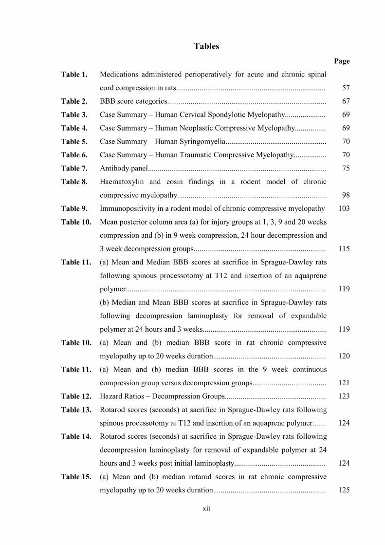
Tables
Page
Table 1. Medications administered perioperatively for acute and chronic spinal
cord compression in rats.............................................................................
57
Table 2. BBB score categories.................................................................................. 67
Table 3. Case Summary – Human Cervical Spondylotic Myelopathy..................... 69
Table 4. Case Summary – Human Neoplastic Compressive Myelopathy................ 69
Table 5. Case Summary – Human Syringomyelia.................................................... 70
Table 6. Case Summary – Human Traumatic Compressive Myelopathy................. 70
Table 7. Antibody panel............................................................................................ 75
Table 8. Haematoxylin and eosin findings in a rodent model of chronic
compressive myelopathy.............................................................................
98
Table 9. Immunopositivity in a rodent model of chronic compressive myelopathy 103
Table 10. Mean posterior column area (a) for injury groups at 1, 3, 9 and 20 weeks
compression and (b) in 9 week compression, 24 hour decompression and
3 week decompression groups....................................................................
115
Table 11. (a) Mean and Median BBB scores at sacrifice in Sprague-Dawley rats
following spinous processotomy at T12 and insertion of an aquaprene
polymer.......................................................................................................
119
(b) Median and Mean BBB scores at sacrifice in Sprague-Dawley rats
following decompression laminoplasty for removal of expandable
polymer at 24 hours and 3 weeks................................................................
119
Table 10. (a) Mean and (b) median BBB score in rat chronic compressive
myelopathy up to 20 weeks duration..........................................................
120
Table 11. (a) Mean and (b) median BBB scores in the 9 week continuous
compression group versus decompression groups......................................
121
Table 12. Hazard Ratios – Decompression Groups.................................................... 123
Table 13. Rotarod scores (seconds) at sacrifice in Sprague-Dawley rats following
spinous processotomy at T12 and insertion of an aquaprene polymer.......
124
Table 14. Rotarod scores (seconds) at sacrifice in Sprague-Dawley rats following
decompression laminoplasty for removal of expandable polymer at 24
hours and 3 weeks post initial laminoplasty...............................................
124
Table 15. (a) Mean and (b) median rotarod scores in rat chronic compressive
myelopathy up to 20 weeks duration..........................................................
125
xiii
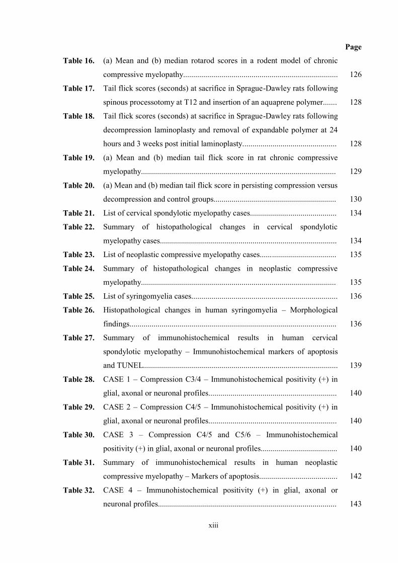
Page
Table 16. (a) Mean and (b) median rotarod scores in a rodent model of chronic
compressive myelopathy.............................................................................
126
Table 17. Tail flick scores (seconds) at sacrifice in Sprague-Dawley rats following
spinous processotomy at T12 and insertion of an aquaprene polymer.......
128
Table 18. Tail flick scores (seconds) at sacrifice in Sprague-Dawley rats following
decompression laminoplasty and removal of expandable polymer at 24
hours and 3 weeks post initial laminoplasty...............................................
128
Table 19. (a) Mean and (b) median tail flick score in rat chronic compressive
myelopathy.................................................................................................
129
Table 20. (a) Mean and (b) median tail flick score in persisting compression versus
decompression and control groups.............................................................
130
Table 21. List of cervical spondylotic myelopathy cases........................................... 134
Table 22. Summary of histopathological changes in cervical spondylotic
myelopathy cases........................................................................................
134
Table 23. List of neoplastic compressive myelopathy cases...................................... 135
Table 24. Summary of histopathological changes in neoplastic compressive
myelopathy.................................................................................................
135
Table 25. List of syringomyelia cases......................................................................... 136
Table 26. Histopathological changes in human syringomyelia – Morphological
findings.......................................................................................................
136
Table 27. Summary of immunohistochemical results in human cervical
spondylotic myelopathy – Immunohistochemical markers of apoptosis
and TUNEL.................................................................................................
139
Table 28. CASE 1 – Compression C3/4 – Immunohistochemical positivity (+) in
glial, axonal or neuronal profiles................................................................
140
Table 29. CASE 2 – Compression C4/5 – Immunohistochemical positivity (+) in
glial, axonal or neuronal profiles................................................................
140
Table 30. CASE 3 – Compression C4/5 and C5/6 – Immunohistochemical
positivity (+) in glial, axonal or neuronal profiles......................................
140
Table 31. Summary of immunohistochemical results in human neoplastic
compressive myelopathy – Markers of apoptosis.......................................
142
Table 32. CASE 4 – Immunohistochemical positivity (+) in glial, axonal or
neuronal profiles.........................................................................................
143

xiv
Page
Table 33. CASE 5 – Immunohistochemical positivity (+) in glial, axonal or
neuronal profiles.........................................................................................
143
Table 34. CASE 6 – Immunohistochemical positivity (+) in glial, axonal or
neuronal profiles.........................................................................................
143
Table 35. CASE 7 – Immunohistochemical positivity (+) in glial, axonal or
neuronal profiles.........................................................................................
144
Table 36. CASE 8 – Immunohistochemical positivity (+) in glial, axonal or
neuronal profiles.........................................................................................
144
Table 37. CASE 9 – Immunohistochemical positivity (+) in glial, axonal or
neuronal profiles.........................................................................................
145
Table 38. CASE 10 – Immunohistochemical positivity (+) in glial, axonal or
neuronal profiles.........................................................................................
145
Table 39. Immunohistochemical staining using a panel of markers of apoptosis in
human syringomyelia – Morphological findings........................................
146
Table 40. CASE 19 – Syringomyelia series – Immunohistochemical positivity (+)
in glial, axonal or neuronal profiles............................................................
147
Table 41. CASE 20 – Syringomyelia series – Immunohistochemical positivity (+)
in glial, axonal or neuronal profiles............................................................
147
Table 42. CASE 21 – Syringomyelia series – Immunohistochemical positivity (+)
in glial, axonal or neuronal profiles............................................................
147
Table 43. Immunohistochemical and histopathological findings in normal human
spinal cord cases. .......................................................................................
148
Table 44. Summary of immunohistochemical results in human cervical
spondylotic myelopathy – APP, Caspase-3, CMAP and Amyloid-beta
antibodies....................................................................................................
149
Table 45. CASE 1 – Compression C3/4 – Immunohistochemical positivity (+) in
glial, axonal or neuronal profiles................................................................
154
Table 46. CASE 2 – Compression C4/5 – Immunohistochemical positivity (+) in
glial, axonal or neuronal profiles................................................................
154
Table 47. CASE 3 – Compression C4/5 and C5/6 – Immunohistochemical
positivity (+) in glial, axonal or neuronal profiles......................................
154
xv
Page
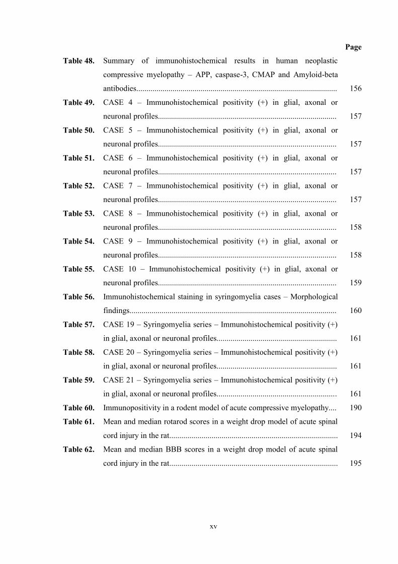
Table 48. Summary of immunohistochemical results in human neoplastic
compressive myelopathy – APP, caspase-3, CMAP and Amyloid-beta
antibodies....................................................................................................
156
Table 49. CASE 4 – Immunohistochemical positivity (+) in glial, axonal or
neuronal profiles.........................................................................................
157
Table 50. CASE 5 – Immunohistochemical positivity (+) in glial, axonal or
neuronal profiles.........................................................................................
157
Table 51. CASE 6 – Immunohistochemical positivity (+) in glial, axonal or
neuronal profiles.........................................................................................
157
Table 52. CASE 7 – Immunohistochemical positivity (+) in glial, axonal or
neuronal profiles.........................................................................................
157
Table 53. CASE 8 – Immunohistochemical positivity (+) in glial, axonal or
neuronal profiles.........................................................................................
158
Table 54. CASE 9 – Immunohistochemical positivity (+) in glial, axonal or
neuronal profiles.........................................................................................
158
Table 55. CASE 10 – Immunohistochemical positivity (+) in glial, axonal or
neuronal profiles.........................................................................................
159
Table 56. Immunohistochemical staining in syringomyelia cases – Morphological
findings.......................................................................................................
160
Table 57. CASE 19 – Syringomyelia series – Immunohistochemical positivity (+)
in glial, axonal or neuronal profiles............................................................
161
Table 58. CASE 20 – Syringomyelia series – Immunohistochemical positivity (+)
in glial, axonal or neuronal profiles............................................................
161
Table 59. CASE 21 – Syringomyelia series – Immunohistochemical positivity (+)
in glial, axonal or neuronal profiles............................................................
161
Table 60. Immunopositivity in a rodent model of acute compressive myelopathy.... 190
Table 61. Mean and median rotarod scores in a weight drop model of acute spinal
cord injury in the rat....................................................................................
194
Table 62. Mean and median BBB scores in a weight drop model of acute spinal
cord injury in the rat....................................................................................
195
xvi
Page
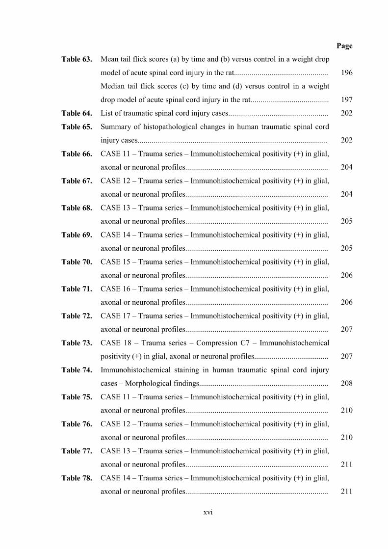
Table 63. Mean tail flick scores (a) by time and (b) versus control in a weight drop
model of acute spinal cord injury in the rat................................................
196
Median tail flick scores (c) by time and (d) versus control in a weight
drop model of acute spinal cord injury in the rat........................................
197
Table 64. List of traumatic spinal cord injury cases................................................... 202
Table 65. Summary of histopathological changes in human traumatic spinal cord
injury cases.................................................................................................
202
Table 66. CASE 11 – Trauma series – Immunohistochemical positivity (+) in glial,
axonal or neuronal profiles.........................................................................
204
Table 67. CASE 12 – Trauma series – Immunohistochemical positivity (+) in glial,
axonal or neuronal profiles.........................................................................
204
Table 68. CASE 13 – Trauma series – Immunohistochemical positivity (+) in glial,
axonal or neuronal profiles.........................................................................
205
Table 69. CASE 14 – Trauma series – Immunohistochemical positivity (+) in glial,
axonal or neuronal profiles.........................................................................
205
Table 70. CASE 15 – Trauma series – Immunohistochemical positivity (+) in glial,
axonal or neuronal profiles.........................................................................
206
Table 71. CASE 16 – Trauma series – Immunohistochemical positivity (+) in glial,
axonal or neuronal profiles.........................................................................
206
Table 72. CASE 17 – Trauma series – Immunohistochemical positivity (+) in glial,
axonal or neuronal profiles.........................................................................
207
Table 73. CASE 18 – Trauma series – Compression C7 – Immunohistochemical
positivity (+) in glial, axonal or neuronal profiles......................................
207
Table 74. Immunohistochemical staining in human traumatic spinal cord injury
cases – Morphological findings..................................................................
208
Table 75. CASE 11 – Trauma series – Immunohistochemical positivity (+) in glial,
axonal or neuronal profiles.........................................................................
210
Table 76. CASE 12 – Trauma series – Immunohistochemical positivity (+) in glial,
axonal or neuronal profiles.........................................................................
210
Table 77. CASE 13 – Trauma series – Immunohistochemical positivity (+) in glial,
axonal or neuronal profiles.........................................................................
211
Table 78. CASE 14 – Trauma series – Immunohistochemical positivity (+) in glial,
axonal or neuronal profiles.........................................................................
211

xvii
Page
Table 79. CASE 15 – Trauma series – Immunohistochemical positivity (+) in glial,
axonal or neuronal profiles.........................................................................
212
Table 80. CASE 16 – Trauma series – Immunohistochemical positivity (+) in glial,
axonal or neuronal profiles.........................................................................
212
Table 81. CASE 17 – Trauma series – Immunohistochemical positivity (+) in glial,
axonal or neuronal profiles.........................................................................
213
Table 82. CASE 18 – Trauma series – Compression C7 – Immunohistochemical
positivity (+) in glial, axonal or neuronal profiles......................................
213
Table 83. Immunohistochemistry human traumatic spinal cord injury –
Morphological findings..............................................................................
214

xviii
ABSTRACT
Evidence suggests that apoptosis of neurons and glia may play an important role in the
pathophysiology and functional outcome of spinal cord compression. In the current thesis,
chronic and acute rodent experimental models analysed the functional, cellular and
apoptotic marker changes produced by compression and subsequent surgical
decompression.
In experimental mild chronic compression there was a loss of posterior white matter
maximal at the compression site. Total cross-sectional area decreased with a longer
duration of compression (3 weeks) but resolved with decompression (e.g. 3 week group
mean 3.05mm2 increasing following decompression at 3 weeks to 5.75 mm2). A significant
increase in posterior white matter area was found above and below the site at 3 weeks.
Caspase-9, PARP, AIF and active caspase-3 staining was found in glia at, above and below
the site in all groups. Caspase-3 was greater expressed in the 24 hour (mean 0.32, p = 0.01)
and 3 week (mean 0.31, p = 0.02) decompression groups when compared with the 9 week
compression group (mean 0.19). APP axonal immunopositivity was frequently seen after
decompression.
Following experimental acute compression, central necrosis was seen, surrounded by
axonal swellings and inflammatory infiltrate. Glial positivity using TUNEL occurred at 24
hours and 1 week post-injury. PARP, DNA-PKcs and AIF immunopositivity occurred in
glia at, above and below the site. APP immunopositivity was present in axonal swellings.
In human chronic compression, axonal swellings, loss of anterior horn cells, and cystic
change were seen in severe cases. TUNEL, DNA-PKcs, PARP and AIF immunopositivity
in glia wereas seen at, above and below the compression. APP immunopositivity was seen
in axonal swellings.
In human acute compression, the central cord showed haemorrhagic necrosis and
inflammatory cells. TUNEL, DNA-PKcs and PARP immunopositive glia were found at,
above and below the site. Axonal swellings, a subset of which were APP immunopositive,

xix
occurred in the penumbra. APP immunopositive axonal swellings were found above and
below the site of compression, indicating widespread changes in fast axoplasmic transport.
We conclude that mild, chronic, fixed posterior compression results in a potentially
reversible reduction of white matter at the site and increased white matter above and below
the site of compression. This, combined with evidence of axonal injury, may indicate
altered axoplasmic transport. Decompressive surgery results in increased immunostaining
for apoptotic markers and increased axonal injury despite restoration of spinal cord
anatomy. These studies provide novel evidence that neuronal and glial apoptosis occurs in
acute and chronic compressive myelopathy at various time points of compression, maximal
at the site of injury.