A glyphosate-based herbicide induces necrosis and apoptosis in ...
11
A glyphosate-based herbicide induces necrosis and apoptosis in mature rat testicular cells in vitro, and testosterone decrease at lower levels Émilie Clair a,b , Robin Mesnage a,b , Carine Travert a , Gilles-Éric Séralini a,b,⇑ a Université de Caen Basse-Normandie, EA2608, Institute of Biology, Esplanade de la Paix, 14032 Caen Cedex, France b Université de Caen Basse-Normandie, Risk Pole MRSH-CNRS, and CRIIGEN, 40 rue de Monceau, 75008 Paris, France article info Article history: Received 23 June 2011 Accepted 9 December 2011 Available online 19 December 2011 Keywords: Roundup Glyphosate Testicular cells Cytotoxicity Endocrine disruption abstract The major herbicide used worldwide, Roundup, is a glyphosate-based pesticide with adjuvants. Glyphos- ate, its active ingredient in plants and its main metabolite (AMPA) are among the first contaminants of surface waters. Roundup is being used increasingly in particular on genetically modified plants grown for food and feed that contain its residues. Here we tested glyphosate and its formulation on mature rat fresh testicular cells from 1 to 10000 ppm, thus from the range in some human urine and in environ- ment to agricultural levels. We show that from 1 to 48 h of Roundup exposure Leydig cells are damaged. Within 24–48 h this formulation is also toxic on the other cells, mainly by necrosis, by contrast to gly- phosate alone which is essentially toxic on Sertoli cells. Later, it also induces apoptosis at higher doses in germ cells and in Sertoli/germ cells co-cultures. At lower non toxic concentrations of Roundup and gly- phosate (1 ppm), the main endocrine disruption is a testosterone decrease by 35%. The pesticide has thus an endocrine impact at very low environmental doses, but only a high contamination appears to provoke an acute rat testicular toxicity. This does not anticipate the chronic toxicity which is insufficiently tested, and only with glyphosate in regulatory tests. Ó 2011 Elsevier Ltd. All rights reserved. 1. Introduction An environmentally-linked syndrome called testicular dysgene- sis has emerged (Bay et al., 2006; Skakkebaek et al., 2001). It in- cludes a decrease in sperm quantity and quality (Auger et al., 1995; Carlsen et al., 1992), an increase in congenital malformations such as cryptorchidism and hypospadias (Toppari et al., 2010), and a preoccupying increase of testicular cancer incidence (Bergstrom et al., 1996). This indicates that the testis is a sensitive target for xenobiotics. The food/water/air intake of xenobiotics in the young as well as in adults may lead to endocrine disruption at a reproduc- tive and more specifically testicular level (Anway et al., 2006; Savitz et al., 1997). In vitro, ex vivo and in vivo experiments are nec- essary approaches to help us understand the mechanisms of xeno- biotics actions at a developmental and/or adult stage. In this work, we have chosen to test one of the most used pesti- cides round the world. Roundup (R) formulations are non selective herbicides composed of mixtures of glyphosate (G) and adjuvants such as polyoxyethylene tallowamine (POEA) (Benachour et al., 2007b). These compounds, with the G metabolite aminomethyl- phosphonic acid (AMPA), are major contaminants in surface waters with levels reaching for instance 24 ppb for G in groundwater (IFEN, 2007). Moreover, these residues also concentrate in approximately 80% genetically modified plants grown for food and feed, which are rendered R tolerant, up to 400 ppm (maximal residual levels, U.S. EPA, 1998). We tested here R from 1 ppm to agricultural working dilutions on rat testicular cells. It is known that G is a weed killer inhibiting the shikimic acid pathway in plants, essential for aromatic amino acids synthesis, and it penetrates and is stabilized in the cells with the help of the adjuvants (Cox, 1998, 2004). R used in our study contains 360 g/l of G and various xenobiotics added as adjuvants. Therefore R is a good model to study in vitro combined effects, and especially synergistic ones for xenobiotics (Benachour et al., 2007a; Richard et al., 2005). In fact, G and/or R can also induce mortality in human cells, revealed by disruptions of mitochondrial succinate dehydro- genase, caspases 3/7, and adenylate kinase (Benachour and Séralin- i, 2009). R is even responsible for oxidative damage in human epidermal cells (Gehin et al., 2005). G and/or R also have side tar- gets in mammals such as cytochrome P450 reductase, StAR, aroma- tase and sexual steroid receptors of cells involved in reproduction or in transfected human cells (Gasnier et al., 2009; Richard et al., 2005; Stocco et al., 1995; Walsh et al., 2000). In mammals, and rats in particular, the respiratory and hepatic systems (Adam et al., 1997; Beuret et al., 2005) can be altered by this herbicide, as well as hepatobiliary and reproductive functions including sperm production or libido, and even fetal development 0887-2333/$ - see front matter Ó 2011 Elsevier Ltd. All rights reserved. doi:10.1016/j.tiv.2011.12.009 ⇑ Corresponding author at: Université de Caen Basse-Normandie, EA2608, Institute of Biology, Esplanade de la Paix, 14032 Caen Cedex, France. Tel.: +33 (0) 2 31 56 54 89; fax: +33 (0) 2 31 56 53 20. E-mail address: [email protected] (G-É. Séralini). Toxicology in Vitro 26 (2012) 269–279 Contents lists available at SciVerse ScienceDirect Toxicology in Vitro journal homepage: www.elsevier.com/locate/toxinvit
Transcript of A glyphosate-based herbicide induces necrosis and apoptosis in ...
Toxicology in Vitro 26 (2012) 269–279
Contents lists available at SciVerse ScienceDirect
Toxicology in Vitro
journal homepage: www.elsevier .com/locate / toxinvi t
A glyphosate-based herbicide induces necrosis and apoptosis in mature rattesticular cells in vitro, and testosterone decrease at lower levels
Émilie Clair a,b, Robin Mesnage a,b, Carine Travert a, Gilles-Éric Séralini a,b,⇑a Université de Caen Basse-Normandie, EA2608, Institute of Biology, Esplanade de la Paix, 14032 Caen Cedex, Franceb Université de Caen Basse-Normandie, Risk Pole MRSH-CNRS, and CRIIGEN, 40 rue de Monceau, 75008 Paris, France
a r t i c l e i n f o a b s t r a c t
Article history:Received 23 June 2011Accepted 9 December 2011Available online 19 December 2011
Keywords:RoundupGlyphosateTesticular cellsCytotoxicityEndocrine disruption
0887-2333/$ - see front matter � 2011 Elsevier Ltd. Adoi:10.1016/j.tiv.2011.12.009
⇑ Corresponding author at: Université de CaenInstitute of Biology, Esplanade de la Paix, 14032 Caen2 31 56 54 89; fax: +33 (0) 2 31 56 53 20.
E-mail address: [email protected] (G-É. Séralini).
The major herbicide used worldwide, Roundup, is a glyphosate-based pesticide with adjuvants. Glyphos-ate, its active ingredient in plants and its main metabolite (AMPA) are among the first contaminants ofsurface waters. Roundup is being used increasingly in particular on genetically modified plants grownfor food and feed that contain its residues. Here we tested glyphosate and its formulation on maturerat fresh testicular cells from 1 to 10000 ppm, thus from the range in some human urine and in environ-ment to agricultural levels. We show that from 1 to 48 h of Roundup exposure Leydig cells are damaged.Within 24–48 h this formulation is also toxic on the other cells, mainly by necrosis, by contrast to gly-phosate alone which is essentially toxic on Sertoli cells. Later, it also induces apoptosis at higher dosesin germ cells and in Sertoli/germ cells co-cultures. At lower non toxic concentrations of Roundup and gly-phosate (1 ppm), the main endocrine disruption is a testosterone decrease by 35%. The pesticide has thusan endocrine impact at very low environmental doses, but only a high contamination appears to provokean acute rat testicular toxicity. This does not anticipate the chronic toxicity which is insufficiently tested,and only with glyphosate in regulatory tests.
� 2011 Elsevier Ltd. All rights reserved.
1. Introduction
An environmentally-linked syndrome called testicular dysgene-sis has emerged (Bay et al., 2006; Skakkebaek et al., 2001). It in-cludes a decrease in sperm quantity and quality (Auger et al.,1995; Carlsen et al., 1992), an increase in congenital malformationssuch as cryptorchidism and hypospadias (Toppari et al., 2010), anda preoccupying increase of testicular cancer incidence (Bergstromet al., 1996). This indicates that the testis is a sensitive target forxenobiotics. The food/water/air intake of xenobiotics in the youngas well as in adults may lead to endocrine disruption at a reproduc-tive and more specifically testicular level (Anway et al., 2006;Savitz et al., 1997). In vitro, ex vivo and in vivo experiments are nec-essary approaches to help us understand the mechanisms of xeno-biotics actions at a developmental and/or adult stage.
In this work, we have chosen to test one of the most used pesti-cides round the world. Roundup (R) formulations are non selectiveherbicides composed of mixtures of glyphosate (G) and adjuvantssuch as polyoxyethylene tallowamine (POEA) (Benachour et al.,2007b). These compounds, with the G metabolite aminomethyl-phosphonic acid (AMPA), are major contaminants in surface waters
ll rights reserved.
Basse-Normandie, EA2608,Cedex, France. Tel.: +33 (0)
with levels reaching for instance 24 ppb for G in groundwater (IFEN,2007). Moreover, these residues also concentrate in approximately80% genetically modified plants grown for food and feed, which arerendered R tolerant, up to 400 ppm (maximal residual levels, U.S.EPA, 1998). We tested here R from 1 ppm to agricultural workingdilutions on rat testicular cells.
It is known that G is a weed killer inhibiting the shikimic acidpathway in plants, essential for aromatic amino acids synthesis,and it penetrates and is stabilized in the cells with the help ofthe adjuvants (Cox, 1998, 2004). R used in our study contains360 g/l of G and various xenobiotics added as adjuvants. ThereforeR is a good model to study in vitro combined effects, and especiallysynergistic ones for xenobiotics (Benachour et al., 2007a; Richardet al., 2005). In fact, G and/or R can also induce mortality in humancells, revealed by disruptions of mitochondrial succinate dehydro-genase, caspases 3/7, and adenylate kinase (Benachour and Séralin-i, 2009). R is even responsible for oxidative damage in humanepidermal cells (Gehin et al., 2005). G and/or R also have side tar-gets in mammals such as cytochrome P450 reductase, StAR, aroma-tase and sexual steroid receptors of cells involved in reproductionor in transfected human cells (Gasnier et al., 2009; Richard et al.,2005; Stocco et al., 1995; Walsh et al., 2000).
In mammals, and rats in particular, the respiratory and hepaticsystems (Adam et al., 1997; Beuret et al., 2005) can be altered bythis herbicide, as well as hepatobiliary and reproductive functionsincluding sperm production or libido, and even fetal development
270 É. Clair et al. / Toxicology in Vitro 26 (2012) 269–279
(Chan and Mahler, 1992; Dallegrave et al., 2003, 2007; Yousef et al.,1995). Therefore its impact in mammalian reproduction is docu-mented, but not its direct possible testicular impact nor the mech-anism of action or the sensitivity of adult gonadal cells.
R contamination may come from air (dermal or pulmonary dur-ing spraying), water, feed and food. At present few studies havebeen conducted to know the tissue concentration of G after expo-sure and its possible bioaccumulation. However it was reportedthat occupational exposure, primarily via the oral route, resultsin urinary concentrations of G in the order of ppm (Acquavellaet al., 2004), in the range of our lower concentrations tested(1 ppm of R corresponding to 0.36 ppm of G). More recently, itwas observed that after oral administration to rats of 10 ppm ofG, 30% were absorbed in males and 36% in females, with a peak ob-served 2 h after administration. If the majority of G (90% after 72 h)appears to be excreted via the feces or urine before metabolism,this does not exclude the bioaccumulation in some tissues. Onepercent of G persists after 7 days, located in particular in the colonbut also primarily in bone (Brewster et al., 1991). It is known that Gcan bind to calcium ions, this occurs also in the soil (Sprankle et al.,1975). More recently it has been shown that after oral ingestion of10 ppm of the herbicide, it diffuses in mammalian tissues, with ahalf-life of 15 h in rats, and G is then found in plasma at 5 ppm(Anadon et al., 2009).
Typically these assays are performed after administration of asingle or a few doses of G in a short time, and this does not quantifythe bioaccumulation in the body of a long-term real environmentalexposure by air, water, food or feed, like through consumption ofRoundup-tolerant edible plants, such as most agricultural geneti-cally modified organisms. In addition, only the active ingredientin plants is well studied thus there is little information availableon its metabolites, and regarding R adjuvants and theirtoxicokinetics.
To date, very few studies have been conducted on R effects onprimary cells; one study in particular was conducted by our groupon human umbilical cord cells (Benachour and Séralini, 2009),where we demonstrated the necrotic and apoptotic capacities ofR at environmental levels. Consequently, in this work, we mea-sured the differential specificities of R and G actions on adult ratfreshly separated testicular cells in order to know the thresholdof toxicity. These are Leydig, Sertoli, Sertoli and germ cells, andgerm cells alone. An increased mortality of these cells or a disrup-tion in enzyme or hormone production could lead to a deleteriouseffect on reproduction. Necrosis and apoptosis were assayed atsub-agricultural dilutions of the herbicide R, and G, its compoundwithout adjuvants, and the endocrine disruption was tested atnon cytotoxic levels from 1 ppm. This work is the first study ofthe side effects of the main herbicide of the world on primary tes-ticular mammalian cells.
2. Materials and methods
2.1. Animals
Healthy adult male albino Sprague–Dawley rats (70 days ± 5)were obtained from Janvier (Le Genest-Saint-Isle, France) or fromthe University Center of Biological Resources (Caen, France), andwere maintained on a 12 h light/dark cycle at 20–22 �C. Standardfood and water were provided to the animals ad libitum.
2.2. Chemicals
Dulbecco’s Modified Eagle’s Medium (DMEM) and Ham F12were purchased from PAN (Biotech GmbH, Dutscher, Brumath,France); and collagenase/dispase from Vibrio alginolyticus/Bacillus
polymyxa was from Roche (Mannheim, Germany). Soybean trypsininhibitor (STI), deoxyribonuclease I from bovine pancreas (DNAseI), glyphosate (G) and serum replacement 3 were purchased fromSigma–Aldrich (Saint-Quentin Fallavier, France). The 40,60-Di Ami-dino-2-PhenylIndole (DAPI) was from Lonza (Verviers, Belgium).Percoll was from GE Healthcare (Saclay, France). All other reagentswere of analytical grade. The herbicide R Bioforce� containing360 g/l of acid G (R, homologation 9800036 corresponding to100%) is a commercial formulation. Solutions of G (2% or 7.2 g/lof G final) and R Bioforce� (diluted also to 2% final) were preparedin DMEM/Ham F12 medium and adjusted to pH 7.4 and serially di-luted in the same medium.
2.3. Isolation, purification and culture of Leydig cells
The rats were sacrificed and the testes were quickly decapsulat-ed and placed in DMEM/Ham F12 nutrient medium (1:1, v/v). Thecrude interstitial cells were separated from seminiferous tubulesby incubation in a medium containing collagenase/dispase(0.05%), STI (0.005%), and DNase I (0.001%) at 32 �C for 15 min ina shaking water bath, followed by several decantations and a filtra-tion through 30-mesh nylon. The Leydig cells were purified on adiscontinuous gradient of Percoll (20–80%) prepared in mediumas previously described (Lefevre et al., 1983). Leydig cell fractionswere collected, washed with the medium and their purity wasappreciated by histochemical staining for the specific 3b-hydroxy-steroid dehydrogenase activity; 85–90% of positive cells were la-beled. Leydig cells viability was determined by Trypan blueexclusion test and was near 90%. After purification, Leydig cellswere maintained in DMEM/Ham F12 nutrient medium (1:1, v/v)at 32 �C (5% CO2, 95% air) with or without hCG, human homologof LH physiologically involved in endocrine regulation of Leydigcells.
2.4. Isolation, purification and culture of Sertoli and germ cells
Sertoli and germ cells were isolated from the same rat testes bythree enzymatic digestions on pellets obtained after previously de-scribed decantations. These pellets contain seminiferous tubules.Briefly, after the first enzymatic digestion described above (32 �C,15 min), a second one was performed in the same conditions dur-ing 30 min. The third and last one was in a solution with 0.1% hyal-uronidase and 0.005% STI at 37 �C for 30 min. After centrifugation(900 rpm, 2 min), pellets contained Sertoli and germ cells. A secondcentrifugation (2500 rpm, 10 min) was necessary to isolate germcells. Around 106 germ cells/well were seeded in 96-wells platesbefore treatments. The Sertoli and germ cells mixture was at a den-sity of 2 � 106 cells/well in the same plates and cultured for 48 h inHam’s F12/DMEM medium (1:1, v/v) supplemented with serumreplacement 3 at 32 �C (5% CO2 and 95% air). On day 3, to obtainpurified Sertoli cells cultures when necessary, germ cells were re-moved with an osmotic shock using a 20 mM Tris–HCl solution (pH7.2). Treatments with different dilutions were applied on day 5 onSertoli cells.
2.5. Adenylate kinase measurement
The bioluminescent ToxiLightTM bioassay (Lonza, Verviers,Belgium), developed by Crouch et al. (1993), is a non-destructiveenzymatic bioassay. It measures quantitatively the luminescenceof adenylate kinase (AK) of mammalian injured cells in culture(Crouch et al., 1993). The AK is a membranous enzyme present inall eukaryotic cells, and is released into culture medium when cellsare damaged (the membrane integrity is disrupted during necrosisor secondary necrosis that occurs as a result of apoptosis). The AKrelease in the medium converts ATP from ADP, which is then
É. Clair et al. / Toxicology in Vitro 26 (2012) 269–279 271
measured on a luminometer (Mithras LB 940, Berthold, Thoiry,France); the bioluminescent reaction is produced by luciferase.When the cytolysis increases, AK increases in the supernatants,resulting in higher light intensity.
Before the assay, 105 cells per well in 96-well plates (Dutsher,Brumath, France) or 3 � 105 cells per well in 24-well plates(Dutsher, Brumath, France) were treated with different dilutionsof R or G ± 1 UI/ml of hCG during 3, 6, 9, 12, 18, 24 or 48 h. Theadenylate kinase detection reagent (AKDR) was prepared in a buf-fer (5 g/10 ml). Then 50 ll of supernatant were transferred to anopaque black 96-well plate. Fifty microliter of AKDR reagent weredeposited into each well. The plates were then placed under agita-tion for 15 min in the dark, and light was measured using theluminometer.
2.6. Caspase 3/7 activity
The Caspase-GloTM 3/7 assay (Promega, Paris, France) measuresthe activities, in 96-well white plates (GBO, Dutscher, France), ofcaspases 3 and 7, key-caspases of apoptosis, in cell cultures usinga bioluminescence-based method. The reagent contained a pro-luminescent substrate of caspases 3/7, containing the tetrapeptidesequence Z-DEVD-aminoluciferin, in a buffer including a detergentfor cell lysis and other components stabilizing caspases activities(Bondzio et al., 2008; Liu et al., 2005; Riss and Moravec, 2004).After cell lysis, the cleavage of the substrate by caspases releasedaminoluciferin, which was then able to generate a ‘‘glow-type’’luminescence, produced by luciferase related to the caspase 3/7activity in the sample. This assay was designed for automatedhigh-throughput screening of caspases 3/7 activities, specific forapoptosis.
Before the assay, 105 cells per well in 96-well white transparentbackground plate were treated with different dilution of R orG ± 1 UI/ml of hCG during 3, 6, 9, 12, 18, 24 or 48 h. The Caspase-Glo� 3/7 reagent was prepared in the buffer provided (Promega,Paris, France). After 30 min at room temperature, 50 ll of Cas-pase-Glo� 3/7 reagent was added to 50 ll of culture medium con-taining the cells previously treated in each well. After shaking theplate during 15 min, an incubation period of 45 min at room tem-perature in the dark was needed to stabilize the signal before lumi-nescence measurement with the luminometer.
2.7. Analysis of DNA in situ by DAPI
DAPI is a fluorescent stain that binds strongly to DNA after pass-ing through cell membrane. After a 24 h incubation in presence ofvarious dilutions of G or R, 24-wells plates were centrifuged(900 rpm, 15 min) and the medium was removed slowly. Leydigcells (3 � 105/well) were fixed for a day in absolute ethanol–chloroform–acetic acid (6:3:1, v/v/v) at �20 �C. The wells werewashed with PBS (pH 7.4) and incubated with 1 lg/ml of a solutioncontaining DAPI during 30 min (Travert et al., 2006). Each well waswashed with water and then examined with a microscope using afluorescent mode (DMLB, Leica). Labeled DNA of viable cells wasscattered throughout the nucleus, and bright condensation of chro-matin revealed apoptotic cells (magnification 400�).
2.8. 3b-HSD activity
Leydig cells, previously prepared and seeded in 96-well platesas described above, were exposed for 24 h to different concentra-tions of R Bioforce�, or equivalent non cytotoxic concentrationsof G, in medium DMEM/HamF12 to 32 �C (5% CO2, 95% air). The3b-hydroxysteroid dehydrogenase (3b-HSD), a Leydig cell specificenzyme involved in particular in testosterone synthesis, was mea-sured at the end of the treatment. The 3b-HSD reagent containing
DHEA (substrate), NAD (cofactor), NBT and nicotinamide wasadded to wells containing Leydig cells pretreated and then incu-bated at 37 �C for 45–60 min. Once the cells are stained brown, asolution of acetic acid (10%) was added to each well to solubilizeformazan crystals previously formed. The 3b-HSD enzyme activitywas then evaluated by reading the optical density of each well at560 nm (formazan) through a plate reader (Mithras LB 940,Berthold, Thoiry, France).
2.9. Radioimmunoassay (RIA) of testosterone
The radioimmunoassay was performed on the same Leydig cellsby competition and stopped using the method of activated char-coal. Indeed, the steroid dose is in competition with its tritiatedcounterpart by incubating 200 ll of standard solution of unlabeledtestosterone (7.5–800 pg of testosterone/200 ll phosphate buffer),phosphate buffer (0.1 M Na2HPO4, 0.9% NaCl, 0.5% BSA, 0.01% NaN3
– pH 7.4) or culture supernatant with 100 ll of radioactive testos-terone (3000 cpm/100 ll phosphate buffer) and 100 ll of rabbitanti-testosterone antibody (final concentration 1/18000). After30 min at room temperature, the mixture is placed at 4 �C untilthe next day. Then 500 ll of charcoal/dextran (50%/5%) at 4 �Care added. After 10 min of incubation at 4 �C, the tubes were cen-trifuged 10 min at 2400 rpm at 4 �C and the radioactivity of thesupernatant was then counted. The sensitivity of the assay was12 pg of testosterone per tube.
2.10. Measurement of mRNA expression of aromatase, androgenreceptor and estrogen receptor a and b by real-time PCR
After exposure of Leydig cells for 24 h at different non cytotoxicconcentrations of R or G in 6-well plates cell pellets were recoveredand placed in the presence of Trizol to degrade the cells. Then chlo-roform was added to recover the aqueous phase containing theRNA. Precipitation of RNA is done by adding isopropanol and wash-ing by adding ethanol (70%). After a denaturation step (10 min at55–60 �C), the integrity of total RNA was controlled by dosing(260–280 nm) and by electrophoresis on agarose gel (1.5%) labeledby bromide ethidium. To achieve the reverse transcription (RT)250 ng of RNA were used and placed in the presence of 200 U ofMMLV-RT (Moloney murine leukemia virus reverse transcriptase),0.2 g of random primers, 500 mM of each dNTP and 20 U of recom-binant RNasin�. The samples were then placed 90 min at 37 �C toobtain the cDNA, the reaction was stopped by 5 min at 75 �C. Thepolymerase chain reaction was performed on cDNA using themethod GoTaq� qPCR Master Mix (Promega). The PCR primersused are: L19 50-GGA ATC TAA GAA GAT TGA CCG TC-30 and30-GCC TTG TCT GCC TTC AGT TT-50; aromatase 50- CGT CAT GTTGCT TCT CAT CG-30 and 30-TAC CGC AGG CTC TCG TTA AT-50; estro-gen a receptor 50-AAT TCT GAC AAT CGA CGC CAG-30 and 30-GTGCTT CAA CAT TCT CCC TCC TC-50; estrogen b receptor 50-CTT GCCCAC TTG GAA ACA TC-30 and 30-CCA AAG GTT GAT TTT ATG GCC-50; androgen receptor 50-TGG GGA CAT GCG TTT GGA CAG T-30
and 30-GCT GCC ACA AGT GAG AGC TCC G-50. The PCR conditionswere an initial step at 95 �C for 3 min, then 40 cycles of 30 s at95 �C and 60 �C for 60 s. mRNA levels of aromatase, estrogen recep-tor a and b and androgen receptor were normalized using the L19control gene.
2.11. Statistical analysis
All data are presented as the means ± Standard Errors (SEM).The experiments were repeated in triplicates on different monthsfrom three independent cultures each time (n = 9) unless other-wise specified. To compare the results, statistically significant dif-ferences from controls were determined by an Anova test followed
272 É. Clair et al. / Toxicology in Vitro 26 (2012) 269–279
by Bonferroni post-test with p < 0.001 (⁄⁄⁄⁄), p < 0.005 (⁄⁄⁄), p < 0.01(⁄⁄) and p < 0.05 (⁄).
3. Results
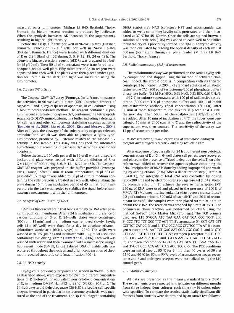
Cytotoxicities of G alone and with adjuvants (R), from 50 ppm toagricultural working dilutions (200 times more), were simulta-neously measured on 70-day isolated adult rat testicular cells: Ley-dig alone, Sertoli with germ cells, purified Sertoli, and finally germcells alone. Significant membrane degradations were provoked byR in Leydig cells within 24 h from 0.1% (1000 ppm), thus at relativelyhigh levels, however 10 times below the lowest agricultural dilu-tions (Fig. 1a). This tendency was visible very rapidly from 1 h of
Fig. 1. Effects of Roundup or Glyphosate (empty diamonds) in DMEM/Ham F12 mediumb), Sertoli with germ cells (c and d), purified Sertoli (e and f) and germ (g and h) cellsadenylate kinase activities indicating membrane degradations in primary cultures of tequivalent doses of glyphosate in DMEM/HamF12 medium in 96-wells plate at 32 �C (5%All studies for each concentration were repeated three times and in three different expecompared to controls. SEMs are shown in all instances (Anova test p < 0.001 ⁄⁄⁄⁄; p < 0.0
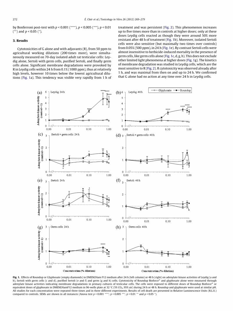
treatment and was persistent (Fig. 2). This phenomenon increasesup to five times more than in controls at higher doses; only at thesedoses Leydig cells reacted as though they were around 50% moreresistant after 48 h of treatment (Fig. 1b). Moreover, isolated Sertolicells were also sensitive (but maximally two times over controls)from 0.05% (500 ppm), in 24 h (Fig. 1e). By contrast Sertoli cells werealmost insensitive to herbicide-induced mortality in the presence ofgerm cells, like germ cells alone (Fig. 1c, d, g, h). This does not excludeother limited light phenomena at higher doses (Fig. 1g). The kineticsof membrane degradation was studied in Leydig cells, which are themost sensitive to R (Fig. 2). R cytotoxicity was observed already after1 h, and was maximal from then on and up to 24 h. We confirmedthat G alone had no action at any time over 24 h in Leydig cells.
after 24 h (left column) or 48 h (right) on adenylate kinase activities of Leydig (a and. Cytotoxicity of Roundup Bioforce� and glyphosate alone were measured throughesticular cells. The cells were exposed to different doses of Roundup Bioforce� orCO2, 95% air) during 24 h or 48 h. Roundup and glyphosate were used at similar pH.riments. Results of cell death are presented in Relative Luminescence Units (R.L.U.)05 ⁄⁄⁄; p < 0.01 ⁄⁄ and p < 0.05 ⁄).
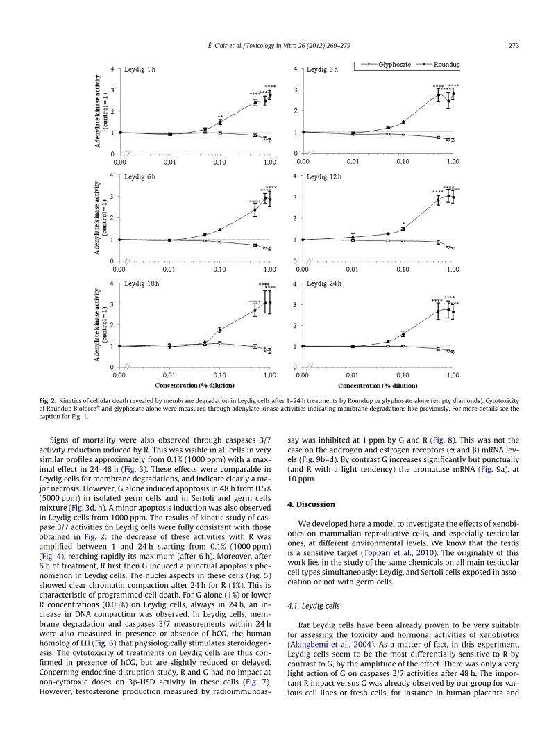
Fig. 2. Kinetics of cellular death revealed by membrane degradation in Leydig cells after 1–24 h treatments by Roundup or glyphosate alone (empty diamonds). Cytotoxicityof Roundup Bioforce� and glyphosate alone were measured through adenylate kinase activities indicating membrane degradations like previously. For more details see thecaption for Fig. 1.
É. Clair et al. / Toxicology in Vitro 26 (2012) 269–279 273
Signs of mortality were also observed through caspases 3/7activity reduction induced by R. This was visible in all cells in verysimilar profiles approximately from 0.1% (1000 ppm) with a max-imal effect in 24–48 h (Fig. 3). These effects were comparable inLeydig cells for membrane degradations, and indicate clearly a ma-jor necrosis. However, G alone induced apoptosis in 48 h from 0.5%(5000 ppm) in isolated germ cells and in Sertoli and germ cellsmixture (Fig. 3d, h). A minor apoptosis induction was also observedin Leydig cells from 1000 ppm. The results of kinetic study of cas-pase 3/7 activities on Leydig cells were fully consistent with thoseobtained in Fig. 2: the decrease of these activities with R wasamplified between 1 and 24 h starting from 0.1% (1000 ppm)(Fig. 4), reaching rapidly its maximum (after 6 h). Moreover, after6 h of treatment, R first then G induced a punctual apoptosis phe-nomenon in Leydig cells. The nuclei aspects in these cells (Fig. 5)showed clear chromatin compaction after 24 h for R (1%). This ischaracteristic of programmed cell death. For G alone (1%) or lowerR concentrations (0.05%) on Leydig cells, always in 24 h, an in-crease in DNA compaction was observed. In Leydig cells, mem-brane degradation and caspases 3/7 measurements within 24 hwere also measured in presence or absence of hCG, the humanhomolog of LH (Fig. 6) that physiologically stimulates steroidogen-esis. The cytotoxicity of treatments on Leydig cells are thus con-firmed in presence of hCG, but are slightly reduced or delayed.Concerning endocrine disruption study, R and G had no impact atnon-cytotoxic doses on 3b-HSD activity in these cells (Fig. 7).However, testosterone production measured by radioimmunoas-
say was inhibited at 1 ppm by G and R (Fig. 8). This was not thecase on the androgen and estrogen receptors (a and b) mRNA lev-els (Fig. 9b–d). By contrast G increases significantly but punctually(and R with a light tendency) the aromatase mRNA (Fig. 9a), at10 ppm.
4. Discussion
We developed here a model to investigate the effects of xenobi-otics on mammalian reproductive cells, and especially testicularones, at different environmental levels. We know that the testisis a sensitive target (Toppari et al., 2010). The originality of thiswork lies in the study of the same chemicals on all main testicularcell types simultaneously: Leydig, and Sertoli cells exposed in asso-ciation or not with germ cells.
4.1. Leydig cells
Rat Leydig cells have been already proven to be very suitablefor assessing the toxicity and hormonal activities of xenobiotics(Akingbemi et al., 2004). As a matter of fact, in this experiment,Leydig cells seem to be the most differentially sensitive to R bycontrast to G, by the amplitude of the effect. There was only a verylight action of G on caspases 3/7 activities after 48 h. The impor-tant R impact versus G was already observed by our group for var-ious cell lines or fresh cells, for instance in human placenta and
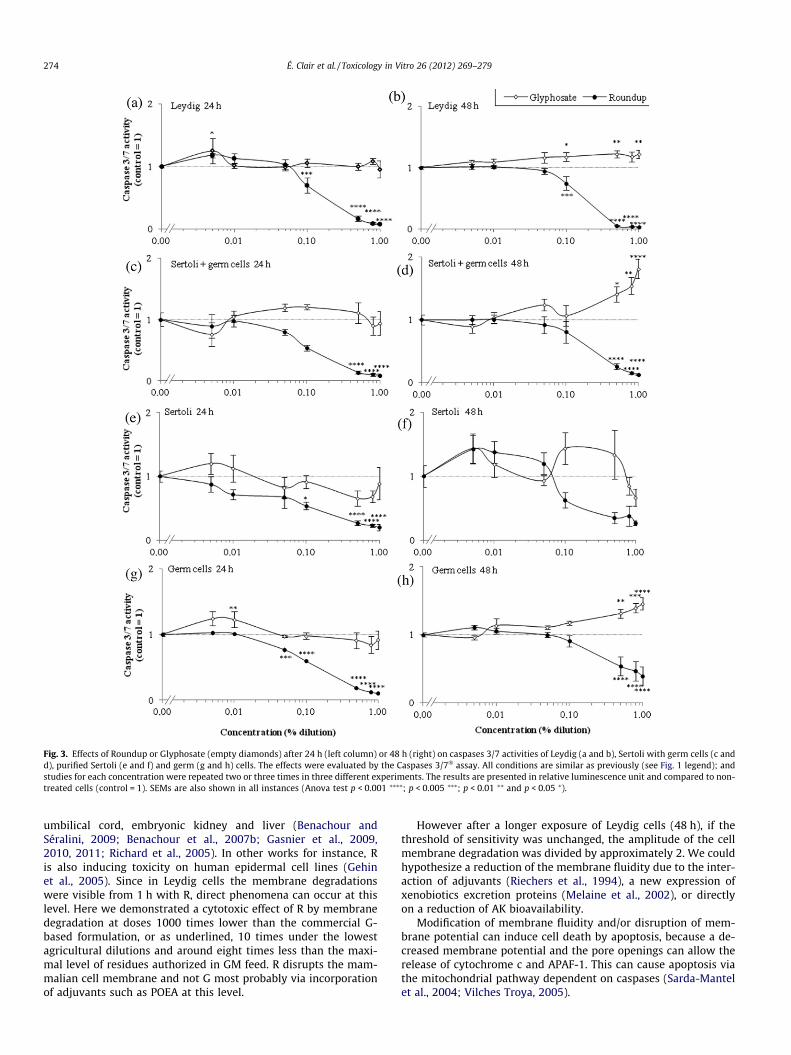
Fig. 3. Effects of Roundup or Glyphosate (empty diamonds) after 24 h (left column) or 48 h (right) on caspases 3/7 activities of Leydig (a and b), Sertoli with germ cells (c andd), purified Sertoli (e and f) and germ (g and h) cells. The effects were evaluated by the Caspases 3/7� assay. All conditions are similar as previously (see Fig. 1 legend); andstudies for each concentration were repeated two or three times in three different experiments. The results are presented in relative luminescence unit and compared to non-treated cells (control = 1). SEMs are also shown in all instances (Anova test p < 0.001 ⁄⁄⁄⁄; p < 0.005 ⁄⁄⁄; p < 0.01 ⁄⁄ and p < 0.05 ⁄).
274 É. Clair et al. / Toxicology in Vitro 26 (2012) 269–279
umbilical cord, embryonic kidney and liver (Benachour andSéralini, 2009; Benachour et al., 2007b; Gasnier et al., 2009,2010, 2011; Richard et al., 2005). In other works for instance, Ris also inducing toxicity on human epidermal cell lines (Gehinet al., 2005). Since in Leydig cells the membrane degradationswere visible from 1 h with R, direct phenomena can occur at thislevel. Here we demonstrated a cytotoxic effect of R by membranedegradation at doses 1000 times lower than the commercial G-based formulation, or as underlined, 10 times under the lowestagricultural dilutions and around eight times less than the maxi-mal level of residues authorized in GM feed. R disrupts the mam-malian cell membrane and not G most probably via incorporationof adjuvants such as POEA at this level.
However after a longer exposure of Leydig cells (48 h), if thethreshold of sensitivity was unchanged, the amplitude of the cellmembrane degradation was divided by approximately 2. We couldhypothesize a reduction of the membrane fluidity due to the inter-action of adjuvants (Riechers et al., 1994), a new expression ofxenobiotics excretion proteins (Melaine et al., 2002), or directlyon a reduction of AK bioavailability.
Modification of membrane fluidity and/or disruption of mem-brane potential can induce cell death by apoptosis, because a de-creased membrane potential and the pore openings can allow therelease of cytochrome c and APAF-1. This can cause apoptosis viathe mitochondrial pathway dependent on caspases (Sarda-Mantelet al., 2004; Vilches Troya, 2005).
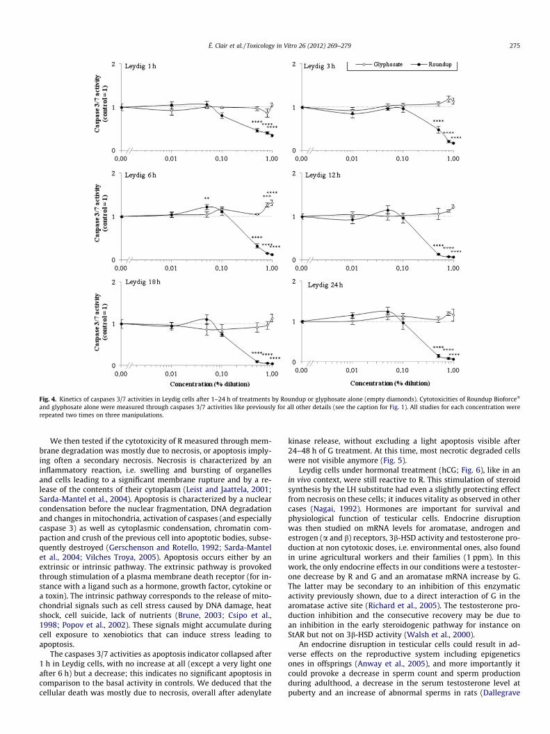
Fig. 4. Kinetics of caspases 3/7 activities in Leydig cells after 1–24 h of treatments by Roundup or glyphosate alone (empty diamonds). Cytotoxicities of Roundup Bioforce�
and glyphosate alone were measured through caspases 3/7 activities like previously for all other details (see the caption for Fig. 1). All studies for each concentration wererepeated two times on three manipulations.
É. Clair et al. / Toxicology in Vitro 26 (2012) 269–279 275
We then tested if the cytotoxicity of R measured through mem-brane degradation was mostly due to necrosis, or apoptosis imply-ing often a secondary necrosis. Necrosis is characterized by aninflammatory reaction, i.e. swelling and bursting of organellesand cells leading to a significant membrane rupture and by a re-lease of the contents of their cytoplasm (Leist and Jaattela, 2001;Sarda-Mantel et al., 2004). Apoptosis is characterized by a nuclearcondensation before the nuclear fragmentation, DNA degradationand changes in mitochondria, activation of caspases (and especiallycaspase 3) as well as cytoplasmic condensation, chromatin com-paction and crush of the previous cell into apoptotic bodies, subse-quently destroyed (Gerschenson and Rotello, 1992; Sarda-Mantelet al., 2004; Vilches Troya, 2005). Apoptosis occurs either by anextrinsic or intrinsic pathway. The extrinsic pathway is provokedthrough stimulation of a plasma membrane death receptor (for in-stance with a ligand such as a hormone, growth factor, cytokine ora toxin). The intrinsic pathway corresponds to the release of mito-chondrial signals such as cell stress caused by DNA damage, heatshock, cell suicide, lack of nutrients (Brune, 2003; Csipo et al.,1998; Popov et al., 2002). These signals might accumulate duringcell exposure to xenobiotics that can induce stress leading toapoptosis.
The caspases 3/7 activities as apoptosis indicator collapsed after1 h in Leydig cells, with no increase at all (except a very light oneafter 6 h) but a decrease; this indicates no significant apoptosis incomparison to the basal activity in controls. We deduced that thecellular death was mostly due to necrosis, overall after adenylate
kinase release, without excluding a light apoptosis visible after24–48 h of G treatment. At this time, most necrotic degraded cellswere not visible anymore (Fig. 5).
Leydig cells under hormonal treatment (hCG; Fig. 6), like in anin vivo context, were still reactive to R. This stimulation of steroidsynthesis by the LH substitute had even a slightly protecting effectfrom necrosis on these cells; it induces vitality as observed in othercases (Nagai, 1992). Hormones are important for survival andphysiological function of testicular cells. Endocrine disruptionwas then studied on mRNA levels for aromatase, androgen andestrogen (a and b) receptors, 3b-HSD activity and testosterone pro-duction at non cytotoxic doses, i.e. environmental ones, also foundin urine agricultural workers and their families (1 ppm). In thiswork, the only endocrine effects in our conditions were a testoster-one decrease by R and G and an aromatase mRNA increase by G.The latter may be secondary to an inhibition of this enzymaticactivity previously shown, due to a direct interaction of G in thearomatase active site (Richard et al., 2005). The testosterone pro-duction inhibition and the consecutive recovery may be due toan inhibition in the early steroidogenic pathway for instance onStAR but not on 3b-HSD activity (Walsh et al., 2000).
An endocrine disruption in testicular cells could result in ad-verse effects on the reproductive system including epigeneticsones in offsprings (Anway et al., 2005), and more importantly itcould provoke a decrease in sperm count and sperm productionduring adulthood, a decrease in the serum testosterone level atpuberty and an increase of abnormal sperms in rats (Dallegrave
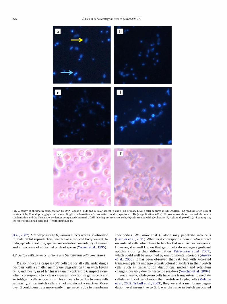
Fig. 5. Study of chromatin condensation by DAPI-labeling (a–d) and cellular aspect (e and f) on primary Leydig cells cultures in DMEM/Ham F12 medium after 24 h oftreatment by Roundup or glyphosate alone. Bright condensation of chromatin revealed apoptotic cells (magnification 400�). Yellow arrow shows normal chromatincondensation and the blue arrow evidences compacted chromatin. DAPI labeling in (a) control cells, (b) cells treated with glyphosate 1%, (c) Roundup 0.05%, (d) Roundup 1%;(e) control unstained cells and (f) with Roundup 1%.
276 É. Clair et al. / Toxicology in Vitro 26 (2012) 269–279
et al., 2007). After exposure to G, various effects were also observedin male rabbit reproductive health like a reduced body weight, li-bido, ejaculate volume, sperm concentration, osmolarity of semen,and an increase of abnormal or dead sperm (Yousef et al., 1995).
4.2. Sertoli cells, germ cells alone and Sertoli/germ cells co-cultures
R also induces a caspases 3/7 collapse for all cells, indicating anecrosis with a smaller membrane degradation than with Leydigcells, and mostly in 24 h. This is again in contrast to G impact alone,which corresponds to a clear caspases induction in germ cells andSertoli/germ cells associations. This appears to be due to germ cellssensitivity, since Sertoli cells are not significantly reactive. More-over G could penetrate more easily in germ cells due to membrane
specificities. We know that G alone may penetrate into cells(Gasnier et al., 2011). Whether it corresponds to an in vitro artifacton isolated cells which have to be checked in in vivo experiments.However, it is well known that germ cells do undergo significantapoptosis during their differentiation (Petre-Lazar et al., 2007),which could well be amplified by environmental stressors (Anwayet al., 2006). It has been observed that rats fed with R-treatedtransgenic plants undergo ultrastructural disorders in their Sertolicells, such as transcription disruptions, nuclear and reticulumchanges, possibly due to herbicide residues (Vecchio et al., 2004).
Surprisingly, while germ cells have less transporters to mediatecellular efflux of xenobiotics than Sertoli or Leydig cells (Melaineet al., 2002; Tribull et al., 2003), they were at a membrane degra-dation level insensitive to G. It was the same in Sertoli associated
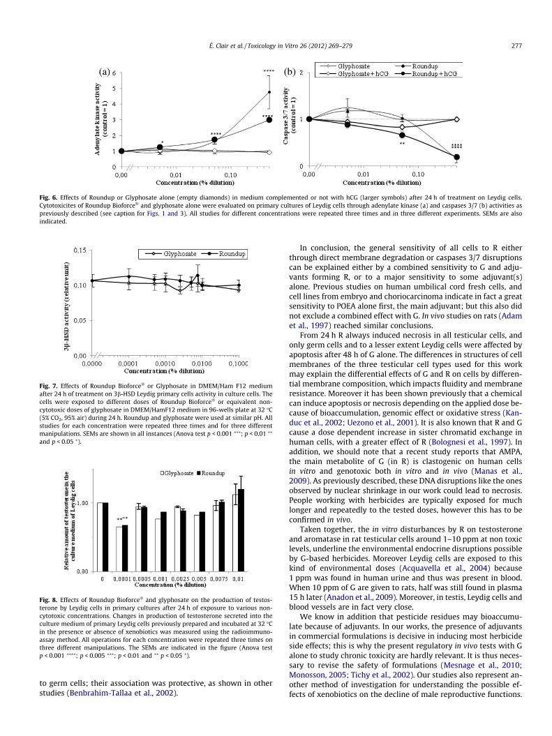
Fig. 6. Effects of Roundup or Glyphosate alone (empty diamonds) in medium complemented or not with hCG (larger symbols) after 24 h of treatment on Leydig cells.Cytotoxicites of Roundup Bioforce� and glyphosate alone were evaluated on primary cultures of Leydig cells through adenylate kinase (a) and caspases 3/7 (b) activities aspreviously described (see caption for Figs. 1 and 3). All studies for different concentrations were repeated three times and in three different experiments. SEMs are alsoindicated.
Fig. 7. Effects of Roundup Bioforce� or Glyphosate in DMEM/Ham F12 mediumafter 24 h of treatment on 3b-HSD Leydig primary cells activity in culture cells. Thecells were exposed to different doses of Roundup Bioforce� or equivalent non-cytotoxic doses of glyphosate in DMEM/HamF12 medium in 96-wells plate at 32 �C(5% CO2, 95% air) during 24 h. Roundup and glyphosate were used at similar pH. Allstudies for each concentration were repeated three times and for three differentmanipulations. SEMs are shown in all instances (Anova test p < 0.001 ⁄⁄⁄; p < 0.01 ⁄⁄
and p < 0.05 ⁄).
Fig. 8. Effects of Roundup Bioforce� and glyphosate on the production of testos-terone by Leydig cells in primary cultures after 24 h of exposure to various non-cytotoxic concentrations. Changes in production of testosterone secreted into theculture medium of primary Leydig cells previously prepared and incubated at 32 �Cin the presence or absence of xenobiotics was measured using the radioimmuno-assay method. All operations for each concentration were repeated three times onthree different manipulations. The SEMs are indicated in the figure (Anova testp < 0.001 ⁄⁄⁄⁄; p < 0.005 ⁄⁄⁄; p < 0.01 and ⁄⁄ p < 0.05 ⁄).
É. Clair et al. / Toxicology in Vitro 26 (2012) 269–279 277
to germ cells; their association was protective, as shown in otherstudies (Benbrahim-Tallaa et al., 2002).
In conclusion, the general sensitivity of all cells to R eitherthrough direct membrane degradation or caspases 3/7 disruptionscan be explained either by a combined sensitivity to G and adju-vants forming R, or to a major sensitivity to some adjuvant(s)alone. Previous studies on human umbilical cord fresh cells, andcell lines from embryo and choriocarcinoma indicate in fact a greatsensitivity to POEA alone first, the main adjuvant; but this also didnot exclude a combined effect with G. In vivo studies on rats (Adamet al., 1997) reached similar conclusions.
From 24 h R always induced necrosis in all testicular cells, andonly germ cells and to a lesser extent Leydig cells were affected byapoptosis after 48 h of G alone. The differences in structures of cellmembranes of the three testicular cell types used for this workmay explain the differential effects of G and R on cells by differen-tial membrane composition, which impacts fluidity and membraneresistance. Moreover it has been shown previously that a chemicalcan induce apoptosis or necrosis depending on the applied dose be-cause of bioaccumulation, genomic effect or oxidative stress (Kan-duc et al., 2002; Uezono et al., 2001). It is also known that R and Gcause a dose dependent increase in sister chromatid exchange inhuman cells, with a greater effect of R (Bolognesi et al., 1997). Inaddition, we should note that a recent study reports that AMPA,the main metabolite of G (in R) is clastogenic on human cellsin vitro and genotoxic both in vitro and in vivo (Manas et al.,2009). As previously described, these DNA disruptions like the onesobserved by nuclear shrinkage in our work could lead to necrosis.People working with herbicides are typically exposed for muchlonger and repeatedly to the tested doses, however this has to beconfirmed in vivo.
Taken together, the in vitro disturbances by R on testosteroneand aromatase in rat testicular cells around 1–10 ppm at non toxiclevels, underline the environmental endocrine disruptions possibleby G-based herbicides. Moreover Leydig cells are exposed to thiskind of environmental doses (Acquavella et al., 2004) because1 ppm was found in human urine and thus was present in blood.When 10 ppm of G are given to rats, half was still found in plasma15 h later (Anadon et al., 2009). Moreover, in testis, Leydig cells andblood vessels are in fact very close.
We know in addition that pesticide residues may bioaccumu-late because of adjuvants. In our works, the presence of adjuvantsin commercial formulations is decisive in inducing most herbicideside effects; this is why the present regulatory in vivo tests with Galone to study chronic toxicity are hardly relevant. It is thus neces-sary to revise the safety of formulations (Mesnage et al., 2010;Monosson, 2005; Tichy et al., 2002). Our studies also represent an-other method of investigation for understanding the possible ef-fects of xenobiotics on the decline of male reproductive functions.
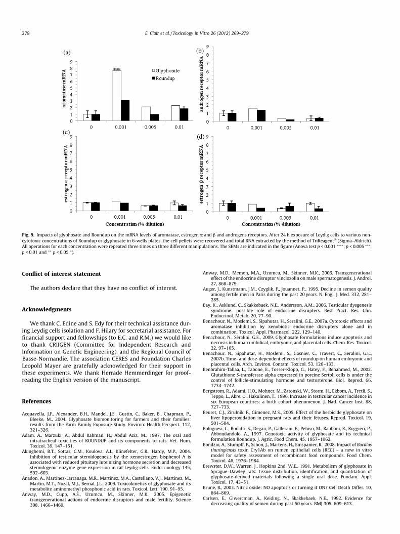
Fig. 9. Impacts of glyphosate and Roundup on the mRNA levels of aromatase, estrogen a and b and androgens receptors. After 24 h exposure of Leydig cells to various non-cytotoxic concentrations of Roundup or glyphosate in 6-wells plates, the cell pellets were recovered and total RNA extracted by the method of TriReagent� (Sigma–Aldrich).All operations for each concentration were repeated three times on three different manipulations. The SEMs are indicated in the figure (Anova test p < 0.001 ⁄⁄⁄⁄; p < 0.005 ⁄⁄⁄;p < 0.01 and ⁄⁄ p < 0.05 ⁄).
278 É. Clair et al. / Toxicology in Vitro 26 (2012) 269–279
Conflict of interest statement
The authors declare that they have no conflict of interest.
Acknowledgments
We thank C. Edine and S. Edy for their technical assistance dur-ing Leydig cells isolation and F. Hilary for secretarial assistance. Forfinancial support and fellowships (to E.C. and R.M.) we would liketo thank CRIIGEN (Committee for Independent Research andInformation on Genetic Engineering), and the Regional Council ofBasse-Normandie. The association CERES and Foundation CharlesLeopold Mayer are gratefully acknowledged for their support inthese experiments. We thank Herrade Hemmerdinger for proof-reading the English version of the manuscript.
References
Acquavella, J.F., Alexander, B.H., Mandel, J.S., Gustin, C., Baker, B., Chapman, P.,Bleeke, M., 2004. Glyphosate biomonitoring for farmers and their families:results from the Farm Family Exposure Study. Environ. Health Perspect. 112,321–326.
Adam, A., Marzuki, A., Abdul Rahman, H., Abdul Aziz, M., 1997. The oral andintratracheal toxicities of ROUNDUP and its components to rats. Vet. Hum.Toxicol. 39, 147–151.
Akingbemi, B.T., Sottas, C.M., Koulova, A.I., Klinefelter, G.R., Hardy, M.P., 2004.Inhibition of testicular steroidogenesis by the xenoestrogen bisphenol A isassociated with reduced pituitary luteinizing hormone secretion and decreasedsteroidogenic enzyme gene expression in rat Leydig cells. Endocrinology 145,592–603.
Anadon, A., Martinez-Larranaga, M.R., Martinez, M.A., Castellano, V.J., Martinez, M.,Martin, M.T., Nozal, M.J., Bernal, J.L., 2009. Toxicokinetics of glyphosate and itsmetabolite aminomethyl phosphonic acid in rats. Toxicol. Lett. 190, 91–95.
Anway, M.D., Cupp, A.S., Uzumcu, M., Skinner, M.K., 2005. Epigenetictransgenerational actions of endocrine disruptors and male fertility. Science308, 1466–1469.
Anway, M.D., Memon, M.A., Uzumcu, M., Skinner, M.K., 2006. Transgenerationaleffect of the endocrine disruptor vinclozolin on male spermatogenesis. J. Androl.27, 868–879.
Auger, J., Kunstmann, J.M., Czyglik, F., Jouannet, P., 1995. Decline in semen qualityamong fertile men in Paris during the past 20 years. N. Engl. J. Med. 332, 281–285.
Bay, K., Asklund, C., Skakkebaek, N.E., Andersson, A.M., 2006. Testicular dysgenesissyndrome: possible role of endocrine disrupters. Best Pract. Res. Clin.Endocrinol. Metab. 20, 77–90.
Benachour, N., Moslemi, S., Sipahutar, H., Seralini, G.E., 2007a. Cytotoxic effects andaromatase inhibition by xenobiotic endocrine disrupters alone and incombination. Toxicol. Appl. Pharmacol. 222, 129–140.
Benachour, N., Séralini, G.E., 2009. Glyphosate formulations induce apoptosis andnecrosis in human umbilical, embryonic, and placental cells. Chem. Res. Toxicol.22, 97–105.
Benachour, N., Sipahutar, H., Moslemi, S., Gasnier, C., Travert, C., Seralini, G.E.,2007b. Time- and dose-dependent effects of roundup on human embryonic andplacental cells. Arch. Environ. Contam. Toxicol. 53, 126–133.
Benbrahim-Tallaa, L., Tabone, E., Tosser-Klopp, G., Hatey, F., Benahmed, M., 2002.Glutathione S-transferase alpha expressed in porcine Sertoli cells is under thecontrol of follicle-stimulating hormone and testosterone. Biol. Reprod. 66,1734–1742.
Bergstrom, R., Adami, H.O., Mohner, M., Zatonski, W., Storm, H., Ekbom, A., Tretli, S.,Teppo, L., Akre, O., Hakulinen, T., 1996. Increase in testicular cancer incidence insix European countries: a birth cohort phenomenon. J. Natl. Cancer Inst. 88,727–733.
Beuret, C.J., Zirulnik, F., Gimenez, M.S., 2005. Effect of the herbicide glyphosate onliver lipoperoxidation in pregnant rats and their fetuses. Reprod. Toxicol. 19,501–504.
Bolognesi, C., Bonatti, S., Degan, P., Gallerani, E., Peluso, M., Rabboni, R., Roggieri, P.,Abbondandolo, A., 1997. Genotoxic activity of glyphosate and its technicalformulation Roundup. J. Agric. Food Chem. 45, 1957–1962.
Bondzio, A., Stumpff, F., Schon, J., Martens, H., Einspanier, R., 2008. Impact of Bacillusthuringiensis toxin Cry1Ab on rumen epithelial cells (REC) – a new in vitromodel for safety assessment of recombinant food compounds. Food Chem.Toxicol. 46, 1976–1984.
Brewster, D.W., Warren, J., Hopkins 2nd, W.E., 1991. Metabolism of glyphosate inSprague–Dawley rats: tissue distribution, identification, and quantitation ofglyphosate-derived materials following a single oral dose. Fundam. Appl.Toxicol. 17, 43–51.
Brune, B., 2003. Nitric oxide: NO apoptosis or turning it ON? Cell Death Differ. 10,864–869.
Carlsen, E., Giwercman, A., Keiding, N., Skakkebaek, N.E., 1992. Evidence fordecreasing quality of semen during past 50 years. BMJ 305, 609–613.
É. Clair et al. / Toxicology in Vitro 26 (2012) 269–279 279
Chan, P., Mahler, J., 1992. NTP technical report on the toxicity studies of Glyphosate(CAS No. 1071-83-6) Administered in Dosed Feed to F344/N Rats and B6C3F1Mice. Toxic Rep Ser 16, 1-D3.
Cox, C., 1998. Herbicide factsheet – glyphosate (Roundup). J. Pesticide Reform 18, 3–15.
Cox, C., 2004. Herbicide factsheet – glyphosate. J. Pesticide Reform 24, 10–15.Crouch, S.P., Kozlowski, R., Slater, K.J., Fletcher, J., 1993. The use of ATP
bioluminescence as a measure of cell proliferation and cytotoxicity. J.Immunol. Methods 160, 81–88.
Csipo, I., Montel, A.H., Hobbs, J.A., Morse, P.A., Brahmi, Z., 1998. Effect of Fas+ andFas� target cells on the ability of NK cells to repeatedly fragment DNA andtrigger lysis via the Fas lytic pathway. Apoptosis 3, 105–114.
Dallegrave, E., Mantese, F.D., Coelho, R.S., Pereira, J.D., Dalsenter, P.R., Langeloh, A.,2003. The teratogenic potential of the herbicide glyphosate-Roundup in Wistarrats. Toxicol. Lett. 142, 45–52.
Dallegrave, E., Mantese, F.D., Oliveira, R.T., Andrade, A.J., Dalsenter, P.R., Langeloh, A.,2007. Pre- and postnatal toxicity of the commercial glyphosate formulation inWistar rats. Arch. Toxicol. 81, 665–673.
Gasnier, C., Benachour, N., Clair, E., Travert, C., Langlois, F., Laurant, C., Decroix-Laporte,C., Seralini, G.E., 2010. Dig1 protects against cell death provoked by glyphosate-based herbicides in human liver cell lines. J. Occup. Med. Toxicol. 5, 29.
Gasnier, C., Dumont, C., Benachour, N., Clair, E., Chagnon, M.C., Seralini, G.E., 2009.Glyphosate-based herbicides are toxic and endocrine disruptors in human celllines. Toxicology 262, 184–191.
Gasnier, C., Laurant, C., Decroix-Laporte, C., Mesnage, R., Clair, E., Travert, C., Seralini,G.E., 2011. Defined plant extracts can protect human cells against combinedxenobiotic effects. J. Occup. Med. Toxicol. 6, 3.
Gehin, A., Guillaume, Y.C., Millet, J., Guyon, C., Nicod, L., 2005. Vitamins C and Ereverse effect of herbicide-induced toxicity on human epidermal cells HaCaT: abiochemometric approach. Int. J. Pharm. 288, 219–226.
Gerschenson, L.E., Rotello, R.J., 1992. Apoptosis: a different type of cell death. FASEBJ. 6, 2450–2455.
IFEN, 2007. Report on pesticides in waters. Data 2005.Kanduc, D., Mittelman, A., Serpico, R., Sinigaglia, E., Sinha, A.A., Natale, C.,
Santacroce, R., Di Corcia, M.G., Lucchese, A., Dini, L., Pani, P., Santacroce, S.,Simone, S., Bucci, R., Farber, E., 2002. Cell death: apoptosis versus necrosis(review). Int. J. Oncol. 21, 165–170.
Lefevre, A., Saez, J.M., Finaz, C., 1983. HCG responsiveness of purified Leydig cellsfrom immature and mature rats. Horm. Res. 17, 114–120.
Leist, M., Jaattela, M., 2001. Four deaths and a funeral: from caspases to alternativemechanisms. Nat. Rev. Mol. Cell Biol. 2, 589–598.
Liu, J.J., Wang, W., Dicker, D.T., El-Deiry, W.S., 2005. Bioluminescent imaging ofTRAIL-induced apoptosis through detection of caspase activation followingcleavage of DEVD-aminoluciferin. Cancer Biol. Ther. 4, 885–892.
Manas, F., Peralta, L., Raviolo, J., Garcia Ovando, H., Weyers, A., Ugnia, L., GonzalezCid, M., Larripa, I., Gorla, N., 2009. Genotoxicity of AMPA, the environmentalmetabolite of glyphosate, assessed by the Comet assay and cytogenetic tests.Ecotoxicol. Environ. Saf. 72, 834–837.
Melaine, N., Lienard, M.O., Dorval, I., Le Goascogne, C., Lejeune, H., Jegou, B., 2002.Multidrug resistance genes and p-glycoprotein in the testis of the rat, mouse,Guinea pig, and human. Biol. Reprod. 67, 1699–1707.
Mesnage, R., Clair, E., Séralini, G.-E., 2010. Roundup in genetically modified crops:regulation and toxicity in mammals. Theorie in der Ökologie 16, 31–33.
Monosson, E., 2005. Chemical mixtures: considering the evolution of toxicology andchemical assessment. Environ. Health Perspect. 113, 383–390.
Nagai, A., 1992. Study on ethane dimethane sulphonate (EDS)-inducedspermatogenic damage and protective drugs against this damage in the rat.Nippon Hinyokika Gakkai Zasshi 83, 1322–1329.
Petre-Lazar, B., Livera, G., Moreno, S.G., Trautmann, E., Duquenne, C., Hanoux, V.,Habert, R., Coffigny, H., 2007. The role of p63 in germ cell apoptosis in thedeveloping testis. J. Cell. Physiol. 210, 87–98.
Popov, S.G., Villasmil, R., Bernardi, J., Grene, E., Cardwell, J., Wu, A., Alibek, D., Bailey,C., Alibek, K., 2002. Lethal toxin of Bacillus anthracis causes apoptosis ofmacrophages. Biochem. Biophys. Res. Commun. 293, 349–355.
Richard, S., Moslemi, S., Sipahutar, H., Benachour, N., Seralini, G.E., 2005. Differentialeffects of glyphosate and roundup on human placental cells and aromatase.Environ. Health Perspect. 113, 716–720.
Riechers, D.E., Wax, L.M., Liebl, R.A., Bush, D.R., 1994. Surfactant-increasedglyphosate uptake into plasma membrane vesicles isolated from commonLambsquarters leaves. Plant Physiol. 105, 1419–1425.
Riss, T.L., Moravec, R.A., 2004. Use of multiple assay endpoints to investigate theeffects of incubation time, dose of toxin, and plating density in cell-basedcytotoxicity assays. Assay Drug Dev. Technol. 2, 51–62.
Sarda-Mantel, L., Couturier, O., Rouzet, F., Vrigneaud, J.M., Bleichner-Perez, S., LeGuludec, D., Chérel, M., 2004. Imagerie scintigraphique de l’apoptose. MédecineNucléaire – Imagerie fonctionnelle et métabolique 28, 383–391.
Savitz, D.A., Arbuckle, T., Kaczor, D., Curtis, K.M., 1997. Male pesticide exposure andpregnancy outcome. Am. J. Epidemiol. 146, 1025–1036.
Skakkebaek, N.E., Rajpert-De Meyts, E., Main, K.M., 2001. Testicular dysgenesissyndrome: an increasingly common developmental disorder withenvironmental aspects. Hum. Reprod. 16, 972–978.
Sprankle, P., Meggitt, W.F., Penner, D., 1975. Adsorption, mobility, and microbialdegradation of glyphosate in the soil. Weed Sci. 23, 229–234.
Stocco, D.M., King, S., Clark, B.J., 1995. Differential effects of dimethylsulfoxide onsteroidogenesis in mouse MA-10 and rat R2C Leydig tumor cells. Endocrinology136, 2993–2999.
Tichy, M., Borek-Dohalsky, V., Rucki, M., Reitmajer, J., Feltl, L., 2002. Risk assessmentof mixtures: possibility of prediction of interaction between chemicals. Int.Arch. Occup. Environ. Health 75 (Suppl.), S133–S136.
Toppari, J., Virtanen, H.E., Main, K.M., Skakkebaek, N.E., 2010. Cryptorchidism andhypospadias as a sign of testicular dysgenesis syndrome (TDS): environmentalconnection. Birth Defects Res. A Clin. Mol. Teratol. 88, 910–919.
Travert, C., Carreau, S., Le Goff, D., 2006. Induction of apoptosis by 25-hydroxycholesterol in adult rat Leydig cells: protective effect of 17beta-estradiol. Reprod. Toxicol. 22, 564–570.
Tribull, T.E., Bruner, R.H., Bain, L.J., 2003. The multidrug resistance-associatedprotein 1 transports methoxychlor and protects the seminiferous epitheliumfrom injury. Toxicol. Lett. 142, 61–70.
U.S. EPA, 1998. Environmental Protection Agency. Endocrine Disruptor Screeningand Testing Advisory Committee (ECSTAC) Final Report. Washington, DC.
Uezono, T., Maruyama, W., Matsubara, K., Naoi, M., Shimizu, K., Saito, O., Ogawa, K.,Mizukami, H., Hayase, N., Shiono, H., 2001. Norharman, an indoleamine-derivedbeta-carboline, but not Trp-P-2, a gamma-carboline, induces apoptoticcell death in human neuroblastoma SH-SY5Y cells. J. Neural Transm. 108,943–953.
Vecchio, L., Cisterna, B., Malatesta, M., Martin, T.E., Biggiogera, M., 2004.Ultrastructural analysis of testes from mice fed on genetically modifiedsoybean. Eur. J. Histochem. 48, 448–454.
Vilches Troya, J., 2005. Understanding cell death: a challenge for biomedicine. An. RAcad. Nac. Med. (Madr.) 122, 631–656, discussion 656–639.
Walsh, L.P., McCormick, C., Martin, C., Stocco, D.M., 2000. Roundup inhibitssteroidogenesis by disrupting steroidogenic acute regulatory (StAR) proteinexpression. Environ. Health Perspect. 108, 769–776.
Yousef, M.I., Salem, M.H., Ibrahim, H.Z., Helmi, S., Seehy, M.A., Bertheussen, K., 1995.Toxic effects of carbofuran and glyphosate on semen characteristics in rabbits. J.Environ. Sci. Health B 30, 513–534.