A Genome-Wide Association Meta-Analysis of...
13
A Genome-Wide Association Meta-Analysis of Circulating Sex Hormone-Binding Globulin Reveals Multiple Loci Implicated in Sex Steroid Hormone Regulation Coviello, Andrea D.; Haring, Robin; Wellons, Melissa; Vaidya, Dhananjay; Lehtimaki, Terho; Keildson, Sarah; Lunetta, Kathryn L.; He, Chunyan; Fornage, Myriam; Lagou, Vasiliki; Mangino, Massimo; Onland-Moret, N. Charlotte; Chen, Brian; Eriksson, Joel; Garcia, Melissa; Mei, Yong; Koster, Annemarie; Lohman, Kurt; Lyytikainen, Leo-Pekka; Petersen, Ann-Kristin; Prescott, Jennifer; Stolk, Lisette; Vandenput, Liesbeth; Wood, Andrew R.; Zhuang, Wei Vivian; Ruokonen, Aimo; Hartikainen, Anna-Liisa; Pouta, Anneli; Bandinelli, Stefania; Biffar, Reiner; Brabant, Georg; Cox, David G.; Chen, Yuhui; Cummings, Steven; Ferrucci, Luigi; Gunter, Marc J.; Hankinson, Susan E.; Martikainen, Hannu; Hofman, Albert; Homuth, Georg; Illig, Thomas; Jansson, John-Olov; Johnson, Andrew D.; Karasik, David; Karlsson, Magnus; Kettunen, Johannes; Kiel, Douglas P.; Kraft, Peter; Liu, Jingmin; Ljunggren, Osten Published in: PLoS Genetics DOI: 10.1371/journal.pgen.1002805 2012 Link to publication Citation for published version (APA): Coviello, A. D., Haring, R., Wellons, M., Vaidya, D., Lehtimaki, T., Keildson, S., ... Perry, J. R. B. (2012). A Genome-Wide Association Meta-Analysis of Circulating Sex Hormone-Binding Globulin Reveals Multiple Loci Implicated in Sex Steroid Hormone Regulation. PLoS Genetics, 8(7). DOI: 10.1371/journal.pgen.1002805 General rights Copyright and moral rights for the publications made accessible in the public portal are retained by the authors and/or other copyright owners and it is a condition of accessing publications that users recognise and abide by the legal requirements associated with these rights. • Users may download and print one copy of any publication from the public portal for the purpose of private study or research. • You may not further distribute the material or use it for any profit-making activity or commercial gain • You may freely distribute the URL identifying the publication in the public portal Take down policy If you believe that this document breaches copyright please contact us providing details, and we will remove access to the work immediately and investigate your claim. Download date: 16. Feb. 2019
Transcript of A Genome-Wide Association Meta-Analysis of...
LUND UNIVERSITY
PO Box 117221 00 Lund+46 46-222 00 00
A Genome-Wide Association Meta-Analysis of Circulating Sex Hormone-BindingGlobulin Reveals Multiple Loci Implicated in Sex Steroid Hormone Regulation
Coviello, Andrea D.; Haring, Robin; Wellons, Melissa; Vaidya, Dhananjay; Lehtimaki, Terho;Keildson, Sarah; Lunetta, Kathryn L.; He, Chunyan; Fornage, Myriam; Lagou, Vasiliki;Mangino, Massimo; Onland-Moret, N. Charlotte; Chen, Brian; Eriksson, Joel; Garcia, Melissa;Mei, Yong; Koster, Annemarie; Lohman, Kurt; Lyytikainen, Leo-Pekka; Petersen, Ann-Kristin;Prescott, Jennifer; Stolk, Lisette; Vandenput, Liesbeth; Wood, Andrew R.; Zhuang, WeiVivian; Ruokonen, Aimo; Hartikainen, Anna-Liisa; Pouta, Anneli; Bandinelli, Stefania; Biffar,Reiner; Brabant, Georg; Cox, David G.; Chen, Yuhui; Cummings, Steven; Ferrucci, Luigi;Gunter, Marc J.; Hankinson, Susan E.; Martikainen, Hannu; Hofman, Albert; Homuth, Georg;Illig, Thomas; Jansson, John-Olov; Johnson, Andrew D.; Karasik, David; Karlsson, Magnus;Kettunen, Johannes; Kiel, Douglas P.; Kraft, Peter; Liu, Jingmin; Ljunggren, OstenPublished in:PLoS Genetics
DOI:10.1371/journal.pgen.1002805
2012
Link to publication
Citation for published version (APA):Coviello, A. D., Haring, R., Wellons, M., Vaidya, D., Lehtimaki, T., Keildson, S., ... Perry, J. R. B. (2012). AGenome-Wide Association Meta-Analysis of Circulating Sex Hormone-Binding Globulin Reveals Multiple LociImplicated in Sex Steroid Hormone Regulation. PLoS Genetics, 8(7). DOI: 10.1371/journal.pgen.1002805
General rightsCopyright and moral rights for the publications made accessible in the public portal are retained by the authorsand/or other copyright owners and it is a condition of accessing publications that users recognise and abide by thelegal requirements associated with these rights.
• Users may download and print one copy of any publication from the public portal for the purpose of private studyor research. • You may not further distribute the material or use it for any profit-making activity or commercial gain • You may freely distribute the URL identifying the publication in the public portalTake down policyIf you believe that this document breaches copyright please contact us providing details, and we will removeaccess to the work immediately and investigate your claim.
Download date: 16. Feb. 2019

A Genome-Wide Association Meta-Analysis of CirculatingSex Hormone–Binding Globulin Reveals Multiple LociImplicated in Sex Steroid Hormone RegulationAndrea D. Coviello1,2,3., Robin Haring4., Melissa Wellons5., Dhananjay Vaidya6., Terho Lehtimaki7.,
Sarah Keildson8., Kathryn L. Lunetta9, Chunyan He10,11, Myriam Fornage12, Vasiliki Lagou8,13,
Massimo Mangino14, N. Charlotte Onland-Moret15, Brian Chen16, Joel Eriksson17, Melissa Garcia18, Yong
Mei Liu19,20, Annemarie Koster21, Kurt Lohman19, Leo-Pekka Lyytikainen7, Ann-Kristin Petersen22,
Jennifer Prescott23,24, Lisette Stolk25,26, Liesbeth Vandenput17, Andrew R. Wood27, Wei Vivian Zhuang9,
Aimo Ruokonen28, Anna-Liisa Hartikainen29, Anneli Pouta30, Stefania Bandinelli31, Reiner Biffar32,
Georg Brabant33, David G. Cox34,35, Yuhui Chen8, Steven Cummings36, Luigi Ferrucci37, Marc J. Gunter35,
Susan E. Hankinson24,38,39, Hannu Martikainen29, Albert Hofman26,40, Georg Homuth41, Thomas Illig42,43,
John-Olov Jansson17, Andrew D. Johnson3, David Karasik44, Magnus Karlsson45, Johannes Kettunen46,47,
Douglas P. Kiel44, Peter Kraft48, Jingmin Liu49, Osten Ljunggren50, Mattias Lorentzon17,
Marcello Maggio51, Marcello R. P. Markus52, Dan Mellstrom17, Iva Miljkovic53, Daniel Mirel54,
Sarah Nelson55, Laure Morin Papunen29, Petra H. M. Peeters15, Inga Prokopenko8,13, Leslie Raffel56,
Martin Reincke57, Alex P. Reiner58, Kathryn Rexrode59, Fernando Rivadeneira25,26,
Stephen M. Schwartz60, David Siscovick60, Nicole Soranzo14,61, Doris Stockl62,63, Shelley Tworoger24,39,
Andre G. Uitterlinden25,26,40, Carla H. van Gils15, Ramachandran S. Vasan1,3, H.-Erich Wichmann64,65,66,
Guangju Zhai14,67, Shalender Bhasin2, Martin Bidlingmaier57, Stephen J. Chanock68, Immaculata De
Vivo23,24, Tamara B. Harris21, David J. Hunter23,24, Mika Kahonen69, Simin Liu70, Pamela Ouyang71,
Tim D. Spector14, Yvonne T. van der Schouw15, Jorma Viikari72, Henri Wallaschofski4,
Mark I. McCarthy8,73,74, Timothy M. Frayling27, Anna Murray27, Steve Franks75, Marjo-
Riitta Jarvelin76,77,78,79", Frank H. de Jong25", Olli Raitakari80", Alexander Teumer41", Claes Ohlsson17",
Joanne M. Murabito3,81"*, John R. B. Perry8,14,27"*
1 Section of Preventive Medicine and Epidemiology, Boston University School of Medicine, Boston, Massachusetts, United States of America, 2 Section of Endocrinology, Diabetes,
and Nutrition, Boston University School of Medicine, Boston, Massachusetts, United States of America, 3 National Heart, Lung, and Blood Institute’s The Framingham Heart Study,
Framingham, Massachusetts, United States of America, 4 Institute for Clinical Chemistry and Laboratory Medicine, University Medicine, Ernst-Moritz-Arndt University of Greifswald,
Greifswald, Germany, 5 Department of Medicine and Department of Obstetrics and Gynecology, The University of Alabama at Birmingham, Birmingham, Alabama, United States of
America, 6 Department of Medicine, Johns Hopkins University, Baltimore, Maryland, United States of America, 7 Department of Clinical Chemistry, Fimlab Laboratories, Tampere
University Hospital and University of Tampere School of Medicine, Tampere, Finland, 8 Wellcome Trust Centre for Human Genetics, University of Oxford, Oxford, United Kingdom,
9 Department of Biostatistics, Boston University School of Public Health, Boston, Massachusetts, United States of America, 10 Department of Public Health, Indiana University
School of Medicine, Indianapolis, Indiana, United States of America, 11 Melvin and Bren Simon Cancer Center, Indiana University, Indianapolis, Indiana, United States of America,
12 University of Texas Health Sciences Center at Houston, Houston, Texas, United States of America, 13 Oxford Centre for Diabetes, Endocrinology, and Metabolism, University of
Oxford, Oxford, United Kingdom, 14 Department of Twin Research and Genetic Epidemiology, King’s College London, London, United Kingdom, 15 Julius Center for Health
Sciences and Primary Care, University Medical Center Utrecht, Utrecht, The Netherlands, 16 Program on Genomics and Nutrition and the Center for Metabolic Disease Prevention,
School of Public Health, University of California Los Angeles, Los Angeles, California, United States of America, 17 Center for Bone and Arthritis Research, Institute of Medicine,
Sahlgrenska Academy, University of Gothenburg, Gothenburg, Sweden, 18 Laboratory of Epidemiology, Demography, and Biometry, National Institute on Aging, Bethesda,
Maryland, United States of America, 19 Wake Forest University School of Medicine, Winston-Salem, North Carolina, United States of America, 20 Department of Epidemiology and
Prevention, Division of Public Health Sciences, Wake Forest University Health Sciences, Winston-Salem, North Carolina, United States of America, 21 Laboratory of Epidemiology,
Demography, and Biometry, National Institute on Aging, Bethesda, Maryland, United States of America, 22 Institute of Genetic Epidemiology, Helmholtz Zentrum Munchen,
Neuherberg, Germany, 23 Program in Molecular and Genetic Epidemiology, Department of Epidemiology, Harvard School of Public Health, Boston, Massachusetts, United States of
America, 24 Channing Laboratory, Department of Medicine, Brigham and Women’s Hospital, and Harvard Medical School, Boston, Massachusetts, United States of America,
25 Department of Internal Medicine, Erasmus MC, Rotterdam, The Netherlands, 26 Netherlands Consortium of Healthy Aging, Rotterdam, The Netherlands, 27 Genetics of
Complex Traits, Peninsula Medical School, University of Exeter, Exeter, United Kingdom, 28 Institute of Diagnostics, University of Oulu, Oulu, Finland, 29 Department of Obstetrics
and Gynecology, University Hospital of Oulu, Oulu, Finland, 30 National Institute for Health and Welfare and Institute of Health Sciences, University of Oulu, Oulu, Finland,
31 Geriatric Unit, Azienda Sanitaria di Firenze, Florence, Italy, 32 Department of Prosthetic Dentistry, Gerostomatology, and Dental Materials, University of Greifswald, Greifswald,
Germany, 33 Experimental and Clinical Endocrinology, University of Lubeck, Lubeck, Germany, 34 Cancer Research Center of Lyon, INSERM U1052, Lyon, France, 35 Department of
Epidemiology and Biostatistics, School of Public Health, Imperial College, London, United Kingdom, 36 California Pacific Medical Center, San Francisco, California, United States of
America, 37 Longitudinal Studies Section, Clinical Research Branch, National Institute on Aging, Baltimore, Maryland, United States of America, 38 Division of Biostatistics and
Epidemiology, University of Massachusetts, Amherst, Massachusetts, United States of America, 39 Department of Epidemiology, Harvard School of Public Health, Boston,
Massachusetts, United States of America, 40 Department of Epidemiology, Erasmus MC, Rotterdam, The Netherlands, 41 Interfaculty Institute for Genetics and Functional
Genomics, University of Greifswald, Greifswald, Germany, 42 Research Unit of Molecular Epidemiology, Helmholtz Zentrum Munchen, Neuherberg, Germany, 43 Hannover Unified
Biobank, Hannover Medical School, Hannover, Germany, 44 Hebrew SeniorLife Institute for Aging Research and Harvard Medical School, Boston, Massachusetts, United States of
America, 45 Clinical and Molecular Osteoporosis Research Unit, Department of Clinical Sciences and Department of Orthopaedics, Lund University, Malmo, Sweden, 46 Institute for
Molecular Medicine Finland (FIMM), University of Helsinki, Helsinki, Finland, 47 Department of Chronic Disease Prevention, National Institute for Health and Welfare, Helsinki,
PLoS Genetics | www.plosgenetics.org 1 July 2012 | Volume 8 | Issue 7 | e1002805
Finland, 48 Program in Molecular and Genetic Epidemiology, Department of Epidemiology, Harvard School of Public Health, Boston, Massachusetts, United States of America,
49 Women’s Health Initiative Clinical Coordinating Center, Division of Public Health Sciences, Fred Hutchinson Cancer Research Center, Seattle, Washington, United States of
America, 50 Department of Medical Sciences, University of Uppsala, Uppsala, Sweden, 51 Department of Internal Medicine and Biomedical Sciences, Section of Geriatrics,
University of Parma, Parma, Italy, 52 Institute for Community Medicine, University of Greifswald, Greifswald, Germany, 53 University of Pittsburgh, Pittsburgh, Pennsylvania, United
States of America, 54 Gene Environment Initiative, Program in Medical and Population Genetics, Broad Institute of Harvard and MIT, Boston, Massachusetts, United States of
America, 55 Department of Biostatistics, University of Washington, Seattle, Washington, United States of America, 56 Medical Genetics Institute, Cedars-Sinai Medical Center, Los
Angeles, California, United States of America, 57 Medizinische Klinik and Poliklinik IV, Ludwig-Maximilians University, Munich, Germany, 58 Division of Public Health Sciences, Fred
Hutchinson Cancer Research Center, Seattle, Washington, United States of America, 59 Brigham and Women’s Hospital, Harvard Medical School, Boston, Massachusetts, United
States of America, 60 Cardiovascular Health Research Unit, Department of Epidemiology, University of Washington, Seattle, Washington, United States of America, 61 Human
Genetics, Wellcome Trust Sanger Institute, Hinxton, United Kingdom, 62 Institute of Epidemiology II, Helmholtz Zentrum Munchen, Neuherberg, Germany, 63 Department of
Obstetrics and Gynaecology, Ludwig-Maximilians-University, Munich, Germany, 64 Institute of Epidemiology I, Helmholtz Zentrum Munchen, Neuherberg, Germany, 65 Institute of
Medical Informatics, Biometry, and Epidemiology, Ludwig-Maximilians-Universitat, Munich, Germany, 66 Klinikum Großhadern, Munich, Germany, 67 Discipline of Genetics, Faculty
of Medicine, Memorial University of Newfoundland, St. John’s, Newfoundland, Canada, 68 Division of Cancer Epidemiology and Genetics, National Cancer Institute, National
Institutes of Health, Bethesda, Maryland, United States of America, 69 Department of Clinical Physiology, Tampere University Hospital and University of Tampere School of
Medicine, Tampere, Finland, 70 Program on Genomics and Nutrition, Department of Epidemiology, University of California Los Angeles, Los Angeles, California, United States of
America, 71 Division of Cardiology, Johns Hopkins Bayview Medical Center, Baltimore, Maryland, United States of America, 72 Department of Medicine, Turku University Hospital
and University of Turku, Turku, Finland, 73 Oxford Centre for Diabetes, Endocrinology, and Metabolism, University of Oxford, Churchill Hospital, Oxford, United Kingdom, 74 Oxford
NIHR Biomedical Research Centre, Churchill Hospital, Oxford, United Kingdom, 75 Institute of Reproductive and Developmental Biology, Imperial College London, London, United
Kingdom, 76 Department of Biostatistics and Epidemiology, School of Public Health, MRC-HPA Centre for Environment and Health, Faculty of Medicine, Imperial College London,
London, United Kingdom, 77 Institute of Health Sciences, University of Oulu, Oulu, Finland, 78 Biocenter Oulu, University of Oulu, Oulu, Finland, 79 National Institute of Health and
Welfare, University of Oulu, Oulu, Finland, 80 Department of Clinical Physiology and Nuclear Medicine, Turku University Hospital and Research Centre of Applied and Preventive
Cardiovascular Medicine, University of Turku, Turku, Finland, 81 Section of General Internal Medicine, Boston University School of Medicine, Boston, Massachusetts, United States of
America
Abstract
Sex hormone-binding globulin (SHBG) is a glycoprotein responsible for the transport and biologic availability of sex steroidhormones, primarily testosterone and estradiol. SHBG has been associated with chronic diseases including type 2 diabetes(T2D) and with hormone-sensitive cancers such as breast and prostate cancer. We performed a genome-wide associationstudy (GWAS) meta-analysis of 21,791 individuals from 10 epidemiologic studies and validated these findings in 7,046individuals in an additional six studies. We identified twelve genomic regions (SNPs) associated with circulating SHBGconcentrations. Loci near the identified SNPs included SHBG (rs12150660, 17p13.1, p = 1.86102106), PRMT6 (rs17496332, 1p13.3,p = 1.4610211), GCKR (rs780093, 2p23.3, p = 2.2610216), ZBTB10 (rs440837, 8q21.13, p = 3.4610209), JMJD1C (rs7910927, 10q21.3,p = 6.1610235), SLCO1B1 (rs4149056, 12p12.1, p = 1.9610208), NR2F2 (rs8023580, 15q26.2, p = 8.3610212), ZNF652 (rs2411984,17q21.32, p = 3.5610214), TDGF3 (rs1573036, Xq22.3, p = 4.1610214), LHCGR (rs10454142, 2p16.3, p = 1.3610207), BAIAP2L1(rs3779195, 7q21.3, p = 2.7610208), and UGT2B15 (rs293428, 4q13.2, p = 5.5610206). These genes encompass multiple biologicpathways, including hepatic function, lipid metabolism, carbohydrate metabolism and T2D, androgen and estrogen receptorfunction, epigenetic effects, and the biology of sex steroid hormone-responsive cancers including breast and prostate cancer.We found evidence of sex-differentiated genetic influences on SHBG. In a sex-specific GWAS, the loci 4q13.2-UGT2B15 wassignificant in men only (men p = 2.5610208, women p = 0.66, heterogeneity p = 0.003). Additionally, three loci showed strongsex-differentiated effects: 17p13.1-SHBG and Xq22.3-TDGF3 were stronger in men, whereas 8q21.12-ZBTB10 was stronger inwomen. Conditional analyses identified additional signals at the SHBG gene that together almost double the proportion ofvariance explained at the locus. Using an independent study of 1,129 individuals, all SNPs identified in the overall or sex-differentiated or conditional analyses explained ,15.6% and ,8.4% of the genetic variation of SHBG concentrations in menand women, respectively. The evidence for sex-differentiated effects and allelic heterogeneity highlight the importance ofconsidering these features when estimating complex trait variance.
Citation: Coviello AD, Haring R, Wellons M, Vaidya D, Lehtimaki T, et al. (2012) A Genome-Wide Association Meta-Analysis of Circulating Sex Hormone–BindingGlobulin Reveals Multiple Loci Implicated in Sex Steroid Hormone Regulation. PLoS Genet 8(7): e1002805. doi:10.1371/journal.pgen.1002805
Editor: Greg Gibson, Georgia Institute of Technology, United States of America
Received February 20, 2012; Accepted May 19, 2012; Published July 19, 2012
This is an open-access article, free of all copyright, and may be freely reproduced, distributed, transmitted, modified, built upon, or otherwise used by anyone forany lawful purpose. The work is made available under the Creative Commons CC0 public domain dedication.
Funding: This meta-analysis is a collaborative effort involving data from many individual studies and many sources of funding. The details of funding sources for eachstudy are detailed in Text S1 as well as below. Framingham Heart Study (FHS): The phenotype-genotype association analyses were funded through grants from the NIAR21AG032598 (JM Murabito, KL Lunetta), R01HL094755 (AD Coviello, RS Vasan, S Bandinelli), and R01AG31206 (RS Vasan, S Bandinelli), R01 AR/AG 41398 (DP Kiel). Thisresearch was conducted in part using data and resources from the Framingham Heart Study of the National Heart, Lung, and Blood Institute of the National Institutes ofHealth and Boston University School of Medicine. The analyses reflect intellectual input and resource development from the Framingham Heart Study investigatorsparticipating in the SNP Health Association Resource (SHARe) project. This work was partially supported by the National Heart, Lung, and Blood Institute’s FraminghamHeart Study (Contract No. N01-HC-25195) and its contract with Affymetrix for genotyping services (Contract No. N02-HL-6-4278). A portion of this research utilized theLinux Cluster for Genetic Analysis (LinGA-II), funded by the Robert Dawson Evans Endowment of the Department of Medicine at Boston University School of Medicineand Boston Medical Center. Gothenburg Osteoporosis and Obesity Determinants (GOOD) Study: Financial support was received from the Swedish Research Council(K2010-54X-09894-19-3, 2006-3832, and K2010-52X-20229-05-3), the Swedish Foundation for Strategic Research, the ALF/LUA research grant in Gothenburg, theLundberg Foundation, the Torsten and Ragnar Soderberg’s Foundation, Petrus and Augusta Hedlunds Foundation, the Vastra Gotaland Foundation, the GoteborgMedical Society, the Novo Nordisk foundation, and the European Commission grant HEALTH-F2-2008-201865-GEFOS. We would like to acknowledge Maria Nethander atthe genomics core facility at University of Gothenburg for statistical analyses. We would also like to thank Dr. Tobias A. Knoch, Luc V. de Zeeuw, Anis Abuseiris, and Robde Graaf as well as their institutions the Erasmus Computing Grid, Rotterdam, The Netherlands, and especially the national German MediGRID and Services@MediGRIDpart of the German D-Grid, both funded by the German Bundesministerium fuer Forschung und Technology under grants #01 AK 803 A-H and #01 IG 07015 G foraccess to their grid resources. We would also like to thank Karol Estrada, Department ofInternal Medicine, Erasmus MC, Rotterdam, The Netherlands, for advice regarding
Genetic Influences of Sex Hormone–Binding Globulin
PLoS Genetics | www.plosgenetics.org 2 July 2012 | Volume 8 | Issue 7 | e1002805

the grid resources. Health, Aging, and Body Composition (Health ABC) Study: This Health ABC Study was supported by NIA contracts N01AG62101, N01AG62103, andN01AG62106. The genome-wide association study was funded by NIA grant 1R01AG032098-01A1 to Wake Forest University Health Sciences and genotyping services wereprovided by the Center for Inherited Disease Research (CIDR). CIDR is fully funded through a federal contract from the National Institutes of Health to The Johns HopkinsUniversity, contract number HHSN268200782096C. This research was also supported in part by the Intramural Research Program of the National Institute on Aging, NIH,Bethesda, Maryland. Invecchiare in Chianti (InCHIANTI): The InCHIANTI study baseline (1998–2000) was supported as a ‘‘targeted project’’ (ICS110.1/RF97.71) by the ItalianMinistry of Health and in part by the U.S. National Institute on Aging (Contracts: 263 MD 9164 and 263 MD 821336); the InCHIANTI Follow-up 1 (2001–2003) was funded bythe U.S. National Institute on Aging (Contracts: N.1-AG-1-1 and N.1-AG-1-2111); the InCHIANTI Follow-ups 2 and 3 studies (2004–2010) were financed by the U.S. NationalInstitute on Aging (Contract: N01-AG-5-0002), supported in part by the Intramural research program of the National Institute on Aging, National Institutes of Health,Baltimore, Maryland. JRB Perry is a Sir Henry Wellcome Postdoctoral Research Fellow (092447/Z/10/Z). Cooperative Health Research in the Region of Augsburg (KORA): TheKORA research platform was initiated and financed by the Helmholtz Zentrum Munich, German Research Center for Environmental Health, which is funded by the GermanFederal Ministry of Education and Research (BMBF) and by the State of Bavaria. Part of this work was financed by the German National Genome Research Network (NGFN-2and NGFNPlus: 01GS0823). Our research was supported within the Munich Center of Health Sciences (MC Health) as part of LMUinnovativ. Multi-Ethnic Study ofAtherosclerosis (MESA): MESA and the MESA SHARe project are conducted and supported by the National Heart, Lung, and Blood Institute (NHLBI) in collaboration with MESAinvestigators. Support is provided by grants and contracts N01 HC-95159, N01-HC-95160, N01-HC-95161, N01-HC-95162, N01-HC-95163, N01-HC-95164, N01-HC-95165, N01-HC-95166, N01-HC-95167, N01-HC-95168, N01-HC-95169, and RR-024156. Funding support for the sex hormone dataset was provided by grants HL074406 and HL074338.Funding for SHARe genotyping was provided by NHLBI Contract N02-HL-6-4278. Genotyping was performed at the Broad Institute of Harvard and MIT (Boston,Massachusetts, USA) and at Affymetrix (Santa Clara, California, USA) using the Affymetric Genome-Wide Human SNP Array 6.0. Northern Finland Birth Cohort 1966 Study(NFBC-66): NFBC1966 received financial support from the Academy of Finland (project grants 104781, 120315, 129269, 1114194, Center of Excellence in Complex DiseaseGenetics, and SALVE), University Hospital Oulu, Biocenter, University of Oulu, Finland (75617), the European Commission (EURO-BLCS, Framework 5 award QLG1-CT-2000-01643), NHLBI grant 5R01HL087679-02 through the STAMPEED program (1RL1MH083268-01), NIH/NIMH (5R01MH63706:02), ENGAGE project and grant agreement HEALTH-F4-2007-201413, the Medical Research Council UK (G0500539, G0600705, PrevMetSyn/SALVE), and the Wellcome Trust (project grant GR069224). We acknowledge thesupport of U.S. National Heart, Lung, and Blood Institute grant HL087679 through the STAMPEED program; grants MH083268, GM053275-14, and U54 RR020278 from theU.S. National Institutes of Health; grant DMS-0239427 from the National Science Foundation; the Medical Research Council of the UK, EURO-BLCS, QLG1-CT-2000-01643 andthe European Community’s Seventh Framework Programme (FP7/2007–2013); ENGAGE project and grant agreement HEALTH-F4-2007-201413. The authors would like tothank the Center of Excellence in Common Disease Genetics of the Academy of Finland and Nordic Center of Excellence in Disease Genetics, the Sydantautisaatio (FinnishFoundation of Heart Diseases), the Broad Genotyping Center, D. Mirel, H. Hobbs, J. DeYoung, P. Rantakallio, M. Koiranen, and M. Isohanni for advice and assistance. Rotterdamstudy (RS1): The generation and management of GWAS genotype data for the Rotterdam Study are supported by the Netherlands Organisation of Scientific Research NWOInvestments (nr. 175.010.2005.011, 911-03-012). This study is funded by the Research Institute for Diseases in the Elderly (014-93-015; RIDE2), the Netherlands GenomicsInitiative (NGI)/Netherlands Organisation for Scientific Research (NWO) project nr. 050-060-810, and funding from the European Commision (HEALTH-F2-2008-201865,GEFOS; HEALTH-F2-2008-35627, TREAT-OA). The Rotterdam Study is funded by Erasmus Medical Center and Erasmus University, Rotterdam; Netherlands Organization for theHealth Research and Development (ZonMw); the Research Institute for Diseases in the Elderly (RIDE); the Ministry of Education, Culture, and Science; the Ministry for Health,Welfare, and Sports; the European Commission (DG XII); and the Municipality of Rotterdam. We thank Pascal Arp, Mila Jhamai, Dr. Michael Moorhouse, Marijn Verkerk, andSander Bervoets for their help in creating the GWAS database. The authors are grateful to the study participants, the staff from the Rotterdam Study and the participatinggeneral practitioners and pharmacists. We would like to thank Dr. Tobias A. Knoch, Karol Estrada, Luc V. de Zeeuw, Anis Abuseiris, and Rob de Graaf as well as their institutionsthe Erasmus Computing Grid, Rotterdam, The Netherlands, and especially the national German MediGRID and Services@MediGRID part of the German D-Grid, both fundedby the German Bundesministerium fuer Forschung und Technology under grants #01 AK 803 A-H and # 01 IG 07015 Gfor access to their grid resources. Study of Health inPomerania (SHIP): SHIP is part of the Community Medicine Research Net of the University of Greifswald, Germany, which is funded by the Federal Ministry of Education andResearch (grants no. 01ZZ9603, 01ZZ0103, and 01ZZ0403), and the Ministry of Cultural Affairs, as well as the Social Ministry of the Federal State of Mecklenburg, WestPomerania. Genome-wide data have been supported by the Federal Ministry of Education and Research (grant no. 03ZIK012) and a joint grant from Siemens Healthcare,Erlangen, Germany, and the Federal State of Mecklenburg, West Pomerania. The University of Greifswald is a member of the ‘‘Center of Knowledge Interchange’’ program ofthe Siemens AG. This work is also part of the research project Greifswald Approach to Individualized Medicine (GANI_MED). The GANI_MED consortium is funded by theFederal Ministry of Education and Research and the Ministry of Cultural Affairs of the Federal State of Mecklenburg, West Pomerania (03IS2061A). The SHBG reagents usedwere sponsored by Siemens Healthcare Diagnostics, Eschborn, formerly DPC Biermann GmbH, Bad Nauheim, Germany. Novo Nordisk provided partial grant support for thedetermination of serum samples and data analysis. R Haring received honorarium for lectures by Bayer Pharma AG. H Wallaschofski has received research grants from NovoNordisk and Pfizer for research unrelated to the contents of this manuscript and honorarium for lectures by Bayer Pharma AG. TWINS UK: The study was funded by theWellcome Trust, European Community’s Seventh Framework Programme (FP7/2007–2013) grant agreement HEALTH-F2-2008-201865-GEFOS and (FP7/2007–2013), ENGAGEproject grant agreement HEALTH-F4-2007-201413, and the FP-5 GenomEUtwin Project (QLG2-CT-2002-01254). The study also receives support from the Department ofHealth via the National Institute for Health Research (NIHR) comprehensive Biomedical Research Centre award to Guy’s and St. Thomas’ NHS Foundation Trust in partnershipwith King’s College London. TD Spector is an NIHR senior Investigator. The project also received support from a Biotechnology and Biological Sciences Research Council(BBSRC) project grant (G20234). The authors acknowledge the funding and support of the National Eye Institute via an NIH/CIDR genotyping project (PI: Terri Young). Wethank the staff from the Genotyping Facilities at the Wellcome Trust Sanger Institute for sample preparation, quality control, and genotyping led by Leena Peltonen andPanos Deloukas; Le Centre National de Genotypage, France, led by Mark Lathrop, for genotyping; Duke University, North Carolina, USA, led by David Goldstein, forgenotyping; and the Finnish Institute of Molecular Medicine, Finnish Genome Center, University of Helsinki, led by Aarno Palotie. Genotyping was also performed by CIDR aspart of an NEI/NIH project grant. The Cardiovascular Risk in Young Finns Study (YFS): The Young Finns Study has been financially supported by the Academy of Finland: grants134309 (Eye), 126925, 121584, 124282, 129378 (Salve), 117787 (Gendi), and 41071 (Skidi); the Social Insurance Institution of Finland; Kuopio, Tampere, and Turku UniversityHospital Medical Funds (grant 9M048 for 9N035 for TeLeht); Juho Vainio Foundation; Paavo Nurmi Foundation; Finnish Foundation of Cardiovascular Research and FinnishCultural Foundation; Tampere Tuberculosis Foundation; and Emil Aaltonen Foundation (T Lehtimaki). The expert technical assistance in the statistical analyses by Irina Lisinenand Ville Aalto are gratefully acknowledged. Women’s Health Initiative (WHI): Genotyping was performed at the Broad Institute (Cambridge, MA) through the NHGRI-fundedGenomics and Randomized Clinical Network (U01 HG005152) or GARNET. The WHI program is funded by the National Heart, Lung, and Blood Institute, National Institutes ofHealth, U.S. Department of Health and Human Services through contracts N01WH22110, 24152, 32100–2, 32105–6, 32108–9, 32111–13, 32115, 32118–32119, 32122, 42107–26, 42129–32, and 44221. The authors thank the WHI investigators and staff for their dedication, and the study participants for making the program possible. A listing of WHIinvestigators can be found at http://www.whiscience.org/publications/WHI_investigators_shortlist.pdf. Coronary Artery Risk Development in Young Adults (CARDIA)Women’s Study: The CARDIA study is funded by contracts N01-HC-95095, N01-HC-48047, N01-HC-48048, N01-HC-48049, N01-HC-48050, N01-HC-45134, N01-HC-05187, N01-HC-45205, and N01-HC-45204 and by the CARDIA Women’s study by R01-HL065611 from the National Heart, Lung, and Blood Institute to the CARDIA investigators.Genotyping of the CARDIA participants was supported by grants U01-HG-004729, U01-HG-004446, and U01-HG-004424 from the National Human Genome ResearchInstitute. Statistical analyses were supported by grants U01-HG-004729 and R01-HL-084099 to M Fornage. M Wellons is supported by the Career Development Award 5-K23-HL087114. European Prospective Investigation into Cancer and Nutrition (Prospect-EPIC): The Prospect-EPIC study was funded by ‘‘Europe against Cancer’’ Programme of theEuropean Commission (SANCO); the Dutch Ministry of Health, Welfare, and Sports (VWS); and ZONMw. Osteoporotic fractures in men (MrOS) study Sweden: Financial supportwas received from the Swedish Research Council (2006-3832), the Swedish Foundation for Strategic Research, the ALF/LUA research grant in Gothenburg, the LundbergFoundation, the Torsten and Ragnar Soderberg’s Foundation, Petrus and Augusta Hedlunds Foundation, the Vastra Gotaland Foundation, the Goteborg Medical Society, theNovo Nordisk Foundation, and the European Commission grant HEALTH-F2-2008-201865-GEFOS. Nurses’ Health Study (NHS): The NHS breast cancer GWAS was performedas part of the Cancer Genetic Markers of Susceptibility (CGEMS) initiative of the NCI. We particularly acknowledge the contributions of R. Hoover, A. Hutchinson, K. Jacobs andG. Thomas. The current research is supported by CA87969, CA49449, CA40356, CA128034, and U01-CA98233 from the National Cancer Institute. We acknowledge the studyparticipants in the NHS for their contribution in making this study possible. The funders had no role in study design, data collection and analysis, decision to publish, orpreparation of the manuscript.
Competing Interests: The authors have declared that no competing interests exist.
* E-mail: [email protected] (JRB Perry); [email protected] (JM Murabito)
. These authors contributed equally to this work.
" These authors also contributed equally to this work.
Genetic Influences of Sex Hormone–Binding Globulin
PLoS Genetics | www.plosgenetics.org 3 July 2012 | Volume 8 | Issue 7 | e1002805
Introduction
Sex hormone-binding globulin (SHBG) is a protein secreted
mainly by the liver that binds to the sex steroids, testosterone,
dihydrotestosterone, and estradiol, transports them in the circu-
lation, and influences their action in target tissues by regulating
their bioavailability. SHBG thereby influences the expression of
sex hormone sensitive phenotypes including sexual characteristics
and reproductive function in men and women [1]. In addition to
regulating sex steroid hormone effects, SHBG may exert
independent effects through its own receptor [2]. Variation in
SHBG concentration has also been associated with various chronic
diseases including cancers [3], polycystic ovary syndrome (PCOS)
[4,5] and type 2 diabetes (T2D) [6,7]. Although SHBG is
estimated to have a heritable component (,50%) [8], little is
known about the genetic regulation of SHBG. Polymorphisms at
the SHBG gene locus have been associated with SHBG concen-
trations [9,10], but much remains unknown about specific genetic
variants that may determine circulating SHBG concentrations.
Identifying genetic factors that influence SHBG may provide
insights into the biology of sex steroid hormone regulation,
metabolism and tissue effects that underlie their relationship with
chronic diseases such as T2D as well as hormone-sensitive cancers
such as breast and prostate cancer.
Results
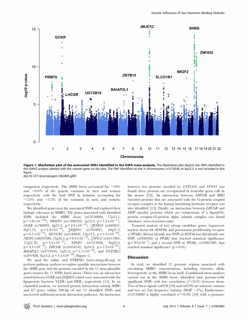
We identified nine loci associated with SHBG concentrations at
the genome-wide significance threshold of p = 561028 (Table 1
and Figure 1) in a genome-wide association study (GWAS) meta-
analysis of circulating SHBG concentrations in 21,791 men and
women from 10 studies (Table S1). All nine lead SNPs at these loci
had effects in the same direction (seven with p,0.05) in the
validation dataset of 7,046 men and women from six additional
studies (Table S2). The strongest association was within the SHBG
locus (rs12150660, p = 26102106). Together, these nine lead SNPs
explained 7.2% of the genetic variance (assuming 50% heritability)
in SHBG concentrations.
We next performed a series of additional analyses to explain
more of the phenotypic variance (Figure 2). First, we hypothesized
that genetic effects may be different in men and women, as SHBG
concentrations are .50% higher in females than males, and may
be differentially regulated between sexes. In a sex stratified
analysis, three of the nine loci showed evidence of sex-differen-
tiated effects at p,0.02 when we would not expect any signals to
have reached this level of significance by chance. The associations
at the 17p13.1-SHBG and Xq22.3 loci were stronger in males
whereas the association at the 8q21.13 locus was stronger in
females. To investigate the apparent differential sex effect for the
X chromosome further we ran a recessive regression model for the
X chromosome SNP rs1573036 in women in the Framingham
Heart Study and found no association with SHBG suggesting the
sex-differentiated effect is not the result of a recessive inheritance
pattern. Sex stratified GWAS identified one novel signal in men,
which showed no association in women (4q13.2: men
p = 2.561028, women p = 0.66, heterogeneity p = 0.003).
A series of conditional analyses were performed to identify
statistically independent signals. At the SHBG locus, three
apparently independent additional signals separate from the main
index SNP were observed, based on low (r2,0.05) pairwise
correlations in HapMap (rs6258 p = 2.7610246, rs1625895
p = 1.2610214 and rs3853894 p = 2.5610211). A series of iterative
conditional analyses (Table 2) involving SNPs at the SHBG locus
generated a final regression model including six statistically
independent SHBG SNPs. Four of these SNPs (#1–4 Table 2)
retained GWS when conditioned against the other five, and two
were nominally associated (SNP#5 p = 0.0001, SNP#6 p = 0.01).
Re-running the GWAS meta-analysis adjusting for these six SNPs
revealed evidence for three additional statistically independent
(pairwise HapMap r2,0.01) signals at the SHBG locus (SNP#7
p = 1.561027, SNP#8 p = 4.661025, SNP#9 p = 9.961026)
(Figure 3). There were also two additional trans signals located at
2p16.3 and 7q21.3 (Table 1). Although the 2p16.3 signal dropped
below GWS when combined with follow-up samples (p = 161027),
the index SNP at 2p16.3 is ,300 kb away from a strong candidate
gene, the luteinizing hormone receptor gene (LHCGR).
The majority of pair-wise correlations for the nine SHBG locus
SNPs highlighted by our conditional analyses showed very low
HapMap r2 values. However, the pairwise D9 values are often high
(Table S3) indicating that no or few recombination events have
occurred between some SNPs, and that combinations of SNPs
may be tagging un-typed variants on a common haplotype. To
investigate this possibility, we performed more extensive analyses
in a single study (NFBC1966, n = 4467). We used a denser set of
SNPs imputed from the June 2011 version of the 1000 Genomes
data and performed model selection analyses. Model selection
identifies a set of SNPs that best explain phenotypic variation,
while simultaneously penalizing each SNP included in this set, and
therefore correlated SNPs tend to be excluded from the final
model. These analyses consistently included at least seven SNPs in
the model, although it is hard to estimate the false-negative rate of
using the reduced sample size. While we are underpowered to
accurately pinpoint the exact number of independent signals, these
analyses support the results of the conditional analysis and suggest
that multiple variants at the SHBG locus are independently
associated with SHBG concentrations.
Data from an independent study, the InCHIANTI study, was
used to calculate the proportion of genetic variance in SHBG
concentrations explained when accounting for sex specific effects,
the multiple signals of association at the SHBG locus, and the
additional trans signals identified post conditional analysis. In men
and women we explained ,15.6% and ,8.4% of the heritable
Author Summary
Sex hormone-binding globulin (SHBG) is the key proteinresponsible for binding and transporting the sex steroidhormones, testosterone and estradiol, in the circulatorysystem. SHBG regulates their bioavailability and thereforetheir effects in the body. SHBG has been linked to chronicdiseases including type 2 diabetes and to hormone-sensitive cancers such as breast and prostate cancer. SHBGconcentrations are approximately 50% heritable in familystudies, suggesting SHBG concentrations are under signif-icant genetic control; yet, little is known about the specificgenes that influence SHBG. We conducted a large study ofthe association of SHBG concentrations with markers in thehuman genome in ,22,000 white men and women todetermine which loci influence SHBG concentrations.Genes near the identified genomic markers in addition tothe SHBG protein coding gene included PRMT6, GCKR,ZBTB10, JMJD1C, SLCO1B1, NR2F2, ZNF652, TDGF3, LHCGR,BAIAP2L1, and UGT2B15. These genes represent a widerange of biologic pathways that may relate to SHBGfunction and sex steroid hormone biology, including liverfunction, lipid metabolism, carbohydrate metabolism andtype 2 diabetes, and the development and progression ofsex steroid hormone-responsive cancers.
Genetic Influences of Sex Hormone–Binding Globulin
PLoS Genetics | www.plosgenetics.org 4 July 2012 | Volume 8 | Issue 7 | e1002805

Ta
ble
1.
SNP
sre
pre
sen
tin
glo
cias
soci
ate
dw
ith
circ
ula
tin
gSH
BG
con
cen
trat
ion
s.
Dis
cov
ery
Sa
mp
les
Dis
cov
ery
+Fo
llo
w-u
p
Ind
ex
SN
PA
na
lysi
sR
eg
ion
Nr
Ge
ne
Ch
rP
osi
tio
nE
ffe
ctA
lle
leO
the
rA
lle
leE
AF
Be
taS
Ep
Be
taS
Ep
rs1
74
96
33
2M
ain
1p
13
.3P
RM
T6
11
07
34
78
98
ag
0.6
72
0.0
26
0.0
04
61
.0E-
08
20
.02
80
.00
41
1.4
E-1
1
rs7
80
09
3M
ain
2p
23
.3G
CK
R2
27
59
61
07
tc
0.4
02
0.0
33
0.0
04
35
.8E-
14
20
.03
20
.00
39
2.2
E-1
6
rs4
40
83
7M
ain
8q
21
.13
ZB
TB
10
88
16
24
52
9a
g0
.78
20
.03
00
.00
52
6.7
E-0
92
0.0
28
0.0
04
73
.4E-
09
rs7
91
09
27
Mai
n1
0q
21
.3JM
JD1
C1
06
48
08
91
6t
g0
.51
20
.04
40
.00
43
7.4
E-2
52
0.0
48
0.0
03
96
.1E-
35
rs4
14
90
56
Mai
n1
2p
12
.1SL
CO
1B
11
22
12
22
81
6t
c0
.82
0.0
32
0.0
05
71
.5E-
08
0.0
29
0.0
05
21
.9E-
08
rs8
02
35
80
Mai
n1
5q
26
.2N
R2
F21
59
45
09
29
5t
c0
.72
20
.02
90
.00
49
2.8
E-0
92
0.0
30
.00
44
8.3
E-1
2
rs1
21
50
66
0M
ain
17
p1
3.1
SHB
G1
77
46
26
40
tg
0.2
40
.10
00
.00
53
1.2
E-7
90
.10
30
.00
47
1.8
E-1
06
rs2
41
19
84
Mai
n1
7q
21
.32
ZN
F65
21
74
48
00
75
0a
g0
.28
0.0
34
0.0
04
91
.5E-
12
0.0
33
0.0
04
43
.5E-
14
rs1
57
30
36
Mai
nX
q2
2.3
TD
GF3
23
10
97
06
72
4t
c0
.39
0.0
31
0.0
04
35
.1E-
13
0.0
28
0.0
03
74
.1E-
14
rs1
04
54
14
2C
on
dit
ion
al2
p1
6.3
LHC
GR
24
84
99
90
3t
c0
.69
0.0
26
0.0
04
72
.8E-
08
0.0
23
0.0
04
41
.3E-
07
rs3
77
91
95
Co
nd
itio
nal
7q
21
.3B
AIA
P2
L17
97
83
12
98
at
0.1
72
0.0
33
0.0
05
71
.2E-
08
20
.02
80
.00
51
2.7
E-0
8
rs2
93
42
8Se
x-sp
eci
fic
4q
13
.2U
GT
2B
15
46
96
26
37
1a
g0
.69
20
.02
30
.00
47
1.6
E-0
62
0.0
19
0.0
04
25
.5E-
06
Dis
cov
ery
Me
nD
isco
ve
ry+F
oll
ow
-up
Me
nD
isco
ve
ryW
om
en
Dis
cov
ery
+Fo
llo
w-u
pW
om
en
He
tero
ge
ne
ity
Ind
ex
SN
PB
eta
SE
pB
eta
SE
pB
eta
SE
pB
eta
SE
pp
Se
x
rs1
74
96
33
22
0.0
27
0.0
05
49
.6E-
07
20
.02
70
.00
51
1.5
E-0
72
0.0
20
.00
80
.00
32
0.0
29
0.0
06
71
.8E-
05
0.7
9Fe
mal
es
rs7
80
09
32
0.0
29
0.0
05
21
.8E-
08
20
.02
60
.00
49
7.0
E-0
82
0.0
40
.00
76
1.3
E-0
72
0.0
41
0.0
06
38
.6E-
11
0.0
7Fe
mal
es
rs4
40
83
72
0.0
21
0.0
06
20
.00
09
20
.01
90
.00
58
0.0
01
20
.04
90
.00
93
1.2
E-0
72
0.0
42
0.0
07
87
.2E-
08
0.0
2Fe
mal
es
rs7
91
09
27
20
.04
90
.00
51
5.3
E-2
22
0.0
50
0.0
04
81
.2E-
25
20
.03
80
.00
75
6.4
E-0
72
0.0
46
0.0
06
31
.7E-
13
0.6
3M
ale
s
rs4
14
90
56
0.0
28
0.0
06
70
.00
00
30
.02
70
.00
63
1.5
E-0
50
.04
90
.01
03
0.0
00
00
20
.03
70
.00
86
1.7
E-0
50
.36
Fem
ale
s
rs8
02
35
80
20
.02
40
.00
57
0.0
00
02
20
.02
50
.00
54
5.1
E-0
62
0.0
38
0.0
08
70
.00
00
12
0.0
38
0.0
07
17
.8E-
08
0.1
3Fe
mal
es
rs1
21
50
66
00
.10
60
.00
63
1.8
E-6
30
.11
00
.00
58
3.7
E-8
00
.08
50
.00
94
1.8
E-1
90
.08
70
.00
77
5.8
E-3
00
.02
Mal
es
rs2
41
19
84
0.0
34
0.0
05
85
.9E-
09
0.0
34
0.0
05
42
.3E-
10
0.0
32
0.0
08
40
.00
01
0.0
29
0.0
07
3.2
E-0
50
.54
Mal
es
rs1
57
30
36
0.0
40
.00
48
9.1
E-1
70
.03
50
.00
43
2.8
E-1
60
.01
20
.00
83
0.1
50
.01
60
.00
70
.02
0.0
2M
ale
s
rs1
04
54
14
2
rs3
77
91
95
rs2
93
42
82
0.0
32
0.0
05
61
.5E-
08
20
.02
90
.00
53
2.5
E-0
82
0.0
05
0.0
08
50
.57
20
.00
30
.00
71
0.6
60
.00
3M
ale
s
All
SNP
sar
eo
nth
e+s
tran
dan
dp
osi
tio
ns
are
bas
ed
on
bu
ild3
6.E
AF
=‘e
ffe
ctal
lele
fre
qu
en
cy’.
Be
tau
nit
sar
ep
er-
alle
lee
ffe
cte
stim
ate
sin
nat
ura
llo
gtr
ansf
orm
ed
nm
ol/
L.Se
xco
lum
ng
ive
sth
ese
xw
ith
the
larg
est
pe
r-al
lele
be
tae
stim
ate
.M
issi
ng
valu
es
for
con
dit
ion
alSN
Ps
asse
x-sp
eci
fic
con
dit
ion
alan
alys
isw
asn
ot
pe
rfo
rme
d.
do
i:10
.13
71
/jo
urn
al.p
ge
n.1
00
28
05
.t0
01
Genetic Influences of Sex Hormone–Binding Globulin
PLoS Genetics | www.plosgenetics.org 5 July 2012 | Volume 8 | Issue 7 | e1002805
component respectively. The SHBG locus accounted for ,10%
and ,6.6% of the genetic variation in men and women
respectively with the lead SNP in isolation accounting for
,7.8% and ,3.3% of the variation in men and women,
respectively.
We identified genes near the associated SNPs and explored their
biologic relevance to SHBG. The genes associated with identified
SNPs included the SHBG locus (rs12150660, 17p13.1,
p = 1.86102106), PRMT6 (rs17496332, 1p13.3, p = 1.4610211),
GCKR (rs780093, 2p23.3, p = 2.2610216), ZBTB10 (rs440837,
8q21.13, p = 3.4610209), JMJD1C (rs7910927, 10q21.3,
p = 6.1610235), SLCO1B1 (rs4149056, 12p12.1, p = 1.9610208),
NR2F2 (rs8023580, 15q26.2, p = 8.3610212), ZNF652 (rs2411984,
17q21.32, p = 3.5610214), TDGF3 (rs1573036, Xq22.3,
p = 4.1610214), LHCGR (rs10454142, 2p16.3, p = 1.3610207),
BAIAP2L1 (rs3779195, 7q21.3, p = 2.7610208), and UGT2B15
(rs293428, 4q13.2, p = 5.5610206) (Figure 1).
We used the online tool STRING (www.string-db.org) to
perform pathway analyses to explore possible interactions between
the SHBG gene and the proteins encoded by the 11 most plausible
genes nearest the 11 SNPs listed above. There was an interaction
noted between GCKR and JMJD1C which were associated with the
lipoprotein fractions VLDL and HDL, respectively [11]. In an
expanded analysis, we assessed protein interactions among SHBG
and 67 genes within 500 kb of our 11 identified SNPs and
uncovered additional protein interaction pathways. An interaction
between two proteins encoded by GTF2A1L and STON1 was
found; these proteins are co-expressed in testicular germ cells in
the mouse [12]. An interaction between LHCGR and BRI3
encoded proteins that are associated with the G-protein coupled
receptor complex in the human luteinizing hormone receptor was
also identified [13]. Finally, an interaction between LHCGR and
IAPP (amylin) proteins which are components of a ligand/G-
protein receptor/G-protein alpha subunit complex was found
(database: www.reactome.com).
Targeted analysis of two strong candidate genes, hepatocyte
nuclear factor-4a (HNF4a) and peroxisome-proliferating receptor
c (PPARc) did not identify any SNPs at HNF4a but did identify one
SNP, rs2920502, at PPARc that reached statistical significance
(p = 9.961025) and a second SNP at PPARc, rs13081389, that
reached nominal significance (p = 0.01).
Discussion
In total, we identified 12 genomic regions associated with
circulating SHBG concentrations, including extensive allelic
heterogeneity at the SHBG locus itself. Conditional meta-analyses
carried out at the SHBG locus, identified nine genome-wide
significant SNPs with low correlation (r2,0.01) between them.
Two of these signals (rs6258 [10] and rs6259) are missense variants
and two are low frequency variants (MAF ,2%). Furthermore,
rs12150660 is highly correlated (r2.0.95) [10] with a pentanu-
Figure 1. Manhattan plot of the autosomal SNPs identified in the GWA meta-analysis. The Manhattan plot depicts the SNPs identified inthe GWAS analysis labeled with the nearest gene on the plot. The SNP identified on the X chromosome, rs1573036, at Xq22.3, is not included in thisfigure.doi:10.1371/journal.pgen.1002805.g001
Genetic Influences of Sex Hormone–Binding Globulin
PLoS Genetics | www.plosgenetics.org 6 July 2012 | Volume 8 | Issue 7 | e1002805
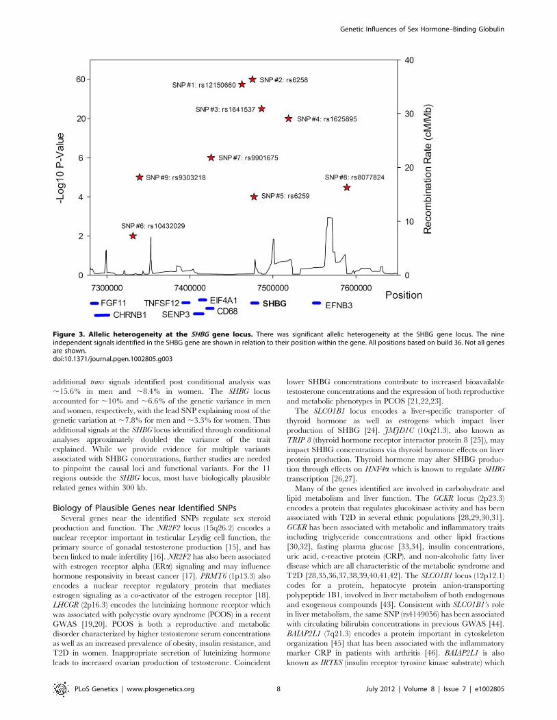
cleotide repeat, which affects SHBG expression in-vitro [14]. To our
knowledge, the magnitude of secondary signals observed at this
locus are the largest seen for any complex trait.
The proportion of genetic variance in SHBG serum concen-
trations explained when accounting for sex specific effects, the
multiple signals of association at the SHBG locus, and the
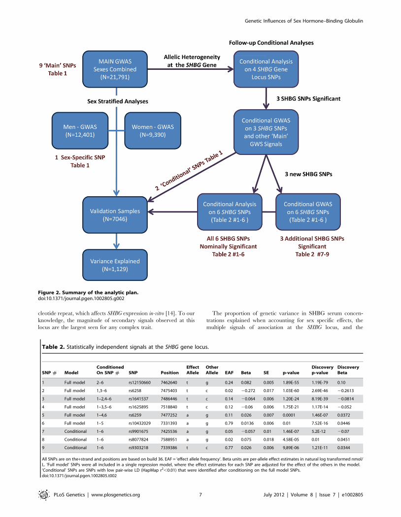
Figure 2. Summary of the analytic plan.doi:10.1371/journal.pgen.1002805.g002
Table 2. Statistically independent signals at the SHBG gene locus.
SNP # ModelConditionedOn SNP # SNP Position
EffectAllele
OtherAllele EAF Beta SE p-value
Discoveryp-value
DiscoveryBeta
1 Full model 2–6 rs12150660 7462640 t g 0.24 0.082 0.005 1.89E-55 1.19E-79 0.10
2 Full model 1,3–6 rs6258 7475403 t c 0.02 20.272 0.017 1.03E-60 2.69E-46 20.2613
3 Full model 1–2,4–6 rs1641537 7486446 t c 0.14 20.064 0.006 1.20E-24 8.19E-39 20.0814
4 Full model 1–3,5–6 rs1625895 7518840 t c 0.12 20.06 0.006 1.75E-21 1.17E-14 20.052
5 Full model 1–4,6 rs6259 7477252 a g 0.11 0.026 0.007 0.0001 1.46E-07 0.0372
6 Full model 1–5 rs10432029 7331393 a g 0.79 0.0136 0.006 0.01 7.52E-16 0.0446
7 Conditional 1–6 rs9901675 7425536 a g 0.05 20.057 0.01 1.46E-07 5.2E-12 20.07
8 Conditional 1–6 rs8077824 7588951 a g 0.02 0.075 0.018 4.58E-05 0.01 0.0451
9 Conditional 1–6 rs9303218 7339386 t c 0.77 0.026 0.006 9,89E-06 1.21E-11 0.0344
All SNPs are on the+strand and positions are based on build 36. EAF = ‘effect allele frequency’. Beta units are per-allele effect estimates in natural log transformed nmol/L. ‘Full model’ SNPs were all included in a single regression model, where the effect estimates for each SNP are adjusted for the effect of the others in the model.‘Conditional’ SNPs are SNPs with low pair-wise LD (HapMap r2,0.01) that were identified after conditioning on the full model SNPs.doi:10.1371/journal.pgen.1002805.t002
Genetic Influences of Sex Hormone–Binding Globulin
PLoS Genetics | www.plosgenetics.org 7 July 2012 | Volume 8 | Issue 7 | e1002805
additional trans signals identified post conditional analysis was
,15.6% in men and ,8.4% in women. The SHBG locus
accounted for ,10% and ,6.6% of the genetic variance in men
and women, respectively, with the lead SNP explaining most of the
genetic variation at ,7.8% for men and ,3.3% for women. Thus
additional signals at the SHBG locus identified through conditional
analyses approximately doubled the variance of the trait
explained. While we provide evidence for multiple variants
associated with SHBG concentrations, further studies are needed
to pinpoint the causal loci and functional variants. For the 11
regions outside the SHBG locus, most have biologically plausible
related genes within 300 kb.
Biology of Plausible Genes near Identified SNPsSeveral genes near the identified SNPs regulate sex steroid
production and function. The NR2F2 locus (15q26.2) encodes a
nuclear receptor important in testicular Leydig cell function, the
primary source of gonadal testosterone production [15], and has
been linked to male infertility [16]. NR2F2 has also been associated
with estrogen receptor alpha (ERa) signaling and may influence
hormone responsivity in breast cancer [17]. PRMT6 (1p13.3) also
encodes a nuclear receptor regulatory protein that mediates
estrogen signaling as a co-activator of the estrogen receptor [18].
LHCGR (2p16.3) encodes the luteinizing hormone receptor which
was associated with polycystic ovary syndrome (PCOS) in a recent
GWAS [19,20]. PCOS is both a reproductive and metabolic
disorder characterized by higher testosterone serum concentrations
as well as an increased prevalence of obesity, insulin resistance, and
T2D in women. Inappropriate secretion of luteinizing hormone
leads to increased ovarian production of testosterone. Coincident
lower SHBG concentrations contribute to increased bioavailable
testosterone concentrations and the expression of both reproductive
and metabolic phenotypes in PCOS [21,22,23].
The SLCO1B1 locus encodes a liver-specific transporter of
thyroid hormone as well as estrogens which impact liver
production of SHBG [24]. JMJD1C (10q21.3), also known as
TRIP 8 (thyroid hormone receptor interactor protein 8 [25]), may
impact SHBG concentrations via thyroid hormone effects on liver
protein production. Thyroid hormone may alter SHBG produc-
tion through effects on HNF4a which is known to regulate SHBG
transcription [26,27].
Many of the genes identified are involved in carbohydrate and
lipid metabolism and liver function. The GCKR locus (2p23.3)
encodes a protein that regulates glucokinase activity and has been
associated with T2D in several ethnic populations [28,29,30,31].
GCKR has been associated with metabolic and inflammatory traits
including triglyceride concentrations and other lipid fractions
[30,32], fasting plasma glucose [33,34], insulin concentrations,
uric acid, c-reactive protein (CRP), and non-alcoholic fatty liver
disease which are all characteristic of the metabolic syndrome and
T2D [28,35,36,37,38,39,40,41,42]. The SLCO1B1 locus (12p12.1)
codes for a protein, hepatocyte protein anion-transporting
polypeptide 1B1, involved in liver metabolism of both endogenous
and exogenous compounds [43]. Consistent with SLCO1B1’s role
in liver metabolism, the same SNP (rs4149056) has been associated
with circulating bilirubin concentrations in previous GWAS [44].
BAIAP2L1 (7q21.3) encodes a protein important in cytoskeleton
organization [45] that has been associated with the inflammatory
marker CRP in patients with arthritis [46]. BAIAP2L1 is also
known as IRTKS (insulin receptor tyrosine kinase substrate) which
Figure 3. Allelic heterogeneity at the SHBG gene locus. There was significant allelic heterogeneity at the SHBG gene locus. The nineindependent signals identified in the SHBG gene are shown in relation to their position within the gene. All positions based on build 36. Not all genesare shown.doi:10.1371/journal.pgen.1002805.g003
Genetic Influences of Sex Hormone–Binding Globulin
PLoS Genetics | www.plosgenetics.org 8 July 2012 | Volume 8 | Issue 7 | e1002805
is involved in insulin receptor signaling [47] and may relate to
insulin resistant states including obesity and T2D [48,49,
50,51,52,53,54]. We conducted a targeted analysis of PPARc, a
gene that influences SHBG gene expression in the liver [1,55] and
is associated with T2D [56,57]. Our analysis identified one
significant SNP (rs2920502, p = 9.961025) and a second nomi-
nally significant SNP (rs13081389, p = 0.01) at PPARc. Some of
the identified genes involved in hepatic metabolism of lipids and
carbohydrates may be affect SHBG concentrations indirectly
through effects on the SHBG transcription regulator HNF4aalthough HNF4a itself was not identified in this meta-analyses
[27,58,59,60].
The UGT2B15 locus (4q13.2) was significantly associated with
SHBG concentrations in men but not women in this meta-
analysis. UGT2B15 belongs to a family of genes (the UGT2B gene
family) that code for enzymes involved in the metabolism of sex
hormones through glucuronidation which allows for excretion of
sex steroids through the kidney and the gut via bile excretion
[61,62], primary clearance mechanisms for sex steroids [63].
UGT2B15 is involved in the conjugation and inactivation of
testosterone [64]. An association between rs293428 in the
UGT2B15 locus and circulating SHBG concentrations in men is
supported by a previous study demonstrating that a non-
synonymous SNP in UGT2B15 (rs1902023; D85Y) is associated
with serum SHBG concentrations in younger adult men [65].
UGT2B15 is thought to play a significant role in local tissue
inactivation of androgens in androgen dependent prostate cancer
[66,67]. The mechanism behind the influence of genetic variants
in UGT2B15 on SHBG concentrations is unknown, but one may
speculate that UGT2B15 affects the local androgenic environment
in selected tissues, which in turn results in regulation of SHBG
concentrations.
In addition to UGT2B15, three other genes near the identified
SNPs are associated with carcinogenesis, particularly in the
prostate and breast. ZBTB10 (8q21.13), has been linked to breast
cancer [68]. In breast cancer cell lines ZBTB10 is suppressed by
ROS-microRNA27a thereby enhancing ERa alpha expression
and mediating estrogen effects [17]. The ZNF652 (17q21.32) locus
codes for a DNA binding protein thought to act as a tumor
suppressor gene in breast cancer [69,70,71] that is also co-
expressed with the androgen receptor in prostate cancer [72].
TDGF3, teratocarcinoma derived growth factor 3, is the only
significant region identified on the X chromosome ((Xq22.3).
TDGF3 is a pseudogene of TDGF1 located on chromosome 3p23-p21
that has been associated with testicular germ cell tumors [73].
Strengths and LimitationsThis GWAS meta-analysis incorporated data from approxi-
mately 22,000 men and women from 16 epidemiologic cohorts.
The overall size of the study yields power but the meta-analysis of
data from different epidemiologic studies requires the inclusion of
different laboratory methods. The different studies used a variety
of assay methodologies to measure serum SHBG concentrations
although the vast majority were immunoassays (Tables S1 and S2,
Text S1) with similar methodologies. Variation introduced by the
use of different SHBG assays would result in loss of statistical
power and likely bias toward the null. Additionally, the majority of
women were post-menopausal as ascertained by self-report in all
studies (Table S1). SHBG concentrations, like testosterone, decline
only slightly across the menopause [74] so adjustment for
menopause status is not necessary. SHBG may also increase with
ovulation and be slightly higher in the luteal versus the follicular
phase of the menstrual cycle in premenopausal women, but most
studies did not collect data on menstrual phase at the time of
SHBG measurement so adjustment for menstrual phase was not
possible [75]. Finally, individuals were not excluded based on
health status, therefore some individuals with chronic conditions
that may affect hepatic production of or clearance of proteins
including SHBG such as liver disease, renal disease, or severe
malnutrition, may have been included in this analysis.
ConclusionSHBG synthesis in the liver is known to be affected directly or
indirectly by estrogens, androgens and thyroid hormones and has
been observed to be inversely associated with the higher insulin
concentrations characteristic of insulin resistant states such as T2D
[1,6]. In summary, the results of this GWAS reflect these
influences. Three regions map to proteins related to hepatic
function (12p12.1-SLCO1B1 [76], 2p23.3-GCKR [77] and 10q21.3-
JMJD1C [77]). In addition, 2p23.3-GCKR and 7q21.3-BAIAP2L1
[alias insulin receptor tyrosine kinase substrate (IRTKS)] are
involved in susceptibility to T2D [48] and insulin signaling [47],
respectively. Two signals also mapped to loci involved in thyroid
hormone regulation (10q21.3-JMJD1C and 12p12.1-SLCO1B1).
One signal mapped to the receptor for luteinizing hormone
2p16.3-LHCGR [20], the hormone that stimulates testosterone
production. Five regions mapped to genes previously implicated in
androgen and estrogen signaling (1p13.3-PRMT6 [18], 8q21.13-
ZBTB10 [17], 12p12.1-SLCO1B1 [76], 15q26.2-NR2F2 [78],
4q13.2-UGT2B15 [63]).
We have combined a conventional GWAS approach with
detailed additional analyses, including sex stratification, condi-
tional analysis and imputation from 1000 Genomes. Our results
demonstrate that these approaches can lead to an appreciable gain
in heritable variance explained. It does however highlight the
complexity of elucidating individual variant causality through
statistical approaches. In addition to the extensive allelic hetero-
geneity at the SHBG locus, our data identify loci with a role in sex
steroid hormone metabolism, which may help elucidate the role of
sex steroid hormones in disease, particularly T2D and hormone-
sensitive cancers.
Methods
We performed a genome wide association study (GWAS) meta-
analysis of 21,791 individuals (Table S1: 9,390 women, 12,401
men) from ten observational studies. Data from an additional six
studies totaling 7,046 individuals (Table S2: 4,509 women; 2,537
men) were used for validation. The proportion of variance
explained was estimated in an independent study (InCHIANTI,
n = 1,129). The individual study protocols were approved by their
respective institution’s ethics committee/institutional review board
and all participants provided informed consent prior to participa-
tion. Individuals known to be taking hormonal contraceptives or
hormone replacement therapy at time of SHBG measurement
were excluded from analysis. Age, sex and body mass index (BMI)
were included as covariates. After applying standard quality
control measures, imputed genotypes were available for approx-
imately 2.5 M SNPs. See Figure 2 for an overview of the analytic
plan and the Text S1 for further information for individual studies
included in this meta-analysis.
GWAS Conditional Meta-Analysis StepsConditional analysis #1. The initial starting point for the
conditional analysis was the four SHBG locus SNPs that all showed
low Hapmap LD (r2,0.05) with each other: rs12150660 (lead
SNP Table 1), rs6258 p = 2.7610246, rs1625895 p = 1.2610214
and rs3853894 p = 2.5610211. Each cohort fitted a single
Genetic Influences of Sex Hormone–Binding Globulin
PLoS Genetics | www.plosgenetics.org 9 July 2012 | Volume 8 | Issue 7 | e1002805
regression model, fitting SHBG concentrations against these four
genome-wide significant SHBG locus SNPs (rs12150660, rs6258,
rs1625895 and rs3853894), in addition to age, sex and BMI. After
meta-analyzing the results from all cohorts, three of the SNPs
retained genome wide significance when regressed against each
other, with the fourth SNP narrowly missing that threshold
(rs3853894, p = 4.161026).
Conditional GWAS #1 (Table 1, conditional analysis). We
next performed a conditional GWAS meta-analysis, where each
study included, as additional covariates to the original analysis plan,
the ten genome-wide significant autosomal SNPs (the eight ‘Main’
signals from Table 1 and the two unique SHBG locus signals
described above in addition to the lead SNP rs12150660: rs6258 and
rs1625895). Three additional signals (independence based on
HapMap r2,0.05) at the SHBG locus reached genome-wide
significance (rs1641537 p = 7.8610232, rs6259 p = 1.5610212 and
rs10432029 p = 361028), giving a total of six independent signals in
this gene region. In addition, two novel signals reached genome-wide
significance in the conditional analysis, at 7q21.3 (rs3779195
p = 161028) and 2p16.3 (rs10454142 p = 361028). After replication,
only rs3779195 at the BAIAP2L1 locus retained genome-wide
significance.
Conditional analysis #2 (Table 2, full model). Given the
six signals observed at the SHBG locus (three through conditional
analysis #1 rs12150660, rs6258, rs1625895, three through LD
estimates from conditional GWAS #1: rs1641537, rs6259,
rs10432029), we sought to confirm which of these six were truly
independent by a second round of conditional analysis. All
discovery and replication cohorts fitted a single regression model
of the six SNPs (SNPs # 1–6, Table 2) against SHBG
concentrations, using the same parameters and covariates as
conditional analysis #1. Four of the six SNPs (#1–4: rs12150660,
rs6258, rs1641537, and rs1625895) retained genome-wide signif-
icance when conditioned against each other, with two showing
nominal evidence of association (SNP #5 rs6259, p = 0.0001; SNP
#6 rs10432029, p = 0.01).
Conditional GWAS #2 (Table 2, conditional model). Finally,
we performed a second conditional GWAS analysis, adjusting for the
six SHBG locus SNPs which had evidence of association from
conditional analysis #2. All the discovery cohorts were used in this
analysis, in addition to three replication cohorts (total sample size
24,354). This analysis revealed evidence for a further three
independent signals at the SHBG locus (based on HapMap
r2,0.01), SNP #7 rs9901675 p = 1.561027, SNP #8 rs8077824
p = 4.661025, and SNP #9 rs9393218 p = 9.961026.
Sensitivity Analysis—Allelic Heterogeneity at the SHBGLocus
We performed a sensitivity analysis using samples from the 1966
Northern Finland Birth Cohort (NFBC1966) study to further
investigate allelic heterogeneity at the SHBG locus (Text S1). The
conditional meta-analysis showed evidence for up to nine signals at
the SHBG locus, but it is possible that these signals could be
explaining a much smaller number of causal variants in the region.
Since 1000 Genomes imputation allows us to assess the genetic
variation associated with a phenotype across a much denser set of
markers, it increases our power to detect allelic heterogeneity
within a region. Therefore, 1000 Genomes imputation was carried
out on all the samples in the NFBC1966 study and forward
selection was used to identify the set of SNPs that best explain the
variation in the SHBG phenotype. 1000 Genomes imputation was
carried out using IMPUTE2. The mean genotype probabilities for
each SNP were calculated and used in the model selection step.
Only SNPs 250 kb upstream and 250 kb downstream from the
SHBG locus (7283453–7786700 bp) were used in the analysis. All
SNPs with MAF ,0.1% or an imputation quality score less than
0.4 were excluded from the analysis. In total, 1978 SHBG region
SNPs measured or imputed in 4467 samples from the NFBC1966
study were used in the sensitivity analysis. Forward selection was
implemented in R (version 2.13.0) using the stepAIC package to
estimate the Akaikie Information Criterion (AIC), an inclusion
parameter. Given the high degree of correlation between the SNPs
in this region, we increased the penalty (k) on the number of terms
included in the model to 12 (where it is usually two), to minimize
possible over fitting. The final model included seven SNPs,
adjusted for sex and BMI.
Pathway AnalysisWe examined potential interactions among the proteins
encoded by the SHBG locus and the proteins encoded by the 11
genes (ZBT10, TDGF1, ZNF652, PRMT6, JMJD1C, GCKR,
BAIAP2L1, LHCGR, SLCO1B1, UGT2B15, NR2F2) closest to the
11 identified SNPs using pathway analysis with Search Tool for
the Retrieval of Interacting Genes/Proteins (STRING) Pathways
Analysis (www.string-db.org). The interactions explored by
STRING include direct (physical) and indirect (functional)
associations. We then expanded the analysis to examine protein
interactions among the SHBG gene and the proteins encoded by
67 genes within 500 kb of the 11 identified SNPs.
Targeted Candidate Gene AnalysisWe conducted targeted analysis of two strong candidate genes,
hepatocyte nuclear factor-4a (HNF4a) and peroxisome-proliferat-
ing receptor c (PPARc). Statistical significance thresholds were set
correcting for the number of SNPs tested in each gene region
(6100 kb).
Supporting Information
Table S1 Characteristics of 21,791 individuals from 10 discov-
ery cohorts included in the meta-analysis.
(DOC)
Table S2 Characteristics of 8,175 individuals from the six
cohorts included in the validation analysis (WHI, CARDIA,
Prospect-EPIC, MrOs, NHS, YFS) and the independent cohort
used to estimate the proportion of genetic variance explained by
the indentified SNPs (InChianti).
(DOC)
Table S3 Hapmap (release 22) linkage disequilibrium estimates
for the nine SHBG gene locus single nucleotide polymorphisms.
(DOC)
Text S1 Supplementary Methods with Specific Cohort Infor-
mation.
(DOC)
Acknowledgments
We are grateful to all study participants and staff in our participating
studies. Full study acknowledgements are available in Text S1.
Author Contributions
Conceived and designed the experiments: FH de Jong, O Raitakari, A
Teumer, C Ohlsson, JM Murabito, JRB Perry. Analyzed the data: JRB
Perry, AD Coviello, R Haring, M Wellons, D Vaidya, T Lehtimaki, S
Keildson, KL Lunetta, C He, M-R Jarvelin. Wrote the paper: AD
Coviello, R Haring, M Wellons, D Vaidya, T Lehtimaki, S Keildson, KL
Lunetta, C He, TM Frayling, A Murray, S Franks, M-R Jarvelin, FH de
Genetic Influences of Sex Hormone–Binding Globulin
PLoS Genetics | www.plosgenetics.org 10 July 2012 | Volume 8 | Issue 7 | e1002805
Jong, O Raitakari, A Teumer, C Ohlsson, JM Murabio, JRB Perry.
Individual cohort phenotyping/genotyping: KL Lunetta, C He, M
Fornage, V Lagou, M Mangino, NC Onland-Moret, B Chen, J Eriksson,
M Garcia, YM Liu, A Koster, K Lohman, L-P Lyytikainen, A-K Petersen,
J Prescott, L Stolk, L Vandenput, AR Wood, WV Zhuang, A Ruokonen,
A-L Hartikainen, A Pouta, S Bandinelli, AD Coviello, R Biffar, G Brabant,
DG Cox, Y Chen, S Cummings, L Ferrucci, MJ Gunter, SE Hankinson, H
Martikainen, A Hofman, G Homuth, T Illig, J-O Jansson, AD Johnson, D
Karasik, M Karlsson, J Kettunen, DP Kiel, P Kraft, J Liu, O Ljunggren, M
Lorentzon, M Maggio, MRP Markus, D Mellstrom, I Miljkovic, D Mirel,
S Nelson, L Morin Papunen, PHM Peeters, I Prokopenko, L Raffel, M
Reincke, AP Reiner, K Rexrode, F Rivadeneira, SM Schwartz, D
Siscovick, N Soranzo, D Stockl, S Tworoger, AG Uitterlinden, CH van
Gils, RS Vasan, H-E Wichmann, G Zhai, S Bhasin, M Bidlingmaier, SJ
Chanock, I De Vivo, TB Harris, DJ Hunter, M Kahonen, S Liu, P
Ouyang, TD Spector, YT van der Schouw, J Viikari, H Wallaschofski, MI
McCarthy, C Ohlsson, TM Frayling, A Murray, S Franks, M-R Jarvelin.
References
1. Hammond GL (2011) Diverse roles for sex hormone-binding globulin in
reproduction. Biol Reprod 85: 431–441.
2. Hammes A, Andreassen TK, Spoelgen R, Raila J, Hubner N, et al. (2005) Role
of endocytosis in cellular uptake of sex steroids. Cell 122: 751–762.
3. Thompson DJ, Healey CS, Baynes C, Kalmyrzaev B, Ahmed S, et al. (2008)
Identification of common variants in the SHBG gene affecting sex hormone-
binding globulin levels and breast cancer risk in postmenopausal women. Cancer
Epidemiol Biomarkers Prev 17: 3490–3498.
4. Wassell J, Michail M, Soliman N, Wardle PG (2011) The value of sex hormone
binding globulin (SHBG) in predicting treatment response in polycystic ovary
syndrome (PCOS). Clin Lab 57: 95–98.
5. Xita N, Tsatsoulis A, Chatzikyriakidou A, Georgiou I (2003) Association of the
(TAAAA)n repeat polymorphism in the sex hormone-binding globulin (SHBG)
gene with polycystic ovary syndrome and relation to SHBG serum levels. J Clin
Endocrinol Metab 88: 5976–5980.
6. Ding EL, Song Y, Manson JE, Hunter DJ, Lee CC, et al. (2009) Sex hormone-
binding globulin and risk of type 2 diabetes in women and men. N Engl J Med
361: 1152–1163.
7. Perry JR, Weedon MN, Langenberg C, Jackson AU, Lyssenko V, et al. (2010)
Genetic evidence that raised sex hormone binding globulin (SHBG) levels reduce
the risk of type 2 diabetes. Hum Mol Genet 19: 535–544.
8. Coviello AD, Zhuang WV, Lunetta KL, Bhasin S, Ulloor J, et al. (2011)
Circulating testosterone and SHBG concentrations are heritable in women: the
Framingham Heart Study. J Clin Endocrinol Metab 96: E1491–1495.
9. Melzer D, Perry JR, Hernandez D, Corsi AM, Stevens K, et al. (2008) A
genome-wide association study identifies protein quantitative trait loci (pQTLs).
PLoS Genet 4: e1000072. doi:10.1371/journal.pgen.1000072
10. Ohlsson C, Wallaschofski H, Lunetta KL, Stolk L, Perry JR, et al. (2011)
Genetic determinants of serum testosterone concentrations in men. PLoS Genet
7: e1002313. doi:10.1371/journal.pgen.1002313
11. Chasman DI, Pare G, Mora S, Hopewell JC, Peloso G, et al. (2009) Forty-three
loci associated with plasma lipoprotein size, concentration, and cholesterol
content in genome-wide analysis. PLoS Genet 5: e1000730. doi:10.1371/
journal.pgen.1000730
12. Han SY, Zhou L, Upadhyaya A, Lee SH, Parker KL, et al. (2001) TFIIAalpha/
beta-like factor is encoded by a germ cell-specific gene whose expression is up-
regulated with other general transcription factors during spermatogenesis in the
mouse. Biol Reprod 64: 507–517.
13. Kudo M, Osuga Y, Kobilka BK, Hsueh AJ (1996) Transmembrane regions V
and VI of the human luteinizing hormone receptor are required for constitutive
activation by a mutation in the third intracellular loop. J Biol Chem 271: 22470–
22478.
14. Hogeveen KN, Talikka M, Hammond GL (2001) Human sex hormone-binding
globulin promoter activity is influenced by a (TAAAA)n repeat element within
an Alu sequence. J Biol Chem 276: 36383–36390.
15. Martin LJ, Tremblay JJ (2010) Nuclear receptors in Leydig cell gene expression
and function. Biol Reprod 83: 3–14.
16. Hu Z, Xia Y, Guo X, Dai J, Li H, et al. (2011) A genome-wide association study
in Chinese men identifies three risk loci for non-obstructive azoospermia. Nat
Genet 44: 183–186.
17. Li X, Mertens-Talcott SU, Zhang S, Kim K, Ball J, et al. (2010) MicroRNA-27a
Indirectly Regulates Estrogen Receptor {alpha} Expression and Hormone
Responsiveness in MCF-7 Breast Cancer Cells. Endocrinology 151: 2462–
2473.
18. Harrison MJ, Tang YH, Dowhan DH (2010) Protein arginine methyltransferase
6 regulates multiple aspects of gene expression. Nucleic Acids Res 38: 2201–
2216.
19. Franks S, Gharani N, McCarthy M (2001) Candidate genes in polycystic ovary
syndrome. Hum Reprod Update 7: 405–410.
20. Chen ZJ, Zhao H, He L, Shi Y, Qin Y, et al. (2011) Genome-wide association
study identifies susceptibility loci for polycystic ovary syndrome on chromosome
2p16.3, 2p21 and 9q33.3. Nat Genet 43: 55–59.
21. Cousin P, Calemard-Michel L, Lejeune H, Raverot G, Yessaad N, et al. (2004)
Influence of SHBG gene pentanucleotide TAAAA repeat and D327N
polymorphism on serum sex hormone-binding globulin concentration in hirsute
women. J Clin Endocrinol Metab 89: 917–924.
22. Ehrmann DA (2005) Polycystic ovary syndrome. N Engl J Med 352: 1223–1236.
23. Franks S (1995) Polycystic ovary syndrome. N Engl J Med 333: 853–861.
24. Abe T, Kakyo M, Tokui T, Nakagomi R, Nishio T, et al. (1999) Identification of
a novel gene family encoding human liver-specific organic anion transporter
LST-1. J Biol Chem 274: 17159–17163.
25. Lee JW, Choi HS, Gyuris J, Brent R, Moore DD (1995) Two classes of proteins
dependent on either the presence or absence of thyroid hormone for interaction
with the thyroid hormone receptor. Mol Endocrinol 9: 243–254.
26. Selva DM, Hammond GL (2009) Thyroid hormones act indirectly to increase
sex hormone-binding globulin production by liver via hepatocyte nuclear factor-
4alpha. J Mol Endocrinol 43: 19–27.
27. Janne M, Hammond GL (1998) Hepatocyte nuclear factor-4 controls
transcription from a TATA-less human sex hormone-binding globulin gene
promoter. J Biol Chem 273: 34105–34114.
28. Reiling E, van ’t Riet E, Groenewoud MJ, Welschen LM, van Hove EC, et al.
(2009) Combined effects of single-nucleotide polymorphisms in GCK, GCKR,
G6PC2 and MTNR1B on fasting plasma glucose and type 2 diabetes risk.
Diabetologia 52: 1866–1870.
29. Qi Q, Wu Y, Li H, Loos RJ, Hu FB, et al. (2009) Association of GCKR rs780094,
alone or in combination with GCK rs1799884, with type 2 diabetes and related
traits in a Han Chinese population. Diabetologia 52: 834–843.
30. Ling Y, Li X, Gu Q, Chen H, Lu D, et al. (2011) Associations of common
polymorphisms in GCKR with type 2 diabetes and related traits in a Han Chinese
population: a case-control study. BMC Med Genet 12: 66.
31. Tanaka D, Nagashima K, Sasaki M, Yamada C, Funakoshi S, et al. (2011)
GCKR mutations in Japanese families with clustered type 2 diabetes. Mol Genet
Metab 102: 453–460.
32. Varbo A, Benn M, Tybjaerg-Hansen A, Grande P, Nordestgaard BG (2011)
TRIB1 and GCKR polymorphisms, lipid levels, and risk of ischemic heart disease
in the general population. Arterioscler Thromb Vasc Biol 31: 451–457.
33. Takeuchi F, Katsuya T, Chakrewarthy S, Yamamoto K, Fujioka A, et al. (2010)
Common variants at the GCK, GCKR, G6PC2-ABCB11 and MTNR1B loci are
associated with fasting glucose in two Asian populations. Diabetologia 53: 299–
308.
34. Onuma H, Tabara Y, Kawamoto R, Shimizu I, Kawamura R, et al. (2010) The
GCKR rs780094 polymorphism is associated with susceptibility of type 2
diabetes, reduced fasting plasma glucose levels, increased triglycerides levels and
lower HOMA-IR in Japanese population. J Hum Genet 55: 600–604.
35. Hadarits F, Kisfali P, Mohas M, Maasz A, Duga B, et al. (2011) Common
functional variants of APOA5 and GCKR accumulate gradually in association
with triglyceride increase in metabolic syndrome patients. Mol Biol Rep.
36. Hu C, Zhang R, Wang C, Yu W, Lu J, et al. (2010) Effects of GCK, GCKR,
G6PC2 and MTNR1B variants on glucose metabolism and insulin secretion.
PLoS ONE 5: e11761. doi:10.1371/journal.pone.0011761
37. Kozian DH, Barthel A, Cousin E, Brunnhofer R, Anderka O, et al. (2010)
Glucokinase-activating GCKR polymorphisms increase plasma levels of
triglycerides and free fatty acids, but do not elevate cardiovascular risk in the
Ludwigshafen Risk and Cardiovascular Health Study. Horm Metab Res 42:
502–506.
38. Mohas M, Kisfali P, Jaromi L, Maasz A, Feher E, et al. (2010) GCKR gene
functional variants in type 2 diabetes and metabolic syndrome: do the rare
variants associate with increased carotid intima-media thickness? Cardiovasc
Diabetol 9: 79.
39. Orho-Melander M, Melander O, Guiducci C, Perez-Martinez P, Corella D,
et al. (2008) Common missense variant in the glucokinase regulatory protein
gene is associated with increased plasma triglyceride and C-reactive protein but
lower fasting glucose concentrations. Diabetes 57: 3112–3121.
40. Perez-Martinez P, Delgado-Lista J, Garcia-Rios A, Mc Monagle J, Gulseth HL,
et al. (2011) Glucokinase regulatory protein genetic variant interacts with omega-
3 PUFA to influence insulin resistance and inflammation in metabolic syndrome.
PLoS ONE 6: e20555. doi:10.1371/journal.pone.0020555
41. Tam CH, Ma RC, So WY, Wang Y, Lam VK, et al. (2009) Interaction effect of
genetic polymorphisms in glucokinase (GCK) and glucokinase regulatory protein
(GCKR) on metabolic traits in healthy Chinese adults and adolescents. Diabetes
58: 765–769.
42. Yang Z, Wen J, Tao X, Lu B, Du Y, et al. (2011) Genetic variation in the
GCKR gene is associated with non-alcoholic fatty liver disease in Chinese
people. Mol Biol Rep 38: 1145–1150.
43. Seithel A, Klein K, Zanger UM, Fromm MF, Konig J (2008) Non-synonymous
polymorphisms in the human SLCO1B1 gene: an in vitro analysis of SNP
c.1929A.C. Mol Genet Genomics 279: 149–157.
Genetic Influences of Sex Hormone–Binding Globulin
PLoS Genetics | www.plosgenetics.org 11 July 2012 | Volume 8 | Issue 7 | e1002805
44. Buch S, Schafmayer C, Volzke H, Seeger M, Miquel JF, et al. (2010) Loci from a
genome-wide analysis of bilirubin levels are associated with gallstone risk andcomposition. Gastroenterology 139: 1942–1951 e1942.
45. Miyahara A, Okamura-Oho Y, Miyashita T, Hoshika A, Yamada M (2003)
Genomic structure and alternative splicing of the insulin receptor tyrosine kinasesubstrate of 53-kDa protein. J Hum Genet 48: 410–414.
46. Galligan CL, Baig E, Bykerk V, Keystone EC, Fish EN (2007) Distinctive geneexpression signatures in rheumatoid arthritis synovial tissue fibroblast cells:
correlates with disease activity. Genes Immun 8: 480–491.
47. Millard TH, Dawson J, Machesky LM (2007) Characterisation of IRTKS, anovel IRSp53/MIM family actin regulator with distinct filament bundling
properties. J Cell Sci 120: 1663–1672.48. Dupuis J, Langenberg C, Prokopenko I, Saxena R, Soranzo N, et al. (2010) New
genetic loci implicated in fasting glucose homeostasis and their impact on type 2diabetes risk. Nat Genet 42: 105–116.
49. Wang L, Muromoto N, Hayashi H, Mitani Y, Uehara H, et al. (1997)
Hyperinsulinemia but no diabetes in transgenic mice homozygously expressingthe tyrosine kinase-deficient human insulin receptor. Biochem Biophys Res
Commun 240: 446–451.50. Hotamisligil GS, Budavari A, Murray D, Spiegelman BM (1994) Reduced
tyrosine kinase activity of the insulin receptor in obesity-diabetes. Central role of
tumor necrosis factor-alpha. J Clin Invest 94: 1543–1549.51. Kusari J, Takata Y, Hatada E, Freidenberg G, Kolterman O, et al. (1991)
Insulin resistance and diabetes due to different mutations in the tyrosine kinasedomain of both insulin receptor gene alleles. J Biol Chem 266: 5260–5267.
52. Block NE, Komori K, Robinson KA, Dutton SL, Lam CF, et al. (1991)Diabetes-associated impairment of hepatic insulin receptor tyrosine kinase
activity: a study of mechanisms. Endocrinology 128: 312–322.
53. Odawara M, Kadowaki T, Yamamoto R, Shibasaki Y, Tobe K, et al. (1989)Human diabetes associated with a mutation in the tyrosine kinase domain of the
insulin receptor. Science 245: 66–68.54. Arner P, Pollare T, Lithell H, Livingston JN (1987) Defective insulin receptor
tyrosine kinase in human skeletal muscle in obesity and type 2 (non-insulin-
dependent) diabetes mellitus. Diabetologia 30: 437–440.55. Selva DM, Hammond GL (2009) Peroxisome-proliferator receptor gamma
represses hepatic sex hormone-binding globulin expression. Endocrinology 150:2183–2189.
56. Altshuler D, Hirschhorn JN, Klannemark M, Lindgren CM, Vohl MC, et al.(2000) The common PPARgamma Pro12Ala polymorphism is associated with
decreased risk of type 2 diabetes. Nat Genet 26: 76–80.
57. Hakonarson H, Grant SF (2011) GWAS and its impact on elucidating theetiology of diabetes. Diabetes Metab Res Rev.
58. Selva DM, Hogeveen KN, Innis SM, Hammond GL (2007) Monosaccharide-induced lipogenesis regulates the human hepatic sex hormone-binding globulin
gene. J Clin Invest 117: 3979–3987.
59. Shih DQ, Bussen M, Sehayek E, Ananthanarayanan M, Shneider BL, et al.(2001) Hepatocyte nuclear factor-1alpha is an essential regulator of bile acid and
plasma cholesterol metabolism. Nat Genet 27: 375–382.60. Yin L, Ma H, Ge X, Edwards PA, Zhang Y (2011) Hepatic hepatocyte nuclear
factor 4alpha is essential for maintaining triglyceride and cholesterol homeosta-sis. Arterioscler Thromb Vasc Biol 31: 328–336.
61. Wilson W, 3rd, Pardo-Manuel de Villena F, Lyn-Cook BD, Chatterjee PK, Bell
TA, et al. (2004) Characterization of a common deletion polymorphism of theUGT2B17 gene linked to UGT2B15. Genomics 84: 707–714.
62. Turgeon D, Carrier JS, Levesque E, Hum DW, Belanger A (2001) Relativeenzymatic activity, protein stability, and tissue distribution of human steroid-
metabolizing UGT2B subfamily members. Endocrinology 142: 778–787.
63. Menard V, Eap O, Harvey M, Guillemette C, Levesque E (2009) Copy-number
variations (CNVs) of the human sex steroid metabolizing genes UGT2B17 and
UGT2B28 and their associations with a UGT2B15 functional polymorphism.
Hum Mutat 30: 1310–1319.
64. Levesque E, Beaulieu M, Green MD, Tephly TR, Belanger A, et al. (1997)
Isolation and characterization of UGT2B15(Y85): a UDP-glucuronosyltransfer-
ase encoded by a polymorphic gene. Pharmacogenetics 7: 317–325.
65. Swanson C, Mellstrom D, Lorentzon M, Vandenput L, Jakobsson J, et al. (2007)
The uridine diphosphate glucuronosyltransferase 2B15 D85Y and 2B17 deletion
polymorphisms predict the glucuronidation pattern of androgens and fat mass in
men. J Clin Endocrinol Metab 92: 4878–4882.
66. Chouinard S, Barbier O, Belanger A (2007) UDP-glucuronosyltransferase 2B15
(UGT2B15) and UGT2B17 enzymes are major determinants of the androgen
response in prostate cancer LNCaP cells. J Biol Chem 282: 33466–33474.
67. Chouinard S, Pelletier G, Belanger A, Barbier O (2004) Cellular specific
expression of the androgen-conjugating enzymes UGT2B15 and UGT2B17 in
the human prostate epithelium. Endocr Res 30: 717–725.
68. Pathi SS, Jutooru I, Chadalapaka G, Sreevalsan S, Anand S, et al. (2011) GT-
094, a NO-NSAID, inhibits colon cancer cell growth by activation of a reactive
oxygen species-microRNA-27a: ZBTB10-specificity protein pathway. Mol
Cancer Res 9: 195–202.
69. Kumar R, Manning J, Spendlove HE, Kremmidiotis G, McKirdy R, et al.
(2006) ZNF652, a novel zinc finger protein, interacts with the putative breast
tumor suppressor CBFA2T3 to repress transcription. Mol Cancer Res 4: 655–
665.
70. Kumar R, Selth L, Schulz RB, Tay BS, Neilsen PM, et al. (2011) Genome-wide
mapping of ZNF652 promoter binding sites in breast cancer cells. J Cell
Biochem.
71. Kumar R, Cheney KM, Neilsen PM, Schulz RB, Callen DF (2010) CBFA2T3-
ZNF651, like CBFA2T3-ZNF652, functions as a transcriptional corepressor
complex. FEBS Lett 584: 859–864.
72. Callen DF, Ricciardelli C, Butler M, Stapleton A, Stahl J, et al. (2010) Co-
expression of the androgen receptor and the transcription factor ZNF652 is
related to prostate cancer outcome. Oncol Rep 23: 1045–1052.
73. Baldassarre G, Romano A, Armenante F, Rambaldi M, Paoletti I, et al. (1997)
Expression of teratocarcinoma-derived growth factor-1 (TDGF-1) in testis germ
cell tumors and its effects on growth and differentiation of embryonal carcinoma
cell line NTERA2/D1. Oncogene 15: 927–936.
74. Burger HG, Dudley EC, Cui J, Dennerstein L, Hopper JL (2000) A prospective
longitudinal study of serum testosterone, dehydroepiandrosterone sulfate, and
sex hormone-binding globulin levels through the menopause transition. J Clin
Endocrinol Metab 85: 2832–2838.
75. Schijf CP, van der Mooren MJ, Doesburg WH, Thomas CM, Rolland R (1993)
Differences in serum lipids, lipoproteins, sex hormone binding globulin and
testosterone between the follicular and the luteal phase of the menstrual cycle.
Acta Endocrinol (Copenh) 129: 130–133.
76. Link E, Parish S, Armitage J, Bowman L, Heath S, et al. (2008) SLCO1B1
variants and statin-induced myopathy–a genomewide study. N Engl J Med 359:
789–799.
77. Chambers JC, Zhang W, Sehmi J, Li X, Wass MN, et al. (2011) Genome-wide
association study identifies loci influencing concentrations of liver enzymes in
plasma. Nat Genet 43: 1131–1138.
78. Lee DK, Kurihara I, Jeong JW, Lydon JP, DeMayo FJ, et al. (2010) Suppression
of ERalpha activity by COUP-TFII is essential for successful implantation and
decidualization. Mol Endocrinol 24: 930–940.
Genetic Influences of Sex Hormone–Binding Globulin
PLoS Genetics | www.plosgenetics.org 12 July 2012 | Volume 8 | Issue 7 | e1002805


![Genome-wide meta-analysis identifies five new ...eprints.qut.edu.au/91749/1/Law2015NatGenet[MainText&Figure1&2].pdf · Genome-wide meta-analysis identifies five new susceptibility](https://static.fdocuments.in/doc/165x107/5af3fdc17f8b9a9e598c0aba/genome-wide-meta-analysis-identifies-five-new-maintextfigure12pdfgenome-wide.jpg)