A Dynamic molecular basis for malfunction in disease ...
25
*For correspondence: anns@ pound.med.utoronto.ca (AKS); [email protected] (LEK) Competing interests: The authors declare that no competing interests exist. Funding: See page 21 Received: 30 July 2016 Accepted: 25 October 2016 Published: 09 November 2016 Reviewing editor: Volker Do ¨ tsch, Goethe University, Germany Copyright Schuetz and Kay. This article is distributed under the terms of the Creative Commons Attribution License, which permits unrestricted use and redistribution provided that the original author and source are credited. A Dynamic molecular basis for malfunction in disease mutants of p97/ VCP Anne K Schuetz 1,2,3 *, Lewis E Kay 1,2,3,4 * 1 Department of Molecular Genetics, University of Toronto, Toronto, Canada; 2 Department of Biochemistry, University of Toronto, Toronto, Cananda; 3 Department of Chemistry, University of Toronto, Toronto, Canada; 4 Program in Molecular Structure and Function, Hospital for Sick Children, Toronto, Canada Abstract p97/VCP is an essential, abundant AAA+ ATPase that is conserved throughout eukaryotes, with central functions in diverse processes ranging from protein degradation to DNA damage repair and membrane fusion. p97 has been implicated in the etiology of degenerative diseases and in cancer. Using Nuclear Magnetic Resonance spectroscopy we reveal how disease- causing mutations in p97 deregulate dynamics of the N-terminal domain that binds adaptor proteins involved in controlling p97 function. Our results provide a molecular basis for understanding how malfunction occurs whereby mutations shift the ADP-bound form of the enzyme towards an ATP-like state in a manner that correlates with disease severity. This deregulation interferes with the two-pronged binding of an adaptor that affects p97 function in lysosomal degradation of substrates. Subtle structural changes propagate from mutation sites to regions distal in space, defining allosteric networks that facilitate inter-domain communication, with potential implications for modulation of enzyme activity by drug molecules. DOI: 10.7554/eLife.20143.001 Introduction The valosin containing protein (VCP) or p97 is a highly conserved enzyme in mammalian cells, with orthologues in yeast (Cdc48), in flies (TER94) and in archaea (VAT) (Rabouille et al., 1995). It is involved in various processes in the cell (Figure 1A) including membrane fusion (Rabouille et al., 1995), chromatin-associated functions (Moreno et al., 2014; Dantuma and Hoppe, 2012), cell cycle progression (Cao et al., 2003), and apoptosis (Madeo et al., 1997) and it is active in proteasomal degradation (Richly et al., 2005), autophagy (Ju et al., 2009; Buchan et al., 2013), and in endoso- mal pathways (Ritz et al., 2011). The involvement of p97 in all major proteolysis pathways makes it a central player in cellular homeostasis (Meyer et al., 2012). p97 is a 540 kDa homo-hexamer (6x89 kDa) with each monomer comprising an N-terminal domain (NTD) and a pair of ATPase domains, D1 and D2, arranged in primary sequence as NTD-D1-D2. Both D1 and D2 are organized as rings that stack coaxially, with the NTD located at the periphery of the D1 ring (DeLaBarre and Brunger, 2003)(Figure 1B,C). p97 converts the energy obtained via ATP hydrolysis to remove sub- strates from complexes or membranes and to structurally remodel or unfold them (Barthelme and Sauer, 2016). Its diverse activities result from various cofactors that recruit it to specific functions (Buchberger et al., 2015), with more than 40 adaptors discovered in mammalian cells. Many of these contain highly conserved p97 binding domains such as UBX (Buchberger et al., 2001) and VIM (Stapf et al., 2011). Given its prominent role in the eukaryotic cell, p97 misfunction is associ- ated with human disease. While p97 deletion results in early embryonic lethality (Mu ¨ller et al., 2007), a series of missense mutations lead to very specific malfunctions in protein homeostasis Schuetz and Kay. eLife 2016;5:e20143. DOI: 10.7554/eLife.20143 1 of 25 RESEARCH ARTICLE
Transcript of A Dynamic molecular basis for malfunction in disease ...
*For correspondence: anns@
pound.med.utoronto.ca (AKS);
(LEK)
Competing interests: The
authors declare that no
competing interests exist.
Funding: See page 21
Received: 30 July 2016
Accepted: 25 October 2016
Published: 09 November 2016
Reviewing editor: Volker
Dotsch, Goethe University,
Germany
Copyright Schuetz and Kay.
This article is distributed under
the terms of the Creative
Commons Attribution License,
which permits unrestricted use
and redistribution provided that
the original author and source are
credited.
A Dynamic molecular basis formalfunction in disease mutants of p97/VCPAnne K Schuetz1,2,3*, Lewis E Kay1,2,3,4*
1Department of Molecular Genetics, University of Toronto, Toronto, Canada;2Department of Biochemistry, University of Toronto, Toronto, Cananda;3Department of Chemistry, University of Toronto, Toronto, Canada; 4Program inMolecular Structure and Function, Hospital for Sick Children, Toronto, Canada
Abstract p97/VCP is an essential, abundant AAA+ ATPase that is conserved throughout
eukaryotes, with central functions in diverse processes ranging from protein degradation to DNA
damage repair and membrane fusion. p97 has been implicated in the etiology of degenerative
diseases and in cancer. Using Nuclear Magnetic Resonance spectroscopy we reveal how disease-
causing mutations in p97 deregulate dynamics of the N-terminal domain that binds adaptor
proteins involved in controlling p97 function. Our results provide a molecular basis for
understanding how malfunction occurs whereby mutations shift the ADP-bound form of the enzyme
towards an ATP-like state in a manner that correlates with disease severity. This deregulation
interferes with the two-pronged binding of an adaptor that affects p97 function in lysosomal
degradation of substrates. Subtle structural changes propagate from mutation sites to regions
distal in space, defining allosteric networks that facilitate inter-domain communication, with
potential implications for modulation of enzyme activity by drug molecules.
DOI: 10.7554/eLife.20143.001
IntroductionThe valosin containing protein (VCP) or p97 is a highly conserved enzyme in mammalian cells, with
orthologues in yeast (Cdc48), in flies (TER94) and in archaea (VAT) (Rabouille et al., 1995). It is
involved in various processes in the cell (Figure 1A) including membrane fusion (Rabouille et al.,
1995), chromatin-associated functions (Moreno et al., 2014; Dantuma and Hoppe, 2012), cell cycle
progression (Cao et al., 2003), and apoptosis (Madeo et al., 1997) and it is active in proteasomal
degradation (Richly et al., 2005), autophagy (Ju et al., 2009; Buchan et al., 2013), and in endoso-
mal pathways (Ritz et al., 2011). The involvement of p97 in all major proteolysis pathways makes it a
central player in cellular homeostasis (Meyer et al., 2012). p97 is a 540 kDa homo-hexamer
(6x89 kDa) with each monomer comprising an N-terminal domain (NTD) and a pair of ATPase
domains, D1 and D2, arranged in primary sequence as NTD-D1-D2. Both D1 and D2 are organized
as rings that stack coaxially, with the NTD located at the periphery of the D1 ring (DeLaBarre and
Brunger, 2003) (Figure 1B,C). p97 converts the energy obtained via ATP hydrolysis to remove sub-
strates from complexes or membranes and to structurally remodel or unfold them (Barthelme and
Sauer, 2016). Its diverse activities result from various cofactors that recruit it to specific functions
(Buchberger et al., 2015), with more than 40 adaptors discovered in mammalian cells. Many of
these contain highly conserved p97 binding domains such as UBX (Buchberger et al., 2001) and
VIM (Stapf et al., 2011). Given its prominent role in the eukaryotic cell, p97 misfunction is associ-
ated with human disease. While p97 deletion results in early embryonic lethality (Muller et al.,
2007), a series of missense mutations lead to very specific malfunctions in protein homeostasis
Schuetz and Kay. eLife 2016;5:e20143. DOI: 10.7554/eLife.20143 1 of 25
RESEARCH ARTICLE

linked to degenerative disorders (Watts et al., 2004), among them Inclusion Body Myopathy associ-
ated with Paget disease of the bone and Frontotemporal Dementia (IBMPFD), a lethal autosomal
dominant disorder with onset in midlife. These mutations, mostly occurring at the NTD-D1 interface
or in the linker region between these domains (Figure 2), also lead to an increased occurrence of
amyotrophic lateral sclerosis (Johnson et al., 2011) and of familial Parkinson’s disease (Chan et al.,
2012).
Of particular functional relevance is the mobility of the NTD, which has been shown to undergo
large displacements during the nucleotide cycle (Tang et al., 2010). NTDs are the major binding
sites for cofactors along with substrates and cross-linking NTD to D1 via disulfides abrogates all
ATPase activity (both in D1 and in D2) (Niwa et al., 2012). Crystallographic studies (Tang et al.,
2010) conducted on a truncated form of p97 comprising NTD and D1 show that NTD is coplanar rel-
ative to D1 in both wild-type (wt) and disease mutants of p97-ADP (referred to in what follows as
the down state, Figure 1B, Figure 1—figure supplement 1A,B) and elevated relative to D1 in p97-
ATPgS disease mutants (up state, Figure 1C, Figure 1—figure supplement 1A,B). Complementary
B
A
188 209 460 481 762
D2NTD D1
ADP down
ATPγS up
C
Nucleic acid repair
and replication
Proteasome
Autophagy
Endoplasmic reticulum
associated degradation
Endosomal trafficking
Aggregate
handlingp97
Lysosome
Chromatin
associated
degradation
Mitochondria
associated
degradationCell cycle
control
Control of
apoptosisGolgi membrane
dynamicsUBXD1
p47
Disease
Mutants
Figure 1. p97 structure and function. (A) Schematic illustration of a set of p97 cellular functions adapted from
(Meyer and Weihl, 2014) highlighting pathways (orange square) that may be affected by IBMPFD disease-related
mutations. Substrates are shown in blue and adaptors studied herein in green. (B,C) Ribbon-diagram
representation of ND1Lp97 (residues 1–480) [PDB: 1E32 for ADP state, 4KO8 for ATPgS state of R155H] with NTD
(blue), linker (green) and D1 (red in ADP state, B; grey in ATPgS state, C) color-coded. Shown to the side are
cartoons of ND1Lp97-ADP and ND1Lp97-ATP that are used in other figures, with a single NTD highlighted in the
down (ADP) and up (ATP) position, as well as the domain structure of p97.
DOI: 10.7554/eLife.20143.002
The following figure supplement is available for figure 1:
Figure supplement 1. Impact of nucleotide on the structure of ND1Lp97.
DOI: 10.7554/eLife.20143.003
Schuetz and Kay. eLife 2016;5:e20143. DOI: 10.7554/eLife.20143 2 of 25
Research article Biophysics and Structural Biology

static pictures recently obtained from electron microscopy on full-length wt p97 (22) confirm that the
nucleotide state in D1, but not in D2, determines the NTD up/down conformation and establish that
the up-conformation in the ATPgS state is not an anomaly of the disease mutants but also found in
wt protein. Thus, a picture emerges whereby NTDs of wt or mutant p97 exist in one of two confor-
mations, controlled by the nucleotide state of D1 and not by mutation. A structural understanding of
how disease mutations deregulate the ATP cycle and modulate p97 function is, therefore, lacking
from the available static models. Herein this is addressed by using methyl-TROSY Nuclear Magnetic
Resonance (NMR) spectroscopy, that is sensitive to protein dynamics, to show that IBMPFD disease
mutations deregulate the up/down NTD equilibrium, leading to impaired binding of an adaptor
involved in the lysosomal degradation pathway.
Results
Methyl-TROSY NMR of p97We have used methyl-TROSY NMR spectroscopy that is optimized for studies of high molecular
weight complexes (Tugarinov et al., 2003) to study a 320 kDa construct of p97 containing the NTD,
D1 and the linker between D1-D2 (ND1Lp97, 6*53 kDa, residues 1–480) that has been used previ-
ously for crystallographic studies (Tang et al., 2010). Samples of highly deuterated, Id1-13CH3, proR
L,V-13CH3, Me-13CH3 (referred to as ILVM-13CH3-) p97 have been prepared following standard pro-
tocols (Tugarinov and Kay, 2004; Gelis et al., 2007), with methyl groups exploited as probes of
molecular structure and dynamics. A high level of deuteration is required to improve spectral sensi-
tivity and resolution by minimizing peak broadening that results from 1H-1H spin relaxation interac-
tions that would otherwise dominate in protonated samples of high molecular weight complexes
(Tugarinov et al., 2003; Sprangers and Kay, 2007).
Well-resolved resonances in 13C-1H HMQC spectra of ILVM-13CH3-ND1Lp97 and full length p97
(6*89 kDa) labeled in the same manner are superimposable (Figure 3—figure supplement 1), estab-
lishing that ND1Lp97 is a good model system for structural studies. Notably, some peaks in spectra
of full-length p97 are missing in the comparison that reflects the slower tumbling of the larger com-
plex, leading to inferior spectra relative to the 320 kDa construct. Substantial changes in spectra are
noted for ND1Lp97 as a function of nucleotide (ADP or ATPgS) that are detected across the entire
protein, consistent with a major rearrangement of the NTD from up to down as ATP is converted to
ADP (Tang et al., 2010; Banerjee et al., 2016), Figure 3, Figure 3—figure supplement 2. Having
established that high quality NMR spectra of ND1Lp97 could be recorded, we next assigned methyl
cross-peaks to specific sites using a combination of mutagenesis and nuclear Overhauser effect spec-
troscopy (Wuthrich, 1986), taking advantage of the X-ray structures of this construct, as described
previously (Sprangers and Kay, 2007; Ruschak and Kay, 2012). Approximately 97% and 79% of
ILVM-methyl assignments in ADP and ATPgS states, respectively, were obtained in this manner (Fig-
ure 3—figure supplement 3).
Figure 2. IBMPFD mutation sites. (A) Residues that are mutated in IBMPFD patients and investigated in this study
are indicated in yellow and ADP (grey) is shown with a space-filling model. (B) Table summarizing the distribution
of 20 IBMPFD disease mutation sites across three NTD/D1 interfaces and the linker.
DOI: 10.7554/eLife.20143.004
Schuetz and Kay. eLife 2016;5:e20143. DOI: 10.7554/eLife.20143 3 of 25
Research article Biophysics and Structural Biology
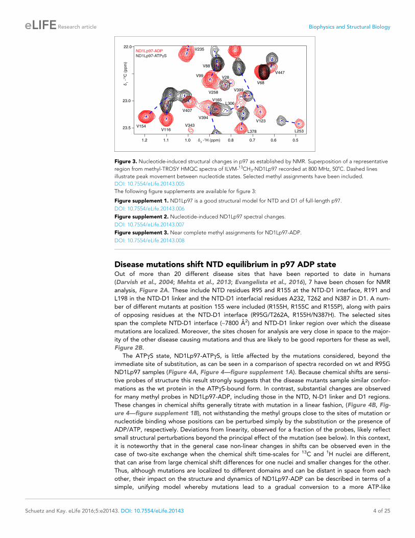
Disease mutations shift NTD equilibrium in p97 ADP stateOut of more than 20 different disease sites that have been reported to date in humans
(Darvish et al., 2004; Mehta et al., 2013; Evangelista et al., 2016), 7 have been chosen for NMR
analysis, Figure 2A. These include NTD residues R95 and R155 at the NTD-D1 interface, R191 and
L198 in the NTD-D1 linker and the NTD-D1 interfacial residues A232, T262 and N387 in D1. A num-
ber of different mutants at position 155 were included (R155H, R155C and R155P), along with pairs
of opposing residues at the NTD-D1 interface (R95G/T262A, R155H/N387H). The selected sites
span the complete NTD-D1 interface (~7800 A2) and NTD-D1 linker region over which the disease
mutations are localized. Moreover, the sites chosen for analysis are very close in space to the major-
ity of the other disease causing mutations and thus are likely to be good reporters for these as well,
Figure 2B.
The ATPgS state, ND1Lp97-ATPgS, is little affected by the mutations considered, beyond the
immediate site of substitution, as can be seen in a comparison of spectra recorded on wt and R95G
ND1Lp97 samples (Figure 4A, Figure 4—figure supplement 1A). Because chemical shifts are sensi-
tive probes of structure this result strongly suggests that the disease mutants sample similar confor-
mations as the wt protein in the ATPgS-bound form. In contrast, substantial changes are observed
for many methyl probes in ND1Lp97-ADP, including those in the NTD, N-D1 linker and D1 regions.
These changes in chemical shifts generally titrate with mutation in a linear fashion, (Figure 4B, Fig-
ure 4—figure supplement 1B), not withstanding the methyl groups close to the sites of mutation or
nucleotide binding whose positions can be perturbed simply by the substitution or the presence of
ADP/ATP, respectively. Deviations from linearity, observed for a fraction of the probes, likely reflect
small structural perturbations beyond the principal effect of the mutation (see below). In this context,
it is noteworthy that in the general case non-linear changes in shifts can be observed even in the
case of two-site exchange when the chemical shift time-scales for 13C and 1H nuclei are different,
that can arise from large chemical shift differences for one nuclei and smaller changes for the other.
Thus, although mutations are localized to different domains and can be distant in space from each
other, their impact on the structure and dynamics of ND1Lp97-ADP can be described in terms of a
simple, unifying model whereby mutations lead to a gradual conversion to a more ATP-like
0.60.70.81.01.11.2
22.0
23.0
23.5
ND1Lp97-ADP
ND1Lp97-ATPγS
0.5
V235
V68
V447
V88
V99 V28
V258V399
L306
V123
L253
V154V116
V407
V165
V394
V343
L378
δ2 -1H (ppm)
δ1 -
13C
(ppm
)
Figure 3. Nucleotide-induced structural changes in p97 as established by NMR. Superposition of a representative
region from methyl-TROSY HMQC spectra of ILVM-13CH3-ND1Lp97 recorded at 800 MHz, 50˚C. Dashed lines
illustrate peak movement between nucleotide states. Selected methyl assignments have been included.
DOI: 10.7554/eLife.20143.005
The following figure supplements are available for figure 3:
Figure supplement 1. ND1Lp97 is a good structural model for NTD and D1 of full-length p97.
DOI: 10.7554/eLife.20143.006
Figure supplement 2. Nucleotide-induced ND1Lp97 spectral changes.
DOI: 10.7554/eLife.20143.007
Figure supplement 3. Near complete methyl assignments for ND1Lp97-ADP.
DOI: 10.7554/eLife.20143.008
Schuetz and Kay. eLife 2016;5:e20143. DOI: 10.7554/eLife.20143 4 of 25
Research article Biophysics and Structural Biology

0.60.70.8
I189
I146
ND1Lp97-ATPγS
R95G & T262A
R155H & N387H
R95G
R191Q
L198W
N387H
R155H
ND1Lp97-ADP
I27
0.9
13.0
13.5
14.5
15.0
DI274
0.8
11.0
δ2 -1H (ppm)
δ1 -
13C
(pp
m)
D
1.2
V87
22.0
22.5
0.9V99
B
16.0
17.0
1.2
I100
I206
V87
0.70.7
22.0
23.0
1.2
V154
V116
ND1Lp97-ADP
R95G
full-length
p97-ADP
R95G
ND1L
p97-ADP
R95G
full-length
p97-ADP
R95G
C
ND1Lp97-ATPγS
R95G ND1Lp97-
ATPγS
A
I233
I146
I182
I437
I189
I303
Figure 4. A dynamic NTD equilibrium between up/down states is affected by mutations in ND1Lp97-ADP.
(A) Similar spectra are obtained for wt (black, single contour) and R95G (blue, multiple contours) ND1Lp97-ATPgS.
(B) Superposition of selected 13C-1H HMQC spectral regions of wt (red) and mutant ND1Lp97-ADP (colored as
indicated) and of wt ND1Lp97-ATPgS (black), focusing on V87, V99, I146 from the NTD, I189 from the NTD-D1
linker and I274 (D1), showing the progressive titration of cross-peak chemical shifts. Note that the I274 peak for wt
ND1Lp97-ATPgS is not shown; a large CSP is noted from the ADP to ATPgS substitution due to the proximity of
I274 to the nucleotide. (C) Superposition of selected regions of 13C-1H HMQC spectra of full-length wt and R95G
p97 (ADP state, right) showing analogous changes as for ND1Lp97-ADP (left).
DOI: 10.7554/eLife.20143.009
The following figure supplement is available for figure 4:
Figure supplement 1. Disease mutation-induced ND1Lp97 spectral changes.
DOI: 10.7554/eLife.20143.010
Schuetz and Kay. eLife 2016;5:e20143. DOI: 10.7554/eLife.20143 5 of 25
Research article Biophysics and Structural Biology

conformation (red to black in Figure 4B, despite the absence of ATP), consistent with a gradual
upward movement of the NTD. This conversion is readily apparent for I189, for example, located in
the NTD-D1 linker, that is sensitive to a loop to helix conformational change that accompanies the
down to up NTD conformation and has the largest chemical shift perturbation (CSP) between ADP-
(red) and ATPgS- (black) bound states. The chemical shifts in each of these states provide the limiting
values from which the fraction of NTDs that are up/down can be readily calculated for the different
mutants, as described below.
A concern in studies of smaller fragments of intact proteins is the possibility that the absence of
part of the structure may bias the results. As a control we have also studied full-length p97.
Figure 4C shows the superposition of regions of 13C-1H HMQC spectra recorded of both wt and
R95G variants of ND1Lp97-ADP and full-length p97-ADP. It is clear that the CSPs for both ND1Lp97
and full-length p97 are identical so that the upwards movement of the NTD upon introduction of dis-
ease mutations, characterized in ND1Lp97-ADP, is also observed in the intact molecule. Thus, unlike
the picture from X-ray studies of disease mutants, our NMR results establish that the NTD is not
static in p97-ADP, but rather exchanges rapidly between up/down states, with the relative mixture
of each state changing with mutation. The exchange rate is estimated to be greater than 2000 s�1,
based on the chemical shift differences between ADP and ATPgS-bound states, as measured for the
I189 Cd1 that has the largest CSPs. Moreover, less severe mutations (see below), such as R155H,
have a relatively small effect on the NTD equilibrium (orange peaks in Figure 4B), which is skewed
to the down state, but the severe R95G mutation (blue) shifts the relative up/down populations to
approximately equal.
Figure 5A shows all ILVM methyl groups that have been used as probes superimposed on the
structure of ND1Lp97, with each methyl indicated by a sphere that is color-coded by the difference
in chemical shifts in spectra recorded of R95G ND1Lp97-ADP and wt ND1Lp97-ADP constructs.
CSPs extend from the site of mutation (yellow) to the nucleotide-binding region and beyond to the
p97 central pore, identifying potential allosteric pathways of communication throughout the protein
(see Discussion). The 22 residues in ND1Lp97 that are mutated in IBMPFD disease patients are
shown in stick representation in red in Figure 5B. These mutation sites localize to regions with large
CSPs. Notably, regions of the NTD that are associated with UBX and VIM domain binding (see
below) are not perturbed by the disease mutations (no CSPs).
Disease mutations decrease interactions at the NTD-D1 interfaceFurther insight into the NTD equilibrium can be obtained from experiments that measure the overall
tumbling time, tc, of the NTD in the context of the ND1Lp97 hexamer in different nucleotide states
and for different mutations. We have obtained NTD tc values by quantifying the build-up of methyl1H triple-quantum coherence that occurs efficiently only for high molecular weight complexes and
rigid methyl probes (Sun et al., 2011) (see Materials and methods for details, Figure 6—figure sup-
plement 1). Figure 6A illustrates a number of build-up profiles focusing on examples from p97 con-
structs at different ends of the NTD dynamics spectrum. In the limit of a rigidly attached NTD tc is
predicted to be approximately 120 ns (as detailed in Materials and methods), while for a fully inde-
pendent domain a value of approximately 13 ns is expected. Correlation times of 91 ± 3 ns and
68 ± 2 ns were determined for ADP and ATPgS bound wt states, respectively, while a value of 13 ns
was measured for the isolated NTD. This implies that the NTD is only partially docked to D1, as
expected on the basis of X-ray and cryo-EM models of p97 (20, 22), with more motional freedom in
p97-ATPgS. A value of tc = 100 ± 3 ns was obtained for an intra-protomer cross-linked variant that
tethers the NTD to D1 (Figure 6A).
To quantify how the NTD tumbling time is correlated with the up/down equilibrium we have cal-
culated the fractional population of NTD in the up state, pU , for each of the variants examined. Since
plots of chemical shifts vs mutation are linear with little broadening during the course of the chemi-
cal shift vs mutation trajectory (Figure 4B) it is reasonable to assume a two-site exchange mechanism
between up/down NTD states that is fast on the NMR chemical shift time-scale. In this limit, pU val-
ues can be calculated according to the relation (Palmer et al., 2001)
dmutant ¼ pD dDþ pU dU (1)
where dmutant is the chemical shift of a methyl probe in the mutant, dD and dU are the corresponding
Schuetz and Kay. eLife 2016;5:e20143. DOI: 10.7554/eLife.20143 6 of 25
Research article Biophysics and Structural Biology

chemical shifts in the ADP (down) and ATPgS (up) states and pD=U is the fractional population of the
D/U state (pU þ pD ¼ 1). Values of pU for a range of disease mutants are listed in Figure 6B (purple)
along with tc values (blue), with tc decreasing (mobility increasing) as the percentage of NTD in the
up conformation grows, Figure 6C. It is worth noting that I189, L229 and M388 all have large CSPs
due to mutation and the pU values obtained from the trajectories of all 3 residues are consistent (see
legend to Figure 4—figure supplement 1B).
Both the chemical shift and relaxation data provide strong evidence that the NTDs are dynamic,
rapidly interconverting between up/down conformers. In further support of this interpretation we
have carried out chemical cross-linking experiments by introducing cysteine residues at positions
155 (NTD) and 387 (D1) that are within 3 A in the down state (Niwa et al., 2012), yet greater than
20 A in the up conformation. The R95G mutant has been chosen for cross-linking because it is effec-
tively in a ‘50% up’ conformation (Figure 6B) so that in the limit of a static structure a disulfide bond
connecting C155 and C387 would not be formed and CSPs would not be observed upon oxidation
of the sample. The 13C-1H HMQC spectrum of R95G,R155C,N387C ND1Lp97-ADP, Figure 6D,
under oxidizing conditions shows large CSPs for I146 and I189, consistent with disulfide bond forma-
tion. The presence of a disulfide bond is further established by comparing the chemical shifts of
these peaks (grey) with those for R155C,N387C ND1Lp97-ADP upon crosslinking, Figure 6E (grey),
where a locked, down NTD orientation is readily formed upon oxidation. Taken together our data
indicate that the disease mutants affect the up/down equilibrium in p97-ADP, with the NTD almost
entirely up in the R95G/T262A double mutant, similar to the ATPgS-conformation (Figure 6B).
Finally, it is worth noting that the positions of the I146 and I189 cross peaks under reducing
R95G
UBX & VIM
binding
0 ppm
1
0.4
A
B
Figure 5. Methyl CSPs introduced by the R95G mutation. (A) Methyl groups are shown as spheres and color-
coded according to the size of the CSP, as indicated. The UBX/VIM (Buchberger et al., 2001; Stapf et al., 2011)
binding groove between the two NTD subdomains is unaffected by disease mutations. (B) For reference, the
locations of the 22 identified sites of IBMPFD mutations in p97 ND1L are shown in red.
DOI: 10.7554/eLife.20143.011
Schuetz and Kay. eLife 2016;5:e20143. DOI: 10.7554/eLife.20143 7 of 25
Research article Biophysics and Structural Biology

conditions (i.e., absence of disulfide bond) are very different for R95G,R155C,N387C ND1Lp97-ADP
(blue) and R155C,N387C ND1Lp97-ADP (red) that reflects the different pU values for the NTDs, as
discussed above.
Implications of mutations on UBXD1 bindingWe next sought to establish the consequences of the mutations that perturb the up/down equilib-
rium for adaptor binding. The UBXD1 adaptor protein is abundant in neuronal cells (Madsen et al.,
2008; Nagahama et al., 2009) and a complex with p97 binds to the plasma membrane protein cav-
eolin-1, mediating sorting of ubiquitylated cargo in endosomal degradation (Ritz et al., 2011)
(Figure 1A, region highlighted in the orange square). Importantly, the UBXD1-p97 complex is specif-
ically disrupted in disease-associated mutations and function is impaired (Ritz et al., 2011) but the
molecular details underlying this disruption are not understood. An elegant biophysical study of the
isolated NTD with the N-terminal portion of UBXD1 (UBXD1-N, residues 1–133, Figure 7A)
(Trusch et al., 2015) shows an interaction involving VIM (residues 52–63) that is localized to the
canonical VIM/UBX binding site on the NTD (Stapf et al., 2011). Moreover, the authors suggest that
the N-terminus of UBXD1 may engage the NTD-D1 interface and NTD-D1 linker, where disease
mutations occur, and hypothesize that this secondary interaction may favor a more compact confor-
mation of p97. We have used methyl-TROSY NMR to study the binding of UBXD1-N to wt and
mutant forms of ND1Lp97-ADP, Figure 7, Figure 7—figure supplement 1–3. In Figure 7 the focus
for simplicity is on three residues. First, V68 that is localized to the UBX/VIM binding region of the
NTD and that, therefore, is a proxy for binding of VIM, and, second, I146 and I189 that report on
ATPADP Disease mutations
91 τc [ns]
NTD up [%]0
cross
linked
7888 72
10014 27 42 45 79
688091
R155H L198W
39
R95G&
T262AR191Q
R95G
R155H&
N387H
wt ADP
100
R155P
51
R155C
B
A EC
NTD up [%]
wt ATP
Disulfide
bond
ADP ADP
Disulfide
bond
Disulfide
bond
N387H
A232E
18
I146
I189
R155C
N387C
NTD in isolation
ND1Lp97
cross-linked
I189
I146
cross-linked
0.1
0.2
0.3
L96
0
0.1
0.2
0.3
V108
0
0.1
0.2
0
0.1
0.2
0.3
I119
τc [n
s]
120
100
13
0
60
80
0.80.9
V68
0 4 8 16T [ms]
0 4 8 16T [ms]
I TQ/I
SQ
92±8 ns
98±9 ns
88±8 ns
66±3 ns62±3 ns
53±4 ns
93±8 ns
70±7 ns
15±1 ns 15±1 ns
13±1 ns16±1 ns
isolated NTD
cross-linked ADP
ATPγS
0.80.9
13.5
14.0
15.0
0 20 80 100
D
δ2 -1H (ppm)
δ1 -
13C
(pp
m)
R95G
R155C
N387C
Figure 6. Probing NTD dynamics in ND1Lp97-ADP. (A) Measurement of NTD tumbling times for ND1Lp97-ADP cross-linked to the down position via a
disulfide link between positions 155 (NTD) and 387 (D1) (320 kDa, 50˚C, red), ND1Lp97-ATPgS (NTD up, 50˚C, black) and isolated NTD (24 kDa, 37˚C,grey). Correlation times were obtained via an approach that monitors the build-up of triple quantum coherence (see Materials and methods). (B) NTD
tumbling times (tc) and fraction NTD up (pU ), quantified by the 13C chemical shift of I189 for different mutants and nucleotide states of ND1Lp97.
Increased motion corresponds to lower tc. (C) Linear correlation of % NTD up vs NTD tc for different disease mutants. tc values for the isolated NTD
(adjusted for 50˚C) and ND1Lp97 (50˚C), calculated from peaks in D1 (see Materials and methods), are given by dashed grey lines for reference.
Chemical crosslinking via disulfide bond formation between cysteine residues at positions 155 and 387 (21) forces NTDs of both R95G ND1Lp97-ADP
(D) and ND1Lp97-ADP (E, wt at position 95) to the down position, and increases tc to 100 ns in both cases.
DOI: 10.7554/eLife.20143.012
The following figure supplement is available for figure 6:
Figure supplement 1. Histograms of tc distributions as obtained from per-residue fits of methyl 1H spin relaxation data.
DOI: 10.7554/eLife.20143.013
Schuetz and Kay. eLife 2016;5:e20143. DOI: 10.7554/eLife.20143 8 of 25
Research article Biophysics and Structural Biology

E0.80.9
0.7
δ2 -1H (ppm)
δ1 -
13C
(ppm
)
0.80.9
V68 V68 V68
I146 I146 I146
I189
I189
I189
R155H
R155H + UBXD1-N
wt + UBXD1-N
B C DND1Lp97-ADP
+ UBXD1-N
R95G
R95G + UBXD1-N
wt + UBXD1-N
A
0.80.9
14.0
14.5
15.0
ADP
ADP
ADP
22.5
VIMH1/H2
52 631 25 133ADP ADP
V68
I146
I189
Figure 7. Disease mutants impair binding of UBXD1-N. (A) (left) Domain organization of UBXD1-N (residues 1- 133
(35)), (right) schematic of binding reaction highlighting 3 key residues that are used as probes of binding in what
follows. (B) Selected regions of 13C-1H HMQC spectra of wt ND1Lp97-ADP without (red) and with (grey) 3-fold
excess UBXD1-N over protomer, focusing on V68, reporting on VIM domain binding, as well as I146 and I189 that
serve as proxies for binding of the H1/H2 motif of UBXD1 (see text). As the NTD is in the down position prior to
UBXD1-N binding, only small CPSs are observed for I146/I189, that reflect binding of H1/H2. (C) Addition of 3 fold
excess UBXD1-N to R155H results in CSPs (orange to black) that do not extend to the fully bound state observed
in wt (grey), indicating only a partial shifting of the up/down equilibrium to the down conformation (cartoon inset).
Note that I146/I189 peaks for unbound R155H are shifted upfield relative to wt (compare orange with red peaks)
reflecting the increased up population of the NTD for R155H (14%); differences in peak positions for wt and
mutant p97 in other panels also reflect changes in the up/down equilibrium. (D) Addition of UBXD1-N to R95G
ND1Lp97-ADP (42% up in the unbound state) results in partial binding of VIM but no binding of H1/H2 and
subsequently no shift in the up/down equilibrium. (E) Chemical shift perturbations caused by binding of VIM
(yellow) and H1/H2 (dark red) of UBXD1 to wt ND1Lp97-ADP mapped onto a surface representation of the NTD
(blue) and neighboring D1 structure (inset shows complete hexameric structure from which the surface was taken).
DOI: 10.7554/eLife.20143.014
The following figure supplements are available for figure 7:
Figure supplement 1. Positions of 3 key reporting residues, V68 (sensitive to VIM binding), I146 and I189 (both Ile
are sensitive to the NTD up/down equilibrium) superimposed on a structural model of a complex of the VIM
domain (yellow) and ND1Lp97-ADP.
DOI: 10.7554/eLife.20143.015
Figure supplement 2. Adaptor binding of UBXD1 to wt and mutant p97.
DOI: 10.7554/eLife.20143.016
Figure supplement 3. Titration of wt ND1Lp97-ADP with UBXD1-N to obtain an effective macroscopic KdðKd;macroÞ
for the binding reaction.
DOI: 10.7554/eLife.20143.017
Schuetz and Kay. eLife 2016;5:e20143. DOI: 10.7554/eLife.20143 9 of 25
Research article Biophysics and Structural Biology
binding of the H1/H2 region of UBXD1-N (Figure 7, Figure 7—figure supplement 1). Neither I146
nor I189 show CSPs from the addition of p47 UBX (Figure 8—figure supplement 1A,B), consistent
with the fact that these residues are not sensitive to binding at the canonical UBX/VIM groove. Bind-
ing of UBXD1-N causes small shifts to I146 and I189 of the wt protein (Figure 7B) that are consistent
with locking the NTD in the down conformation, as observed upon chemical cross-linking
(Figure 6E). Larger shifts are observed for V68 and I175 that report on VIM binding, from which a Kd
value of 22 ± 2 mM for the VIM-NTD interaction is calculated (see Materials and methods Figure 7—
figure supplement 3). This value is in good agreement with affinities measured for the binding of
full-length wt p97 to full length UBXD1, 3.5 mM (Hanzelmann and Schindelin, 2011), and for the
binding of the isolated NTD with UBXD1-N, 9 mM (Trusch et al., 2015), especially considering that
the latter pair of studies were carried out at 25˚C in comparison to 50˚C for the NMR results
reported here. Studies of R155H and N387H mutants of ND1Lp97-ADP that mildly perturb the up/
down equilibrium (Figure 7C, Figure 7—figure supplement 2D) show that two-pronged binding
involving VIM and H1/H2 occurs partially but that a complete down conformation is not obtained
with a 3-fold excess of adaptor over p97 protomer (compare dashed and solid arrows that corre-
spond to movements for peaks from wt and mutant p97, respectively). A Kd value of ~80 mM is
obtained for the VIM-NTD interaction in this class of mild disease mutants. Notably, while the VIM
domain weakly binds to the NTD of the strong mutants R95G and R155P ND1Lp97-ADP, Figure 7D
and Figure 7—figure supplement 2E, (Kd ~ 200 mM, see Materials and methods) there is no binding
of H1/H2 and no shift in the conformational equilibrium towards the down state, as evidenced by a
lack of CSPs for I146 and I189 (3:1 UBXD1-N:p97 monomer ratio). Supporting data from further
probes are highlighted in Figure 7—figure supplement 2. These include V38 and V108 in the UBX/
VIM binding site of NTD that are sensitive to the interaction with the VIM domain (Stapf et al.,
2011), and V116, V133 and V154 reporting on H1/H2 binding. Finally, it is worth emphasizing that
the similar trajectories of CSPs as a function of added UBXD1-N observed for wt and all disease
mutants (note the collinearity of dashed and solid arrows) provides strong evidence that the mecha-
nism of binding is identical in all cases, with the differences in the extent of shift changes reflecting
differing affinities, as calculated in the present study.
Our results thus establish a link between the severity of the disease mutant and the ability of
UBXD1 to shift the up/down equilibrium so as to lock the NTD in the down conformation. Figure 7E
plots CSPs obtained from VIM (yellow) and from H1/H2 (purple) binding on a space filling model of
the NTD, showing that the canonical VIM binding site is affected by the interaction with VIM
(Hanzelmann and Schindelin, 2011), while the NTD-D1 interface serves as the binding site for H1/
H2. A simple binding model for the UBXD1-N - p97 interaction is provided in the Materials and
methods.
Effects of disease mutations are more severe for specific adaptorsIt is notable that IBMPFD disease mutants affect lysosome-related functions but not others, such as
endoplasmic reticulum associated degradation and Golgi membrane fusion (Tresse et al., 2010;
Meyer and Weihl, 2014; Johnson et al., 2015) (Figure 1A). We therefore sought to establish that
the mutants considered here would not disturb complex formation with p47, one of the best-charac-
terized adaptors and known inhibitors of p97, that is involved in regrowth of Golgi from membrane
fragments (Kondo et al., 1997). As a first test we studied the p47 UBX domain exclusively, which
bound to wt and R95G ND1Lp97-ADP identically (Figure 8—figure supplement 1A,B), in agree-
ment with the literature (Fernandez-Saiz and Buchberger, 2010). Very similar CSPs were further
obtained for wt and R95G proteins upon binding full-length p47 (Figure 8A, Figure 8—figure sup-
plement 1C), which interacts as a trimer (Beuron et al., 2006). Notably, the CSPs upon binding p47
are distinct both in magnitude and in direction from those that result from perturbation to the NTP
up/down equilibrium (Figure 8A, Figure 8—figure supplement 1C). For example, addition of p47
leads to shifts in I189 that are predominantly horizontal (solid arrows), compared with near vertical
shifts (dashed arrows) that are observed as the NTDs become progressively detached from the D1
domain. Significant differences in CSPs are also observed between the I146 response to p47 binding
and to changes in the up/down equilibrium as reported by I146 (compare dashed and solid arrows
for I146). This suggests a different binding mechanism for p47 relative to UBXD1-N, that is made
clear when methyl probes showing CSPs from p47 binding are highlighted on the ND1Lp97 struc-
ture, Figure 8B. Binding occurs at two sites, with one region corresponding to the canonical UBX
Schuetz and Kay. eLife 2016;5:e20143. DOI: 10.7554/eLife.20143 10 of 25
Research article Biophysics and Structural Biology

binding site of the NTD (yellow), as observed for the binding of the VIM domain of UBXD1-N. A sec-
ond unique site on the NTD (purple) is also involved in binding that has been shown in a previous
structural study to engage the SHP domain of p47 (Hanzelmann et al., 2016). Unlike the second
site for UBXD1-N binding that bridges the NTD and D1 domains, the site of engagement of SHP
does not, so that it is not surprising that the p47-p97 interaction is not significantly perturbed by dis-
ease mutations. Figure 8C illustrates schematically the binding of p47 SHP and UBX domains to
p97.
I146
A
I189
I146
I189
UBX
SHP
SEP
UBA45
244
263
368
1
179
246
291ADP
ND1Lp97-ADP
ND1Lp97-ADP + p47
R95G
R95G + p47
I189
B
0.8
14.0
14.5
15.0C
δ2 -1H (ppm)
δ1 -
13C
(ppm
)
Figure 8. Binding of p47 is not impaired by disease mutations. (A) Superposition of selected regions of 13C-1H
HMQC spectra focusing on I146 and I189 of wt ND1Lp97-ADP and R95G ND1Lp97-ADP without (red for wt, blue
for R95G) and with (grey for wt, black for R95G) 1.25 fold excess p47. Similar CSPs indicate that binding to p47 has
not been impaired by the R95G mutation, nor is the up/down equilibrium changed. For reference, the ‘mutant
titration’ from Figure 4B is provided at the sides of the main spectrum for residues I146 and I189, emphasizing
that binding of p47 results in changes that are distinct from NTD up/down. (B) CSPs caused by binding of full-
length p47 to wt ND1Lp97-ADP as mapped onto a surface representation of the NTD (blue) and neighboring D1
structure (light red), with the inset showing the complete hexameric structure from which the surface was taken.
Residues affected by UBX binding are color-coded in yellow and those perturbed by SHP binding indicated in
dark red. (C) Domain organization of p47 and cartoon of the p47-p97 complex, highlighting the regions of
interaction.
DOI: 10.7554/eLife.20143.018
The following figure supplement is available for figure 8:
Figure supplement 1. Adaptor binding of p47 to wt and mutant p97.
DOI: 10.7554/eLife.20143.019
Schuetz and Kay. eLife 2016;5:e20143. DOI: 10.7554/eLife.20143 11 of 25
Research article Biophysics and Structural Biology

DiscussionThe development of significantly improved NMR instrumentation, new labeling methodologies
(Tugarinov and Kay, 2004; Gelis et al., 2007; Kainosho et al., 2006; Gans et al., 2010) and novel
NMR experiments that exploit the labeling in ways that preserve signal intensity (Tugarinov et al.,
2003; Fiaux et al., 2002) has significantly impacted on the size of protein complexes that are now
amenable for detailed solution NMR spectroscopy studies (Rosenzweig and Kay, 2014). Here we
have used experiments that rely on a methyl-TROSY effect to quantify inter-domain dynamics in a
series of IBMPFD mutants of human p97 and to study interactions with adaptor molecules. Many
studies of molecular machines by NMR have focused on homo-oligomeric systems where each pro-
tomer is relatively small (20–30 kDa) (Sprangers and Kay, 2007; Shi and Kay, 2014). In the case of
ND1Lp97, that is the subject of the present work, protomeric molecular weights in excess of 50 kDa
challenge resolution in spectra, necessitating the use of an elegant labeling strategy developed by
Boisbouvier and coworkers whereby methyl groups are 13CH3-labeled in a stereospecific manner
(Gans et al., 2010). Near complete assignments for ILVM methyl groups (pro-R for LV) are reported
for both ADP and ATP states of the enzyme (Figure 3—figure supplement 3).
To date over 40 different IBMPFD disease mutations have been identified in 22 different positions
throughout ND1Lp97 (Evangelista et al., 2016), of which 7 representative sites were studied here.
From our dynamics studies a picture emerges whereby the NTDs of p97-ADP interconvert between
up/down conformations that can be modulated by IBMPFD disease mutations. X-ray and cryo-EM
studies have established that for the wt protein the up/down NTD equilibrium is highly skewed to
the down state in the ADP bound form (Tang et al., 2010; Banerjee et al., 2016; Zhang et al.,
2000), and our NMR results show that this arrangement facilitates the two-pronged binding of
UBXD1 to the enzyme. Our NMR studies further show that IBMPFD disease mutations perturb this
equilibrium in the direction of the up state, leading to an impaired biological ‘readout’, namely
UBXD1 adaptor binding.
A key strength of NMR in studies of proteins lies in the fact that the easiest parameter to measure
– the chemical shift – is also amongst the most powerful in detecting subtle conformational changes
of the sort that can give rise to allosteric pathways of communication, for example. Detailed crystal-
lographic studies of p97 have established that the NTD/D1 interface and the conformation of the
linker between NTD and D1 are coupled to the nucleotide state in D1 (20, 50). Notably, in the wt
protein the nucleotide state in D1 determines the NTD up/down conformation and, conversely, per-
turbing the NTD down state by the introduction of disease mutations in the interface region propa-
gates to the nucleotide-binding pocket (Figure 5). CSPs from the R95G mutation are indicated in
Figure 5A; these provide some insight into the communication between distal regions of p97 at the
level of individual amino acids, as highlighted in Figure 9. Here we show a number of pathways (I-III)
defined by CSPs that arise upon perturbing the up/down equilibrium. A first pathway (I) includes res-
idues in the linker connecting NTD with D1, with large CSPs observed for I189 and L198 at opposite
ends of the linker; CSPs are also observed for I206 and L213 proximal to the nucleotide binding site.
The arginine finger (R359) and Walker B (E305) residues, at the opposite face of the nucleotide bind-
ing site, sense the up/down equilibrium (in the case of the wt protein, ADP versus ATP) leading to
CSPs close to the central pore, as observed for I274. Pathway II involves residues from an extensive
interface between NTD and D1, including numerous disease mutation sites such as R155 and N387,
and perturbations to the up/down equilibrium lead to changes far into the D1 domain. Finally, the
NTD conformational change is communicated to the adjacent protomer at the I27/L429 interface via
pathway III and this network eventually connects to the same cluster of residues as in pathway II.
Despite small patient groups and phenotypic variability it is possible to classify mutants as moder-
ate or severe, based on age of onset and relative elevation of disease markers (Figure 10A), with
R155H and R155C, the two most frequent mutations in patients (Mehta et al., 2013), defined as
weak and strong, respectively. An important result from this study is the strong correlation between
disease severity and the extent to which the up/down equilibrium is skewed in the ND1Lp97-ADP
state with more severe mutants unable to engage the H1/H2 region of UBXD1 that is required to
lock the NTD conformation in the down state, illustrated schematically in Figure 10B. Because a
complex of UBXD1-p97 is required for recruitment of ubiquitylated caveolin-1 to the lysosome for
degradation this pathway becomes impaired in the disease mutants, leading to the accumulation of
caveolin-1 positive endolysosomes in IBMPFD patients (Ritz et al., 2011). Notably, significantly
Schuetz and Kay. eLife 2016;5:e20143. DOI: 10.7554/eLife.20143 12 of 25
Research article Biophysics and Structural Biology

impaired binding to IBMPFD disease mutants of p97 is not observed for all adaptors. For example,
we have shown that binding of p47 is not perturbed by mutation (see below), consistent with the
location of observed CSPs showing that p47-p97 interactions only engage the NTD and do not
include the NTD-D1 interface, as for UBXD1 (compare Figure 7E and Figure 8B).
It is of interest to ask why these structural changes have not been observed via other high-resolu-
tion techniques. The answer partly lies in the fact that the mild disease mutants introduce only subtle
changes to the equilibrium, with R155H and N387H mutations shifting the NTD equilibrium from 0%
up to ~15% up in p97-ADP, for example. Moreover the differences in free energies between the up
and down conformations are relatively small, with 4Gup/down(R155H) ~ 1200 cal/mol and 4Gup/
down(R95G) ~ 200 cal/mol based on the measured pU values (Figure 6B). The equilibrium may well
be affected by the liquid nitrogen temperatures used in x-ray and cryo-EM studies and by crystal
packing forces that could skew populations. The significant conformational dynamics associated with
the up/down interconversion may also make it difficult to observe this effect by crystallography. For
example, while crystal structures of weak disease mutants in the ADP state (R155H, L198W) have
been published and all show the down NTD conformation, as for the wt, structures of strong
mutants such as R95G, R155C/P in the ADP state (4Gup/down~0 cal/mol) have not been reported.
On an atomic scale, only localized changes were observed in the crystal structures of mutants of
p97-ADP relative to the wt conformation (Tang et al., 2010; Tang and Xia, 2013;
Tang and Xia, 2016). For example, the sidechain of R359, which serves as a sensor of the nucleotide
state of an adjacent protomer, is slightly shifted and this shift is thought to lead to a destabilization
of the ADP state in mutants. Moreover, the sidechain orientation of F360 differs between ADP states
of wt and disease mutants (Tang and Xia, 2013). Together, these structural differences are thought
to result in defective communication between subunits in mutant p97 molcules.
R95G
III
II
I
L198I189
I274
I27
L429
Central
poreI206
E305
R359
L213
R155
N387
Figure 9. Putative allosteric networks illustrating ‘pathways of communication’ (I-III) from the site of the R95G
disease mutation to regions distal in the structure. R95 is highlighted in yellow. Chemical shift perturbations upon
R95G mutation above 0.3 ppm are shown as red spheres. Key residues that form continuous pathways are shown
as black sticks, as discussed in the main text.
DOI: 10.7554/eLife.20143.020
Schuetz and Kay. eLife 2016;5:e20143. DOI: 10.7554/eLife.20143 13 of 25
Research article Biophysics and Structural Biology

The effects of IBMPFD disease mutations on p97 adaptor interactions, nucleotide binding, and
nucleotide hydrolysis rates have been extensively investigated in a series of papers (Fernandez-
Saiz and Buchberger, 2010; Tang and Xia, 2013; Manno et al., 2010; Zhang et al., 2015;
Bulfer et al., 2016). Beyond the primary defect in UBXD1 binding (Ritz et al., 2011), altered cofac-
tor interactions have been reported in a number of studies, including those of p47, p37, Ufd1-Npl4,
E4B and ataxin-3 (41, 53, 54). Due to the NTD conformational equilibrium shift in disease mutants,
changes in interactions are expected whenever adaptors bind differentially to the up and down con-
formations of the NTD, which are characteristic of ATP and ADP states of wt p97, respectively. Our
experiments, that do not detect differences in p47 interactions with wt or disease mutants of p97,
are consistent with biochemical studies of p47 binding to both wt and mutant variants of p97, for
which affinities in the nM range are obtained (Bulfer et al., 2016). In these studies intact or even
increased p47 binding of mutants has been detected (Fernandez-Saiz and Buchberger, 2010;
Bulfer et al., 2016) and no differences in ATP hydrolysis kinetics between wt and disease mutant
p97-p47 complexes in the limit of saturated binding have been observed (Zhang et al., 2015). Our
NMR experiments are not sensitive to affinity differences for p47 and wt or mutant p97 since dissoci-
ation constants (nano-molar range) are below the threshold for which differences can be detected.
They do establish, however, that the p97-p47 complex is formed analogously in wt and disease
mutants (Figure 8A), and, unique to the NMR, that the skewed NTD conformational equilibrium
found in disease mutants of p97-ADP is conserved in the p47 bound complex.
Several biochemical studies have reported altered nucleotide binding properties for disease
mutants in terms of affinity and the amount of prebound nucleotide along with elevated ATPase
activities (Tang and Xia, 2013). In general, the literature consensus is that disease mutations slightly
Re
lative
ele
va
tio
n
Disease markers
Disease
0
1
2
3
4
5
30
40
50
60
Ag
e o
f o
nse
t [y
ea
rs]
ALP PYD DPD
A
PDB IBM FTD
B‘weak’
R155H
‘strong’
R155C/P
R95G
~80 ns~15% ~50%~90 ns
NTD upτ
c
ADP
ADPADPD
ise
ase
se
ve
rity
Disease severity
Figure 10. Correlation between disease severity and up/down equilibrium. (A) Disease (PDB=Paget disease of
bone, IBM=inclusion body myopathy, FTD=frontotemporal dementia) onset and elevation of biochemical markers
relative to normal (ALP=alkaline phosphatase, PYD=pyridinoline, DPD=deoxypyridinoline) (Mehta et al., 2013), is
correlated with the extent of perturbation of the NTD up/down equilibrium. Data from R155H (R155C/P, R95G)
patients are used for ‘weak’ (‘strong’) mutants; R155C is associated with a significantly earlier onset of symptoms
compared to R155H and showed significantly reduced mean survival (Mehta et al., 2013). (B) Schematic of
UBXD1-N binding to ND1Lp97-ADP.
DOI: 10.7554/eLife.20143.021
Schuetz and Kay. eLife 2016;5:e20143. DOI: 10.7554/eLife.20143 14 of 25
Research article Biophysics and Structural Biology

decrease the affinity of D1 for ADP (by factors between 2–4) but not for ATP, while increasing the
ADP release rate from D1 by approximately 2-fold (Tang et al., 2010; Tang and Xia, 2013;
Bulfer et al., 2016). We have observed no simple correlation between biophysical properties of vari-
ous disease mutants and either perturbation of the NTD conformational equilibrium or empirical dis-
ease severity in patients. As an example, ATPase activities of disease mutants were reported to be
normal in some (Fernandez-Saiz and Buchberger, 2010) and elevated in other studies (Tang et al.,
2010; Niwa et al., 2012; Tang and Xia, 2013) but not in a manner that correlates with disease
severity. Further, R155H is a weak disease mutation in patients (Mehta et al., 2013), with only a
modest impact on the NTD up/down equilibrium, yet R155H p97 has been reported to have a partic-
ularly high ATPase activity and a low affinity for ADP (Tang and Xia, 2013). Thus, it is difficult to
understand presently how altered nucleotide interactions can be the prevalent cause of disease,
especially since it becomes hard to explain why only a subset of p97 functions would then be
affected. In contrast, our results provide strong support for a model whereby perturbation of the
up/down NTD equilibrium in the p97-ADP state due to mutation couples to disease by impairing
p97 binding to the UBXD1 adaptor. Stronger perturbations lead to more acute disease (Figure 10).
The binding of other adaptors, such as p47, that are involved in processes distinct from that of
UBXD1, are much less affected, if at all, by the IBMPFD mutations.
In summary, this work provides a further demonstration of the utility of methyl-TROSY NMR in
studies of high molecular weight complexes where dynamics play a critical role in both function and
misfunction. While we report near-complete methyl group assignments for both ADP and ATP states
of ND1Lp97 it is worth mentioning that the key conclusions about the disease mutants can be
obtained without extensive assignment, by recording simple 2D 13C-1H HMQC spectra of the
IBMPFD mutant series, with quantification of the NTD up/down populations from the CSPs of only a
few key reporter residues. Our NMR results paint a picture whereby the primary effect of disease
causing mutations is a change in NTD dynamics accompanied by more subtle structural changes that
propagate to regions distal from the sites of mutation. Perturbations to the up/down NTD equilib-
rium are observed in the form of CSPs that extend across the NTD/D1 interface and include the
linker region, which in turn connects to the nucleotide-binding site (Figure 9). Residues showing
large CSPs comprise a network that mediates communication between the NTD and D1 in wt p97
that extends to the central pore. Indeed, many of the 22 mutation sites reported to date in
ND1Lp97 are localized to regions with large CSPs, suggesting that these loci serve as ‘hot-spots’ for
propagation of conformational changes. Such sites may well serve as starting points for the rational
design of drug molecules that modulate NTD-D1 interactions, potentially reversing the effects of the
disease mutations or inhibiting the wt protein, a strategy currently explored in clinical cancer studies
(Anderson et al., 2015).
Materials and methods
Plasmids and constructsDNA encoding the p97 protein from Homo sapiens/Mus musculus (which are identical, Uniprot entry
P55072 versus Q01853) was obtained from GenScript (Piscataway, NJ, USA) with an N-terminal
6xHis tag and TEV cleavage site, sub-cloned into the NdeI and XhoI sites of pET29b+ (Novagen,
Madison, WI, USA). Point mutations and stop codons were introduced using Quikchange site-
directed mutagenesis (Agilent, Santa Clara, CA, USA). The truncations used for this study were p97
ND1L 1–480 (Song et al., 2003) and p97 NTD 1–213 (Isaacson et al., 2007). DNA encoding adap-
tors, including full-length p47, p47 UBX (residues 282–371 [Yuan et al., 2001]) and UBXD1-N (resi-
dues 1–133 [Trusch et al., 2015]) from Mus musculus were also obtained from GenScript, with
affinity tags, TEV cleavage sites and sub-cloning as for p97.
Protein expression and purificationThe isolated NTD of p97, ND1Lp97, full-length p97, p47 UBX, full-length p47 and UBXD1-N were
overexpressed in Escherichia coli BL21(DE3) cells in minimal M9 D2O media supplemented with15NH4Cl and [2H,12C]-glucose as the only nitrogen and carbon sources (Cambridge Isotope Labora-
tories (Tewksbury, MA, USA). Selective labeling with I-d1-[13CH3], V/L-g1/d1(proR)-[
13CH3,12CD3] and
M-e1[13CH3] was achieved as detailed previously (Gelis et al., 2007; Gans et al., 2010;
Schuetz and Kay. eLife 2016;5:e20143. DOI: 10.7554/eLife.20143 15 of 25
Research article Biophysics and Structural Biology

Tugarinov et al., 2006). Cells were induced at OD600 » 0.8 by addition of 1 mM IPTG and expres-
sion was performed for ca. 20 hr overnight at 18˚C (25˚C for UBXD1-N). p97 was purified using Ni-
affinity chromatography and the affinity tag was cleaved using TEV protease. The cleaved proteins
were concentrated using an Amicon Ultra-15 100 kDa molecular weight cutoff filter (Millipore, Etobi-
coke, ON, Canada) and nucleotide removed using apyrase (New England Biolabs, Ipswich, MA,
USA, 2 units per NMR sample) for at least 2 hr (room temperature). The apo protein was further
purified on a HiLoad 16/60 Superdex 200 gel filtration column (GE Healthcare, Pittsburgh, PA, USA).
Buffer compositions were adapted from (Chou et al., 2014). BME (2 mM) and DTT (2 mM) were
used as reducing agents during Ni-affinity chromatography and TEV cleavage, nucleotide digestion,
gel filtration and for storage (DTT), respectively. Purification schemes for full-length p47 and p47
UBX were analogous to that used for p97, without reducing agent as there are no Cys residues in
p47. In order to avoid proteolysis of full-length p47, protease inhibitor cocktail tablets (Roche, Basel,
Switzerland) were added during all steps of purification except during TEV cleavage. Full-length p47
eluted as a trimer on a HiLoad 16/60 Superdex 200 gel filtration column and p47 UBX as a monomer
on a HiLoad 16/60 Superdex 75 gel filtration column.
Purification of UBXD1-N was achieved with cell lysis and Ni-affinity chromatography under dena-
turing conditions (6 M guanidine), followed by TEV cleavage and size exclusion chromatography
using a HiLoad 16/60 Superdex 75 column under native conditions.
Protein concentrations were determined based on absorbance at 280 nm for p97 and full-length
p47. Neither p47 UBX nor UBXD1-N contains W or Y residues; protein concentrations for these con-
structs were thus determined via quantitative amino acid analysis. Proteins were subsequently
exchanged into the appropriate NMR buffer or frozen at �80˚C after addition of 5% glycerol for
future use.
NMR sample preparationProtein samples of the isolated NTD were generated via exchange (10 kDa MWCO filters) into a
buffer comprising 25 mM HEPES pH 7.5, 50 mM NaCl, 10 mM MgCl2, 5 mM TCEP. Solvents that
were either 90%/10% H2O/D2O (15N-1H based experiments) or 100% D2O (13C-1H) were used along
with protein concentrations up to 1.5 mM.
Optimal buffer conditions for full-length p97 and ND1Lp97 were identified via screening salt and
nucleotide concentrations using dynamic scanning fluorimetry (using Sypro Orange (Sigma) as a
reporter dye) for maximal melting temperature. ND1Lp97 samples were prepared for NMR via
buffer exchange (100 kDa MWCO filters) into D2O buffer containing 25 mM HEPES, pH 7.5(7.0), 25
(50) mM NaCl, 5 mM ATPgS(10 mM ADP), 4(0) mM MgCl2, 5 mM TCEP for ND1Lp97-ATPgS
(ND1Lp97-ADP) at protein concentrations between 100 mM (for assignment by mutagenesis), 600
mM (dynamics data) and 1 mM (NOESY based assignment). To limit ATP hydrolysis, ATPgS, a com-
mercially available non-hydrolyzable analogue of ATP (Sigma/Roche), was employed in combination
with the Walker B mutant of p97 (Wendler et al., 2012), in which the glutamate residue competent
for ATP hydrolysis (E305Q) is replaced by glutamine. Samples were stable at 50˚C for ~3 days in the
ATPgS state, limited by ATPgS hydrolysis, and for about one week in the ADP form. It is worth
emphasizing that under the conditions of our NMR experiments samples are fully nucleotide bound.
For example, reported Kd values for ADP binding to wt and disease mutants range from 0.1 mM – 5
mM, with the lower affinities associated with the disease mutants (Tang et al., 2010; Tang and Xia,
2013). Interestingly there is not a correlation between lower ADP affinity and disease severity as
Kd(wt) = 0.88 ± 0.18 mM < Kd(R95G) = 2.3 ± 0.1 mM < Kd(R155H) = 4.3 ± 0.5 mM. Assuming Kd = 5
mM, a concentration of p97 monomer of 600 mM (highest used) and [ADP]= 10 mM the amount of
apo-p97 would be on the order of 0.01% of the nucleotide bound species.
NMR sample conditions for studies involving adaptors were identical to those used for p97. Con-
centrations (monomer) used for binding experiments were as follows: UBXD1-N 150 mM, wt/R95G/
R155H/R155P/N387H ND1Lp97-ADP 50 mM; full-length p47 300 mM, wt/R95G NDL1p97-ADP 250
mM; p47 UBX 300 mM, ND1Lp97-ADP 200 mM. All p97 binding partners were perdeuterated. Our
NMR studies necessitate monomer protein concentrations in the tens-hundreds of mM range,
depending on the application. Interestingly, as protein concentrations can reach 200 mg/mL
(Ellis, 2001) or ~5 mM in the cytosol, with p97 accounting for an estimated 1% of cytosolic proteins,
the NMR experiments are carried out under conditions that resemble physiological. Of note, sam-
ples of the ND1Lp97-ADP/UBXD1-N complex were stable for ca. 5 hr at 50˚C, at which point
Schuetz and Kay. eLife 2016;5:e20143. DOI: 10.7554/eLife.20143 16 of 25
Research article Biophysics and Structural Biology

precipitation of UBXD1-N leads to increasing amounts of unbound p97. In contrast, the tightly
bound p47-p97 complexes were stable for at least 12 hr.
Cross-linkingCovalent cross-linking of NTD to D1 in ND1Lp97 was achieved by mutating R155 (NTD) and N387
(D1) to Cys (Niwa et al., 2012). The resulting protein was purified as described above using reduc-
ing conditions and dialyzed into non-reducing buffer for 3 days at 4˚C prior to NMR experiments.
For R95G ND1Lp97 oxidation was more difficult than for the wt protein and required the addition of
the oxidizing agent Cu phenanthroline (McIntosh and Freedman, 1980; Barthelme et al., 2014).
The increased difficulty in oxidation of the R95G sample is consistent with the fact that the NTD
equilibrium in the mutant is much further in the up direction so that cross-linking, which only occurs
from the NTD down position, is less efficient. The completion of the reaction was monitored by
NMR spectroscopy via chemical shift changes of the proR methyl group of V154, adjacent to C155.
Data acquisition and analysisNMR experiments were performed at field strengths of 14.0 T and 18.8 T using either Varian or
Bruker spectrometers. With the exception of several 3D data sets and the adaptor data all experi-
ments at 18.8 T were measured on a system with a room temperature probe; the remaining experi-
ments were obtained using cryogenically cooled probes. Experiments were recorded at 37˚C on the
isolated NTD and at 50˚C for all samples involving ND1Lp97 and full-length p97. Spectra were proc-
essed using the NMRPipe suite of programs (Delaglio et al., 1995) with chemical shift assignments
obtained with the aid of the CcpNMR (Vranken et al., 2005) program. Peak fitting was performed
using FuDA (http://pound.med.utoronto.ca/~flemming/fuda/), with remaining data analysis achieved
via home-built scripts executed in MATLAB.
NMR assignmentsAssignment of p97 NTDThe NTD domain of p97 is stable and monomeric (24 kDa) and sequential backbone resonance
assignments have been obtained before (Isaacson et al., 2007). These were kindly provided to the
authors by Professor Rivka Isaacson (King’s College, London). Resonance assignments were repeated
using standard TROSY-based (Pervushin et al., 1997) triple resonance experiments (Sattler and
Schleucher, 1999) (HNCO, HNCACO, HNCACB, CBCA(CO)NH) on a uniformly [13C,15N,2H] labeled
sample and subsequently extended to the methyl sidechains using (H)C(CO)NH-TOCSY, H(CCO)NH-
TOCSY schemes (Gardner et al., 1996) ([13C,15N, ~70% 2H] labeled sample). As a final step, assign-
ments were confirmed via analysis of a 3D 13C-edited NOESY data set (200 ms mixing time) that was
modified to include the methyl-TROSY approach (sequence available upon request). The experiment
was recorded as 13C(t1) NOE 13C(t2),1H(t3) where ti is a chemical shift acquisition period. Assigned
NOEs were cross-validated based on the X-ray structure of the domain (Hanzelmann et al., 2011)
(pdb ID 3QQ7). Near complete assignments of methyl groups of ILVM residues were obtained (56/
59), while those that could not be assigned (L17, I32, M158) did not give rise to peaks in spectra.
Stereospecific assignment of V and L methyl groups was achieved as reported by Neri et al.
(Neri et al., 1989).
Assignment of methyl groups of ND1Lp97NTD assignments were subsequently transferred to spectra of ND1Lp97 (6x53 kDa) in ADP and
ATPgS nucleotide states via an NOE-based approach using 3D 13C-edited NOESY data sets
(Sprangers and Kay, 2007). Methyl chemical shifts for residues in the D1 domain were obtained via
a mutagenesis strategy (Sprangers and Kay, 2007), whereby assignments are generated by mutat-
ing methyl containing residues and comparing the resulting spectra with the corresponding data set
recorded on the wt protein. Assignments were extended via analysis of NOE data sets where mea-
sured distances between proximal methyl groups in solution, as estimated qualitatively by NOE
cross-peak intensities, were compared with those predicted on the basis of p97 X-ray structures
(ADP state pdb ID 1E32 [Zhang et al., 2000]; ATP state pdb 4KO8 [Tang and Xia, 2013]). Spectra
of 40 point-mutants of residues distributed throughout D1 were recorded in both ADP and ATPgS
states, with remaining residues assigned based on NOEs (150 ms mixing time) to these 40
Schuetz and Kay. eLife 2016;5:e20143. DOI: 10.7554/eLife.20143 17 of 25
Research article Biophysics and Structural Biology
‘anchoring’ positions. In the ADP state, 128/132 (97%) of all methyl probes could be assigned,
including 54/54 in the NTD, 4/4 in the linker region and 70/74 in the D1 domain. The total of 132
methyl groups is based on including only 1 methyl for each V/L residue as labeling of these residues
was proR (Gans et al., 2010). Assignments for 104/132 (79%) of the methyl groups in ND1Lp97-
ATPgS were obtained (54/54, 4/4 and 46/74 in the NTD, linker and D1, respectively). The point
mutants utilized for assignment were chosen based on prediction of mutant stability from 44G cal-
culations (Kellogg et al., 2011) and sequence homology alignment.
Dynamics measurementsNTD correlation time15N R1 and R1� relaxation rates and steady state 15N-{1H} NOE values (Farrow et al., 1994) for the
isolated NTD were acquired at 600 MHz, 37 oC using relaxation delays of 10, 110, 240, 380, 550,
750, 1000 ms (R1) and 2, 7, 14, 22, 31, 42, 55 ms (R1�) along with a 1.8 kHz spin-lock field (n1). R1�
values were converted to R2 rates via the relation, R1� = R2 sin2(q) + R1 cos2(q), where tan(q)=n1/4,
and 4 is the 15N chemical shift offset from the carrier. Per-residue values of tc; S2 and te were
obtained via a spectral density mapping approach (Farrow et al., 1995), where tc is the assumed
isotropic molecular tumbling time, S2 is the square of an order parameter that is related to the
amplitude of the amide bond vector motion and te is the correlation time for rapid bond vector
dynamics. Briefly, values of the spectral density function at three frequencies were obtained for each
residue and these were fit to extract (t; S2; te). A value of tc = 13.6 ± 0.9 ns was obtained, where
the error is given as 1 standard deviation of the range of values, using only those residues in struc-
tured regions of the protein. This value is in keeping with expectations for a 24 kDa protein, 37˚C(Jarymowycz and Stone, 2006). The value of tc so obtained was subsequently used to calculate
methyl axis order parameters, as described below.
Measurement of isolated NTD domain methyl S2axistc valuesMethyl 1H spin relaxation data sets were recorded (37˚C) as described in (Sun et al., 2011) using an
approach that quantifies the time dependencies of sums and differences of magnetization that give
rise to 1H single- (ISQ) and triple- (ITQ) quantum coherences, respectively. Relaxation delay values T of
2, 7, 12, 17, 22, 27, 32, 37, 42, 50 ms were recorded in an interleaved manner and intensities of
methyl cross peaks fit according to
ISQ
ITQ
�
�
�
�
�
�
�
�
¼0:75h tanhðT
ffiffiffiffiffiffiffiffiffiffiffiffiffiffiffi
h2 þ d2p
Þffiffiffiffiffiffiffiffiffiffiffiffiffiffiffi
h2 þ d2p
� dtanhðTffiffiffiffiffiffiffiffiffiffiffiffiffiffiffi
h2þ d2p
Þ(2)
with S2axistc obtained via
hffi9
10
�0
4p
� �2
P2 cos�axis;HH� �� �2S
2
axisg4
H�h2tC
r6HH(3)
In Equation 3 P2ðxÞ ¼ ð3x2� 1Þ=2 with �axis;HH ¼ 90� the angle between the methyl threefold axis
and the vector connecting a pair of methyl protons, S2axis is the square of an order parameter quanti-
fying the amplitude of motion of the methyl threefold symmetry axis, �h is Planck’s constant divided
by 2p; gH is the gyromagnetic ratio of a proton spin, and rHH is the distance between pairs of methyl
protons (1.813 A). The parameter d in Equation 2 accounts for the proton density around each
methyl group. Values of S2axis were obtained for each methyl group from the quantified S2axistc values,
assuming tc =13.6 ns as obtained above from 15N spin relaxation experiments. We did not attempt
to take into account any motional anisotropy, as the orientation of methyl groups from X-ray struc-
tures must be considered to be approximate, especially in cases where sidechains are dynamic.
Determination of tcfor ND1Lp97
Methyl 1H spin relaxation data sets, as described above, were recorded on samples of ND1Lp97 (for
wt and mutant proteins in ADP, ATPgS states) using relaxation delay values of 0.5, 1, 2, 3, 4, 5, 6, 7,
8, 10, 12, 15, 18 ms. Twenty NTD residues were considered for analysis based on minimal chemical
shift changes between corresponding methyl groups in the isolated NTD and in the NTD of
Schuetz and Kay. eLife 2016;5:e20143. DOI: 10.7554/eLife.20143 18 of 25
Research article Biophysics and Structural Biology

ND1Lp97. This ensures that individual NTD methyl group dynamics are little affected by the sur-
rounding D1L in ND1Lp97 constructs. Thus, S2axis values measured for isolated NTD would be
expected to be similar for the corresponding NTD methyl groups in ND1Lp97. S2axis values obtained
for residues in the isolated NTD (37˚C) were extrapolated to 50˚C by assuming a temperature-
dependence as described by Wand and coworkers (Song et al., 2007). These extrapolated order
parameters were then used to obtain a global estimate of the tc for the NTD of ND1Lp97, Figure 6—
figure supplement 1, via fitting measured S2axistcvalues for ND1Lp97 using values for S2axis obtained
as described above. A tc value for the hexameric barrel formed by the D1 domains was obtained
from maximum S2axistc values measured for methyl groups of D1, as described previously
(Sprangers and Kay, 2007). The value of 120 ns obtained in this manner is in good agreement with
that obtained for the half-proteasome, a7a7, that is of a similar size (360 kDa, 50˚C) to NDL1p97
(Sprangers and Kay, 2007). Interestingly, HYDRONMR (Garcıa de la Torre et al., 2000) predictions
for tc of ND1Lp97 were 150–200 ns, depending on which X-ray structure was used in the calculations
(1E32 [Zhang et al., 2000], 3HU1 [Niwa et al., 2012], 4KOD [Tang and Xia, 2013]).
Analysis of UBXD1-N adaptor titrationA previous study has established that UBXD1-N has two p97 binding regions (Trusch et al., 2015).
These include a well-characterized VIM domain that forms a single ~10 residue helix upon binding to
a groove between NTD sub-domains (Stapf et al., 2011; Hanzelmann and Schindelin, 2011) and an
H1/H2 helical domain that has been shown through biochemical studies to fix the NTDs of wt p97-
ADP in the down position (referred in our work as the locked, down position). The results presented
in Figure 7 of the main text establish a two-pronged binding mode with NTD residues at the canoni-
cal UBX/VIM binding interface reporting on VIM binding (for example V38, V68, V108, see Figure 7,
Figure 7—figure supplement 2D,E), with residues such as I146 and I189 reporting on H1/H2
binding.
The CSPs of V68 from the addition of UBXD1-N to R95G ND1Lp97-ADP has been used to esti-
mate a Kd for the VIM ND1Lp97-ADP interaction. This mutant allows the deconvolution of VIM and
H1/H2 binding because the latter interaction does not occur (CSPs are not observed for I146/I189 of
the R95G mutant). The fraction of bound VIM for [NTD]T = 6x[ND1Lp97-ADP] = 50 mM, [UBXD1-N]T= 150 mM considered here, where [NTD]T and [UBXD1-N]T are the total (T) concentrations of NTD
and UBXD1-N, respectively, can be calculated from the shift (di) of V68 (black peak, Figure 7D inset)
and the difference in chemical shifts between free and bound states, dB � dF, via,
fB ¼di� dF
dB � dF: (4)
Note that the affinity of VIM for the R95G mutant is relatively weak so that VIM binding was not
saturated with the 3:1 ligand to p97 (monomer) ratio used and calculations establish that a very high
excess of UBXD1-N would be required for saturation. The value of dB used in the calculation was,
therefore, obtained from the wt protein with a 3:1 ratio (same dB with a 5:1 ratio) since the wt pro-
tein has a higher net affinity for UBXD1-N (compare grey and black peaks in the inset to Figure 7D
for V68). In this manner fB ¼ ½B�=ð½F�þ ½B�Þ for the R95G mutant was obtained (~0.4) from which a Kd
of ~200 mM was calculated, 50˚C. The poor sample stability (aggregation of UBXD1-N after ca. 5 hr)
precluded a typical titration, as is routinely done for stable samples. The obtained Kd is a micro-
scopic value for the interaction of the VIM domain of UBXD1-N with the NTD of ND1Lp97-ADP.
We have also calculated an effective macroscopic KdðKd;macroÞ for the binding of UBXD1-N to wt
ND1Lp97-ADP. CSPs for residues that report on the VIM - NTD interaction are influenced by a num-
ber of different pathways, including direct binding as well as binding that follows the H1/H2 domain-
NTD association (see Scheme 1 and discussion below). In this case Kd;macro = 22 ± 2 mM, 50˚C, isobtained from analysis of chemical shift titration profiles for V68, L140, I175, I182 (Figure 7, Fig-
ure 7—figure supplement 3). Because of the instability of UBXD1-N at 50˚C, each concentration of
ligand required a new sample, limiting the number of titration points. The corresponding Kd;macro
value for the moderate disease mutant, R155H, has been obtained from a 1 point titration, as for
R95G above, yielding a value of ~80 mM, 50˚C.The CSPs for I146/I189 of the different disease mutants provide valuable information about how
the individual IBMPFD mutations affect binding of the H1/H2 motif, as illustrated below. In the case
Schuetz and Kay. eLife 2016;5:e20143. DOI: 10.7554/eLife.20143 19 of 25
Research article Biophysics and Structural Biology

of wt ND1Lp97-ADP small shifts of I146/I189 occur (as well as for other residues, see Figure 7—fig-
ure supplement 2B,C) that are similar to those observed upon locking the down state via disulfide
bond formation. For moderate disease mutants such as R155H and N387H where pU ~ 15% in the
free state, the observed I146/I189 CSPs correspond to a slight shift in pU to ~10% that reflects bind-
ing of H1/H2 (although the calculated shift is larger for I146 than for I189 based on CSPs), with no
CSPs, and hence effectively no H1/H2 binding for the R95G mutant. We consider below a simple
model in which the disease mutants are assumed to have little effect on the microscopic binding of
the VIM domain to the NTD (equilibrium defined by K2 below) but where H1/H2 binding becomes
increasingly impaired with severity of mutation. In this manner UBXD1 binding locks the NTD in the
down state for wt ND1Lp97-ADP, while for moderate mutants (R155H, Figure 7C; N387H, Fig-
ure 7—figure supplement 2D) an incomplete fraction of the down state is locked (both VIM and
H1/H2 bound). In contrast, the binding affinity for H1/H2 becomes sufficiently weak in the case of
strong disease mutants (R95G, Figure 7D; R155P, Figure 7—figure supplement 2E) so that the up/
down equilibrium cannot be shifted to the down, locked state at all. The binding model of Scheme 1
can be used to quantify this further, providing a picture that is consistent with experiment.
U D DH
UV
DV
DHV
K1
K3
K2
K2
K4
K1
K5
Scheme 1.
DOI: 10.7554/eLife.20143.022
Here UðDÞ and UV ðDV Þ correspond to NTD in the up(down) states, either free (U;D) or bound
(UV ;DV ) to the VIM motif of UBXD1-N, and DH ; DHV denote the down state that is bound to
UBXD1-N via the H1/H2 motif (DH ) or via both VIM and H1/H2 motifs (DHV ). The Ki values are given
by: K1 ¼ ½U�=½D� ¼ ½UV �=½DV �; K2 ¼ ½U�½L�=½UV � ¼ ½D½L��=½DV �; K3 ¼ ½D�½L�=½DH �; K4 ¼ ½DH �=½DHV �; K5 ¼
½DV �=½DHV �; L=UBXD1-N, from which it follows that K4=K5 ¼ K2=K3 ¼ 1=g. This model assumes (i)
that each NTD acts independently, (ii) that the up/down equilibrium is unaffected by binding V, (iii)
that binding of H can only occur for an NTD down state and (iv) equivalent binding affinities of V to
p97 when NTDs are in the up or down states. Condition (iii) follows from the fact that it is only possi-
ble for H1/H2 to bind to the NTD-D1 interface when the NTDs are close to the interface (i.e., down).
It is worth noting that Kd;macro as measured in the UBXD1-N titration of wt ND1Lp97-ADP and
reported by the chemical shifts of methyl probes that are sensitive to VIM binding (such as V68) is
given by the relation
Kd;macro ¼ð½D� þ ½U�þ ½DH �Þ � ½L�
ð½UV �þ ½DV � þ ½DHV �Þ¼K2ð1þK1Þþ
½L�g
1þK1 þ1
K5
: (5)
It is straightforward to show that in Scheme 1, above, ½L� is given by the positive root of the equa-
tion A½L�2 þB½L�þC¼ 0 where
A ¼ a 1
K2
þ 1
K1K2
þ 1
K1K3
þ 1
K1K3K4
� �
; a¼ K1
1þK1
� �
B ¼ A PT �LTð Þþ 1
C ¼�LT
(6)
and PT, LT are the total protein and ligand concentrations (50 mM and 150 mM in the present study).
For wt, moderate (R155H and N387H) and severe (R95G, R155P) mutants, K1 << 1, K1 ~ 0.15, K1 ~
Schuetz and Kay. eLife 2016;5:e20143. DOI: 10.7554/eLife.20143 20 of 25
Research article Biophysics and Structural Biology

1, respectively (Figure 6). A value of K2 ~ 200 mM has been measured, as described above, and
although a value for K3 is not available from our studies (too weak) it has been measured to be
approximately g=25 fold larger than K2 in studies of an isolated NTD (Trusch et al., 2015). We have
carried out simulations assuming K5=0.1 (wt), K5=0.5 (R155H, N387H) and K5=10 (R95G, R155P) (i.
e., locking of the NTD becomes progressively more difficult with severity of mutation), with K3 and
K4 obtained from K2 ¼K3=g and K4 ¼K5=g, respectively, assuming g=25 for wt and g increasing for
the mutants in proportion to K5(mutant)/K5(wt) so that K2 and K4 are fixed (microscopic binding of
VIM unperturbed by mutation). Thus, there is only a single free parameter in scheme 1 (K5). Simula-
tions (for PT= 50 mM, LT= 150 mM, as in experiments) show that in the case of wt ND1Lp97-ADP the
down conformation is pushed to the locked-form upon binding UBXD1-N, as expected from experi-
ment. For moderate disease mutants a considerable fraction of the down state is locked (~40%). In
addition, the up/down equilibrium becomes more skewed towards down (~90%, pU ~ 10%), as
observed experimentally (Figure 7C, Figure 7—figure supplement 2D). In contrast, in the case of
severe mutations, the populations of the up and down protein states remain essentially unchanged
(as observed experimentally), with only a few percent of the locked, down state produced. Notably,
the results change little for g=5 and even when LT is increased to 300 mM (6-fold over PT ) there is
still little locked, down state produced. Values of Kd;macro obtained from the model are ~ 20 mM, 80
mM and 200 mM for wt, moderate and severe disease mutants, respectively, consistent with experi-
ment. Indeed, in the case of wt ND1Lp97-ADP where 1=K5 � 1þK1;K1 �;1 Kd;macro ~K5K2, while for
severe disease mutants, 1=K5 � 1þK1;g is very large and Kd;macro ~K2 (Equation 5). The proposed
model can explain the CSPs of Figure 7 and, importantly, it shows that K5 must increase with disease
severity because keeping K5 constant cannot explain the observation that the up/down equilibrium
is shifted somewhat for moderate mutants but not at all for severe mutations with addition of
UBXD1-N.
AcknowledgementThe authors thank Drs. R Rosenzweig, A Sekhar, A Velyvis, and R Vernon for expert advice, Dr. R
Muhandiram for excellent technical support and Dr. R Isaacson (King’s College) for assignments of
the isolated p97 NTD. AKS acknowledges funding from the Swiss National Science Foundation and
the European Molecular Biology Organization. This work was supported by a grant from the Cana-
dian Institutes of Health Research. LEK hold a Canada Research Chair in Biochemistry.
Additional information
Funding
Funder Grant reference number Author
European Molecular BiologyOrganization
ALTF 100-2013 Anne K Schuetz
Swiss National Science Foun-dation
P2EZP2_148754 Anne K Schuetz
Canadian Institutes of HealthResearch
RN203972 - 310401 Lewis E Kay
The funders had no role in study design, data collection and interpretation, or the decision tosubmit the work for publication.
Author contributions
AKS, LEK, Conceived the study, Produced samples, Obtained NMR spectra and analyzed the data,
Wrote and edited the paper, Involved in all aspects of the work
Author ORCIDs
Lewis E Kay, http://orcid.org/0000-0002-4054-4083
Schuetz and Kay. eLife 2016;5:e20143. DOI: 10.7554/eLife.20143 21 of 25
Research article Biophysics and Structural Biology
ReferencesAnderson DJ, Le Moigne R, Djakovic S, Kumar B, Rice J, Wong S, Wang J, Yao B, Valle E, Kiss von Soly S,Madriaga A, Soriano F, Menon MK, Wu ZY, Kampmann M, Chen Y, Weissman JS, Aftab BT, Yakes FM, ShawverL, et al. 2015. Targeting the AAA ATPase p97 as an approach to treat cancer through disruption of proteinhomeostasis. Cancer Cell 28:653–665. doi: 10.1016/j.ccell.2015.10.002, PMID: 26555175
Banerjee S, Bartesaghi A, Merk A, Rao P, Bulfer SL, Yan Y, Green N, Mroczkowski B, Neitz RJ, Wipf P, FalconieriV, Deshaies RJ, Milne JL, Huryn D, Arkin M, Subramaniam S. 2016. 2.3 A resolution cryo-EM structure of humanp97 and mechanism of allosteric inhibition. Science 351:871–875. doi: 10.1126/science.aad7974, PMID: 26822609
Barthelme D, Chen JZ, Grabenstatter J, Baker TA, Sauer RT. 2014. Architecture and assembly of the archaealCdc48*20S proteasome. PNAS 111:E1687–E1694. doi: 10.1073/pnas.1404823111
Barthelme D, Sauer RT. 2016. Origin and functional evolution of the Cdc48/p97/VCP AAA+ protein unfoldingand remodeling machine. Journal of Molecular Biology 428:1861–1869. doi: 10.1016/j.jmb.2015.11.015,PMID: 26608813
Beuron F, Dreveny I, Yuan X, Pye VE, McKeown C, Briggs LC, Cliff MJ, Kaneko Y, Wallis R, Isaacson RL, LadburyJE, Matthews SJ, Kondo H, Zhang X, Freemont PS. 2006. Conformational changes in the AAA ATPase p97-p47adaptor complex. The EMBO Journal 25:1967–1976. doi: 10.1038/sj.emboj.7601055, PMID: 16601695
Buchan JR, Kolaitis RM, Taylor JP, Parker R. 2013. Eukaryotic stress granules are cleared by autophagy andCdc48/VCP function. Cell 153:1461–1474. doi: 10.1016/j.cell.2013.05.037, PMID: 23791177
Buchberger A, Howard MJ, Proctor M, Bycroft M. 2001. The UBX domain: a widespread ubiquitin-like module.Journal of Molecular Biology 307:17–24. doi: 10.1006/jmbi.2000.4462, PMID: 11243799
Buchberger A, Schindelin H, Hanzelmann P. 2015. Control of p97 function by cofactor binding. FEBS Letters589:2578–2589. doi: 10.1016/j.febslet.2015.08.028, PMID: 26320413
Bulfer SL, Chou TF, Arkin MR. 2016. p97 disease mutations modulate nucleotide-induced conformation to alterprotein-protein interactions. ACS Chemical Biology 11:2112–2116. doi: 10.1021/acschembio.6b00350,PMID: 27267671
Cao K, Nakajima R, Meyer HH, Zheng Y. 2003. The AAA-ATPase Cdc48/p97 regulates spindle disassembly at theend of mitosis. Cell 115:355–367. doi: 10.1016/S0092-8674(03)00815-8, PMID: 14636562
Chan N, Le C, Shieh P, Mozaffar T, Khare M, Bronstein J, Kimonis V. 2012. Valosin-containing protein mutationand Parkinson’s disease. Parkinsonism and Related Disorders 18:107–109. doi: 10.1016/j.parkreldis.2011.07.006, PMID: 21816654
Chou TF, Bulfer SL, Weihl CC, Li K, Lis LG, Walters MA, Schoenen FJ, Lin HJ, Deshaies RJ, Arkin MR. 2014.Specific inhibition of p97/VCP ATPase and kinetic analysis demonstrate interaction between D1 and D2 ATPasedomains. Journal of Molecular Biology 426:2886–2899. doi: 10.1016/j.jmb.2014.05.022, PMID: 24878061
Dantuma NP, Hoppe T. 2012. Growing sphere of influence: Cdc48/p97 orchestrates ubiquitin-dependentextraction from chromatin. Trends in Cell Biology 22:483–491. doi: 10.1016/j.tcb.2012.06.003, PMID: 22818974
DeLaBarre B, Brunger AT. 2003. Complete structure of p97/valosin-containing protein reveals communicationbetween nucleotide domains. Nature Structural Biology 10:856–863. doi: 10.1038/nsb972, PMID: 12949490
Delaglio F, Grzesiek S, Vuister GW, Zhu G, Pfeifer J, Bax A. 1995. NMRPipe: a multidimensional spectralprocessing system based on UNIX pipes. Journal of Biomolecular NMR 6:277–293. doi: 10.1007/BF00197809,PMID: 8520220
Dreveny I, Kondo H, Uchiyama K, Shaw A, Zhang X, Freemont PS. 2004. Structural basis of the interactionbetween the AAA ATPase p97/VCP and its adaptor protein p47. The EMBO Journal 23:1030–1039. doi: 10.1038/sj.emboj.7600139, PMID: 14988733
Ellis RJ. 2001. Macromolecular crowding: an important but neglected aspect of the intracellular environment.Current Opinion in Structural Biology 11:114–119. doi: 10.1016/S0959-440X(00)00172-X, PMID: 11179900
Evangelista T, Weihl CC, Kimonis V, Lochmuller H, VCP related diseases Consortium. 2016. 215th ENMCInternational Workshop VCP-related multi-system proteinopathy (IBMPFD) 13-15 November 2015, Heemskerk,The Netherlands. Neuromuscular Disorders 26:535–547. doi: 10.1016/j.nmd.2016.05.017, PMID: 27312024
Farrow NA, Muhandiram R, Singer AU, Pascal SM, Kay CM, Gish G, Shoelson SE, Pawson T, Forman-Kay JD, KayLE. 1994. Backbone dynamics of a free and phosphopeptide-complexed Src homology 2 domain studied by15N NMR relaxation. Biochemistry 33:5984–6003. doi: 10.1021/bi00185a040, PMID: 7514039
Farrow NA, Zhang O, Szabo A, Torchia DA, Kay LE. 1995. Spectral density function mapping using 15Nrelaxation data exclusively. Journal of Biomolecular NMR 6:153–162. doi: 10.1007/BF00211779, PMID: 8589604
Fernandez-Saiz V, Buchberger A. 2010. Imbalances in p97 co-factor interactions in human proteinopathy. EMBOReports 11:479–485. doi: 10.1038/embor.2010.49, PMID: 20414249
Fiaux J, Bertelsen EB, Horwich AL, Wuthrich K. 2002. NMR analysis of a 900K GroEL GroES complex. Nature418:207–211. doi: 10.1038/nature00860, PMID: 12110894
Gans P, Hamelin O, Sounier R, Ayala I, Dura MA, Amero CD, Noirclerc-Savoye M, Franzetti B, Plevin MJ,Boisbouvier J. 2010. Stereospecific isotopic labeling of methyl groups for NMR spectroscopic studies of high-molecular-weight proteins. Angewandte Chemie 122:2002–2006. doi: 10.1002/ange.200905660
Garcıa de la Torre J, Huertas ML, Carrasco B. 2000. HYDRONMR: prediction of NMR relaxation of globularproteins from atomic-level structures and hydrodynamic calculations. Journal of Magnetic Resonance 147:138–146. doi: 10.1006/jmre.2000.2170, PMID: 11042057
Schuetz and Kay. eLife 2016;5:e20143. DOI: 10.7554/eLife.20143 22 of 25
Research article Biophysics and Structural Biology

Gardner KH, Konrat R, Rosen MK, Kay LE. 1996. An (H)C(CO)NH-TOCSY pulse scheme for sequentialassignment of protonated methyl groups in otherwise deuterated (15)N, (13)C-labeled proteins. Journal ofBiomolecular NMR 8:351–356. doi: 10.1007/BF00410333, PMID: 20686885
Gelis I, Bonvin AM, Keramisanou D, Koukaki M, Gouridis G, Karamanou S, Economou A, Kalodimos CG. 2007.Structural basis for signal-sequence recognition by the translocase motor SecA as determined by NMR. Cell131:756–769. doi: 10.1016/j.cell.2007.09.039, PMID: 18022369
Hanzelmann P, Buchberger A, Schindelin H. 2011. Hierarchical binding of cofactors to the AAA ATPase p97.Structure 19:833–843. doi: 10.1016/j.str.2011.03.018, PMID: 21645854
Hanzelmann P, Schindelin H. 2011. The structural and functional basis of the p97/valosin-containing protein(VCP)-interacting motif (VIM): mutually exclusive binding of cofactors to the N-terminal domain of p97. TheJournal of Biological Chemistry 286:38679–38690. doi: 10.1074/jbc.M111.274506, PMID: 21914798
Hanzelmann P, Schindelin H. 2016. Characterization of an additional binding surface on the p97 N-terminaldomain involved in bipartite cofactor interactions. Structure 24:140–147. doi: 10.1016/j.str.2015.10.027,PMID: 26712280
Isaacson RL, Pye VE, Simpson P, Meyer HH, Zhang X, Freemont PS, Matthews S. 2007. Detailed structuralinsights into the p97-Npl4-Ufd1 interface. Journal of Biological Chemistry 282:21361–21369. doi: 10.1074/jbc.M610069200, PMID: 17491009
Jarymowycz VA, Stone MJ. 2006. Fast time scale dynamics of protein backbones: NMR relaxation methods,applications, and functional consequences. Chemical Reviews 106:1624–1671. doi: 10.1021/cr040421p,PMID: 16683748
Johnson AE, Shu H, Hauswirth AG, Tong A, Davis GW. 2015. VCP-dependent muscle degeneration is linked todefects in a dynamic tubular lysosomal network in vivo. eLife 4:e07366. doi: 10.7554/eLife.07366
Johnson JO, Mandrioli J, Benatar M, Abramzon Y, Van Deerlin VM, Trojanowski JQ, Gibbs JR, Brunetti M,Gronka S, Wuu J, Ding J, McCluskey L, Martinez-Lage M, Falcone D, Hernandez DG, Arepalli S, Chong S,Schymick JC, Rothstein J, Landi F, et al. 2011. Exome sequencing reveals VCP mutations as a cause of familialALS. Neuron 69:397. doi: 10.1016/j.neuron.2011.01.009
Ju JS, Fuentealba RA, Miller SE, Jackson E, Piwnica-Worms D, Baloh RH, Weihl CC. 2009. Valosin-containingprotein (VCP) is required for autophagy and is disrupted in VCP disease. The Journal of Cell Biology 187:875–888. doi: 10.1083/jcb.200908115, PMID: 20008565
Kainosho M, Torizawa T, Iwashita Y, Terauchi T, Mei Ono A, Guntert P. 2006. Optimal isotope labelling for NMRprotein structure determinations. Nature 440:52–57. doi: 10.1038/nature04525, PMID: 16511487
Kellogg EH, Leaver-Fay A, Baker D. 2011. Role of conformational sampling in computing mutation-inducedchanges in protein structure and stability. Proteins: Structure, Function, and Bioinformatics 79:830–838. doi: 10.1002/prot.22921, PMID: 21287615
Kondo H, Rabouille C, Newman R, Levine TP, Pappin D, Freemont P, Warren G. 1997. p47 is a cofactor for p97-mediated membrane fusion. Nature 388:75–78. doi: 10.1038/40411, PMID: 9214505
Madeo F, Frohlich E, Frohlich KU. 1997. A yeast mutant showing diagnostic markers of early and late apoptosis.The Journal of Cell Biology 139:729–734. doi: 10.1083/jcb.139.3.729, PMID: 9348289
Madsen L, Andersen KM, Prag S, Moos T, Semple CA, Seeger M, Hartmann-Petersen R. 2008. Ubxd1 is a novelco-factor of the human p97 ATPase. The International Journal of Biochemistry & Cell Biology 40:2927–2942.doi: 10.1016/j.biocel.2008.06.008, PMID: 18656546
Manno A, Noguchi M, Fukushi J, Motohashi Y, Kakizuka A. 2010. Enhanced ATPase activities as a primary defectof mutant valosin-containing proteins that cause inclusion body myopathy associated with Paget disease ofbone and frontotemporal dementia. Genes to Cells : Devoted to Molecular & Cellular Mechanisms 15:911–922.doi: 10.1111/j.1365-2443.2010.01428.x, PMID: 20604808
McIntosh PR, Freedman RB. 1980. Characteristics of a copper-dependent cross-linking reaction between twoforms of cytochrome P-450 in rabbit-liver microsomal membranes. Biochemical Journal 187:227–237. doi: 10.1042/bj1870227, PMID: 6773520
Mehta SG, Khare M, Ramani R, Watts GD, Simon M, Osann KE, Donkervoort S, Dec E, Nalbandian A, Platt J,Pasquali M, Wang A, Mozaffar T, Smith CD, Kimonis VE. 2013. Genotype-phenotype studies of VCP-associatedinclusion body myopathy with Paget disease of bone and/or frontotemporal dementia. Clinical Genetics 83:422–431. doi: 10.1111/cge.12000, PMID: 22909335
Meyer H, Bug M, Bremer S. 2012. Emerging functions of the VCP/p97 AAA-ATPase in the ubiquitin system.Nature Cell Biology 14:117–123. doi: 10.1038/ncb2407, PMID: 22298039
Meyer H, Weihl CC. 2014. The VCP/p97 system at a glance: connecting cellular function to diseasepathogenesis. Journal of Cell Science 127:3877–3883. doi: 10.1242/jcs.093831, PMID: 25146396
Moreno SP, Bailey R, Campion N, Herron S, Gambus A. 2014. Polyubiquitylation drives replisome disassembly atthe termination of DNA replication. Science 346:477–481. doi: 10.1126/science.1253585, PMID: 25342805
Muller JM, Deinhardt K, Rosewell I, Warren G, Shima DT. 2007. Targeted deletion of p97 (VCP/CDC48) in mouseresults in early embryonic lethality. Biochemical and Biophysical Research Communications 354:459–465.doi: 10.1016/j.bbrc.2006.12.206, PMID: 17239345
Nagahama M, Ohnishi M, Kawate Y, Matsui T, Miyake H, Yuasa K, Tani K, Tagaya M, Tsuji A. 2009. UBXD1 is aVCP-interacting protein that is involved in ER-associated degradation. Biochemical and Biophysical ResearchCommunications 382:303–308. doi: 10.1016/j.bbrc.2009.03.012, PMID: 19275885
Neri D, Szyperski T, Otting G, Senn H, Wuthrich K. 1989. Stereospecific nuclear magnetic resonance assignmentsof the methyl groups of valine and leucine in the DNA-binding domain of the 434 repressor by biosyntheticallydirected fractional 13C labeling. Biochemistry 28:7510–7516. doi: 10.1021/bi00445a003, PMID: 2692701
Schuetz and Kay. eLife 2016;5:e20143. DOI: 10.7554/eLife.20143 23 of 25
Research article Biophysics and Structural Biology

Niwa H, Ewens CA, Tsang C, Yeung HO, Zhang X, Freemont PS. 2012. The role of the N-domain in the ATPaseactivity of the mammalian AAA ATPase p97/VCP. Journal of Biological Chemistry 287:8561–8570. doi: 10.1074/jbc.M111.302778, PMID: 22270372
Palmer AG, Kroenke CD, Loria JP. 2001. Nuclear magnetic resonance methods for quantifying microsecond-to-millisecond motions in biological macromolecules. Methods in Enzymology 339:204–238. doi: 10.1016/s0076-6879(01)39315-1, PMID: 11462813
Pervushin K, Riek R, Wider G, Wuthrich K. 1997. Attenuated T2 relaxation by mutual cancellation of dipole-dipole coupling and chemical shift anisotropy indicates an avenue to NMR structures of very large biologicalmacromolecules in solution. PNAS 94:12366–12371. doi: 10.1073/pnas.94.23.12366
Rabouille C, Levine TP, Peters JM, Warren G. 1995. An NSF-like ATPase, p97, and NSF mediate cisternalregrowth from mitotic Golgi fragments. Cell 82:905–914. doi: 10.1016/0092-8674(95)90270-8, PMID: 7553851
Richly H, Rape M, Braun S, Rumpf S, Hoege C, Jentsch S. 2005. A series of ubiquitin binding factors connectsCDC48/p97 to substrate multiubiquitylation and proteasomal targeting. Cell 120:73–84. doi: 10.1016/j.cell.2004.11.013, PMID: 15652483
Ritz D, Vuk M, Kirchner P, Bug M, Schutz S, Hayer A, Bremer S, Lusk C, Baloh RH, Lee H, Glatter T, Gstaiger M,Aebersold R, Weihl CC, Meyer H. 2011. Endolysosomal sorting of ubiquitylated caveolin-1 is regulated by VCPand UBXD1 and impaired by VCP disease mutations. Nature Cell Biology 13:1116–1123. doi: 10.1038/ncb2301,PMID: 21822278
Rosenzweig R, Kay LE. 2014. Bringing dynamic molecular machines into focus by methyl-TROSY NMR. AnnualReview of Biochemistry 83:291–315. doi: 10.1146/annurev-biochem-060713-035829, PMID: 24905784
Ruschak AM, Kay LE. 2012. Proteasome allostery as a population shift between interchanging conformers. PNAS109:E3454–E3462. doi: 10.1073/pnas.1213640109
Sattler M, Schleucher J. 1999. Heteronuclear multidimensional NMR experiments for the structure determinationof proteins in solution employing pulsed field gradients. Progress in Nuclear Magnetic ResonanceSpectroscopy 34:93–158. doi: 10.1016/S0079-6565(98)00025-9
Shi L, Kay LE. 2014. Tracing an allosteric pathway regulating the activity of the HslV protease. PNAS 111:2140–2145. doi: 10.1073/pnas.1318476111
Song C, Wang Q, Li CC. 2003. ATPase activity of p97-valosin-containing protein (VCP). D2 mediates the majorenzyme activity, and D1 contributes to the heat-induced activity. The Journal of Biological Chemistry 278:3648–3655. doi: 10.1074/jbc.M208422200, PMID: 12446676
Song XJ, Flynn PF, Sharp KA, Wand AJ. 2007. Temperature dependence of fast dynamics in proteins. BiophysicalJournal 92:L43–45. doi: 10.1529/biophysj.106.102160, PMID: 17218465
Sprangers R, Kay LE. 2007. Quantitative dynamics and binding studies of the 20S proteasome by NMR. Nature445:618–622. doi: 10.1038/nature05512, PMID: 17237764
Stapf C, Cartwright E, Bycroft M, Hofmann K, Buchberger A. 2011. The general definition of the p97/valosin-containing protein (VCP)-interacting motif (VIM) delineates a new family of p97 cofactors. Journal of BiologicalChemistry 286:38670–38678. doi: 10.1074/jbc.M111.274472, PMID: 21896481
Sun H, Kay LE, Tugarinov V. 2011. An optimized relaxation-based coherence transfer NMR experiment for themeasurement of side-chain order in methyl-protonated, highly deuterated proteins. The Journal of PhysicalChemistry B 115:14878–14884. doi: 10.1021/jp209049k, PMID: 22040035
Tang WK, Li D, Li CC, Esser L, Dai R, Guo L, Xia D. 2010. A novel ATP-dependent conformation in p97 N-D1fragment revealed by crystal structures of disease-related mutants. The EMBO Journal 29:2217–2229. doi: 10.1038/emboj.2010.104, PMID: 20512113
Tang WK, Xia D. 2013. Altered intersubunit communication is the molecular basis for functional defects ofpathogenic p97 mutants. Journal of Biological Chemistry 288:36624–36635. doi: 10.1074/jbc.M113.488924,PMID: 24196964
Tang WK, Xia D. 2016. Role of the D1-D2 Linker of Human VCP/p97 in the Asymmetry and ATPase Activity ofthe D1-domain. Scientific Reports 6:20037. doi: 10.1038/srep20037, PMID: 26818443
Tresse E, Salomons FA, Vesa J, Bott LC, Kimonis V, Yao TP, Dantuma NP, Taylor JP. 2010. VCP/p97 is essentialfor maturation of ubiquitin-containing autophagosomes and this function is impaired by mutations that causeIBMPFD. Autophagy 6:217–227. doi: 10.4161/auto.6.2.11014, PMID: 20104022
Trusch F, Matena A, Vuk M, Koerver L, Knævelsrud H, Freemont PS, Meyer H, Bayer P. 2015. The N-terminalregion of the ubiquitin regulatory X (UBX) domain-containing protein 1 (UBXD1) modulates interdomaincommunication within the valosin-containing protein p97. Journal of Biological Chemistry 290:29414–29427.doi: 10.1074/jbc.M115.680686, PMID: 26475856
Tugarinov V, Hwang PM, Ollerenshaw JE, Kay LE. 2003. Cross-correlated relaxation enhanced 1H[bond]13CNMR spectroscopy of methyl groups in very high molecular weight proteins and protein complexes. Journal ofthe American Chemical Society 125:10420–10428. doi: 10.1021/ja030153x, PMID: 12926967
Tugarinov V, Kanelis V, Kay LE. 2006. Isotope labeling strategies for the study of high-molecular-weight proteinsby solution NMR spectroscopy. Nature Protocols 1:749–754. doi: 10.1038/nprot.2006.101, PMID: 17406304
Tugarinov V, Kay LE. 2004. An isotope labeling strategy for methyl TROSY spectroscopy. Journal ofBiomolecular NMR 28:165–172. doi: 10.1023/B:JNMR.0000013824.93994.1f, PMID: 14755160
Vranken WF, Boucher W, Stevens TJ, Fogh RH, Pajon A, Llinas M, Ulrich EL, Markley JL, Ionides J, Laue ED.2005. The CCPN data model for NMR spectroscopy: development of a software pipeline. Proteins: Structure,Function, and Bioinformatics 59:687–696. doi: 10.1002/prot.20449, PMID: 15815974
Schuetz and Kay. eLife 2016;5:e20143. DOI: 10.7554/eLife.20143 24 of 25
Research article Biophysics and Structural Biology

Watts GD, Wymer J, Kovach MJ, Mehta SG, Mumm S, Darvish D, Pestronk A, Whyte MP, Kimonis VE. 2004.Inclusion body myopathy associated with Paget disease of bone and frontotemporal dementia is caused bymutant valosin-containing protein. Nature Genetics 36:377–381. doi: 10.1038/ng1332, PMID: 15034582
Wendler P, Ciniawsky S, Kock M, Kube S. 2012. Structure and function of the AAA+ nucleotide binding pocket.Biochimica Et Biophysica Acta (BBA) - Molecular Cell Research 1823:2–14. doi: 10.1016/j.bbamcr.2011.06.014
Wuthrich K. 1986. NMR of Proteins and Nucleic Acids. Wiley.Yuan X, Shaw A, Zhang X, Kondo H, Lally J, Freemont PS, Matthews S. 2001. Solution structure and interactionsurface of the C-terminal domain from p47: a major p97-cofactor involved in SNARE disassembly. Journal ofMolecular Biology 311:255–263. doi: 10.1006/jmbi.2001.4864, PMID: 11478859
Zhang X, Gui L, Zhang X, Bulfer SL, Sanghez V, Wong DE, Lee Y, Lehmann L, Lee JS, Shih P-Y, Lin HJ, IacovinoM, Weihl CC, Arkin MR, Wang Y, Chou T-F. 2015. Altered cofactor regulation with disease-associated p97/VCPmutations. PNAS 112:E1705–E1714. doi: 10.1073/pnas.1418820112
Zhang X, Shaw A, Bates PA, Newman RH, Gowen B, Orlova E, Gorman MA, Kondo H, Dokurno P, Lally J, LeonardG, Meyer H, van Heel M, Freemont PS. 2000. Structure of the AAA ATPase p97.Molecular Cell 6:1473–1484.doi: 10.1016/S1097-2765(00)00143-X, PMID: 11163219
Schuetz and Kay. eLife 2016;5:e20143. DOI: 10.7554/eLife.20143 25 of 25
Research article Biophysics and Structural Biology