A de novo silencer causes elimination of MITF-M expression and ...
15
RESEARCH ARTICLE Open Access A de novo silencer causes elimination of MITF-M expression and profound hearing loss in pigs Lei Chen 1,3† , Weiwei Guo 2† , Lili Ren 2† , Mingyao Yang 4 , Yaofeng Zhao 1 , Zongyi Guo 3 , Haijin Yi 2 , Mingzhou Li 4 , Yiqing Hu 1 , Xi Long 3 , Boyuan Sun 4 , Jinxiu Li 1 , Suoqiang Zhai 2 , Tinghuan Zhang 3 , Shilin Tian 4 , Qingyong Meng 1 , Ning Yu 2 , Dan Zhu 3 , Guoqing Tang 4 , Qianzi Tang 4 , Liming Ren 1 , Ke Liu 2 , Shihua Zhang 3 , Tiandong Che 4 , Zhengquan Yu 1 , Nan Wu 2 , Lan Jing 3 , Ran Zhang 1 , Tao Cong 2 , Siqing Chen 3 , Yiqiang Zhao 1 , Yue Zhang 2 , Xiaoqing Bai 3 , Ying Guo 1 , Lidong Zhao 2 , Fengming Zhang 3 , Hui Zhao 2 , Liang Zhang 3 , Zhaohui Hou 2 , Jiugang Zhao 3 , Jianan Li 2 , Lijuan Zhang 3 , Wei Sun 5 , Xiangang Zou 3 , Tao Wang 3 , Liangpeng Ge 3 , Zuohua Liu 3 , Xiaoxiang Hu 1 , Jingyong Wang 3* , Shiming Yang 2* and Ning Li 1* Abstract Background: Genesis of novel gene regulatory modules is largely responsible for morphological and functional evolution. De novo generation of novel cis-regulatory elements (CREs) is much rarer than genomic events that alter existing CREs such as transposition, promoter switching or co-option. Only one case of de novo generation has been reported to date, in fish and without involvement of phenotype alteration. Yet, this event likely occurs in other animals and helps drive genetic/phenotypic variation. Results: Using a porcine model of spontaneous hearing loss not previously characterized we performed gene mapping and mutation screening to determine the genetic foundation of the phenotype. We identified a mutation in the non-regulatory region of the melanocyte-specific promoter of microphthalmia-associated transcription factor (MITF) gene that generated a novel silencer. The consequent elimination of expression of the MITF-M isoform led to early degeneration of the intermediate cells of the cochlear stria vascularis and profound hearing loss, as well as depigmentation, all of which resemble the typical phenotype of Waardenburg syndrome in humans. The mutation exclusively affected MITF-M and no other isoforms. The essential function of Mitf-m in hearing development was further validated using a knock-out mouse model. Conclusions: Elimination of the MITF-M isoform alone is sufficient to cause deafness and depigmentation. To our knowledge, this study provides the first evidence of a de novo CRE in mammals that produces a systemic functional effect. Keywords: De novo silencer, MITF-M, Hearing loss, Waardenburg syndrome, cis-regulatory element, Pig * Correspondence: [email protected]; [email protected]; [email protected]; [email protected] † Equal contributors 3 Key Laboratory of Pig Industry Sciences (Ministry of Agriculture), Chongqing Academy of Animal Science, Chongqing 402460, China 2 Department of Otolaryngology, Head & Neck Surgery, Institute of Otolaryngology, Chinese PLA General Hospital, Beijing 100853, China 1 State Key Laboratory for Agrobiotechnology, College of Biological Sciences, National Engineering Laboratory for Animal Breeding, China Agricultural University, Beijing 100193, China Full list of author information is available at the end of the article © 2016 Chen et al. Open Access This article is distributed under the terms of the Creative Commons Attribution 4.0 International License (http://creativecommons.org/licenses/by/4.0/), which permits unrestricted use, distribution, and reproduction in any medium, provided you give appropriate credit to the original author(s) and the source, provide a link to the Creative Commons license, and indicate if changes were made. The Creative Commons Public Domain Dedication waiver (http://creativecommons.org/publicdomain/zero/1.0/) applies to the data made available in this article, unless otherwise stated. Chen et al. BMC Biology (2016) 14:52 DOI 10.1186/s12915-016-0273-2
Transcript of A de novo silencer causes elimination of MITF-M expression and ...

RESEARCH ARTICLE Open Access
A de novo silencer causes elimination ofMITF-M expression and profound hearingloss in pigsLei Chen1,3†, Weiwei Guo2†, Lili Ren2†, Mingyao Yang4, Yaofeng Zhao1, Zongyi Guo3, Haijin Yi2, Mingzhou Li4,Yiqing Hu1, Xi Long3, Boyuan Sun4, Jinxiu Li1, Suoqiang Zhai2, Tinghuan Zhang3, Shilin Tian4, Qingyong Meng1,Ning Yu2, Dan Zhu3, Guoqing Tang4, Qianzi Tang4, Liming Ren1, Ke Liu2, Shihua Zhang3, Tiandong Che4,Zhengquan Yu1, Nan Wu2, Lan Jing3, Ran Zhang1, Tao Cong2, Siqing Chen3, Yiqiang Zhao1, Yue Zhang2,Xiaoqing Bai3, Ying Guo1, Lidong Zhao2, Fengming Zhang3, Hui Zhao2, Liang Zhang3, Zhaohui Hou2,Jiugang Zhao3, Jianan Li2, Lijuan Zhang3, Wei Sun5, Xiangang Zou3, Tao Wang3, Liangpeng Ge3, Zuohua Liu3,Xiaoxiang Hu1, Jingyong Wang3*, Shiming Yang2* and Ning Li1*
Abstract
Background: Genesis of novel gene regulatory modules is largely responsible for morphological and functionalevolution. De novo generation of novel cis-regulatory elements (CREs) is much rarer than genomic events that alterexisting CREs such as transposition, promoter switching or co-option. Only one case of de novo generation hasbeen reported to date, in fish and without involvement of phenotype alteration. Yet, this event likely occurs inother animals and helps drive genetic/phenotypic variation.
Results: Using a porcine model of spontaneous hearing loss not previously characterized we performed genemapping and mutation screening to determine the genetic foundation of the phenotype. We identified a mutationin the non-regulatory region of the melanocyte-specific promoter of microphthalmia-associated transcription factor(MITF) gene that generated a novel silencer. The consequent elimination of expression of the MITF-M isoform led toearly degeneration of the intermediate cells of the cochlear stria vascularis and profound hearing loss, as well asdepigmentation, all of which resemble the typical phenotype of Waardenburg syndrome in humans. The mutationexclusively affected MITF-M and no other isoforms. The essential function of Mitf-m in hearing development wasfurther validated using a knock-out mouse model.
Conclusions: Elimination of the MITF-M isoform alone is sufficient to cause deafness and depigmentation. To ourknowledge, this study provides the first evidence of a de novo CRE in mammals that produces a systemicfunctional effect.
Keywords: De novo silencer, MITF-M, Hearing loss, Waardenburg syndrome, cis-regulatory element, Pig
* Correspondence: [email protected]; [email protected]; [email protected];[email protected]†Equal contributors3Key Laboratory of Pig Industry Sciences (Ministry of Agriculture), ChongqingAcademy of Animal Science, Chongqing 402460, China2Department of Otolaryngology, Head & Neck Surgery, Institute ofOtolaryngology, Chinese PLA General Hospital, Beijing 100853, China1State Key Laboratory for Agrobiotechnology, College of Biological Sciences,National Engineering Laboratory for Animal Breeding, China AgriculturalUniversity, Beijing 100193, ChinaFull list of author information is available at the end of the article
© 2016 Chen et al. Open Access This article is distributed under the terms of the Creative Commons Attribution 4.0International License (http://creativecommons.org/licenses/by/4.0/), which permits unrestricted use, distribution, andreproduction in any medium, provided you give appropriate credit to the original author(s) and the source, provide a link tothe Creative Commons license, and indicate if changes were made. The Creative Commons Public Domain Dedication waiver(http://creativecommons.org/publicdomain/zero/1.0/) applies to the data made available in this article, unless otherwise stated.
Chen et al. BMC Biology (2016) 14:52 DOI 10.1186/s12915-016-0273-2

BackgroundGenome variation in non-coding regions may not onlydisrupt regulatory elements but can also create them [1].Due to the sequence-specific nature of transcriptionfactors’ binding to DNA, introduction of a single-nucleotide polymorphism (SNP) or a small-sized inser-tion or deletion (indel) in the target regulatory sequencecan incapacitate or inhibit binding [1, 2]. Moreover,these relatively small mutations can be sufficient togenerate a novel cis-regulatory element (CRE). A grow-ing body of evidence has shown that new regulatory be-havior can accompany the modification of existingfunctional elements [3–5], with point mutations and de-letions being sufficient to generate cis-regulatory diver-gences [6, 7].One of the most well studied genetic events capable of
creating new regulatory behaviors is the transposon, atransposable genetic element that has been shown togenerate functional changes in existing enhancers; theextensive studies on transposons have revealed their ac-tivities in insects and plants as well as mammals [8, 9].In contrast, de novo generation of a novel CRE is lesswell studied. Only one case has been reported to date,that of an enhancer generated by whole-genome duplica-tion in sequences in fish that were demonstrated asformerly lacking cis-regulatory activity [10]. No suchcases have been reported for mammals, and there are noreports of minor changes driving either de novo genesisof regulatory elements or systemic functional alterations.Waardenburg syndrome type 2 (WS2; OMIM
#193510) is a hereditary sensorineural deafness syn-drome caused by gene mutations; additional physicaltraits of the disease in humans include heterochromiairidis and white forelock [11, 12]. A common mutationfound in WS2 sufferers involves the microphthalmia-associated transcription factor (MITF), which plays acritical role in melanocytes and melanoma [13, 14]. TheMitf multi-promoter gene encodes at least seven iso-forms of MITF, each with a distinct N-termini. Theseseven isoforms have been identified in humans and mice,and are known to be translated from different transcrip-tional variants with differing first and/or second 5’-endexons [15]. The various promoters associated with eachisoform contribute to their tissue-specific expression andfunctions [16].In the present study, we described a porcine model
with spontaneous deafness, which exhibits WS2-likephenotypes, including depigmentation (Fig. 1). Weperformed whole-genome mapping and detected a shortinsertion in the distal melanocyte-specific regulatoryregion of MITF. We showed that this insertion creates ade novo silencer that completely eliminated the expres-sion of the transcripts for the MITF-M isoform both invivo and in vitro. Therefore, the present study provides
the first evidence to demonstrate that minor mutationsin non-coding regions that lack cis-regulatory activityare able to generate a systemic, functional de novo silen-cer and result in dramatic phenotypic alterations. Add-itionally, we determined that among all of the MITFisoforms only MITF-M was affected in this porcinemodel and confirmed the phenotype relationship usingan M-exon knock-out mouse model; thus, we concludethat MITF-M is vital for normal hearing and may playan important role in WS2 of mammals, includinghumans.
ResultsProfound hearing loss in a porcine modelAlbino pigs (Fig. 1a) spontaneously arising from a nativebreed of swine in Southwest China (Rongchang pigs[17]) are well-studied for their observed phenotypes ofdeafness and depigmentation, similar to the phenotypeof WS2 [11, 12]. Results from auditory brainstem re-sponse (ABR) tests show that the albino pigs producedno recognizable waveforms up to 100 dB sound pressurelevel (SPL) stimuli in the range from 4–32 kHz, whereasnormal pigs produced ABR thresholds at 5–10 dB SPL(Fig. 1b). Loss of hair cells and stereocilia bundles wereobserved in the cochleae of the albino pigs by scanningelectron microscopy (SEM; Fig. 1c). Because the hearingloss observed in human cases of WS2 is attributed to ab-normal cochlear stria vascularis (SV) [18], the morph-ology of SV was examined in our study’s albino pigsusing light microscopy and transmission electron mi-croscopy (TEM). As shown in Fig. 1d and e, the albinopigs lacked intermediate cells and had thinner SVs, con-sisting of two layers of cells only. Because the majorfunctions of the SV are secretion of potassium ions andproduction of endolymphatic potential (EP), we recordedEPs and measured the [K+] in the scala media of thecochlea. The EP and [K+] were significantly lower thanthose of normal pigs (P < 0.001, Student’s t-test; Fig. 1f,g). Since EP and high [K+] in the endolymph are report-edly the driving force for mechanotransduction in coch-lear hair cells [19], a reduction in EP can lead toprofound hearing loss. All these phenotypes were testedat postnatal day 13. Thus, these results confirmed thephenotype of profound hearing loss related to cochlearmorphology defects in our study’s albino pigs. Consider-ing that eye defects have also been observed in someWS2 patients, we also assessed the morphology of por-cine eyes. The irises of the albino pigs presented withpale coloration due to lack of pigmentation (Additionalfile 1: Figure S1A). The paraffin-embedded sections ofretinae showed hypopigmentation in the choroid, butthe retinal pigment epithelium was normal in both thenormal and albino pigs (Additional file 1: Figure S1B).
Chen et al. BMC Biology (2016) 14:52 Page 2 of 15

Gene mapping and mutation screeningTo investigate whether the hearing loss in our study’s al-bino pigs was caused by genetic factors, a genetic ana-lysis was performed with the aim of identifying thehereditary pattern of the deafness. The deafness inci-dence rate in offspring of albino × albino mating was100 %, whereas almost all offspring of normal × albinomating yielded normal hearing offspring. Moreover,there was no significant difference in the prevalence of
deafness between males and females. These results im-plied an autosomal recessive inheritance pattern; to con-firm this, 11 pairs of putative heterozygous boars andsows were selected from the normal herd according tothe selection criteria of having produced at least one al-bino offspring. In total, 74 piglets from 11 litters of het-erozygous × heterozygous matings were phenotyped toinvestigate the Mendelian segregation ratio; the resultswere 25 piglets with hearing loss and 49 piglets with
Fig. 1 Cochlear morphology and auditory electrophysiology defects of albino pigs. a Gross image of a normal pig and an albino pig. b Results ofauditory brainstem response tests showing profound hearing loss of albino pigs. The raw data is provided in Additional file 5: sheet 1 Data ofABR tests (pigs). c Scanning electron microscopy images showing missing or fused (star) stereocilias of inner (arrow) and outer (arrowhead) haircells in albino pigs. d Images showing that the stria vascularis (SV) of albino pig are remarkably thinner than that of normal pig. e Image showinglack of intermediate cells in the SV of albino pigs. Marginal cell layer, arrowheads; intermediate cell, stars; basal cell, arrows; spiral ligament, Spl. fand g The average values of endolymphatic potential and scala media potassium concentration in albino pigs were significantly lower than innormal pigs (raw data in Additional file 5: sheet 2 Data of EP and sheet 3 Data of K+ concentration). Error bars indicate the standard deviations.Scale bars in c = 100 μm, in d = 50 μm, and in e = 5 μm
Chen et al. BMC Biology (2016) 14:52 Page 3 of 15

normal hearing. Results of χ2 goodness-of-fit test indi-cated that the segregating ratio of the hearing loss traitwas 3:1 (P < 0.01; Additional file 2: Table S1), confirmingthat the trait’s inheritance mode was autosomal reces-sive. This result led us to hypothesize that all of the al-bino pigs had inherited a mutant allele (r allele) from acommon ancestor, instead of the wild-type allele (R al-lele) of normal pigs. We then applied a whole genomeassociation approach with a phenotype-segregatedpopulation (Additional file 1: Figure S2). The strongestassociation signals were detected at two markers(ASGA0057578 and ALGA0070138, Pgenome = 0.00242;Fig. 2a) on Sus scrofa chromosome 13 (SSC 13). Then,by using haplotype association analysis we detectedstrong concordance of a haplotype block that was com-posed of five markers, with the hearing loss phenotypein the mapping population (Fig. 2b). All homozygotes ofthe “GGGGA” haplotype were hearing impaired and thehomozygotes of the “AAAAG” haplotype had normalhearing (Praw = 3.72 × 10⁻¹²; chromosome-wide signifi-cance, Pchr = 1.74 × 10⁻⁵, 25,000 permutations; Fig. 2b).Among the heterozygotes, a small percentage of the pigs(4/51) were hearing impaired, indicating an autosomalsemi-recessive transmission mode for hearing loss. Basedon these results, we mapped the causative mutant geneof hearing loss to a 763 kb interval (SSC 13: 56,170,062to 56,933,573), which was defined by the haplotype blockand the proximal recombinant markers (Fig. 2c). Themelanogenesis- and hearing-related gene Mitf was theonly annotated gene located in this sequence interval(Fig. 2c). A search of the literature determined that pre-vious studies had associated mutations in Mitf withauditory-pigmentary syndrome in humans, mice, cattle,horses and dogs [20–24], all of which have reported phe-notypes similar to those of the albino pigs used in ourstudy. Based on these results we speculated that Mitfwas likely the causative mutant gene of hearing loss inthese albino pigs.To achieve more fine mapping of the causative
mutation, we carried out gene screening for 12 mutant(Mitf r/r) and 12 wild-type (Mitf R/R) pigs. The porcineMitf gene was not completely annotated in the referencegenome (Sus scrofa genome 10.2) due to the poor as-sembly quality in this region. Therefore, we performed ahomology annotation using the human Mitf mRNA fromthe Reference Sequence (RefSeq) database and the scaf-fold (TP_scaffold_24421) from our recently reportedgenome of Tibetan wild boars [25]. We found that 15exons, spanning 243 kb of consecutive sequence, had ahigh syntenic relationship with the Mitf genes in bothhuman and mouse (Additional file 2: Table S2). We nextamplified and sequenced all exons, exon-intron bound-aries and proximal promoters of the full Mitf gene, outto 10 kb upstream of the transcription start site of the
encoded MITF-M isoform. In total, 21 co-segregatedvariants were identified, including 16 SNPs, four inser-tions, and one deletion (Additional file 2: Table S3). AC > A non-synonymous variant, which induced a N106Ktransition, was detected in the protein-coding region(exon 3). However, because only the Mitf R/R pigs carriedthis variant, it seemed unlikely to be correlated with thehearing loss phenotype. The other 20 variants were lo-cated in regulatory regions.As the associated region we discovered was too large
for complete screening, the causative mutation couldhave been missed due to incomplete coverage by Sangersequencing. To help rule out this possibility, we usedwhole-genome re-sequencing data for three of the Mitf r/r
pigs and three of the Mitf R/R pigs (Additional file 2: TableS4). A total of 1711 SNPs were detected in the associatedregion (SSC13: 56,170,062 to 56,933,573; Additional file 3:Table S5) and 961 of these co-segregated with the hearingloss phenotype, including 362 ambiguous SNPs withmissing data (Additional file 3: Table S6). Furthermore,103 open-access porcine whole genome re-sequencingdata sets (Additional file 2: Table S4) were included in theanalysis to filter out common variants, which are not ex-pected to be related to the hearing loss phenotype. Be-cause this phenotype has not been reported in any otherpig breed beyond the Rongchang breed, the 103 pigs fromthe open-access database were regarded as wild-type pigs(MITF R/R). Next, 946 of the abovementioned 961 co-segregated SNPs were sought in the 103 MITF R/R pigs(Additional file 3: Table S6); only 15 of the co-segregatedSNPs were identified as carried exclusively by the Mitf r/r
pigs (Additional file 3: Table S7). Additionally, nine ofthose 21 co-segregated variants detected in the mutationscreening noted above were excluded due to their existencein any Mitf R/R pigs (marked with bold text in Additionalfile 2: Table S3). Combining the data from our mutationscreening and re-sequencing analysis provided a totalof 26 co-segregated variants that were deemed as candi-date mutations for further study (Additional file 4: TableS8).
Mitf expression analysisBecause an association assay was incapable of furtheridentifying the causative mutation, we next investigatedthe differential expression of Mitf between the mutantand wild-type pigs. In humans, at least seven transcriptvariants (encoding seven isoforms) with the same num-ber of promoters and first exons have been identified[15]; in pigs, the Mitf gene has not yet been fully charac-terized and only one transcript has been identified [26].Thus, the techniques of reverse transcription-PCR and5’-rapid amplification of cDNA ends (commonly knownas 5’-RACE) were used to investigate the transcript vari-ants in the porcine Mitf gene. The transcript variants of
Chen et al. BMC Biology (2016) 14:52 Page 4 of 15
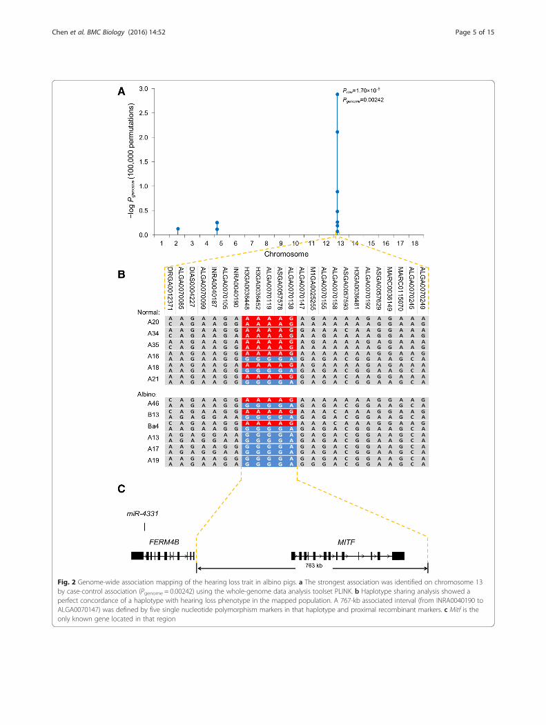
Fig. 2 Genome-wide association mapping of the hearing loss trait in albino pigs. a The strongest association was identified on chromosome 13by case-control association (Pgenome = 0.00242) using the whole-genome data analysis toolset PLINK. b Haplotype sharing analysis showed aperfect concordance of a haplotype with hearing loss phenotype in the mapped population. A 767-kb associated interval (from INRA0040190 toALGA0070147) was defined by five single nucleotide polymorphism markers in that haplotype and proximal recombinant markers. c Mitf is theonly known gene located in that region
Chen et al. BMC Biology (2016) 14:52 Page 5 of 15

Mitf-m, Mitf-a and Mitf-h were identified in the cDNAfrom the porcine inner ear (Fig. 3a).Moreover, we found that the Mitf-m transcript was nor-
mally expressed in Mitf R/R cochlea, but not in Mitf r/r
cochlea at any developmental stage (Fig. 3b). No obviousdifferences were found in the expression levels of Mitf-aor Mitf-h between the Mitf R/R and Mitf r/r cochlea(Fig. 3b). RNA-seq assay was used to detect differencesbetween the transcriptome profiles of the Mitf R/r andMitf r/r SVs at embryonic day 85; at this time in the de-velopment, melanocytes remained in the Mitf r/r SVs(Additional file 1: Figure S3). A total of 28 genes showedtwo-fold differential expression, including some geneswith known functions related to pigmentation andmelanogenesis, but the Mitf gene was not among them(Additional file 4: Table S9). Because the algorithm usedfor calculating the gene RPKM values (reads per kilobase
per million mapped reads, which serve to estimate geneexpression) cannot separate the expression of Mitf-mfrom other equally expressed transcript variants, we ob-tained the normalized read count for each exon individu-ally, and found that expression of the M-exon in wild-typeSVs was approximately 11.5-fold higher than in mutantSVs (Fig. 3d). In addition, RNA-seq data indicated thatexpression of some marker genes of melanocytes orthe SV intermediate cells remained in the MITF r/r
SVs; these genes included S100, KIT, MUM1, andKir1.2 (Additional file 4: Table S10). Most of thegenes expressed in MITF r/r SVs, however, did notshow significantly lower expression than the genes inthe MITF R/r SVs. When we considered these resultsalong with those from TEM analysis of prenatal SVs(Additional file 1: Figure S3), we determined that theintermediate cells remained in the MITF r/r SVs at the
Fig. 3 Expression analysis of Mitf transcriptional variants and isoforms. a Schematics of splicing structure in the porcine Mitf transcript variants(leading to MITF-A, MITF-H and MITF-M) detected in the cochlea. The specific fragments of transcript variants used for quantitative PCR in thisstudy are indicated by blue lines. b Expression profiles of MITF-A, MITF-H and MITF-M during cochlear development were examined by reversetranscription-PCR. MITF-M expression was present at detectable levels in the Mitf R/R cochlea, but not in the Mitf r/r cochlea. c Immunoblottinganalysis of MITF isoforms in the cochlea and skin. MITF-M expression was detectable in the Mitf R/R cochlea and skin, but not in samples from theMitf r/r pigs. d Differential level of expression of the Mitf exons in Mitf R/r and Mitf r/r stria vascularis (SV). The M-exon showed a 11.5-fold decreasein the Mitf r/r SV. The fold change of each exon is estimated by comparing the normalized read count of each exon between Mitf R/r and Mitf r/r SVin the RNA-seq assay. Raw data for this is provided in Additional file 5; sheet 4, Data of Mitf exon fold change
Chen et al. BMC Biology (2016) 14:52 Page 6 of 15
embryo stage and disappeared around birth. Thus, theexpression differences between MITF R/r and MITF r/r
SVs that we observed were indeed caused by Mitfmutation, rather than a lack of intermediate cells.Subsequent immunoblotting assay showed an un-
detectable level of polypeptides of 55–70 kDa (the re-ported size range of the MITF-M isoform in melanoma[27]) in the Mitf r/r cochleae (Fig. 3c). Consistent withthe previous data, the levels of MITF-A and MITF-H de-tected by immunoblot (Fig. 3c) were similar between theMitf R/R and Mitf r/r cochleae. These results indicatedthat the expression of MITF-M was eliminated in theMitf r/r pigs at both the transcript and protein levels, andthis differential expression pattern itself indicated the ex-istence of regulatory mutations in M isoform-specific re-gions. Consistently, eight of the 26 co-segregatedvariants were located in the M isoform-specific pro-moter (M-promoter; Additional file 4: Table S8).
Transcriptional activity analysis of the MITF M-promoterTo test whether the variants in the M-promoter werecapable of altering transcriptional activity, a transienttransfection assay was performed using mouse B16 mel-anoma cells. The luciferase reporter constructs con-tained varying lengths (7.8, 6.4, 5.2, 3.7 or 1.2 kb) oftruncated M-promoter from the R and r alleles, asshown in Fig. 4a. The construct pGL3-r-7.8 k, whichcontained a 7852-bp promoter region of the r allele, ex-hibited significantly lower luciferase activity than the Rallele construct (pGL3-R-7.8 k; Fig. 4a). There was nosignificant difference in activity between the pGL3-r-7.8 k construct and the null construct (pGL3-Basic vec-tor), the pGL3-R-6.4 k and pGL3-r-6.4 k constructs, orthe pGL3-R-1.2 k and pGL3-r-1.2 k constructs (Fig. 4a).Together, these results suggested that the sequence vari-ations involving the sequences between –7852 and –6416 bp, relative to the transcription start site of Mitf-m,were responsible for the elimination of MITF-M expres-sion. Four co-segregated variants were located withinthat region, including two insertions (9 and 14 bp, re-spectively) and two continuous SNPs (Fig. 4b, c, redbox). The mutations were densely clustered within a96 bp region (Fig. 4a, b, black box). To further validatetheir effects on transcription, we knocked out the 96-bpfragment from the pGL3-r-7.8 k construct (namedpGL3-r-7.8D) and detected restoration of the transcrip-tional activity (Fig. 4a). Using TFSEARCH [28], a tran-scription factor binding site searching tool, we predictedthat the 9- and 14-bp insertions would create two puta-tive binding sites for SOX family proteins (Fig. 4c, redunderlined in red). As SOX proteins can act as suppres-sors of gene expression [29], we speculated that these in-sertions and the SNPs could be functional mutations.
To address whether these candidate mutations wereable to alter the protein binding landscape in the involvedsequence region, we used electrophoretic mobility shiftassay (EMSA) to compare the binding capacity of thewild-type and mutant sequences. Two sets of oligonucleo-tides, R1 and r1 (Fig. 4c, red and yellow highlight, respect-ively), which differed only in the 9-bp insertion and theGC>TT replacement, and R2 and r2, which differed onlyin the 14-bp insertion (Fig. 4c, Additional file 4: TableS11), were incubated with nuclear extracts from mouseB16 melanoma cells. Only one differential complex (C1 inFig. 4d) was formed, namely that with the r2 probe andlacking the R2 probe. The specificity of the complex wasconfirmed by competition EMSA, wherein a 50- and 100-fold molar excess of unlabeled r2 probe showed effectivebinding competition but a 50- and 2100-fold excess of un-labeled R2 probe did not. Another complex (C2 in Fig. 4d)formed was a non-specific complex, because no competi-tion was observed with even 100-fold molar excess r2 coldprobe. No difference in protein binding was observed be-tween the R1 and r1 probes (Additional file 1: Figure S4).Collectively, these results show that only the 14-bp inser-tion can induce specific transcription factor bindingevents, while the 9-bp insertion and the two continuousSNPs do not. Thus, the 14-bp insertion is the only vari-ation detected in our study that was considered as poten-tially responsible for the observed down-regulation ofMitf-m transcription activity.SOX proteins (SOX2, SOX3 and SOX9) can regulate
inner ear development [30–32]. Our RNA-seq datashowed that Sox2 and Sox9, but not Sox3, wereexpressed in porcine SVs (Additional file 4: Table S12),suggesting that SOX2 and/or SOX9 is capable of bindingectopically to a new negative CRE (i.e. a silencer), whichmay have been generated by the 14-bp short insertion inthe Mitf regulatory region and which may have causedthe abrogation of Mitf-m expression (Fig. 6). Thus, thephenotypes of hearing loss and depigmentation in the al-bino pigs appear to be caused, at least partially, by the14-bp insertion that is located at –7532 bp relative tothe transcription start site of Mitf-m. Additionally, weperformed a further genotyping analysis of the causativemutant in 311 individual Rongchang pigs (Additionalfile 1: Figure S5). Both the 14- and 9-bp insertionswere found to be carried by all albino pigs, and toco-segregate with the albino phenotype completely(Additional file 4: Table S13). No recombination eventinvolving the two insertions was observed.These results provide convincing evidence that even a
small insertion in a region that lacks regulatory activityin the melanocyte lineage can create a transcription fac-tor binding site (TFBS). Therefore, we next investigatedwhether this region was a non-regulatory sequence inother cell/species lineages. By exploiting the available
Chen et al. BMC Biology (2016) 14:52 Page 7 of 15

Fig. 4 A new generated silencer in the M-promoter eliminated Mitf-m transcription. a Transcriptional activity analysis of the Mitf-m promoter from theR and r alleles. The reporter constructs are shown on the left, and the corresponding relative luciferase activity measured in transient transfection assaysis shown on the right. The luciferase activity of pGL3-r-7.8 k was significantly lower than that of pGL3-R-7.8 k. There was no significant differencebetween the R and r alleles when constructs were shorter than 7.8 k. Error bars indicate the standard deviations. The results shown are for oneexperiment with four technical replicates. Raw data for this and three additional experiments with similar results are provided in Additional file 5; sheet5, Data of reporter assay. b Schematics of the M-promoter. The sequence variations between the R and r alleles are labeled. INS, insertion; DEL, deletion.c Schematics of the M-promoter from –7513 bp to –7609 bp relative to the transcription start site of the M-exon. Sequence differences between R andr allele are indicated with a red box. The new sites showing consensus sequence for SOX protein binding are underlined in red. The oligonucleotideprobes designed for electrophoretic mobility shift assay (EMSA) are highlighted. d EMSA shows the specific binding of the nuclear proteins to the r2probe, and absence of binding to the R2 probe. In vitro incubation was performed using the indicated nuclear extracts, probe and unlabeledoligonucleotides (cold probes). C1, complex 1; C2, complex 2; R, R2 probe; r, r2 probe; N, random (negative control) probe
Chen et al. BMC Biology (2016) 14:52 Page 8 of 15

data from the human ENCODE project, which had pre-viously identified large numbers of regulatory elements,we were able to obtain the human ortholog of the mu-tant region identified in pigs and investigate whether thisregion overlapped with the CREs reported in the EN-CODE data (Additional file 1: Figure S6A). The pub-lished RNA-seq data indicated that the wild-type M-exon was indeed transcribed in the human melanocytelineage (Additional file 1: Figure S6C).Using the UCSC browser to search the ENCODE data,
we noted the following characteristics of these putativeCREs. (1) The flanking regions of the causative mutantpoint has a relatively low level of conservation in mam-mals, suggesting a low probability of conserved func-tional CREs (Additional file 1: Figure S6B). (2) DNAse Ihypersensitivity data indicate that chromatin accessibilityaround the mutant point is low in melanocytes and vari-ous cell lineages (Additional file 1: Figure S6D). (3)Chromatin immunoprecipitation and DNAse I footprint-ing data provide no evidence of protein binding sitesnear the mutant point (Additional file 1: Figure S6E). (4)The H3K4Me1 and H3K27Ac histone markers show noevidence of CREs in the flanking region of the mutantpoint (Additional file 1: Figure S6F). Because none ofthese data supported the existence of CREs near the mu-tant point, we concluded that the 14-bp insertion foundin the albino pigs resulted in the de novo genesis of a si-lencer in the M-promoter (Fig. 6).
Mitf-m-specific mutations result in hearing loss in amouse modelTo investigate whether a loss-of-function mutation inMitf-m is sufficient to cause the deafness and depigmen-tation phenotypes, we constructed a mouse model withnull Mitf-m alleles (Mitf mi-ΔM/mi-ΔM mouse; Fig. 5a). Aquantitative PCR assay confirmed that the expression ofMitf transcriptional variants in the Mitf mi-ΔM/mi-ΔM-targeted mouse was similar to those detected in the albinopigs (Fig. 5d). The Mitf mi-ΔM/mi-ΔM mice displayed pro-found hypopigmentation, with white hair and skin(Fig. 5b). ABR testing also revealed profound hearing lossin the Mitf mi-ΔM/mi-ΔM mice (n = 10; Fig. 5c). Finally, theMitf mi-ΔM/mi-ΔM mice showed thinner SVs, compared tothe wild-type mice, and fused or missing stereocilias ofhair cells (Fig. 5e, f ), similar to the cochlear morphologyseen in the albino pigs. Together, these data demonstratethat an exclusive malfunction in the m transcript iso-form is sufficient to cause the auditory-pigmentaryphenotypes.
DiscussionGenetic variations can create regulatory elements.Within the 10 million years of evolution involving mod-ern organisms, hundreds of non-functional sequences
have gained de novo regulatory functions [33]. Inducedde novo cis-regulatory behaviors have been reported inthe literature as having emerged via the following fourgenetic mechanisms. (1) Duplicated coding genes maylose their coding functions and gain de novo regulatoryfunctions through a whole-genome duplication event;these de novo enhancers are consequently known as “re-cycle regions” [10]. (2) Point mutations can create newregulatory behaviors and are commonly observed, fromflies to primates [6, 34], indicating their evolutionarybenefit. (3) Deletions can also generate new enhancersby bringing together flanking sequences that createnovel binding sites for activators [7]. (4) Transposonscan serve as a source for acquiring new regulatory be-haviors [8, 9, 35]. However, to our knowledge, short in-sertions acting as the source of de novo CREs have notbeen reported previously. Thus, our results provide evi-dence that short insertions can also create systemicfunctional silencers by introducing novel binding sites.Previous studies have shown that transposons contain-
ing existing TFBS can facilitate de novo regulatory be-havior and increase the transcriptional activity of targetgenes [9, 35]. In the current study, we found a de novosilencer in a distal melanocyte-specific regulatory regionthat had been naturally created by short insertions.Moreover, this de novo silencer acted as a negative CRE,blocking the expression of Mitf-m. This novel enhancerfeature, introduced by an insertion event, comprised anin silico predicted SOX protein-binding consensus se-quence. Our evidence indicated that a transcription fac-tor (predicted to be SOX2 or SOX9) interacts with thisnovel binding site to repress the expression of MITF-M.Both the 9-bp and the 14-bp insertions were predictedto create new TFBS, and although only the 14-bp inser-tion interacted directly with nucleoproteins in vitro, wecannot rule out the possibility that the 9-bp insertion isthe causative mutation, because the distance betweenthe two insertion regions is as close as 50 bp and theHMG binding domain of SOX family proteins can bindto a sequence as large as 100 bp [36]. Thus, the 9-bp in-sertion region may act as a flanking sequence, affectingthe robustness of the de novo element [37, 38].Previous studies of the coding and regulatory regions
of the Mitf gene have identified various functional muta-tions associated with congenital hearing loss in mam-mals [13, 20, 23, 24, 39]. Most of these mutations arelocated in the exons and introns that are conservedbetween species, and have been shown to cause equalgenetic effects on multiple isoforms. Dysfunction in me-lanocytes in the SV is the major pathology of WS2.Apart from MITF-M, isoforms MITF-A and MITF-Hare also expressed in melanocytes and melanoma cells[40–42]. The particular function(s) of each isoform inhearing loss remains to be fully elucidated. In a previous
Chen et al. BMC Biology (2016) 14:52 Page 9 of 15

study of Mitf mi-bw mice, insertion of a retrotransposing L1element into intron 3 of the MITF gene abolished MITF-M, but also affected the expression of MITF-A and MITF-H [43]. Thus, the phenotypes on hearing loss and pigmen-tation cannot be exclusively attributable to the eliminationof MITF-M, and there remains a possibility that MITF-Aor MITF-H may play functional roles in the pathogenesisof WS2 and its manifested symptoms. Our study mapped
a novel mutation located in the MITF-M-specific regula-tory region that completely eliminated MITF-M expres-sion, but which apparently had no effect on any otherisoform (Fig. 3b, c). A search of the literature revealed thatmutations in this region have also been shown to affectcoat color in dogs [21] and horses [22]. Furthermore, wegenerated the first M-exon specific knock-out mousemodel, we found phenotypes that were consistent with
Fig. 5 Phenotypes of the Mitf-m knock-out mice. a Schematic of the Mitf-m targeting technical process. The region of the Mitf gene containingexons M, 2, 3, and 4 are shown at the top. The targeting vector with a floxed-neomycin cassette in the M-promoter/M-exon region is shown inthe middle. The resultant Mitf gene portion after targeting (Mitf mi-ΔM allele) is shown at the bottom. b Mitf +/+ had a black coat color, andMitf mi-ΔM/mi-ΔM had a white coat color and black eyes. c The auditory brainstem response thresholds were 20–30 dB SPL for the Mitf +/+ mice, and100–110 dB SPL for the Mitf mi-ΔM/mi-ΔM mice (from 4 to 32 kHz). The raw data is provided in Additional file 5: sheet 6 Data of ABR tests (mice).d Mitf-m was not expressed at detectable levels in the Mitf mi-ΔM/mi-ΔM cochlea (red arrow), but was expressed at detectable levels in the Mitf +/+
cochlea. There was no difference observed between the expression levels of Mitf-a and Mitf-h in Mitf mi-ΔM/mi-ΔM and Mitf +/+ mice. Error bars inc and d indicate the standard deviations. Raw data in Additional file 5: sheet 7 Data of mouse Mitf qPCR (Ct) and sheet 8 Data of mouse MitfqPCR (FC). e In the Mitf mi-ΔM/mi-ΔM cochlea, most of the stereocilias of inner hair cells (arrows) and outer hair cells (arrowheads) were fused ormissing (stars). f The stria vascularis of Mitf mi-ΔM/mi-ΔM cochlea are significantly thinner and shorter than that of Mitf +/+ cochlea
Chen et al. BMC Biology (2016) 14:52 Page 10 of 15

Mitfr/r pigs, as well as similar Mitf expression profiles.Considering the collective results from the specificnaturally-arising porcine mutation and the artificially-induced mouse mutation, it appears that MITF-M exerts aunique function in the inner ear and that dysfunction ofthe MITF-M isoform alone is sufficient to cause deafness.Mitf is a well-described and frequent causative gene of
WS2 [11, 12]. Most of the mutations found in Mitf arelocated in the coding region and the flanking splicedonor/acceptor sites, accounting for the phenotype ofhearing loss in approximately 15 % of WS2 cases; how-ever, the mutation profile has not been determined inapproximately 70 % of WS2 patients [13]. Our resultssuggest that the upstream region of the M-exon, whichserves to regulate the expression of Mitf-m, is a novelpotential mutation region in WS2 patients. Identifyingand characterizing such regulatory regions will have clin-ical implications for identifying causative mutations forgenetic diseases such as WS2. New techniques, such astargeted, capture-based sequencing and whole-genomere-sequencing, provide the possibility of mutationscreening for large-scale regulatory regions. Finally, gen-etic hearing loss is almost exclusively studied in mousemodels. The lack of a large animal model for genetichearing loss has impaired the development of gene ther-apy for clinical application. Our Mitf r/r pigs should serve
as a valuable model to be tested for such therapeuticintervention both by traditional gene therapy and bynew CRISPR/Cas9-mediated genome editing [44].
ConclusionsIn summary, we provide evidence, for the first time, thatshort insertions in non-coding regions, previously lack-ing cis-regulatory activity, are capable of creating sys-temic functional de novo CREs, resulting in dramaticphenotypic alterations in mammals (Fig. 6). We alsoidentified the essential role of MITF-M in cochlear de-velopment and demonstrated that loss-of-function muta-tions of MITF-M are sufficient to cause deafness. Thus,it is important to include the regulatory regions in clin-ical gene screening for WS2.
MethodsStudy population of Rongchang pigsChinese Rongchang pigs were chosen from the animalbreeding facility of the Chongqing Academy of AnimalScience. Pigs from 1 month to 1.5 years old (male andfemale) were used. All Rongchang pigs underwent audi-tory electrophysiology diagnoses to determine the hear-ing phenotype. Pigs with normal hearing were assignedto the control group and albino pigs with no ABR re-sponse were used as the case group. Use of the pigs and
Fig. 6 Schematics showing the genetic effect of the causative mutation. In Mitf R/R stria vascularis (SVs) (before duplication). SOX proteins cannotrecognize and bind to the M-promoter and MITF-M was normally transcribed. In Mitf r/r SVs, a new consensus site for SOX protein binding, whichresulted from the 14-bp duplication, created a de novo silencer in the M-promoter. SOX proteins ectopically binding to that silencer may repressthe transcription of Mitf-m (after duplication)
Chen et al. BMC Biology (2016) 14:52 Page 11 of 15
mice was approved by the Institutional Animal Care andUse Committee of China Agricultural University inBeijing.
Auditory electrophysiologyABR, EP and [K+] recording were used to evaluate hear-ing function of pigs and mice. Details of these tests havebeen described in our previous publications [45]. Briefly,ABRs were evoked with clicks and tone pips at 4, 8, 16,and 20 kHz. The ABR threshold was determined by vis-ual inspection. EP and [K+] in the endolymph were re-corded using double-barreled microelectrodes filled with150 mM KCl and a potassium ion exchanger. The refer-ence barrel was filled with 0.5 M NaCl for EP recordingand the electrodes were calibrated in a 37 °C water bathchamber using a series of solutions of KCl and NaCl thathad [K+] of 1, 5, 25, 50, 100, and 150 mM. At least fourindependent animals were tested to confirm the results.
Cochlear morphologyThe morphology of the stereocilia of cochlear hair cellswas examined using SEM. Samples for SEM were pre-pared following procedures described in our previouspublications [46]. Briefly, the cochleae were fixed with2.5 % glutaraldehyde. After dehydration, the sampleswere critical-point dried, mounted on aluminum stubs,sputter-coated with gold particles, and examined using aHitachi S-3700N SEM (Japan). The cochlear SV morph-ology was observed on cochlear sections using light mi-croscopy and TEM. For TEM, the cochleae afterovernight fixation were decalcified, embedded in Eponresin, and sectioned on a Reichert Ultracut E ultramicro-tome (Boeckeler Instruments, Tucson, NM). Ultrathinsections were mounted on formvar-coated slot grids,stained with lead citrate and uranyl acetate, and exam-ined using a Philips CM 120 TEM. At least three inde-pendent samples were performed to confirm the results.
Genetic analysis and whole genome association studyEleven pairs of putative heterozygous boars and sowswere selected from the normal herd if they had albinooffspring. In total, 74 piglets from 11 litters of heterozy-gous × heterozygous matings were phenotyped to inves-tigate the Mendel segregation ratio. DNA from 28 pigsshowing profound hearing loss and 73 pigs with normalhearing was isolated from tissue samples taken frompinnas. The DNA samples were genotyped using theIllumina PorcineSNP60 BeadChip. The PLINK softwarepackage was used for genome-wide mapping; all SNPmarkers with MAF > 0.05 and call rate > 75 % were usedfor a case-control association analysis. Genome-wide Pvalues were ascertained through phenotype permutationtesting (n = 100,000).
Mutation screeningAll exons, exon-intron boundaries and promoters (10-kbfragment upstream of TSS) were sequenced in 12 nor-mal pigs and 12 albino pigs using the primers listed inAdditional file 4: Table S11. Long-range PCR with Long-AmpTaq DNA polymerase (NEB) was used to amplifythese fragments. PCR fragments were gel-purified withan EZNA Gel Extraction kit (Omega Biotek) and thensubjected to Sanger sequencing.
Expression analysisThe mRNA expression levels of MITF transcript variantsand β-actin were analyzed by a qPCR method using theprimers in Additional file 4: Table S11. The cDNAsamples (100 ng) and primers for the target genes weremixed with Power SYBR Green PCR Master Mix (AppliedBiosystems) in 25-μL final volumes, and amplified usingan ABI7900 instrument (Applied Biosystems). All sampleswere analyzed in triplicate. Protein expression levels ofMITF isoforms and tubulin were examined by immuno-blotting assays using lysed tissue from MITF R/R andMITF r/r cochleae or skin. MITF antibody C5 (#ab80651,Abcam) was used. The loading amounts were verified bydetermining levels of a housekeeping protein, β-tublin(Chemicon, Temecula, CA).
Transcriptome analysis of SVsThe lateral walls of the cochlea containing the SVs weredissected from pigs, and preserved with the RNAlaterreagent (Ambion, Austin, TX). Total RNA was extractedusing the RNeasy Micro Kit (#74004, Qiagen, GRE).RNA concentration and integrity was measured using aQubit 2.0 Flurometer (Life Technologies, CA, USA) anda Bioanalyzer 2100 system (Agilent Technologies, CA,USA), respectively. The IlluminaTruSeq RNA SamplePreparation Kit (Illumina, San Diego, USA) was used togenerate sequencing libraries following the manufac-turer’s recommendations. The clustering of samples wasperformed with the cBot Cluster Generation System ac-cording to the manufacturer’s instructions. Clustered li-braries were sequenced on a Hiseq 2000 platform(Illumina) and 100-bp paired-end reads were generated.After quality control, clean reads with high quality wereobtained for downstream analysis. Genome assemblySscrofa10.2 was used as the reference genome for readmapping. Clean reads were aligned to the reference gen-ome using TopHat (ver. 2.0.7) [33, 47]. RPKM values(reads per kilobase of exon model per million mappedreads) were calculated to measure the expression level ofgenes.
Transcription activity analysisThe 7.8-kb fragments of the M-promoter of the R and ralleles were inserted into a luciferase reporter plasmid
Chen et al. BMC Biology (2016) 14:52 Page 12 of 15
pGL3-basic (Promega, Madison, USA) to yield thepGL3-R-7.8 k and pGL3-r-7.8 k constructs, respectively.Similarly, truncated constructs including pGL3-R-6.4 k,pGL3-r-6.4 k, pGL3-R-1.2 k, and pGL3-r-1.2 k were gen-erated. Murine B16 melanoma cells with high endogen-ous MITF-M expression were cultured for plasmidtransfection. Luciferase reporter constructs (2.0 μg) andRenilla luciferase vector (0.5 μg) were co-transfected intoB16 cells using Lipofectamine 2000 Transfection Re-agent according to the manufacturer’s protocol (#11668-019, Invitrogen). At 48 h after transfection, luciferaseactivity was determined with a Dual-Luciferase ReporterAssay Kit (#E2920, Promega, Madison, USA). TheRenilla luciferase activity was used to normalize thetransfection efficiency. At least three independent exper-iments were performed.
EMSANuclear extracts from B16 cells were prepared using theNuclear and Cytoplasmic Protein Extraction Kit(#P0027, Beyotime). Oligonucleotides representing the Rand r allele fragments (Additional file 4: Table S11) were3’ end-labeled with biotin and incubated with nuclearextract in the absence or presence of homologous un-labeled DNA (50-fold molar excess). The products wereresolved by electrophoresis on an 8 % polyacrylamide gelwith × 0.5 TBE at room temperature for 2 h at 150 V. Atleast three independent replicates were performed toconfirm the results.
Mitf-m-targeted miceMitf mi-ΔM/mi-ΔM-targeted mice were generated using the“recombineering” technology. Briefly, a targeting constructwith flanking regions (10,868 bp) of the M-promoter/M-exon was used for standard targeting of mouse embryonicstem cells. Positive cell clones were microinjected intoeight-cell embryos to obtain chimeric mice. Chimericmice that could transmit the modified Mitf m-exon alleleto their progeny were crossed with wild-type mice to gen-erate Mitf mi-ΔM/+ mice. Mitf mi-ΔM/mi-ΔM-targeted micewere obtained by Mitf mi-ΔM/+ ×Mitf mi-ΔM/+ mating.Genotyping of the mice was performed by PCR usingprimers flanking the m-exon of Mitf (as shown in Add-itional file 4: Table S11). Use of the mice was approved bythe Institutional Animal Care and Use Committee of theGeneral Hospital of PLA in Beijing.
Additional files
Additional file 1: Figure S1. Eye morphology defects of albino pigs.Figure S2. Three family pedigrees of mapping population. Figure S3.Images showing presence of intermediate cells in the stria vascularis ofalbino pigs at the embryo stage. Figure S4. Results of EMSA using probeR1 and r1. Figure S5. Genotyping of Rongchang pigs for causative mutation.
Figure S6. The human orthologous of the causative mutant region found inMITF r/r pigs are formerly lack of regulatory activity. (DOCX 6667 kb)
Additional file 2: Table S1. The goodness of fit test for Mendelianratios of the hearing loss. Table S2. Re-annotation of porcine MITF genein Genome of Tibet pig. Table S3. Co-segregated mutations detected inmutation screening. Table S4. Summary and mapping statistics of thepig genome re-sequencing data. (DOCX 58 kb)
Additional file 3: Table S5. SNPs detected by re-sequencing in theassociated region of Rongchang pigs. Table S6. SNPs co-segregated withhearing loss phenotype in three MITF r/r pigs and three MITF R/R Rong-chang pigs. Table S7. SNPs uniquely detected in Rongchang pigs, and homo-zygous in MITF r/r. (XLSX 465 kb)
Additional file 4: Table S8. Co-segregated variants detected inre-sequencing and mutation screening. Table S9. Differential expressedgenes between MITF R/r and MITF r/r stria vascularis (SVs). Table S10.Expression levels of melanocyte marker genes in porcine SVs. Table S11.Primer pairs used for screening the MITF gene, for qPCR and for micegenotyping. Table S12. Expression levels of SOX family members inporcine SVs. Table S13. Distribution of hearing loss phenotype andgenotype in a large Rongchang pig population. (DOCX 55 kb)
Additional file 5: Supporting data. 1 Data of ABR tests (pigs). 2 Dataof EP. 3 Data of K+ concentration. 4 Data of Mitf exon fold change. 5Data of reporter assay. 6 Data of ABR tests (mice). 7 Data of mouse MitfqPCR (Ct). 8 Data of mouse Mitf qPCR (FC). (XLSX 27 kb)
AbbreviationsABR, auditory brainstem response; CRE, cis-regulatory element; EMSA,electrophoretic mobility shift assays; EP, endolymphatic potential; MITF,microphthalmia-associated transcription factor; SEM, scanning electronmicroscopy; SNP, single-nucleotide polymorphism; SPL, sound pressure level;SSC 13, Sus scrofa chromosome 13; SV, stria vascularis; TEM, transmissionelectron microscopy; TFBS, transcription factor binding site; WGD, whole-genome duplication; WS2, Waardenburg syndrome type 2
AcknowledgementsWe thank Dr. David ZZ He at Creighton University and Dr. Zhengyi Chen atHarvard Medical School for critical review of the manuscript. This work wassupported by grants the National Basic Research Program of China (973Program) (#2012CB967900) to SY, the National High Technology Researchand Development Program of China (863 Program) (#2013AA102502), the973 Program (#2011CBA01000) to NL, the 863 Program (#2014AA020510 and#2015SKLAB6-16), the National Transgenic Research Project (#2011ZX08009-001-003), the National Science Foundation of China (#31101701, #81470700,#31372284, #81400472 and #81271082), the China Postdoctoral ScienceFoundation to LC and the Chongqing Fund of application and development(#cstc2013yykfC80003) to LC.
Availability of data and materialsSupporting data of quantification results are available in Additional file 5. Thehigh-throughput data sets supporting the results of this article are available inthe NCBI Sequence Read Archive and Gene Expression Omnibus repository. TheRongchang pig whole-genome re-sequencing data has been deposited at theNCBI Sequence Read Archive, (SRA:SRX397138, SRA:SRX397142, SRA:SRX397141,SRA:SRX397138, SRA:SRX397139, SRA:SRX397137). Raw whole-genome geno-type data of the mapping population have been deposited in the Gene Expres-sion Omnibus (GEO:GSE67618). Data of porcine SV transcriptome have beendeposited in GEO: GSE80665.
Authors’ contributionsLC, WG, LR, JW, SY, and NL led the experiments. ZG, SC, XB, LZ, XZ, LG, ZL, andLZ performed animal work and prepared biological samples. MY, YZ, YH, BS, JL,and XL performed transcriptional activity analysis. DZ, LR, SZ, ZY, TZ, and JLperformed sanger sequencing. ML, ST, QM, QT, YZ, and TC performed the high-throughput sequencing. HY, SZ, KL, NW, TC, YZ, and LZ performed the cochlearmorphology experiments. NY, RZ, YG, FZ, HZ, ZH, JL, and WS performed theauditory electrophysiology experiments. JZ, TW and XH mapped the causativegene. LC, WG and WS wrote the paper. JW, SY, MY, ZY, and NL revised thepaper. All authors read and approved the final manuscript.
Chen et al. BMC Biology (2016) 14:52 Page 13 of 15
Competing interestsThe authors declare that they have no competing interests.
Additional informationAdditional information is available in the online version of the paper.
Author details1State Key Laboratory for Agrobiotechnology, College of Biological Sciences,National Engineering Laboratory for Animal Breeding, China AgriculturalUniversity, Beijing 100193, China. 2Department of Otolaryngology, Head &Neck Surgery, Institute of Otolaryngology, Chinese PLA General Hospital,Beijing 100853, China. 3Key Laboratory of Pig Industry Sciences (Ministry ofAgriculture), Chongqing Academy of Animal Science, Chongqing 402460,China. 4Institute of Animal Genetics and Breeding, College of Animal Scienceand Technology, Sichuan Agricultural University, Ya’an, Sichuan 625014,China. 5Department of Communicative Disorders and Sciences, Center forHearing and Deafness, State University of New York at Buffalo, Buffalo, NewYork, USA.
Received: 15 February 2016 Accepted: 10 June 2016
References1. Wray GA. The evolutionary significance of cis-regulatory mutations. Nat Rev
Genet. 2007;8(3):206–16.2. Herz HM, Hu D, Shilatifard A. Enhancer malfunction in cancer. Mol Cell.
2014;53(6):859–66.3. Rebeiz M, Jikomes N, Kassner VA, Carroll SB. Evolutionary origin of a novel
gene expression pattern through co-option of the latent activities ofexisting regulatory sequences. Proc Natl Acad Sci U S A. 2011;108(25):10036–43.
4. Frankel N, Erezyilmaz DF, McGregor AP, Wang S, Payre F, Stern DL.Morphological evolution caused by many subtle-effect substitutions inregulatory DNA. Nature. 2011;474(7353):598–603.
5. Wittkopp PJ, Kalay G. Cis-regulatory elements: molecular mechanisms andevolutionary processes underlying divergence. Nat Rev Genet. 2012;13(1):59–69.
6. Prabhakar S, Visel A, Akiyama JA, Shoukry M, Lewis KD, Holt A, et al. Human-specific gain of function in a developmental enhancer. Science. 2008;321(5894):1346–50.
7. Shirangi TR, Dufour HD, Williams TM, Carroll SB. Rapid evolution of sexpheromone-producing enzyme expression in Drosophila. PLoS Biol. 2009;7(8):e1000168.
8. Schlenke TA, Begun DJ. Strong selective sweep associated with atransposon insertion in Drosophila simulans. Proc Natl Acad Sci U S A.2004;101(6):1626–31.
9. Studer A, Zhao Q, Ross-Ibarra J, Doebley J. Identification of a functionaltransposon insertion in the maize domestication gene tb1. Nat Genet.2011;43(11):1160–3.
10. Eichenlaub MP, Ettwiller L. De novo genesis of enhancers in vertebrates.PLoS Biol. 2011;9(11):e1001188.
11. Chang T, Hashimoto K, Bawle EV. Spontaneous contraction of leukodermicpatches in Waardenburg syndrome. J Dermatol. 1993;20(11):707–11.
12. Read AP, Newton VE. Waardenburg syndrome. J Med Genet. 1997;34(8):656–65.13. Pingault V, Ente D, Dastot-Le Moal F, Goossens M, Marlin S, Bondurand N.
Review and update of mutations causing Waardenburg syndrome. HumMutat. 2010;31(4):391–406.
14. Levy C, Khaled M, Fisher DE. MITF: master regulator of melanocytedevelopment and melanoma oncogene. Trends Mol Med. 2006;12(9):406–14.
15. Steingrimsson E, Copeland NG, Jenkins NA. Melanocytes and the microphthalmiatranscription factor network. Annu Rev Genet. 2004;38:365–411.
16. Vachtenheim J, Borovansky J. "Transcription physiology" of pigment formationin melanocytes: central role of MITF. Exp Dermatol. 2010;19(7):617–27.
17. Li M, Wu H, Luo Z, Xia Y, Guan J, Wang T, et al. An atlas of DNA methylomes inporcine adipose and muscle tissues. Nat Commun. 2012;3:850.
18. Steel KP, Barkway C. Another role for melanocytes: their importance fornormal stria vascularis development in the mammalian inner ear.Development. 1989;107(3):453–63.
19. Koshikawa S, Giorgianni MW, Vaccaro K, Kassner VA, Yoder JH, Werner T, et al.Gain of cis-regulatory activities underlies novel domains of wingless geneexpression in Drosophila. Proc Natl Acad Sci U S A. 2015;112(24):7524–9.
20. Tassabehji M, Newton VE, Read AP. Waardenburg syndrome type 2 causedby mutations in the human microphthalmia (MITF) gene. Nat Genet. 1994;8(3):251–5.
21. Karlsson EK, Baranowska I, Wade CM, Salmon Hillbertz NH, Zody MC,Anderson N, et al. Efficient mapping of Mendelian traits in dogs throughgenome-wide association. Nat Genet. 2007;39(11):1321–8.
22. Hauswirth R, Haase B, Blatter M, Brooks SA, Burger D, Drogemuller C, et al.Mutations in MITF and PAX3 cause "splashed white" and other whitespotting phenotypes in horses. PLoS Genet. 2012;8(4):e1002653.
23. Philipp U, Lupp B, Momke S, Stein V, Tipold A, Eule JC, et al. A MITFmutation associated with a dominant white phenotype and bilateraldeafness in German Fleckvieh cattle. PLoS One. 2011;6(12):e28857.
24. Tachibana M, Kobayashi Y, Matsushima Y. Mouse models for four types ofWaardenburg syndrome. Pigm Cell Res. 2003;16(5):448–54.
25. Li M, Tian S, Jin L, Zhou G, Li Y, Zhang Y, et al. Genomic analyses identifydistinct patterns of selection in domesticated pigs and Tibetan wild boars.Nat Genet. 2013;45(12):1431–8.
26. Okumura N, Hayashi T, Sekikawa H, Matsumoto T, Mikawa A, Hamasima N,et al. Sequencing, mapping and nucleotide variation of porcine coat colourgenes EDNRB, MYO5A, KITLG, SLC45A2, RAB27A, SILV and MITF. Anim Genet.2006;37(1):80–2.
27. Li KK, Goodall J, Goding CR, Liao SK, Wang CH, Lin YC, et al. The melanocyteinducing factor MITF is stably expressed in cell lines from human clear cellsarcoma. Brit J Cancer. 2003;89(6):1072–8.
28. Akiyama Y. TFSEARCH: searching transcription factor binding sites. Japan:Real World Computing Partnership; 1995.
29. Chew LJ, Gallo V. The Yin and Yang of Sox proteins: activation andrepression in development and disease. J Neurosci Res. 2009;87(15):3277–87.
30. Uwanogho D, Rex M, Cartwright EJ, Pearl G, Healy C, Scotting PJ, et al.Embryonic expression of the chicken Sox2, Sox3 and Sox11 genessuggests an interactive role in neuronal development. Mech Develop.1995;49(1-2):23–36.
31. Neves J, Kamaid A, Alsina B, Giraldez F. Differential expression of Sox2 andSox3 in neuronal and sensory progenitors of the developing inner ear ofthe chick. J Comp Neurol. 2007;503(4):487–500.
32. Trowe MO, Shah S, Petry M, Airik R, Schuster-Gossler K, Kist R, et al. Loss ofSox9 in the periotic mesenchyme affects mesenchymal expansion anddifferentiation, and epithelial morphogenesis during cochlea developmentin the mouse. Dev Biol. 2010;342(1):51–62.
33. Arnold CD, Gerlach D, Spies D, Matts JA, Sytnikova YA, Pagani M, et al.Quantitative genome-wide enhancer activity maps for five Drosophilaspecies show functional enhancer conservation and turnover duringcis-regulatory evolution. Nat Genet. 2014;46(7):685–92.
34. Gompel N, Prud'homme B, Wittkopp PJ, Kassner VA, Carroll SB. Chancecaught on the wing: cis-regulatory evolution and the origin of pigmentpatterns in Drosophila. Nature. 2005;433(7025):481–7.
35. Chung H, Bogwitz MR, McCart C, Andrianopoulos A, Ffrench-Constant RH,Batterham P, et al. Cis-regulatory elements in the Accord retrotransposonresult in tissue-specific expression of the Drosophila melanogasterinsecticide resistance gene Cyp6g1. Genetics. 2007;175(3):1071–7.
36. Pil PM, Chow CS, Lippard SJ. High-mobility-group 1 protein mediates DNAbending as determined by ring closures. Proc Natl Acad Sci U S A. 1993;90(20):9465–9.
37. Goode DK, Callaway HA, Cerda GA, Lewis KE, Elgar G. Minor change, majordifference: divergent functions of highly conserved cis-regulatory elementssubsequent to whole genome duplication events. Development. 2011;138(5):879–84.
38. Erceg J, Saunders TE, Girardot C, Devos DP, Hufnagel L, Furlong EE. Subtlechanges in motif positioning cause tissue-specific effects on robustness ofan enhancer's activity. PLoS Genet. 2014;10(1):e1004060.
39. Markakis MN, Soedring VE, Dantzer V, Christensen K, Anistoroaei R.Association of MITF gene with hearing and pigmentation phenotype inHedlund white American mink (Neovison vison). J Genet. 2014;93(2):477–81.
40. Amae S, Fuse N, Yasumoto K, Sato S, Yajima I, Yamamoto H, et al.Identification of a novel isoform of microphthalmia-associated transcriptionfactor that is enriched in retinal pigment epithelium. Biochem Bioph ResCommun. 1998;247(3):710–5.
41. Fuse N, Yasumoto K, Takeda K, Amae S, Yoshizawa M, Udono T, et al.Molecular cloning of cDNA encoding a novel microphthalmia-associatedtranscription factor isoform with a distinct amino-terminus. J Biochem.1999;126(6):1043–51.
Chen et al. BMC Biology (2016) 14:52 Page 14 of 15

42. Vachtenheim J, Novotna H. Expression of genes for microphthalmia isoforms,Pax3 and MSG1, in human melanomas. Cell Mol Biol. 1999;45(7):1075–82.
43. Yajima I, Sato S, Kimura T, Yasumoto K, Shibahara S, Goding CR, et al. An L1element intronic insertion in the black-eyed white (Mitf[mi-bw]) gene: theloss of a single Mitf isoform responsible for the pigmentary defect andinner ear deafness. Hum Mol Genet. 1999;8(8):1431–41.
44. Zuris JA, Thompson DB, Shu Y, Guilinger JP, Bessen JL, Hu JH, et al. Cationiclipid-mediated delivery of proteins enables efficient protein-based genomeediting in vitro and in vivo. Nat Biotechnol. 2015;33(1):73–80.
45. Guo W, Yi H, Ren L, Chen L, Zhao L, Sun W, et al. The morphology andelectrophysiology of the cochlea of the miniature pig. Anat Res.2015;298(3):494–500.
46. Yang SM, Guo WW, Hu YY, Sun YX, Hou ZH, Sun JH, et al. Smad5haploinsufficiency leads to hair cell and hearing loss. Dev Neurobiol. 2009;69(2-3):153–61.
47. Carroll SB. Evolution at two levels: on genes and form. PLoS Biol. 2005;3(7):e245.
• We accept pre-submission inquiries
• Our selector tool helps you to find the most relevant journal
• We provide round the clock customer support
• Convenient online submission
• Thorough peer review
• Inclusion in PubMed and all major indexing services
• Maximum visibility for your research
Submit your manuscript atwww.biomedcentral.com/submit
Submit your next manuscript to BioMed Central and we will help you at every step:
Chen et al. BMC Biology (2016) 14:52 Page 15 of 15