A DAY IN THE LIFE OF AN OPHTHALMIC SURGEONOphthalmic Consultants of Long Island and Connecticut...
12
A DAY IN THE LIFE OF AN OPHTHALMIC SURGEON Expert Recommendations for Optimal Cataract and Refractive Outcomes Original Release: April 1, 2019 Expiration: April 30, 2020 This continuing medical education activity is supported through an unrestricted educational grant from Bausch & Lomb Incorporated. This continuing medical education activity is jointly provided by New York Eye and Ear Infirmary of Mount Sinai and MedEdicus LLC. Distributed with CME MONOGRAPH VISIT HTTPS://TINYURL.COM/OPTIMALCATARACTOUTCOMES FOR ONLINE TESTING AND INSTANT CME CERTIFICATE. Eric D. Donnenfeld, MD (Chair) • John Berdahl, MD • Preeya K. Gupta, MD • Edward J. Holland, MD FACULTY
Transcript of A DAY IN THE LIFE OF AN OPHTHALMIC SURGEONOphthalmic Consultants of Long Island and Connecticut...

A DAY I N T H E L I F E O F A N O P H T H A L M I C S U R G E O N
Expert Recommendations for Optimal Cataract and Refractive Outcomes
Original Release: April 1, 2019 Expiration: April 30, 2020
This continuing medical education activity is supported through an unrestricted educational grant from Bausch & Lomb Incorporated.
This continuing medical education activity is jointly provided by New York Eye and Ear Infirmary of Mount Sinai and MedEdicus LLC.
Distributed with
C M E M O N O G R A P HV I S I T H T T P S : / / T I N Y U R L . C O M / O P T I M A L C ATA R AC T O U T C O M E S
F O R O N L I N E T E S T I N G A N D I N S TA N T C M E C E R T I F I C AT E .
Eric D. Donnenfeld, MD (Chair) • John Berdahl, MD • Preeya K. Gupta, MD • Edward J. Holland, MD
FAC U LT Y
2
LEARNING METHOD AND MEDIUM This educational activity consists of a supplement and ten (10) study questions. The participant should, in order, read the learning objectives contained at the beginning of this supplement, read the supplement, answer all questions in the post test, and complete the Activity Evaluation/Credit Request form. To receive credit for this activity, please follow the instructions provided on the post test and Activity Evaluation/Credit Request form. This educational activity should take a maximum of 1.5 hours to complete.
ACTIVITY DESCRIPTION This program will provide a case-based learning experience on cataract surgery, taking into account individual patient goals and comorbidities. Topics include identifying and addressing ocular surface disorders, glaucoma management, intraocular lens (IOL) selection, toric IOL rotation, femtosecond laser-assisted surgery, infection prophylaxis, and inflammation control. The desired results of this activity are the optimization of outcomes of cataract surgery.
TARGET AUDIENCE This educational activity is intended for ophthalmologists.
LEARNING OBJECTIVES Upon completion of this activity, participants will be better able to: • Appraise the optimal preparation of the ocular surface preoperatively for
optimal outcomes in patients undergoing cataract surgery • Explain appropriate medication regimens for inflammation and infection
control in patients undergoing cataract surgery • Summarize optimal IOL selection criteria in individual patients • Compare femtosecond cataract surgery technology with conventional
cataract surgery technology • Apply evidence-based approaches for achieving optimal outcomes in
cataract surgery in patients with comorbidities
ACCREDITATION STATEMENT This activity has been planned and implemented in accordance with the accreditation requirements and policies of the Accreditation Council for Continuing Medical Education (ACCME) through the joint providership of New York Eye and Ear Infirmary of Mount Sinai and MedEdicus LLC. The New York Eye and Ear Infirmary of Mount Sinai is accredited by the ACCME to provide continuing medical education for physicians.
In July 2013, the Accreditation Council for Continuing Medical Education (ACCME) awarded New York Eye and Ear Infirmary of Mount Sinai “Accreditation with Commendation,” for six years as a provider of continuing medical education for physicians, the highest accreditation status awarded by the ACCME.
AMA CREDIT DESIGNATION STATEMENT The New York Eye and Ear Infirmary of Mount Sinai designates this enduring material for a maximum of 1.5 AMA PRA Category 1 Credits™. Physicians should claim only the credit commensurate with the extent of their participation in the activity.
GR ANTOR STATEMENT This continuing medical education activity is supported through an unrestricted educational grant from Bausch & Lomb Incorporated.
DISCLOSURE POLICY STATEMENT It is the policy of New York Eye and Ear Infirmary of Mount Sinai that the faculty and anyone in a position to control activity content disclose any real or apparent conflicts of interest relating to the topics of this educational activity, and also disclose discussions of unlabeled/unapproved uses of drugs or devices during their presentation(s). New York Eye and Ear Infirmary of Mount Sinai has established policies in place that will identify and resolve all conflicts of interest prior to this educational activity. Full disclosure of faculty/planners and their commercial relationships, if any, follows.
DISCLOSURES John Berdahl, MD, had a financial agreement or affiliation during the past year with the following commercial interests in the form of Royalty: Imprimis Pharmaceuticals, Inc; Consultant/Advisory Board: Alcon; Allergan; Aurea Medical; Avedro, Inc; Bausch & Lomb Incorporated; ClarVista Medical; CorneaGen; Envisia Therapeutics; Equinox; Glaukos Corporation; IanTECH; Imprimis Pharmaceuticals, Inc; Johnson & Johnson Vision Care, Inc; New World Medical, Inc; Ocular Surgical Data; Ocular Therapeutix, Inc; Omega Ophthalmics; RxSIGHT; Verana Health, Inc; Vittamed; and Zeiss; Honoraria from promotional, advertising or non-CME services received directly from commercial interests or their Agents (eg, Speakers Bureaus): Alcon; Allergan; and Glaukos Corporation; Ownership Interest (Stock options, or other holdings, excluding diversified mutual funds): CorneaGen; Equinox; IanTECH; Ocular Surgical Data; Omega Ophthalmics; Verana Health, Inc; and Zeiss.
Eric D. Donnenfeld, MD, had a financial agreement or affiliation during the past year with the following commercial interests in the form of Consultant/Advisory Board: AcuFocus, Inc; Alcon; Allergan; AqueSys, Inc; Avedro, Inc; Bausch & Lomb Incorporated; Beaver-Visitec International; ELENZA, Inc; EyePoint Pharmaceuticals; ForSight Labs, LLC; Glaukos Corporation; Icon Bioscience, Inc; Johnson & Johnson Vision Care, Inc; Kala Pharmaceuticals; Katena Products, Inc; LacriSciences LLP; LensGen; Mati Therapeutics, Inc; Merck & Co., Inc.; Mimetogen Pharmaceuticals; NovaBay Pharmaceuticals, Inc; Novaliq GmbH Germany; OcuHub LLC; Odyssey Medical, Inc; Omega Ophthalmics; Omeros Corporation; Oyster Point Pharma, Inc; Pfizer Inc; PogoTec; PRN; Rapid Pathogen Screening, Inc; Shire; Strathspey Crown; Sun Pharmaceutical Industries, Inc; TearLab Corporation; TrueVision; Veracity Innovations LLC; and Zeiss; Ownership Interest (Stock options, or other holdings, excluding diversified mutual funds): AcuFocus, Inc; AqueSys, Inc; Avedro, Inc; ELENZA, Inc; Glaukos Corporation; LacriSciences LLP; LensGen; Mati Pharmaceuticals, Inc; Mimetogen Pharmaceuticals; NovaBay Pharmaceuticals, Inc; OcuHub LLC; PogoTec; Rapid Pathogen Screening, Inc; SARcode Bioscience, Inc; Strathspey Crown; TearLab Corporation; TrueVision; and Veracity Innovations LLC.
Preeya K. Gupta, MD, had a financial agreement or affiliation during the past year with the following commercial interests in the form of Consultant/Advisory Board: Alcon; Allergan; Aurea Medical; Bio-Tissue; Johnson & Johnson Vision Care, Inc; NovaBay Pharmaceuticals, Inc; Ocular Science; Shire; Sight Sciences; TearLab Corporation; and Zeiss.
Edward J. Holland, MD, had a financial agreement or affiliation during the past year with the following commercial interests in the form of Consultant/Advisory Board: Aerie Pharmaceuticals, Inc; Azura Ophthalmics Ltd; CorneaGen; Glaukos Corporation; Kala Pharmaceuticals; Katena Products, Inc; Mati Therapeutics, Inc; Novartis Pharmaceuticals Corporation; Omeros Corporation; Precision Lens; Senju Pharmaceutical Co, Ltd; Shire; Sight Sciences; TearLab Corporation; and Vomaris Innovations, Inc; Contracted Research: Mati Therapeutics, Inc; Novartis Pharmaceuticals Corporation; Omeros Corporation; and Senju Pharmaceutical Co, Ltd; Honoraria from promotional, advertising or non-CME services received directly from commercial interests or their Agents (eg, Speakers Bureaus): Novartis Pharmaceuticals Corporation; Omeros Corporation; Senju Pharmaceutical Co, Ltd; and Shire.
NEW YORK EYE AND EAR INFIRMARY OF MOUNT SINAI PEER REVIEW DISCLOSURE Angie E. Wen, MD, has no relevant commercial relationships to disclose.
EDITORIAL SUPPORT DISCLOSURES Cheryl Guttman Krader; Cynthia Tornallyay, RD, MBA, CHCP; Melissa Carter-Ozhan; Kimberly Corbin, CHCP; Barbara Aubel; and Michelle Ong have no relevant commercial relationships to disclose.
DISCLOSURE ATTESTATION The contributing physicians listed above have attested to the following: 1) that the relationships/affiliations noted will not bias or otherwise influence
their involvement in this activity; 2) that practice recommendations given relevant to the companies with whom
they have relationships/affiliations will be supported by the best available evidence or, absent evidence, will be consistent with generally accepted medical practice; and
3) that all reasonable clinical alternatives will be discussed when making practice recommendations.
OFF-LABEL DISCUSSION This CME activity includes discussion of unlabeled and/or investigative uses of drugs. Please refer to the official prescribing information for each drug discussed in this activity for FDA-approved dosing, indications, and warnings.
NEW YORK EYE AND EAR INFIRMARY OF MOUNT SINAI PRIVACY & CONFIDENTIALITY POLICIES http://www.nyee.edu/health-professionals/cme/enduring-activities
CME PROVIDER CONTACT INFORMATION For questions about this activity, call 212-870-8127.
TO OBTAIN AMA PR A CATEGORY 1 CREDIT™ To obtain AMA PRA Category 1 Credit™ for this activity, read the material in its entirety and consult referenced sources as necessary. Please take this post test and evaluation online by going to https://tinyurl.com/OptimalCataractOutcomes. Upon passing, you will receive your certificate immediately. You must score 70% or higher to receive credit for this activity, and may take the test up to 2 times. Upon registering and successfully completing the post test, your certificate will be made available online and you can print it or file it.
DISCLAIMER The views and opinions expressed in this educational activity are those of the faculty and do not necessarily represent the views of New York Eye and Ear Infirmary of Mount Sinai, MedEdicus LLC, Bausch & Lomb Incorporated, or Ophthalmology Times.
For instant processing, complete the CME Post Test online
3
INTRODUCTION Patients requiring cataract surgery can present with a variety of challenges that might compromise surgical outcomes. In this activity, expert faculty provide insights for addressing a range of preoperative, intraoperative, and postoperative issues, with the aim of helping surgeons achieve success and patient satisfaction.
CASE 1: PATIENT WITH HIGH MYOPIA, DRY EYE, AND CATARACT F R O M T H E F I L E S O F P R E E YA K . G U P TA , M D A 56-year-old man presented with decreased vision in his right eye plus glare and halos when driving at night. He has been wearing rigid gas permeable (RGP) contact lenses for 30 years, and had LASIK (laser assisted in situ keratomileusis) in his right eye in 1999 to treat high myopia that was reduced from -18.0 D to -6.0 D.
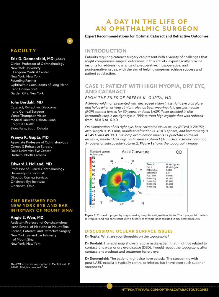
On examination of his right eye, best-corrected visual acuity (BCVA) is 20/150, axial length is 35.1 mm, manifest refraction is -12.0 D sphere, and keratometry is 42.49 D and 48.38 D. Slit-lamp examination reveals 1+ punctate epithelial erosions, visible LASIK flap, and a dense cataract (3+ nuclear sclerotic cataract, 3+ posterior subcapsular cataract). Figure 1 shows the topography image.
DISCUSSION: OCULAR SURFACE ISSUES Dr Gupta: What are your thoughts on the topography?
Dr Berdahl: The axial map shows irregular astigmatism that might be related to contact lens wear or dry eye disease (DED). I would repeat the topography after contact lens washout and treatment for dry eye.
Dr Donnenfeld: The patient might also have ectasia. The steepening with post-LASIK ectasia is typically central or inferior, but I have seen such superior steepness.1
Figure 1. Corneal topography map showing irregular astigmatism. Note: The topographic pattern is irregular and not consistent with a history of myopic laser assisted in situ keratomileusis.
Eric D. Donnenfeld, MD (Chair) Clinical Professor of Ophthalmology New York University Langone Medical Center New York, New York Founding Partner Ophthalmic Consultants of Long Island and Connecticut Garden City, New York
John Berdahl, MD Cataract, Refractive, Glaucoma, and Corneal Surgeon Vance Thompson Vision Medical Director, Dakota Lions Sight & Health Sioux Falls, South Dakota
Preeya K. Gupta, MD Associate Professor of Ophthalmology Cornea & Refractive Surgery Duke University Eye Center Durham, North Carolina
Edward J. Holland, MD Professor of Clinical Ophthalmology University of Cincinnati Director, Cornea Services Cincinnati Eye Institute Cincinnati, Ohio
FAC U LT Y
C M E R E V I E W E R F O R N E W YO R K E Y E A N D E A R INFIRMARY OF MOUNT SINAI
Angie E. Wen, MD Assistant Professor of Ophthalmology Icahn School of Medicine at Mount Sinai Cornea, Cataract, and Refractive Surgery New York Eye and Ear Infirmary of Mount Sinai New York, New York
This CME activity is copyrighted to MedEdicus LLC ©2019. All rights reserved. 164
A DAY I N T H E L I F E O F A N O P H T H A L M I C S U R G E O N
Expert Recommendations for Optimal Cataract and Refractive Outcomes
HTTPS://TINYURL.COM/OPTIMALCATARACTOUTCOMES
4
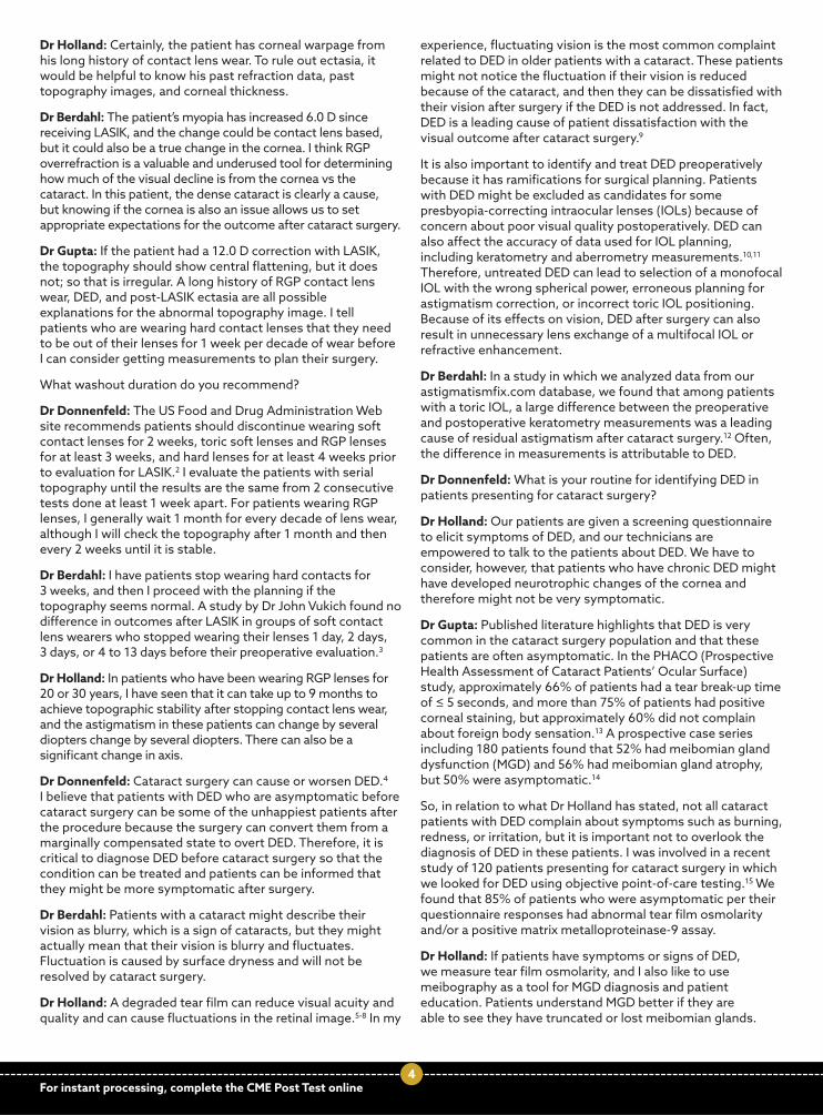
Dr Holland: Certainly, the patient has corneal warpage from his long history of contact lens wear. To rule out ectasia, it would be helpful to know his past refraction data, past topography images, and corneal thickness.
Dr Berdahl: The patient’s myopia has increased 6.0 D since receiving LASIK, and the change could be contact lens based, but it could also be a true change in the cornea. I think RGP overrefraction is a valuable and underused tool for determining how much of the visual decline is from the cornea vs the cataract. In this patient, the dense cataract is clearly a cause, but knowing if the cornea is also an issue allows us to set appropriate expectations for the outcome after cataract surgery.
Dr Gupta: If the patient had a 12.0 D correction with LASIK, the topography should show central flattening, but it does not; so that is irregular. A long history of RGP contact lens wear, DED, and post-LASIK ectasia are all possible explanations for the abnormal topography image. I tell patients who are wearing hard contact lenses that they need to be out of their lenses for 1 week per decade of wear before I can consider getting measurements to plan their surgery.
What washout duration do you recommend?
Dr Donnenfeld: The US Food and Drug Administration Web site recommends patients should discontinue wearing soft contact lenses for 2 weeks, toric soft lenses and RGP lenses for at least 3 weeks, and hard lenses for at least 4 weeks prior to evaluation for LASIK.2 I evaluate the patients with serial topography until the results are the same from 2 consecutive tests done at least 1 week apart. For patients wearing RGP lenses, I generally wait 1 month for every decade of lens wear, although I will check the topography after 1 month and then every 2 weeks until it is stable.
Dr Berdahl: I have patients stop wearing hard contacts for 3 weeks, and then I proceed with the planning if the topography seems normal. A study by Dr John Vukich found no difference in outcomes after LASIK in groups of soft contact lens wearers who stopped wearing their lenses 1 day, 2 days, 3 days, or 4 to 13 days before their preoperative evaluation.3
Dr Holland: In patients who have been wearing RGP lenses for 20 or 30 years, I have seen that it can take up to 9 months to achieve topographic stability after stopping contact lens wear, and the astigmatism in these patients can change by several diopters change by several diopters. There can also be a significant change in axis.
Dr Donnenfeld: Cataract surgery can cause or worsen DED.4 I believe that patients with DED who are asymptomatic before cataract surgery can be some of the unhappiest patients after the procedure because the surgery can convert them from a marginally compensated state to overt DED. Therefore, it is critical to diagnose DED before cataract surgery so that the condition can be treated and patients can be informed that they might be more symptomatic after surgery.
Dr Berdahl: Patients with a cataract might describe their vision as blurry, which is a sign of cataracts, but they might actually mean that their vision is blurry and fluctuates. Fluctuation is caused by surface dryness and will not be resolved by cataract surgery.
Dr Holland: A degraded tear film can reduce visual acuity and quality and can cause fluctuations in the retinal image.5-8 In my
experience, fluctuating vision is the most common complaint related to DED in older patients with a cataract. These patients might not notice the fluctuation if their vision is reduced because of the cataract, and then they can be dissatisfied with their vision after surgery if the DED is not addressed. In fact, DED is a leading cause of patient dissatisfaction with the visual outcome after cataract surgery.9
It is also important to identify and treat DED preoperatively because it has ramifications for surgical planning. Patients with DED might be excluded as candidates for some presbyopia-correcting intraocular lenses (IOLs) because of concern about poor visual quality postoperatively. DED can also affect the accuracy of data used for IOL planning, including keratometry and aberrometry measurements.10,11 Therefore, untreated DED can lead to selection of a monofocal IOL with the wrong spherical power, erroneous planning for astigmatism correction, or incorrect toric IOL positioning. Because of its effects on vision, DED after surgery can also result in unnecessary lens exchange of a multifocal IOL or refractive enhancement.
Dr Berdahl: In a study in which we analyzed data from our astigmatismfix.com database, we found that among patients with a toric IOL, a large difference between the preoperative and postoperative keratometry measurements was a leading cause of residual astigmatism after cataract surgery.12 Often, the difference in measurements is attributable to DED.
Dr Donnenfeld: What is your routine for identifying DED in patients presenting for cataract surgery?
Dr Holland: Our patients are given a screening questionnaire to elicit symptoms of DED, and our technicians are empowered to talk to the patients about DED. We have to consider, however, that patients who have chronic DED might have developed neurotrophic changes of the cornea and therefore might not be very symptomatic.
Dr Gupta: Published literature highlights that DED is very common in the cataract surgery population and that these patients are often asymptomatic. In the PHACO (Prospective Health Assessment of Cataract Patients’ Ocular Surface) study, approximately 66% of patients had a tear break-up time of ≤ 5 seconds, and more than 75% of patients had positive corneal staining, but approximately 60% did not complain about foreign body sensation.13 A prospective case series including 180 patients found that 52% had meibomian gland dysfunction (MGD) and 56% had meibomian gland atrophy, but 50% were asymptomatic.14
So, in relation to what Dr Holland has stated, not all cataract patients with DED complain about symptoms such as burning, redness, or irritation, but it is important not to overlook the diagnosis of DED in these patients. I was involved in a recent study of 120 patients presenting for cataract surgery in which we looked for DED using objective point-of-care testing.15 We found that 85% of patients who were asymptomatic per their questionnaire responses had abnormal tear film osmolarity and/or a positive matrix metalloproteinase-9 assay.
Dr Holland: If patients have symptoms or signs of DED, we measure tear film osmolarity, and I also like to use meibography as a tool for MGD diagnosis and patient education. Patients understand MGD better if they are able to see they have truncated or lost meibomian glands.
For instant processing, complete the CME Post Test online
5
Topography is routine for all patients needing cataract surgery. Irregularities, such as poor image quality, data dropout, or astigmatism, are a sign of DED, and I also compare the topographic keratometry and astigmatism data with the optical biometry measurements. Lack of agreement between the devices is a sign of ocular surface disease.
Although older patients with DED can be asymptomatic, there is a cohort of patients, particularly younger individuals, who can be very symptomatic but have minimal to no signs of DED. It is my impression that many clinicians think that absence of fluorescein staining rules out DED, but corneal fluorescein staining does not develop until there is moderate-to-severe DED.16 Lissamine green is more useful for detecting earlier disease.16
Dr Donnenfeld: I rely on corneal topography to identify DED and am looking at the quality of the image and at the color codes to evaluate tear film structure. A small white area of dropout represents a location where the ocular surface is so irregular that the device could not pick up the image, and that is an absolute indication that the patient needs to be evaluated for ocular surface disease before planning cataract surgery. Diagnoses to consider aside from DED include Salzmann nodular degeneration or epithelial basement membrane dystrophy.
I also measure tear break-up time and do a careful eyelid examination to look for MGD as part of my DED evaluation. Tear film osmolarity and the matrix metalloproteinase-9 assay are improving our ability to diagnose DED, but they are not standalone tests.
How do you manage DED before cataract surgery?
Dr Holland: I think corticosteroids are the best option for rapid rehabilitation of the ocular surface, and I like to use loteprednol because it is very effective and safe.17–19 I start loteprednol 2 to 4 times a day with a preservative-free artificial tear. If the patient has MGD, I add an in-office thermal pulsation treatment and have started doing microblepharoexfoliation, which is a new technique that debrides the eyelid margins, removes the bacterial biofilm, and unroofs the meibomian glands.20 I think the loteprednol helps minimize discomfort from the mechanical treatments.
Dr Donnenfeld: I, too, start patients on loteprednol gel, artificial tears, and microblepharoexfoliation for MGD. The loteprednol gel is good for MGD because it adheres to the lid margin. I add a topical immunomodulator when patients have significant DED. I prefer lifitegrast because in premarketing clinical trials, it has worked to improve symptoms within just 2 weeks.21,22
The role of oral omega-3 supplements for managing patients with MGD has become controversial because of some conflicting results in clinical trials. What are your thoughts on their value?
Dr Gupta: The National Eye Institute–supported DREAM (Dry Eye Assessment and Management) study found that dietary supplementation with oral omega-3 fatty acids in re-esterified triglyceride form was no better than placebo in relieving signs and symptoms of DED.23 The placebo, however, was olive oil, and after 6 and 12 months, both groups had statistically significant improvement in the Ocular Surface Disease Index.
I think the study showed that olive oil also has value, and I am continuing to recommend omega-3 supplements.
Dr Donnenfeld: My experience using oral omega-3 supplementation is also positive, and I prescribe it long term as maintenance treatment for DED. I think there are really no downsides to using it.
Dr Gupta: I also use intense pulsed light followed by manual gland expression to treat MGD, particularly in patients with rosacea.24 As a caveat, however, I do not use it in patients with dark skin (Fitzpatrick skin types V or VI) because they are at an increased risk for depigmentation.
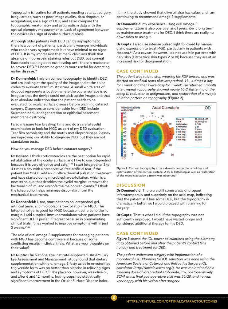
CASE CONTINUED The patient was told to stop wearing his RGP lenses, and was started on artificial tears plus loteprednol, 1%, 4 times a day for 1 week and then twice daily for 1 week. He returned 1 month later; repeat topography showed nearly 10-D flattening of the steep K, reduction in astigmatism, and restoration of a myopic ablation pattern on topography (Figure 2).
DISCUSSION Dr Donnenfeld: There are still some areas of dropout inferotemporally and superiorly on the axial map, indicating that the patient still has some DED, but the topography is dramatically better, so I would proceed with planning for surgery.
Dr Gupta: That is what I did. If the topography was not sufficiently improved, I would have waited longer and considered additional therapy for his DED.
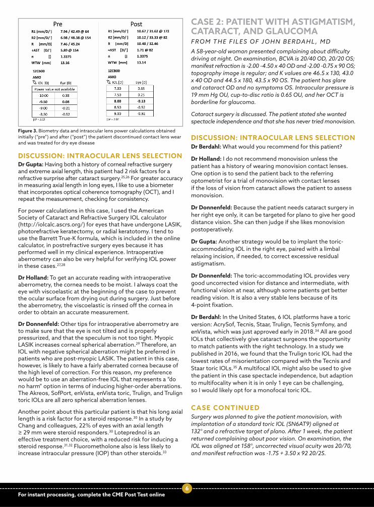
CASE CONTINUED Figure 3 shows the IOL power calculations using the biometry data obtained before and after the patient’s contact lens holiday and treatment for DED.
The patient underwent surgery with implantation of a monofocal IOL. Planning for IOL selection was done using the American Society of Cataract and Refractive Surgery IOL calculator (http://iolcalc.ascrs.org/). He was maintained on a tapering dose of loteprednol etabonate, 1%, postoperatively. BCVA at his final postoperative visit was 20/20, and he was very happy with his vision after surgery.
Figure 2. Corneal topography after a 4-week contact lens holiday and optimization of the corneal surface. A 10-D flattening as well as restoration of the myopic ablation pattern was observed.
HTTPS://TINYURL.COM/OPTIMALCATARACTOUTCOMES
6
DISCUSSION: INTRAOCULAR LENS SELECTION Dr Gupta: Having both a history of corneal refractive surgery and extreme axial length, this patient had 2 risk factors for a refractive surprise after cataract surgery.25,26 For greater accuracy in measuring axial length in long eyes, I like to use a biometer that incorporates optical coherence tomography (OCT), and I repeat the measurement, checking for consistency.
For power calculations in this case, I used the American Society of Cataract and Refractive Surgery IOL calculator (http://iolcalc.ascrs.org/) for eyes that have undergone LASIK, photorefractive keratectomy, or radial keratotomy. I tend to use the Barrett True-K formula, which is included in the online calculator, in postrefractive surgery eyes because it has performed well in my clinical experience. Intraoperative aberrometry can also be very helpful for verifying IOL power in these cases.27,28
Dr Holland: To get an accurate reading with intraoperative aberrometry, the cornea needs to be moist. I always coat the eye with viscoelastic at the beginning of the case to prevent the ocular surface from drying out during surgery. Just before the aberrometry, the viscoelastic is rinsed off the cornea in order to obtain an accurate measurement.
Dr Donnenfeld: Other tips for intraoperative aberrometry are to make sure that the eye is not tilted and is properly pressurized, and that the speculum is not too tight. Myopic LASIK increases corneal spherical aberration.29 Therefore, an IOL with negative spherical aberration might be preferred in patients who are post-myopic LASIK. The patient in this case, however, is likely to have a fairly aberrated cornea because of the high level of correction. For this reason, my preference would be to use an aberration-free IOL that represents a “do no harm” option in terms of inducing higher-order aberrations. The Akreos, SofPort, enVista, enVista toric, Trulign, and Trulign toric IOLs are all zero spherical aberration lenses.
Another point about this particular patient is that his long axial length is a risk factor for a steroid response.30 In a study by Chang and colleagues, 22% of eyes with an axial length ≥ 29 mm were steroid responders.30 Loteprednol is an effective treatment choice, with a reduced risk for inducing a steroid response.31,32 Fluorometholone also is less likely to increase intraocular pressure (IOP) than other steroids.33
CASE 2: PATIENT WITH ASTIGMATISM, CATARACT, AND GLAUCOMA F R O M T H E F I L E S O F J O H N B E R DA H L , M D A 58-year-old woman presented complaining about difficulty driving at night. On examination, BCVA is 20/40 OD, 20/20 OS; manifest refraction is -2.00 -4.50 x 40 OD and -2.00 -0.75 x 90 OS; topography image is regular; and K values are 46.5 x 130, 43.0 x 40 OD and 44.5 x 180, 43.5 x 90 OS. The patient has glare and cataract OD and no symptoms OS. Intraocular pressure is 19 mm Hg OU, cup-to-disc ratio is 0.65 OU, and her OCT is borderline for glaucoma.
Cataract surgery is discussed. The patient stated she wanted spectacle independence and that she has never tried monovision.
DISCUSSION: INTRAOCULAR LENS SELECTION Dr Berdahl: What would you recommend for this patient?
Dr Holland: I do not recommend monovision unless the patient has a history of wearing monovision contact lenses. One option is to send the patient back to the referring optometrist for a trial of monovision with contact lenses if the loss of vision from cataract allows the patient to assess monovision.
Dr Donnenfeld: Because the patient needs cataract surgery in her right eye only, it can be targeted for plano to give her good distance vision. She can then judge if she likes monovision postoperatively.
Dr Gupta: Another strategy would be to implant the toric-accommodating IOL in the right eye, paired with a limbal relaxing incision, if needed, to correct excessive residual astigmatism.
Dr Donnenfeld: The toric-accommodating IOL provides very good uncorrected vision for distance and intermediate, with functional vision at near, although some patients get better reading vision. It is also a very stable lens because of its 4-point fixation.
Dr Berdahl: In the United States, 6 IOL platforms have a toric version: AcrySof, Tecnis, Staar, Trulign, Tecnis Symfony, and enVista, which was just approved early in 2018.34 All are good IOLs that collectively give cataract surgeons the opportunity to match patients with the right technology. In a study we published in 2016, we found that the Trulign toric IOL had the lowest rates of misorientation compared with the Tecnis and Staar toric IOLs.35 A multifocal IOL might also be used to give the patient in this case spectacle independence, but adaption to multifocality when it is in only 1 eye can be challenging, so I would likely opt for a monofocal toric IOL.
CASE CONTINUED Surgery was planned to give the patient monovision, with implantation of a standard toric IOL (SN6AT9) aligned at 132° and a refractive target of plano. After 1 week, the patient returned complaining about poor vision. On examination, the IOL was aligned at 158°, uncorrected visual acuity was 20/70, and manifest refraction was -1.75 + 3.50 x 92 20/25.
For instant processing, complete the CME Post Test online
Figure 3. Biometry data and intraocular lens power calculations obtained initially (“pre”) and after (“post”) the patient discontinued contact lens wear and was treated for dry eye disease

7
DISCUSSION: TORIC INTR AOCULAR LENS ROTATION Dr Berdahl: Improper IOL alignment (wrong location), incorrect toric power (wrong lens), and unrecognized ocular pathology (wrong eye) are all possible causes for residual astigmatism after toric IOL implantation. Several scenarios can lead to each of these situations (Table 1).
Poor preoperative measurements, poor calculations, surgically induced astigmatism (SIA) that is different from what is expected, and failure to account for the posterior cornea can all lead to incorrect alignment or toric power selection. Improper intraoperative alignment and postoperative rotation are other causes of wrong positioning of the IOL.
Poor calculations have become less of an issue now than they were in the past because of newer toric IOL calculators that account for posterior corneal astigmatism. These calculators, however, use a population average, and some patients can be outliers.
Surprises with SIA occur more often than surgeons might realize. Surgeons input their average SIA for the toric IOL power calculation, but for approximately 30% of surgeons, the standard deviation might be ≥ 0.5 D.36
If the power is wrong, the IOL will need to be exchanged or the patient will need a refractive enhancement with laser vision correction to treat the residual astigmatism. Intraocular lens rotation can correct residual astigmatism in cases in which the IOL is the proper power but in the wrong location.
When rotating the IOL, the ideal axis, which is the axis that will minimize astigmatism, can be determined using the astigmatismfix.com Web site by inputting the patient’s postoperative refraction, the toric IOL model, and its actual axis. The program determines how many degrees the IOL should be rotated from its current position, which can be marked on the cornea preoperatively using the IOL’s alignment marks.
Dr Gupta: Do you put in a capsular tension ring to prevent the IOL from rotating again?
Dr Berdahl: Some people think it can help, and findings from a published randomized controlled study support its use.37 I think it can be argued that a capsular tension ring might help by stretching out the capsular bag, but it might also cause the bag to collapse, which would not be good, so I do not use one. In a case I had in which a lens rotated a second time, I did a reverse optic capture to fix its position.
If the rotation is done early enough after the primary surgery, it is not necessary to viscodissect the IOL from the capsule.
If the capsule has already contracted over the IOL and viscodissection is needed, the viscoelastic will have to be completely removed or else the IOL might rotate again.
CASE CONTINUED The IOL was rotated, and the patient was very happy with her vision. She returned 5 years later at age 63 complaining about difficulty driving at night. Her uncorrected visual acuity was 20/20 OD, but in her left eye, she had glare, cataract, and 20/40 BCVA, with an unchanged refraction (-2.00 - 0.75 x 90). Both eyes had worsened IOP (23 mm Hg OD and 25 mm Hg OS), cup-to-disc ratio (0.75 OU), and OCT (nerve fiber layer thinning). The patient stated she liked monovision and wanted spectacle independence.
DISCUSSION: INTR AOCULAR LENS SELECTION AND GLAUCOMA MANAGEMENT Dr Berdahl: For this patient who now has glaucoma, which IOL would you consider?
Dr Holland: I would plan to implant a monofocal IOL to give her monovision and manage her astigmatism. She does not have a high amount of astigmatism, but considering the potential effect of the posterior cornea, I would use intraoperative aberrometry to determine whether or not to use a toric IOL.
Dr Gupta: Checking the refraction intraoperatively with aberrometry is a good idea because she might not need a toric lens. I also agree about giving her monovision, and I would aim for the -2.0 D of myopia that she has been used to and has been happy with for the past 5 years.
Would you do anything to manage her glaucoma?
Dr Holland: Cataract surgery lowers IOP.38 Therefore, I would wait to see what her IOP is postoperatively.
Dr Berdahl: A study by Poley and colleagues showed that IOP was lowered by an average of 6.5 mm Hg (27%) after cataract surgery in patients with a preoperative IOP of 23 to 31 mm Hg.39 Therefore, I agree that doing cataract surgery alone is reasonable.
I have not done her surgery yet, but my plan is to do combined cataract and minimally invasive glaucoma surgery (MIGS). Because the patient has -0.75 D of refractive against-the-rule astigmatism, she probably has 1.5 D of total corneal astigmatism. Therefore, I think she needs a toric IOL, but I will use intraoperative aberrometry to guide the decision.40
Because she is relatively young, I think she probably still has accommodation in her left eye that gives her some range of vision. With the excellent uncorrected distance vision in her right eye, the patient might be happy with a monovision approach using a monofocal IOL in the left eye, with a myopic target. My plan, however, is to give her monovision with the accommodating toric IOL, which will give her more range of vision. My refractive target will be -2.0 D to -2.25 D. My choice for a corticosteroid to control inflammation in this patient would be loteprednol etabonate because it has less potential to elevate IOP than other corticosteroids.31,32
Wrong Location Wrong Lens Wrong Eye
• Poor measurements • Poor calculations • Surprising SIA • Posterior Ks • IOL rotation • Poor IOL placement
• Poor measurements • Poor calculations • Surprising SIA • Posterior Ks
• Ocular surface disease
• Anterior basement membrane dystrophy
• Irregular astigmatism
Table 1. Causes of Residual Astigmatism After Toric IOL Implantation
Abbreviations: IOL, intraocular lens; SIA, surgically induced astigmatism.
HTTPS://TINYURL.COM/OPTIMALCATARACTOUTCOMES

8
Options for managing IOP in the right eye include topical medication, selective laser trabeculoplasty, or a MIGS procedure. Our medication choices for IOP lowering were recently expanded with the approval of latanoprostene bunod, 0.024%, and netarsudil, 0.02%. In a phase 3 study, once-daily latanoprostene bunod, 0.024%, was significantly more effective than twice-daily timolol, 0.5%, for lowering IOP.41 In phase 3 efficacy studies, once-daily netarsudil, 0.02%, was noninferior to twice-daily timolol, 0.5%, for lowering IOP in patients with baseline IOP up to < 30 mm Hg and up to < 25 mm Hg.42 Both latanoprostene bunod and netarsudil were pretty well tolerated.41,42 Both can cause ocular redness, particularly netarsudil. In phase 3 trials, the incidence of conjunctival hyperemia was approximately 54% with netarsudil and 6.0% with latanoprostene bunod.
I would be reluctant to treat this patient with a prostaglandin analogue, considering that she might develop asymmetric eyelash growth from using the medication unilaterally, but she could be treated with a beta blocker, carbonic anhydrase inhibitor, or alpha agonist. Selective laser trabeculoplasty can be a good option for primary treatment of glaucoma, although at least 1 study has presented conflicting data on its efficacy in patients who are pseudophakic.43,44
CASE 3: MANAGING/PREVENTING INFECTION AFTER CATARACT SURGERY F R O M T H E F I L E S O F E R I C D. D O N N E N F E L D, M D A 67-year-old woman presented with a visually significant cataract OU and requested decreased dependence on glasses. She has glaucoma, underwent trabeculectomy in her right eye in 2012, and developed blebitis in 2015. She is noncompliant with her prescribed prostaglandin analogue, and she has a history of being a steroid responder. Findings on examination are IOP of 19 mm Hg OD and 16 mm Hg OS, 2.5 D of against-the-rule astigmatism, and an average endothelial cell count of 1523 cells/mm2 OD and 2462 cells/mm2 OS.
DISCUSSION Dr Donnenfeld: This patient presents with multiple issues that mandate special attention for planning and carrying out cataract surgery. Care must be taken to avoid trauma to the bleb and corneal endothelium, which can become compromised because of posttrabeculectomy endothelial cell loss.45 Inflammation control is important to minimize scarring and trabeculectomy failure.46 There is concern about using a steroid in this case because the patient has glaucoma and is a steroid responder. With her history of blebitis, she is also at increased risk for postoperative endophthalmitis.47 In addition, she has significant astigmatism that will need to be addressed to satisfy her desire for reduced dependence on glasses. Consideration might also be given to surgical opportunities for long-term IOP control.
Dr Berdahl, are there any advantages for doing femtosecond laser-assisted cataract surgery (FLACS) in this patient?
Dr Berdahl: Compared with conventional surgery, FLACS reduces ultrasound energy use and anterior chamber inflammation.48-51 Therefore, it might have advantages for preserving trabeculectomy function and the endothelium. I am concerned about increased IOP and trauma to the bleb
from placing the patient interface, but FLACS has been performed safely in posttrabeculectomy patients.52,53 Even though it has potential benefits, I would not use FLACS. I would be very careful placing the patient interface. I would not use FLACS because of the bleb. I would also be sure that when doing phacoemulsification, the phaco tip is kept deep and away from the corneal endothelium.
Dr Donnenfeld: A femtosecond laser system with a liquid optic interface is preferred in eyes with glaucoma because it is associated with less of an IOP rise than are contact corneal applanation systems.54
Dr Holland: Nuclear disassembly with the microfilament loop device is an alternative to FLACS that can also reduce ultrasound energy use in cataract surgery.
Dr Donnenfeld: Raising IOP at the end of the case to promote incision sealing is also best avoided in eyes with glaucoma. Another advantage of FLACS is the reliability of the laser for creating reproducibly architecturally stable incisions.55
What are other considerations for preventing endophthalmitis in this patient?
Dr Gupta: She needs an antibiotic with good coverage against gram-positive and gram-negative pathogens. Coagulase-negative staphylococci and Staphylococcus aureus are the most common causes of early blebitis, whereas streptococci and gram-negative bacteria are the leading isolates in delayed blebitis.47 Coagulase-negative staphylococci and S aureus are also the most common causes of endophthalmitis after cataract surgery. A recent analysis of endophthalmitis isolates found that approximately one-half of the coagulase-negative staphylococci and approximately one-third of the S aureus strains were methicillin resistant (Table 2).56
Dr Donnenfeld: Bacteria causing endophthalmitis after cataract surgery most often come from the lids and conjunctiva.57,58 Staphylococcus epidermidis and S aureus are the predominant organisms colonizing these tissues.58 The likelihood of finding methicillin-resistant strains increases with age and is a concern in all cataract-aged patients, not only health care workers.58
For instant processing, complete the CME Post Test online
AntibioticMIC90, μg/mL
MSSA MRSA MSCoNS MRCoNS
Vancomycin 1 1 2 2
Besifloxacin 0.03 2 1 4
Gatifloxacin 0.12 32 16 64
Moxifloxacin 0.06 32 16 64
Ciprofloxacin 0.5 256 64 64
Tobramycin 0.5 > 256 8 4
Azithromycin 512 > 512 > 512 > 512
Table 2. ARMOR Surveillance MIC90 Values for Presumed Endophthalmitis Isolates56
Abbreviations: ARMOR, Antibiotic Resistance Monitoring in Ocular Microorganisms; MIC90, minimum inhibitory concentration that inhibits the growth of 90% of indicated isolates; MRCoNS, methicillin-resistant coagulase-negative staphylococci; MRSA, methicillin-resistant Staphyloccocus aureus; MSCoNS, methicillin-susceptible coagulase-negative staphylococci; MSSA, methicillin-susceptible S aureus.

9
Among the antibiotics that are used in ophthalmology, vancomycin has the most potent activity against methicillin-resistant staphylococci, according to data reported from the ARMOR (Antibiotic Resistance Monitoring in Ocular Microorganisms) surveillance study in 2016.56 To prevent the emergence of bacterial resistance, however, vancomycin is generally reserved for infection treatment rather than for prophylaxis. Of the remaining ophthalmic antibiotics, besifloxacin has the best activity (lowest minimum inhibitory concentration that inhibits the growth of 90% of indicated isolates) against the most common causes of endophthalmitis after cataract surgery (Table 2).56 A 2018 report from the ARMOR surveillance study showed that among available fluoroquinolones, the activity of besifloxacin against staphylococci was the most comparable with that of vancomycin.59
Dr Holland, is there anything else you would do for infection prophylaxis in this patient?
Dr Holland: Infectious complications, including endophthalmitis, are a concern for patients with a keratoprosthesis. As a prophylactic strategy in these patients, we have been performing a povidone/iodine rinse at each clinic visit. With that in mind, we would recommend this patient come in to the clinic both 1 week and 1 day prior to surgery for a 5% povidone/iodine rinse.
Dr Donnenfeld: What strategies would you use in this patient to control postoperative inflammation?
Dr Berdahl: First, thorough removal of lens epithelial cells is important because these cells release proinflammatory cytokines.60 Regarding anti-inflammatory medications, I think it is important to use a steroid even though the patient is a steroid responder because I believe the risk of an IOP increase is reduced in a posttrabeculectomy eye, in which there is an alternate aqueous outflow pathway, and controlling inflammation to prevent scarring of the trabeculectomy is a greater concern. Thorough removal of viscoelastic at the end of the case is also critical for avoiding a postoperative IOP spike.
Dr Holland: I would use loteprednol because it has a < 3% risk of steroid response compared with the > 10% risk associated with other topical corticosteroids.61
Dr Donnenfeld: A prospective randomized study sponsored by the European Society of Cataract and Refractive Surgeons provided definitive evidence supporting the benefit of using a topical nonsteroidal anti-inflammatory drug (NSAID) in addition to a steroid to control inflammation and to reduce the risk of cystoid macular edema after cataract surgery.62 In this study, 914 patients were randomized to receive topical bromfenac, topical dexamethasone, or both medications. At 12 weeks after surgery, the lowest incidence of clinically significant cystoid macular edema (CSME) was in patients receiving combination therapy (1.5%; 4 out of 275 patients) and it was lower in patients receiving the NSAID than in those receiving the corticosteroid (3.6% vs 5.1%; 10 out of 274 and 14 out of 273 patients, respectively).
New and emerging corticosteroids have the anti-inflammatory benefit with a reduced dosing frequency. Loteprednol etabonate suspension, 1%, formulated in mucus-penetrating particle technology, and loteprednol etabonate gel, 0.38%, that formulates the corticosteroid in a submicron particle size, recently became available; both are intended for twice-daily use.
Ocular surface disease must be identified and treated prior to cataract surgery.
Patients must discontinue contact lens wear for at least 2 weeks prior to having measurements taken for cataract surgery. • The length of washout will depend on the type and
duration of contact lens worn
Educating patients about any unique circumstances that can affect their outcome is critical so that they have appropriate expectations.
Patients with glaucoma deserve to have their astigmatism fixed. • A toric IOL can be an appropriate option
Consider multifocal IOLs carefully in patients with glaucoma. • An accommodating IOL is a better option for presbyopia
correction
Minimally invasive glaucoma surgical devices can be a good option for controlling IOP in patients undergoing cataract surgery and might reduce the need for topical medications and their associated problems.
It is extremely important to protect the cornea and to control inflammation when performing cataract surgery in a posttrabeculectomy eye. • A FLACS procedure might bring advantages
Evidence from a randomized clinical trial supports the benefit of using a topical NSAID in addition to a steroid to control inflammation and reduce the risk of CSME after cataract surgery.
TAKE-HOME POINTS
In addition, dexamethasone intraocular suspension, 9%, is now approved, with an indication for treating inflammation after ocular surgery.63 It is injected behind the iris at the end of surgery and releases the steroid for up to 21 days using a biodegradable platform. A dexamethasone intracanalicular insert, 0.4 mg, is also now approved to treat pain after ocular surgery.64
CASE CONTINUED The patient had FLACS, with implantation of a toric IOL combined with MIGS. Postoperatively, the filtering bleb was functioning well, the cornea was clear, IOP was reduced from 19 to 14 mm Hg, astigmatism was reduced to < 1.0 D, and the patient was very happy with the outcome.
DISCUSSION Dr Donnenfeld: A toric IOL can be used to reduce significant regular astigmatism in an eye with glaucoma. If presbyopia correction is desired and there is nerve fiber layer thinning, an accommodating IOL is a better choice than a multifocal IOL or even an extended depth-of-focus IOL, because both of the latter IOL types can reduce contrast sensitivity.65
Because the patient was not compliant with her topical treatment for IOP lowering, I chose to combine the cataract surgery with a MIGS procedure. A MIGS procedure can reduce or eliminate the need for IOP-lowering medications and therefore address the issues of poor compliance and ocular surface toxicity that can occur with topical treatment.66,67
HTTPS://TINYURL.COM/OPTIMALCATARACTOUTCOMES

10For instant processing, complete the CME Post Test online
1. Binder PS. Ectasia after laser in situ keratomileusis. J Cataract Refract Surg. 2003;29(12):2419-2429.
2. U.S. Food and Drug Administration. What should I expect before, during, and after surgery? https://www.fda.gov/ medicaldevices/productsandmedicalprocedures/surgeryandlifesupport/lasik/ucm061270.htm. Updated July 11, 2018. Accessed March 8, 2019.
3. Vukich J. How long do contact lenses need to be out prior to LASIK evaluation? Paper presented at: Hawaiian Eye 2017; January 14-20, 2017; Koloa, HI.
4. Sutu C, Fukuoka H, Afshari NA. Mechanisms and management of dry eye in cataract surgery patients. Curr Opin Ophthalmol. 2016;27(1):24-30.
5. Albarrán C, Pons AM, Lorente A, Montés R, Artigas JM. Influence of the tear film on optical quality of the eye. Cont Lens Anterior Eye. 1997;20(4):129-135.
6. Wang Y, Xu J, Sun X, Chu R, Zhuang H, He JC. Dynamic wavefront aberrations and visual acuity in normal and dry eyes. Clin Exp Optom. 2009;92(3):267-273.
7. Tutt R, Bradley A, Begley C, Thibos LN. Optical and visual impact of tear break-up in human eyes. Invest Ophthalmol Vis Sci. 2000;41(13):4117-4123.
8. Goto E, Yagi Y, Matsumoto Y, Tsubota K. Impaired functional visual acuity of dry eye patients. Am J Ophthalmol. 2002;133(2): 181-186.
9. Woodward MA, Randleman JB, Stulting RD. Dissatisfaction after multifocal intraocular lens implantation. J Cataract Refract Surg. 2009;35(6):992-997.
10. Epitropoulos AT, Matossian C, Berdy GJ, Malhotra RP, Potvin R. Effect of tear osmolarity on repeatability of keratometry for cataract surgery planning. J Cataract Refract Surg. 2015;41(8): 1672-1677.
11. Montés-Micó R. Role of the tear film in the optical quality of the human eye. J Cataract Refract Surg. 2007;33(9):1631-1635.
12. Potvin R, Kramer BA, Hardten DR, Berdahl JP. Factors associated with residual astigmatism after toric intraocular lens implantation reported in an online toric intraocular lens back-calculator. J Refract Surg. 2018;34(6):366-371.
13. Trattler WB, Majmudar PA, Donnenfeld ED, McDonald MB, Stonecipher KG, Goldberg DF. The Prospective Health Assessment of Cataract Patients’ Ocular Surface (PHACO) study: the effect of dry eye. Clin Ophthalmol. 2017;11:1423-1430.
14. Cochener B, Cassan A, Omiel L. Prevalence of meibomian gland dysfunction at the time of cataract surgery. J Cataract Refract Surg. 2018;44(2):144-148.
15. Gupta PK, Drinkwater OJ, VanDusen KW, Brissette AR, Starr CE. Prevalence of ocular surface dysfunction in patients presenting for cataract surgery evaluation. J Cataract Refract Surg. 2018;44(9):1090-1096.
16. Cunningham DN, Whitley WO. The how and why of diagnosing dry eye. Rev Optometry. https://www.reviewofoptometry.com/ article/the-how-and-why-of-diagnosing-dry-eye. March 15, 2016. Accessed March 8, 2019.
17. Foulks GN, Forstot SL, Donshik PC, et al. Clinical guidelines for management of dry eye associated with Sjögren disease. Ocul Surf. 2015;13(2):118-132.
18. Pflugfelder SC, Maskin SL, Anderson B, et al. A randomized, double-masked, placebo-controlled, multicenter comparison of loteprednol etabonate ophthalmic suspension, 0.5%, and placebo for treatment of keratoconjunctivitis sicca in patients with delayed tear clearance. Am J Ophthalmol. 2004;138(3): 444-457.
19. Avunduk AM, Avunduk MC, Varnell ED, Kaufman HE. The comparison of efficacies of topical corticosteroids and nonsteroidal anti-inflammatory drops on dry eye patients: a clinical and immunocytochemical study. Am J Ophthalmol. 2003;136(4):593-602.
20. Rynerson JM, Perry HD. DEBS – a unification theory for dry eye and blepharitis. Clin Ophthalmol. 2016;10:2455-2467.
21. Tauber J, Karpecki P, Latkany R, et al; OPUS-2 Investigators. Lifitegrast ophthalmic solution 5.0% versus placebo for treatment of dry eye disease: results of the randomized phase III OPUS-2 study. Ophthalmology. 2015;122(12):2423-2431.
22. Holland EJ, Luchs J, Karpecki PM, et al. Lifitegrast for the treatment of dry eye disease: results of a phase III, randomized, double-masked, placebo-controlled trial (OPUS-3). Ophthalmology. 2017;124(1):53-60.
23. Asbell PA, Maguire MG, Pistilli M, et al; Dry Eye Assessment and Management Study Research Group. n-3 fatty acid supplementation for the treatment of dry eye disease. N Engl J Med. 2018;378(18):1681-1690.
24. Vora GK, Gupta PK. Intense pulsed light therapy for the treatment of evaporative dry eye disease. Curr Opin Ophthalmol. 2015;26(4):314-318.
25. Wang L, Shirayama M, Ma XJ, Kohnen T, Koch DD. Optimizing intraocular lens power calculations in eyes with axial lengths above 25.0 mm. J Cataract Refract Surg. 2011;37(11):2018-2027.
26. Abulafia A, Hill WE, Koch DD, Wang L, Barrett GD. Accuracy of the Barrett True-K formula for intraocular lens power prediction after laser in situ keratomileusis or photorefractive keratectomy for myopia. J Cataract Refract Surg. 2016;42(3): 363-369.
27. Yesilirmak N, Palioura S, Culbertson W, Yoo SH, Donaldson K. Intraoperative wavefront aberrometry for toric intraocular lens placement in eyes with a history of refractive surgery. J Refract Surg. 2016;32(1):69-70.
28. Fisher B, Potvin R. Clinical outcomes with distance-dominant multifocal and monofocal intraocular lenses in post-LASIK cataract surgery planned using an intraoperative aberrometer. Clin Exp Ophthalmol. 2018;46(6):630-636.
29. He L, Liu A, Manche EE. Wavefront-guided versus wavefront-optimized laser in situ keratomileusis for patients with myopia: a prospective randomized contralateral eye study. Am J Ophthalmol. 2014;157(6):1170-1178.
30. Chang DF, Tan JJ, Tripodis Y. Risk factors for steroid response among cataract patients. J Cataract Refract Surg. 2011;37(4): 675-681.
31. Bartlett JD, Horwitz B, Laibovitz R, Howes JF. Intraocular pressure response to loteprednol etabonate in known steroid responders. J Ocul Pharmacol. 1993;9(2):157-165.
32. Holland EJ, Djalilian AR, Sanderson JP. Attenuation of ocular hypertension with the use of topical loteprednol etabonate 0.5% in steroid responders after corneal transplantation. Cornea. 2009;28(10):1139-1143.
33. Leibowitz HM, Ryan WJ Jr, Kupferman A. Comparative anti-inflammatory efficacy of topical corticosteroids with low glaucoma-inducing potential. Arch Ophthalmol. 1992;110(1): 118-120.
34. U.S. Food and Drug Administration. enVista® one piece hydrophobic acrylic toric intraocular lens - P910056/S027. https://www.fda.gov/MedicalDevices/ProductsandMedical Procedures/DeviceApprovalsandClearances/Recently-ApprovedDevices/ucm612788.htm. Updated July 9, 2018. Accessed March 8, 2019.
REFERENCES

11
35. Potvin R, Kramer BA, Hardten DR, Berdahl JP. Toric intraocular lens orientation and residual refractive astigmatism: an analysis. Clin Ophthalmol. 2016;10:1829-1836.
36. Dalton M. Determining your surgically induced astigmatism. EyeWorld. https://www.eyeworld.org/article-determining-your-surgically-induced-astigmatis. February 2012. Accessed March 8, 2019.
37. Rastogi A, Khanam S, Goel Y, Kamlesh, Thacker P, Kumar P. Comparative evaluation of rotational stability and visual outcome of toric intraocular lenses with and without a capsular tension ring. Indian J Ophthalmol. 2018;66(3):411-415.
38. Masis M, Mineault PJ, Phan E, Lin SC. The role of phacoemulsification in glaucoma therapy: a systematic review and meta-analysis. Surv Ophthalmol. 2018;63(5):700-710.
39. Poley BJ, Lindstrom RL, Samuelson TW. Long-term effects of phacoemulsification with intraocular lens implantation in normotensive and ocular hypertensive eyes. J Cataract Refract Surg. 2008;34(5):735-742.
40. Goggin M, Zamora-Alejo K, Esterman A, van Zyl L. Adjustment of anterior corneal astigmatism values to incorporate the likely effect of posterior corneal curvature for toric intraocular lens calculation. J Refract Surg. 2015;31(2):98-102.
41. Weinreb RN, Scassellati Sforzolini B, Vittitow J, Liebmann J. Latanoprostene bunod 0.024% versus timolol maleate 0.5% in subjects with open-angle glaucoma or ocular hypertension: the APOLLO study. Ophthalmology. 2016;123(5):965-973.
42. US Food and Drug Administration. Dermatologic and Ophthalmic Drugs Advisory Committee Meeting Briefing Document. https://www.fda.gov/downloads/advisory committees/committeesmeetingmaterials/drugs/dermatologic andophthalmicdrugsadvisorycommittee/ucm579731.pdf. Published October 13, 2017. Accessed March 13, 2019.
43. De Keyser M, De Belder M, De Groot V. Selective laser trabeculoplasty in pseudophakic and phakic eyes: a prospective study. Int J Ophthalmol. 2017;10(4):593-598.
44. Shazly TA, Latina MA, Dagianis JJ, Chitturi S. Effect of prior cataract surgery on the long-term outcome of selective laser trabeculoplasty. Clin Ophthalmol. 2011;5:377-380.
45. Janson BJ, Alward WL, Kwon YH, et al. Glaucoma-associated corneal endothelial cell damage: a review. Surv Ophthalmol. 2018;63(4):500-506.
46. Husain R, Liang S, Foster PJ, et al. Cataract surgery after trabeculectomy: the effect on trabeculectomy function. Arch Ophthalmol. 2012;130(2):165-170.
47. Yassin SA. Bleb-related infection revisited: a literature review. Acta Ophthalmol. 2016;94(2):122-134.
48. Popovic M, Campos-Möller X, Schlenker B, Ahmed II. Efficacy and safety of femtosecond laser-assisted cataract surgery compared with manual cataract surgery: a meta-analysis of 14 567 eyes. Ophthalmology. 2016;123(10):2113-2126.
49. Chen X, Xiao W, Ye S, Chen W, Liu Y. Efficacy and safety of femtosecond laser-assisted cataract surgery versus conventional phacoemulsification for cataract: a meta-analysis of randomized controlled trials. Sci Rep. 2015;5:13123.
50. Abell RG, Allen PL, Vote BJ. Anterior chamber flare after femtosecond laser-assisted cataract surgery. J Cataract Refract Surg. 2013;39(9):1321-1326.
51. Ang RET, Quinto MMS, Cruz EM, Rivera MCR, Martinez GHA. Comparison of clinical outcomes between femtosecond laser-assisted versus conventional phacoemulsification. Eye Vis (Lond). 2018;5:8.
52. Jones J. Laser cataract surgery for glaucoma patients. Glaucoma Today. http://glaucomatoday.com/2013/04/ laser-cataract-surgery-for-glaucoma-patients/March/. Published March/April 2013. Accessed March 8, 2019.
53. Noecker RJ. Laser cataract surgery in the glaucoma population. Glaucoma Today. May/June 2014:36-38. http://glaucomatoday.com/2014/06/laser-cataract-surgery-in-the-glaucoma-population/. Accessed March 8, 2019.
54. Williams GP, Ang HP, George BL, et al. Comparison of intra-ocular pressure changes with liquid or flat applanation interfaces in a femtosecond laser platform. Sci Rep. 2015;5:14742.
55. Masket S, Sarayba M, Ignacio T, Fram N. Femtosecond laser-assisted cataract incisions: architectural stability and reproducibility. J Cataract Refract Surg. 2010;36(6):1048-1049.
56. Asbell PA, Mah FS, Sanfilippo CM, DeCory HH. Antibiotic susceptibility of bacterial pathogens isolated from the aqueous and vitreous humor in the Antibiotic Resistance Monitoring in Ocular Microorganisms (ARMOR) surveillance study. J Cataract Refract Surg. 2016;42(12):1841-1843.
57. Speaker MG, Menikoff JA. Prophylaxis of endophthalmitis with topical povidone-iodine. Ophthalmology. 1991;98(12):1769-1775.
58. Olson R, Donnenfeld E, Bucci FA Jr, et al. Methicillin resistance of Staphylococcus species among health care and nonhealth care workers undergoing cataract surgery. Clin Ophthalmol. 2010;4:1505-1514.
59. Asbell PA, Pandit RT, Sanfilippo CM. Antibiotic resistance rates by geographic region among ocular pathogens collected during the ARMOR surveillance study. Ophthalmol Ther. 2018;7(2):417-429.
60. Jiang J, Shihan MH, Wang Y, Duncan MK. Lens epithelial cells initiate an inflammatory response following cataract surgery. Invest Ophthalmol Vis Sci. 2018;59(12):4986-4997.
61. Sheppard JD, Comstock TL, Cavet ME. Impact of the topical ophthalmic corticosteroid loteprednol etabonate on intraocular pressure. Adv Ther. 2016;33(4):532-552.
62. Wielders LHP, Schouten JSAG, Winkens B, et al; ESCRS PREMED Study Group. European multicenter trial of the prevention of cystoid macular edema after cataract surgery in nondiabetics: ESCRS PREMED study report 1. J Cataract Refract Surg. 2018;44(4):429-439.
63. Dexycu [package insert]. Sunnyvale, CA: Icon Bioscience, Inc; 2018.
64. Dextenza [package insert]. Bedford, MA: Ocular Therapeutix, Inc; 2018.
65. Braga-Mele R, Chang D, Dewey S, et al; ASCRS Cataract Clinical Committee. Multifocal intraocular lenses: relative indications and contraindications for implantation. J Cataract Refract Surg. 2014;40(2):313-322.
66. Nordstrom BL, Friedman DS, Mozaffari E, Quigley HA, Walker AM. Persistence and adherence with topical glaucoma therapy. Am J Ophthalmol. 2005;140(4):598-606.
67. Saini M, Vanathi M, Dada T, Agarwal T, Dhiman R, Khokhar S. Ocular surface evaluation in eyes with chronic glaucoma on long term topical antiglaucoma therapy. Int J Ophthalmol. 2017;10(6):931-938.
REFERENCES (CONTINUED)
HTTPS://TINYURL.COM/OPTIMALCATARACTOUTCOMES
1. A patient presents for cataract surgery with decreased and fluctuating vision. He has 2.25 D of topographic astigmatism, with small areas of dropout and moderate MGD. The patient wants to reduce his need for glasses after surgery. How would you treat him?
a. Plan surgery with a toric presbyopia-correcting IOL and recommend use of artificial tears to decrease fluctuating vision
b. Plan surgery with intraoperative aberrometry to check IOL power
c. Treat the MGD and bring the patient back for another preoperative evaluation
d. Tell the patient he is not a good candidate for a presbyopia-correcting IOL
2. What treatment would you use to rapidly rehabilitate the ocular surface in a patient with DED needing cataract surgery?
a. Preservative-free artificial tears 6 times a day b. Punctal plugs c. Topical corticosteroid d. Topical cyclosporine
3. A patient with mild glaucoma that recently progressed needs bilateral cataract surgery. His refraction is -2.50 + 1.75 at 135°. He requests reduced dependence on spectacles. Which IOL would you consider?
a. Monofocal toric IOL b. Multifocal IOL with limbal relaxing incisions c. Multifocal toric IOL d. Accommodating IOL
4. According to results of the prospective, randomized European Society of Cataract and Refractive Surgeons–sponsored study, adding a topical NSAID to a topical corticosteroid in patients undergoing cataract surgery ____________________________________________.
a. Is no more effective than using either agent alone for reducing the risk of CSME
b. Reduces CSME risk more than NSAID monotherapy, but is no more effective than corticosteroid monotherapy
c. Reduces the risk of CSME only in patients at increased risk for CSME
d. Is more effective than either agent alone for reducing the risk of CSME
5. What is the most common cause of endophthalmitis after cataract surgery?
a. Coagulase-negative staphylococci b. Methicillin-resistant S aureus c. Haemophilus influenzae d. Streptococcus pyogenes
6. Compared with cataract surgery performed using conventional manual techniques, cataract surgery using the femtosecond laser has been proven to:
a. Improve surgical workflow b. Minimize intraoperative miosis risk c. Reduce ultrasound energy use d. Avoid IOP increase
7. Which corticosteroid(s) would you most likely consider for controlling inflammation after cataract surgery in a patient with glaucoma and a history of being a steroid responder?
a. Prednisolone acetate or loteprednol etabonate b. Loteprednol etabonate or fluorometholone c. Difluprednate or fluorometholone d. Intracameral triamcinolone
8. In a study by Poley and colleagues, eyes with a preoperative IOP ranging from 23 to 29 mm Hg had an average ___ reduction in IOP after cataract surgery.
a. No change b. 15% c. 27% d. 34%
9. According to data from the ARMOR surveillance study reported in 2018, which fluoroquinolone had activity against staphylococci that was most comparable to that of vancomycin?
a. Besifloxacin b. Ciprofloxacin c. Gatifloxacin d. Moxifloxacin
10. To use the astigmatismfix.com calculator to determine the degrees of rotation needed for a misaligned toric IOL, surgeons need to know all the following data, EXCEPT:
a. Actual axis b. Initially intended axis c. Postoperative refraction d. Toric IOL model
HTTPS://TINYURL.COM/OPTIMALCATARACTOUTCOMESFor instant processing, complete the CME Post Test online at
CME POST TEST QUESTIONS To obtain AMA PRA Category 1 Credit™ for this activity, complete the CME Post Test and course evaluation online at https://tinyurl.com/OptimalCataractOutcomes. Upon successful completion of the post test and evaluation, you will be able to generate an instant certificate of credit.
See detailed instructions under To Obtain AMA PRA Category 1 Credit™ on page 2.