![[Open Drugs] GHB Manual](https://static.fdocuments.in/doc/165x107/5449ce0baf79598c188b460e/open-drugs-ghb-manual.jpg)
A Comprehensive Review of MDMA and GHB: Two Common Club Drugs
28
R EVIEWS F T HERAPEUTICS _ A Comprehensive Review of MDMA and GHB: Two Common Club Drugs Christian J. Teter, Pharm.D., and Sally K. Guthrie, Pharm.D., FCCP “Club drugs” have become alarmingly popular. The use of 3,4- methylenedioxymethamphetamine (MDMA, Ecstasy) and g-hydroxybutyrate (GHB), in particular, has increased dramatically from 1997–1999. The pharmacokinetics of MDMA and GHB appear to be nonlinear, making it difficult to estimate a dose-response relationship. The drug MDMA is an amphetamine analog with sympathomimetic properties, whereas GHB is a g- aminobutyric acid analog with sedative properties. Symptoms of an MDMA toxic reaction include tachycardia, sweating, and hyperthermia. Occasional severe sequelae include disseminated intravascular coagulation, rhabdomyolysis, and acute renal failure. Treatment includes lowering the body temperature and maintaining adequate hydration. Symptoms of GHB intoxication include coma, respiratory depression, unusual movements, confusion, amnesia, and vomiting. Treatment includes cardiac and respiratory support. Because of the popularity of these agents and their potentially dangerous effects, health care professionals must be familiar with these substances and the treatment options for patients who present with symptoms of a toxic reaction. (Pharmacotherapy 2001;21(12):1486–1513) OUTLINE MDMA History Availability Chemistry Pharmacokinetics Pharmacology Compound Lethality Preclinical Neurotoxic Reactions Clinical Neurotoxic Reactions Subjective Effects Pharmacologic Pretreatment Adverse Effects and Acute Toxic Reactions Treatment Summary GHB History Availability Pharmacokinetics Pharmacology Therapeutic Applications Adverse Effects and Acute Toxic Reactions Treatment Conclusion The increase in the popularity of a socially designated class of drugs known as “club drugs” has been alarming. These drugs acquired this label because they are used most often at all- night dance parties known as “raves” or in dance clubs and bars. Drugs commonly included in this group are 3,4-methylenedioxymetham- phetamine (MDMA, “Ecstasy”), g-hydroxybutyrate (GHB), flunitrazepam (“Roofies”), ketamine, methamphetamine, and lysergic acid diethyl- amide (LSD). Many of these drugs are colorless, From the College of Pharmacy, the University of Michigan, Ann Arbor, Michigan (both authors). Address reprint requests to Christian J. Teter, Pharm.D., College of Pharmacy, the University of Michigan, 428 Church Street, Room 4017, Ann Arbor, MI 48109-1065; e- mail: [email protected].
Transcript of A Comprehensive Review of MDMA and GHB: Two Common Club Drugs
R E V I E W S F T H E R A P E U T I C S_
A Comprehensive Review of MDMA and GHB: Two Common Club Drugs
Christian J. Teter, Pharm.D., and Sally K. Guthrie, Pharm.D., FCCP
“Club drugs” have become alarmingly popular. The use of 3,4-methylenedioxymethamphetamine (MDMA, Ecstasy) and g-hydroxybutyrate(GHB), in particular, has increased dramatically from 1997–1999. Thepharmacokinetics of MDMA and GHB appear to be nonlinear, making itdifficult to estimate a dose-response relationship. The drug MDMA is anamphetamine analog with sympathomimetic properties, whereas GHB is a g-aminobutyric acid analog with sedative properties. Symptoms of an MDMAtoxic reaction include tachycardia, sweating, and hyperthermia. Occasionalsevere sequelae include disseminated intravascular coagulation,rhabdomyolysis, and acute renal failure. Treatment includes lowering thebody temperature and maintaining adequate hydration. Symptoms of GHBintoxication include coma, respiratory depression, unusual movements,confusion, amnesia, and vomiting. Treatment includes cardiac and respiratorysupport. Because of the popularity of these agents and their potentiallydangerous effects, health care professionals must be familiar with thesesubstances and the treatment options for patients who present with symptomsof a toxic reaction.(Pharmacotherapy 2001;21(12):1486–1513)
OUTLINE
MDMAHistoryAvailabilityChemistryPharmacokineticsPharmacologyCompound LethalityPreclinical Neurotoxic ReactionsClinical Neurotoxic ReactionsSubjective EffectsPharmacologic PretreatmentAdverse Effects and Acute Toxic ReactionsTreatmentSummary
GHBHistoryAvailabilityPharmacokineticsPharmacologyTherapeutic ApplicationsAdverse Effects and Acute Toxic ReactionsTreatment
Conclusion
The increase in the popularity of a sociallydesignated class of drugs known as “club drugs”has been alarming. These drugs acquired thislabel because they are used most often at all-night dance parties known as “raves” or in danceclubs and bars. Drugs commonly included inthis group are 3,4-methylenedioxymetham-phetamine (MDMA, “Ecstasy”), g-hydroxybutyrate(GHB), flunitrazepam (“Roofies”), ketamine,methamphetamine, and lysergic acid diethyl-amide (LSD). Many of these drugs are colorless,
From the College of Pharmacy, the University ofMichigan, Ann Arbor, Michigan (both authors).
Address reprint requests to Christian J. Teter, Pharm.D.,College of Pharmacy, the University of Michigan, 428Church Street, Room 4017, Ann Arbor, MI 48109-1065; e-mail: [email protected].

MDMA AND GHB, TWO COMMON CLUB DRUGS Teter and Guthrie
tasteless, and odorless and can be placed covertlyinto beverages. Therefore, some of these drugs,in particular GHB and flunitrazepam, have beenused to intoxicate or sedate unsuspectingindividuals to facilitate sexual assault and havebeen given the name “date rape” drugs.1
Epidemiologic studies have shown that use ofclub drugs is on the rise.2, 3 According to theMonitoring the Future Study,2 the use of MDMAhas risen sharply over the last couple of years. In2000, 8.2% of 12th graders reported takingMDMA in the prior 12 months, an increase from5.6% in 1999. Use also increased markedlyamong American college students, from 0.5% in1994 to 5.5% in 1999. According to the principalinvestigator for the Monitoring the Future Study,two reasons explain why MDMA use may still beon the rise despite a great deal of attention in themedia. First, young people may not yet seeMDMA as a dangerous drug. Second, theperceived availability of MDMA has increaseddramatically. By 2000, 51% of 12th gradersstated that they could buy MDMA fairly or veryeasily. The same study reported that theprevalence rates of GHB and ketamine use are1–2.5% in grades 8, 10, and 12. Ketamine andGHB were added to the ongoing Monitoring theFuture Study in 2000, so it is not possible toobtain usage trends for these drugs. The use offlunitrazepam, crystal methamphetamine, andLSD has actually declined from peak levels in themid-1990s through 2000.
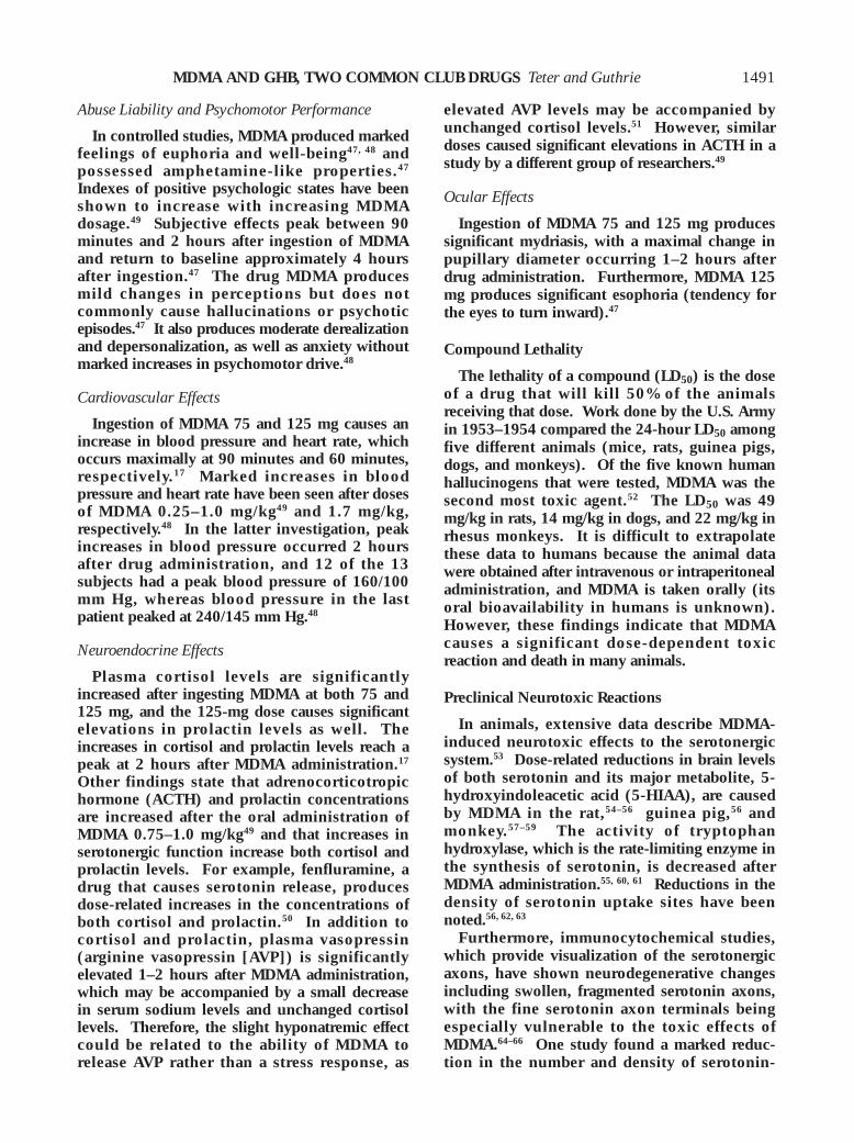
The increase in the use of club drugs has beenobserved by medical care providers. Accordingto the Drug Abuse Warning Network (DAWN)
report,3 emergency department episodessignificantly increased for MDMA (p<0.01), GHB(p<0.01), and ketamine (p<0.05) from1994–1999. These increases were especiallydramatic from 1997–1999 for MDMA and GHB(Figures 1 and 2). Apparent increases in the useof flunitrazepam over the same time period werenot statistically significant. Methamphetamineand LSD account for the largest number ofemergency department “mentions” in this reportoverall; however, mentions of methamphetaminedropped significantly from 1994–1999, whereasmentions of LSD remained stable over these 6years. (A mention refers to a specific substancethat was mentioned in a drug abuse episode.)
The use of these drugs poses a serious publichealth problem, and health care professionalsneed to be familiar with these substances.
MDMA
History
The drug MDMA (Ecstasy, “E”) is a ring-substituted amphetamine analog commonlytaken as a recreational drug of abuse. It wassynthesized in 1912 by Merck Pharmaceuticalsand patented in 1914.4 However, MDMA did notbecome popular until the 1970s when it waspromoted as a useful adjunct to psychotherapy.It allegedly improved self-esteem and enhancedcommunication within significant emotionalrelationships.5 Therapeutic applications for
1487
Figure 2. Drug Abuse Warning Network (DAWN) statisticsof the number of emergency department mentions of g-hydroxybutyrate (GHB) from 1994–1999. (From reference3.)
Figure 1. Drug Abuse Warning Network (DAWN) statisticsof the number of emergency department mentions of 3,4-methylenedioxymethamphetamine (MDMA) from1994–1999. (From reference 3.)
0
500
1000
1500
2000
2500
3000
1994 1995 1996 1997 1998 1999
No.
of E
mer
genc
y D
epar
tmen
t Men
tions
of M
DM
A
Year
0
500
1000
1500
2000
2500
3000
1994 1995 1996 1997 1998 1999
Year
No.
of E
mer
genc
y D
epar
tmen
t Men
tions
of G
HB

PHARMACOTHERAPY Volume 21, Number 12, 2001
MDMA were not well established, however, andthe Drug Enforcement Administration (DEA)classified MDMA as a schedule I drug in 1985after its recreational use became more widespreadand publicized.6 It also was shown that 3,4-methylenedioxyamphetamine (MDA), an analogand metabolite of MDMA, had a neurotoxic effectin animals.7
Availability
The drug MDMA is commonly distributed assmall tablets, capsules, or white powder. Ecstasytablets may contain various chemicals other thanpure MDMA, including MDA, 3,4-methylene-dioxyethylamphetamine (MDEA), caffeine,dextromethorphan, ephedrine, phenyl-propanolamine, methamphetamine, ampheta-mine, diphenhydramine, ketamine, cocaine, anddiazepam. Some have contained no active drugsat all.8 Results of analyses of tablets from all overthe United Kingdom, given to the LeedsAddiction Unit, confirm that there are manyingredients in Ecstasy tablets other than MDMA.9
In another report, MDMA concentrations in 25tablets varied 70-fold, and 9 of the tablets did notcontain either MDMA or any related MDMAanalog.10 Furthermore, MDMA tablets collectedby the Haight Ashbury Free Medical Cliniccontained from 16–150 mg of MDMA/tablet.Owing to the great variability in the dose ofMDMA in any given tablet, it is very difficult for
users of MDMA to control their dose. Larger-than-expected doses of MDMA may be takenaccidentally, leading to adverse effects.11
Furthermore, because of the variety of substancesthat may be found in any given MDMA tablet, theclinical presentation of acute intoxication mayvary significantly.
Chemistry
The chemical designation of MDMA is N-methyl-1-(3,4-methylenedioxyphenyl)-2-aminopropane (Figure 3). Structurally itresembles both the stimulant amphetamine andthe hallucinogen mescaline.12 The drug MDMAis optically active, with the dextrorotatory isomer(S+) having higher central activity than thelevorotatory isomer.4, 13
Pharmacokinetics
Pharmacokinetic Parameters
The pharmacokinetics of MDMA after oralingestion have been studied by variousresearchers.14–17 The time to maximumconcentration (Tmax) is 2 hours after oralingestion of MDMA 50, 75, or 125 mg.14–17 Thehalf-life shows little variation after a wide rangeof doses. After a 50-, 75-, or 125-mg dose, thehalf-life is 8 hours.16, 17 Other studies found thehalf-life to be 9.53 hours after a 75-mg dose and9.12 hours after a 125-mg dose.14 The maximum
1488
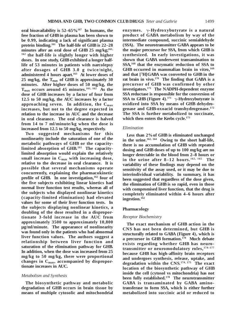
Figure 3. Mechanism of MDMA elimination. The parent compound MDMA and its metabolites are excreted in the urine tovarying degrees (see text). Also, MDMA may be metabolized by ring hydroxylation and demethylenation to potentialserotonergic neurotoxins.

MDMA AND GHB, TWO COMMON CLUB DRUGS Teter and Guthrie
concentration (Cmax) after oral ingestion appearsto be dose dependent. A Cmax of 105.6 ng/ml wasreported in a single subject who took a 50-mgdose,16 whereas a Cmax of 330 ng/ml was found inanother subject who took MDMA 135 mg.18 In agroup of eight subjects, the Cmax values afteringestion of MDMA 75 mg and 125 mg were126.5 and 226.3 ng/ml, respectively,14 whereas inanother group of eight subjects, Cmax values of130.9 ng/ml and 236.4 ng/ml were obtained afteringestion of MDMA 75 mg and 125 mg,respectively.17 In these studies, the Cmax exhibitsa slightly greater-than-expected increasecompared with the increase in dose. Accordingto these observations, after the usual recreationaldose of 100–150 mg, the Cmax should be 200–300ng/ml. The area under the concentration-timecurve (AUC) data from these studies also suggestnonlinearity. The AUC measured over 24 hoursafter ingestion of a 125-mg dose (2235.9µg/L•hr) is more than twice the AUC afteringestion of a 75-mg dose (995.4 µg/L•hr).14
Nonlinearity is further supported by otherevidence, in which the dose ratio of MDMA was1:3 (50 mg and 150 mg), whereas the AUC ratioover 24 hours after ingestion was greater than1:10. The authors suggested that the nonrenalclearance of MDMA is dose dependent (i.e.,HMMA, one of the many metabolites of MDMAmetabolism [Figure 3], was the major product inplasma at lower doses, whereas MDMA was thepredominant product at higher doses). Thisresulted in a disproportionate increase in plasmaAUC and an increase in the proportion of MDMAexcreted in the urine as the dose increased. It ispossible that demethylenation may be inhibitedas MDMA accumulates or one of the MDMAmetabolites may inhibit cytochrome P450 (CYP)2D6, which is responsible for a substantialproportion of MDMA nonrenal clearance.Alternatively, there might be an increase in thefraction of drug bioavailable as the doseincreases.15 Unfortunately, to our knowledge, theoral bioavailability of MDMA has not beendetermined in humans.
Primary Metabolism
The primary metabolic pathways for MDMAhave been elucidated, with a number ofmetabolites having been identified in bothanimals and humans (Figure 3). The main meta-bolic pathway appears to be demethylenation tothe catechol metabolite 3,4-dihydroxy-methamphetamine (DHMA; also called N-
methyl-a-methyldopamine).19 The metaboliteDHMA is the major metabolite of MDMA in ratliver19, 20 and in rat brain microsomes.21
Microsomes from yeast expressing humanCYP2D6 demethylenate MDMA to the metaboliteDHMA.22, 23
Furthermore, using human liver microsomes,CYP2D6 is the primary isoenzyme responsiblefor the demethylenation of MDMA.24 If CYP2D6is the isoenzyme responsible for the majority ofMDMA metabolism in humans, then poormetabolizers could be sensitive to the acutephysiologic effects of MDMA, but less prone toany long-term toxic effects of MDMA arisingfrom metabolites. However, case reports haveindicated that fatal MDMA intoxications haveoccurred in patients who were shown to beCYP2D6 extensive metabolizers,25 and it also hasbeen shown in vivo that in the absence offunctional CYP2D6 a considerable amount ofmetabolism of MDMA analogs occurs bydemethylenation.24 It may be that more than onemetabolic pathway can lead to an MDMA-induced toxic reaction.
Secondary Metabolism
A second pathway of MDMA metabolism is N-demethylation to MDA, which appears to be aminor metabolite of MDMA14 and is an abuseddrug in its own right. Concentrations of MDA inplasma range from 3–5% of those correspondingto MDMA.14 When formed from MDMA, theMDA formation rate constant is approximately0.75/hour and the half-life is 16–28 hours,depending on the dose of MDMA given. TheCmax for MDA occurs at 5–7 hours, and, on thebasis of plasma AUC comparisons of MDMA andMDA, 8–9% of MDMA is converted to MDA,which may be further metabolized beforeelimination. The urinary recovery of unchangedMDA accounts for approximately 1% of the doseof MDMA.17 It is unlikely that significantaccumulation of MDA would occur after a singledose of MDMA. Given the prolonged half-life ofMDA, however, it could accumulate in anindividual taking MDMA 3 or more times/week.
Toxic Metabolites
It has been hypothesized that some of theneurotoxic actions of MDMA may result fromquinones formed from the metabolism of DHMA,which can combine with glutathione and otherthiol compounds.19, 22 A 6-hydroxy-dopamineanalog is formed by the aromatic hydroxylation
1489

PHARMACOTHERAPY Volume 21, Number 12, 2001
and demethylenation of MDMA that also couldbe neurotoxic.22, 26 Catecholamines formed fromMDMA, such as DHMA, are highly polarcompounds that cannot cross the blood-brainbarrier. However, these highly polar compoundshave been detected in the brain after peripheraladministration of MDMA,27 indicating that someMDMA metabolism may occur in the brain.
Drug Interactions
There is a single report, to our knowledge, of apossible drug interaction involving MDMA andritonavir.28 A patient receiving ritonavir for thetreatment of human immunodeficiency virusingested MDMA in an estimated dose of 180 mg.The resultant blood MDMA level was 4.56 µg/ml,which is much higher than would be expectedfrom this dose of MDMA. The authors suggestthat the coadministration of MDMA and ritonavir(an inhibitor of CYP2D6) is the explanation forthe unusually high levels of MDMA after acommonly used recreational dose.28
Preincubation of MDMA with human livermicrosomes and nicotinamide adeninedinucleotide phosphate (NADPH) resulted insignificant inhibition of CYP2D6 activity.Therefore, MDMA may be a potent inhibitor ofCYP2D6 in vivo, and the interaction of MDMAwith this metabolic pathway may cause long-lasting drug interactions with other CYP2D6substrates.29 No clinical data are available insupport of this theory.
Elimination
In humans, approximately 50–70%15, 16 of thetotal MDMA dose is recovered in the urine asMDMA and other metabolites. Although MDMAis metabolized in the body, a large proportion isexcreted unchanged in the urine. A report basedon one patient indicated that after a single oralingestion of MDMA 50 mg, 32.52 mg (65%) ofunchanged drug was excreted in the urine over72 hours.16 In another study,15 urine collectionshowed an increase in the amount of unchangedMDMA excreted by a factor of 20, from the 50-mg to the 150-mg dose, whereas the urinaryrecovery of 4-hydroxy-3-methoxymetham-phetamine (HMMA), a metabolite of MDMAmetabolism, remained unchanged. Nosignificant changes in the urinary pH orcreatinine clearance occurred during this study.Although the renal clearance remained fairlyconstant, the nonrenal clearance appeared to bedose dependent.15
Pharmacology
Receptor Biochemistry
The drug MDMA is a potent indirectmonoaminergic agonist, which is thought to actby both increasing the release and inhibiting thereuptake of serotonin and, to a lesser extent,dopamine.30 Serotonin is involved in theregulation of a variety of behavioral functions,including mood, anxiety, aggression, appetite,and sleep. Dopamine is the primary neuro-transmitter of the “reward pathway” and isinvolved in motivational processes such asreward and reinforcement. Norepinephrine hasimportant roles in the processes of attention andarousal. In vitro, MDMA causes release ofserotonin, dopamine, and norepinephrine fromsynaptosomes31, 32 and rat brain slices.33, 34 Invivo, in freely moving rats, MDMA increases bothserotonin and dopamine release in the caudate.35
In a similar study, MDMA increased dopaminerelease in vivo in awake rats, resulting in region-,time-, and dose-dependent behavior.36 In ratbrain synaptosomes, MDMA inhibited the uptakeof serotonin and norepinephrine and, to a lesserextent, dopamine.37 The local administration ofMDMA to the rat nucleus accumbens resulted inincreases in the extracellular levels of bothserotonin and dopamine in this region,38 which ispart of the reward pathway activated by otherabused substances such as amphetamine andcocaine. These actions in the nucleus accumbensmay account for the euphoric effects produced byMDMA.
In addition to causing the release of serotoninand inhibiting its reuptake, MDMA may havedirect agonist effects on serotonin and dopaminereceptors.39 It has affinities for a broad range ofneurotransmitter recognition sites39, 40 and mayact at both serotonin receptors, 5-HT2A and 5-HT2C.41 Selective serotonin reuptake inhibitors(SSRIs) such as fluoxetine and citalopram blockthe release of serotonin induced by MDMA, bothin vitro42, 43 and in vivo.44, 45 They also reportedlyblock the subjective effects produced by MDMAin humans.46 Consequently, the release ofserotonin by MDMA may be dependent on theserotonin transporter. Different potencies for theneurotransmitter systems are shown by MDMAthan by either amphetamines or thehallucinogens. Owing to its individualbiochemical profile and the subjective effects itproduces in humans, MDMA has been called anentactogen, which means, “producing a touchingwithin.”13
1490
MDMA AND GHB, TWO COMMON CLUB DRUGS Teter and Guthrie
Abuse Liability and Psychomotor Performance
In controlled studies, MDMA produced markedfeelings of euphoria and well-being47, 48 andpossessed amphetamine-like properties.47
Indexes of positive psychologic states have beenshown to increase with increasing MDMAdosage.49 Subjective effects peak between 90minutes and 2 hours after ingestion of MDMAand return to baseline approximately 4 hoursafter ingestion.47 The drug MDMA producesmild changes in perceptions but does notcommonly cause hallucinations or psychoticepisodes.47 It also produces moderate derealizationand depersonalization, as well as anxiety withoutmarked increases in psychomotor drive.48
Cardiovascular Effects
Ingestion of MDMA 75 and 125 mg causes anincrease in blood pressure and heart rate, whichoccurs maximally at 90 minutes and 60 minutes,respectively.17 Marked increases in bloodpressure and heart rate have been seen after dosesof MDMA 0.25–1.0 mg/kg49 and 1.7 mg/kg,respectively.48 In the latter investigation, peakincreases in blood pressure occurred 2 hoursafter drug administration, and 12 of the 13subjects had a peak blood pressure of 160/100mm Hg, whereas blood pressure in the lastpatient peaked at 240/145 mm Hg.48
Neuroendocrine Effects
Plasma cortisol levels are significantlyincreased after ingesting MDMA at both 75 and125 mg, and the 125-mg dose causes significantelevations in prolactin levels as well. Theincreases in cortisol and prolactin levels reach apeak at 2 hours after MDMA administration.17
Other findings state that adrenocorticotropichormone (ACTH) and prolactin concentrationsare increased after the oral administration ofMDMA 0.75–1.0 mg/kg49 and that increases inserotonergic function increase both cortisol andprolactin levels. For example, fenfluramine, adrug that causes serotonin release, producesdose-related increases in the concentrations ofboth cortisol and prolactin.50 In addition tocortisol and prolactin, plasma vasopressin(arginine vasopressin [AVP]) is significantlyelevated 1–2 hours after MDMA administration,which may be accompanied by a small decreasein serum sodium levels and unchanged cortisollevels. Therefore, the slight hyponatremic effectcould be related to the ability of MDMA torelease AVP rather than a stress response, as
elevated AVP levels may be accompanied byunchanged cortisol levels.51 However, similardoses caused significant elevations in ACTH in astudy by a different group of researchers.49
Ocular Effects
Ingestion of MDMA 75 and 125 mg producessignificant mydriasis, with a maximal change inpupillary diameter occurring 1–2 hours afterdrug administration. Furthermore, MDMA 125mg produces significant esophoria (tendency forthe eyes to turn inward).47
Compound Lethality
The lethality of a compound (LD50) is the doseof a drug that will kill 50% of the animalsreceiving that dose. Work done by the U.S. Armyin 1953–1954 compared the 24-hour LD50 amongfive different animals (mice, rats, guinea pigs,dogs, and monkeys). Of the five known humanhallucinogens that were tested, MDMA was thesecond most toxic agent.52 The LD50 was 49mg/kg in rats, 14 mg/kg in dogs, and 22 mg/kg inrhesus monkeys. It is difficult to extrapolatethese data to humans because the animal datawere obtained after intravenous or intraperitonealadministration, and MDMA is taken orally (itsoral bioavailability in humans is unknown).However, these findings indicate that MDMAcauses a significant dose-dependent toxicreaction and death in many animals.
Preclinical Neurotoxic Reactions
In animals, extensive data describe MDMA-induced neurotoxic effects to the serotonergicsystem.53 Dose-related reductions in brain levelsof both serotonin and its major metabolite, 5-hydroxyindoleacetic acid (5-HIAA), are causedby MDMA in the rat,54–56 guinea pig,56 andmonkey.57–59 The activity of tryptophanhydroxylase, which is the rate-limiting enzyme inthe synthesis of serotonin, is decreased afterMDMA administration.55, 60, 61 Reductions in thedensity of serotonin uptake sites have beennoted.56, 62, 63
Furthermore, immunocytochemical studies,which provide visualization of the serotonergicaxons, have shown neurodegenerative changesincluding swollen, fragmented serotonin axons,with the fine serotonin axon terminals beingespecially vulnerable to the toxic effects ofMDMA.64–66 One study found a marked reduc-tion in the number and density of serotonin-
1491

PHARMACOTHERAPY Volume 21, Number 12, 2001
immunoreactive axons throughout the cerebralcortex in monkeys who also displayed reductionsin brain concentrations of serotonin and 5-HIAA.This is important as it provides some evidencethat reductions in serotonin and 5-HIAA may bedirectly associated with morphologic findings ofdamage to serotonergic axons.59
In one study that used positron emissiontomography (PET) in the nonhuman primate,67 asubstantial loss of serotonin transporters wasfound in the central nervous system (CNS) afterMDMA treatment. Reasonably good agreementwas noted between the PET data and reductionsin serotonin, 5-HIAA, and serotonin transporterdensity, measured neurochemically in postmortembrain tissue 3 weeks after the last PET study wasperformed.67 The neurotoxic effects of MDMAon the serotonergic system of the monkey may belong-lasting and still evident from 18 months68 to7 years69 after the administration of MDMA.There is evidence of some regrowth of the axons,but it may be abnormal and incomplete,69 withsome reorganization of ascending serotonergicprojections.70
Clinical Neurotoxic Reactions
In addition to the extensive animal dataproviding evidence for serotonergic neurotoxiceffects, there is evidence of possible neurotoxicreactions in human users of MDMA. Theserotonergic neurotoxic evidence can beclassified into three domains: neurobiologic (i.e.,neuroendocrine and brain imaging), psychologicand somatic, and psychiatric.
Neurobiologic Domain
Studies have shown dose-dependent decreasesin the concentrations of the serotonin metabolite5-HIAA in the cerebrospinal fluid of individualstaking MDMA.71, 72 Another technique to assessserotonergic damage in humans is to administerserotonergic agonists and examine neuro-endocrine responses. Various serotonergicagonists such as L-tryptophan, m-chlorophenyl-piperazine (m-CPP), and D-fenfluramine (D-fen)have been used to assess the prolactin response.71,
73–75 Two studies evaluated the prolactin responseafter the administration of L-tryptophan, aserotonin precursor known to increase serumprolactin concentration. One investigation founda nonsignificant trend toward a blunted prolactinresponse in those who took MDMA, as comparedwith control subjects,74 whereas the other studydid not find significant differences in the
prolactin response to L-tryptophan in those wholast took MDMA 18 weeks–2 years before thebeginning of the study, in comparison with thecontrol group.71 With use of m-CPP, 25individuals who took MDMA were less sensitiveto the anxiogenic effects of m-CPP comparedwith 25 controls, and the men who took MDMAhad a diminished prolactin and cortisol responseto m-CPP.73 A blunted neuroendocrine responsealso was found with the serotonin releaser, D-fen,in 15 individuals abstaining from taking MDMAin comparison with control subjects. Prolactinresponse to D-fen was significantly reduced inindividuals taking MDMA at both 3 weeks and12 months after last MDMA use. In contrast,cortisol response to D-fen was reduced at 3 weeksbut had recovered by 12 months in those takingMDMA. The authors suggest that the cortisolresponse at 12 months may indicate either partialrecovery to the neurotoxic actions of MDMA orselective neurotoxic actions of MDMA ondifferent serotonin receptors and pathways.75
A study using PET with [11C] McN5652 (aradioligand selective for the 5-HT transporter)found that individuals who abstained from takingMDMA (at least 3 weeks since last use) showedsignificant reductions in 5-HT transporterbinding compared with that of control subjectswho had never taken MDMA. This wasattributed to a reduced density of serotoninuptake sites. Furthermore, reduced binding waspositively correlated with the amount of previousMDMA use.76 Another study using PET imagingfound a reduction in the brain glucosemetabolism in individuals taking MDMA. ThePET scans were obtained in seven individualswho had taken MDMA from 1–39 months andseven age-matched control subjects with nohistory of illicit drug use. Glucose uptake waslowered in the hippocampus, amygdala, andcingulate bilaterally in the MDMA group.77 Adecrease in the density of serotonin uptake siteshas been found with use of other brain imagingtechniques as well. Ten men who had takenMDMA long term were compared with 10individuals who had not taken MDMA, matchedfor the consumption of other drugs. Eachsubject was examined with single photonemission computed tomography (SPECT) with a5-HT transporter ligand. The MDMA groupshowed a reduction in cortical serotonintransporter binding.78 In another SPECT study,79
cortical 5-HT2A receptor densities in the occipitalcortex were increased in five individuals whoabstained from taking MDMA (at least 2 months
1492

MDMA AND GHB, TWO COMMON CLUB DRUGS Teter and Guthrie
since last use) compared with nine healthycontrol subjects. The authors suggested anupregulation of postsynaptic 5-HT2A receptorsdue to serotonin depletion.80 Only the subjectswith apparent high densities of postsynaptic 5-HT2 receptors in the occipital area showeddetectable decreases in memory function.
Psychologic and Somatic Domain
Memory decrements are more pronounced inthose taking MDMA regularly (10 or moreoccasions) than in those just beginning (9 orfewer occasions).81 In addition, both those justbeginning to take MDMA and those regularlytaking MDMA exhibit significantly lowerimmediate word recall and delayed word recallcompared with control subjects.81 Significantmemory impairment has been reported in thosewho take only MDMA compared with those whotake many drugs but who had never takenMDMA, suggesting that the memory impairmentsare caused primarily by the MDMA rather thanthe various other drugs consumed by theseindividuals.82
Psychiatric Domain
Two studies suggest a depression of mood inthe days after taking MDMA.83, 84 In one study,those who took MDMA scored in the mild-to-moderate clinical range for depression on theBeck Depression Inventory.83 In the other study,visual analog mood scales were used to assess 16mood states. Those taking MDMA reportedfeeling significantly more depressed, abnormal,unsociable, unpleasant, and less good tempered 2days after the ingestion of MDMA than did thecontrol subjects.84
Subjective Effects
There are very few controlled studiesevaluating MDMA in humans, mostly becauseMDMA is a schedule I drug in the U.S. andtherefore is difficult to obtain for study purposes.Also, the safety of human research subjects whotake MDMA cannot be guaranteed. Theinformation that is available, with the exceptionof a few small controlled trials, comes frominformation collected retrospectively from peoplewho have taken MDMA outside of a controlledresearch environment. The high rate of concurrentmultisubstance abuse, the uncontrolled contentof active MDMA in any given pill, as well as thehistorical accuracy of information reported by the
person who takes recreational drugs lend someuncertainty to the conclusions drawn from thesereports. According to these reports, MDMA isingested orally in a dose of approximately100–150 mg, with an onset of effects usuallyaround 30 minutes, which is described as anamphetamine-like rush.
The earliest reports of MDMA effects wereprimarily anecdotal. Although users stressed thepositive feelings associated with MDMA, negativeeffects were also reported. Some of the positiveeffects include a sense of “closeness” towardothers, heightened alertness, increased ability tointeract with others, decreased defensiveness,decreased fear, decreased sense of alienation fromothers, increased awareness of emotions,decreased aggression, euphoria, increased energy,and sexual arousal.85–87 The negative effectsinclude tachycardia, trismus (jaw clenching),bruxism (teeth grinding), decreased appetite,lower back pain, and decreased desire to performmental or physical activities.85–87 Aftereffects(“hangover”) most often described by those whohave taken MDMA include lethargy, anorexia,decreased motivation, sleepiness, depressedmood, and fatigue.85, 86 Of interest, many of thesubjects reported that with regular MDMA usage(≥ six separate doses), the positive effectslessened while the negative effects increased.85
Consequently, many individuals space theirusage.5
Two early reports on the effects of MDMA wereprospective studies, involving 50 patients, thatwere completed before the governmentrestriction of MDMA to schedule 1.5, 88 In bothstudies, patients provided positive and negativedescriptions of the experience. The positiveexperiences included a perception of enhancedcommunication, increased feelings of intimacy,cognitive enhancement, euphoria, increased self-confidence, a heightened sense of sensualawareness (with some subjects reportingincreased sexual arousal and an increase inphysical and emotional energy). Adverse effectsthat were described by all of the subjects includethose similar to amphetamines such as tachycardia,dry mouth, palpitations, bruxism, trismus,nausea, anorexia, headaches, eyelid twitches, andinsomnia. Unlike with amphetamines, thereappeared to be no “crash” or depression up to 24hours after ingestion.5, 88
Pharmacologic Pretreatment
Three studies have investigated the feasibility
1493

PHARMACOTHERAPY Volume 21, Number 12, 2001
of giving pharmacologic agents to block orattenuate MDMA effects. Citalopram is aserotonin reuptake inhibitor that should blockthe uptake of MDMA into the neuronal terminal.Pretreatment with intravenous administration ofcitalopram 40 mg attenuated the acutepsychologic effects of MDMA 1.5 mg/kg inhealthy volunteers. Some of the effectsattenuated by citalopram included MDMA-induced increases in positive mood, derealizationand depersonalization phenomena, and the lossof thought and body control. The attenuation ofthe psychologic effects induced by MDMA as aresult of citalopram pretreatment suggests thatMDMA actions are at least partly dependent on acarrier-mediated release of serotonin.46 The sameinvestigators performed another study designedto test the effect of haloperidol 1.4 mgintravenously (dopamine D2 antagonist) on thepsychologic and physiologic responses toMDMA. Haloperidol treatment before MDMAadministration reduced the positive mood andeuphoria induced by MDMA, but not thecardiovascular effects. The authors suggestedthat there may be a role for dopamine in theeuphoria-producing effects of MDMA and thatserotonin or norepinephrine may mediate thephysiologic effects.89 The final study, also by thisgroup, used ketanserin (5-HT2 antagonist) toexamine the role of 5-HT2 receptors on MDMA’sactions. Ketanserin 50 mg was given orally tohealthy volunteers before the oral administrationof MDMA 1.5 mg/kg. Ketanserin attenuatedperceptual changes and emotional excitationinduced by MDMA but had little effect onMDMA-induced positive mood, well-being, andextroversion. Furthermore, body temperaturewas lower after the MDMA-ketanserincombination than with MDMA alone.90
Adverse Effects and Acute Toxic Reactions
Acute Syndrome
One of the dangers of MDMA is the apparentlack of relationship between alleged dose andseverity of acute toxic reaction.91, 92 Althoughone person attempted suicide after reportedlytaking 42 pills of Ecstasy with a resultant plasmaMDMA level of 7.72 µg/ml and displayed onlyhypertension and tachycardia,93 others have diedwith much lower plasma MDMA levels rangingfrom 0.05–1.26 µg/ml.91 Furthermore, serumMDMA levels do not correlate well with clinicalsymptoms.94 Acute toxic reactions usuallydevelop within 15 minutes–6 hours after the
ingestion of MDMA.91 Symptoms of an acuteMDMA toxic reaction include agitation,tachycardia, hypertension, dilated pupils,trismus, and sweating, whereas the more severecases may be characterized by hyperthermia,disseminated intravascular coagulation (DIC),rhabdomyolysis, and acute renal failure.93 Inmore severe cases, elevated creatine kinase levelsare often present,95–98 with levels as high as122,341–555,000 IU/L being reported.97, 98 Otherfrequently reported acute adverse effectsoccurring after the ingestion of MDMA includelack of appetite, difficulty concentrating,impaired balance, and restless legs.48
The toxic effects of MDMA were divided intothree categories in one investigation to helpdistinguish acute toxic reactions from long-termresidual effects. These categories were acutereactions at therapeutic doses, overdosereactions, and residual effects.11 At moderatedoses (85–100 mg), acute effects includedtransient nausea occurring about 30 minutesafter ingestion and lasting about 30 minutes,increases in both blood pressure and heart rate,and symptoms related to increased muscletonicity, such as jaw clenching and teethgrinding. In those subjects who were particularlysensitive to MDMA, higher doses (≥ 100 mg)caused numbness and tingling in the extremities,luminescence of objects, increased sensitivity tocold, increased color acuity, and vomiting.Residual effects occurring from 2 hours–2 weeksafter ingestion included exhaustion, fatigue, andnausea. Doses higher than 200 mg result in aclassic toxic psychosis with symptoms ofparanoia and auditory and visual hallucinations.
Hyperthermia
Hyperthermia (temperature > 40°C) is themost common adverse effect associated with asevere acute toxic reaction to MDMA. Theincrease in body temperature is probably due toserotonergic actions of MDMA in the thermo-regulatory center in the hypothalamus99 becauseanimal studies have shown that the hyperthermiacaused by compounds such as MDMA ismediated by actions at serotonin receptors in theCNS.100 Hyperthermia also may be caused byexcessive heat production due to sustainedmuscle hyperactivity, increased metabolic rate,rigidity, and seizures.101 Hyperthermia is believedto be the beginning of the cascade leading toDIC, rhabdomyolysis, myoglobinuria, and acuterenal failure. However, the exact pathophysiology
1494

MDMA AND GHB, TWO COMMON CLUB DRUGS Teter and Guthrie
of this cascade after MDMA intoxication has notbeen fully elucidated.
Cardiovascular Effects
Similar to cocaine and amphetamine, MDMAmay cause sympathetic stimulation and increasemyocardial oxygen demand, leading to varyingdegrees of tachycardia, vasoconstriction, changesin blood pressure, and arrhythmias. In severecases, vasospasm leading to acute myocardialinfarction and irreversible dilated cardio-myopathy may occur.102 Abnormal electro-cardiographic changes that show widespread STsegment elevation indicating acute myocardialinfarction have been seen with laboratoryevidence in the urine of MDMA users.103 Duringpostmortem evaluations, necrosis of the heart(contraction band necrosis or widespread foci ofnecrosis) has been seen104 and may be due toexcessive catecholamines.102 These findings donot necessarily establish a cause and effectrelationship, since other substances orcircumstances may have contributed.
Cerebrovascular Effects
“Designer drugs” such as MDMA are associatedwith intracerebral hemorrhage, often inconjunction with an underlying vascularmalformation.105 Other investigators havepostulated that those who take MDMA are at anincreased risk for cerebrovascular accidents dueto the altered 5-HT system because postsynaptic5-HT receptors are involved in the regulation ofthe brain microvasculature.106 Other cerebro-vascular adverse effects that have been associatedwith MDMA include subarachnoid hemorrhage,cerebral infarction, and cerebral venous sinusthrombosis.107 Magnetic resonance imagingrevealed a left basal ganglia hematoma after theingestion of MDMA in a patient with no apparentcardiovascular risk factors.108
Neuroendocrine Effects
Numerous cases of hyponatremia have beenassociated with MDMA use, often in combinationwith seizures, catatonic stupor, and incontinenceof urine.109–114 It is possible that hyponatremia isa direct result of MDMA neuroendocrine effectsor from massive water intake leading todilutional hyponatremia. Since many users takeMDMA during all-night dancing parties, largeamounts of fluid are ingested, both as a naturalconsequence of physical activity and because ofMDMA-induced hyperthermia. Hyponatremia
may be due to the syndrome of inappropriateantidiuretic hormone,111, 112 because MDMAcauses the release of AVP.51 In addition, theextreme dehydration caused by sweating and/orvomiting associated with MDMA use combinedwith massive water intake could lead tohyponatremia.115 In one report of a fatality dueto MDMA, hyponatremia leading to cerebraledema appeared to be the main cause of death.116
Contamination of the MDMA tablets with othersubstances has been postulated as the cause ofhyponatremia associated with MDMA use.110
Postcards have been distributed in some clubsand bars advising patrons who take MDMA thatthey should drink about a pint of water an hourand eat or drink something salty, such as a sportsdrink, to replace lost sodium.117
Hepatotoxicity
Hepatotoxic effects have been associated withMDMA.91, 93, 104, 118, 119 In one case series of sevenfatalities associated with the use of ring-substituted amphetamines, including MDMA,necrosis of the liver was seen in all cases.104 Twoof the most likely mechanisms for causing ahepatotoxic reaction are immune-mediatedreaction or injury secondary to hyperthermia.119
Hepatotoxic reaction arising from drugimpurities or MDMA metabolites is also possible.Liver transplantation has been required becauseof hepatic damage associated with MDMA use.118
Psychopathology
A psychotic syndrome characterized bydelusions, usually of the persecutory type, maybe caused by MDMA. Other nonpsychoticconditions include visual phenomena,depersonalization and derealization, panicattacks, and depression. Persons who displaysuch symptoms may have at least one first-degreerelative with a history of psychiatric illness andbe predisposed to have psychiatric symptoms.120
Anxiety attacks, persistent insomnia, ragereactions, and psychosis (especially at higherdoses) have occurred after MDMA use, althoughin most cases the premorbid psychiatric status ofthese patients was not known.11 Compared withcontrol subjects who do not take MDMA, thosewho frequently take MDMA have significantlyhigher scores on scales used to assesssomatization, obsessionality, anxiety, hostility,phobic-anxiety, paranoid ideation, psychoticism,poor appetite, and restless or disturbed sleep.They also showed greater impulsiveness.121
1495

PHARMACOTHERAPY Volume 21, Number 12, 2001
Death
Conditions commonly contributing to deathdue to MDMA include dehydration, hyper-thermia, disseminated intravascular coagulation,rhabdomyolysis, acute renal failure, tachycardiaand other cardiac arrhythmias, and convulsions.93,
122 In other fatal cases involving MDMA, necrosisof the liver and heart were found at autopsy aswere various injuries to the brain such as focalhemorrhages and severe cerebral edemaconsistent with water intoxication.104
Treatment
The diagnosis of acute toxic reaction to MDMAis made based on the history and clinical featuresof intoxication. Initial examination shouldinclude blood chemistry analysis, complete bloodcount, liver function tests, cardiac enzyme andcreatine kinase measurements, and a urinetoxicology screen. Quantitative serum levels donot correlate well with severity of symptoms andare not generally available.94 A complete historyand physical examination should be performed,and the patient should be assessed for hyper-tensive crisis or life-threatening arrhythmias. Anelectrocardiogram for chest pain or a computedtomographic (CT) scan of the brain for persistentmental status changes should be obtained.123, 124
Amphetamines and related drugs (i.e., metham-phetamine, MDMA) can be detected in the urine,but there is a high degree of cross-reactivitybetween amphetamine derivatives and adrenergicamines. Therefore, confirmatory testing usuallyis required.94
Resuscitation
There is no antidote for MDMA intoxication,and in general, recommended treatment ofMDMA overdose is similar to the treatment ofamphetamine or methamphetamine overdose.The first priority should be maintaining theairway, breathing, and circulation.123, 124
Treatment will then be aimed at reducing varioussymptoms, including hyperthermia, agitation,cardiovascular and cerebrovascular incidents,neuroendocrine abnormalities, and neurologicproblems.
Decontamination and Elimination
Decontamination of the gastrointestinal tractwith lavage, activated charcoal, and cathartictechniques has been used. Induction of emesis isnot appropriate because of the potential for CNS
depression and seizures.94 Because approxi-mately 50–70%15, 16 of MDMA is recovered in theurine, renal failure would significantly decreasethe elimination of MDMA from the body, somaintaining adequate hydration is essential.Because MDMA is a weak base and a significantproportion is eliminated in the urine, acidifyingthe urine is likely to be an effective means ofincreasing renal elimination, but it mayprecipitate acute renal failure in patients withmyoglobinuria and is not recommended.94
Hyperthermia
Although fatalities may be due to manydifferent causes, hyperthermia is probably thesingle most important condition to treat becauseit may lead to further severe complications, suchas rhabdomyolysis and DIC.125 Mortality hasbeen correlated to both the extent of hyper-thermia and the duration, and active coolingmeasures are indicated in cases of MDMA-induced hyperthermia (see Supportive Caresection).99 It is important to control agitation tolimit further heat production.126 Neuromuscularblockers, such as pancuronium, have been given,but their use requires ventilation andendotracheal intubation.94
Dantrolene sodium, a drug that is indicated forthe treatment of malignant hyperthermia and thatinhibits the release of calcium from thesarcoplasmic reticulum, is recommended bymany clinicians to treat hyperthermia secondaryto MDMA use.93, 95, 96, 99, 127–132 Speculativehypotheses notwithstanding, the use ofdantrolene for the treatment of MDMAintoxication remains controversial. The efficacyof dantrolene in treating this condition has beenquestioned, as some patients have improved withsupportive care only133 and some clinicians assertthat there is insufficient evidence to recommenddantrolene in cases of MDMA acute toxicreaction.134–136
To determine if MDMA caused an increase ofcalcium within the muscle, which would suggestthat an inhibitor of calcium release in skeletalmuscle, such as dantrolene, might be efficaciousin treating MDMA intoxication, in vitroexperiments using human muscle subjected tohalothane and caffeine contracture tests wereperformed (used to test for susceptibility tomalignant hyperthermia). It was hypothesizedthat if MDMA raised the calcium levels withinthe muscle, then dantrolene should be effectivein treating MDMA acute toxic reaction, since it is
1496

MDMA AND GHB, TWO COMMON CLUB DRUGS Teter and Guthrie
effective in treating malignant hyperthermia. Theresults indicated that the hyperthermia fromMDMA intoxication is associated with anelevation in the myoplasmic calcium concen-trations, similar to that seen in malignanthyperthermia, which suggests that dantrolenemight be a helpful agent in treating MDMA-induced hyperthermia.137 It has been argued thatMDMA-induced hyperthermia results fromaugmentation of central serotonin, and sincedantrolene has no central activity (inhibitscalcium peripherally in the skeletal muscle), itshould not be effective. Therefore, a non-depolarizing neuromuscular blocker may be justas effective in treating MDMA acute toxicreaction.92 As stated previously, however,ventilation and intubation may be required.94
Although, there are not sufficient data in humansto confirm that the hyperpyrexia associated withMDMA is a centrally mediated effect, the use ofdantrolene should not be precluded because itdoes appear to reduce pyrexia secondary toexertional heatstroke.138 It is hypothesized thatthe unpredictable hyperthermia associated withMDMA may result from an underlying metabolicmyopathy, similar to that seen with exertionalheatstroke, and associated with a skeletal muscleabnormality similar to malignant hyperthermia.139
Cardiovascular Treatment
Tachycardia without hemodynamiccompromise does not need to be treated.Sedative dosages of benzodiazepines may behelpful by reducing blood pressure and heartrate, which may reduce myocardial oxygendemand.140 b-Blockers should be avoided whentreating stimulant-induced hypertension becausethis may result in unopposed a-adrenergicvasoconstriction. Hypertension can be treatedwith an a-blocker such as phentolamine or witha direct-acting vasodilator such as nitroprusside.94,
123 Another option is the use of a b-blockerconcurrently with phentolamine.126 Myocardialischemia caused by stimulants should be treatedwith oxygen, aspirin, and benzodiazepines. Ifthese options do not reverse the ischemia, thenvasodilators or phentolamine should be given.102
Arterial spasm may be treated with sublingual orintravenous nitroglycerin.94 Arrhythmias shouldbe treated according to advanced cardiac lifesupport guidelines.123, 126 Thrombolytic agentshave been given safely to patients with stimulant-induced myocardial infarction.140
Cerebrovascular Treatment
Patients with altered mental status, lethargy, orobtundation should undergo CT of the brainbecause of the risk for intracranial hemorrhageand infarct.123 In patients with nontraumaticintracranial hemorrhage, arteriography should beperformed and a thorough history of the use ofillicit substances should be evaluated.105
Neurologic Treatment
Patients who are agitated may requiretreatment with a benzodiazepine, such asdiazepam, lorazepam, or midazolam.94, 123 It isvery important to control agitation as this maydecrease further heat production.126 Some of theconditions associated with MDMA acute toxicreaction (mental status changes, hyperthermia,autonomic instability, increased motorrestlessness, myoclonus, elevated creatine kinaselevel, diaphoresis, and death due to renal failure)are similar to the findings in both neurolepticmalignant syndrome and serotonin syndrome.91,
127 Pharmacologic treatments effective in thesesyndromes are recommended by some clinicians,127
including methysergide maleate (nonspecificserotonin antagonist),141 b-blockers (5-HT1Aantagonists),142 or bromocriptine (a dopamineagonist).143 However, none of these drugs hasbeen prospectively evaluated for the treatment ofMDMA acute toxic reaction.
Caution may be warranted in usingantipsychotic agents when treating MDMAintoxication. Antipsychotics decrease the seizurethreshold, and blocking dopamine receptors mayaffect the thermoregulatory system leading tohyperthermia or exacerbation of existinghyperthermia. In addition, SSRIs may furtherincrease serotonergic transmission by blockingthe reuptake of synaptic serotonin, possiblyraising the risk for development of the serotoninsyndrome or aggravating already existinghyperthermia.144
Hepatotoxicity
Owing to the risk for hepatotoxicity, it wouldbe prudent to monitor liver function in personssuspected of taking MDMA,119 and any personwith unexplained jaundice or hepatomegalyshould be screened for a history of MDMA use.93
Treatment will be primarily supportive (seeSupportive Care section). If severe hepaticnecrosis has occurred, transplantation may be theonly option101 and has been performedsuccessfully in patients with acute liver failuredue to MDMA use.118
1497

PHARMACOTHERAPY Volume 21, Number 12, 2001
Supportive Care
Supportive therapy includes rehydration withintravenous fluids and lowering the temperatureof the patient with use of cooling blankets or icebaths.143 In some cases, lowering the bodytemperature may require infusion of coldintravenous fluids or peritoneal lavage with cooldialysate.92 Crystalloids may be given to helptreat both the profuse sweating that oftenaccompanies MDMA acute toxic reaction as wellas prophylaxis against acute renal failuresecondary to rhabdomyolysis and myoglobinuria.95
Furthermore, judicious fluid support may helpwith symptoms of hepatotoxicity as it mayincrease liver blood flow and prevent furtherhepatic damage.119
Summary
The use of MDMA is on the rise, especiallyover the last couple of years. Although it causespleasant sensations, MDMA can be a verydangerous drug when used recreationally.Particularly severe adverse reactions includehyperthermia, rhabdomyolysis, DIC, renalfailure, cardiac complications, intracranialhemorrhage, and hepatotoxicity. The long-termneurotoxic effects, particularly in theserotonergic system, of MDMA have not beenfully elucidated. It is imperative that cliniciansbe familiar with the symptoms and treatmentoptions for acute toxic reaction to MDMA.
GHB
History
g-Hydroxybutyric acid (GHB) is a CNSdepressant that has become increasingly popularas a drug of abuse over the last 10 years. Manynames are used for GHB such as sodium oxybate,sodium oxybutyrate, g-hydroxybutyrate sodium,g-OH, 4-hydroxy butyrate, and g-hydrate, as wellas others. Names used on the street includeLiquid Ecstasy, Liquid X, Liquid E, Georgia HomeBoy, Grievous Bodily Harm, G-Riffick, Soap,Scoop, Salty Water, Somatomax, and OrganicQuaalude. In the 1960s, a French researchersynthesized GHB in an attempt to create a g-aminobutyric acid (GABA) analog that would,unlike GABA, cross the blood-brain barrier.145
Somewhat simultaneously, in 1963, GHB wasfound to be a naturally occurring metabolite inthe human brain.146 The first accepted medicalapplication of GHB was for intravenousinduction of anesthesia.145 However, its use was
limited due to the high frequency of vomiting,147
seizure-like activity in animals,148, 149 and inabilityto produce analgesia.150 In the 1970s, GHB wasrecommended for narcolepsy because it increasesslow-wave sleep and consolidates sleep at night,therefore decreasing sleep during the day.151 Inthe 1980s, GHB was commonly sold over-the-counter in health food stores where it was allegedto increase the effect of growth hormone.152 Inthe late 1980s and early 1990s, GHB wasadvocated for the treatment of alcoholdependence153 and opiate withdrawal.154 Duringthe same time period, GHB was illicitlyadvertised as a hypnotic to replace tryptophan,which had been removed from the market due toits connection with eosinophilia-myalgiasyndrome.155 Since 1990, an increasing numberof cases of both abuse and toxic reaction hasbeen noted, and in 1997 GHB was labeled a “daterape” drug by the press.156 In March 2000, GHBbecame a schedule I controlled substance in theU.S.157
Availability
Most of the GHB available in the U.S. ismanufactured clandestinely. Many Internet sitesand books that describe the process of makingGHB are available.158 Commonly offered for saleon Internet sites, GHB kits provide the chemicalsand recipes used to produce GHB.159, 160
Currently, GHB is only legally available in theU.S. for the investigational treatment ofnarcolepsy. The drug is synthesized by using acombination of sodium hydroxide and g-butyrolactone (GBL; another commonly abuseddrug). Because sodium hydroxide is very caustic,severe toxic reactions may result if GHB ismanufactured improperly. The drug GHB isavailable as a powder or a colorless, odorlessliquid with a salty or soapy taste. Its taste caneasily be masked by adding it to flavoredbeverages. As GHB is colorless and odorless, andbecause small quantities are required to achieve adesired effect, GHB has been used as a date rapedrug. The amnesia produced by GHB oftenmakes victims unable to serve as valid witnesses.
Pharmacokinetics
Pharmacokinetic Parameters
The pharmacokinetics of GHB are nonlinear inhumans over the therapeutic dosage range.161–164
The drug is rapidly absorbed orally,165 with anonset of action within 15 minutes.166 In the rat,
1498
MDMA AND GHB, TWO COMMON CLUB DRUGS Teter and Guthrie
oral bioavailability is 52–65%.167 In humans, thefree fraction of GHB in plasma has been shown tobe 0.99, indicating a lack of significant plasmaprotein binding.161 The half-life of GHB is 22–28minutes after an oral dose of GHB 25 mg/kg;161,
162 the half-life is slightly longer with higherdoses. In one study, GHB exhibited a longer half-life of 53 minutes in patients with narcolepsyafter dosages of GHB 3.0 g twice/night,administered 4 hours apart.163 At lower doses of25 mg/kg, the Tmax of GHB is approximately 30minutes. After higher doses of 50 mg/kg, theTmax occurs around 45 minutes.161, 162 As thedose of GHB increases by a factor of four from12.5 to 50 mg/kg, the AUC increases by a factorapproaching seven. In addition, the Cmaxincreases, but not to the degree expected inrelation to the increase in AUC and the decreasein oral clearance. The oral clearance is halvedfrom 14 to 7 ml/minute/kg when the dose isincreased from 12.5 to 50 mg/kg, respectively.
Two suggested mechanisms for thisnonlinearity include the saturation of one of themetabolic pathways of GHB or the capacity-limited absorption of GHB.161 The capacity-limited absorption would explain the relativelysmall increase in Cmax with increasing dose,relative to the decrease in oral clearance. It ispossible that several mechanisms operateconcurrently, explaining the pharmacokineticprofile of GHB. In one investigation,162 four ofthe five subjects exhibiting linear kinetics hadnormal liver function test results, whereas all ofthe subjects who displayed nonlinear kinetics(capacity-limited elimination) had elevatedvalues for some of their liver function tests. Inthe subjects displaying nonlinear kinetics, adoubling of the dose resulted in a dispropor-tionate 3-fold increase in the AUC fromapproximately 3500 to approximately 10,800µg/ml/minute. The appearance of nonlinearitywas found only in the patients who had abnormalliver function values. The authors suggest arelationship between liver function andsaturation of the elimination pathway for GHB.In addition, when the dose was increased from 25mg/kg to 50 mg/kg, there were proportionalchanges in Cmax, accompanied by dispropor-tionate increases in AUC.
Metabolism and Synthesis
The biosynthetic pathway and metabolicdegradation of GHB occurs in brain tissue bymeans of multiple cytosolic and mitochondrial
enzymes. g-Hydroxybutyrate is a naturalproduct of GABA metabolism by way of theintermediate compound, succinic semialdehyde(SSA). The neurotransmitter GABA appears to bethe major precursor for SSA, from which GHB issynthesized. In early investigations, it wasshown that GABA underwent transamination toSSA,168 that the enzymatic reduction of SSA toGHB occurred in mammalian brain in vitro,169
and that [3H]GABA was converted to GHB in therat brain in vivo.170 The finding that GABA is aprecursor of GHB was confirmed by otherinvestigators.171 The NADPH-dependent enzymeSSA reductase is responsible for the conversion ofSSA to GHB (Figure 4).171 g-Hydroxybutyrate isoxidized into SSA by means of GHB dehydro-genase and GHB-oxoacid transhydrogenase.172
The SSA is further metabolized to succinate,which then enters the Krebs cycle.173
Elimination
Less than 2% of GHB is eliminated unchangedin the urine.162, 164 Owing to the short half-life,there is no accumulation of GHB with repeateddosing and GHB doses of up to 100 mg/kg are nolonger detectable in the blood from 2–8 hours orin the urine after 8–12 hours.162, 165 Thevariability of these findings may depend on thesensitivity of the assay used, or it may be due tointerindividual variability. In summary, it hasbeen suggested that regardless of the dose given,the elimination of GHB is so rapid, even in thosewith compromised liver function, that the drug iscompletely eliminated within 4–6 hours afteringestion.162
Pharmacology
Receptor Biochemistry
The exact mechanism of GHB action in theCNS has not been determined, but GHB isstructurally related to GABA (Figure 4), which isa precursor in GHB formation.170 Much debateexists regarding whether GHB has neuro-transmitter or neuromodulatory roles,174–177
because GHB has high-affinity brain receptorsand undergoes synthesis, release, uptake, anddegradation within the CNS.174, 175 The exactlocation of the biosynthetic pathway of GHBinside the cell (cytosol vs mitochondria) has notbeen fully established.178 The neurotransmitterGABA is transaminated by GABA amino-transferase to form SSA, which is either furthermetabolized into succinic acid or reduced to
1499

PHARMACOTHERAPY Volume 21, Number 12, 2001
form GHB by the enzyme SSA reductase, aNADPH-dependent enzyme.171, 173 The highestconcentrations of GHB in the brain are found inthe substantia nigra179 and hypothalamus,whereas the highest turnover rate of GHB occursin the hippocampus.180 The uptake of GHBappears to be the highest in the striatum,181, 182
and this uptake is dependent on a specificsodium-dependent active transport system forGHB.181 In addition to being found in the CNS,GHB is found in the kidney, heart, skeletalmuscle, and brown fat.183
g-Hydroxybutyrate appears to have affinity fortwo receptor sites in the CNS. It binds to GHBreceptors, which may be linked to cyclicguanosine 3 ′5′ -monophosphate and inositolphosphate intracellular pathways184, 185 and aremost numerous in the hippocampus andcortex.186 It also binds to GABAB receptors,187, 188
but not to GABAA receptors.189 The relevance ofthis remains unknown but suggests that some ofthe pharmacologic actions of GHB are mediatedby the GABAB receptor.
The drug GHB alters dopaminergic activity, insome cases increasing and in others decreasingthe amount of dopamine released.178 Thesystemic administration of GHB to animalsresults in increased dopamine accumulation inthe extrapyramidal system of the brain, whichreaches its highest values 1–2 hours afterinjection, without parallel increases in serotoninor norepinephrine.190 The administration of a-methyltyrosine, which blocks the activity oftyrosine hydroxylase, almost completely blocksthe rise in brain dopamine induced by GHB,which occurred within 1 hour in control mice.Therefore, GHB mediates the accumulation ofdopamine by increasing the activity of tyrosinehydroxylase.191 In addition, GHB may inhibit therelease of newly synthesized dopamine192 anddecrease the firing rate of dopaminergic neuronsin the substantia nigra with maximal inhibitionwithin 8 minutes.179 The end result seems to be atissue accumulation of dopamine in the brain,175
which is supported by results of the short-termstudies described above. Dopamine release in the
1500
Figure 4. Mechanism of GHB elimination. g-Butyrolactone (GBL) and 1,4-butanediol (1,4-BD) are converted in the body toGHB.

MDMA AND GHB, TWO COMMON CLUB DRUGS Teter and Guthrie
striatum may be accompanied by the release ofendogenous opioids.193 The exact interactionsbetween GHB and the opioid system are not fullyunderstood, but the administration of naloxoneor nalorphine, opioid receptor antagonists,blocks some of the effects of GHB.193, 194
Dose-Related Effects
The primary dose-related effects of GHB arerelated to CNS depression. At 10 mg/kg, GHB iscapable of producing amnesia195 and hypotonia ofthe skeletal muscles196, 197 resulting from thedepression of neurons in the spinal cord.166 At20–30 mg/kg, GHB promotes a normal sequenceof rapid eye movement (REM) and non-REM(slow-wave) sleep, which lasts from 2–3 hours.151,
198 At 40–50 mg/kg intravenously, GHB producesa state of somnolence, which appears within5–15 minutes, and an oral dose of approximatelythe same amount will produce similar results.166
Anesthesia is associated with doses of 50mg/kg,164, 166, 199 and doses higher than 50 mg/kghave been associated with profound coma,199 aswell as decreased cardiac output, respiratorydepression, and seizures. These effects are morepronounced with the coingestion of CNSdepressants, particularly ethanol.155 Larger dosesof 60–70 mg/kg produce a state of unarousablecoma that lasts about 1–2 hours.166 Theinvestigators who initially discovered that GHBwas a natural metabolite of the brain reportedthat GHB 100 mg/kg administered intravenouslyproduced sleep that begins within 15 minutes ofadministration and lasts about 1.5–2 hours.146
Serum Concentrations
Oral ingestion of GHB 75–100 mg/kg inhumans results in peak blood levels ofapproximately 90–100 µg/ml at 1–2 hours afteringestion.165 Intravenous administration of GHB50 and 165 mg/kg results in peak blood levelsthat reach 180 and 412 µg/ml, respectively. Themean blood GHB level at the commonly useddose of 100 mg/kg is 304 µg/ml.164 When theblood GHB levels exceed 258 µg/ml, subjects fallinto a state of deep sleep, characterized bynonresponse to various stimuli such as touch,pinprick, deep pressure, skin preparations, orvaginal examinations, although there is stillreflex response to surgical incision. During thisstage of deep sleep, blinking stops and the eyesremain central and fixed with small pupils. Amoderate level of sleep is associated with bloodGHB levels ranging from 155–258 µg/ml. This
moderate stage of sleep is characterized byspontaneous blinking and responses to deeppressure. Blood GHB levels ranging from 52–155µg/ml are associated with a light sleep charac-terized by spontaneous movements and occasionalopening of the eyes. When the blood GHB levelsdecrease below 52 µg/ml, subjects wake up.164
Abuse Potential and Intoxication
Factors that seem to contribute to the abusepotential of GHB include its intoxicating effects,its purported anabolic effects, its hypnotic effects,and its ability to incapacitate women forpurposes of sexual assault.157, 200, 201 One of themain reasons GHB became a popular drug ofabuse is its ability to produce a “high.”155, 202
Those who take GHB describe it as producing astate of relaxation and tranquility accompaniedby feelings of calmness, mild euphoria, atendency to verbalize, mild numbing, andpleasant disinhibition. Despite these positivefeelings attributed to the use of GHB, the dose-response curve for GHB has been described asbeing remarkably steep. Therefore, as the dose ofGHB is increased, a steep increase in adverseeffects may occur.143 The effects of GHB havebeen described as being similar to those ofalcohol, and the two agents may act syner-gistically, further increasing the risk forintoxication or overdose.203
Cardiovascular Effects
Moderate bradycardia appears after theadministration of GHB204, 205 and is likely due tocentral vagal activity.166 In addition tobradycardia, GHB reduces stroke volume as wellas cardiac output, which reaches a nadir around30 minutes after ingestion. Atropine reverses thedecreases in both heart rate and stroke volume.205
The autonomic centers are fully active duringGHB-induced coma, and surgical stimuli result ina cardiovascular response, such as tachycardia,hypertension, and raised cardiac output.150, 166, 204
Respiration
Respiratory rate is often reduced, but this isusually accompanied by an increase in tidalvolume.150, 204 The drug GHB also produces aslowing and deepening of respiration sometimesleading to a Cheyne-Stokes pattern.166, 204
Neuroendocrine Effects
In an early study that stimulated much interest
1501

PHARMACOTHERAPY Volume 21, Number 12, 2001
in the use of GHB by the bodybuildingpopulation, intravenous administration of GHB2.5 g significantly increased plasma growthhormone levels, which peaked at 60 minutes.152
In a more recent study, after bedtime oralingestion of GHB 2.5, 3.0, and 3.5 g, asignificant increase occurred in the normalsecretory pulse of growth hormone during thefirst 2 hours after sleep onset. The authorssuggest that agents such as GHB may increase therelease of growth hormone by increasing slow-wave sleep, because there is a large pulse ingrowth hormone secretion during the first stageof slow-wave sleep more than 90% of the time.206
Sedation and Anesthesia
The principal actions of GHB have not beenfully elucidated. However, the results of earlyinvestigations suggest that GHB appears to act onthe cerebral cortex with little or no depression ofthe reticular activating system.150 Some authorsspeculate that there is depression of the limbichippocampal structures166 and subcorticalcenters.199 The anesthetic effects of GHB areprimarily hypnotic204 as GHB provides little or noanalgesia.150, 204 The transition from wakefulnessis described as being a sudden shift fromresponsivity to unconsciousness.199
Sleep Physiology
The drug GHB stimulates slow-wave sleep.199,
207–210 It does not appear to suppress REMsleep207, 209 and may even decrease fragmentationof REM sleep.208 It appears to increase “slow”sleep as evidenced by a slow synchronizedelectroencephalographic recording.199 Inaddition, GHB increases slow-wave sleep (stages3 and 4), whereas light sleep (stage 1) isdecreased, and the frequency of awakenings isreduced.210 In healthy subjects, under double-blind conditions, single oral doses of GHB 2.25 gsignificantly increased the time spent in slow-wave sleep, while sacrificing stage 1 sleep andsignificantly decreasing slow-wave sleep latency.The efficiency of REM sleep is increased, but theREM latency and time spent in REM sleep do notchange.211
Therapeutic Applications
Most of the therapeutic applications of GHBresult from its sedative and hypnotic effects onthe CNS. There are no currently acceptedmedical applications for GHB in the U.S.,although it is being evaluated for the symptoms
of narcolepsy.157 However, GHB has beenextensively administered and studied for a varietyof indications in other countries.
Sedation and Anesthesia
The first clinical application of GHB was as ahypnotic anesthetic agent.145 It is still given forsedation and anesthesia in Germany, where it isconsidered safe and effective as long as the dosesgiven are limited to the clinical needs.212 In dosesof 10–20 mg/kg, GHB demonstrates hemodynamicstability and lack of severe respiratorydepression, while control and recovery areacceptable for clinical purposes.213 However,bradycardia, hypotension, arrhythmias, andsevere respiratory depression have been reportedduring GHB intoxication (see Adverse Effectssection).
Cellular and Cerebral Protection
g-Hydroxybutyrate may be an endogenousinhibitor of energy metabolism, protecting tissueswhen energy supplies are low. Evidence suggeststhat GHB reduces cellular activity, whiledepressing the utilization of glucose as well asother energy substrates. This may result intissues being less sensitive to the damagingeffects of anoxia or during periods of excessivemetabolic demand. Therefore, the naturalfunction of GHB may include a role as a tissueprotective substance.214 g-Hydroxybutyratereduces tissue oxygenation demands and protectscells during hypoxic states, which has beendemonstrated in both human and animal studiesas well as in various organ systems. It exerts aprotective effect and reduces cellular damageduring sepsis, hemorrhagic shock, great vessel orcoronary artery occlusion, stroke, organtransplantation, and myocardial infarction. Inaddition, in humans with brain tumors, GHBdecreases intracranial pressure and increasescerebral blood flow. A thorough review of thesetopics involving the cellular protective effects andcerebral protective effects of GHB, as well asvarious applications for GHB in anesthesia, hasbeen published.215
Narcolepsy and Insomnia
Owing to the ability of GHB to increase slow-wave sleep and facilitate REM sleep efficiency,GHB may improve nighttime sleep and thereforeimprove alertness during the day, which couldalleviate some of the symptoms of narcolepsy.151,
208–210, 216 In addition, administration of GHB to
1502

MDMA AND GHB, TWO COMMON CLUB DRUGS Teter and Guthrie
patients with narcolepsy revealed significantimprovements in sleep attacks, daytimedrowsiness, cataplexy, hypnogogic hallucinations,and sleep paralysis.209, 217 Because GHB is a CNSdepressant, it has been investigated for treatingthe symptoms of insomnia,165, 207 and in oneinvestigation it was rated by the subjects as beingan “excellent hypnotic.”165 However, when beingused as a hypnotic, an oral dose of GHB 100mg/kg resulted in frequent awakenings at either1.5 or 4–5 hours after ingestion, whichaccounted for 14 of the 25 adverse effectsreported in this dose group.165 Furthermore,GHB reportedly produced sleep paralysis, sleepwalking, and cataplexy.151
Alcohol and Opiate Withdrawal
The drug GHB 50 mg/kg/day has been givenorally to treat the symptoms of acute alcoholwithdrawal and to facilitate both short- and long-term abstinence from alcohol. It also was givento treat opiate withdrawal, often in higherdosages of 50–300 mg/kg/day. These applicationsof GHB were discussed extensively in a recentreview of this topic during a symposium hostedby the Italian Society on Biological Psychiatry.218
Despite a possible benefit of taking GHB for theseconditions, craving for GHB developed duringthese trials, with some subjects increasing theirdosage up to 6-7 times the recommendedlevels.219
Anabolism
Although GHB is commonly taken for itsproposed anabolic effects (related to the ability ofGHB to stimulate the release of growthhormone), especially by the bodybuildingcommunity,152, 206, 220 no definitive evidence existsthat it increases muscle mass or fat catabolism.In addition, in patients with chronic alcoholism,long-term administration of GHB did not affectmuscular mass.221
Adverse Effects and Acute Toxic Reactions
Acute Syndrome
The Centers for Disease Control and Prevention(CDC) released two reports describing the toxiceffects of GHB.155, 222 These reports documentover 120 poisonings and one fatality inindividuals from various regions of the U.S. whobecame ill secondary to taking GHB. The usualcourse of illness was very similar from case tocase. Approximately 15–60 minutes after
ingestion, one or more of the followingsymptoms occurred: vomiting, drowsiness,soporific state, hypotonia, or vertigo. Dependingon the dosage taken and concurrent use of otherCNS depressants, such as alcohol, any of thefollowing occurred as well: loss of consciousness,respiratory depression, tremors, myoclonus,seizure-like activity, bradycardia, hypotension, orrespiratory arrest. In many of these cases, thesymptoms spontaneously resolved within 2–96hours.155, 222 As a result of the increased rate ofGHB abuse since the first CDC report in 1990,the number of acute intoxications due to GHBhas increased.158, 160, 200, 202, 222–235 Some of themore common and better documented conditionsthat appear in various reports include coma,respiratory depression, seizure-like activity(uncontrollable or unusual movements),bradycardia, drowsiness or dizziness, confusion,amnesia, headache, nausea, vomiting, mildhypothermia, acidosis, and psychiatriccomplications (e.g., agitation, delirium).
Since 1992, the DEA has documented over9600 adverse reactions, overdoses, and othercases reported by various law enforcementagencies, poison control centers, and hospitals in46 states.157 The Food and Drug Administrationhas issued warnings to inform consumers aboutthe dangers of ingesting two potentiallydangerous GHB precursors, g-butyrolactone(GBL) and 1,4 butanediol (BD), which areconverted to GHB in the body.236, 237 The doses ofGHB that elicit adverse effects vary greatly fromreport to report and range from 0.25 teaspoon(1.25 ml) to 4 tablespoons (60 ml)200 up to 16ounces (480 ml).223 However, GHB often isproduced in clandestine laboratories, resulting inpreparations with a wide range of purity andstrength. Therefore, the quantities reported to beingested in cases of acute intoxications may notbe that informative. A 99% pure sample of GHBweighs 2.8 g/level teaspoon (5 ml).200 However,40 ml of clandestinely produced GHB may weighfrom 3–20 g.224 One aspect of GHB that makes itdangerous is that the response to oral ingestionseems to vary within the same patient as well asbetween patients.
The adverse effects described in the followingsections were found in experimental investigationsand in reports of intoxications. The drug GHBaffects the CNS, cardiovascular system, andrespiratory system but does not have a toxiceffect on the kidneys or the liver.151, 166
CNS Effects
1503

PHARMACOTHERAPY Volume 21, Number 12, 2001
Drowsiness and dizziness induced by GHB arereported frequently in both investigational andtoxicity reports. Subjects receiving oral doses ofGHB 25–50 mg/kg in a controlled studycomplained of dizziness and drowsiness.161
Other common CNS adverse effects includevertigo and headache.165 More serious CNSdepression during intoxication with GHBcommonly occurs. Numerous reports ofintoxication with GHB describe patients whopresent with Glasgow Coma Scale (GCS) scoresas low as 3–5.224–231 Recovery appears to beinversely related to GCS score, with a lower GCSscore resulting in a longer time to recover.228
Coma induced by GHB usually appears rapidlyafter ingestion, followed by a rapid and apparentfull recovery. Often in the cases of intoxication,the unconsciousness will resolve within 6–7hours.200, 223, 225–230, 232 One of the distinctivelycharacteristic aspects of GHB intoxication is therapid recovery, which is often uneventful andmay create a false sense of security in the user.223
Cardiovascular Effects
Bradycardia has occurred when GHB was givenfor anesthesia150, 166, 204 as well as in overdosesituations.200, 222, 224, 228, 231 In a retrospectivereview of GHB intoxication, 36% of patients hadpulse rates defined as bradycardia (heart rate< 55 beats/minute) and one patient required asingle dose of atropine for a heart rate of 24beats/minute.228 In the same case series, 10patients had hypotension (systolic blood pressure≤ 90 mm Hg) at presentation. Six of the patientswith hypotension also had concurrent bradycardia,and in all six cases alcohol and/or another drugof abuse were present. In another case series ofseven patients, the authors reported that fivepatients developed U waves on their electro-cardiograms after GHB exposure, although noneof them was significantly hypokalemic. Three ofthese five patients had significant abnormalitiesthat included first-degree heart block, rightbundle branch block, and ventricular ectopy.233
Respiratory System
Respiratory depression, difficulty breathing,and apnea have been reported after theadministration of GHB.155, 166, 200, 202, 222, 224, 228, 231,
233, 235 The respiratory depression may be verysevere, and in some cases the respiratory ratemay drop to as low as four breaths/minute.224, 231
Abnormal patterns of breathing such as Cheyne-Stokes breathing may result.204
Psychopathology
Under the influence of GHB, some individualsmay become hostile, belligerent, and agitated.223,
225 Patients display loss of consciousness and areextremely combative when stimulated, despiteprofound respiratory depression. Furthermore,they may require physical restraints to protectthemselves and hospital personnel.233 Psychiatriccomplications such as delirium, paranoia,depression, and hallucinations have beenreported in a small number of patients.158, 234
Ocular Effects
During intoxication with GHB, the pupils havebeen described as being miotic and sluggishlyreactive to light,225 and during coma induced byGHB the eyes have been found to be miotic andunresponsive to light.199
Acidosis
Mild acute respiratory acidosis is a commonfinding when GHB has been used as ananesthetic, as well as when it has been abused.228,
233 In one review, 93% of patients had a pH lessthan 7.40, and 30% had a pH less than 7.30. Inaddition, 70% of patients had a partial pressure ofcarbon dioxide of 45 mm Hg or greater.228
Gastrointestinal System
A high frequency of vomiting is associated withthe use of GHB,147, 238 especially during inductionand on emergence from intravenously inducedanesthesia.166, 238 In an early investigation, 52% ofpatients receiving GHB for anesthesiaexperienced nausea or vomiting.204 According toone case series, vomiting was also very commonand occurred in 30% of 88 cases of GHBintoxication. It typically occurred as the patientswere regaining consciousness.228 In anotherreview of 78 cases of GHB overdose, vomitingwas reported in 22% of the cases.229
Body Temperature
Although hypothermia has not been auniversal finding during GHB intoxication,233
mild hypothermia has been observed in patientsafter a GHB overdose.223, 225, 227, 228 In one study of70 patients, 31% had an initial body temperatureof less than 35°C, and the mean bodytemperature was 35.8 ± 1.1°C.228 In an additionalsmall series of five patients, hypothermia wasreported in three patients, with the lowesttemperature being 32.8°C.227
1504

MDMA AND GHB, TWO COMMON CLUB DRUGS Teter and Guthrie
Movements
There have been many reports of unusual,random clonic movements and uncontrollableshaking associated with GHB use.150, 155, 166, 200, 202,
225, 227, 229, 234, 235 In anesthesia studies, abnormalmovements occurred during induction with GHBbut were not accompanied by any seizure-likeelectroencephalographic tracings and could bereduced by administering a phenothiazinedrug.150 Administration of GHB will notnecessarily result in abnormal epileptiformelectroencephalographic changes212 or seizure-like activity.228
Miscellaneous
Cold and heavy extremities have been reportedafter oral ingestion of GHB 50 mg/kg.165
Diaphoresis was reported in 35% of the 78 casesof GHB overdose in one investigation.229 Homebrewing of GHB, often from kits sold on Internetsites or from mail order sources, can lead tovarious adverse effects due to impropermanufacturing of GHB. The manufacture of GHBinvolves the mixture of g-butyrolactone and thealkaline substance, sodium hydroxide. Theinappropriate manufacture of GHB may lead to avery alkaline mixture, resulting in esophagealdamage.239 In New York, a 20-year-old manaspirated during vomiting, resulting in damage tohis lung tissue that was attributed to the mixtureof gastric contents containing sodium hydroxide.222
Hematuria has also occurred after the ingestionof improperly manufactured GHB. Home-brewedGHB was being made with swimming poolchlorine tablets instead of the required sodiumhydroxide.159
Withdrawal and Tolerance
Data gathered by the DEA indicate that thosewho take GHB have exhibited chronic self-administration, compulsive abuse regardless ofadverse consequences, as well as drug-seekingbehaviors. These data suggest individuals maybecome psychologically dependent on GHB.157
Physical dependence may develop, with awithdrawal syndrome occurring on abruptdiscontinuation.157, 201, 240–242 Tolerance to theeffects of GHB results in an increase in dosageand a withdrawal syndrome on cessation of GHBingestion.201, 241–243 The withdrawal syndrome ischaracterized by insomnia, tremor, and anxietythat may last approximately 1 week.201 Inaddition, more severe symptoms have beenreported, including confusion, hallucinations,
delirium, and autonomic stimulation withtachycardia. The symptoms of withdrawal maybegin within 1–6 hours after the last dose of GHBand may last from 5–15 days.242
One case of Wernicke-Korsakoff syndrome hasbeen attributed to the use of GHB.244 Accordingto the authors, the patient had not imbibedalcohol for several months before admission,although there was no mention of an ethanolscreen. The patient presented with the classictriad of symptoms of Wernicke-Korsakoffsyndrome: global confusion, sixth nerve palsies,and ataxic gait. In addition, paranoid delusionsand hallucinations were present. According tothe authors, the atypical mental featuresrepresented GHB withdrawal and were similar, inpart, to delirium tremens without the seriousautonomic dysfunction. The patient’s symptomsresolved quickly with thiamine treatment, withthe eye movement abnormalities resolvingrapidly, followed by resolution of the abnormalgait and mentation.244 The clinical picture ofGHB withdrawal appears to range from anxiety,tremor, and insomnia to more severe symptomssuch as disorientation, paranoia, hallucinations,tachycardia, and possibly extraocular motorimpairment.
Death
Fatalities have been associated with GHBuse.157, 222, 245–247 The DEA has collected inves-tigative, toxicology, and autopsy reports fromcases in which GHB was found in biologicalsamples of the deceased. Since 1990, the DEAreports that they are aware of 68 deathsassociated with the use of GHB, most of whichhave occurred in the last 4 years. Details of thecases are not given.157 In an article discussingpre- and postmortem GHB blood and urinelevels, the authors refer to four fatalitiesattributed to the use of GHB.246 Three of thefatalities had postmortem blood GHB levelsranging from 52–121 mg/L. In a series offorensic samples submitted for laboratoryanalysis, blood GHB levels ranging from 3.2–168mg/L were found in 15 of 20 autopsy specimens,although the deaths were not thought to be GHBrelated. Furthermore, GHB was not found insamples from living subjects who did not takeGHB. Because of these findings, the authorssuggest that GHB may be a natural product ofpostmortem decomposition occurring in blood.246
Other investigators suggest that the magnitude ofGHB levels found in many fatality cases is too signif-icant to be attributed to postmortem decomposition.247
1505

PHARMACOTHERAPY Volume 21, Number 12, 2001
Treatment
Resuscitation
The mainstay of treatment for GHBintoxication is protection of the airway andassisted ventilation if needed. Intubation, toprotect the airway, is a common treatmentprocedure during GHB intoxication, and assistedventilation may be required in some cases.233, 248
Laboratory monitoring should include serumelectrolytes and blood glucose levels insymptomatic patients, and additional monitoring,such as pulse oximetry and arterial blood gases,in patients with respiratory depression. Becauseof the increased prevalence of GHB abuse, itshould be considered as a causal agent in anypatient with coma of unknown origin atpresentation. Since GHB is rapidly cleared fromthe body, it is often difficult to confirm thedefinite use of GHB. Furthermore, GHB will bemissed by many conventional first-line urinedrug screens,249 and analysis with gaschromatography mass spectrometry is requiredfor detection and quantification.250 Therefore, ahistory from the patient, or others who witnessedthe GHB use, may be important diagnosticinformation. However, because GHB hasamnestic properties, the patient may not be ableto provide a very reliable history. Some suggestthat a history of bodybuilding or athleticphysique may aid in the diagnosis of GHBabuse,158, 226 as this drug is commonly used in thispatient population.
Decontamination and Elimination
The roles of gastric lavage and activatedcharcoal have been questioned as the volumes ofGHB are very small and GHB is rapidly absorbedfrom the gut,251 but these treatments may behelpful when GHB is coingested with other drugsof abuse. Activated charcoal may be of benefitfor recent, large ingestions of GHB.252 Inductionof emesis is not recommended because the CNSdepression and diminished gag reflex may lead topulmonary aspiration.252
Neurologic Treatment
Because many of the symptoms of GHBintoxication are so rapidly reversed, it is difficultto determine if purported helpful pharmacologictreatments have been successful or if the GHBintoxication has simply worn off. In clinical casesof GHB intoxication, both naloxone andflumazenil have been found to be of no benefit in
reversing unconsciousness.155, 200, 223–225, 230, 231, 233, 234
Owing to the association between GHB andabsence epilepsy in animals, variousanticonvulsant agents have been used as GHB-reversal agents, but there are no data in theliterature indicating that any of these agents havebeen useful in experimental or clinical situationsin humans. Monitoring neurologic function andapplying GCS scores are essential. Patients withan initial GCS of 8 or less may have a moreserious clinical course, requiring a longerrecovery time, so they should be monitored veryclosely.228 It has been suggested that if a patienthas stable mental status and vital signs after 6hours of observation in the emergencydepartment, he or she could be discharged unlessthere is some other indication for hospitaladmission.233
Case reports231 and clinical trials253–255 indicatethat neostigmine or physostigmine may behelpful in the treatment of the symptoms of GHBintoxication. Physostigmine is given clinically toreverse the toxic CNS effects caused byanticholinergic agents. Three trials in humansundergoing GHB anesthesia have evaluated theuse of physostigmine or neostigmine as reversalagents.253–255 Two of these studies included theuse of a neuromuscular blocker in addition toGHB, which complicate the results. In one study,effective reversal of GHB-induced sedationoccurred after the administration ofphysostigmine alone, given intravenously as 2-mg single or repeated doses.253
Cardiovascular Treatment
Symptomatic bradycardia associated with GHBintoxication should be treated with atropine.230,
233 However, although a single case reportindicated that atropine was successful in treatinga case of severe bradycardia,228 this approach hasnot been adequately evaluated.
Withdrawal
Benzodiazepines may be given to treat the GHBwithdrawal syndrome.252 In one reported case,the withdrawal symptoms were so severe that,over a 9-day detoxification period, the patientreceived propranolol, benzodiazepines, andphenothiazines for paranoia, agitation, anddelirium.241 In another report, the patientdisplayed agitation, hallucinations, tachycardia,and elevated blood pressure after the cessation ofGHB. Over the course of this patient’s treatment,he received lorazepam 507 mg and diazepam 120
1506

MDMA AND GHB, TWO COMMON CLUB DRUGS Teter and Guthrie
mg for agitation over a 90-hour period.243
Although benzodiazepines and other agents havebeen given to treat the signs and symptoms ofGHB withdrawal, no standard treatment protocolexists.
Supportive Care
The treatment of GHB intoxication is mainlysupportive because no specific GHB antidote hasbeen proved effective in humans. Because ofreduced respiratory function, the patient mayrequire intubation or mechanical ventilation. Asvomiting is a common symptom of GHBintoxication, airway protection becomes evenmore important to avoid the risk of aspiration.The improper manufacture of GHB can lead to amixture of GHB and sodium hydroxide, which isvery caustic and, if aspirated, is likely to causesevere damage to the lung tissue. Therefore, it isimportant to maintain the airway and establishintravenous access.
Conclusion
The use of MDMA and GHB has risendramatically over the last couple of years.Evidence indicates that MDMA is toxic toserotonergic neurons in animals. Furtherevidence indicates that MDMA may be aneurotoxin in humans as well.
The drug MDMA is the most popular of theclub drugs and continues to gain popularitydespite adverse effects that have been associatedwith it, such as agitation, tachycardia,hypertension, dilated pupils, trismus, bruxism,sweating, hyperthermia, DIC, rhabdomyolysis,and acute renal failure. Hyperthermia appears tobe the most serious complication, sometimesleading to a cascade of events including DIC,rhabdomyolysis, and acute renal failure.
No standard protocols exist for treating MDMAintoxication. However, the most importanttechniques include lowering the bodytemperature and maintaining adequate hydrationto avoid acute renal failure due to rhabdomyolysisand myoglobinuria. The cases of hyponatremiaassociated with MDMA that have resulted inseizures, coma, and cerebral edema indicate thatit is extremely important to avoid overhydrationof the patient during treatment.
The use of GHB, although not as popular asMDMA, is also a significant problem. It is usedfor its sedating, intoxicating, and allegedbodybuilding properties. In addition, in recentyears, GHB has been used to facilitate sexual
assault and has been labeled a “date rape” drugby the press. Regardless of the initial motive forusing GHB, it is a dangerous substance whenused outside of a controlled setting. It may causeacute toxic reactions and possibly physicaldependence, especially if used at high doses for aprolonged period of time. Acute symptoms of aGHB toxic reaction include coma (often with aGCS score < 8), respiratory depression, seizure-like activity, bradycardia, drowsiness, confusion,amnesia, nausea, vomiting, mild hypothermia,acidosis, and psychiatric complications.Symptoms associated with acute withdrawal inthose who are physically dependent range fromanxiety, tremor, and insomnia in milder cases, toconfusion, delirium, and hallucinations in moresevere cases.
No standard treatment protocols exist for GHBintoxication or withdrawal. However,physostigmine has been suggested as a possibletreatment option to reduce the CNS andrespiratory depression induced by GHB.Atropine may be beneficial in cases of severebradycardia. Benzodiazepines are commonlygiven for GHB withdrawal symptoms; although,to our knowledge no prospective studies haveevaluated their use for this indication.
Because of the increasing popularity of clubdrugs and their dangerous adverse effects, healthcare professionals must be familiar with thesesubstances. Early identification and treatment ofsymptoms associated with these agents is ofparamount importance. No standard treatmentprotocols exist for the intoxication syndromesassociated with MDMA or GHB, and supportivecare is currently the standard of treatment.Pharmacologic treatments have been givensuccessfully for treating the symptoms associatedwith MDMA and GHB toxic reactions. However,larger clinical trials evaluating the use ofpharmacotherapy during intoxication with, orwithdrawal from, either of these agents arelacking. A knowledge of the expected adverseeffects and the course and duration ofintoxication or withdrawal will help health careproviders to identify and treat the consequencesof MDMA or GHB abuse.
References
1. Leshner AI. Club drugs community drug alert bulletin.Bethesda, MD: National Institute on Drug Abuse, NationalInstitutes of Health. Available from www.nida.nih.gov/ClubAlert/ClubDrugAlert.html. Updated January 25, 2001.Accessed October 9, 2001.
2. Johnston LD, O’Malley PM, Bachman JG. “Ecstasy” use risessharply among teens in 2000; use of many other drugs steady,
1507

PHARMACOTHERAPY Volume 21, Number 12, 2001
but significant declines are reported for some [press release].Ann Arbor, MI: University of Michigan News and InformationServices, December 14, 2000. Available fromhttp://monitoringthefuture.org/press.html. Accessed October9, 2001.
3. Drug Abuse Warning Network. Club drugs: the DAWNreport. Office of Applied Studies, Substance Abuse and MentalHealth Service Administration (SAMHSA), Rockville, MD,December 2000.
4. Shulgin AT. The background and chemistry of MDMA. JPsychoactive Drugs 1986;18:291–304.
5. Greer G, Tolbert R. Subjective reports of the effects of MDMAin a clinical setting. J Psychoactive Drugs 1986;18:319–27.
6. Lawn JC. Schedules of controlled substances: scheduling of3,4-methylenedioxymethamphetamine (MDMA) intoSchedule I. Federal Register 1988;51:36552–60.
7. Ricaurte G, Bryan G, Strauss L, Seiden L, Schuster C.Hallucinogenic amphetamine selectively destroys brainserotonin nerve terminals. Science 1985;229:986–8.
8. DanceSafe.Search laboratory pill test results. UpdatedSeptember 11, 2001. Available from http://www.dancesafe.org/labtesting/. Accessed October 9, 2001.
9. Wolff K, Hay AWM, Sherlock K, Conner M. Contents of“ecstasy.” Lancet 1995;346:1100–1.
10. Sherlock K, Wolff K, Hay AWM, Conner M. Analysis of illicitecstasy tablets: implications for clinical management in theaccident and emergency department. J Accid Emerg Med1999;16:194–7.
11. Hayner GN, McKinney H. MDMA: the dark side of ecstasy. JPsychoactive Drugs 1986;18:341–7.
12. Steele TD, McCann UD, Ricaurte GA . 3,4-methylenedioxymethamphetamine (MDMA, “Ecstasy”):pharmacology and toxicology in animals and humans.Addiction 1994;89:539–51.
13. Nichols DE. Differences between the mechanism of action ofMDMA, MBDB, and the classic hallucinogens: identificationof a new therapeutic class—entactogens. J Psychoactive Drugs1986;18:305–13.
14. Cami J, de la Torre R, Ortuno J, et al. Pharmacokinetics ofecstasy (MDMA) in healthy subjects [abstr]. Eur J ClinPharmacol 1997;52(suppl):A168.
15. de la Torre R, Farre M, Ortuno J, et al . Non-linearpharmacokinetics of MDMA (‘ecstasy’) in humans. Br J ClinPharmacol 2000;49:104–9.
16. Verebey K, Alrazi J, Jaffee JH. The complications of ‘Ecstasy’(MDMA). JAMA 1988;259:1649–50.
17. Mas M, Farre M, de la Torre R, et al. Cardiovascular andneuroendocrine effects and pharmacokinetics of 3,4-methylenedioxymethamphetamine in humans. J PharmacolExp Ther 1999;290:136–45.
18. Helmlin HJ, Bracher K, Bourquin D, Vonlanthen D,Brenneisen R. Analysis of 3,4- methylenedioxymeth-amphetamine (MDMA) and its metabolites in plasma andurine by HPLC-DAD and GC-MS. J Anal Toxicol1996;20:432–40.
19. Hiramatsu M, Kumagai Y, Unger SE, Cho AK. Metabolism ofmethylenedioxymethamphetamine: formation of dihydroxy-methamphetamine and a quinone identified as its glutathioneadduct. J Pharmacol Exp Ther 1990;254:521–7.
20. Hiramatsu M, DiStefano EW, Cho AK. Stereochemicaldifferences in the in vivo and in vitro metabolism of MDMA[abstr]. FASEB 1989;3:A1035.
21. Lin LY, Kumagai Y, Cho AK. Enzymatic and chemicaldemethylenation of (methylenedioxy)amphetamine and(methylenedioxy)methamphetamine by rat brain microsomes.Chem Res Toxicol 1992;5:401–6.
22. Tucker GT, Lennard MS, Ellis SW, et al . Thedemethylenation of methylenedioxymethamphetamine(“Ecstasy”) by debrisoquine hydroxylase (CYP2D6). BiochemPharmacol 1994;47:1151–6.
23. Lin LY, DiStefano EW, Schmitz DA, et al. Oxidation ofmethamphetamine and methylenedioxymethamphetamine byCYP2D6. Drug Metab Dispos 1997;25:1059–64.
24. Kreth KP, Kovar KA, Schwab M, Zanger UM. Identification ofthe human cytochrome P450 involved in the oxidativemetabolism of “Ecstasy”-related designer drugs. BiochemPharmacol 2000;59:1563–71.
25. Schwab M, Seyringer E, Brauer RB, Hellinger A, Griese EU.Fatal MDMA intoxication. Lancet 1999;353:593–4.
26. Zhao ZY, Castagnoli N Jr, Ricaurte GA, Steele T, Martello M.Synthesis and neurotoxicological evaluation of putativemetabolites of the serotonergic neurotoxin 2-(methylamino)-1-[3,4-(methylenedioxy)phenyl] propane [(methylenedioxy)-methamphetamine]. Chem Res Toxicol 1992;5:89–94.
27. Lim HK, Foltz RL. In vivo and in vitro metabolism of 3,4-(methylenedioxy)methamphetamine in the rat: identificationof metabolites using an ion trap detector. Chem Res Toxicol1988;1:370–8.
28. Henry JA, Hill IR. Fatal interaction between ritonavir andMDMA. Lancet 1998;352:1751–2.
29. Wu D, Otton SV, Inaba T, Kalow W, Sellers EM. Interactionsof amphetamine analogs with human liver CYP2D6. BiochemPharmacol 1997;53:1605–12.
30. Morgan MJ. Ecstasy (MDMA): a review of its possiblepersistent psychological effects. Psychopharmacology (Berl)2000;152:230–48.
31. Nichols DE, Lloyd DH, Hoffman AJ, Nichols MB, Yim GKW.Effects of certain hallucinogenic amphetamine analogues onthe release of [3H]serotonin from rat brain synaptosomes. JMed Chem 1982;25:530–5.
32. Berger UV, Gu XF, Azmitia EC . The substitutedamphetamines 3,4-methylenedioxymethamphetamine,methamphetamine, p-chloroamphetamine and fenfluramineinduce 5-hydroxytryptamine release via a commonmechanism blocked by fluoxetine and cocaine. Eur JPharmacol 1992;215:153–60.
33. Johnson MP, Hoffman AJ, Nichols DE. Effects of theenantiomers of MDA, MDMA and related analogues on[3H]serotonin and [3H]dopamine release from superfused ratbrain slices. Eur J Pharmacol 1986;132:269–76.
34. Fitzgerald JL, Reid JJ . Effects of methylenedioxy-methamphetamine on the release of monoamines from ratbrain slices. Eur J Pharmacol 1990;191:217–20.
35. Gough B, Ali SF, Slikker W Jr, Holson RR. Acute effects of3,4-methylenedioxymethamphetamine (MDMA) onmonoamines in rat caudate. Pharmacol Biochem Behav1991;39:619–23.
36. Yamamoto BK, Spanos LJ . The acute effects ofmethylenedioxymethamphetamine on dopamine release inthe awake-behaving rat. Eur J Pharmacol 1988;148:195–203.
37. Steele TD, Nichols DE, Yim GK. Stereochemical effects of3,4-methylenedioxymethamphetamine (MDMA) and relatedamphetamine derivatives on inhibition of uptake of[3H]monoamines into synaptosomes from different regions ofrat brain. Biochem Pharmacol 1987;36:2297–303.
38. White SR, Duffy P, Kalivas PW . Methylenedioxy-methamphetamine depresses glutamate-evoked neuronalfiring and increases extracellular levels of dopamine andserotonin in the nucleus accumbens in vivo. Neuroscience1994;62:41–50.
39. Battaglia G, Yeh SY, De Souza EB . MDMA-inducedneurotoxicity: parameters of degeneration and recovery ofbrain serotonin neurons. Pharmacol Biochem Behav1988;29:269–74.
40. Battaglia G, De Souza EB . Pharmacologic profile ofamphetamine derivatives at various brain recognition sites:selective effects on serotonergic systems. NIDA Res Monogr1989;94:240–58.
41. Nash JF, Roth BL, Brodkin JD, Nichols DE, Gudelsky GA.Effect of the R(-) and S(+) isomers of MDA and MDMA onphosphatidyl inositol turnover in cultured cells expressing 5-HT2A or 5- HT2C receptors. Neurosci Lett 1994;177:111–15.
42. Hekmatpanah CR, Peroutka SJ. 5-hydroxytryptamine uptakeblockers attenuate the 5-hydroxytryptamine-releasing effect of3,4-methylenedioxymethamphetamine and related agents. EurJ Pharmacol 1990;177:95–8.
1508

MDMA AND GHB, TWO COMMON CLUB DRUGS Teter and Guthrie
43. Gu XF, Azmitia EC. Integrative transporter-mediated releasefrom cytoplasmic and vesicular 5-hydroxytryptamine stores incultured neurons. Eur J Pharmacol 1993;235:51–7.
44. Gudelsky GA, Nash JF. Carrier-mediated release of serotoninby 3,4-methylenedioxymethamphetamine: implications forserotonin-dopamine interactions. J Neurochem1996;66:243–9.
45. Koch S, Galloway MP. MDMA-induced dopamine release invivo: role of endogenous serotonin. J Neural Transm1997;104:135–46.
46. Liechti ME, Baumann C, Gamma A, Vollenweider FX. Acutepsychological effects of 3,4-methylenedioxymethamphet-amine (MDMA, “Ecstasy”) are attenuated by the serotoninuptake inhibitor citalopram. Neuropsychopharmacology2000;22:513–21.
47. Cami J, Farre M, Mas M, et al. Human pharmacology of 3,4-methylenedioxymethamphetamine (“Ecstasy”): psychomotorperformance and subjective effects. J Clin Psychopharmacol2000;20:455–66.
48. Vollenweider FX, Gamma A, Liechti M, Huber T .Psychological and cardiovascular effects and short-termsequelae of MDMA (“Ecstasy”) in MDMA-naïve healthyvolunteers. Neuropsychopharmacology 1998;19:241–51.
49. Grob CS, Poland RE, Chang L, Ernst T. Psychobiologiceffects of 3,4-methylenedioxymethamphetamine in humans:methodological considerations and preliminary observations.Behav Brain Res 1996;73:103–7.
50. Nurnberger JI Jr, Simmons-Alling S, Kessler L, et al.Separate mechanisms for behavioral, cardiovascular, andhormonal responses to dextroamphetamine in man.Psychopharmacology (Berl) 1984;84:200–4.
51. Henry JA, Fallon JK, Kicman AT, et al. Low-dose MDMA(“Ecstasy”) induces vasopressin secretion [letter]. Lancet1998;351:1784.
52. Hardman HF, Haavik CO, Seevers MH. Relationship of thestructure of mescaline and seven analogs to toxicity andbehavior in five species of laboratory animals. Toxicol ApplPharmacol 1973;25:299–309.
53. Seiden LS, Sabol KE. Methamphetamine and methylene-dioxymethamphetamine neurotoxicity: possible mechanismsof cell destruction. NIDA Res Monogr 1996;163:251–76.
54. Schmidt CJ, Wu L, Lovenberg W . Methylenedioxy-methamphetamine: a potentially neurotoxic amphetamineanalogue. Eur J Pharmacol 1986;124:175–8.
55. Stone DM, Stahl DC, Hanson GR, Gibb JW. The effects of3,4-methylenedioxymethamphetamine (MDMA) and 3,4-methylenedioxyamphetamine (MDA) on monoaminergicsystems in the rat brain. Eur J Pharmacol 1986;128:41–8.
56. Commins DL, Vosmer G, Virus RM, Woolverton WL,Schuster CR, Seiden LS. Biochemical and histologicalevidence that methylenedioxymethylamphetamine (MDMA)is toxic to neurons in the rat brain. J Pharmacol Exp Ther1987;241:338–45.
57. Ricaurte GA, DeLanney LE, Irwin I, Langston JW. Toxiceffects of MDMA on central serotonergic neurons in theprimate: importance of route and frequency of drugadministration. Brain Res 1988;446:165–8.
58. Ricaurte GA, DeLanney LE, Wiener SG, Irwin I, LangstonJW. 5-Hydroxyindoleacetic acid in the cerebrospinal fluidreflects serotonergic damage induced by 3,4-methylene-dioxymethamphetamine in CNS of non-human primates.Brain Res 1988;474:359–63.
59. Ricaurte GA, Forno LS, Wilson MA, et al . (±)3,4-methylenedioxymethamphetamine selectively damagescentral serotonergic neurons in non-human primates. JAMA1988;260:51–5.
60. Schmidt CJ, Taylor VL. Depression of rat brain tryptophanhydroxylase activity following the acute administration ofmethylenedioxymethamphetamine. Biochem Pharmacol1987;36:4095–102.
61. Stone DM, Merchant KM, Hanson GR, Gibb JW. Immediateand long-term effects of 3,4-methylenedioxymeth-amphetamine on serotonin pathways in brain of rat.
Neuropharmacology 1987;26:1677–83.62. Battaglia G, Yeh SY, O’Hearn E, Molliver ME, Kuhar MJ, De
Souza EB. 3,4-Methylenedioxymethamphetamine and 3,4-methylenedioxyamphetamine destroy serotonin terminals inrat brain: quantification of neurodegeneration bymeasurement of [3H]paroxetine-labeled serotonin uptakesites. J Pharmacol Exp Ther 1987;242:911–6.
63. Insel TR, Battaglia G, Johannessen JN, Marra S, De SouzaEB. 3,4-methylenedioxymethamphetamine (“Ecstasy”)selectively destroys brain serotonin terminals in Rhesusmonkeys. J Pharmacol Exp Ther 1989;249:713–20.
64. O’Hearn E, Battaglia G, De Souza EB, Kuhar MJ, MolliverME . Methylenedioxyamphetamine (MDA) andmethylenedioxymethamphetamine (MDMA) cause selectiveablation of serotonergic axon terminals in forebrain:immunocytochemical evidence for neurotoxicity. J Neurosci1988;8:2788–803.
65. Wilson MA, Ricaurte GA, Molliver ME. Distinct morphologicclasses of serotonergic axons in primates exhibit differentialvulnerability to the psychotropic drug 3,4-methylene-dioxymethamphetamine. Neuroscience 1989;28:121–37.
66. Molliver ME, Berger UV, Mamounas LA, Molliver DC,O’Hearn E, Wilson MA. Neurotoxicity of MDMA and relatedcompounds: anatomic studies. Ann N Y Acad Sci1990;600:649–61.
67. Scheffel U, Szabo Z, Mathews WB, et al. In vivo detection ofshort- and long-term MDMA neurotoxicity: a positronemission tomography study in the living baboon brain.Synapse 1998;29:183–92.
68. Ricaurte GA, Martello AL, Katz JL, Martello MB. Lastingeffects of (±)-3,4-methylenedioxymethamphetamine (MDMA)on central serotonergic neurons in nonhuman primates:neurochemical observations. J Pharmacol Exp Ther1992;261:616–22.
69. Hatzidimitriou G, McCann UD, Ricaurte GA. Alteredserotonin innervation patterns in the forebrain of monkeystreated with MDMA seven years previously: factorsinfluencing abnormal recovery. J Neurosci 1999;19:5096–107.
70. Fischer C, Hatzidimitriou G, Wlos J, Katz J, Ricaurte G.Reorganization of ascending 5-HT axon projections in animalspreviously exposed to the recreational drug ±3,4-methylenedioxymethamphetamine (MDMA, “Ecstasy”). JNeurosci 1995;15:5476–85.
71. McCann UD, Ridenour A, Shaham Y, Ricaurte GA. Serotoninneurotoxicity after (±)3,4- methylenedioxymeth-amphetamine (MDMA;”Ecstasy”): a controlled study inhumans. Neuropsychopharmacology 1994;10:129–38.
72. Bolla KI, McCann UD, Ricaurte GA. Memory impairment inabstinent MDMA (“Ecstasy”) users. Neurology1998;51:1532–7.
73. McCann UD, Eligulashvili V, Mertl M, Murphy DL, RicaurteGA. Altered neuroendocrine and behavioral responses to m-chlorophenylpiperazine in 3,4-methylenedioxymeth-amphetamine (MDMA) users. Psychopharmacology (Berl)1999;147:56–65.
74. Price LH, Ricaurte GA, Krystal JH, Heninger GR .Neuroendocrine and mood responses to intravenous L-tryptophan in 3,4-methylenedioxymethamphetamine(MDMA) users. Arch Gen Psychiatry 1989;46:20–2.
75. Gerra G, Zaimovic A, Ferri M, et al. Long-lasting effects of(±)3,4-methylenedioxymethamphetamine (Ecstasy) onserotonin system function in humans. Biol Psychiatry2000;47:127–36.
76. McCann UD, Szabo Z, Scheffel U, Dannals RF, Ricaurte GA.Positron emission tomographic evidence of toxic effect ofMDMA (“Ecstasy”) on brain serotonin neurons in humanbeings. Lancet 1998;352:1433–7.
77. Obrocki J, Buchert R, Vaterlein O, Thomasius R, Beyer W,Schiemann T. Ecstasy: long-term effects on the human centralnervous system revealed by positron emission tomography. BrJ Psychiatry 1999;175:186–8.
78. Semple DM, Ebmeier KP, Glabus MF, O’Carroll RE,Johnstone EC. Reduced in vivo binding to the serotonin
1509

PHARMACOTHERAPY Volume 21, Number 12, 2001
transporter in the cerebral cortex of MDMA (‘Ecstasy’) users.Br J Psychiatry 1999;175:63–9.
79. Reneman L, Booij J, Schmand B, van den Brink W, GunningB. Memory disturbances in “Ecstasy” users are correlated withan altered brain serotonin neurotransmission.Psychopharmacology (Berl) 2000;148:322–4.
80. Heal DJ, Philpot J, Molyneux SG, Metz A. Intracerebro-ventricular administration of 5,7- dihydroxy-tryptamine tomice increases both head-twitch response and the number ofcortical 5-HT2 receptors. Neuropharmacology 1985;24:1201–5.
81. Parrott AC, Lees A, Garnham NJ, Jones M, Wesnes K.Cognitive performance in recreational users of MDMA or‘Ecstasy’: evidence for memory deficits. J Psychopharmacol1998;12:79–83.
82. Morgan JM. Memory deficits associated with recreational useof “Ecstasy” (MDMA). Psychopharmacology (Berl)1999;141:30–6.
83. Curran HV, Travill RA. Mood and cognitive effects of 3,4-methylenedioxymethamphetamine (MDMA, ‘Ecstasy’):weekend ‘high’ followed by mid-week low. Addiction1997;92:821–31.
84. Parrott AC, Lasky J. Ecstasy (MDMA) effects upon mood andcognition: before, during and after a Saturday night dance.Psychopharmacology (Berl) 1998;139:261–8.
85. Peroutka SJ . Incidence of recreational use of 3,4-methylenedioxymethamphetamine (MDMA, ’ecstasy’) on anundergraduate campus. N Engl J Med 1987;317:1542–3.
86. Liester MB, Grob CS, Bravo GL, Walsh RN. Phenomenologyand sequelae of 3,4-methylenedioxymethamphetamine use. JNerv Ment Dis 1992;180:345–52.
87. Cohen RS. Subjective reports on the effects of the MDMA(‘Ecstasy’) experience in humans. Prog Neuropsycho-pharmacol Biol Psychiatry 1995;19:1137–45.
88. Downing J. The psychological and physiological effects ofMDMA on normal volunteers. J Psychoactive Drugs1986;18:335–40.
89. Liechti ME, Vollenweider FX. Acute psychological andphysiological effects of MDMA (“Ecstasy”) after haloperidolpretreatment in healthy humans. Eur Neuropsychopharmacol2000;10:289–95.
90. Liechti ME, Saur MR, Gamma A, Hell D, Vollenweider FX.Psychological and physiological effects of MDMA (“Ecstasy”)after pretreatment with the 5-HT2 antagonist ketanserin inhealthy humans. Neuropsychopharmacology 2000;23:396–404.
91. Demirkiran M, Jankovic J, Dean JM. Ecstasy intoxication: anoverlap between serotonin syndrome and neurolepticmalignant syndrome. Clin Neuropharmacol 1996;19:157–64.
92. Dar KJ, McBrien ME. MDMA-induced hyperthermia: reportof a fatality and review of current therapy. Intensive Care Med1996;22:995–6.
93. Henry JA, Jeffreys KJ, Dawling S. Toxicity and deaths from3,4-methylenedioxymethamphetamine (“Ecstasy”). Lancet1992;340:384–7.
94. Benowitz NL. Amphetamines. In: Olson KR, ed. Poisoningand drug overdose, 3rd ed. Stamford, CT: Appleton & Lange,1999:68–70.
95. Singarajah C, Lavies NG. An overdose of ecstasy: a role fordantrolene. Anaesthesia 1992;47:686–7.
96. Mallick A, Bodenham AR. MDMA-induced hyperthermia: asurvivor with an initial body temperature of 42.9°C. J AccidEmerg Med 1997;14:336–8.
97. Murthy BVS, Wilkes RG, Roberts NB. Creatine kinaseisoform changes following ecstasy overdose. AnaesthIntensive Care 1997;25:156–9.
98. Hall AP, Lyburn ID, Spears FD, Riley B. An unusual case ofEcstasy poisoning. Intensive Care Med 1996;22:670–1.
99. Hall AP. “Ecstasy” and the anaesthetist. Br J Anaesth1997;79:697–8.
100. Schmidt CJ, Black CK, Abbate GM, Taylor VL. MDMA-induced hyperthermia and neurotoxicity are independentlymediated by 5-HT2 receptors. Brain Res 1990;529:85–90.
101. Olson KR. Comprehensive evaluation and treatment. In:Olson KR, ed. Poisoning and drug overdose, 3rd ed. Stamford,CT: Appleton & Lange, 1999:1–61.
102. Ghuran A, Nolan J. Recreational drug misuse: issues for thecardiologist. Heart 2000;83:627–33.
103. Qasim A, Townend J, Davies MK. Ecstasy induced acutemyocardial infarction. Heart 2001;85:E10.
104. Milroy CM, Clark JC, Forrest ARW. Pathology of deathsassociated with “Ecstasy” and “Eve” misuse. J Clin Pathol1996;49:149–53.
105. McEvoy AW, Kitchen ND, Thomas DG . Intracerebralhaemorrhage and drug abuse in young adults. Br J Neurosurg2000;14:449–54.
106. Reneman L, Habraken JB, Majoie CB, Booij J, den HeetenGJ . MDMA (“Ecstasy”) and its association withcerebrovascular accidents: preliminary findings. Am JNeuroradiol 2000;21:1001–7.
107. McCann UD, Slate SO, Ricaurte GA. Adverse reactions with3,4-methylenedioxymethamphetamine (MDMA; ‘Ecstasy’).Drug Saf 1996;15:107–15.
108. Ranalli E, Bouton R. Intracerebral haemorrhage associatedwith ingestion of “Ecstasy” [abstr]. Eur Neuropsycho-pharmacol 1997;7:S263.
109. Maxwell DL, Polkey MI, Henry JA. Hyponatraemia andcatatonic stupor after taking “Ecstasy.” BMJ 1993;307:1399.
110. Kessel B. Hyponatraemia after ingestion of “Ecstasy.” BMJ1994;308:414.
111. Holden R, Jackson MA. Near-fatal hyponatraemic coma dueto vasopressin over-secretion after “Ecstasy” (3,4-MDMA)[letter]. Lancet 1996;347:1052.
112. Matthai SM, Davidson DC. Cerebral oedema after ingestionof MDMA (“Ecstasy”) and unrestricted intake of water[letter]. BMJ 1996;312:1359.
113. Box SA, Prescott LF, Freestone S. Hyponatraemia at a rave.Postgrad Med J 1997;73:53-4.
114. Holmes SB, Banerjee AK, Alexander WD. Hyponatraemiaand seizures after ecstasy use. Postgrad Med J 1999;75:32–3.
115. Wilkins B. Cerebral oedema after MDMA (“Ecstasy”) andunrestricted water intake: hyponatraemia must be treatedwith low water input. BMJ 1996;313:689–90.
116. Parr MJA, Low HM, Botterill P. Hyponatremia and deathafter “Ecstasy” ingestion. Med J Aust 1997;166:136–7.
117. Finch E, Sell L, Arnold D. Cerebral oedema after MDMA(“Ecstasy”) and unrestricted water intake: drug workersemphasize that water is not an antidote to drug [letter]. BMJ1996;313:690.
118. Ellis AJ, Wendon JA, Portmann B, Williams R. Acute liverdamage and ecstasy ingestion. Gut 1996;38:454–8.
119. Jones AL, Simpson KJ. Review article: mechanisms andmanagement of hepatotoxicity in Ecstasy (MDMA) andamphetamine intoxications. Aliment Pharmacol Ther1999;13:129–33.
120. McGuire PK, Cope H, Fahy TA. Diversity of psychopathologyassociated with use of 3,4-methylenedioxymethamphetamine(‘Ecstasy’). Br J Psychiatry 1994;165:391–5.
121. Parrott AC, Sisk E, Turner JJ. Psychobiological problems inheavy ‘Ecstasy’ (MDMA) polydrug users. Drug AlcoholDepend 2000;60:105–10.
122. Dowling GP, McDonough ET, Bost RO. ‘Eve’ and ‘Ecstasy’: areport of five deaths associated with the use of MDEA andMDMA. JAMA 1987;257:1615–17.
123. Perrone J. Amphetamines. In: Viccellio P, ed. Emergencytoxicology, 2nd ed. Philadelphia: Lippincott-Raven,1998:899–902.
124. Rochester JA, Kirchner JT . Ecstasy (3,4-methylene-dioxymethamphetamine): history, neurochemistry, andtoxicology. J Am Board Fam Pract 1999;12:137–42.
125. Screaton GR, Singer M, Cairns HS, Thrasher A, Sarner M,Cohen SL. Hyperpyrexia and rhabdomyolysis after MDMA(“Ecstasy”) abuse [letter]. Lancet 1992;339:677–8.
126. McKinney PE. Designer drugs. In: Haddad LM, Shannon MW,Winchester JF, ed. Poisoning and drug overdose, 3rd ed.Philadelphia: WB Saunders, 1998:569–80.
1510

MDMA AND GHB, TWO COMMON CLUB DRUGS Teter and Guthrie
127. Ames D, Wirshing WC. Ecstasy, the serotonin syndrome, andneuroleptic malignant syndrome: a possible link? [letter].JAMA 1993;269:869–70.
128. Tehan B. Ecstasy and dantrolene [letter]. BMJ 1993;306:146. 129. Webb C, Williams V. Ecstasy intoxication: appreciation of
complications and the role of dantrolene. Anaesthesia1993;48:542–3.
130. Logan ASC, Stickle B, O’Keefe N, Hewitson H. Survivalfollowing ‘Ecstasy’ ingestion with a peak temperature of 42°C.Anaesthesia 1993;48:1017–18.
131. Larner AJ. Dantrolene and ‘Ecstasy’ overdose. Anaesthesia1993;48:179–80.
132. McCauley JC. Deaths attributed to an “Ecstasy” overdose[letter]. Med J Aust 1996;164:56.
133. Campkin NTA, Davies UM. Treatment of ‘Ecstasy’ overdosewith dantrolene [letter]. Anaesthesia 1993;48:82–3.
134. Watson JD, Ferguson C, Hinds CJ, Skinner R, Coakley JH.Exertional heat stroke induced by amphetamine analogues.Anaesthesia 1993;48:1057–60.
135. Dowsett RP. Deaths attributed to “Ecstasy” overdose [letter].Med J Aust 1996;164:700.
136. Chadwick IS, Linsley A, Freemont AJ, Doran B. Ecstasy, 3,4-methylenedioxymethamphetamine (MDMA), a fatalityassociated with coagulopathy and hyperthermia [letter]. JRoyal Soc Med 1991;84:371.
137. Denborough MA, Hopkinson KC. Dantrolene and “Ecstasy”[letter]. Med J Aust 1997;166:165–6.
138. Larner AJ. Dantrolene for exertional heatstroke [letter].Lancet 1992;339:182.
139. Hopkins PM, Ellis FR, Halsall PJ. Evidence for relatedmyopathies in exertional heat stroke and malignanthyperthermia. Lancet 1991;338:1491–2.
140. Hollander JE. The management of cocaine-associatedmyocardial ischemia. N Engl J Med 1995;333:1267–72.
141. Sandyk R . L-dopa induced ‘serotonin syndrome’ in aparkinsonian patient on bromocriptine. J ClinPsychopharmacol 1986;6:194–5.
142. Guze BH, Baxter LR Jr. The serotonin syndrome: caseresponsive to propranolol [letter]. J Clin Psychopharmacol1986;6:119–20.
143. McDowell DM. MDMA, ketamine, GHB, and the “club drug”scene. In: Galanter M, Kleber HD, ed. Textbook of substanceabuse treatment, 2nd ed. Washington, DC: AmericanPsychiatric Press, 1999:295–305.
144. Green AR, Cross AJ, Goodwin GM . Review of thepharmacology and clinical pharmacology of 3,4-methylenedioxymethamphetamine (MDMA or “Ecstasy”).Psychopharmacology (Berl) 1995;119:247–60.
145. Laborit H. Sodium 4-hydroxybutyrate. Int J Neuropharmacol1964;3:433–52.
146. Bessman SP, Fishbein WN. g-Hydroxybutyrate, a normalbrain metabolite. Nature 1963;200:1207–8.
147. Root B . Oral premedication of children with 4-hydroxybutyrate. Anesthesiology 1965;26:259–60.
148. Winters WD, Spooner CE . Various seizure activitiesfollowing g-hydroxybutyrate. Int J Neuropharmacol1965;4:197–200.
149. Godschalk M, Dzoljic MR, Bonta IL. Slow wave sleep and astate resembling absence epilepsy induced in the rat by g-hydroxybutyrate. Eur J Pharmacol 1977;44:105–11.
150. Solway J, Sadove MS. 4-Hydroxybutyrate: a clinical study.Anesth Analg 1965;44:532–9.
151. Mamelak M, Scharf MB, Woods M. Treatment of narcolepsywith g-hydroxybutyrate: a review of clinical and sleeplaboratory findings. Sleep 1986;9:285–9.
152. Takahara J, Yunoki S, Yakushiji W, Yamauchi J, Yamane Y,Ofuji T. Stimulatory effects of g-hydroxybutyric acid ongrowth hormone and prolactin release in humans. J ClinEndocrinol Metab 1977;44:1014–17.
153. Gallimberti L, Canton G, Gentile N, et al. g-Hydroxybutyricacid for treatment of alcohol withdrawal syndrome. Lancet1989;2:787–9.
154. Gallimberti L, Cibin M, Pagnin P, et al. g-Hydroxybutyric
acid for treatment of opiate withdrawal syndrome.Neuropsychopharmacology 1993;9:77–81.
155. Centers for Disease Control and Prevention. Multistateoutbreak of poisonings associated with illicit use of g-hydroxybutyrate. MMWR 1990;39:861–3.
156. Anonymous . ‘Date-rape’ drug linked to a death, 69poisonings. Wall Street Journal. April 4, 1997; sect B8.
157. U.S. Department of Justice, Drug EnforcementAdministration . WHO questionnaire for review ofdependence-producing psychoactive substances by the 32ndExpert Committee on Drug Dependence. Washington, DC:U.S. Department of Justice, Drug EnforcementAdministration, May 17, 2000.
158. Sanguineti VR, Angelo A, Frank MR. GHB: A home brew. AmJ Drug Alcohol Abuse 1997;23:637–42.
159. Wiley J, Dick R, Arnold T . Hematuria from home-manufactured GHB. J Toxicol Clin Toxicol 1998;36:502–3.
160. Henretig F, Vassalluzo C, Osterhoudt K, et al. “Rave by net”:g-hydroxybutyrate (GHB) toxicity from kits sold to minorsvia the internet [abstr]. J Toxicol Clin Toxicol 1998;36:503.
161. Palatini P, Tedeschi L, Frison G, et al. Dose-dependentabsorption and elimination of g-hydroxybutyric acid inhealthy volunteers. Eur J Clin Pharmacol 1993;45:353–6.
162. Ferrara SD, Zotti S, Tedeschi L, et al. Pharmacokinetics of g-hydroxybutyric acid in alcohol dependent patients after singleand repeated oral doses. Br J Clin Pharmacol 1992;34:231–5.
163. Scharf MB, Lai AA, Branigan B, Stover R, Berkowitz DB.Pharmacokinetics of g-hydroxybutyrate (GHB) in narcolepticpatients. Sleep 1998;21:507–14.
164. Helrich M, McAslan TC, Skolnik S, Bessman SP. Correlationof blood levels of 4-hydroxybutyrate with state ofconsciousness. Anesthesiology 1964;25:771–5.
165. Hoes MJ, Vree TB, Guelen PJ. g-Hydroxybutyric acid ashypnotic. Encephale 1980;6:93–9.
166. Vickers MD. g-Hydroxybutyric acid. Int Anesthesiol Clin1969;7:75–89.
167. Lettieri J, Fung H. Absorption and first-pass metabolism of(14)C-g-hydroxybutyric acid. Res Commun Chem PatholPharmacol 1976;13:425–37.
168. Bessman SP, Rossen J, Layne EC. g-Aminobutyric acid-glutamic acid transamination in brain. J Biol Chem1953;201:385–91.
169. Fishbein WN, Bessman SP . g-Hydroxybutyrate inmammalian brain. Reversible oxidation by lacticdehydrogenase. J Biol Chem 1964;239:357–61.
170. Roth RH, Giarman NJ. Conversion in vivo of g-aminobutyricto g-hydroxybutyric acid in the rat. Biochem Pharmacol1969;18:247–50.
171. Anderson RA, Ritzmann RF, Tabakoff B. Formation of g-hydroxybutyrate in brain. J Neurochem 1977;28:633–9.
172. Nelson T, Kaufman EE. Developmental time courses in thebrain and kidney of two enzymes that oxidize g-hydroxybutyrate. Dev Neurosci 1994;16:352–8.
173. Doherty JD, Roth RH. Metabolism of g-hydroxy-[1-14C]butyrate by rat brain: relationship to the Krebs cycle andmetabolic compartmentation of amino acids. J Neurochem1978;30:1305–9.
174. Mandel P, Maitre M, Vayer P, Hechler V. Function of g-hydroxybutyrate: a putative neurotransmitter. Biochem SocTrans 1987;15:215–7.
175. Vayer P, Mandel P, Maitre M. g-Hydroxybutyrate, a possibleneurotransmitter. Life Sci 1987;41:1547–57.
176. Tunnicliff G. Significance of g-hydroxybutyric acid in thebrain. Gen Pharmacol 1992;23:1027–34.
177. Cash CD. g-Hydroxybutyrate: an overview of the pros andcons for it being a neurotransmitter and/or a usefultherapeutic agent. Neurosci Biobehav Rev 1994;18:291–304.
178. Tunnicliff G. Sites of action of g-hydroxybutyrate (GHB): aneuroactive drug with abuse potential. J Toxicol Clin Toxicol1997;35:581–90.
179. Roth RH, Doherty JD, Walters JR. g-Hydroxybutyrate: a rolein the regulation of central dopaminergic neurons? Brain Res1980;189:556–60.
1511

PHARMACOTHERAPY Volume 21, Number 12, 2001
180. Vayer P, Ehrhardt JD, Gobaille S, Mandel P, Maitre M. g-Hydroxybutyrate distribution and turnover rates in discretebrain regions of the rat. Neurochem Int 1988;12:53–9.
181. Benavides J, Rumigny JF, Bourguignon JJ, et al. High-affinitybinding site for g-hydroxybutyric acid in rat brain. Life Sci1982;30:953–61.
182. Hechler V, Bourguignon JJ, Wermuth CG, Mandel P, MaitreM. g-Hydroxybutyrate uptake by rat brain striatal slices.Neurochem Res 1985;10:387–96.
183. Nelson T, Kaufman E, Kline J, Sokoloff L. The extraneuraldistribution of g-hydroxybutyrate. J Neurochem1981;37:1345–8.
184. Vayer P, Maitre M. g-Hydroxybutyrate stimulation of theformation of cyclic GMP and inositol phosphates in rathippocampal slices. J Neurochem 1989;52:1382–7.
185. Maitre M, Hechler V, Vayer P, et al . A specific g-hydroxybutyrate receptor ligand possesses both antagonisticand anticonvulsant properties. J Pharmacol Exp Ther1990;255:657–63.
186. Hechler V, Gobaille S, Maitre M. Selective distributionpattern of g-hydroxybutyrate receptors in the rat forebrainand midbrain as revealed by quantitative autoradiography.Brain Res 1992;572:345–8.
187. Bernasconi R, Lauber J, Marescaux C, et al. Experimentalabsence seizures: potential role of g-hydroxybutyric acid andGABAB receptors. J Neural Transm Suppl 1992;35:155–77.
188. Xie X, Smart TG . g-Hydroxybutyrate hyperpolarizeshippocampal neurones by activating GABAB receptors. Eur JPharmacol 1992;212:291–4.
189. Serra M, Sanna E, Foddi C, Concas A, Biggio G. Failure of g-hydroxybutyrate to alter the function of the GABAA receptorcomplex in the rat cerebral cortex. Psychopharmacology(Berl) 1991;104:351–5.
190. Gessa GL, Vargiu L, Crabai F, Boero GC, Caboni F, CambaR. Selective increase of brain dopamine induced by g-hydroxybutyrate. Life Sci 1966;5:1921–30.
191. Spanos PF, Tagliamonte A, Tagliamonte P, Gessa GL.Stimulation of brain dopamine synthesis by g-hydroxybutyrate. J Neurochem 1971;18:1831–6.
192. Bustos G, Roth RH. Effect of g-hydroxybutyrate on therelease of monoamines from the rat striatum. Br J Pharmacol1972;44:817–20.
193. Hechler V, Gobaille S, Bourguignon JJ, Maitre M .Extracellular events induced by g-hydroxybutyrate instriatum: a microdialysis study. J Neurochem 1991;56:938–44.
194. Snead OC III, Bearden LJ . Naloxone overcomes thedopaminergic, EEG, and behavioral effects of g-hydroxybutyrate. Neurology 1980;30:832–8.
195. Grove-White IG, Kelman GR. Effect of methohexitone,diazepam and sodium 4-hydroxybutyrate on short-termmemory. Br J Anaesth 1971;43:113–16.
196. Mamelak M, Sowden K. The effect of g-hydroxybutyrate onthe H-reflex: pilot study. Neurology 1983;33:1497–500.
197. Van Woert M, Sethy V, Roth R. Clinical studies of g-hydroxybutyrate in cerebral palsy [abstr]. In: Proceedings ofthe 6th international congress of pharmacology, 1975:386.
198. Lapierre O, Lamarre M, Montplaisir J, Lapierre G. The effectof g-hydroxybutyrate: a double-blind study of normal subjects[abstr]. Sleep Res 1988;17:99.
199. Metcalf DR, Emde RN, Stripe JT. An EEG-behavioral studyof sodium hydroxybutyrate in humans. Electroenceph ClinNeurophysiol 1966;20:506–12.
200. Dyer JE. g-Hydroxybutyrate: a health-food product producingcoma and seizure-like activity. Am J Emerg Med1991;9:321–4.
201. Galloway GP, Frederick SL, Staggers FE Jr, Gonzales M,Stalcup SA, Smith DE. g-Hydroxybutyrate: an emerging drugof abuse that causes physical dependence. Addiction1997;92:89–96.
202. Chin MY, Kreutzer RA, Dyer JE. Acute poisoning from g-hydroxybutyrate in California. West J Med 1992;156:380–4.
203. McCabe ER, Layne EC, Sayler DF, Slusher N, Bessman SP.Synergy of ethanol and a natural soporific: g-
hydroxybutyrate. Science 1971;171:404–6.204. Appleton PJ, Burn JM. A neuroinhibitory substance: g-
hydroxybutyric acid. Anesth Analg 1968;47:164–70.205. Virtue RW, Lund LO, Beckwitt HJ, Vogel JH. Cardiovascular
reactions to g-hydroxybutyrate in man. Can Anaesth Soc J1966;13:119–23.
206. Van Cauter E, Plat L, Scharf MB, Leproult R, Cespedes S,L’Hermite-Baleriaux M. Simultaneous stimulation of slow-wave sleep and growth hormone secretion by g-hydroxy-butyrate in normal young men. J Clin Invest 1997;100:745–53.
207. Mamelak M, Escriu JM, Stokan O . The effects of g-hydroxybutyrate on sleep. Biol Psychiatry 1977;12:273–88.
208. Broughton R, Mamelak M . Effects of nocturnal g-hydroxybutyrate on sleep/waking patterns in narcolepsy-cataplexy. Can J Neurol Sci 1980;7:23–31.
209. Scharf MB, Brown D, Woods M, Brown L, Hirschowitz J.The effects and effectiveness of g-hydroxybutyrate in patientswith narcolepsy. J Clin Psychiatry 1985;46:222–5.
210. Scrima L, Hartman PG, Johnson FH Jr, Thomas EE, HillerFC. The effects of g-hydroxybutyrate on the sleep ofnarcolepsy patients: A double-blind study. Sleep1990;13:479–90.
211. Lapierre O, Montplaisir J, Lamarre M, Bedard MA. Theeffect of g-hydroxybutyrate on nocturnal and diurnal sleep ofnormal subjects: further considerations on REM sleep-triggering mechanisms. Sleep 1990;13:24–30.
212. Entholzner E, Mielke L, Pichlmeier R, Weber F, Schneck H.EEG changes during sedation with g-hydroxybutyric acid.Anaesthesist 1995;44:345–50.
213. Kleinschmidt S, Schellhase C, Mertzlufft F. Continuoussedation during spinal anaesthesia: g-hydroxybutyrate vspropofol. Eur J Anaesthesiol 1999;16:23–30.
214. Mamelak M. g-Hydroxybutyrate: an endogenous regulator ofenergy metabolism. Neurosci Biobehav Rev 1989;13:187–98.
215. Li J, Stokes SA, Woeckener A. A tale of novel intoxication: areview of the effects of g-hydroxybutyric acid withrecommendations for management. Ann Emerg Med1998;31:729–36.
216. Lammers GJ, Arends J, Declerck AC, Ferrari MD,Schouwink G, Troost J. g-Hydroxybutyrate and narcolepsy: adouble-blind placebo-controlled study. Sleep 1993;16:216–20.
217. Broughton R, Mamelak M. The treatment of narcolepsy-cataplexy with nocturnal g-hydroxybutyrate. Can J Neurol Sci1979;6:1–6.
218. Gessa GL, Addolorato G, Caputo F, et al. Symposium ongamma-hydroxybutyric acid (GHB): a neurotransmitter, amedicine, a drug of abuse. Alcohol 2000;20:213–304.
219. Addolorato G, Caputo F, Stefanini GF, Gasbarrini G. g-Hydroxybutyric acid in the treatment of alcohol dependence:possible craving development for the drug. Addiction1997;92:1035–6.
220. Gerra G, Caccavari R, Fontanesi B, et al. Flumazenil effectson growth hormone response to g-hydroxybutyric acid. IntClin Psychopharmacol 1994;9:211–15.
221. Addolorato G, Capristo E, Gessa GL, Caputo F, Stefanini GF,Gasbarrini G. Long-term administration of GHB does notaffect muscular mass in alcoholics. Life Sci 1999;65:191–6.
222. Centers for Disease Control and Prevention . g-Hydroxybutyrate use: New York and Texas, 1995–1996.MMWR 1997;46:281–3.
223. Ross TM. g-Hydroxybutyrate overdose: two cases illustratethe unique aspects of this dangerous drug. J Emerg Nurs1995;21:374–6.
224. Thomas G, Bonner S, Gascoigne A. Coma induced by abuseof g-hydroxybutyrate (GHB or “Liquid Ecstasy”): a casereport. BMJ 1997;314:35–6.
225. Steele MT, Watson WA . Acute poisoning from g-hydroxybutyrate (GHB). Mo Med 1995;92:354–7.
226. Libetta C. g-Hydroxybutyrate poisoning. J Accid Emerg Med1997;14:411–12.
227. Ryan JM, Stell I. g-Hydroxybutyrate: a coma- inducingrecreational drug. J Accid Emerg Med 1997;14:259–61.
1512
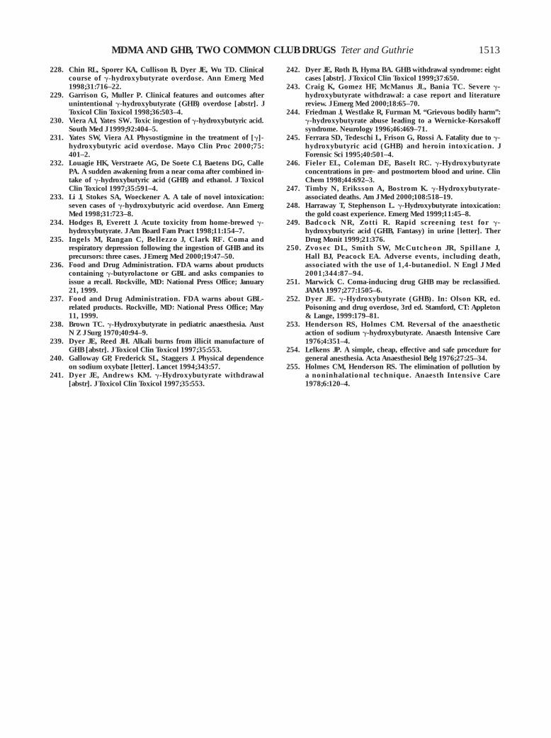
MDMA AND GHB, TWO COMMON CLUB DRUGS Teter and Guthrie
228. Chin RL, Sporer KA, Cullison B, Dyer JE, Wu TD. Clinicalcourse of g-hydroxybutyrate overdose. Ann Emerg Med1998;31:716–22.
229. Garrison G, Muller P. Clinical features and outcomes afterunintentional g-hydroxybutyrate (GHB) overdose [abstr]. JToxicol Clin Toxicol 1998;36:503–4.
230. Viera AJ, Yates SW. Toxic ingestion of g-hydroxybutyric acid.South Med J 1999;92:404–5.
231. Yates SW, Viera AJ. Physostigmine in the treatment of [g]-hydroxybutyric acid overdose. Mayo Clin Proc 2000;75:401–2.
232. Louagie HK, Verstraete AG, De Soete CJ, Baetens DG, CallePA. A sudden awakening from a near coma after combined in-take of g-hydroxybutyric acid (GHB) and ethanol. J ToxicolClin Toxicol 1997;35:591–4.
233. Li J, Stokes SA, Woeckener A. A tale of novel intoxication:seven cases of g-hydroxybutyric acid overdose. Ann EmergMed 1998;31:723–8.
234. Hodges B, Everett J. Acute toxicity from home-brewed g-hydroxybutyrate. J Am Board Fam Pract 1998;11:154–7.
235. Ingels M, Rangan C, Bellezzo J, Clark RF. Coma andrespiratory depression following the ingestion of GHB and itsprecursors: three cases. J Emerg Med 2000;19:47–50.
236. Food and Drug Administration. FDA warns about productscontaining g-butyrolactone or GBL and asks companies toissue a recall. Rockville, MD: National Press Office; January21, 1999.
237. Food and Drug Administration. FDA warns about GBL-related products. Rockville, MD: National Press Office; May11, 1999.
238. Brown TC. g-Hydroxybutyrate in pediatric anaesthesia. AustN Z J Surg 1970;40:94–9.
239. Dyer JE, Reed JH. Alkali burns from illicit manufacture ofGHB [abstr]. J Toxicol Clin Toxicol 1997;35:553.
240. Galloway GP, Frederick SL, Staggers J. Physical dependenceon sodium oxybate [letter]. Lancet 1994;343:57.
241. Dyer JE, Andrews KM. g-Hydroxybutyrate withdrawal[abstr]. J Toxicol Clin Toxicol 1997;35:553.
242. Dyer JE, Roth B, Hyma BA. GHB withdrawal syndrome: eightcases [abstr]. J Toxicol Clin Toxicol 1999;37:650.
243. Craig K, Gomez HF, McManus JL, Bania TC. Severe g-hydroxybutyrate withdrawal: a case report and literaturereview. J Emerg Med 2000;18:65–70.
244. Friedman J, Westlake R, Furman M. “Grievous bodily harm”:g-hydroxybutyrate abuse leading to a Wernicke-Korsakoffsyndrome. Neurology 1996;46:469–71.
245. Ferrara SD, Tedeschi L, Frison G, Rossi A. Fatality due to g-hydroxybutyric acid (GHB) and heroin intoxication. JForensic Sci 1995;40:501–4.
246. Fieler EL, Coleman DE, Baselt RC. g-Hydroxybutyrateconcentrations in pre- and postmortem blood and urine. ClinChem 1998;44:692–3.
247. Timby N, Eriksson A, Bostrom K. g-Hydroxybutyrate-associated deaths. Am J Med 2000;108:518–19.
248. Harraway T, Stephenson L. g-Hydroxybutyrate intoxication:the gold coast experience. Emerg Med 1999;11:45–8.
249. Badcock NR, Zotti R . Rapid screening test for g-hydroxybutyric acid (GHB, Fantasy) in urine [letter]. TherDrug Monit 1999;21:376.
250. Zvosec DL, Smith SW, McCutcheon JR, Spillane J,Hall BJ, Peacock EA. Adverse events, including death,associated with the use of 1,4-butanediol. N Engl J Med2001;344:87–94.
251. Marwick C. Coma-inducing drug GHB may be reclassified.JAMA 1997;277:1505–6.
252. Dyer JE. g-Hydroxybutyrate (GHB). In: Olson KR, ed.Poisoning and drug overdose, 3rd ed. Stamford, CT: Appleton& Lange, 1999:179–81.
253. Henderson RS, Holmes CM. Reversal of the anaestheticaction of sodium g-hydroxybutyrate. Anaesth Intensive Care1976;4:351–4.
254. Lelkens JP. A simple, cheap, effective and safe procedure forgeneral anesthesia. Acta Anaesthesiol Belg 1976;27:25–34.
255. Holmes CM, Henderson RS. The elimination of pollution bya noninhalational technique. Anaesth Intensive Care1978;6:120–4.
1513


![[MDMA]MDMA Neurochemistry](https://static.fdocuments.in/doc/165x107/577dab601a28ab223f8c57f3/mdmamdma-neurochemistry.jpg)


![[Open Drugs] a Complete MDMA Synthesis for the First Time Chemist](https://static.fdocuments.in/doc/165x107/5498dc5eac79592f2e8b5779/open-drugs-a-complete-mdma-synthesis-for-the-first-time-chemist.jpg)