A Comparative Study of Some Staining Properties of - Blood
15
A Comparative Study of Some Staining Properties of Crystals in a Lympho-plasmocytoid Cell, of Russell Bodies in Plasmocytes, and of Amyloids-With Special Emphasis on Their Isoelectric Points Bg ARTHUR F. GOLDBERG AND HELEN WENDLER DEANE T HIS STUDY was made to characterize intracellular crystals found in unusual lympho-plasmocytoid cells, which have been described elsewhere’ and for convenience will be termed crystal cells. For comparison, an examina- tion was made of the related inclusions in plasmocytes ( Russell bodies ) , and also of primary, secondary, and senile cardiac amyloids. Their relative iso- electric points were determined by a staining method that effectively titrates the ionizable radicals of the compounds. Tile results suggest strongly that the crystals and Russell bodies are gamma globulins, whereas the amyloids studied resemble alpha glycoproteins. Two preliminary reports have been made.2” MATERIALS AND METHODS The test objects were: (a) the intracytoplasmic crystals in lympho-plasmocytoid cells found in a lymph node biopsy and in bone marrow smears;1 (b) Russell bodies in plasmo- cytes present in the lamina propria of normal human stomach and small intestine (two cases), as well as in the gastrointestinal tract of a patient with multiple myeloma (one case) and in a biopsy of chronic inflammatory tissue from the external auditory canal; (c) tissues containing secondary amyloids associated with chronic tuberculosis (three cases), chronic osteomyeiitis (one case); and (d) specimens of amyloid associated with multiple myeboma (two cases), as well as generalized primary amyloid and senile cardiac amyloid (one case each). The locations of the amyloids studied were as follows: liver and spleen in the tuberculosis cases; spleen in the osteomyclitis case; blood vessels in stomach, intestine, and spleen of the cases of multiple myeloma; and the heart in general- ized primary and senile cardiac amyloidosis. From the Departments of Pathology and Anatomy, Albert Einstein College of Medicine, New York 61, N. Y. This study was aided by a U.S.PJI.S. Training Grant (2G-97) to the Department of Pathology. We wish to acknowledge the technical assistance of Irene Morvay and Ellen C. Driks. Submitted June 23, 1960; accepted for publication Aug. 13, 1960. #{176}Additional data on the cases in which amyloid were found are as follows. (a) Cases associated with tuberculosis: (1) 73 year old male; total serum protein 4.9 Gm. per cent; albumin 2.8 Gm. per cent, globulin 2.1 Cm. per cent. Serum electro- phoresis showed 29.3 per cent albumin, 7.1 per cent alpha-i, 17.7 per cent alpha-2, 11.8 per cent beta, and 34.1 per cent gamma globulin; BUN 60 mg. per cent; no proteinuria; (2) 23 year old female; total protein 7.2 Gm. per cent; albumin 4.5 Gm. per cent, globulin 2.7 Cm. per cent; BUN 6 mg. per cent; no proteinuria; (3) 50 year old female; total protein 6.9 Cm. per cent; albumin 2.9 Cm. per cent, globulin 4 Cm. per cent. Serum electrophoretic pattern showed 27 per cent albumin, 7 per cent alpha-i, 13.5 per cent alpha-2, 11.5 per cent beta, and 41 per cent gamma globulin. Four months later total protein was 5.3 Gm. per cent; albumin 1 Cm. per cent, globulin 4.3 Gm. per cent. Serum 1708 For personal use only. on January 31, 2018. by guest www.bloodjournal.org From
Transcript of A Comparative Study of Some Staining Properties of - Blood
A Comparative Study of Some Staining Properties ofCrystals in a Lympho-plasmocytoid Cell, of Russell
Bodies in Plasmocytes, and of Amyloids-WithSpecial Emphasis on Their Isoelectric Points
Bg ARTHUR F. GOLDBERG AND HELEN WENDLER DEANE
T HIS STUDY was made to characterize intracellular crystals found in
unusual lympho-plasmocytoid cells, which have been described elsewhere’
and for convenience will be termed crystal cells. For comparison, an examina-
tion was made of the related inclusions in plasmocytes ( Russell bodies ) , and
also of primary, secondary, and senile cardiac amyloids. Their relative iso-
electric points were determined by a staining method that effectively titrates
the ionizable radicals of the compounds. Tile results suggest strongly that
the crystals and Russell bodies are gamma globulins, whereas the amyloids
studied resemble alpha glycoproteins. Two preliminary reports have been
made.2”
MATERIALS AND METHODS
The test objects were: (a) the intracytoplasmic crystals in lympho-plasmocytoid cells
found in a lymph node biopsy and in bone marrow smears;1 (b) Russell bodies in plasmo-
cytes present in the lamina propria of normal human stomach and small intestine (two
cases), as well as in the gastrointestinal tract of a patient with multiple myeloma (one
case) and in a biopsy of chronic inflammatory tissue from the external auditory canal;
(c) tissues containing secondary amyloids associated with chronic tuberculosis (three
cases), chronic osteomyeiitis (one case); and (d) specimens of amyloid associated with
multiple myeboma (two cases), as well as generalized primary amyloid and senile cardiacamyloid (one case each). The locations of the amyloids studied were as follows: liverand spleen in the tuberculosis cases; spleen in the osteomyclitis case; blood vessels in
stomach, intestine, and spleen of the cases of multiple myeloma; and the heart in general-
ized primary and senile cardiac amyloidosis.�
From the Departments of Pathology and Anatomy, Albert Einstein College of Medicine,
New York 61, N. Y.
This study was aided by a U.S.PJI.S. Training Grant (2G-97) to the Department ofPathology.
We wish to acknowledge the technical assistance of Irene Morvay and Ellen C. Driks.Submitted June 23, 1960; accepted for publication Aug. 13, 1960.
#{176}Additional data on the cases in which amyloid were found are as follows.(a) Cases associated with tuberculosis: (1) 73 year old male; total serum protein
4.9 Gm. per cent; albumin 2.8 Gm. per cent, globulin 2.1 Cm. per cent. Serum electro-
phoresis showed 29.3 per cent albumin, 7.1 per cent alpha-i, 17.7 per cent alpha-2, 11.8
per cent beta, and 34.1 per cent gamma globulin; BUN 60 mg. per cent; no proteinuria;(2) 23 year old female; total protein 7.2 Gm. per cent; albumin 4.5 Gm. per cent,
globulin 2.7 Cm. per cent; BUN 6 mg. per cent; no proteinuria; (3) 50 year old female;total protein 6.9 Cm. per cent; albumin 2.9 Cm. per cent, globulin 4 Cm. per cent. Serum
electrophoretic pattern showed 27 per cent albumin, 7 per cent alpha-i, 13.5 per cent
alpha-2, 11.5 per cent beta, and 41 per cent gamma globulin. Four months later totalprotein was 5.3 Gm. per cent; albumin 1 Cm. per cent, globulin 4.3 Gm. per cent. Serum
1708
For personal use only.on January 31, 2018. by guest www.bloodjournal.orgFrom
CRYSTALS, RUSSELL BODIES AND AMYLOIDS 1709
Since Russell bodies are believed to contain gamma globulin,1 human gamma globulin
was also studied.#{176} Mayer’s egg albumin, used for affixing sections to slides, was
examined in some situations.
All blocks of tissue and a portion of the gamma globulin were fixed in 10 per cent
formalin and embedded in paraffin. With the exceptions noted, the various staining pro-
cedures listed below were performed on deparaffinized, hydrated sections ( ca. 5 �s ) , andexcept for the preparations stained for lipides, all sections were mounted in Permount.
To determine the apparent isoelectric points of the test objects, the method of Singer
and Morrison4 was employed. Sections were stained overnight ( i.e. to equilibrium ) atroom temperature, in aqueous solutions of eosin Y and methylene blue at concentrationsof 10-4 and 2 X 10-� M., respectively, at intervals of about one pH unit over the range
3. 1 to 9.3 ( see table 1 ) . These dye concentrations were chosen because they permitted
comparable staining of nuclei, cytoplasm, erythrocytes, and collagen fibers at pH 5 with
that obtained after ethanol or Zenker-acetic fixation.5 The Walpole acetate buffer, 0.1 M.,was used for the solutions from pH 3.1 to 7.5. Propanediol buffer, 0.1 M., was employed
for pH 9.3. The sections were dehydrated in a standardized fashion through tertiary butyl
alcohol and xylene. Sections containing the various test objects were stained together,
so that they could be compared with confidence.
Some sections were subjected to deamination by tile Alfert nlodlficatlon of the Van
Slyke method.6 Blockade of carboxyl groups was produced by n�ethylation.7 After these
pretreatments, the sections and appropriate controls were stained with eosin and methylene
blue as above.Other slides were stained by the periodic acid-Schiff (PAS) procedure, by tile PAS
method after saliva treatment, with crystal violet,8 and with Congo red.9 The last prepara-
tions were examined with a polarizing microscope10 as well as the ordinary light
microscope.
The Baker acid-hematein stain for phosphatides” was performed on material fixed
in calcium-formalin: (a) frozen sections of normal human stomach containing plasmocytes,
(b) bone marrow smears from the case with crystal cells and (c) aqueous, air-dried
smears of human gamma globulin and egg albumin. Control preparations were extracted
overnight with pyridine at 60 C. before mordanting and staining.Frozen sections of the formalin-fixed human stomach having numerous plasmocytes in
the lamina propria and bone marrow smears containing crystal cells were stained with
Sudan black B and with oil red 012 for 10 minutes at room temperature, and for 10
and 60 minutes at 60 C.
electrophoretic pattern showed 19 per cent albumin, 8 per cent alpha-i, 29.2 per cent
alpha-2, 11.8 per cent beta and 32 per cent gamma globulin; BUN 20 mg. per cent;
4 plus proteinuria.
(b) Case associated with osteomyelitis: 59 year old male; total protein 5.2 Gm. percent; albumin 2.2 Cm. per cent, globulin 3 Cm. per cent; BUN 28 mg. per cent; trace
of proteinuria.
(c) Cases associated with multiple myeloma: (1) 90 year old female; BUN 28 mg.
per cent, 3 plus proteinuria; (2) 72 year old female; total protein 8.5 Cm. per cent;albumin 3.4 Cm. per cent, globulin, 5.1 Cm. per cent; BUN 10 mg. per cent; 2 plus
proteinuria, Bence-Jones protein present.
(d) Case with generalized primary amyloidosis: 55 year old female; total protein5.4 Gm. per cent; albumin 4 Gm. per cent, globulin 1.4 Cm. per cent; BUN 40 mg.
per cent; no proteinuria.
(e) Case with senile cardiac amyloidosis: 77 year old male; BUN 24 mg. per cent; traceof proteinuria.
#{176}Asample of purified Fraction II was obtained from the Mann Research Laboratories,
New York.
For personal use only.on January 31, 2018. by guest www.bloodjournal.orgFrom

1710 GOLDBERG AND DEANE
OBSERVATIONS
The intensity of staining of the various test objects with eosin and methylene
blue was judged visually, as indicated in table 1. In addition to the Russell
bodies ( fig. 1 ) , gamma globulin, the crystals ( fig. 2 ) , and amyloids ( fig. 3),
the tabulation includes collagen fibers, erythrocytes, eosinophile granules,
and the basophilic cytoplasm ( ergastoplasm ) of plasmocytes. These addi-
tional objects have been studied by others,�m5 and therefore provided com-
parisons for our results. For each object, the zone of equal, weak staining by
tile acid and basic dyes was taken to represent its isoelectric point under the
conditions of preparation.4
For the Russell bodies and gamma globulin, the average isoelectric range
appeared to lie between p11 6.2 and 7.5 (table 1). The slopes of the curves of
dye uptake fell off for the acid dye between pH 6.2 and 7.5 and for the basic
dye between pH 9.3 and 7.5 (fig. 1). There was no residual staining at pH
9.3 with eosin or at pH 5.1 with methylene blue. The crystals stained quite
similarly, but they seemed to have a slightly more acid isoelectric point (table
1, fig. 2).
Individual crystals and Russell bodies always stained uniformly. At a
given p11 of staining, however, some would appear darker than others. Near
neutrality, some were stained while others were almost unstained (e.g.,
figs. lb and c, 2b).
The eight cases of amyloids studied reacted essentially alike and had ap-
l)�tre11t isoelectric points at about pH 5 (table 1, fig. 3). The slopes of theirstaining curves seemed more gradual than those of the crystals and Russell
bodies.
The remaining data listed in table 1 are compatible with the results of
others who have used the controlled-pH staining method, in that the isoelectric
point of collagen appeared to be near pH 613 (fig. 3); the acidophilia of ery-
throcytes and eosinophile granules extended to high pH’4 (fig. 2); and staining
of ergastoplasm with methylene blue dropped between pH 4 and 315 (fig. 1).
Deamination and methylation treatments were performed on sections con-
taining crystals (lymph node), Russell bodies (gut and external ear), three
examples of secondary amyloid (spleen, in cases with tuberculosis), and two
Table 1.-Staining of various objects with a cid and basic dyes at different pH’s#{176}t
pH 3.1 pH 4.1MB E MB
pH 5.1E MB
pH 6.2E MB
pHE
7.5MB
pE
H 9.3MBOBJECTS
CrystaLs
Russell bodies
Camma globulin
All amyloids
Collagen fibers
Erythrocytes
Eosinophile
granules
Ergastoplasrn of
plasmocytes ± 0
Sections stained with eosin (E) at 10-” M., and methylene blue (MB� at 2 >‘ 1O�1 M.
tScale of dye binding: 0 = no dye binding; ± = negligible dye binding: + to ��--�-�- = slight to veryintense dye binding.
0 ++4- 0 +±+ 0 + ± ± ± 0 4:++0 ++ 0 ++ 0 -f-f--’-�. 0-± + + 0 +++0 ++ 0 ++ 0 + ± + ± 0 ++0 ++ 0-± ± ± ()-± + +± 0 �-�--�-
0 ++ 0 + 0 ± ± 0 ± 0 ++0 ++++ 0 +++ 0 ++ 0 ++ 0 + ±
0 ++++ 0 +++ 0 m+ 0 ++ 0 ++ 0
+ 0 ++ 0 +++ 0 +++ 0 ++++
For personal use only.on January 31, 2018. by guest www.bloodjournal.orgFrom

Fig. 1.-Degree of basophilia of Russell bodies in plasmocytes present in chronic
inflammatory tissue in external ear. Methylene blue, 2 X 10� M.; X 900. Staining is
(a) absent [0] at pH 5.1, (b) negligible I ±] at pH 6.2, (c) slight 1+] at pH7.5, and (d) intense [+++I at pH 9.3. Some plasmocytes contain one or two large
Russell bodies, others contain many smaller ones (arrows). The nucleus tends tobe displaced to the periphery of the cell. Strands of basophilic cytoplasm (ergasto-plasm) surround the Russell bodies; this material remains basophilic at pH 5.1.
CRYSTALS, RUSSELL BODIES AND AMYLOIDS 1711
cases of amvloid associated with multiple myeloma (spleen). For all objects.
deamination consistently increased uptake of metilviene blue at etdl p11.
producing detectable staining even at pH :3.5. In contrast, blockade of car-
boxy! groups increased the uptake of eosin, so that all these Oi)jectS stained
at pH 9:3. No changes appeared in the relative intensity of staining of tile
several test objects.
All of the amyloids stained red-orange with Congo red and exhibited the
typical birefringence 111 � light.’’1’ The crystals, Russell bodies, and
gamma globulin stained univ light orange with Congo red all(l (lid not i)e-
come birefringent.
Likewise, all the ainvloids stained ,netachroniaticallv with crystal violet.
There were no significant differences in the reactivity of these S cases. The
For personal use only.on January 31, 2018. by guest www.bloodjournal.orgFrom

.../�.
�
�e �� -.
�
‘&� �
ky.- .% �
.�s,\
-p..
w
Fig. 2.-Degree of acidophilia of crystals in the lympho-plasmocytoid cellspresent in a lymph node biopsy. Eosin, 10’ M.; X 900. Dye uptake is (a) intenseat pH 5.1, (b) slight at pH 6.2, and (c) negligible at pH 7.5. Arrows point torepresentative crystal cells. Ervthrocytes (e) are still acidophilic at pH 7.5.
1712 GOLDBERG AND DEANE
crystals, Russell bodies, and gamma globulin stained orthochromatically with
crystal violet.
Sections containing crystals and anlyloids and smears of gamma globulin
all stained with moderate intensity by the PAS method (fig. 4). Many Russell
bodies reacted more intensely than the other objects when stained in the
same run.
Russell bodies and crystals showed no dye uptake when exposed to either
Sudan black B or oil red 0. A negative reaction for Russell bodies has been
reported by many others.’62#{176} Grundner-Culemann and Diezel,20� however,
found that some Russell bodies do stain with Sudan black B.It has been reported that cell inclusions in plasmocytes stain by the Smith-
Dietrich procedure for phosphatides.21 Because we believe these inclusions
to have been Russell bodies, we also applied a method for phosphatides. The
Baker method11 has the advantage that it uses a parallel control preparation
in which lipides are pre-extracted, so that staining attributable to nonlipide
acidic substances may be recognized. Frozen sections of stomach including
plasmocytes, smears of bone marrow containing crystal cells, and smears of
gamma globulin and egg albumin were stained by this method. All of the
test objects except egg albumin reacted positively. Moreover, Russell bodies
(fig. 5) and crystals became intensely stained even when the preparations
were extracted with hot pyridine before being exposed to the mordant and
For personal use only.on January 31, 2018. by guest www.bloodjournal.orgFrom

Is’.�’.
(:RYSTALS, RUSSELL BODIES AND AMYLOIDS 171:3
Fig. 3.-Degree of basophilia of amyloid occurring in the walls of splenicarteries from a patient who had multiple myeloma. Methylene blue, 2 X l0� M.;X 400. Staining of the amvloid is (a) virtually absent at pH 4.1, (b) negligible at
pH 5.1, (c) moderate at pH 6.2, and (d) fairly intense, although still much weakerthan that in nuclei, at pH 7.5. The collagen fibers in the adventitia of the arteriesare consistently less basophilic than the amyloid.
stain. Differentiation was conducted for the standard 18-hour period. Pre-
extracted smears of gamma globulin remained stained if differentiated for
only 5 hours but had lost all stain if differentiation was extended to 18 hours.
We did not utilize differentiation periods between 5 and 18 hours.
DIscusSIoN
Staining with dilute solutions of acid and basic dyes at a series of pH’s
allows the determination of the relative isoelectric points of various com-
pounds. It should be emphasized that to do this, staining must be continued
until equilibrium has been attained (18-24 hours ) � Also, the subsequent
washing and dehydration of the sections must be kept uniform so that dye is
not removed in differing amounts. If such preparations are analyzed with a
spectrophotometer, accurate pH “signatures” (titration curves) may be oh-
For personal use only.on January 31, 2018. by guest www.bloodjournal.orgFrom

1714 (X)LI)BEII(. .“. NI) l)EA N i
Fig. 4.-PAS positive reaction in (a) crystals [lymph node biopsy I, (b) Russellbodies I chronic inflammatory tissue in external ear], (c) amyloid associated withmultiple myeloma [wall of splenic artery], and (d) secondary amyloid [spleen,chronic pulmonary tuberculosis]. All preparations lightly counterstained with diluteHarris’s hematoxylin; (a) and (b) X 900, (c) and (d) X 400.
tained,’’’’’-� and these provide even more information about the kinds of
ionizable groups presellt. In this study, however, we have hmite(l ourselves
to visual assessment.
Russell Bodies (Ifl (I (;r!/St(Il.5
For Russell bodies, the isoelectric points ranged between 6.2 and 7.5. This is
appro�a mately the range exhibi ted by undenatu red human gamma glob-
itliiis.�-2� Furthermore, \\‘hell we tested fixed gamma globulin by our pro-
cedure, it exhibited staining ProPerties similar to those of Russell bodies. That
its isoelectric range pro�’ec1 to be the sanie with the staining nlethO(15 aS
biophvsical measurement indicated ti iat the (he concentrations selected were
appropriate. Otherwise the rclatiue isoelectric points of the various objects
in the sections, although valid, might not have conformed to the range exhibited
1w undenatured proteins.
For personal use only.on January 31, 2018. by guest www.bloodjournal.orgFrom
CRYSTALS, RUSSELL BOl)IES ANI) AMYLOIDS 1715
#{149}�P;� %‘�,
,e�
,j s�- ,, S
. � � ;� . S
Si � � �
:� �-‘-p �
‘�‘3. I ..
�4 -“ ..�,
� �S, ,
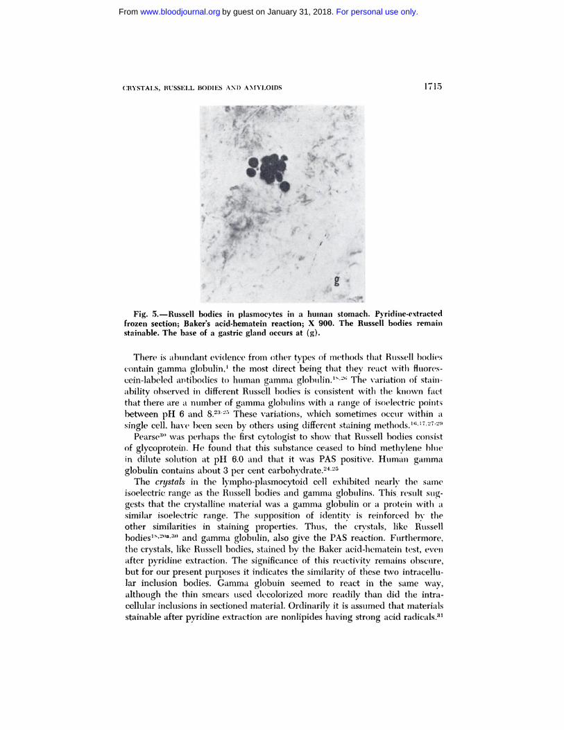
Fig. 5.-Russell bodies in plasmocytes in a human stomach. Pyridine-extractedfrozen section; Baker’s acid-hematein reaction; X 900. The Russell bodies remain
stainable. The base of a gastric gland occurs at (g).
There is al)undant evidence froni other ty�)es of methods that Russell bodies
contain gamma globulin,’ tile most direct being that they react with fluores-
cein-labeled antibodies to human gamma globulin.’�’ The variation of stain-
ability observed in different Russell bo(lies is consistent with the known fact
that there are a number of gamma globulins with a range of isoelectric points
between pH 6 and 8.23� These variations, which sometimes occur within a
single cell, have been seen by others using different staining methods.”
Pearse3’ was perhaps the first cytologist to show that Russell bodies consist
of glycoprotein. He found that this substance ceased to hind methylene blue
in dilute solution at pH 6.0 and that it was PAS positive. Human gamma
globulin contains about 3 Ier cent carbohydrate.24
The crystaLs in the lympho-plasmocytoid cell exhibited nearly the same
isoelectric range as the Russell bodies and gamma globulins. This, result sug-
gests that the crystalline material was a gamma globulin or a protein with a
similar isoeiectric range. The supposition of identity is reinforced h�’ the
other similarities in staining properties. Thus, the crystals, like Russell
hodies�’2”�’3#{176} and gamma globillin, also give tile PAS reaction. Furthermore,
the crystals, like Russell bodies, stained by tile Baker acid-hematem test, even
after pyridine extraction. The significance of this reactwity remains obscure,
but for our present purposes it indicates tile similarity of these two intracellu-
lar inclusion bodies. Gamma globuin seemed to react in the same way,
although the thin smears used dccolorized more readily than did the intra-
cellular inclusions in sectioned material. Ordinarily it is assumed that materials
stainable after pyridine extraction are nonlipides having strong acid radica1s.�
For personal use only.on January 31, 2018. by guest www.bloodjournal.orgFrom

1716 GOLDBERG AND DEANE
The high isoelectric point of the Russell bodies and crystals makes this explana-
tion improbable. And the complete absence of staining by oil-soluble dyes
makes it extremely unlikely that an unextractable acidic lipide accounts for
the stainability with acid hematein. Certainly the results do not support the
assumption that inclusions appearing to be Russell bodies contain phospho-
Iipide.2’
�‘Basophilia” and “acidophilid’
The conflicting reports in the literature concerning the staining properties
of inclusions in plasmocytes warrant comment. It seems to us that the van-
ability in tile staining properties of Russell bodies has been unduly exaggerated.
( a ) As we have shown here, in any one preparation, Russell bodies may
exhibit slightly differing dye uptakes ( fig. 1 ), probably because the constituent
gamma globulins vary slightly in isoelectnic point. (b ) More importantly, the
influence of pH on stainability with Romanovsky stains ( mixtures of methy-
lene blue, azures, and eosin ) is inadequately recognized. Russell bodies have
been described or illustrated as staining pale blue,’9’2029’3234 darker
blue,16’’2”’�3� violet or purple,’7’21”’’1#{176} 1)ink,.�i).:)(),.i7 or red,29’32’M Knowing
the isoelectric point of gamma globulin helps explain these discrepancies,
since Romanovsky mixtures (if buffered at all) are used anywhere between
pH 5.5 and 7. Frequently a buffer is not employed, and \Vnight’s stain may
he quite alkaline because of the bicarbonate present. Thus it is understandable
that sometimes the dye bath is on the alkaline side of the isoelectnic points of
Russell bodies, making them stain basophilically (blue); sometimes near the
isoelectric point, making them appear amphoteric (violet or purple); some-
times on the acid side, making them acidophilic (pink or red). Furthermore,
the Romanovsky mixtures are concentrated, so that staining is not carried to
equilibrium; consequently relative stainability may not be valid.0
\Ve believe, as Bessis concluded on other grounds,3t) that the tendency to
distinguish between the inclusions in plasinocytes associated with different
diseases is unwarranted on the basis of present information. This tendency
still continues.�#{176}’34 Unless preparations have been fixed and stained exactly
alike, tinctorial differences may not be significant. Certainly it is impossible to
compare preparations that have been made in different laboratories when no
standardized procedures exist.
Finally, the fact that Russell bodies often stain somewhat basophilically
with Romanovsky mixtures but are uniformly acidophilic in H & E preparations
is explicable on two grounds. (a) Hematoxylin is not a basic type and general-
ly stains chromatin only. Unless the solution is very old (S. H. Bensley, per-
sonal communication), it has little tendency to stain other basophilic sub-
stances. (b) H & E preparations are generally stained with a strong, alcoholic
solution of eosin, which is taken up by almost all materials in sections, re-
gardless of their isoelectric points.
Amyloids
All specimens of amyloid studied reacted similarly, having an apparent
isoelectnic point at about pH 5. Earlier, Games and Forker4#{176} showed that
For personal use only.on January 31, 2018. by guest www.bloodjournal.orgFrom

CRYSTALS, RUSSELL BODIES AND AMYLOIDS 1717
uptake of another basic dye, toluidine blue, by both primary and secondary
amyloids was extinguished at pH 4.5. These results support the concept that
various amyloids are closely related and that their classification into different
types, although useful, is still quite arbitrary.
The plasma proteins having similar isoelectric points are the alpha-i and
alpha-2 glycoproteins and beta-i globulins.23 In extraction studies on tissues
containing amyloids,4143 it has been shown that amyloids consist of a glyco-
protein fraction and an acid mucopolysaccharide. The glycoprotein stains
by the PAS technic and the acid mucopolysaccharide stains metachro-
matically with toluidine blue or crystal violet. Both of these components
migrate electrophoretically like the alpha and beta globulin fractions. There
is at least one case in the literature in which amyloid was analyzed chem-
ically and found to have an amino acid composition similar to beta globulin.44
Current studies reveal that the acid mucopolysaccharide component of
amyloids does not contain chondroitin sulfate or hyaluronic acid,40’43’45 and
that the acidic moiety is probably carboxylated.45 A known acidic component
of certain mucoproteins and mucopolysaccharides is sialic acid.’” This sub-
stance is present as a significant constituent of the plasma alpha-2 globulins,
and as a lesser constituent of the alpha-i and beta globulin fractions.47 Sialic
acid has been found in extracts of organs containing amyloids as well as in the
appropriate globulin fractions.43’48 Since sialic acid may induce metachromasy
of basic dyes,46 it may be the major substance responsible for the characteristic
metachromatic staining of amyloid.
One of the inherent defects of the controlled-pH staining method, employed
here to determine the isoelectric point of amyloids, is that it measures only the
net available ionizable groups. When a test object contains a mixture of sub-
stances, it is impossible to determine how many ionizable radicals are con-
tributed by each component. Without differential extraction, it would be
impossible to determine the isoelectric points of the protein and carbohydrate
moieties separately.
A long-standing hypothesis has been that tissue amyloids may result from
the local deposition of certain circulating plasma globulin fractions. In the
cases just listed,4’44 the patients had elevated serum globulin fractions with
properties corresponding to those of the amyloids extracted from the tissues.
These cases represented both primary and secondary amyloids. In two of
our cases of secondary amyloidosis, on whom serum electrophoretic patterns
were performed, the alpha globulin fraction was elevated.
Other clinical and laboratory studies on patients or experimental animals
with amyloidosis have shown similar serum electrophoretic patterns.3”4954
Besides the slight-to-moderate elevation of the alpha or beta globulin frac-
tions, the serum albumin fraction is frequently reduced in such patients. The
gamma globulins are usually slightly elevated early in the disease. In long
standing cases, the total globulin is generally not elevated. This is particularly
true in cases of multiple myeloma with amyloidosis.”55
If the protein component of amyloid is an alpha or beta globulin, then
what is its origin? It has long been suggested, especially for cases of amyloid-
osis associated with multiple myeloma, that the amyloid is a product of
For personal use only.on January 31, 2018. by guest www.bloodjournal.orgFrom

1718 GOLDBERG AND DEANE
plasmocytes. While there is no definitive evidence that the plasmocyte pro-
duces alpha or beta globulins, there is indirect evidence that this is so. In
agammaglobulinemia, where plasmocytes are absent, some alpha and beta
globulins may not be produced.’ In multiple myeloma, the reverse situation
may occur, in that there is a marked elevation of serum alpha or beta globu-
lins. It is usually presumed that these abnormal proteins arise from the prolif-
erating myeloma plasmocytes. Even in those cases of multiple myeloma in
which the major protein elevation is gamma or beta globulin, the alpha
globulins may also be moderately elevated,3�’56’57 suggesting that these glob-
nuns too are being produced by the myeloma plasmocytes. In the same cate-
gory are the rare cases of multiple myeloma associated with amyloidosis in
which the amyloid is surrounded by myeloma cells,58’59 suggesting its local
production. Protein inclusions taking positive amyloid stains have also been
described in myeloma cells.58’60 Furthermore, since in multiple myeloma with
amyloidosis there is usually a fail in serum globulins,33’5� there may be a
block in the production of these plasma proteins by myeloma plasmocytes
and possibly a shift in ratio of production of the several globulins.
On the other hand, the liver has been implicated as the primary source of
most alpha and beta globulins.61”12 It is possible, therefore, that certain forms
of amyloidosis may result from a disturbance of protein metabolism in that
organ. The low serum albumin, so frequently found in amyloidosis, may like-
wise be a reflection of this derangement. Possibly hepatic cells also produce the
mucopolysaccharides associated with the globulin fractions and with amyloid.
Finally, it should be pointed out that there is some evidence in the literature
that amyloids may differ from one another. Notably, primary amyloid asso-
ciated with multiple myeloma sometimes exhibits little or no metachromatic
staining (e.g., with crystal violet) � Since patients with multiple myeloma
frequently exhibit reduced serum mucoprotein levels,63 it seems reasonable
to assume that in such cases as described above, little acid mucopolysaccharide
was available to be deposited in the amyloid complex. Our particular cases,
however, all exhibited significant metachromasy with crystal violet. The con-
ditions that may produce secondary amyloidosis all appear to be associated
with an elevation of such serum mucopolysaccharides, thus explaining the
consistent intense metachromasy of the deposits with crystal violet.
SUMMARY
A staining procedure for determining the approximate isoelectric points of
substances has been applied to tissue sections containing (a) intracytoplasmic
crystals in a lympho-plasmocytoid cell, (b) Russell bodies of plasmocytes, and
(c) secondary and primary amyloids. The same method was also applied to
sections of human gamma globulin.
The results indicate the similarity of Russell bodies, the crystals, and gamma
globulin, all of which showed an isoelectric range between pH 6 and 7.5.
Other staining methods employed tended to confirm this identity.
The discrepancies reported in the literature on the staining properties of
Russell bodies are discussed.
For personal use only.on January 31, 2018. by guest www.bloodjournal.orgFrom
CRYSTALS, RUSSELL BODIES ANI) AMYLOIDS 1719
All of the amyloids tested had an apparent isoelectric point near pH 5.
Various suggestions in the literature about the nature and origin of amvloid
are summarized.
SusIsIAmo IN INrERLINGuA
Un methodo de tincturation P”� determinar le approxiniative punctos iso-
electric (Ic substantias esseva applicate a sectiones tissular que contineva (a)
crystallos intracytoplarmic in un cellula lympho-plasmocytoide, (b) corpores
de Russell in plasmocytos, e (c) amyloides secundari e primari. Le mesme
methodo esseva etiam applicate a sectiones de globuhina gamma human.
Le resultatos indica le similaritate de corpores de Russell, de crystallos, e de
globulina gamma que omnes monstrava un gamma isoelectric inter pH 6 e pH
7,5. Altere methodos tincturatori que esseva empleate tendeva a confirmar iste
identitate.
Es discutite le discrepantias que se trova reportate in le litterat� ira con
respecto al ProPrietates tincturatori de corpores de Russell.
Omne le amyloides testate habeva un apparente puncto isoelectric in le
vicinitate de pH 5. Es summarisate he suggestiones in Ic litteratura relative
al natura e al origine de amyloide.
REFERENCES
1. (;�1db.rg, A. 1’.: Au uintisuial lyi�iphouna-
tous disease with lympho-plasunocy-
toid cells containing intracytoplas-
niic crystals. Blood 16:1693-1707,
1960.
2. -- and 1)eane, H. \V.: Staining Pr�Pr-
tks of Russell bodies and crystals in
plasuilocyteS in comparison with amy-bid. J.Histochem.& Cytocheun., vol.
8, Sept. 1960.
:3. - and -: Staining properties of Rus-
sell bodies ill piasmocytes and cry-
stals ill a !yinpho-plasmocyte in com-
parison with amyloids. Bull. New
‘iork Ac�l(lJ5led. (ill ptt’ss).
4. Singer, \I. and Morrison, P. R.: The in-
fluence of pH, (lye, and salt conc:ui-
tration on the dye binding of modi-
fied aul(l unmodified fibrin. j.Bioi.
Chem. 175:133-145, 1948.
5. Fawcett, I). \V. and I)eane, H. XV.: The
(fleet of cortisone on uterme growthin ovariectoniized I ats receiving
estradiol. Quart.J.Micro.Sc. 92:385-
392, 1951.
6. Roseabauni, R. M. and Deane, H. \V.:
Effects of temperature Ofl the bindingof acid and basic dyes. including ref-
erence to unetachromasv. Histochemic
1:213-224, 1959.
7. Fisher, E. B. � Lillic, H. 1).: The ef-
fect of methylaiion ‘)ul 1)lsOphiii:l. J.Histochcm.& Cvtocl cm. 2:81-87,
1954.8. Lieb, E.: Permanent stain for tunyloid.
Am.J.Clin.Path. 17:413-414, 1947.
9. Lilbie, R. D.: Histopathologic Technic
and Practical Histocheiiiistry. See-
ond Edition. New York, Blakiston
Co., Inc., 1954.10. Cohen, S., Calkins, E. and Levene, C.
I.: Studies on experimental amyloid-osis. I. Analysis of histology and
staining reactions of casein-induced
amyloidosis in the ral)bit. Am.J Path..35:971-989, 1959.
11. Pearse, A. G. E.: Histochemistry, Theo-
retical and Applied, Second Edition.
London, j. & A. Churchill, Ltd., 1960.
12. (;omori, C.: Microscopic Histochemis-
try, Principles and Practice. Chicago,
University of Chicago Press, 1952.
13. Sokoloff, L., Mund, A. and Kantor, T.
C.: The affinity of librinoid substances
for acid dyes. Am.J.Path. 27:1037-
1045, 1951.
14. Weiss, L. P.: Binding of acid and basic
dye at varied pH by blood and bonemarrow cells of man. Blood 8:249-
261, 195:3.
For personal use only.on January 31, 2018. by guest www.bloodjournal.orgFrom
1720 GOLDBERG AND DEANE
15. I)empsey, E. W. and Singer, M.: Ob-
servations on the chemical cytologyof the thyroid gland at different func-
tional stages. Endocrinology 38:270-
295, 1946.
16. Leitner, S. T.: Bone Marrow Biopsy:Haematobogy in the Light of Sternal
Puncture. New York, Grune & Strat-
ton, 1949.
17. Di Guglielmo, R.: I Plasmocitomi.
Rome, Abruzzini, 1955.
18. \\‘hite, R. G.: Observations on the for-
mation and nature of Russell bodies.
Brit.J.Exper.Path. 35:365-376, 1954.
19. Rondanelli, E. G., Gorini, P., Stroselli,
E. c Pecorari, E.: Sulla natura dei
globuli endocitoplasmatici delle “eel-
lule morulari” de midolbo osseo
umano. Hematologica 42:1027-1040,
1957.
20. Zlotnick, A., Gerichter, C. B. and Nir,
I.: Experimental production of “grape
cells” and their relation to the serum
gamma globulin and seromucoids.
Blood 14:564-570, 1959.
20a. Crundner-Culeman, A. and Diezel, P.B.: Histochemischc Untersuchungen
an Russellschen KUrperchen liii Gran-
mulationsgewebe chronischer plasma-
cellularer Entzundungen und in
Geschwulstzellen. Frankf.Ztschr.f.Path. 66:161-180, 1955.
21. Van Oye, E. and Peel, E.: Contribution
a l’#{233}tudede la cellule muriforme do
Mott dans le liquide c#{233}phalo-rachidien
des trypanos#{233}s. Acta Trop., Basel, 8:
18-31, 1951.22. Singer, M.: Factors wllich control the
staining of tissue sections with acidand basic dyes. Int.Rev.Cytol. 1:211-
255, 1952.
23. Cohn, E. J., Curd, F. R. N., Surgenor,D. M., Barnes, B. A., Brown, R. K.,
Derouaux, G., Gillespie, J. I’s’I., Kahnt,
F. \V., Lever, �V. F., Liu, C. H., Mit-
telman, D., Mouton, R. F., Schmid,K. and Uroma, E.: A system for the
separation of components of human
l)loOd: Qualitative procedures for
the separation of the protein com-
ponents of human plasma. JAm.
Chem.Soc. 72:465-471, 1950.24. Smith, E. L. and Jager, B. V.: The
characterization of antibodies. Ann.
Rev.Microbiol. 6:207-228, 1952.
25. Isliker, H. C.: The chemical nature of
antibodies. Adv.Prot.Chem. 12:387-463, 1957.
26. Melbors, R. C.: Floureseent antibody
method. In Analytical Cytology, R. C.Mellors, Ed., Second Edition, pp.
1-67. New York, McGraw-Hill, 1959.
27. Brass, K.: Zur Cytologie und Funktionder Plasma und Plasmacytomzellen.
Frankf.Ztschr.f.Path. 57:481-491,
1943.
28. Fruhling, L. et Porte, A.: Contribution
de la microscopie #{233}lectronique aletude d’un sarcome plasmocytaire.
Ann.d’Anat.Path. 3:538-557, 1958.
29. Diggs, L. \V., Strum, D. and Bell, A.:The Morphology of Human BloodCells. Philadelphia & London, Saund-
ers, 1956.30. Pearse, A. G. E.: Nature of Russell
bodies and Kurboff bodies: Observa-tions on cytochemistry of plasma cells
and reticulum cells. J.Clin.Path. 2:
81-90, 1949.
31. i)eane, H. %V.: Intracellular lipides:
Their detection and significance. In
Frontiers in Cytology, S. Palay, Ed.pp. 227-263. New Haven, Conn.,
Yale University Press, 1958.
32. Undritz, E.: Atlas of Haematology.
Basel, Sandoz, 1952.33. Snapper, I., Turner, L. B. and Mosco-
vita, H. L.: Multiple Myeloma. New
York, Grune & Stratton, 1953.34. Zlotnick, A.: The “morula cell” and the
“grape cell” in bone marrow and peri-
pheral blood. Blood 11:1140-1147,
1956.35. Stich, M. W., Swiller, A. I. and Mor-
rison, M.: Tile “grape cell” of multi-
ple myeboma. Am.J.Clin.Path. 25:
601-602, 1955.36. Heilmeyer, L. and Begeinann, H.: Atlas
der klinischen Hamatologie und Cy-tologic. Berlin, Springer, 1955.
37. Daland, G. and Ham, T. H.: A ColorAtlas of Morphologic Hematology.
Cambridge, Mass., Harvard Univer-
sity Press, 1959.38. Whitby, L. E. H. and Britton, C. J. C.:
Disorders of the Blood. New York,Grune & Stratton, 1957.
39. Bessis, M.: Cytology of the Blood and
Blood-Forming Organs. New York,
Grune & Stratton, 1956.
For personal use only.on January 31, 2018. by guest www.bloodjournal.orgFrom

CRYSTALS, RUSSELL BODIES AND AMYLOIDS 1721
40. Carnes, W. H. and Forker, B. R.: Met-
achromasy of Amyloid. Lah.Invest.5:21-43, 1956.
41. Wagner, B. M.: Histochemical studies
of fibrinoid substances and other ab-normal tissue proteins. IV. Protein
character of amyloid. Arch.Path. 60:
221-229, 1955.
42. Larsen, B.: Presence of glycoproteins
in secondary amyloid deposits related
to several glycoproteins. Acta rheu-
mat.scandinav. 3:30-39, 1957.
43. Calkins, E. and Cohen, A. S.: Chemical
composition of amyloid. J.Clm.Invest.
37:882-883, 1958.
44. Permis, B., Schneider, C. and \Vunderly,
C.: Quantitative Aminosiiurenanalyse
von Amyloid-substanz elektrophoretis-
chen Serunl-Eiweissfraktionen smnd
Bindegewebsprotein. Artsliche Fors-
chung 7:454-458, 1953.
45. Braunstein, H. and Buerger, L. A.: Astudy of the histochemical and stain-
ing characteristics of amyloid. Am.J.Path. 35:791-800, 1959.
46. Spicer, S. S. and Warren, L.: Tile histo-
chemistry of sialic acid containing
mucoproterns. J.Histochem.& Cyto-
chem. 8:135-137, 1960.
47. Uzman, L. L. and Rosen, H.: Partition
of neuranlinic acid among human
serum proteins. Science 120:1031-
1032, 1954.
48. Klenk, E. and Faillard, H.: Cher (las
Vorkommen von Neuramins#{228}ure im
Lebereiweiss bei amyloider l)egcner-
ation. Ztschr.f.physiol.Chem. 22.9:
191-192, 1955.
49. H#{252}sselmann, H.: Beitrag zum Amyloid-
problem auf Grund von Untersuch-ungen an menschlichen Herzen. Virch.
Arch.f.Path.Anat. 327:607-628, 1955.
50. Case Records of Massachusetts General
Hospital (Case 42051). New England
J.Med. 254:226-235, 1956.51. Kaufman, H. E. and Thomas, L. B.:
Vitreous opacities diagnostic of
familial primary amyloidosis. NewEngland J.Med. 261:1267-1271,
1959.52. Bohle, A., Hartman, F. and Polo, \V.:
Elektrophoretische Serumweiweiss-
untersuchungen bei experimentellen
Mauseamyloid. Virch.Arch.f. path.
Anat. 319:231-246, 1950.
53. Per#{228}sals, 0. and Latvalahti, J.: Amyloid
degeneration in the light of clinical
and experimental studies. Acta path.
et microbiol.scandinav. 34:208-217,
1954.
54. Catchpole, H. R., Pirani, C. L. and
Bestetti, A.: Serum proteins and mu-
coproteins in experimental amyloid-
osis. Fed.Proc. 17:24, 1958.
55. Eisen, H. N.: Primary systemic amyloid-
osis. Am.j .\Ied. 1:144-160, 1946.
56. Ossermnan, E. F.: Multiple myeloma:
Current clinical and chemical con-cepts. Jim Combined Staff Clinic. Am.
J.Med. 23:283-309, 1957.
57. Reiner, M. 011(1 Stern, K. C.: Electro-
phoretic studies On the protein dis-
tribution in the serum of multiple
myeloma patients. Acta haemat. 9:
19-29, 1953.
58. Ranstr#{246}m,S.: Plasmocytomatosis with
crystalline amyloid deposits in the
tumor tissue. Acta path.et unicrobiol.
scandinav. 28:366-372, 1951.
59. Case Records of the Massachusetts
General Hospital (Case 45062). New
England J.Med. 260:288-292, 1959.
60. Bayrd, E. I). and Bennett, \V. A.:
Amyloidosis complicating multiple
mnyeloma. \I.Clin.N.Am. 34:1151-
1164, 1950.61. Miller, L. L. and Bale, W. F.: Synthesis
of all plasma protein fractions except
gamma globulins by the liver. J.Exper.Med. 99:125-132, 1954.
62. Miller, L. L., Bly, C. G., and Bale, W.
F.: Plasma and tissue proteins pro-
duced by non-hepatic rat organs asstudied with 1ysine-�-C’4. J.Exper.
Med. 99:133-153, 1954.
63. Greenspan, E. M.: Clinical significance
of serum mucoproteins. Adv.Intern.
Med. 7:101-123, 1955.
For personal use only.on January 31, 2018. by guest www.bloodjournal.orgFrom

1960 16: 1708-1721
ARTHUR F. GOLDBERG and HELEN WENDLER DEANE Amyloids-With Special Emphasis on Their Isoelectric PointsLympho-plasmocytoid Cell, of Russell Bodies in Plasmocytes, and of A Comparative Study of Some Staining Properties of Crystals in a
http://www.bloodjournal.org/content/16/6/1708.full.htmlUpdated information and services can be found at:
Articles on similar topics can be found in the following Blood collections
http://www.bloodjournal.org/site/misc/rights.xhtml#repub_requestsInformation about reproducing this article in parts or in its entirety may be found online at:
http://www.bloodjournal.org/site/misc/rights.xhtml#reprintsInformation about ordering reprints may be found online at:
http://www.bloodjournal.org/site/subscriptions/index.xhtmlInformation about subscriptions and ASH membership may be found online at:
Copyright 2011 by The American Society of Hematology; all rights reserved.Hematology, 2021 L St, NW, Suite 900, Washington DC 20036.Blood (print ISSN 0006-4971, online ISSN 1528-0020), is published weekly by the American Society of
For personal use only.on January 31, 2018. by guest www.bloodjournal.orgFrom

![Recovering Bloody Fingerprints from Skin red is a well-known blood-staining agent [7]. Experiments – Blood Preliminary Experiment Before using on human cadaver skin, we conducted](https://static.fdocuments.in/doc/165x107/5b0747f67f8b9ac33f8e0e2d/recovering-bloody-fingerprints-from-skin-red-is-a-well-known-blood-staining-agent.jpg)