A comparative study of root canal shaping using protaper ...Cleaning and shaping of the root canal,...
14
Submitted 19 December 2018 Accepted 5 July 2019 Published 19 August 2019 Corresponding author Gül Çelik, [email protected], [email protected] Academic editor Ali Hassan Additional Information and Declarations can be found on page 9 DOI 10.7717/peerj.7419 Copyright 2019 Çelik et al. Distributed under Creative Commons CC-BY 4.0 OPEN ACCESS A comparative study of root canal shaping using protaper universal and protaper next rotary files in preclinical dental education Gül Çelik 1 , Feyza Özdemir Kısacık 2 , Emir Faruk Yılmaz 3 , Arife Mersinlioğlu 4 , İhsan Furkan Ertuğrul 5 and Hikmet Orhan 6 1 Faculty of Dentistry, Endodontics, Suleyman Demirel University, Isparta, Turkey 2 Vefa Oral and Tooth Health Center, Afyonkarahisar, Turkey 3 Dentplus Oral and Dental Health Center, Bursa, Turkey 4 Araklı Bayram Halil State Hospital, Trabzon, Turkey 5 Faculty of Dentistry, Departmant of Endodontics, Pamukkale University, Denizli, Turkey 6 Faculty of Medicine, Biostatistics and Medical Informatics, Suleyman Demirel University, Isparta, Turkey ABSTRACT Background. Dentistry has undergone an evolution in endodontics practice caused by the advancement of rotary techniques for root canal preparation and their subsequent incorporation into the teaching of dentistry undergraduates. This research aimed to evaluate the shaping ability of third-year dental students as their first experience in rotary instrumentation using ProTaper Universal (PTU) and ProTaper Next (PTN) (Dentsply Maillefer) rotary instruments in simulated curved canals. Methods. Forty students instrumented 200 simulated canals with a 40 ◦ curvature in resin blocks according to the manufacturer’s instructions with PTU and 39 students and 195 canals with PTN files. The canals were prepared at a speed of 300 rpm using a 16:1 reduction hand-piece powered by an electric motor (Xsmart; Dentsply Maillefer). The final apical preparation was set to F2 for the PTU and X2 for the PTN group. The change in canal curvature was evaluated based on Schneider technique using the AutoCAD 2007 software on post-digital photographs. The incidence of instrument fracture and deformation, the incidence of ledge, the change in working length (WL), and the working time were noted. The data were analyzed with Student’s t -test and Chi-Square test at a significance level of 0.05 using SPSS. Results. PTN maintained the original canal curvature better, resulting in fewer fractures and ledges, and shaped the canals faster than the PTU (P < 0.05). The mean curves of the resin canals after the instrumentation for the PTU and PTN groups were 24.03 ◦ ± 3.14 ◦ and 25.64 ◦ ± 2.72 ◦ , respectively. Thirty-three (17.4%) PTU and 18 (9.3%) PTN files fractured (p < 0.05). Nine (4.5%) PTU and 2 (2.6%) PTN deformed (p > 0.05). The change in WL after instrumentation was 0.97 mm ± 0.95 mm in PTU and 0.96 mm ± 0.80 mm in PTN (p < 0.05). The mean times were 627 s ± 18 s for PTU and 379 s ± 18 s for PTN (p < 0.000). Discussion. PTN can be recommended in severely curved root canals in terms of maintenance of the original canal curvature, superior instrument fracture and fewer ledges. Even if training before preparation provides an acceptable level of canal shaping for preclinical students, the use of NiTi rotary instruments should be included in the How to cite this article Çelik G, Özdemir Kısacık F, Emir Faruk Y, Mersinlioğlu A, Ertuğrul IF, Orhan H. 2019. A comparative study of root canal shaping using protaper universal and protaper next rotary files in preclinical dental education. PeerJ 7:e7419 http://doi.org/10.7717/peerj.7419
Transcript of A comparative study of root canal shaping using protaper ...Cleaning and shaping of the root canal,...
Submitted 19 December 2018Accepted 5 July 2019Published 19 August 2019
Corresponding authorGül Çelik, [email protected],[email protected]
Academic editorAli Hassan
Additional Information andDeclarations can be found onpage 9
DOI 10.7717/peerj.7419
Copyright2019 Çelik et al.
Distributed underCreative Commons CC-BY 4.0
OPEN ACCESS
A comparative study of root canalshaping using protaper universal andprotaper next rotary files in preclinicaldental educationGül Çelik1, Feyza Özdemir Kısacık2, Emir Faruk Yılmaz3, Arife Mersinlioğlu4,İhsan Furkan Ertuğrul5 and Hikmet Orhan6
1 Faculty of Dentistry, Endodontics, Suleyman Demirel University, Isparta, Turkey2Vefa Oral and Tooth Health Center, Afyonkarahisar, Turkey3Dentplus Oral and Dental Health Center, Bursa, Turkey4Araklı Bayram Halil State Hospital, Trabzon, Turkey5 Faculty of Dentistry, Departmant of Endodontics, Pamukkale University, Denizli, Turkey6 Faculty of Medicine, Biostatistics and Medical Informatics, Suleyman Demirel University, Isparta, Turkey
ABSTRACTBackground. Dentistry has undergone an evolution in endodontics practice caused bythe advancement of rotary techniques for root canal preparation and their subsequentincorporation into the teaching of dentistry undergraduates. This research aimed toevaluate the shaping ability of third-year dental students as their first experience inrotary instrumentation using ProTaper Universal (PTU) and ProTaper Next (PTN)(Dentsply Maillefer) rotary instruments in simulated curved canals.Methods. Forty students instrumented 200 simulated canals with a 40◦ curvature inresin blocks according to the manufacturer’s instructions with PTU and 39 studentsand 195 canals with PTN files. The canals were prepared at a speed of 300 rpm using a16:1 reduction hand-piece powered by an electric motor (Xsmart; Dentsply Maillefer).The final apical preparation was set to F2 for the PTU and X2 for the PTN group.The change in canal curvature was evaluated based on Schneider technique using theAutoCAD 2007 software on post-digital photographs. The incidence of instrumentfracture and deformation, the incidence of ledge, the change in working length (WL),and the working time were noted. The data were analyzed with Student’s t -test andChi-Square test at a significance level of 0.05 using SPSS.Results. PTNmaintained the original canal curvature better, resulting in fewer fracturesand ledges, and shaped the canals faster than the PTU (P < 0.05). The mean curves ofthe resin canals after the instrumentation for the PTU and PTN groups were 24.03◦ ±3.14◦ and 25.64◦ ± 2.72◦, respectively. Thirty-three (17.4%) PTU and 18 (9.3%) PTNfiles fractured (p< 0.05). Nine (4.5%) PTU and 2 (2.6%) PTN deformed (p> 0.05).The change in WL after instrumentation was 0.97 mm ± 0.95 mm in PTU and 0.96mm ± 0.80 mm in PTN (p< 0.05). The mean times were 627 s ± 18 s for PTU and379 s ± 18 s for PTN (p< 0.000).Discussion. PTN can be recommended in severely curved root canals in terms ofmaintenance of the original canal curvature, superior instrument fracture and fewerledges. Even if training before preparation provides an acceptable level of canal shapingfor preclinical students, the use of NiTi rotary instruments should be included in the
How to cite this article Çelik G, Özdemir Kısacık F, Emir Faruk Y, Mersinlioğlu A, Ertuğrul IF, Orhan H. 2019. A comparativestudy of root canal shaping using protaper universal and protaper next rotary files in preclinical dental education. PeerJ 7:e7419http://doi.org/10.7717/peerj.7419
undergraduate dental curriculum, contributing to an increase in the quality of rootcanal shaping and, consequently, to an improvement of the clinical experience ofstudents.
Subjects Biotechnology, DentistryKeywords Protaper, Preclinic, Rotary techniques, Education, Protaper next
INTRODUCTIONCleaning and shaping of the root canal, which must be performed mechanically andbiologically, is the most important procedure in root canal treatment. A variety oftechniques and instruments have been developed to perform this procedure withoutgenerating undesirable clinical results, such as canal straightening, transportation, ledging,strip perforations, or instrument fractures. The European Society of Endodontics (ESE)has published current articles to lead the undergraduate curriculum in endodontics toimprove dental students’ theoretical and clinical education (ESE, 2001). The ESE believesthat undergraduate students should receive preclinical and clinical training to be able tosuccessfully treat uncomplicated anterior premolar and molar teeth (de Moor et al., 2013).Endodontic education needs to be developed in undergraduate programs (Dummer, 1991;Qualtrough & Dummer, 1997; Unal et al., 2012) and the use of new techniques and devicesthat have proven to be positive contributions to root canal therapy should be included inundergraduate training programs (Unal et al., 2011).
The technical quality of root fillings, assessed radiographically, performed byundergraduate students using hand instruments was investigated in a meta-analysis(Ribeiro et al., 2018). The results revealed a low frequency (48.75%) of acceptable technicalquality of root fillings and drew attention to the major procedural errors created (Ribeiroet al., 2018). Many studies have been conducted to evaluate the performance of studentsin curved root canals (Hänni et al., 2003; Arbab-Chirani & Vulcain, 2004; Alrahabi, 2015;Unal et al., 2011). Despite the multiple benefits of using NiTi rotary instruments, thestep-back technique using stainless steel files is still a conventional teaching method inendodontic programs for undergraduate students in most countries (Alrahabi, 2015).Although students with no experience are reported to be successful in the use of rotaryinstruments (Unal et al., 2012; Kwak et al., 2016; Brito-Júnior et al., 2014), it is evident thaterrors can be minimized by the training to be taken in the curriculum. Georgelin-Gurgel etal. (2008) and Alrahabi (2015) reported that manual instrumentation is safer than rotaryinstrumentation in the hands of inexperienced students. They underlined that acquiringskill in the use of NiTi rotary instrumentation requires specific preclinical training toavert file breakage. Martins et al. (2012) suggested that the use of NiTi rotary instrumentsshould be included in the undergraduate dental curriculum, as this would contribute toan increase in the number of patients assisted and, consequently, improve the students’clinical experience.
Some procedural errors have been minimized after the introduction of the NiTi alloyinto endodontics (Walia, Brantley & Gerstein, 1988; Vaudt et al., 2009; Yang et al., 2011).
Çelik et al. (2019), PeerJ, DOI 10.7717/peerj.7419 2/14
Nevertheless, the fracture of the NiTi rotary root canal instruments suddenly and withoutany warning creates a disappointment for the dentist (Zuolo & Walton, 1997; Arens et al.,2003;Ankrum, Hartwell & Truitt, 2004). The studies are underway to increase the resistanceof the root canal instrument against breakage in order to overcome this problem. A recentnovelty of the alloy is the so-called m-wire alloy. The M-Wire material has been shown tohave longer fatigue resistance and lower fracture risk than conventional NiTi alloy does(Johnson et al., 2008;Montenegro-Santillàn et al.,2013).
The ProTaper Universal (PTU) system (Dentsply Maillefer, Ballaigues, Switzerland)is currently used by endodontists, and it is one of the most revolving instrumentsystems researchers have searched for the most (Peters, Schönenberger & Barbakow, 2003;Schäfer & Vlassis, 2004; Celik Unal, Kececi & Ureyen Kaya, 2006; Yang et al., 2007; Alemam,Dummer & Farnell, 2017). PTU is a nickel titanium (NiTi) rotary system of instrumentsmanufactured with progressive tapering over the length of the cutting blades, convextriangular cross-sections, and noncutting tips. In the PTU system, the file is produced insuch a way that it does not cut the ends, and its cross-section is a convex triangle. The taperangle of the file is not constant. This angle increases parabolically starting from the end(Hieawy et al., 2015).
The design features of ProTaper Next (PTN, Dentsply Maillefer), made from the newM-Wire alloy, include variable tapers and rectangle sections with a remote center. TheM-Wire material has been shown to have longer fatigue resistance and lower fracture riskthan conventional NiTi alloy does (Johnson et al., 2008; Montenegro-Santillàn et al.,2013).The number of instruments is similar to that of the PTU in terms of the order of use andeach instrument. The PTU contains three files (SX, S1, and S2) for the preparation of thecoronal and middle thirds and three files (F1, F2, and F3) for the preparation of the apicalthird. PTN consists of only three files (X1 is #17/.04, X2 is #25/.06, and X3 is #30/.075),close to the diameters of the files used in the apical third in the PTU system (Pérez-Higueraset al., 2014).
This study aimed to compare the shaping ability of PTUandPTNrotary instruments usedby the third-year dental students inexperienced in any rotary instrumentation techniquesin simulated curved canals of resin blocks.
METHODSSeventy-nine third-year undergraduate dental students (2015–2016 intakes) in preclinicalendodontics at the School of the Dentistry of the Suleyman Demirel University, Isparta,Turkey had three 50-min lectures of about the NiTi instruments, their physical properties,and the special constructional features of the files. The present study was based ondata obtained from practical courses, which was compulsory for the students to attend.Therefore, there was no need to sign an acceptance form for participation. The results ofthis study did not affect the course notes of the students. The students instrumented 16simulated canals in acrylic resin blocks in the 2014–2015 academic year and 12 extractedhuman teeth 2015–2016 in the preclinical dental education. The shaping technique wasModified Double Flared Technique using balanced force principle (Saunders & Saunders,
Çelik et al. (2019), PeerJ, DOI 10.7717/peerj.7419 3/14
1992) with stainless steel instrument. The students attended a 2-h lecture on the use ofPTU and PTN files two weeks before the end of the term. The study was started in the lastweek of the term. The students were divided into groups of 10, and an associated professor(G.Ç.) demonstrated the procedures of the shaping of a simulated canal according to theinstructions of the manufacturer. A printed script with the manufacturer’s step-by-stepinstructions for the PTU and PTN rotary systems was given to each student.
The students instrumented a total of 395 simulated resin canals (Endo Training Block02 taper, REFA 0177; Dentsply Maillefer, CH-1338 Ballaigues, Switzerland). All simulatedresin canals had an apical foramen of 0.15 mm, a taper of 0.02, and an angle of curvature of40◦. Of the 79 students, 40 students assigned randomly instrumented 200 canals using PTUfiles and 39 students instrumented 195 canals using PTN files. Each student instrumentedfive simulated canals using just one of the two systems and only one set instrument. Thecanals for each file system were instrumented to a working length of 16 mm (0. five mmfrom the apex) at a speed of 300 rpm and a torque-control level of 2, using a 16:1 reductionhandpiece powered by an electric motor (Xsmart; Dentsply Maillefer). Two mL of distilledwater was used as an irrigant at each change of instrument. After the instrumentation, afinal rinse with distilled water for 1 min was carried out. Each canal was dried using size25 paper points.
The incidence of fractures in the blocks and instrument deformations It were recorded.The blocks which instrument fractured were not taken into account when calculating thecurvature and WL change, and incidence of the ledge.. The working time was calculatedstarting through the insertion of the first file until the end of the instrumentation, includingtotal active instrumentation, cleaning of the flutes of the instruments, and irrigation. Thefinal WL of the canals was determined in mm following instrumentation of canals. An F2PTU or X2 PTN file was inserted into the canal and its WL within the canal measuredto the nearest 0. five mm. The amount of change in WL was determined by subtractingthe final length from 16 mm.. Canal straightening and ledge formation were assessed ondigital images (Figs. 1 and 2). After the canal instrumentation, the images of the resinblocks were obtained with the help of a light microscope camera (Zeiss Axioskop 2; Zeiss,Münich, Germany) to determine the canal curvature. The images were taken from eachblock in one direction (mimicking clinical conditions). In these images, canal curvatureswere measured by a computer program (AutoCAD 2007) according to Schneider (1971).The change in canal curvature was determined by subtracting the value obtained afterinstrumentation from 40 degrees. All students were randomly divided into two groups asthey are at the same educational level. Therefore, it was accepted that the only variable inthe experimental groups was PTU and PTN files. Data were analyzed using SPSS software,version 10.0 (SPSS Inc., Chicago, USA). The level of significance was set at 5%. Student’st -test was used to evaluate differences in canal straightening, working time, and amountof change in WL. The chi-square test was used to evaluate differences in the incidences ofinstrument fracture, instrument deformation, while the Yates corrected chi-square test wasused ledge formation between the groups.
Çelik et al. (2019), PeerJ, DOI 10.7717/peerj.7419 4/14
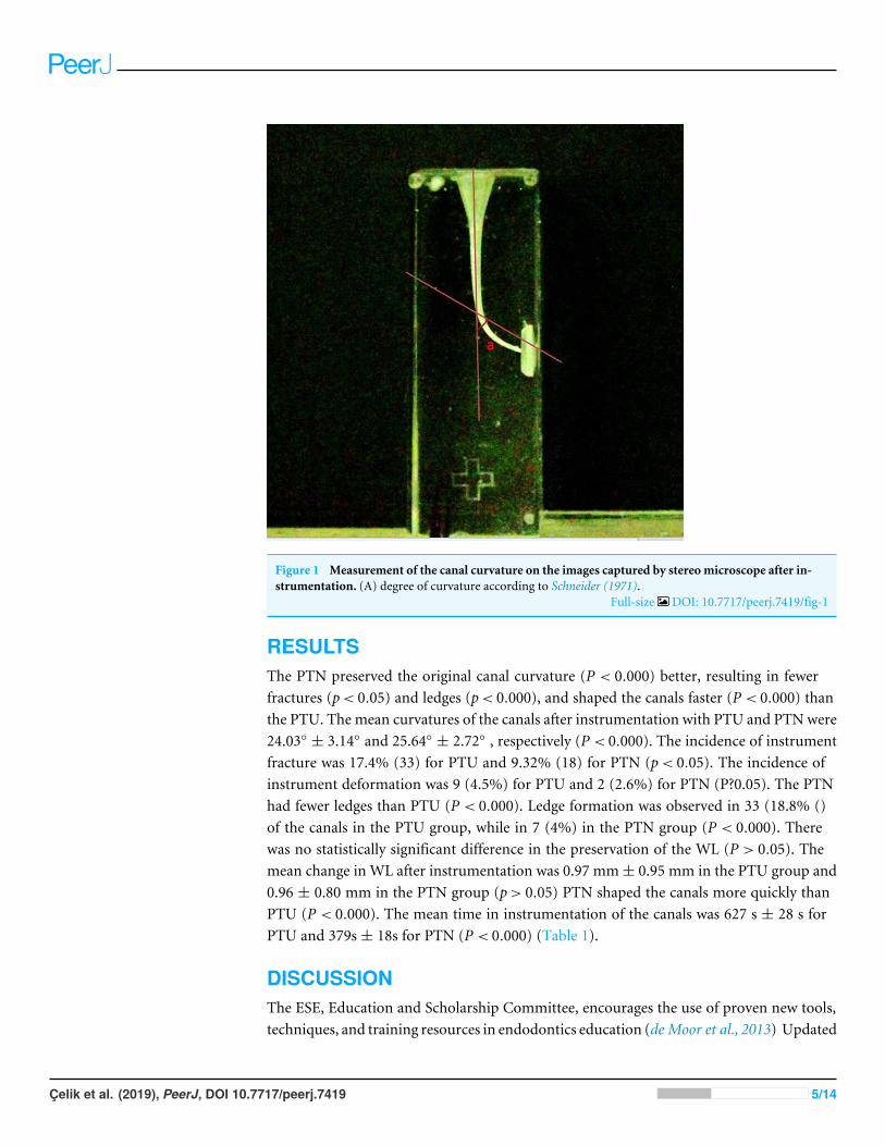
Figure 1 Measurement of the canal curvature on the images captured by stereo microscope after in-strumentation. (A) degree of curvature according to Schneider (1971).
Full-size DOI: 10.7717/peerj.7419/fig-1
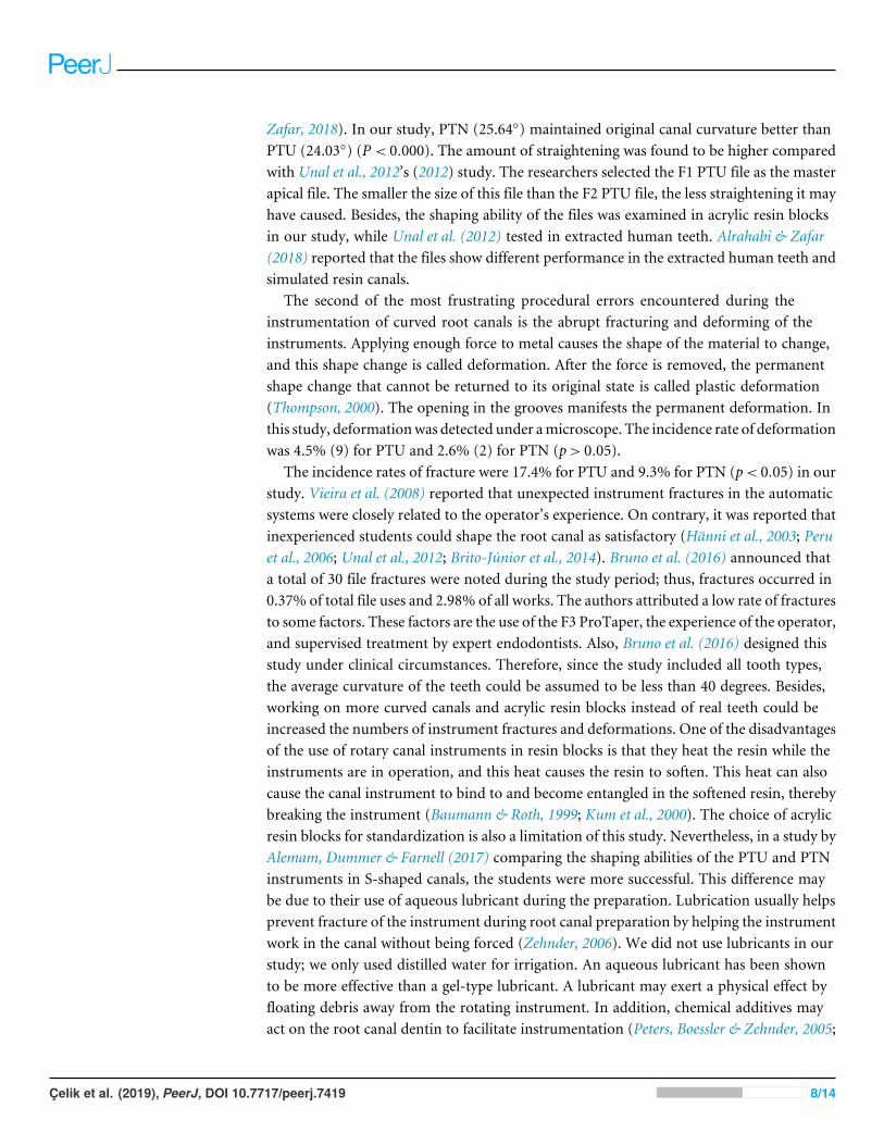
RESULTSThe PTN preserved the original canal curvature (P < 0.000) better, resulting in fewerfractures (p< 0.05) and ledges (p< 0.000), and shaped the canals faster (P < 0.000) thanthe PTU. The mean curvatures of the canals after instrumentation with PTU and PTN were24.03◦ ± 3.14◦ and 25.64◦ ± 2.72◦ , respectively (P < 0.000). The incidence of instrumentfracture was 17.4% (33) for PTU and 9.32% (18) for PTN (p< 0.05). The incidence ofinstrument deformation was 9 (4.5%) for PTU and 2 (2.6%) for PTN (P?0.05). The PTNhad fewer ledges than PTU (P < 0.000). Ledge formation was observed in 33 (18.8% ()of the canals in the PTU group, while in 7 (4%) in the PTN group (P < 0.000). Therewas no statistically significant difference in the preservation of the WL (P > 0.05). Themean change in WL after instrumentation was 0.97 mm± 0.95 mm in the PTU group and0.96 ± 0.80 mm in the PTN group (p> 0.05) PTN shaped the canals more quickly thanPTU (P < 0.000). The mean time in instrumentation of the canals was 627 s ± 28 s forPTU and 379s ± 18s for PTN (P < 0.000) (Table 1).
DISCUSSIONThe ESE, Education and Scholarship Committee, encourages the use of proven new tools,techniques, and training resources in endodontics education (de Moor et al., 2013) Updated
Çelik et al. (2019), PeerJ, DOI 10.7717/peerj.7419 5/14
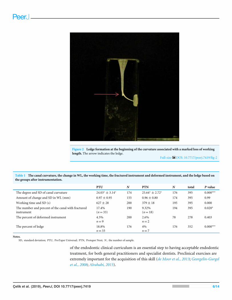
Figure 2 Ledge formation at the beginning of the curvature associated with a marked loss of workinglength. The arrow indicates the ledge.
Full-size DOI: 10.7717/peerj.7419/fig-2
Table 1 The canal curvature, the change inWL, the working time, the fractured instrument and deformed instrument, and the ledge based onthe groups after instrumentation.
PTU N PTN N total P value
The degree and SD of canal curvature 24.03◦ ± 3.14◦ 174 25.64◦ ± 2.72◦ 176 395 0.000∗∗∗
Amount of change and SD in WL (mm) 0.97± 0.95 155 0.96± 0.80 174 395 0.99Working time and SD (s) 627± 28 200 379± 18 195 395 0.000The number and percent of the canal with fracturedinstrument
17.4%(n= 33)
190 9.32%(n= 18)
194 395 0.020∗
The percent of deformed instrument 4.5%n= 9
200 2.6%n= 2
78 278 0.403
The percent of ledge 18.8%n= 33
176 4%n= 7
176 352 0.000∗∗∗
Notes.SD, standard deviation; PTU, ProTaper Universal; PTN, Protaper Next; N , the number of sample.
of the endodontic clinical curriculum is an essential step to having acceptable endodontictreatment, for both general practitioners and specialist dentists. Preclinical exercises areextremely important for the acquisition of this skill (de Moor et al., 2013; Georgelin-Gurgelet al., 2008; Alrahabi, 2015).
Çelik et al. (2019), PeerJ, DOI 10.7717/peerj.7419 6/14
This research presents the experiences of the third-year preclinical students in the Facultyof Dentistry of the University of Suleyman Demirel with rotary instruments followingtraining on root canal preparation with hand instruments. Instrumentation techniqueswith engine-driven instruments are now almost indispensable for root canal treatmentbeyond popularization in root canal instrumentation. Along with hand instruments,which are indispensable, instrumentation with rotary instruments is also important.Hänni et al. (2003) showed that third-year students can use preclinical course profile0.04 taper (Dentsply Maillefer, Ballagues, Switzerland) instruments in a satisfactorymanner. Moreover, Arbab-Chirani & Vulcain (2004) in France and Abu-Tahun et al.(2014) in Jordan reported that there is a consensus on the need for the use of rotaryinstruments in undergraduate education. Following this, the use of rotary instruments inthe dentistry faculty was added to the curriculum in France. Kang et al. (2006) reportedthat the ProFile is the safest, best instrument for root canal shaping for students andbeginners. Peru et al. (2006) reported that curved root canals prepared by non-experiencedundergraduates with rotary instruments exhibit less procedural errors and require less timethan those generated using hand instruments. In addition, Tu et al. (2008) showed thatundergraduate students could generate more successful root canal shaping with ProTaperrotary instruments than they can with hand ProTaper instruments. Sonntag et al. (2008)found that preclinical endodontics training varied significantly according to the curriculum,instructor, and course content, and they announced that, as of 2008, most (63%) facultiesin Germany taught root canal instrumentation with rotary Ni-Ti instruments. Unal et al.(2012) reported that third-year students with no experience with rotating instruments weresuccessful in molar root canal preparations after 2 h of theoretical education about rotaryinstruments. Brito-Júnior et al. (2014) reported that undergraduate students producedlower apical transportation in curved canals with F1 and F2 PTU files. Furthermore, Brunoet al. (2016) stated that the low fracture rates observed in their study indicated that theexamined instruments can be used safely by students.
In the literature, there are many different experiments to evaluate the shaping andcleaning of the root canal. Artificial resin models are preferred in many studies becausehaving a standard canal length, curvature, and form (Kum et al., 2000; Unal et al., 2009;Unal et al., 2012). The CT is emerging in several endodontic research facilities as anondestructive and accurate method to analyze canal geometry and the relative effectsof shaping techniques (Peters, Schönenberger & Barbakow, 2003; Peru et al., 2006). As thecost of the CT assessment method was high, our study was carried out in acrylic resinblocks. Although this experimental model does not fully reflect the morphology of realhuman teeth, it can give an idea of relation to relation to the performance of the root canalinstrument.
The most frequently encountered case for procedural errors during the preparationof curved root canals is the straightening of the root canals instrument fracture anddeformation, and ledge (Nagy et al., 1997). Some of the reasons for these procedural errorsare the instrument type and dimension, the type of alloy and the canal curve of before theinstrumentation (Parashos & Messer, 2006; Lambrianidis, 2009). Further, the test materialin which the instrumentation is carried out may also affect the test results (Alrahabi &
Çelik et al. (2019), PeerJ, DOI 10.7717/peerj.7419 7/14
Zafar, 2018). In our study, PTN (25.64◦) maintained original canal curvature better thanPTU (24.03◦) (P < 0.000). The amount of straightening was found to be higher comparedwith Unal et al., 2012’s (2012) study. The researchers selected the F1 PTU file as the masterapical file. The smaller the size of this file than the F2 PTU file, the less straightening it mayhave caused. Besides, the shaping ability of the files was examined in acrylic resin blocksin our study, while Unal et al. (2012) tested in extracted human teeth. Alrahabi & Zafar(2018) reported that the files show different performance in the extracted human teeth andsimulated resin canals.
The second of the most frustrating procedural errors encountered during theinstrumentation of curved root canals is the abrupt fracturing and deforming of theinstruments. Applying enough force to metal causes the shape of the material to change,and this shape change is called deformation. After the force is removed, the permanentshape change that cannot be returned to its original state is called plastic deformation(Thompson, 2000). The opening in the grooves manifests the permanent deformation. Inthis study, deformationwas detected under amicroscope. The incidence rate of deformationwas 4.5% (9) for PTU and 2.6% (2) for PTN (p> 0.05).
The incidence rates of fracture were 17.4% for PTU and 9.3% for PTN (p< 0.05) in ourstudy. Vieira et al. (2008) reported that unexpected instrument fractures in the automaticsystems were closely related to the operator’s experience. On contrary, it was reported thatinexperienced students could shape the root canal as satisfactory (Hänni et al., 2003; Peruet al., 2006; Unal et al., 2012; Brito-Júnior et al., 2014). Bruno et al. (2016) announced thata total of 30 file fractures were noted during the study period; thus, fractures occurred in0.37% of total file uses and 2.98% of all works. The authors attributed a low rate of fracturesto some factors. These factors are the use of the F3 ProTaper, the experience of the operator,and supervised treatment by expert endodontists. Also, Bruno et al. (2016) designed thisstudy under clinical circumstances. Therefore, since the study included all tooth types,the average curvature of the teeth could be assumed to be less than 40 degrees. Besides,working on more curved canals and acrylic resin blocks instead of real teeth could beincreased the numbers of instrument fractures and deformations. One of the disadvantagesof the use of rotary canal instruments in resin blocks is that they heat the resin while theinstruments are in operation, and this heat causes the resin to soften. This heat can alsocause the canal instrument to bind to and become entangled in the softened resin, therebybreaking the instrument (Baumann & Roth, 1999; Kum et al., 2000). The choice of acrylicresin blocks for standardization is also a limitation of this study. Nevertheless, in a study byAlemam, Dummer & Farnell (2017) comparing the shaping abilities of the PTU and PTNinstruments in S-shaped canals, the students were more successful. This difference maybe due to their use of aqueous lubricant during the preparation. Lubrication usually helpsprevent fracture of the instrument during root canal preparation by helping the instrumentwork in the canal without being forced (Zehnder, 2006). We did not use lubricants in ourstudy; we only used distilled water for irrigation. An aqueous lubricant has been shownto be more effective than a gel-type lubricant. A lubricant may exert a physical effect byfloating debris away from the rotating instrument. In addition, chemical additives mayact on the root canal dentin to facilitate instrumentation (Peters, Boessler & Zehnder, 2005;
Çelik et al. (2019), PeerJ, DOI 10.7717/peerj.7419 8/14
Boessler, Peters & Zehnder, 2007). In our study, this difference between PTU and PTN interms of instrument fracture and instrument deformation can be due to the different levelsof flexibility of the instruments. While PTU is produced from a conventional NiTi alloy,PTN is produced from the more flexible M-Wire. Furthermore, The PTN instrumentshave an innovative off centered rectangular cross-section that gives the file a snake-likeswaggering movement as it progresses through the root canal. The manufacturer claimsthat this asymmetric rotary motion of ProTaper Next allows achieving fully tapered canalswith fewer numbers of files (Tulsa Dental Specialties, 2017). In addition, the X2 of PTN hasan apical cone of 0.06, while PTU’s F2 instrument exhibits a constant taper from D1 to D3(0.08) (Alemam, Dummer & Farnell, 2017).
A ledge is defined as a deviation from the original canal curvature within the apical thirdwhich creates or starts to create a new canal at a tangent to the original canal (Nagy et al.,1997). In our study, ledges occurred in 19% of the canals prepared with PTU, whereasledges formed in 4% of PTN cases. One of the reasons why PTN files produce fewer ledgesthan PTU files can be the superior properties of the m-wire alloy. The alloy structure andcross-sectional shape of the PTU system’s F1 and F2 instruments will result in a hardenedinstrument that can cause more ledges to occur than arise in PTN. Furthermore, Alrahabi& Zafar (2018) considered contributing to the snake-like, swaggering movements of thefiles during advancement into the root canal. This movement serves to minimize theengagement between the file and dentin. Reduced engagement limits any undesirable taperlock, the screw effect, and the torque on any given file (Haapasalo & Shen, 2013).
CONCLUSIONSWithin the limitations of this study, the PTN can be preferred in severly curved rootcanals in terms of maintenance of the original canal curvature, superior fracture, andfewer ledges. Even if short training before preparation provides an acceptable level of canalshaping for preclinical students, the use of NiTi rotary instruments should be included inthe undergraduate dental curriculum, contributing to an increase in the quality of rootcanal shaping, and consequently the improvement of clinical experience of students.
ACKNOWLEDGEMENTSThe authors thank Dentsply R© for the root canal files and resin blocks used in this study.
ADDITIONAL INFORMATION AND DECLARATIONS
FundingThe authors received no funding for this work.
Competing InterestsFeyza Özdemir Kısacık is an employee of Vefa Oral and Tooth Health Center. Emir Yilmazis an employee of Dentplus Oral and Dental Health Center.
Çelik et al. (2019), PeerJ, DOI 10.7717/peerj.7419 9/14

Author Contributions• Gül Çelik conceived and designed the experiments, performed the experiments, analyzedthe data, prepared figures and/or tables, authored or reviewed drafts of the paper,approved the final draft.• Feyza Özdemir Kısacık, Emir Faruk Yılmaz, Arife Mersinlioğlu and İhsan FurkanErtuğrul performed the experiments, contributed reagents/materials/analysis tools.• Hikmet Orhan analyzed the data.
Data AvailabilityThe following information was supplied regarding data availability:
The raw measurements are available as Dataset S1.
Supplemental InformationSupplemental information for this article can be found online at http://dx.doi.org/10.7717/peerj.7419#supplemental-information.
REFERENCESAbu-Tahun I, Al-Rabab’ahMA, HammadM, Khraisat A. 2014. Technical quality of
root canal treatment of posterior teeth after rotary or hand preparation by fifth yearundergraduate students, The University of Jordan. Australian Endodontic Journal40:123–130 DOI 10.1111/aej.12069.
AlemamAAH, Dummer PMH, Farnell DJJ. 2017. A comparative study of protaperuniversal and protaper next used by undergraduate students to prepare root canals.Journal of Endodontics 43:1364–1369 DOI 10.1016/j.joen.2017.03.038.
Alrahabi M. 2015. Comparative study of root-canal shaping with stainless steel androtary NiTi files performed by preclinical dental students. Technology and HealthCare 23:257–265 DOI 10.3233/THC-150895.
Alrahabi M, Zafar MS. 2018. Assessment of apical transportation caused by nickel-titanium rotary systems with full rotation and reciprocating movements usingextracted teeth and resin blocks with simulated root canals: a comparative study.Nigerian Journal of Clinical Practice 21:772–777 DOI 10.4103/njcp.njcp_200_17.
AnkrumMT, Hartwell GR, Truitt JE. 2004. K3 Endo, ProTaper, and ProFile systems:breakage and distortion in severely curved roots of molars. Journal of Endodontics30:234–237 DOI 10.1097/00004770-200404000-00013.
Arbab-Chirani R, Vulcain JM. 2004. Undergraduate teaching and clinical use ofrotary nickel-titanium endodontic instruments: a survey of French dental schools.International Endodontic Journal 37:320–324 DOI 10.1111/j.0143-2885.2004.00805.x.
Arens FC, HoenMM, Steiman HR, Dietz GC. 2003. Evaluation of single-use rotarynickel-titanium instruments. Journal of Endodontics 29:664–666DOI 10.1097/00004770-200310000-00013.
BaumannMA, Roth A. 1999. Effect of experience on quality of canal preparation withrotary nickel-titanium files. Oral Surgery, Oral Medicine, Oral Pathology, OralRadiology, and Endodontology 88:714–718 DOI 10.1016/S1079-2104(99)70015-6.
Çelik et al. (2019), PeerJ, DOI 10.7717/peerj.7419 10/14
Boessler C, Peters OA, Zehnder M. 2007. Impact of lubricant parameters on rotaryinstrument torque and force. Journal of Endodontics 33:280–283DOI 10.1016/j.joen.2006.11.007.
Brito-Júnior M, Faria-E-Silva AL, Camilo CC, Pereira RD, Braga NM, Sousa-NetoMD.2014. Apical transportation associated with ProTaper R© Universal F1, F2 and F3instruments in curved canals prepared by undergraduate students. Journal of AppliedOral Science 22:98–102 DOI 10.1590/1678-775720130464.
Bruno FA, Nunes E, Horta MC, Da Fonseca AM, Silveira FF. 2016. Importance of rotarysystems in dental care by undergraduate students in patients of a public healthservice of Belo Horizonte. Journal of Clinical and Experimental Dentistry 8:e60–3DOI 10.4317/jced.52663.
Celik Unal G, Kececi AD, Ureyen Kaya B. 2006. Comparison of the shaping abilitiesof two rotary and a hand instruments in simulated root canals. Acta OdontologicaTurcica 23:11–16.
deMoor R, HülsmannM, Kirkevang LL, Tanalp J, Whitworth J. 2013. Undergrad-uate curriculum guidelines for endodontology. International Endodontic Journal46:1105–1114 DOI 10.1111/iej.12186.
Dummer PM. 1991. Comparison of undergraduate endodontic teaching programmes inthe United Kingdom and in some dental schools in Europe and the United States. In-ternational Endodontic Journal 24:169–177 DOI 10.1111/j.1365-2591.1991.tb00127.x.
European Society of Endodontology (ESE). 2001. Undergraduate curriculum guidelinesfor endodontology. International Endodontic Journal 34:574–580DOI 10.1046/j.0143-2885.2001.00508.x.
Georgelin-Gurgel M, Devillard R, Lauret ME, Diemer F, Calas P, HennequinM. 2008.Root canal shaping using rotary nickel-titanium files in preclinical teaching. Odonto-stomatologie Tropicale 31:5–11.
Haapasalo M, Shen Y. 2013. Evolution of nickel–titanium instruments: from past tofuture. Endodontic Topics 29:3–17 DOI 10.1111/etp.12049.
Hänni S, Schönenberger K, Peters OA, Barbakow F. 2003. Teaching an engine-driven preparation technique to undergraduates: initial observations. InternationalEndodontic Journal 36:476–482 DOI 10.1046/j.1365-2591.2003.00677.x.
Hieawy A, Haapasalo M, Zhou H,Wang ZJ, Shen Y. 2015. Phase transforma-tion behavior and resistance to bending and cyclic fatigue of protaper goldand protaper universal instruments. Journal of Endodontics 41:1134–1138DOI 10.1016/j.joen.2015.02.030.
Johnson E, Lloyd A, Kuttler S, Namerow K. 2008. Comparison between a novel nickel-titanium alloy and 508 nitinol on the cyclic fatigue life of ProFile 25/.04 rotaryinstruments. Journal of Endodontics 34:1406–1409 DOI 10.1016/j.joen.2008.07.029.
KangMS, KimHC, Hur B, Park JK. 2006. Comparison of shaping ability of rotary Ni-Tifile systems used by undergraduates. Restorative Dentistry & Endodontics 31:1–10DOI 10.5395/JKACD.2006.31.1.001.
KumKY, Spängberg L, Cha BY, Il-Young J, Seung-Jong L, Chan-Young L. 2000. Shap-ing ability of three ProFile rotary instrumentation techniques in simulated resin rootcanals. Journal of Endodontics 26:719–723 DOI 10.1097/00004770-200012000-00013.
Çelik et al. (2019), PeerJ, DOI 10.7717/peerj.7419 11/14
Kwak SW, Cheung GS, Ha JH, Kim SK, Lee H, KimHC. 2016. Preference of under-graduate students after first experience on nickel-titanium endodontic instruments.Restorative Dentistry & Endodontics 41:176–181 DOI 10.5395/rde.2016.41.3.176.
Lambrianidis T. 2009. Ledging and blockage of rootcanals during canal prepara-tion:causes, recognition, prevention,management, and outcomes. Endodontic Topics2009(15):56–7474 DOI 10.1111/j.1601-1546.2009.00235.x.
Martins RC1, Seijo MO, Ferreira EF, Paiva SM, Ribeiro Sobrinho AP. 2012. Dentalstudents’ perceptions about the endodontic treatments performed using NiTi rotaryinstruments and hand stainless steel files. Brazilian Dental Journal 23:729–736DOI 10.1590/S0103-64402012000600018.
Montenegro-Santillàn R, Alegre-Domingo T, Faus-Matoses V, Faus-Llàcer V. 2013.An in vitro comparison of cyclic fatigue resistance of ProTaper universal andGT series X files.Medicina Oral Patologia Oral y Cirugia Bucal 18(3):e533–e536DOI 10.4317/medoral.18595.
Nagy CD, Bartha K, BernáthM, Verdes E, Szabó J. 1997. The effect of root canalmorphology on canal shape following instrumentation using different techniques.International Endodontic Journal 30:133–140 DOI 10.1046/j.1365-2591.1997.00049.x.
Parashos P, Messer HH. 2006. Rotary NiTi instrument fracture and its consequences.Journal of Endodontics 32:1031–1043 DOI 10.1016/j.joen.2006.06.008.
Pérez-Higueras JJ, Arias A, De la Macorra JC, Peters OA. 2014. Differences in cyclic fa-tigue resistance between ProTaper Next and ProTaper Universal instruments at dif-ferent levels. Journal of Endodontics 40:1477–1481 DOI 10.1016/j.joen.2014.02.025.
PeruM, Peru C, Mannocci F, Sherriff M, Buchanan LS, Pitt Ford TR. 2006.Handand nickel-titanium root canal instrumentation performed by dental students: amicro-computed tomographic study. European Journal of Dental Education 10:52–9DOI 10.1111/j.1600-0579.2006.00395.x.
Peters CI, Schönenberger K, Barbakow F. 2003. ProTaper rotary root canal preparation:effects of canal anatomy on final shape analysed by micro C.T. InternationalEndodontic Journal 36:86–92 DOI 10.1046/j.1365-2591.
Peters OA, Boessler C, Zehnder M. 2005. Effect of liquid and paste-type lubricants ontorque values during simulated rotary root canal instrumentation. InternationalEndodontic Journal 38:223–229 DOI 10.1111/j.1365-2591.2005.00937.x.
Qualtrough AJ, Dummer PM. 1997. Undergraduate endodontic teaching in theUnited Kingdom: an update. International Endodontic Journal 30:234–239DOI 10.1046/j.1365-2591.1997.t01-1-00072.x.
Ribeiro DM, Réus JC, FelippeWT, Pacheco-Pereira C, Dutra KL, Santos JN, PorporattiAL, De Luca Canto G. 2018. Technical quality of root canal treatment performed byundergraduate students using hand instrumentation: a meta-analysis. InternationalEndodontic Journal 51:269–283 DOI 10.1111/iej.12853.
SaundersWP, Saunders EM. 1992. Effect of noncutting tipped instruments on thequality of root canal preparation using a modified double-flared technique. Journalof Endodontics 18:32–36 DOI 10.1016/S0099-2399(06)81140-4.
Çelik et al. (2019), PeerJ, DOI 10.7717/peerj.7419 12/14
Schäfer E, Vlassis M. 2004. Comparative investigation of two rotary nickel-titanium in-struments: ProTaper versus RaCe. Part 1. Shaping ability in simulated curved canals.International Endodontic Journal 37:239–248 DOI 10.1111/j.0143-2885.2004.00783.x.
Schneider SW. 1971. A comparison of canal preparations in straight and curved rootcanals. Oral Surgery, Oral Medicine, Oral Pathology and Oral Radiology 32:271–275DOI 10.1016/0030-4220(71)90230-1.
Sonntag D, Bärwald R, HülsmannM, Stachniss V. 2008. Pre-clinical endodontics: a sur-vey amongst German dental schools. International Endodontic Journal 41:863–868DOI 10.1111/j.1365-2591.2008.01438.x.
Thompson SA. 2000. An overview of nickel-titanium alloys used in dentistry. Interna-tional Endodontic Journal 33:297–310 DOI 10.1046/j.1365-2591.2000.00339.x.
TuMG1, Chen SY, Huang HL, Tsai CC. 2008. Endodontic shaping performance usingnickel-titanium hand and motor ProTaper systems by novice dental students. Journalof the Formosan Medical Association 107:381–388DOI 10.1016/S0929-6646(08)60103-5.
Tulsa Dental Specialties. 2017. Protaper Universal. Directions for use. Available athttp://www.tulsadentalspecialties.com/Libraries/Tab_Content-Endo_Access_Shaping/DFUPTNF_Rev1_10-ProTaperNext_DFU.sflb.ashx .
Unal GC, Kececi AD, Kaya BU, Tac AG. 2011. Quality of root canal fillings performed byundergraduate dental students. European Journal of Dentistry 5:324–330.
Unal GC, MadenM, Orhan EO, Sarıtekin E, Teke A. 2012. Root canal shaping usingrotary nickel-titanium files in preclinical dental education in Turkey. Journal ofDental Educutaion 76:509–513.
Unal GC, MadenM, Savgat A, Orhan EO. 2009. ‘‘Comparative investigation of 2 rotarynickel-titanium instruments: protaper universal versus protaper’’. Oral Surgery OralMedicine Oral Pathology Oral Radiology and Endodontology (ISI) 107(6):886–892DOI 10.1016/j.tripleo.2009.01.010.
Vaudt J, Bitter K, Neumann K, Kielbassa AM. 2009. Ex vivo study on root canalinstrumentation of two rotary nickel-titanium systems in comparison tostainless steel hand instruments. International Endodontic Journal 42:22–33DOI 10.1111/j.1365-2591.2008.01489.x.
Vieira EP, Franca EC, Martins RC, Buono VT, Bahia MG. 2008. Influence of multipleclinical use on fatigue resistance of ProTaper rotary nickel-titanium instruments.International Endodontic Journal 41:163–172 DOI 10.1111/j.1365-2591.2007.01336.x.
Walia HM, BrantleyWA, Gerstein H. 1988. An initial investigation of the bending andtorsional properties of Nitinol root canal files. Journal of Endodontics 14:346–351DOI 10.1016/s0099-2399(88)80196-1.
Yang G, Yuan G, Yun X, Zhou X, Liu B,WuH. 2011. Effects of two nickel-titaniuminstrument systems, Mtwo versus ProTaper universal, on root canal geometryassessed by micro-computed tomography. Journal of Endodontics 37:1412–1416DOI 10.1016/j.joen.2011.06.024.
Yang GB, Zhou XD, Zheng YL, Zhang H, Shu Y,WuHK. 2007. Shaping ability of pro-gressive versus constant taper instruments in curved root canals of extracted teeth.International Endodontic Journal 40:707–714 DOI 10.1111/j.1365-2591.2007.01296.x.
Çelik et al. (2019), PeerJ, DOI 10.7717/peerj.7419 13/14

Zehnder M. 2006. Root canal irrigants. Journal of Endodontics 32:389–398DOI 10.1016/j.joen.2005.09.014.
ZuoloML,Walton RE. 1997. Instrument deterioration with usage: nickel-titaniumversus stainless steel. Quintessence International 28:397–402.
Çelik et al. (2019), PeerJ, DOI 10.7717/peerj.7419 14/14