A Common CFH Haplotype, With Deletion of CFHR1 And
6
A common CFH haplotype, with deletion of CFHR1 and CFHR3, is associated with lower risk of age-related macular degeneration Anne E Hughes 1 , Nick Orr 1 , Hossein Esfandiary 1 , Martha Diaz-Torres 2 , Timothy Goodship 2 & Usha Chakravarthy 3 Age-related macular degeneration (AMD; OMIM #603075) is the most frequent cause of visual impairment in the elderly population, with severe disease affecting nearly 10% of individuals of European descent over the age of 75 years. It is a complex disease in which genetic and environmental factors contribute to susceptibility. Complement factor H (CFH) has recently been identified as a major AMD susceptibility gene, and the Y402H polymorphism has been proposed as the likely causative factor. We genotyped polymorphisms spanning the cluster of CFH and five CFH- related genes on chromosome 1q23 in 173 individuals with severe neovascular AMD and 170 elderly controls with no signs of AMD. Detailed analysis showed a common haplotype associated with decreased risk of AMD that was present on 20% of chromosomes of controls and 8% of chromosomes of individuals with AMD. We found that this haplotype carried a deletion of CFHR1 and CFHR3, and the proteins encoded by these genes were absent in serum of homozygotes. The protective effect of the deletion haplotype cannot be attributed to linkage disequilibrium with Y402H and was replicated in an independent sample. CFH and the closely related genes CFHR3, CFHR1, CFHR4, CFHR2 and CFHR5 are arranged in tandem on chromosome 1q23, where they span 355 kb at the proximal end of a cluster of genes involved in regulation of complement activation. They share an extremely high similarity, which reaches 98% between the 3¢ exons of CFH and CFHR1. The complement cascade is implicated in formation of drusen, deposits that form between Bruch’s membrane and the retinal pigment epithelium in eyes showing early signs of AMD 1 . The participants in our experiments were recruited from ophthal- mology clinics and had choroidal neovascularization associated with the more severe exudative or wet form of AMD. Our age-matched controls had no signs of age-related macular disease. We typed 30 SNPs spanning the CFH region selected to ascertain maximum haplotype information for each of the genes. Our data confirmed the strong association between CFH and AMD 2–5 , which was evident by assessing SNPs both individually (Table 1) and by haplotype (Table 2). There was extensive linkage disequilibrium (LD) through- out the region. With the exception of rs512900 at the start of intron 1 of CFH, rs2019727 (CFH intron 10), rs1065489 (CFH exon 19) and rs10922152 (CFHR5 intron 3), all SNPs typed in any of the genes were placed within four large haplotype blocks of 36, 27, 84 and 60 kb, respectively (Fig. 1 and Supplementary Fig. 1 online). LD disinte- grated sharply before F13B, 32 kb distal of CFHR5 (data not shown). Haplotypes ranged in effect from strongly detrimental to highly protective of AMD, with those in blocks 2, 3 and 4 being marginally more strongly associated with disease. In block 1, rs1061170 (Y402H) characterized the haplotype associated with most increased risk of AMD, conferring an odds ratio (OR) of 1.99. Two protective haplo- types were evident. The more strongly protective haplotype (haplo- type 5, marked in Fig. 1b) showed a solid spine of LD across blocks 2 and 3 and was found in nearly 20% of chromosomes of controls and 7.8% of AMD chromosomes, with an OR of 0.35 (inverse OR 2.9). Logistic regression analysis confirmed that variation at Y402H and haplotype 5 each conferred a significant independent effect on AMD status of similar magnitude (Supplementary Table 1 online). We confirmed the effect of Y402H and of block 2 haplotypes by typing rs1061170 and four tagging SNPs for block 2 (rs2274700, rs3753396, rs419137 and rs2284664) in a second group of 192 individuals with end-stage AMD and 192 elderly controls who had not undergone eye examination (Supplementary Table 2 online). The highly protective haplotype 5 was found in 8.7% of chromosomes in this AMD group and in 15.9% of controls, up to 10% of whom may be expected to have AMD. Meta-analysis combining data from all our individuals with AMD and our negative controls with published data from ref. 5 showed an overall OR of 0.4 (95% confidence interval (c.i.) 0.3–0.5). Similarly, meta-analysis of Y402H, which also included data from ref. 6, showed an OR of 2.5 (95% c.i. 2.2–2.8; Supplemen- tary Note online). Received 24 April; accepted 24 August; published online 24 September 2006; corrected after print 14 March 2007; doi:10.1038/ng1890 1 Department of Medical Genetics, Queen’s University, Belfast, Belfast BT12 6BL, UK. 2 Institute of Human Genetics, University of Newcastle upon Tyne, Newcastle upon Tyne NE1 3BZ, UK. 3 Centre for Vision Science and Vascular Biology, Queen’s University, Belfast, Belfast BT12 6BL, UK. Correspondence should be addressed to A.H. ([email protected]). NATURE GENETICS VOLUME 38 [ NUMBER 10 [ OCTOBER 2006 1173 LETTERS © 2007 Nature Publishing Group http://www.nature.com/naturegenetics
Transcript of A Common CFH Haplotype, With Deletion of CFHR1 And
A common CFH haplotype, with deletion of CFHR1 andCFHR3, is associated with lower risk of age-relatedmacular degenerationAnne E Hughes1, Nick Orr1, Hossein Esfandiary1, Martha Diaz-Torres2, Timothy Goodship2 &Usha Chakravarthy3
Age-related macular degeneration (AMD; OMIM #603075)is the most frequent cause of visual impairment in the elderlypopulation, with severe disease affecting nearly 10% ofindividuals of European descent over the age of 75 years.It is a complex disease in which genetic and environmentalfactors contribute to susceptibility. Complement factor H(CFH) has recently been identified as a major AMDsusceptibility gene, and the Y402H polymorphism has beenproposed as the likely causative factor. We genotypedpolymorphisms spanning the cluster of CFH and five CFH-related genes on chromosome 1q23 in 173 individuals withsevere neovascular AMD and 170 elderly controls with nosigns of AMD. Detailed analysis showed a common haplotypeassociated with decreased risk of AMD that was present on20% of chromosomes of controls and 8% of chromosomes ofindividuals with AMD. We found that this haplotype carried adeletion of CFHR1 and CFHR3, and the proteins encoded bythese genes were absent in serum of homozygotes. Theprotective effect of the deletion haplotype cannot be attributedto linkage disequilibrium with Y402H and was replicated in anindependent sample.
CFH and the closely related genes CFHR3, CFHR1, CFHR4, CFHR2and CFHR5 are arranged in tandem on chromosome 1q23, where theyspan 355 kb at the proximal end of a cluster of genes involved inregulation of complement activation. They share an extremely highsimilarity, which reaches 98% between the 3¢ exons of CFH andCFHR1. The complement cascade is implicated in formation ofdrusen, deposits that form between Bruch’s membrane and the retinalpigment epithelium in eyes showing early signs of AMD1.
The participants in our experiments were recruited from ophthal-mology clinics and had choroidal neovascularization associated withthe more severe exudative or wet form of AMD. Our age-matchedcontrols had no signs of age-related macular disease. We typed 30SNPs spanning the CFH region selected to ascertain maximum
haplotype information for each of the genes. Our data confirmed thestrong association between CFH and AMD2–5, which was evident byassessing SNPs both individually (Table 1) and by haplotype(Table 2). There was extensive linkage disequilibrium (LD) through-out the region. With the exception of rs512900 at the start of intron 1of CFH, rs2019727 (CFH intron 10), rs1065489 (CFH exon 19) andrs10922152 (CFHR5 intron 3), all SNPs typed in any of the genes wereplaced within four large haplotype blocks of 36, 27, 84 and 60 kb,respectively (Fig. 1 and Supplementary Fig. 1 online). LD disinte-grated sharply before F13B, 32 kb distal of CFHR5 (data not shown).
Haplotypes ranged in effect from strongly detrimental to highlyprotective of AMD, with those in blocks 2, 3 and 4 being marginallymore strongly associated with disease. In block 1, rs1061170 (Y402H)characterized the haplotype associated with most increased risk ofAMD, conferring an odds ratio (OR) of 1.99. Two protective haplo-types were evident. The more strongly protective haplotype (haplo-type 5, marked in Fig. 1b) showed a solid spine of LD across blocks 2and 3 and was found in nearly 20% of chromosomes of controls and7.8% of AMD chromosomes, with an OR of 0.35 (inverse OR 2.9).Logistic regression analysis confirmed that variation at Y402H andhaplotype 5 each conferred a significant independent effect on AMDstatus of similar magnitude (Supplementary Table 1 online).We confirmed the effect of Y402H and of block 2 haplotypes bytyping rs1061170 and four tagging SNPs for block 2 (rs2274700,rs3753396, rs419137 and rs2284664) in a second group of 192individuals with end-stage AMD and 192 elderly controls who hadnot undergone eye examination (Supplementary Table 2 online). Thehighly protective haplotype 5 was found in 8.7% of chromosomes inthis AMD group and in 15.9% of controls, up to 10% of whom maybe expected to have AMD. Meta-analysis combining data from all ourindividuals with AMD and our negative controls with publisheddata from ref. 5 showed an overall OR of 0.4 (95% confidence interval(c.i.) 0.3–0.5). Similarly, meta-analysis of Y402H, which also includeddata from ref. 6, showed an OR of 2.5 (95% c.i. 2.2–2.8; Supplemen-tary Note online).
Received 24 April; accepted 24 August; published online 24 September 2006; corrected after print 14 March 2007; doi:10.1038/ng1890
1Department of Medical Genetics, Queen’s University, Belfast, Belfast BT12 6BL, UK. 2Institute of Human Genetics, University of Newcastle upon Tyne, Newcastleupon Tyne NE1 3BZ, UK. 3Centre for Vision Science and Vascular Biology, Queen’s University, Belfast, Belfast BT12 6BL, UK. Correspondence should be addressed toA.H. ([email protected]).
NATURE GENETICS VOLUME 38 [ NUMBER 10 [ OCTOBER 2006 1 17 3
LET TERS©
2007
Nat
ure
Pub
lishi
ng G
roup
ht
tp://
ww
w.n
atur
e.co
m/n
atur
egen
etic
s
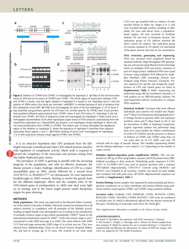
We sought to identify the basis of haplotype 5 associated withdecreased risk of AMD. Examination of quality scores for genotypingrs460897 (reported to encode S1191L within the final exon of CFH)suggested that the assay may have measured a copy number variantrather than the expected SNP. Specifically, we hypothesized that theassay might target a deletion of the final exon of CFHR1, which differsfrom the final exon of CFH only at the position of this putative SNPand at one other base. We selected gene-specific primers to sequencethese exons in DNA from individuals homozygous for each of the fivehaplotypes in block 2 who were also homozygous for the stronglyassociated haplotype in adjoining block 3. We did not find anyvariation in exon 23 of CFH in association with any haplotype;hence, rs460897 does not represent a true SNP. Exon 6 of CFHR1did not amplify in homozygotes for the protective haplotype 5(Fig. 2a,b). In all other haplotypes, exon 6 of CFHR1 differed fromCFH at the expected sites and also showed haplotype-specific variationat SNPs rs4320 (906G-T) and rs414628 (942A-T). All 28 samplessequenced from heterozygous individuals carrying one copy of hap-lotype 5 seemed to be homozygous at all CFHR1 exon 6 SNP sites, ofwhich ten were homozygous for a rare allele. We confirmed deletion inhaplotype 5 homozygotes by coamplification using primers thatannealed to sites common to both CFH and CFHR1. Sequencing ofPCR products showed that haplotype 5 amplified only CFH, whereasall other haplotypes amplified both genes, uncovering ‘pseudoSNPs’at all sites differing between CFH and CFHR1 (Fig. 2c). Similarly, weuncovered deletion of CFHR1 intron 4 by coamplification using
primers common to CFH intron 21 and CFHR1 intron 4, whichamplified products of 324 and 380 bp, respectively (Fig. 2b). Thedeletion also removed CFHR3 from haplotype 5, as indicated byamplification only of CFHR4 sequence in homozygotes using primersspecific to both exon 6 of CFHR3 and CFHR4 flanking a region ofincomplete homology (Fig. 2c).
We used multiplex ligation-dependent probe amplification7 tomeasure copy number of CFH exon 23 and CFHR1 exon 6. Thisrelatively new technique involves hybridization of pairs of adjoiningprobes to genomic DNA followed by ligation, which is achieved only ifboth are perfectly hybridized at the adjacent ends. Our assays centeredon CFH 3572C and CFHR1 869T, and the hybridizing portions of thegene-specific probes varied only at one key base. The copy number ofCFH exon 23 remained constant in male and female DNA samplesfrom individuals carrying 0, 1 or 2 copies of haplotype 5 (who hadcopy numbers of 1.00, 1.04 and 1.06, respectively) when referenced toan autosomal marker in exon 9 of MORF4L1 and an X-linked markerin exon 6 of BCAP31, which was corrected for sex in males. As expec-ted, the copy number of CFHR1 dropped from 1 to 0.44 and from 1 to0 in heterozygotes and homozygotes for haplotype 5, respectively.
Protein blot analysis of serum samples from individuals homo-zygous for each haplotype confirmed the absence of CFHR1 andCFHR3 protein in haplotype 5 homozygotes (Fig. 2d). We did notfind deletions on other haplotypes.
The deletion on haplotype 5 measured 84,682 bases in lengthand occurred between two virtually identical 29-kb segments of
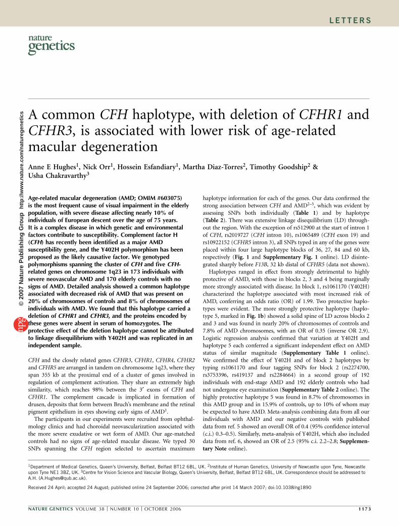
Table 1 Association of SNPs spanning the CFH region with AMD
SNP number SNP name Coding variant Case/control ratios w2 P value
1 rs1292487 308: 38, 263: 77 16.7 4.33 � 10–5
2 rs512900 344: 2, 338: 2 0.0 0.99
3 rs7524776 318: 28, 292: 48 6.3 0.01
4 rs529825 309: 37, 263: 77 17.7 2.61 � 10–5
5 rs800292 CFH I62V 309: 37, 263: 77 17.7 2.61 � 10–5
6 rs1329424 191: 155, 130: 210 19.8 8.47 � 10–6
7 rs1061147 CFH A307A 191: 155, 130: 210 19.8 8.47 � 10–6
8 rs1061170 CFH Y402H 192: 154, 130: 210 20.5 5.96 � 10–6
9 rs10801555 192: 154, 130: 210 20.5 5.96 � 10–6
10 rs2019727 303: 27, 272: 66 17.9 2.29 � 10–5
11 rs2019724 213: 133, 141: 199 27.7 1.41 � 10–7
12 rs203685 212: 134, 141: 199 26.9 2.12 � 10–7
13 rs1831281 293: 37, 269: 69 10.6 0.0011
14 rs2274700 CFH A473A 280: 66, 205: 135 35.2 2.92 � 10–9
15 rs6677604 319: 27, 274: 66 19.7 8.97 � 10–6
16 rs3753396 CFH Q672Q 279: 67, 278: 62 0.1 0.71
17 rs419137 282: 64, 296: 44 4.0 0.046
18 rs2284664 307: 39, 271: 69 10.5 0.001
19 rs1065489 CFH D936E 278: 68, 276: 64 0.1 0.78
20 rs10801560 307: 39, 274: 66 8.8 0.003
21 rs460897 CFH S1191L 319: 27, 270: 68 21.7 3.22 � 10–6
22 rs432007 211: 133, 145: 191 22.5 2.07 � 10–6
23 rs438781 214: 132, 148: 192 23.1 1.54 � 10–6
24 rs408519 212: 134, 147: 193 22.4 2.26 � 10–6
25 rs6428372 282: 64, 281: 59 0.2 0.70
26 rs10922147 296: 48, 261: 79 9.7 0.0018
27 rs1971579 262: 84, 223: 117 8.5 0.0035
28 rs4085749 CFHR2 C140C 298: 48, 263: 77 8.9 0.0029
29 rs10922152 209: 137, 161: 179 11.8 0.0006
30 rs5998 F13B N602N 184: 162, 199: 141 2.0 0.16
P values were generated using a w2 test for association of allele frequencies in 173 individuals with AMD and 170 controls.
1 17 4 VOLUME 38 [ NUMBER 10 [ OCTOBER 2006 NATURE GENETICS
LET TERS©
2007
Nat
ure
Pub
lishi
ng G
roup
ht
tp://
ww
w.n
atur
e.co
m/n
atur
egen
etic
s

duplication in the physical map of the chromosome8 (marked 1 and 1¢in turquoise in Figs. 1a and 2a). The position of the deletion wasmapped by amplifying regions of slight variation between the dupli-cated regions and sequencing to check which base was retained inhomozygotes for haplotype 5 (Fig. 2a,c). A 2.8-kb junction fragmentwas identical in size to that found in nondeleted haplotypes; hence,both breakpoints map to the same point within a 1,618-bp region ofperfect homology centered B6,400 bp after CFH and CFHR1 (Sup-plementary Fig. 2 online).
CFH and related proteins are important regulators of complementactivity. The CFH gene cluster is responsible for numerous alterna-tively spliced transcripts and proteins. Their high homology makesthem extremely difficult to study, and the contribution of each ispoorly understood. CFH and CFHR1 are known to be present in thecirculation at high levels and to act as cofactors for factor I–mediateddegradation of C3b9,10. Notably, our datashow that CFHR3 and CFHR4A (the longform of CFHR4) (ref. 11) are also present inserum at comparable levels. It has not yetbeen possible to assess the relative impor-
tance of deletion of CFHR1 and CFHR3 in contributing to theprotective nature of haplotype 5; however, the products of bothgenes are present in the circulation where they have the potential tocompete with CFH for C3 binding. Mutations in the early exons ofCFH that cause hemolytic uremic syndrome (HUS; see http://www.FH-HUS.org; OMIM #235400) tend to affect CFH proteinsecretion rather than function. Although only semiquantitative, ourprotein blot data suggest that haplotypes 1 and 2, which are associatedwith the harmful C allele of Y402H, may have relatively less serumCFH in comparison with CFH-related proteins. A possible mechanismfor this is through the close association of the C allele with the G alleleof SNP rs10922094, which may permit some alternative splicing of anew exon containing an immediate stop codon. This putative exon isdistinct from exon 10, which is alternatively spliced as the final exon ofthe shorter CFH ‘b’ transcript.
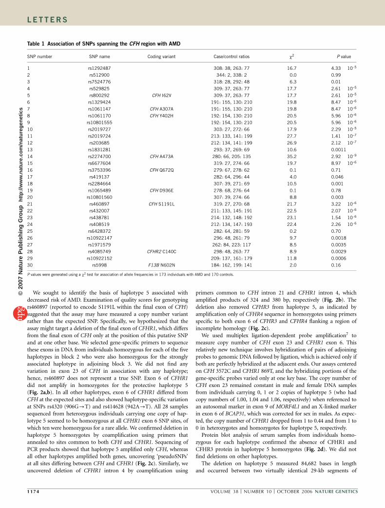
CFH
50 kb
Block 4Block 3Block 2Block 1
CFHR3 CFHR1 CFHR4 CFHR2 CFHR5
1 2 3 1′ 2′ 3′
rs75
2477
6rs
5298
25rs
8002
92rs
1329
424
rs10
6114
7rs
1061
170
rs10
8015
55
rs20
1972
4rs
2036
85rs
1831
281
rs22
7470
0rs
6677
604
rs37
5339
6rs
4191
37rs
2284
664
rs10
8015
60rs
4608
97rs
4320
07rs
4387
81rs
4085
19
rs64
2837
2rs
1092
2147
rs19
7157
9rs
4085
749
0.47
0.16
0.52
0.18
0.15
0.14
0.19
0.36
0.16
0.14
0.36
0.11
0.06
0.52
0.18
0.18
0.11
0.89 0.91 0.89
Haplotype Haplotype Haplotype Haplotype
Block 1 Block 2 Block 3 Block 4
a
b
Table 2 Association of CFH and related gene haplotype blocks with AMD
Haplotype Percentage in affected individuals Percentage in controls Odds ratio w2 P value
Block 1
Markers 3–9 TCGAACA 55.2 38.2 1.99 19.8 8.47 � 10–6
TCGCCTG 33.8 39.1 0.80 2.1 0.15
CTACCTG 8.1 14.1 0.54 6.3 0.012
TTACCTG 2.6 8.5 0.29 11.5 0.0007
Block 2
Markers 11–18 1: AGGCGACG 18.3 12.9 1.51 3.7 0.053
2: AGGCGAAG 43.0 28.5 1.89 15.6 7.72 � 10–5
3: GTGCGGAG 19.5 18.2 1.08 0.2 0.68
4: GTATGAAA 11.3 20.3 0.50 10.3 0.0013
5: GTGTAAAG 7.8 19.4 0.35 19.5 1.03 � 10–5
Block 3
Markers 20–24 CATAC 61.3 43.2 2.08 22.5 2.14 � 10–6
CAGTT 19.5 17.2 1.16 0.6 0.45
AAGTT 11.3 19.4 0.53 8.6 0.003
CGGTT 7.6 19.8 0.33 21.8 2.96 � 10–6
Block 4
Markers 25–28 ACAC 61.5 42.0 2.22 26.1 3.25 � 10–7
CCCC 18.5 17.0 1.10 0.2 0.62
AGAT 13.9 22.6 0.55 8.9 0.003
ACCC 5.8 17.4 0.29 22.5 2.14 � 10–6
An odds ratio 41 indicates a detrimental haplotype, and o1 indicates a protective AMD haplotype.
Figure 1 Haplotype block structure in CFH and
CFH-related genes. (a) Duplicated regions that
share orientation and 496% homology are shown
below the genomic structure of genes. Many
other regions of 80%–95% homology are not
marked. The haplotype block structure of
markers used in this study is shown in blue.
(b) Haplotypes of SNPs are shown in blocks withoverall frequencies (in affected individuals and
controls) and connections from one block to the
next. In the crossing areas, a value of multiallelic
D ¢ is shown. This reflects the level of
recombination between two adjacent blocks.
The black box outlines the protective haplotype 5
in this block.
NATURE GENETICS VOLUME 38 [ NUMBER 10 [ OCTOBER 2006 1 17 5
LET TERS©
2007
Nat
ure
Pub
lishi
ng G
roup
ht
tp://
ww
w.n
atur
e.co
m/n
atur
egen
etic
s
It is an attractive hypothesis that CFH produced from the full-length transcript is beneficial and other CFH-related proteins interferewith regulation of complement activity. Much work is required tounravel the complexity of the transcripts and proteins arising fromthis highly duplicated gene cluster.
The prevalence of AMD is growing in parallel with the increasinglongevity of the population, and with no effective treatment, thedisease presents a major challenge. Identification of CFH as a majorsusceptibility gene in 2005, closely followed by a second locus(LOC387715, or PLEKHA1)12,13 on chromosome 10, were importantbreakthroughs in AMD research. Starting to resolve the genetic basisfor the different haplotypic effects and the complex roles of theCFH-related genes in predisposition to AMD may shed some lighton its etiology and in the future might present useful therapeutictargets for gene silencing.
METHODSStudy populations. This study was approved by the Research Ethics Commit-
tee of the Queen’s University of Belfast. Informed consent was obtained from all
subjects involved, in compliance with all principles of the Helsinki Accord.
Grading of digital fundal color photographs was according to the classification
of mutually exclusive stages of age-related maculopathy (ARM)14 based on the
international classification system for ARM15. Under this system, stages 2 and 3
correspond to early ARM and stage 4 to AMD. Our study subjects with AMD
(173) had end-stage neovascular AMD (stage 4) in at least one eye. They were
selected from Ophthalmology Clinics in the Royal Victoria Hospital, Belfast,
UK, and had an average age of 76 years. The controls in our main study
(170) were age matched with no evidence of early
macular disease in either eye (stages 0 or 1) and
were recruited through random sampling of older
adults (65 years and above) from a population-
based register. All were recruited in Northern
Ireland, UK and were of European descent. The
replication group of 192 subjects matched the
above criteria for subjects. The replication group
of controls consisted of 192 elderly UK individuals
of European descent who had no eye examination.
DNA extraction, genotyping and sequencing.
DNA was extracted from peripheral blood by
standard methods. High-throughput SNP genotyp-
ing was outsourced using Illumina bead technology
based on multiplex PCR and primer extension as
part of a larger project. Additional SNPs were typed
in-house using multiplex PCR followed by multi-
plex SNaPshot (ABI) technology. Primers were
designed using Primer Detective (Clontech). Pri-
mer sequences for specific and nonspecific ampli-
fication of CFH and related genes are listed in
Supplementary Table 3 online. Sequencing was
performed using ABI dye terminator chemistry v3
with analysis on an ABI3100 genetic analyzer. We
used Sequencher software (Genecodes) to compare
DNA sequences.
Statistical methods. Genotype data from affected
individuals and controls were loaded into Haplo-
view16 (http://www.broad.mit.edu/mpg/haploview/)
in linkage format to generate allele and haplotype
numbers, ratios and P values based on the w2 test
for association of allele or haplotype frequencies.
Logistic regression analysis was performed using
Stata v8 to assess further the relative contributions
of rs1061170 (Y402H) and the presence or absence
of deletion of CFHR1 and CFHR3 using Stata on
our main data set of 173 AMD cases and 170
controls with no signs of macular disease. The variables representing His402
and the deletion haplotype 5 were coded 0, 1 or 2 depending on the number of
copies carried.
Multiplex ligation-dependent probe amplification (MLPA). This was per-
formed on 100 ng of DNA using buffers, enzymes and PCR primers from MRC
Holland according to their protocol. Hybridizing probe sequences X-CFH-
1191C or X-CFHR1-1191T were used in separate reactions with a common
PHO-labeled oligonucleotide, CFH1191-Y. Controls using MORF4L1 and
BCAP31 were included in all reactions. Analysis was based on peak heights
and correlated well with peak areas. All MLPA oligonucleotide sequences are
available in Supplementary Table 3.
Protein blotting. Serum samples were separated by polyacrylamide gel electro-
phoresis, transferred on to nylon membrane and protein blotted using poly-
clonal antisera raised against CFHR1 and CFHR3 using standard methods.
Accession codes. GenBank: CFH, NM000186; CFHR1, BC016755; CFHR2,
BC022283; CFHR3, AK124051; CFHR4, BC074957. CFH exons are numbered
to include exon 10, which is alternatively spliced into the shorter transcript of
this gene. Numbering of transcripts starts from the initial ATG.
Note: Supplementary information is available on the Nature Genetics website.
ACKNOWLEDGMENTSWe thank D. McGibbon for assistance with DNA extraction; J. Silvestri,V. McConnell, G. Wright, G. Meenagh and C. Benson for blood samples fromtheir patients; P. Zipfel for antisera for use in protein blotting; C. Cardwell forstatistical help and Illumina for discussions on choice of SNPs for typing. Thiswork was supported by The Health Foundation.
CFHR3 CFHR1CFH
rs10
8015
60
rs43
2007
rs43
8781
rs40
8519
Deleted in haplotype 5
CFH exon 23
CFHR1 exon 6
CFHR1 intron 4CFH intron 21
1 32 4 5
CFHCFHR1
CFHR3CFHR4
Dup1 Dup1′
Anti-CFHR1 Anti-CFHR3
CFH
CFHR1
175
kDa
8362
47.5
32.5
25
1 2 3 4 5 1 2 3 4 5
CFH
CFHR4
CFHR3
a c
db
1 1′
Figure 2 Deletion of CFHR3 and CFHR1 in homozygotes for haplotype 5. (a) Map of the terminal seven
exons of CFH and all six exons of CFHR3 and CFHR1. The 29-kb region of duplication between CFH
and CFHR1 is shown, and the region deleted in haplotype 5 is boxed in red. Haplotype block 3 and the
position of SNPs within this block are indicated. rs460897 is omitted because of lack of evidence thatthis represents a true SNP. (b) DNA from homozygotes for each of the five haplotypes (1–5) of block 2
was amplified using primers specific for CFH exon 23, primers specific for CFHR1 exon 6 and primers
that amplified different-sized products from CFHR1 intron 4 and CFH intron 21. Haplotype 5 did not
amplify from CFHR1. (c) Pairs of sequences show one homozygote for haplotype 5 (lower trace) and a
homozygote representative of all other haplotypes (upper trace) of PCR products coamplifying from two
imperfectly duplicated loci. PseudoSNPs are present in all haplotypes except haplotype 5, which did
not show amplification from CFHR1 or CFHR3. The bottom pair of traces delineates the breakpoint
region of the deletion on haplotype 5, where the sequence of haplotype 5 amplifies from adjacent
duplicated (Dup) regions 1 and 1¢. (d) Protein blotting of serum from homozygotes for haplotypes
1 to 5 with polyclonal antisera raised against CFHR1 and CFHR3.
1 17 6 VOLUME 38 [ NUMBER 10 [ OCTOBER 2006 NATURE GENETICS
LET TERS©
2007
Nat
ure
Pub
lishi
ng G
roup
ht
tp://
ww
w.n
atur
e.co
m/n
atur
egen
etic
s
AUTHOR CONTRIBUTIONSA.E.H., N.O. and H.E. designed the SNP association study, A.E.H. and N.O.analyzed SNP data, T.G. and M.D.-T. performed protein blotting experiments,U.C. provided clinical information and patient samples and A.E.H. performedall other experiments and wrote the paper.
COMPETING INTERESTS STATEMENTThe authors declare competing financial interests (see the Nature Genetics websitefor details).
Published online at http://www.nature.com/naturegenetics
Reprints and permissions information is available online at http://npg.nature.com/
reprintsandpermissions/
1. Mullins, R.F., Aptsiauri, N. & Hageman, G.S. Structure and composition of drusenassociated with glomerulonephritis: implications for the role of complement activationin drusen biogenesis. Eye 15, 390–395 (2001).
2. Klein, R.J. et al. Complement factor H polymorphism in age-related macular degen-eration. Science 308, 385–389 (2005).
3. Edwards, A.O. et al. Complement factor H polymorphism and age-related maculardegeneration. Science 308, 421–424 (2005).
4. Haines, J.L. et al. Complement factor H variant increases the risk of age-relatedmacular degeneration. Science 308, 419–421 (2005).
5. Hageman, G.S. et al. A common haplotype in the complement regulatory gene factor H(HF1/CFH) predisposes individuals to age-related macular degeneration. Proc. Natl.Acad. Sci. USA 102, 7227–7232 (2005).
6. Zareparsi, S. et al. Strong association of the Y402H variant in complement factor H at1q32 with susceptibility to age-related macular degeneration. Am. J. Hum. Genet. 77,149–153 (2005).
7. Schouten, J.P. et al. Relative quantification of 40 nucleic acid sequences bymultiplex ligation-dependent probe amplification. Nucleic Acids Res. 30, e57(2002).
8. Heinen, S. et al. De novo gene conversion in the RCA gene cluster (1q32) causesmutations in complement factor H associated with atypical hemolytic uremic syn-drome. Hum. Mutat. 27, 292–293 (2006).
9. Reid, K.B. et al. Complement system proteins which interact with C3b or C4b; asuperfamily of structurally related proteins. Immunol. Today 7, 230–234 (1986).
10. DiScipio, R.G. Ultrastructures and interactions of complement factors H and I.J. Immunol. 149, 2592–2599 (1992).
11. Jozsi, M. et al. FHR-4A: a new factor H-related protein is encoded by the human FHR-4gene. Eur. J. Hum. Genet. 13, 321–329 (2005).
12. Jakobsdottir, J. et al. Susceptibility genes for age-related maculopathy on chromosome10q26. Am. J. Hum. Genet. 77, 389–407 (2005).
13. Rivera, A. et al. Hypothetical LOC387715 is a second major susceptibility gene forage-related macular degeneration, contributing independently of complement factor Hto disease risk. Hum. Mol. Genet. 14, 3227–3236 (2005).
14. van Leeuwen, R., Klaver, C.C., Vingerling, J.R., Hofman, A. & de Jong, P.T. The risk andnatural course of age-related maculopathy: follow-up at 6 1/2 years in the Rotterdamstudy. Arch. Ophthalmol. 121, 519–526 (2003).
15. The International ARM Epidemiological Study Group. An international classificationand grading system for age-related maculopathy and age-related macular degenera-tion. Surv. Ophthalmol. 39, 367–374 (1995).
16. Barrett, J.C., Fry, B., Maller, J. & Daly, M.J. Haploview: analysis and visualization of LDand haplotype maps. Bioinformatics 21, 263–265 (2005).
NATURE GENETICS VOLUME 38 [ NUMBER 10 [ OCTOBER 2006 1 17 7
LET TERS©
2007
Nat
ure
Pub
lishi
ng G
roup
ht
tp://
ww
w.n
atur
e.co
m/n
atur
egen
etic
s
CORR IGENDA
Corrigendum: A common CFH haplotype, with deletion of CFHR1 and CFHR3, is associated with lower risk of age-related macular degenerationAnne E Hughes, Nick Orr, Hossein Esfandiary, Martha Diaz-Torres, Timothy Goodship & Usha ChakravarthyNat. Genet. 38, 1173–1177 (2006); published online 24 September 2006; corrected after print 14 March 2007
In the version of this article initially published, the G and A alleles of rs1831281 in Figure 1 should be reversed, and the block 2 haplotypes in Figure 1, Table 2 and Supplementary Table 2 should be corrected to 1:AGGCGACG, 2:AGGCGAAG, 3:GTGCGGAG, 4:GTATGAAA and 5:GTGTAAAG. The error has been corrected in the HTML and PDF versions of the article.
©20
07 N
atur
e P
ublis
hing
Gro
up
http
://w
ww
.nat
ure.
com
/nat
ureg
enet
ics