A Closer Look At The Emperor’s New Clothes to be immediately presentable, a ChKM solution was used...
36
Handout on the occasion of 2015 AOD Annual Meeting Synopsis February 26, 2015 Richard V. Tucker Lecture of Excellence A Closer Look At The Emperor’s New Clothes Dr. Dr. Rüdiger Osswald, Munich, Germany Nowadays, countless glossy brochures celebrate modern dentistry as innovation and progress. The protagonists don’t even hesitate to fantasize about a miracle explosion of knowledge in dentistry. If one takes a closer look, it becomes apparent that this progress is a Potemkin Village as it mainly affects prosthodontics: We surely are improving in replacing teeth. But the art of restoration of natural albeit flawed teeth is not only in stagnation since about a 100 years but yet in regression. Our dilemma in restorative dentistry shows that essential knowledge for the solution of difficult and complex cases has been lost. “Once upon a time" we already knew better. Presented by Berufsverband der Allgemeinzahnärzte in Deutschland e.V. (BVAZ) http://www.bvaz.de © · 2015 · Dr. Dr. Rüdiger Osswald, Munich, Germany
Transcript of A Closer Look At The Emperor’s New Clothes to be immediately presentable, a ChKM solution was used...

Handout on the occasion of
2015 AOD Annual Meeting Synopsis February 26, 2015
Richard V. Tucker Lecture of Excellence
A Closer Look At The Emperor’s
New Clothes Dr. Dr. Rüdiger Osswald, Munich, Germany
Nowadays, countless glossy brochures celebrate modern dentistry as innovation and progress. The protagonists don’t even hesitate to fantasize about a miracle explosion of knowledge in dentistry. If one takes a closer look, it becomes apparent that this progress is a Potemkin Village as it mainly affects prosthodontics: We surely are improving in replacing teeth. But the art of restoration of natural albeit flawed teeth is not only in stagnation since about a 100 years but yet in regression. Our dilemma in restorative dentistry shows that essential knowledge for the solution of difficult and complex cases has been lost. “Once upon a time" we already knew better.
Presented by Berufsverband der Allgemeinzahnärzte in Deutschland e.V. (BVAZ) http://www.bvaz.de
© ·2015·Dr. Dr. Rüdiger Osswald, Munich, Germany

The Indication‐Specific Treatment of Bacterial Endodontitis
The “Timbuktu Protocol”(*) PART 1: THE VITAL TOOTH
An Endodontic Case – Mosaicked and Hybrid Forms In the clinical view of endodontics, there are no unambiguously classified findings. Instead, there is an abundant range of hybrid and mosaicked forms. Accordingly, as is the case with any complex protocol, I cannot promise you that you will not have to go through a period of learning. At the beginning it is possible for the patient to report pain or discomfort, if for instance the clinical situation was assessed to be something slightly different than turned out to be the case. However, the wonderful thing about this protocol is that it ensures that nothing gets out of hand, because at any point in the treatment you can simply go back a step and try it again (with more patience). In the course of time, you will be less and less likely to assess the situation
incorrectly, because you will learn relatively quickly which tooth requires which therapy and medication in the specific treatment situation.
That is a promise I will keep. If you follow this protocol precisely, I can make you two promises: On the one hand, the learning curve will be very steep (you will learn very rapidly). This, by the way, lies in extreme contrast to actual treatment under a dental microscope, during which you will encounter endless complications and delays. On the other hand, your treatment results in all your various clinical situations will turn out much better than those
1-01

that appear in the literature. If, until this point, you have followed the treatment protocol as it is currently taught, it will not take long for you to notice the following: Very little if any drama with patients due to exacerbation, and instead, an immediate and lasting freedom from pain. This will be accompanied by strong, nearly complete reduction in requests for pain medications and antibiotics.
Is this a miracle drug?
When endodontists acquaint themselves with my protocol, they often reduce what I teach to the belief that – as long as they do not confuse ChKM withformaldehyde – they simply need to administer a “miracle drug” to the tooth, and everything will be fine. That is not true, and I have never suggested such a thing. Instead, in all three phases of treatment, my protocol is very clearly differentiated from the current, unfortunately dogmatic orthodoxy, which in my eyes now runs counter to better knowledge. This means that I make different preparations for certain clinical situations, disinfect, and apply fillings differently than is commonly practiced.
The mechanism is (only) disinfectant’s servant!E For mechanical preparations, in principle it does not matter with which mechanical tools one prepares. The important thing is that one makes it “neat and tidy.” For me, “neat and tidy” means that one mechanically cleans (and in doing so, expands), as thoroughly as possible, all the main canals one has found after careful inspection, down to the apex. The softer the dentin, the more thorough one’s preparations can and must be – of course without over doing it. The goal is to extend the canals until one creates dry filings. This is an indicator that the side canals
(*) Why is it called the “Timbuktu Protocol”? The name comes from a statement Dr. Osswald made on an internet mailing list. When asked about his special techniques and procedure, he replied, “Give me any workable file and a strong disinfectant, and I will treat (nearly) every root canal successfully, even in Timbuktu!”
State of teeth after chemotherapy and radiation using micro-explosions to treat tooth decay. The prosthetic restoration, the appropriate treatment plan, and a cost evaluation had already been planned with an expert, when "something hard came between the teeth“, resulting in crown fractures to teeth 24 and 25. After caries removal, only the root of the tooth was able to be preserved, as with tooth 26.
Since the remainder of the tooth structure that was able to be preserved lay partially below the level of the surrounding mucosa, there were no previously tested solutions available. Moreover, since the patient was a businessman and needed to be immediately presentable, a ChKM solution was used in the treatment, the root canal was filled, and the screws set in one session, as described in the text.
1-02
Presentation of the tricky cavitysystem of a lower molar by Prof. Dr. Walter Hess, anatomist of the University Zürich, 1917. Published in the textbook „Mein System der medikamentösen Behandlung schwerer Erkrankungen der Zahnpulpa und des Peridontiums“, publishing company of Hermann Meusser, 1928, Berlin. The tubules, representing about 50 % of the cavitysystem, have not been presented because of technical problems.

and the tubules are open, and the path is clear for disinfection. Much in the spirit of Otto Walkhoff – who once remarked, “The mechanism is the disinfectant’s servant!” – I strive to make preparations apically to at least ISO #35. At the end of the process however, I always use a #60 size file, in order to create sufficient room for thorough disinfection. Thus, it should be obvious that I proceed more and more slowly towards the apex with the last files. In cases of a radiologically verified diagnosis of an apical osteitis, I attempt to insert a #15 size file past the apex and into the radiolucency. On the one hand, this is to determine whether or not secretion is present (incipient / manifest abscess formations, cysts). This follows in the footsteps of Hippocrates, who formed a medical theory in 400 B.C. that is still valid today: “Ubi pus, ibi evacua!” (Where [there is] pus, there evacuate!) On the other hand, however, it also improves the reach of the applied disinfectants to the (almost always) bacterially infected periapex or bones, as the “itis” in osteitis implies. In the first step, I myself use hand tools (up to ISO #25/30) and then a Giromatic (rotating it only a quarter turn in quick succession, while the dentist files). In difficult cases I will use both alternatingly. It is only during re-examinations that I use rotary preparation on the first third of the root canal. In the process, I file very precisely – always in circular motions and neatly against the wall –with a file long enough that the next one can be introduced without the application of force. For cases in which I cannot easily advance the first file (usually an ISO #15) down to the apex or
beyond, or if I cannot gain full or partial access to the canal with my fingers due its location, I also use the Giromatic. (In these cases, however, I always use a brand-new file!) This strategy is almost always successful in these cases, and I can then get back into my usual flow of work. In principal, I have nothing against the use of NiTi rotary files. Nevertheless, I think that using conventional files makes more sense. There are several reasons for this. From my “felt” experience, the vast majority of canals are anything but round, rather they are often oval, and usually have recesses and/or offshoots. In order to clean such canals completely using rotation, one would have to prepare the affected canal along its whole length with a file diameter suitable for the largest diameter, thereby weakening the root much more than is necessary. For example, specialists will often find four canals – two mesial and two additional distal canals – in the first lower molar. I frequently find this at the beginning as well. In the vast majority of cases however, it turns out after some filing that the distal canals suddenly merge, such that I have eliminated the isthmus of the most hourglass-shaped distal canal. During rotational preparation, one often does not notice the great deal of debris one has left behind. Another reason I prefer to prepare with filing is because of the inflationary increase of vertical root fractures. In rotational preparation, it is my opinion that one exerts considerably more lateral pressure on canal walls, which are very thin, particularly apically. The fractures almost always run apical to coronal. This risk obviously becomes even greater if one rinses plentifully with sodium hypochlorite and EDTA, both of which can dissolve the organic components of the dentin, causing long-term damage. And it does not take a genius to deduce what might happen when one additionally applies lateral or vertical pressure in order to drive the highly viscous gutta-percha into the small side canals. Today there is plenty of literature documenting the accuracy of these observations. Moreover, I imagine that with rotational preparations (which use instruments much blunter than steel files) a noticeably greater amount of bacterially infected debris is pressed into the small side canals and the tubules. Fundamentally, the following principle applies: The worse the mechanical preparation of the main canals, the more patiently must one disinfect!
-Dr. med. Dr. med dent. Rüdiger Osswald, Munich, www.tarzahn.de
Dr. med. Dr. med dent. Rüdiger Osswald, Munich.
1-03

CLINICAL DIAGNOSIS:
The vital tooth without
clinical symptoms COMPLETE FILLING IN THE FIRST SESSION
Only for teeth that are preoperatively vital and show no clinical symptoms.
Practical only when the pulp chamber is accidentally opened during prosthetic preparation, if the risk for a direct pulp capping should appear too high. Direct pulp capping in the case of restorative treatment and single crowns (long, semi-permanent incorporation) with openings of the pulp chamber that are not overly large.
Only when ample, complication-free preparation down to the apex is possible.
In this sense, it should serve as the exception to the rule.
If at the time of the opening it is not clear whether the caries has been completely removed, it is better to insert a temporary filling with iodoform paste (Walkhoff method), impression, and a temporary tooth. Permanent root canal filling is set when inserting the prosthetic.
Immobilization via thorough occlusal cutting to eliminate contact points, in particular with regard to lateral movements (power centric).
After (nearly) each file, pressure-free rinsing with 3% H2O2
Filling with Endomethasone N using a paste inject instrument with a single-cone-technique.
Naturally, at times there will be situations in which one cannot avoid applying a permanent filling (to clearly vital teeth) in the first session. For example, when a patient's incisor is fractured down to the mucosa level and there is no longer enough substance for a presentable treatment, so that one cannot avoid using a post and core. In such cases, after each file I fill the canal with ChKM (a tip from a fellow dentist). The idea is that during the preparation, the ChKM penetrates into the side canals and the tubules and begins to take effect (in particular with regards to the impregnation of the dead organic tissue there to protect against reinfection across the periodontal gap). With teeth that might be non-vital or in cases of gangrene of the tooth, I provide an interim prosthesis and before the final closure perform the appropriate disinfection protocol for such cases.
=
T H
E I N
D I
C A
T I
O N
- S
P E
C I
F I
C T
R E
A T
M E
N T
O
F
B A
C T
E R
I A
L E
N D
O D
O N
T I
T I
S :
TH
E
“T
I M
B U
K T
U -
P R
OT
O C
O L
” (
*)

The Indication‐Specific Treatment of Bacterial Endodontitis The “Timbuktu Protocol”* SECTION 2: ACUTE PULPITIS
Treatment goals for acute pulpitis As the name indicates, acute pulpitis occurs suddenly and is often very painful; the majority of patients suffering from the disease seek out emergency treatment. Thus, the short-term goal of treatment is the prompt relief of any symptoms. If this goal is accomplished the patient will be highly satisfied, which in turn improves the practice's reputation. Inconsistent results on the other hand, will be highly detrimental to a practice's reputation. The long-term goal of primary care, however, consists in replacing immediate emergency treatment, which brings chaos to the planned course of your day, with regular fixed appointments for treatment, scheduled in advance. Emergency patients should only disrupt one's planned schedule as an exception. In this case, as in any situation, the exceptions are what make the rule.
FUNDAMENTALLY, THE FOLLOWING APPLIES: A VITAL, PAINFUL TOOTH REQUIRES LEDERMIX, WHILE A NON-VITAL TOOTH REQUIRES AS STRONG A DISINFECTANT AS POSSIBLE
Why Ledermix? Ledermix is a combination product made of a corticosteroid (a cortisone derivative, in this case triamcinolone), and an antibiotic (tetracycline). For acute cases, the active, therapeutically relevant substance is the corticosteroid. While the tetracycline surely does not do any harm, it is probably added only because the old principle still applies: In the case of bacterial infections, do not use cortisone without antibiotic protection, because bacteria love cortisones! That is why applying cortisone to treat gangrene is also counter-productive. Cortisone works as an anti-inflammatory because it affects the immune system non-specifically. Accordingly, it reduces any edema which may accompany inflammation [specific signs of inflammation: rubor, calor, tumor (swelling
by edema), Functio laesa]. It intervenes directly in the lymphocytes’ communication and inhibits the release of certain hormones produced by immune system cells, prostaglandins for example, which strengthen one's perception of pain. Some German university professors caution strongly against the use of cortisone. For example, the German dental publication Zahnärztliche Mitteilungen (ZM) reports on an advanced training seminar in Braunlage (6), during which Dr. Edgar Schäfer of Münster warned against using Ledermix in an open root canal. Do not worry too much about this debate. Cortisone is a wonderful medication, and there is absolutely no reason to withhold its beneficial effects from our patients. An old saying also applies here: The difference between medicine and poison is the dose. Not to mention the fact that those who advise against the use of Ledermix would look rather foolish (and get very ill), if every five minutes their adrenal glands did not produce the same amount of cortisone contained in the medication.
That which is intended to heal must be carefully administered! Inflammatory edema is often already present – both in the tooth and periapically– by the time the patient steps into the dentist’s office. Periapical swelling applies pressure from the apex, driving the affected tooth from the alveolus. Additionally, early centric and lateral occlusal points of contact can further damage the “extended” tooth (patients frequently describe the affected tooth as feeling “too high”).
* Why “Timbuktu Protocol”? The name originated in a statement of Dr. Osswald’s on an Internet mailing list. When asked about his special techniques and procedure, he replied, “Give me any workable file and a strong disinfectant, and I will treat (nearly) every root canal successfully, even in Timbuktu!”
2-01

If this condition is not immediately recognized and treated by the dentist, it is highly counterproductive to reaching the primary goal of the treatment: “rapid relief from pain.” Finally, one of the main principles of “barefoot medicine” is also applicable in this case: “That which is supposed to heal must be immobilized.” Over the long-term, a continuous state of stress can even lead to loosening of the tooth. You cannot immobilize a tooth completely unless you splint it. You can, however, eliminate existing early contact points through occlusal cutting, or, if they are not yet present, you can prevent their formation. Those points of occlusion that entail lateral movements on the slopes and ridges of the cusps should receive special attention. Straight posterior teeth are particularly affected by such lateral stress. At best, you should allow the patient to bite down firmly (!) on occlusal paper, then ask him or her to move it laterally in both directions while continuing to bite down (a colleague once very appropriately called this “power centric”). You are familiar with the following situation: You have applied a good filling, had the patient bite down on occlusal paper, and then carefully cut away at the occlusion. At first glance, everything seems perfect. Despite this, after a few days, the patient visits your office again and complains that after treatment, the tooth in question has become sensitive to cold. If you now use the power centric method, you will frequently find lateral early contact points on the ridges of the cusp. If you remove these, the pain is quickly soothed (reversible abacterial pulpitis, evoked through prolonged physical stimulus). Therefore, it is better if you use the power centric method immediately after applying a patient’s filling (or incorporating a prosthetic) so that you won't have to schedule a second appointment. If the sidewalls
of the tooth are especially weak due to extensive caries and the tooth must be crowned, a radical shortening is recommended in order to prevent, as reliably as possible, wedge-shaped fractures that can easily run into the root or can even split the tooth. Crowning is actually always recommendable for treated root canal teeth (except for teeth that have lost only a small amount of the natural tooth structure) in order to physically mount it and thus stabilize it. An alert patient will probably assume a relieving posture (chewing on the contralateral side) of his or her own accord to avoid the pain that comes with pressure. However, it doesn’t hurt to instruct him or her explicitly to take care to protect the provisional cap. To avoid receiving calls in the night or on the weekend, you should clarify some of the main points from the detailed treatment protocol as described below. If you have explained these points to the patient and one of the exceptions you described should occur, the patient will remain calm and say, “Oh, what the doctor said would happen is happening! What a professional!” Without this explanation a patient might feel afraid if something happens that he or she wasn't expecting, and may call to alleviate their concerns. I explain these points in the last phase of the preparation (noting them quickly so as not to prolong the treatment time) when applying the Ledermix and the provisional cap.
AS LONG AS THE PATIENT ONLY EXPERIENCES THOSE PROBLEMS THAT YOU HAVE ALREADY EXPLAINED, YOU WILL BE FINE. OTHERWISE, THE PATIENT’S OPINION MAY UNFORTUNATELY CHANGE QUICKLY.
Upon initial examination in October 1989, teeth 4 and 21 wereembedded in the pulp, tooth 11 showed a need for retreatmentwith a post and core before receiving a prosthetic, and tooth 15was non-vital and gangrenous.
Twenty years after the permanent, initial treatment, in December 2009 all the teeth with various indications were clinically and radiologically unremarkable after being endodontically treated according to the Timbuktu Protocol. It becomes clear how much one can trust the predictability of the method's results that not a single pillar can be neglected without endangering the entire edifice.
.
2-02

Why use additional medications? The first temporary dressing (with Ledermix) necessary in the simplest of cases involving clinical discomfort will be clear to nearly every dentist. If we observe the current orthodoxy, it is more difficult to understand the necessity of the additional medications described below in the treatment protocol. Endodontic teachings that endeavor to appear modern assert that in the case of acute pulpitis, the bacterial infection is restricted to the pulp chamber and at most the coronal third of the root. Accordingly, the bacteria must be completely removed through vital extirpation and the subsequent mechanical preparation of the more accessible root canals. If I remember correctly, the term “partial gangrene” has even been removed from the nomenclature. However, a glance at both old and new literature clearly shows that that is not the case. From the beginning of the last century, histological research showed that acute pulpitis can very well involve the exacerbation of existing partial gangrene. Otto Walkhoff (1) described histologically proven cases in which partial gangrene had formed and microabscesses had even manifested, which resulted however in vital extirpations (VitE). He stated he had never expected such histological findings based on his preliminary clinical examinations. There is also an exemplary study by Gesi, et al (2) from the year 2006. He divided 244 patients with acute pulpitis into two equal groups, whereby the first group was treated in one session while the other was treated with an intermediate, temporary filling of Ca(OH)2. Within just one to three years, approximately 7% of the patients (distributed evenly between the two groups) developed radiologically diagnosed apical osteitis. Gesi concluded, completely correctly, that a temporary dressing with Ca(OH)2 did not seem to have an impact on the treatment’s success. That does not mean, however, that one can or should treat acute pulpitides in a single session or without medications. At least not if – assuming this simplest of all endodontic situations – one strives for a success rate close to 100%, as can be expected for a simple bacterial infection in an anatomical field that has been fully described for 100 years. Walkhoff offers an explanation for these poor results: With very high probability, the cases in which apical periodontitis developed in a relatively short time produced the partial gangrene that he had proven histologically. Under no circumstances, however, can one conclude that
one can forego long-term disinfection in these cases. One must instead draw the conclusion that Ca(OH)2 is not a suitable means for long-term disinfection. The fact that Gesi’s study does not represent an outlier is documented by the meta-analysis by Kojima et al (3) from 2004, whose results were impressively confirmed in 2008 by the meta-analysis conducted by Ng et al. (4,5). Both attest to a failure rate of around 10% in the treatment of acute pulpitis. These meta-analyses also verify that, despite all the mechanical improvements that have since been introduced, the success rates for all stages of bacterial endodontitis have remained unchanged (still rather poor) over the past 60 years. The development of apical osteitis – referred to as a “post-treatment disease” (although “poorly treated disease” might be more appropriate) by German experts who enjoy adorning their language with Anglicisms – does not lead to tooth loss in every case, but is avoidable (and consequently the revision and/or the root tip resection). Furthermore, it is useless to turn an acute infection into a chronic one if one can heal it completely. In a case of clinically determined acute pulpitis, one cannot forego long-term disinfection. One should only avoid using Ca(OH)2 for disinfection. The fact that one should completely forego the use of Ca(OH)2 has since been shown by innumerable individual studies and meta-analyses. In other words: If anything in dental medicine has been sufficiently proven with scientific evidence, it is that calcium hydroxide is not a sufficient endodontic disinfectant. In the context of the overwhelming evidence, it is incomprehensible that the current school of thought should still be valid and virtually dogmatic. This school insists so strongly on its treatment protocol despite the fact that it has been considered a failure internationally for approximately 10 years, and is furthermore ill-equipped to use and research other, clearly more effective disinfectants such as Professor Walkhoff’s original ChKM solution. Instead, it endeavors to condemn these medications in the eyes of the Bundesamt für Arzneimittel (Germany’s Federal Institute for Drugs and Medical Devices) with scientifically untenable arguments, and to disparage those who use them.
-Dr. med. Dr. med dent. Rüdiger Osswald, Munich, www.tarzahn.de
2-03

Acute pulpitis The tooth is not, or only very slightly, sensitive to touch And radiologically yields no suspicion of apical osteitis (apical periodontitis)
This is one of two normal cases with three temporary dressings. Ledermix (applied with a paste inject instrument), cotton wool, Cavit for one to three days, max. one week.(*) ChKM (**), cotton wool, Cavit for approximately one week, max. two weeks. Iodoform paste, cotton wool, cement for two to four weeks, max. three months.(***) Immobilization via good outer contact points, in particular with regard to lateral movements (power centric). After nearly each file, pressure-free rinsing with 3% H2O2. Nothing else.
If, in one of the stages the patient experiences discomfort, go back a step and disinfect with greater patience. In the first stage, this entails: Ledermix, cotton wool, (temporarily) open for one day, followed by ChKM, cotton wool, (temporarily) open for one to two days, repeat until the tooth is either no longer or barely sensitive to the touch.
Explanation for the patient: (*) This might hurt a little today, I have made some major adjustments to the tooth. You may even need to take a pain reliever today, any type is fine. However, it should definitely be better tomorrow and should continue to get better after that. If not, then you'll have to come back, and we will have to treat the tooth (temporarily) openly. As long as it continues to improve, everything will be fine! If, at night or on weekend, the pain becomes so strong that pain relievers don't help, which I do not expect to happen, then you can remove the provisional filling yourself with a strong, blunt needle to relieve the pressure that has built up in the tooth. However, it is exceedingly rare that this happens, I'm only telling you so that you know what you can do in case it should occur.
(**) This might have a strong taste that you will associate with the dentist’s office, but it is a very effective disinfectant. One notices it immediately. If the tooth becomes sensitive when you bite down, then you'll have to come back, and we will have to leave the tooth (temporarily) open for a time.
(***) If you start to feel pain, you'll have to come back and we will take a step back in the process. However, if you tolerate this test root filling without discomfort, which should be the case, then we have an excellent chance of preserving the tooth over the long-term.
=
T H
E I N
D I
C A
T I
O N
- S
P E
C I
F I
C T
R E
A T
M E
N T
O
F B A
C T
E R
I A
L E
N D
O D
O N
T I
S :
T H
E "
T I M
B U
K T
U -
P R
O T
O C
O L
" (
*)

List of literature The Indication-Specific Treatment of Bacterial Endodontitis The “Timbuktu Protocol” Section 2: Acute Pulpitis
1. Walkhoff, O.: Mein System der medikamentösen Behandlung schwerer Erkrankungen der Zahnpulpaund des Periodontiums. Verlag von Hermann Meuser, Berlin 1928.
2. Gesi, A., Hakeberg, M., Warfvinge, J., Bergenholtz, G.: Incidence of periapical lesions and clinicalsymptoms after pulpectomy – a clinical and radiographic evaluation of 1- versus 2- sessiontreatment. Oral Surg Oral Med Oral Pathol Oral Radiol Endod 101, 379 (2006).
3. Kojima, K., Inamoto, I.: Success rate of endodontic treatment of teeth with vital and nonvital pulps. Ametaanalysis. Oral Surg Oral Med Oral Pathol 95, 97 (2004).
4. Ng, Y., Mann, V., Gulabivala, K.: Outcome of secondary root canal treatment: a systematic review of theliterature. Int Endod J 41, 1026 (2008).
5. Ng, Y., Mann, V., Rahbaran, S., Lewsey, J., Gulabivala, K.: Outcome of primary root canal treatment:systematic review of the literature -- Part 2. Influence of clinical factors. Int Endod J 41, 6 (2008).
6. SP.: Lernen in Braunlage - Vom Apex bis zur Krone. ZM (466), (01.03.2014).

The Indication‐Specific treatment of Bacterial Endodontitis The "Timbuktu Protocol(*)" PART 3: PARTIAL GANGRENE
Despite the current orthodoxy's groundless denials, a clinically definable disease – described as “partial gangrene” in older medical literature – does in fact exist. It has been histologically proven, as I have already demonstrated in Part 2. This is just as easily shown in a clinical setting. Every experienced dentist will have, for example, occasionally opened a multi-rooted tooth with pulpitis only to find that two canals were entirely vital, while one was clearly non-vital or already gangrenous. What else can we call this phenomenon but “partial gangrene”? It is usually these same teeth that make accurate diagnosis so difficult due to their atypical symptomatology: inconclusive results on vitality tests, little sensitivity to touch, no mobility, but nevertheless recurrent, atypical discomfort, which often cannot be assigned to one tooth, or even the upper or lower jaw. Even after prosthetic restoration, patients with such symptoms will show up at one's office from time to time complaining of varying degrees of pain, in some extreme cases with a diagnosis of CMD. In this situation, one will continually find more or less apparent cases of early points of contact or malocclusion, and will have to perform occlusal cutting (inflammatory edema, see Part 1). Things will be fine for a while, but then – just when you think you've finally taken care of the problem forever – you'll note in your calendar that the same patient has made yet another appointment because of tooth pain. If at one point you become unnerved and decide for endodontic therapy, over the long term this will give rise to a chronic bacterial infection, which will then often require a lengthy treatment to heal completely. If, in such cases, I suspect a certain tooth of being the culprit, I leave the decision to the patient, saying: “I can probably alleviate this pain with a simple root canal,
you must tell me if that is what you want!” In this way, you quickly find out how much the patient is really suffering. People's sensitivity to pain is remarkably individualized. If one has a treatment protocol in which one can guarantee success even in difficult cases, the decision to proceed with endodontic therapy comes much more easily.
Which does “temporarily open” mean? I use this term to refer to the closure of the trepenated tooth crown with firmly inserted, disinfection-soaked cotton wool pellets. “Ubi pus ibi evacua” is one of the earliest guiding principles of medicine that is still applicable today. Attributed to Hippocrates, the saying was used as early as 400 B.C., and will certainly continue to be used for the next twenty-five hundred years. Then again, it can be true for more than just pus. The same applies for the rotting gases and secretions that arise from the putrid decay of organic tissue. Dead, decaying tissue has an odor because gases arise, and with these gases, pressure. This gas must eventually go somewhere if it is not going to cause pain. If it cannot escape coronally, is the only path the one that leads apically across the foramen and laterally across the side canals and tubules, into the neighboring tissue? Is that medically desirable? Universities teach that after each endodontic procedure one must close each tooth, attempting by any means to prevent the danger of a secondary infection in the oral cavity. Save a gangrenous tooth from saliva, which is bacteriostatic? Does that make any sense? Of course not! This school of thought not only contradicts the ubiquitous and valid medical theorem quoted above - for what is (incipient)
* Why "Timbuktu Protocol"? The name comes from a statement Dr. Osswald made on an internet mailing list. When asked about his special techniques and procedure, he replied, “Give me any workable file and a strong disinfectant, and I will treat (nearly) every root canal successfully, even in Timbuktu!”
3-01

gangrene other than an emergent intradental abscess? –it also contradicts any logical consideration: First, in the oral cavity we find predominately non-pathogenetic viruses, with which the patient has made his peace, which is why they are called “house germs.” Secondly, in the short time that one has the tooth “temporarily” opened for treatment, a cotton wool pellet that has been soaked in a strong disinfectant will take on virtually no bacteria. Moreover, the pellets inserted into the trepanation cavity will reliably block any food particles from entering. It should be obvious that patient must be prevented from eating on the contra-lateral side, thereby also protecting the tooth. (“That which is intended to heal must be carefully administered!”) While inserting the filling and the cotton wool, I tell the patient: “I am now treating your tooth with a very potent medication. It is one of the two most effective disinfectants that we know about in dentistry. You can taste it, can’t you? It's like the chest-compress your mother used to apply. That’s the camphor, which unfortunately we can’t do without. The camphor’s strong smell will be a strong reminder of your visit. We have to press on however, if we want to keep the tooth safe and avoid a root tip resection!” I use Dr. Walkhoff's ChKM solution as a disinfectant. To date I have not met a patient who, when faced with a surgical alternative, decided against ChKM, or regretted his or her decision afterwards. This is an especially attractive solution when one considers the savings on antibiotics and painkillers, which in these cases almost never need to be prescribed. As far as my own nose is concerned: What a pleasure to only smell ChKM on a tooth that had had such a foul odor only two days before. Our office does not reek of ChKM. That however, may be due to the fact
that we ventilate the office well, make sure to keep all ChKM bottles closed, and use the solution with a simple but effective method for application that goes directly into the tooth without spilling a single drop.
High rate of failure despite indication-specific treatment? The treatment of an illness – and this is one of, if not the most important guiding principles of medicine – can only be predictable and successful on a long-term basis if its specific protocol is followed meticulously. With regard to bacterial infections generally, and the treatment of endodontitis and its complications specifically, one must eradicate bacteria responsible for the infection as completely as possible before the permanent root filling. The universally unsatisfactory (if not to say abysmal) results for the endodontic preservation of teeth – unchanged for over 60 years, despite technological advances (1, 2, 3) –
OPT excerpt from December 6, 2011: Acute pulpitis ontooth 29 with very deep distal caries, hypersensitive to cold but barely sensitive to touch. Clinical diagnosis: textbook case of vital extirpation.
Radiograph after three temporary dressings and root fillings on May 14, 2012. The overflow of the sealer leads us to the assumption that - contrary to the clinical diagnosis - partial gangrene with incipient apical variance was already present.
Follow-up from February 26, 2014. The overflowing sealant has, to a large extent, been re-absorbed due to apically normal conditions and continuing lack of pain.
3-02

clearly shows that even this goal has so far not yet been achieved. If one peruses the scientific literature, and in particular the meta-analyses of Kojima et al (1) from 2004 – impressively corroborated by Ng and co-authors (2, 3) in 2008 – one finds no explicit rate of success for the clinical diagnosis discussed here, “a suspicion of partial gangrene.” However, for the approximately 10% of teeth that despite an allegedly perfect vital extirpation soon reveal radiological apical radiolucency – or a chronic bone infection which can exacerbate at any time – it can be assumed that they have already developed partial gangrene.
Beginning with a 0.5% concentration of rinsing agent, today the current school of thought insists on a 5.25% solution of sodium hypochlorite (NaOCl), a solution that carries a high potential for side effects. Surely, one would not continue to increase the concentration of a disinfectant because one is content with the obtained results (4, 10). The question as to how specialists have arrived at the strange concentration of 5.25% has a simple answer: A corresponding solution of hypochlorite is sold as a basic cleaning agent or toilet cleaner at American hardware stores in economical, gallon containers. Still unsatisfied with the results, they have now the increased the duration of the solution's application to a half-hour per canal, heating it up far warmer than body temperature and recommended
ultrasound activation. The central argument for holding on to NaOCl so stubbornly lies in its ability to dissolve tissue. Unfortunately however, living tissue is also dissolved by NaOCl, often with serious, irreversible, and numerous unknown side effects. This has caused the Bundesamt für Arzneimittel to contraindicate the use of concentrated sodium hypochlorite outside of the tooth (open apical foramen) (8,9). NaOCl, then, is anything but suitable for disinfecting the neighboring tissue, which in severe cases is almost always bacterially infected (5). While numerous investigations have scientifically proven that even in the very small concentration of 1% NaOCl is a highly effective disinfectant able to dissolve biofilms in which bacteria persist, it cannot penetrate the side canals and tubules (6), which constitute at least 50% of the endodontic cavity system and which evade any kind of mechanical preparation. It simply cannot reach everywhere that bacteria are located – or it is not allowed to. After a fortunately reversible incident, NaOCl was banned many years ago from our practice – we are all the happier for it, and don't miss it. As a second option for a rinsing agent, ethylenediaminetetraacetic acid (EDTA) is recommended, a solution which possesses virtually no disinfecting properties, but is supposed to dissolve the “smear layer,” or the layer of debris that arises in the course of preparation and blocks access to the side canals and tubules. It is worth noting that depending on how long it is applied, EDTA functions exactly like NaOCl in that it dissolves the organic components within the dentin, thus weakening it on a long-term basis. Moreover, NaOCl quickly disintegrates and becomes ineffective. Both disinfectants are thus unsuitable for long-term disinfection. We do not have any EDTA in our practice and do not miss it. High hopes were initially set on chlorhexidine (CHX), because it worked particularly well in vitro against the bacterium enterococcus faecalis, which appears in almost every tooth with apical osteitis that has been treated for a root canal, and often in teeth with incipient gangrene. Unfortunately, the positive in vitro results have not been confirmed by clinical investigations, as shown, to name only one example, in Paiva’s current 2012 in-vivo study of teeth with apical osteitis (11). This study is particularly interesting as it shows that extensive rinsing with ultrasonically activated hypochlorite paired with CHX was not effective in clearing even the central canals of all bacteria. It is easy, then, to imagine the side canals and tubules swarming with bacteria. As a result, the authors correctly
If the results intended by following professional opinions have been unceasingly poor for more than 60 years, in principal there can be only two alternatives:
One resigns in the belief that endodontitis, with all its complications, is to a large extent an incurable disease. Aided by today's knowledge of etiology, pathogenesis, and an anatomical field that has been completely described for 100 years, such an attitude is unacceptable from a medical perspective. Alternatively, one might finally decide to treat this infection on a case-by-case basis, reacting specifically, instead of acting as many university professors do – like a deer in the headlights – and cling dogmatically to an idea that has proven untenable around the world for many years (4).
3-03

call for an intensified search for alternative antimicrobial substances. According to other studies, CHX unfortunately suddenly seems to lose its effect in the presence of dentin and inflammatory proteins. CHX is therefore unsuitable for long-term disinfection. We do not use CHX in endodontics and do not miss it. Our exclusive rinsing agent is 3% hydrogen peroxide (H2O2), used after (nearly) every file, and before each new temporary dressing, not so much for its disinfecting properties, which are good but not great, but simply as a rinse to keep the canal damp and accessible – H2O2 also foams up quite well, clearing the canals of any debris that has resulted from filing. The side effects are negligible. Nevertheless, if one accidentally uses a little too much 3% H2O2, it is quite painful. In total contrast to a similar incident with NaOCl however, one does not have to call the paramedics, but can simply apologize to the patient and calm him or her down with the fact that the pain will subside in less than one minute, and will cause no permanent damage. According to the conclusions of a systematic study of the existing literature by Fedorowicz et al (7) from 2012, it must be recognized that there is currently no reliable scientific evidence demonstrating that rinsing agents differ sufficiently from each other in efficacy such that one or a specific combination is always preferable. This comes as no surprise when one considers the fact that these disinfectants are not at all able to penetrate entire regions of the infected cavity system and the periapex.
If you really want to advance in endodontics, you have to address the topic of long-term disinfection and potent disinfectants! As I have pointed out and is evident from this article’s illustrations in which the tubules are not even depicted, this is simply a question of anatomy:
The current school of thought recommends, if not to say dogmatically insists that the only long-term disinfectant
one should use is calcium hydroxide (Ca(OH)2)
Considering the meta-analyses of Waltimo et al (12) – which were based on countless studies and show this medication not only to be an unsatisfactory disinfectant, but completely ineffective when treating substantial endodontitis-related bacteria – it can only be termed “permanently unadvisable.” The authors correctly draw the conclusion that their study’s results merely indicate the need to investigate more effective temporary dressings. If one is to put his or her trust in the report from ZM (13) mentioned above in Part 2 however, then Dr. Schäfer is completely unmoved by the evidence. He even allegedly made the bold assertion that temporary dressings only make sense in the case of non-vital teeth with fistules and pronounced symptoms. I am just a humble general dentist, but, with all due respect and requisite modesty, in this connection I would like to point out to him that recent relevant meta-analyses (1, 2, 3) have proven without a doubt that in these cases, even the most modern endodontology has demonstrated a success rate for complete healing of 60% at best. We can only imagine the results from Dr. Schäfer's randomized prospective clinical studies of his newest “secret recipe” (Ca(OH)2 dissolved in water and CHX), as reported in ZM (13). As it appears today, he has in the meantime switched over to the homeopathic camp, which takes a different approach than endodontologists. In light of this camp’s exorbitant increased use of NaOCl, they seem to be of the opinion that diluting a substance that has been scientifically proven to be ineffective makes it noticeably more potent.
Which truly effective medications for long-term disinfection are available to us? Upon closer look, there are actually only two that have been tested scientifically: formaldehyde and Dr. Walkhoff's ChKM solution. They hold in common the fact that they are each considered obsolete by the current school of thought. But more about this in the next section.
-Dr. med. Dr. med dent. Rüdiger Osswald, Munich, www.tarzahn.de
We must use disinfectants that are effective on all kinds of bacteria, and are able not only to penetrate all the regions of the oral cavity but also to disinfect all of the neighboring tissue without causing irreversible side effects. Moreover, we must allow the disinfectants sufficient time for them to take effect.
3-04

Partial gangrene – highly acute pulpitis The tooth is only slightly sensitive to touch, if at all, and based on the radiograph will display no, or at the very most, discrete apical osteitis, for example in the form of a slightly widened periodontal gap, and in some cases (due to functional overuse) clinically discrete loosening, possibly marginal gingivitis, and vestibular sensitivity to touch without swelling.
One of two common cases with three temporary dressings.
Ledermix, cotton wool, “temporarily” open for one to three days.(*)
If it is no longer or only slightly sensitive to touch (as is normal in the case of a correct estimate) ChKM,cotton wool, Cavit for at least one week.(**)
Iodoform paste, cotton wool, cement for at least two weeks, up to three months.(***)
Immobilization via thorough occlusal cutting to eliminate contact points, in particular withregard to lateral movements (power centric).
After (nearly) every file, pressure-free rinsing with 3% H2O2.
If during one of the stages the patient complains of pain, go back a step and proceed more patiently. In this
case, that would mean: ChKM, cotton wool, (temporarily) open for one to three days, then possibly repeating once or twice, until the tooth is no longer or only slightly sensitive to touch.
For cases in which a tooth is simply not be soothed by this method, a trial root filling for an averageof three months with a "magic paste" is a recommendable alternative under simultaneous antibiotic treatment regulation.(****)
Instructions with regards to the patient: (*) In order to allow rotting gases to escape, and thus alleviate pain, the tooth must first be treated "temporarily” openly. Naturally, this will only last for the weekend. If you are not sure about your assessment and do not want to risk receiving any calls, it is generally advisable to leave the suspected tooth "temporarily open" for one night or over the weekend. (**) Patients react very differently to pain. Therefore it makes the most sense to begin by applying only moderate pressure on the neighboring tooth in order to develop a clear picture of the patient's specific reaction. Otherwise, one risks potentially remaining stuck at the same step in the treatment. (***) For example, a trial root filling (which is easily removable) can unexpectedly exacerbate the disease as a consequence of the now tight seal formed by the paste, something which, although rare, can happen from time to time. (****) In the event that such an exacerbation occurs and the situation proves persistently resistant to treatment, all signs point to an abscess between bone and periosteum (highly sensitive to pressure, elastic, vestibular swelling). If the abscess’s complete formation and opening can be impeded but not fully reversed because it is too late, then an antibiotic is recommended. This is all the more advisable in this context, in contrast to treating the bone itself, because the necessary therapeutic levels are reached. If no allergy exists, I prescribe Amoxicillin 1000, 2 x 1 tablet daily for five days, or alternatively Doxycyclin 100, 2x 1. The widespread myth that certain antibiotics that are more or less effective with regards to bones lacks any scientific basis and is solely a marketing ploy. Outside of dentistry, this is almost never asserted. T
H E
I N
D I
C A
T I
ON
- S
P E
C I
F I
C T
R E
A T
M E
N T
O F
B A
C T
E R
I A
L E
N D
O D
O N
T I
T I
S T
H E
"T
I

List of literature The Indication-Specific Treatment of Bacterial Endodontitis The “Timbuktu Protocol” Section 3: Partial Gangrene
1. Kojima, K., Inamoto, I.: Success rate of endodontic treatment of teeth with vital and nonvital pulps. Ametaanalysis. Oral Surg Oral Med Oral Pathol 95, 97 (2004).
2. Ng, Y., Mann, V., Gulabivala, K.: Outcome of secondary root canal treatment: a systematic review of theliterature. Int Endod J 41, 1026 (2008).
3. Ng, Y., Mann, V., Rahbaran, S., Lewsey, J., Gulabivala, K.: Outcome of primary root canal treatment:systematic review of the literature -- Part 2. Influence of clinical factors. Int Endod J 41, 6 (2008).
4. Tronstad, L., Sunde, P.: The evolving new understanding of endodontic infections. Endodontic Topics,(2003).
5. Wu, M., Dummer, P., Wesselink, P.: Consequences of and strategies to deal with residual post-treatment root canal infection. International Endodontic Journal, (2006).
6. Hope, C., Garton, SG., Wang, Q., Burnside, G., Farrelly, PJ.: A direct comparison between extracted toothand filter-membrane biofilm models of endodontic irrigation using Enterococcus faecalis. ArchMicrobiol, (2010).
7. Fedorowicz, Z., Nasser, M., Sequeira-Byron, P., de Souza, RF., Carter, B., Heft, M.: Irrigants for non-surgicalroot canal treatment in mature permanent teeth. Cochrane Database Syst Rev 12, (2012).
8. LegeArtis: Beipackzettel zu Histolith. 2006.
9. Speiko: Beipackzettel zu Hypochlorid-Speiko. (2006).
10. Haapasalo, M., Endal, U., Zandi, H.,Coil, J.M.: Eradication of endodontic infection byinstrumentation and irrigation solutions. Endodontic Topics 10, 77 (2005).
11. Paiva, S., Siqueira, JF Jr., Rôças, IN., Carmo, FL., Ferreira, DC., Curvelo, JA., Soares, RM., Rosado, AS.:Supplementing the antimicrobial effects of chemomechanical debridement with either passive ultrasonicirrigation or a final rinse with chlorhexidine: a clinical study. J Endod 38, (2012).
12. Waltimo, T., Trope, M.,Haapasalp, M.,Orstvik, D.: Clinical Efficacy of Treatment Procedures in EndodonticInfection Control and One Year Follow-Up of Periapical Healing. JOURNAL OF ENDODONTICS 31, 863(2005).
13. SP.: Lernen in Braunlage - Vom Apex bis zur Krone. ZM (466), (01.03.2014).

.
The Indication‐Specific Treatment of Bacterial Endodontitis The “Timbuktu Protocol“* PART 4: MANIFEST GANGRENE
Osteomyelitis is the most feared complication in bone surgery
because this (usually bacterial) infection is so extremely difficult to treat. That is because the bone contains a significant amount of blood, but nevertheless has a poor blood supply (low proliferation rate). Anyone who works with implants can see this for him or herself: When milling the implant tunnel, the blood does not appear as bright red from the arteries, instead it seeps from the veins. In addition, or as a direct consequence, the infected bone is inclined to break down. Subsequently, the intravenous administration of high-dose antibiotic solutions usually does not succeed in producing a sufficiently high local level. And what is apical osteitis other than a (usually mild but not infrequently exacerbated) chronic form of osteomyelitis?
How do surgeons treat this bacterial bone infection?
They create openings, mechanically eliminate the decomposing bone, bring antibiotic granules in direct contact with the infected area, and let it take effect over a very long period of time.
According to university opinions how are we to treat bacterial osteitis?
Without creating openings, very rapidly, and, if possible, in one session, with rinsing solutions whose application is limited to the root canals....in other words, without treating the bacterial bone infection at all over the long term. Knowing this, one may no longer be surprised by the poor results of the current “gold standard” treatment protocols for healing apical periodontitis.
If we want to achieve significantly better results, we should first of all create openings to all the infected areas in order to apply potent disinfectants and grant them ample time to do their work.
In other words: If the treatment of bacterial endodontitis were highly successful even in somewhat difficult cases, one would not need to think about patiently applying extremely potent disinfectants!
When I started my practice, I saw many technically insufficient root canal fillings, even in Munich, and it was not surprising that radiologically verifiable apical osteitis had developed. In just 30 years, that has changed dramatically: Today one sees at least as many root canal fillings that were completed with sufficient technical skill, but that nevertheless show radiologically verifiable apical infections.
4-01

With radiologically verified apical osteitis, I first insert a #15 file into the radiolucency via the apex, in order to determine with certainty whether there is drainage of pus or secretion. Abscesses and cysts cannot always be radiologically distinguished from uncomplicated infections. If there is no exudate, I skip a file so that I do not widen the foramen unnecessarily, and prepare down to the apex, as previously described. If there is drainage of secretion, I prepare with a #30 file at most and let the secretion drain off and/or vacuum it up. If – for whatever reason – it does not work (e.g. with lateral canal openings) to push a file into the radiolucency, I disinfect more patiently. It should be apparent that, as a rinsing agent, sodium hypochlorite (NaOCl) is inappropriate for this indication-specific protocol with regards to use outside of the root canal and/or beyond the (by definition always open) foramen,as contraindicated by the Bundesamt für Arzneimittel.
Potent medications for long-term disinfection in endodontics
In Part 3, I explained that, upon closer examination, formaldehyde and ChKM were the only long-term disinfectants that have been scientifically
analyzed. The example of formaldehyde, which is considered to be a potential carcinogen, reveals how panic-stricken the modern dental orthodoxy is by the application of effective medications, not only with respect to cortisone. With formaldehyde, as with all medications, it is primarily the dosage that makes a substance a poison. A glance at the schematically rendered image of the root canal system makes clear:
how small the amount of sealer is in the main canals, of which only a small part is the disinfectant,
how small the surface is by which the contents of the filled root canals can establish connections with their neighboring tissues, how little formaldehyde from the only slightly soluble sealer can
subsequently be absorbed by the body.
(*) Why is it called the “Timbuktu Protocol”? The name comes from a statement Dr. Osswald made on an internet mailing list. When asked about his special techniques and procedure, he replied, “Give me any workable file and a strong disinfectant, and I will treat (nearly) every root canal successfully, even in Timbuktu!”
01/07/2014 Without draining the exudate, the #15 files are inserted via the apex and pushed forward into the radiolucency. Diagnosis: Extended, diffuse, and therefore rather simple apical osteitis with an extensive anamnesis that at no point shows physical pain. Disinfection protocol: 01/07 ChKM, cotton wool, Cavit 11/09 same 01/14 same 01/22 Iodoform paste, cotton wool, cement.
Immediately after root filling with Endomethasone N on 03/19/2014, or only two months after trepanation. The restoration of bone-density has already made significant progress, as seen radiologically. Slight overflow in the extended periodontal gaps shows that the diagnosis of "diffuse osteitis" was correct, and that despite the long anamnesis, the bone atrophy was contained. If one observes such a fast progression in the healing process, one can perform prosthetic restorations without fear of exacerbation.
4-02

Of all the systems in the human body, the hollow tooth offers the very best conditions for an application of highly potent disinfectants with relatively minimal side effects. The current orthodoxy’s disparagement of these disinfectants contradicts the principles of medical treatment.
Moreover, formaldehyde is ubiquitous in the environment. We are confronted by it on a daily basis. To really drive the issue home, a 2008 study by Milnes(10) shows that the human body itself produces formaldehyde, that it is capable of metabolizing formaldehyde, and that the resulting metabolites are necessary for the synthesis of DNA and RNA. After thorough study of the available scientific publications on the pharmacology of formaldehyde, the author states that the view represented by the health authorities – that formaldehyde is carcinogenic even in the low doses we use in dentistry – is wrong. They are based on erroneous premises that have led to incorrect conclusions. Given credence only by the group’s reputation, a “scientific"”statement issued by the Deutsche Gesellschaft für Zahn-, Mund- und Kieferheilkunde (DGZMK) (the German Society for Dental and Oral Medicine) (11) in 1999 declared the use of formaldehyde as an integral part of sealers to be obsolete. Therefore, it is totally incomprehensible that it can still be found today on the DGZMK website. This is particularly baffling given that the use of formocresol has also continued to be disparaged, while in a 2013 survey, 82% of American university professors stated that they taught students to use formaldehyde in the form of formocresol, even for children.(1)
Professor Walkhoff's original ChKM solution is a very special substance
Of the basic disinfection materials suitable for human use, the most effective, manageable medicine is chlorophenol. The disadvantage of it is that – just like concentrated sodium hypochlorite – it is strongly corrosive. Walkhoff's ChKM(2) is a combination of active substances that includes three potent disinfectants: Paramonochlorphenol (27.1%), camphor (71.2%), and menthol (1.7%). Using camphor as a solvent, a special, technical procedure brings the solution to the saturation point, and this resulting solution is stable at room temperature and will keep for years. It completely neutralizes the corrosivity of the paramonochlorphenol without jeopardizing its antibacterial qualities. Menthol is not very water-soluble and has an
additional anesthetizing and astringent effect. It is crucial that Walkhoff's ChKM contains neither alcohol nor other solvents, since these make the solution volatile and jeopardize its tissue compatibility. In Walkhoff's original solution, the individual components are not chemically bonded, just physically connected. That makes an enormous difference, because the connections between the chlorophenol, camphor, and menthol are very weak and, with the influx of an extremely small quantity of secretion, they are broken. On the one hand, this leads to a separation of camphor from menthol, and on the other hand to the formation of a 1.3% aqueous chlorophenol solution in dynamic equilibrium. Until it is used up (and independent from the amount of the inflowing secretion) the same concentration always occurs. A more highly concentrated solution cannot develop and, as a result, no necrosis either, since the concentration is simply too low. Camphor and menthol separate into an extraordinarily fine crystalline distribution throughout the entire endodontic cavity system. Due to this long-lasting reserve, reinfection through the side canals and tubules is reliably prevented, particularly because the tissue that cannot be completely removed through mechanical preparation is virtually impregnated and thus is no longer attractive to bacteria as sustenance – it can even be deadly.
Furthermore, ChKM is extremely penetrative. When filling a root canal, this is verifiable on the root surface within 24 hours.(3) In 1973, Avny et al(4) radioactively marked the chlorine molecule and found that the camphor-bound chlorophenol cannot penetrate the dentin, but that upon separation, the aqueous chlorophenol solution (see above) penetrates down to the root cementum. ChKM is thus able to penetrate the tubules and medullary canals, reaching the periapex and thus the bone, and to disinfect all the bacterially infected tissue and surfaces occurring with fully developed endodontitis. It is no coincidence that Walkhoff's ChKM has received approval from the Bundesamt für Arzneimittel – in contrast to NaOCl – not only for the disinfection of root canals, but explicitly for the disinfection of apical granuloma.
Cave
Different products are offered under the name “ChKM,” all of which contain chlorophenol, camphor, and menthol. Naturally one can mix cheap parachlorophenol and less expensive camphor in any proportion and add alcohol as a solvent. In each case, one gets a
.
4-03

Dr. med. Dr. med dent. Rüdiger Osswald, Munichparachlorophenol-camphor solution (“camphorated
parachlorophenol”). If one adds menthol to this, CHKM develops. The Speiko company's simple chlorophenol-camphor-menthol solution was refused approval as a medication by the Bundesamt für Arzneimittel in 2005. Since then, the preparation has bypassed the approval procedure and has been distributed as a chemical product, which is why people are urgently warned against it.
Fundamental scientific error
Unfortunately, Walkhoff's original ChKM solution is equated with all the other ChKM preparations and is thus undeservedly brought into disrepute, through no fault of its own. Based on research conducted by Spångberg et al(6) and Byström et al(7) it may have even almost lost its approval as a medication in Germany. In 1973, Spångberg confirmed that “camphorated parachlorophenol” had a superior antiseptic effect, but designated it as too toxic for human use. In 1985, Byström compared the effect of “camphorated parachlorophenol” with that of calcium hydroxide (Ca(OH)2). This research takes a unique position as it is the only study among countless previous and subsequent studies(12) which found that teeth with apical periodontitis had no pathogens resistant to calcium hydroxide, and that Ca(OH)2 – as compared with “camphorated parachlorophenol” – was antiseptically effective. The clear and readily available evidence concerning the insufficiency of Ca(OH)2 for the treatment of bacterial endodontitis has unfortunately not prevented some German university professors from citing these two studies to justify their dogmatic opinion that Ca(OH)2 alone is a “sacred” temporary dressing. The DGZMK website(12) still shows medical nonsense, mentioning “biocompatible, disinfecting agents, (e.g. calcium hydroxide)” as
the only viable temporary dressing. What could be more absurd than demanding that a disinfectant (of all things) be biocompatible? We want to kill highly treatment-resistant bacteria. How does that work if we also use medications thatby definition “cause no damage to the organisms in their environment”? Instead, what's happening here is a classical “contradictio in adjecto,” one that not only explicitly describes the entire dilemma of the high rates of failure in endodontic treatment, but essentially causes them. To boot, all of this is published under the heading “Leitlinie zum Download” (Download Guidelines). Who is going to download it, one wonders. Attorneys perhaps? In the end, something which (scientifically incorrectly) announces itself as a “guideline” – yet gains validity based on reputation and runs counter to the current state of knowledge – cannot appeal to scientifically curious dentists. If one reads Spångberg's and Byström's original publications, however, it becomes clear that the authors did not examine Walkhoff’s fully saturated camphor solution, but rather unsaturated solutions with a lot of (cheap) chlorophenol, a little (expensive) camphor, and alcohol as a solvent. In other words with mixtures that Walkhoff himself rejected as unsuitable 50 years ago.(2) ChKM is not protein precipitating, teratogenic, or carcinogenic.(7, 8,9) The only downside of the application of the Walkhoff solution is that, like NaOCl, it does not smell or taste good. Especially in light of its therapeutic power as a medicinal product without side effects, its bad smell and taste are, from a medical point of view, not grounds on which to withhold it from our patients.
Professor Walkhoff's iodoform paste
Previously, I had also used Ca(OH)2 for a long time in trial root canal fillings as a long-term filling, although after its insufficiency was proven by the meta-analyses of Waltimo et al(12) I have since replaced it with Professor Walkhoff's iodoform paste. My impression, which has been confirmed in the meantime by numerous cases, is that the bone-density restoration of apical osteides (as confirmed radiologically) now clearly proceeds much more quickly. It is also not surprising given its disinfecting components: (iodoform (64.5%), camphor (8.3%), chlorophenol (3.12%), and levomenthol (0.2%)). In complicated processes of bacterial endodontitis, a provisional root filling makes a lot of sense. Quite unlike liquid fillings, the paste completely seals the root canal system. With complicated processes, after using such a thick sealer, within a few days to weeks, exacerbation can occasionally occur. If such a case should arise, one does not have to take a huge amount of time to amend an otherwise well implemented permanent root filling. Instead, one eliminates the iodoform paste with the last file with which one apically prepared, rinses out the
4-04

remainder of it with 3% hydrogen peroxide (H2O2), then goes back a step and disinfects again with greater patience. When removing the paste from the root filling, one does not have to remove 100% of the remaining paste from the tooth. Instead, it is sufficient to press this remaining paste into the side canals with the sealer. Otto Walkhoff used and conceived of iodoform paste as the definitive sealer, even though, or exactly because it is reabsorbed not only in overflow, but also in the root canal. According to Walkhoff's hypothesis however, it is only true for the latter as long as the bacterial infection has not been completely eradicated. Walkhoff assumed that the cavities that developed in the root canal after the absorption of the iodoform paste could be replaced by nothing other than the bacteria-free tissue produced naturally by the body. His hypothesis’s accuracy was later impressively shown by Engel(14) in histological investigations of resected tissue, which was obtained via root tip resections, after completely healed apical osteitis.
How does one apply ChKM into the root canals?
It does not matter how, the important thing is that plentiful amounts are used. Some dentists use pipettes. Others swear by disposable insulin syringes, which can be inexpensively acquired by mail order. I myself (to repeat) use college tweezers, which I close inside the bottle of ChKM, letting the liquid collect between the prongs of the tweezers. Then, without touching the tooth, I open the tweezers over the entrance to the root canal and let the ChKM drip in. Afterwards, I insert a cotton wool pellet the size of the cavity that has been soaked in ChKM, seal it provisionally with Cavit or alternatively leave the trepanation cavity “temporarily open” with additional, firmly inserted pellets. The execution of such a temporary dressing (MED) should thus generally not last more than five minutes.
How exactly does a change in medication work?
I remove the provisional cap and cotton wool pellet(s) taking a narrow tip for the ultrasonic equipment for scaling, which I then insert into the root canals as deep as it goes. I then rinse with 3% H2O2. Then I blow air into the canals using an air syringe. Then I apply the new medication. The cavity system does not have to be absolutely dry. ChKM needs a little dampness in order to disintegrate into its effective parts. I dry just once with paper points, directly before filling with the sealer. Even here, I am not trying to achieve 100% dryness. The idea that one can dry the entire cavity system completely is nothing more than one illusion. A good sealer must be able to absorb the moisture that remains in side canals and tubules, without this preventing it from hardening completely.
“Magic paste”? The name says it all!
No treatment protocol is perfect. In the use of this treatment, there will occasionally be cases that are not completely free of pain. Whether that is because the patient concerned is particularly sensitive, if he or she says that he still notices “something,” or because there is still a residual infection and/or an inflammatory residual infiltrate in the bone remains to be seen. In cases in which I am not completely sure whether the permanent root filling can be applied with close to 100% success over the long-term, I prefer to apply this “magic paste” and seal it with cotton wool pellet(s) and Harvard Cement. It can remain there for up to six months. Those of us working in medicine should not allow ourselves to be influenced by the pressures of time. One cannot disinfect for too long, but one can disinfect with too little patience.
The idea of the “magic paste” and its application protocol does not come from me, but from one my fellow students whose name I unfortunately no longer remember. The “magic paste” is prepared by making a creamy mixture of the sealer N2 so that it does not become too firm in the tooth. Subsequently, one mixes the same quantity of Ledermix, uses the paste inject instrument to apply the mixture deep into the root canal and compact it with a damp cotton wool pellet. If the patient winces, I say: “Very good! Now we know that the medication has gotten to the point where it needs to do its work!”
Assured endodontic success with primary teeth in just a few words.
The name “magic paste” came about through the love of children. A dentist who is truly fond of children avoids using anesthesia on them if possible because of the inherent risk of serious side effects. Therefore, he or she necessarily limits its use to absolute emergencies and says: “Now I'm filling your tooth with magic paste. And when you wake up early tomorrow morning, it'll be all better!”
Imagine a typically gangrenous primary tooth with high or extreme mobility. It sometimes seems to “swim,” exhibiting thick, swollen, bright red or livid gingiva, and many also exhibit the beginning of abscess formation or manifest fistula. In other words, at first glance this appears to be a tooth that one could extract with two fingers. If one should then actually attempt the extraction with all its negative consequences – which is very frequently unnecessarily – and depending on the child’s
4-05

age, you would most likely find that the tooth is still rather strongly attached. In this case, the preservation of the tooth is quite simple:
Trepanning without anesthesia, I prepare the root canals with water cooling using a diamond-coated separator, whereby – taking careful notice of the child's age – I make sure that I keep clear of the apex. To stop possible prolonged bleeding, I perform a pressure-free rinse with 3% H2O2. If the bleeding still does not stop, I insert a cotton wool pellet soaked in H2O2, let the patient bite down on the cotton wool for five to ten minutes and meanwhile treat the next patient. Then I fill the cavity – again here, absolute dryness is not necessary – with magic paste, and, similar to a root canal filling, press it into the root canal with a cotton wool pellet moistened with water or H2O2, seal it with Harvard Cement, and have the child return in two days. I only prescribe pain medications if parents claim that they have an emergency – which is particularly rare with this protocol – or if on thefirst night they don't have ben-u-ron suppositories or liquid ibuprofen on hand. I almost never prescribe antibiotics. A couple of days later, the tooth looks as if it had never been ailing and is firmly in place once again. Then there is nothing more for me to do than remove enough cement to make sufficient room for the permanent filling. The fact that this method – and here I of course use local anesthetic – also works with ailing teeth that are by endodontic treatment standards still vital, goes without saying.
I would like to include images of X-rays, but I almost never X-ray children because of the increased risks radiation poses for them. An endodontic treatment poses no risk. Primum nihil nocere – and all the best to “pediatric dentists”! It's easy to fool yourself when looking back on the past, but I would be surprised if I'd X-rayed more than 20 children under the age of 14 over the past 30 years – if I had to guess I would say fewer than 10.
Just try your hand at treating primary teeth with this endodontic protocol. You will never attempt a different treatment method because you will be just as enthusiastically astonished as I was when I first followed this advice for treatment from a fellow student. -Dr. med. Dr. med dent. Rüdiger Osswald, Munich, www.tarzahn.de
4-06

Manifest gangrene The tooth is clearly sensitive to touch, clearly non-vital, vestibularly and/or orally the patient feels a pressure-sensitive swelling. The tooth exhibits a greater or lesser degree of mobility, radiologically there is a moderate to pronounced apical or periapical radiolucency.
ChKM, cotton wool, (temporarily) open for one to three days, you might need to repeat until the tooth is no longer or is barely sensitive to the touch*,
� Then ChKM (**), cotton wool, Cavit for at least one week.
� Depending on the size of the area and the patient's discomfort, repeat if necessary.
� Iodoform paste, cotton wool, cement for at least three to four
weeks, with larger radiolucencies repeat again,
� Immobilization via good outer contact points, in particular with regard to lateral movements
� After (nearly) each file and with each change in medication, pressure-
free rinsing with 3% H2O2
� If, in one of the stages the patient experiences discomfort, go back a step and disinfect again more patiently.
Explanation for the patient: (*) This will have a strong taste, so you will definitely be thinking of me often
because of it, but we have to disinfect the tooth very carefully if we want to heal the bone infection completely.
T H
E
I N D
I C
A T
I O
N -
S P
E C
I F I
C
T R
E A
T M
E N
T
O F
B A
C T
E R
I A
L
E N
D O
D O
N T
I T
I S
T H
E
.
" T
I M
B U
K T
U P R
O T
O C
O L
" (
*)

List of literature The Indication-Specific Treatment of Bacterial Endodontitis The “Timbuktu Protocol” Section 4: Manifest Gangrene
1. Walker, L., Sanders, BJ., Jones, JE., Williamson, CA., Dean, JA., Legan, JJ., Maupome, G.: Current trends inpulp therapy: a survey analyzing pulpotomy techniques taught in pediatric dental residency programs. JDent Child (Chic) 31, (2013).
2. Walkhoff, O.: Mein System der medikamentösen Behandlung schwerer Erkrankungen der Zahnpulpaund des Periodontiums. Verlag von Hermann Meuser, Berlin 1928.
3. Chang, Y., DDS,MMS, K-W.Tai,DDS,MDS, L Chou, DDs,PhD, and M-Y Chou, PhD: Effects of CamphoratedParachlorphenol on Human Periodontal Ligament Cells In Vitro. J Endodont 25, 779 (1999).
4. Avny et al.: Autoradiographic studies of the intracanal diffusion of aqueous and camphoratedparachlorophenol in endodontics. ORAL SURG ORAL MED ORAL PATHOL 36, (1973).
5. Spångberg L., Engström B, Langeland K: Toxicity and antimicrobial effect of endodontic antisepticsin vitro. Oral Surg 36, 856-871 (1973)
6. Byström A, Claesson R, Sundquist G: The antibacterial effect of camphoratedparamonochlorphenol, camphorated phenol and calcium hydroxide in the treatment of infectedroot canals. Endod Dent Traumatol 1, 170-175 (1985)
7. Ribeiro DA, Marques ME, Salvadori DM: Lack of genotoxicity of formocresol,paramonochlorophenol and calcium hydroxide on mammalian cells by comet assay. J Endod308, 593-596 (2004)
8. Sundqvist G: Användningen av Chlumskyslösning inom tandvården. Tandläkartidningen 91(13), 51-53 (1999)
9. Walkhoff O: Erläuterungen zu den heutigen Behandlungsprinzipien infizierter Zahnwurzeln. ZahnärztlRundsch 38, 485-493 (1929)
10. Milnes, A.: Is formocresol obsolete? A fresh look at the evidence concerning safety issues. J Endod, (2008).
11. Hickel, R., Schäfer, E.: Wurzelkanalfüllpasten und -füllstoffe. WissenschaftlicheStellungnahme der DGZMK, (1999).
12. Waltimo, T., Trope, M.,Haapasalp, M.,Orstvik, D.: Clinical Efficacy of Treatment Procedures in EndodonticInfection Control and One Year Follow-Up of Periapical Healing. JOURNAL OF ENDODONTICS 31, 863(2005).
13. Hülsmann, M., Schäfer, E., Petschelt, A., Raab, W., Weiger, R.: Leitlinie zum Download. Homepageder DGZMK, (2005).
14. Engel, H.: Die Behandlung infizierter Wurzelkanäle und Granulome nach der Methode von Walkhoff.Vergleichend rö-histologische Untersuchungen. Schweiz Monatsschr Zahnmed 11, (1950).

The Indication‐Specific Treatment of Bacterial Endodontitis The "Timbuktu Protocol(*)" PART 5: COMPLICATED GANGRENE
Obturation The requirements for sealers have changed completely in recent years. If before the emphasis was on ease of application, today there are even more technically complex and complicated procedures. If before easy removability was the goal, today the sealer should be as solid as possible, preferably firmly attached to the canal wall and thus ever more difficult to remove. If before the sealer contained a long-lasting disinfectant, today it should be neutral and thus biocompatible. If previously its absorbability outside of the root canal was an important presupposition, today this no longer applies. Such a glaring transformation cries out for a closer examination.
Simple obturation is completely sufficient “There is no scientific evidence that any of the most varied types of obturation improves the prognosis for the treatment of bacterial endodontitis,” reported Haapasalo in 2005 after an extensive study of the literature(1). This already reminds us of mechanical preparation: As it is obviously insignificant with which technique one prepares (see section 1), the technique one uses to fill is equally unimportant. The only thing that seems to matter is that one carry it out as tidily as possible.
Why did our predecessors demand easy removability? The answer is simple: They were highly aware of the shortcomings of their treatment, and in the event of a mishap they wanted to give their successor the chance to make a successful revision. But today? Revisions are not always that simple. They are particularly difficult and complex however, when the root canal filling was completed with a great deal of technical expertise or with “Russian Red,”
(see Part 4) although in this case one does not generally have to revise. There is no scientifically verifiable relationship between the density of the root canal filling and the development of apical osteitis after obturation: “The results of the penetration test were dependent on the quality of the root canal filling, but permitted no conclusion as to whether periapical osteitis would develop or not.” (4) Behind the effort to fill the root canal as compactly as possible and (in the best case) cement the filling to the canal wall lies the hope of sealing the bacteria that have survived the treatment in a mausoleum. This hope, however has long proven illusory. (10)
Why did the sealer previously contain a long-lasting, potent disinfectant? Sterilization means generating a bacteria free environment. In order to do this we would have to place the infected tooth in a sterilizer. That cannot be done – at least as long as the head of the patient is still attached to the tooth. Unfortunately, we can only disinfect. Disinfection thus means generating only a reduction of bacteria. There will always be bacteria that survive any given disinfection protocol. As far as doctors are concerned, we must therefore ensure that there are as few surviving bacteria as possible before applying the filling, and that we make the multiplication and spread of those bacteria that do survive as difficult as possible. In order to achieve this goal, the sealer must include the most potent and long-lasting disinfectant possible.
* Why is it called the "Timbuktu Protocol"? The name comes from a statement Dr. Osswald made on an internet mailing list. When asked about his special technology and procedure, he replied: "If you give me any file on the market and a potent disinfectant, I can treat (nearly) any root canal successfully, even in Timbuktu!"
5-01

Filling with neutral sealers such as cement or gutta percha became known at the beginning of the 20th century in Europe as the “American way.”(2)
… and was rejected as impractical.
In terms of the successful treatment of endodontitis, the Americans haven't learned anything in the past 80 years. One sees that from the fact that they still publicize and employ the “American way.” Europeans however, have taken an inexcusable step backwards: today we use and teach the “American way” – the very method we rejected as inexpedient 80 years ago on extensive scientific and histological grounds. (13)
Why previously was a sealer absorbable outside of the root canal? If one is to provide disinfectants with access to all potentially bacterially infected areas – in other words the periodontium, the granuloma, the fistulas and cysts – but also wants a complete fill, it is not always possible to avoid a light overflow of the root filling material. After all, you are creating entrances to cavities, and in order for there to be overflow, there must first be a cavity. The university professors in Germany insist that one must avoid overflow at all costs, because it worsens one's chances of a successful treatment. As is often the case, they have not tried it out for themselves, although upon first glance, scientific studies from other countries might actually give that impression. If one looks closer however, one cannot avoid the following conclusion:
CASE STUDY 1
Fig. 1 Fig. 2 Fig. 3
Fig. 1: Revision of a root canal on tooth 29 that was only half filled years previous,after abscessed exacerbation in April 2005. As the canal leading out through the completed root filling did not prove to be permeable, an artificial, therapeutic path was created in order to provide access to the radiolucency. Due to their insufficient equipment, earlier practitioners were often forced to carry out such procedures in curved canals. . Fig. 2: Directly after filling in September 2005, the radiolucency has been healed to a large extent. In the meantime however, apical osteitis has formed at tooth 28.
Fig. 4 Fig. 3: Radiological measurement of tooth 28 after insertion of pin at tooth 29. As the healing of such a small, recent, and diffuse radiolucency such as the one at tooth 28 can be nearly 100% guaranteed by strict observation of the prescribed protocol, disinfection can be carefully carried out at the same time as prosthetic treatment.
Fig. 4: A long-term examination in November 2013 radiologically confirmed the restoration of bone density in both cases of apical osteitis. The sealer that had overflowed at tooth 29 has to a large extent been absorbed (detail from OPT).
5-02

Whether or not overflow is harmful or innocuous is to a large degree dependent on the absorbability of the sealer. It also depends on whether or not the filler contains a disinfectant which will continue to kill any bacteria still possibly alive outside the root canal. In this case, the sealer would fulfill the fundamental medical requirement of any treatment, i.e. to support the body's tendency to heal itself. Do you recall how the surgeons heal osteomyelitis? They create access to the infected bone, remove it mechanically as far as possible, then insert locally effective antibiotics in such a way as to bring them into direct contact with the bacteria responsible for the infection.
The consequences of overflow also obviously depend on what is being used in a potentially infected area. It is without doubt a disadvantage to use biocompatible sealer on infected bones, because it increases the chance that the inflammation will remain, or in some cases even worsen. If one overfills using an absorbable sealer with disinfectant after a sufficient scarcity of bacteria has been reached however, the situation suddenly looks very different. In this case, the sterile bone will even tolerate the introduction of implants, which would be entirely impossible for infected bones!
Recommended sealers and reviews AH26: releases formaldehyde upon setting. Cytotoxicity,
misused by university professors as an argument against
the use of truly potent long-lasting disinfection, suddenly no longer plays a role.
MTA: the most expensive and currently most advertised sealer. It is nothing more than Portland cement, however. MTA contains arsenic! And sometimes in quantities which clearly lie over a reasonable limit. Cytotoxicity seems to play no role here either. N2: Obsolete, science has decreed, because it contains
formaldehyde as a long-term disinfectant. N2 however, is certified by the Bundesamt für Arzneimittel, and is the most commonly used sealer worldwide, even in Switzerland! What good fortune I would say, in view of tooth preservation.
For approximately 30 years, with great success and consistency, I have relied exclusively on Endomethasone N when filling cavities, where the “N” stands for “new.” When this sealer was still called just endomethasone, it contained formaldehyde just like N2, which due to the hysteria of that time however was then replaced by thymol iodide. As fate would have it, thymol iodideis highly effective against enterococcus faecalis,(3) which is a chief pathogenic germ in endodontics because it is facultatively anaerobic but still forms biofilms, and consequently is able to survive under the most adverse conditions. Endomethasone N is highly absorbable outside of the canal system in the case of radiologically confirmed restoration of bone density. A small quantity of cortisone is also added, which ensures that the patients will experience no pain if overflow should occur. For me, Endomethasone N is the ideal sealer.
CASE STUDY 2
Fig. 1 Fig. 2 Fig. 3
Fig. 1: Length X-ray combined with a fistula from the source to the opening of a two year old vestibular fistulization of apical osteitis at tooth 13 in February 2004. The fistula was closed within two weeks. Fig. 2:
Directly after filling in April 2004, the radiolucency was healed to a large extent .
Fig. 3: For nine years, the inconspicuous findings at the tip of the root of tooth 13 persisted. Tooth 15 meanwhile unfortunately has developed a deep distal cavity, which will require further predictably successful endodontic treatment (detail from OPT).
5-03

I remove the previous temporary dressing from the main canal, rinsing with 3% H2O2 and drying briefly with paper points. I then apply Endomethasone N by rapidly drilling into all canals using a high speed handpiece and a paste inject instrument, and removing the surplus with a cotton wool pellet. Then I compact with one, or in the case of particularly wide canals two or three gutta percha points, where the first point should be slightly larger than the last file I used in apical preparation. If necessary I insert the gutta percha point upside down. It is not important to me whether the end of the gutta percha point comes to rest exactly at, or say 2 mm before, the apex. What matters is not to push the gutta percha point over the apex, because gutta percha is not absorbable. I separate the coronally excess material with a hot instrument at the cavity floor, rinse the cavity with a cotton wool pellet soaked in H2O2, and cover the canal entrances with a thin layer Harvard Cement or Ketac Bond. It is not unusual for the patient to react briefly when inserting the gutta percha points. Therefore I forewarn them about this, and should it happen, I say the following: “Good. Now we know that the canals have been
completely filled!” Then I make preparations for the control radiograph. Cooling it down with water, I clean the cavity and its edges with a diamond-tipped pear-shaped bur, thereby cleaning the dentin and enamel, in order to insert the (structural) filling with the acid-etch technique in the same session.
Micro-Mega's paste inject instrument is a variation on the common lentulo spiral, one which is, however, much more effective. It comes in different ISO sizes, almost never breaks, carries any paste whatsoever extremely well, and practically eliminates the possibility of air bubbles. In each case I use the size which corresponds to the largest file with which I completed apical preparation. If you reject all my recommendations for treatment on principle because you do not want to or cannot abandon the current orthodoxy, at the very least you would be well advised to use paste inject instruments in the future. That is truly a $100 tip.
Fundamentals in scientific studies Nearly every scientific study of success rates in endodontology shares a common, currently unacknowledged, but nonetheless fundamental
CASE STUDY 3
Fig. 1 Fig. 2 Fig. 3
Fig. 1: Abscessed exacerbation of tooth 19, which had been resected years ago with a retrograde cap in May 2007. Fig. 2: Radiograph taken five months after treatment in October 2007, clinically free of pain. The translucency visible on the radiograph has not been reduced, but the iodoform paste - as an indication of persistent inflammatory activity in the cavity system - has been almost completely absorbed. The subsequent treatment was a new temporary dressing with "magic paste."
Fig. 3: Immediately after the root filling in May 2008 - clinically free of pain and the radiograph shows an almost complete healing of the apical osteitis.
Fig. 4 Fig. 4: The five year check-up in September 2013 radiologically confirms restoration of bone density and lasting elimination of pain (detail from OPT).
5-04

weakness: Only those cases are researched that have already reached the stage of the root filling, and thus to a large extent have already been successfully treated. Any case which does not reach this stage, leading instead either to extraction or at least resection, is not included. In Germany, that is more than 20% of cases (see statistics in the KZBV yearbooks). It is as if, in a study of success rates in implantology, one were to exclude any implant losses from the outset, and only include implants that had minimally reached the stage of osseointegration. Moreover, in these studies it is impossible to deduce the authors' respective efforts to preserve the tooth. Thus, one never knows how close they were to preserving it. In this connection, Sjögren et al(5) have coined the term “intelligent case selection.” It is a perfect description: The more astute the author is, the closer he or she will follow the indications for tooth preservation – the faster he or she will be with the forceps – and therefore the better the results will be.
In order to give a practical example of this type of situation, consider Richard Stoll and colleagues' 2005 retrospective study of long-term results for root canal treatments at the Philipps University in Marburg.(17) In 1990 and 1991, 965 teeth were endodontically treated, of which 51 (5.3%) were first resected and therefore not even included in the study, so that only the remaining 914 teeth were included. Compared with other studies, a mere 191 (21%) of the teeth exhibited a radiologically confirmed apical periodontitis. The number of revisions was also relatively very small with 13.2% (121). These three numbers clearly demonstrate that the indication-specific preservation of the tooth was highly restricted in Marburg at this time. Nevertheless, 105 teeth went “missing” during the investigation period, which with its average of less than three years, was also very short. If one compares this number against the 914 teeth included by Stoll in his study, then we calculate a rate of serious complications of 11.5%. If one adds the teeth that were initially resected – which in a preserving sense represent serious errors – to the teeth that were lost, then compares that total of 156 teeth to the initial set of 965 teeth, a rate for serious complications of 16.2% is calculated. Additionally we would have to include those teeth that underwent a root canal resection during the period of investigation, as well as those that had to be revised during the same
period. Also, those teeth that developed apical osteitis during the treatment, or whose pathological apical condition either did not heal completely or even worsened. Unfortunately, all of these important figures – essential if one is to judge the success rates reported in this study and be able to arrange them correctly – are missing. All in all, it is to be assumed that it is not every 11th tooth which suffers a serious complication from endodontic treatment as reported in the study, but rather – despite a restricted indication for tooth preservation –about every fifth tooth. One begins to see how many different ways statistics can be construed, and how critically one must analyze the results published in studies.
Nevertheless, when compared with other bacterial infections the success rates are simply not convincing – not to mention the fact that they have been invariably abysmal for over 60 years.(6) Comprehensive studies of scientific research reaching back to the middle of the last century by Kojima et al. from the year 2004,(7) whose results were impressively confirmed results by Ng et al. in 2007 and 2008,(7, 8) yielded the following results:
Unfortunately, that is not even the whole story. Clinical bacteriological studies nearly always place paper points in the main canals before and after their respective treatments in order to incubate them. Medically, this is a completely preposterous procedure. We have known for over 100 years that the main canals form only around 50% of the endodontic cavity system. Nevertheless, the now countless endodontic journals are filled every year with such unreasonable and inconclusive studies. Always with the same result, that not even the
Acute, radiologicallyunremarkable pulpitis
Nearly 10% of the teeth developed
radiologically verifiable apical
osteitis within one to three
years after root canal filling.
Non-vital, radiologicallyunremarkable teeth
In approximately 20% of the cases, radiologically verifiable apical osteitis developed.
Non-vital teeth with In approximately 30% of theradiologically verifiable cases, the bone infection did periapical radiolucency not heal. The radiolucency
persisted or grew larger.
Revision of non-vital teeth In approximately 40% of the
with radiologically verifiable cases the bone infection did
periapical radiolucency. not heal. The radiolucency
persisted or grew larger.
5-05

exclusive focus of study – the main canals – are bacteria-free. Histologically proven clinical studies, which are alone able to evaluate the results of treatment by their inclusion of the entire cavity system, are today the very rare exception to the rule – completely in contrast to the time of our professional predecessors.(10) Those few examples that have been recently published(11, 12) show that in practically every case, bacteria survived conventional disinfection not only in the main canals, but especially in the side canals, the tubules, the periapical area, and in the granuloma. That is to say they are in the precise location Otto Walkoff identified nearly 100 years ago and for which he developed his ChKM fully saturated camphor solution. He once described his histological findings in a remarkably vivid and memorable way(13):
“We greatly underestimate pathogens in terms of their behavior, their ability to resist, and their capacity for survival in neglected areas, even in the treatment of a simple gangrenous pulpa. Merely applying a few temporary dressings for a few days, as they are usually done, will not destroy them. Only a substance that is effective for as long as possible – weeks or months if necessary – with a sufficiently strong effect against the micro-organisms will work.”
There is therefore no doubt that more than 80 years ago, Otto Walkhoff meticulously investigated what the newest histological investigations and molecular bacterial identifications have now proven scientifically for the second time. It must be conceded that it would have been exceedingly unfortunate if this concentrated, tooth-preserving insight had disappeared forever. One might even be inclined – in order to effect lasting change in opinion – to lock up all of the endodontologists in the world and only let them out again when they are able to say this sentence spontaneously and without error, even if they are woken in the middle of the night. In this light, it is not surprising that except in Germany stronger and stronger disinfectants and more effective disinfection protocols are desperately sought out.(1, 6, 7, 8, 9, 10)
The treatment of increasingly large radiolucency This treatment differs from manifest gangrene (see Part 4) only in the greater degree of patience that patients and dentists must exercise during the disinfection phase.
In this case, while one may disinfect too little or for too short a time, it can never be too much for too long. With experience and regular reference to the clinical picture (a decrease in pain, buccal swelling, bone swelling, mobility, accompanying abscess, etc.) one quickly develops a sure sense of when to move onto the next step in treatment. As a rule, I prescribe antibiotics only in the case of manifest abscesses, in order to prevent the risk of the abscess breaking through to deeper areas, which would be potentially life-threatening and would place the patient in the intensive care unit. I also prescribe antibiotics in those rare cases that a tooth will not be soothed despite patient treatment or the use of “magic paste,” in other words if it remains vestibularly sensitive to touch, and an incision on the mucous membrane or the periosteum for relief is either not sufficient, or refused by the patient.
Treatment of apical fistulation Fistulas of dental origin illustrate a successful attempt on the part of the body to heal itself, in the sense of “ubi pus, ibi evacua.” If the pus can flow out, the patient is out of danger and usually pain-free. Fistulas can usually be healed with some predictability if it is possible to flush them out via the root canal with 3% H2O2. If rinsing fluid proceeds from the opening in the fistula, it means nothing more than that there is now sufficient access to the fistula's point of origin in the infected bone, and thus to the center of the infection intended for treatment. The radiological diagnosis of fistula is usually successful; one inserts a gutta percha point towards the opening using light pressure, which causes only minor discomfort in the patient. Since the secretion, the pus, or at least the pressure can escape outward via the fistula, after further preparation I use ChKM but generally do not leave it “temporarily open” – I close it, primarily with Cavit. I repeat this every three days to one week, until the fistula is closed. Subsequently, I apply iodoform paste for at least three months, and close with cement. In the rare event that the fistula does not heal with this treatment, I apply a plentiful amount of iodoform paste into the canals and the periapical bone infection with a paste inject instrument, then repeat every four to six weeks, while waiting patiently for nature to do its work. If it is not possible to rinse the fistula via the root canal, it must be disinfected particularly patiently, because in these cases one has not found an optimal entrance to the bone infection and must rely on the ChKM's ability to penetrate.
5-06

The treatment of apical cysts
Periapical cysts cannot be diagnosed with certainty either radiologically or clinically. Histologically however, between 15% to 40% (according to the author of the study) of all inflammatory periapical changes indicate radicular cysts, whereby periapical pocket cysts, which open on to the root canal, as opposed to true perapical cysts, which have epithilial lining, are histologically much more commonly diagnosed.(16) Periapical pocket cysts are considered very difficult to treat, while periapical true cysts, from a preservation point of view, are rarely treatable, if at all. I cannot justify this conception with a view to my own experience with patients, in which I set wide parameters for tooth preservation. But then again, maybe patients with cysts steer clear of my practice. Approximately 50% of all cysts
are bacterially infected. In my view, everything indicates that these difficulties arise from a failure to follow indication-specific protocol. We must simply proceed in a different manner, always beginning by creating access to the cyst in order to empty and disinfect it, and then applying potent disinfectant directly on the bacterial infection and the epithelial lining. That must be the first basic condition! A cyst can only heal completely if its epithelial lining is destroyed. Moreover, the cytotoxicity of a potent disinfectant, the subject of almost hysterical warnings in the universities, proves to be the conditio sine qua non for a successful therapeutic approach. Because just as it kills the bacteria, it will also kill the epithelial lining, if used for a sufficient period of time and is brought into contact with the lining in sufficient concentration,
CASE STUDY 4
Fig. 1 Fig. 2 Fig. 3
Fig. 1: Incidental discovery during a radiological inspection of a severely damaged tooth 31, with a periapical pocket cyst clinically confirmed by over-instrumentation, out of which a large amount of foul smelling, reddish-black secretion emptied in April 2000. Fig. 2: Directly after root canal filling in August 2000, the cyst is clinically free of pain and is healing. Fig. 3: Upon annual examination before prosthetic treatment in November 2001, radiologically confirmed restoration of bone density has progressed with continuous freedom from pain, and the overflow of sealer has been re-absorbed.
Fig. 4 Fig. 4: Seven years after root canal filling and six years after prosthetic treatment with asingle crown, by April 2008 the radiologically confirmed restoration of bone density in the cyst is mostly complete, and the overflow of the sealer has been further re-absorbed. Fig. 5: Fourteen years after root filling and 13 years after prosthetic treatment, in April 2014 one finds an almost complete absorption of sealer overflow, with continuous freedom from pain, and radiologically unremarkable apical bone conditions.
Fig. 5
5-07

CASE STUDY 5
Fig. 1
Fig. 4
Fig. 2 Fig. 3
Fig. 1: Very large diffuse radiolucency on a non-vital tooth 30, vestibularly sensitive to pressure, taken March 2000. The fistula leading from the apical osteitis into the interdental space was only noticed during rinsing, when H2O2 came out through the opening of the fistula.
Fig. 2: Directly after the root canal filling in May 2000 healing of the diffuse radiolucency is well underway. The sealer is pressed into the passage of the fistula - widened by the cyst – but the fistula's opening has long been closed. Take the unsatisfactory X-ray quality as proof that we are dealing with an everyday case. One sees what one needs to see. There is no reason therefore to subject the patient to a new, unnecessary dose of radiation.
Fig. 3: At the five-year inspection before prosthetic treatment in January 2005, the radiologically confirmed restoration of bone density has progressed considerably, with continuous clinical freedom from pain. The overflow of sealer has already clearly been absorbed.
Fig. 4: Twelve years after the root canal filling and seven years after treatment with a single crown, the absorption of the sealer overflow at tooth 30 has further progressed, with radiologically confirmed restoration of bone density. In the meantime, apical osteitis has developed at tooth 29, which makes a revision with careful disinfection necessary for further reliable healing (detail from OPT).
thereby first creating the precondition for predictable treatment.
Treatment in the case of a broken instrument If you want to make an omelet, you have to break some eggs. It is no wonder that from time to time a file will break off. While is certainly annoying, it is not the end of the world. Whenever this happens however, it is essential to explain the complication to the patient, preferably in front of a witness, and also to document it. Just to be on the safe side, forensically speaking. A meta-analysis of scientifically reliable studies on the endodontic treatment of teeth in which an instrument has broken, published in 2010 by Panitvisai and colleagues,(14) shows that the prognosis for the tooth is not thereby affected. The rate of success is only affected negatively, as always, by the presence of apical periodontitis before the beginning of treatment. These findings contradict
the hysteria of some specialists who, not wanting to pass up the opportunity, strongly encourage the tooth's removal – and therefore receive a paycheck. Of course, I too remove fragments that are easy to extract, but not those located deep in the canal. The risk of doing more harm in this situation clearly outweighs the chance of doing good.(15) A fragment of a broken instrument does represent a mechanical impediment to further therapeutic measures, but due to its form will not block the canal completely. The larger the canal’s diameter, the less it will be blocked. The necessary treatment therefore, is obvious: Disinfect with (even more) patience and apply particularly penetrative medication, for example Walkhoff's ChKM solution.
Should one let sleeping dogs lie? Sleeping dogs are teeth that, while highly diseased, appear symptom-free and are only occasionally awoken by a routine radiological examination. If one treats
5-08
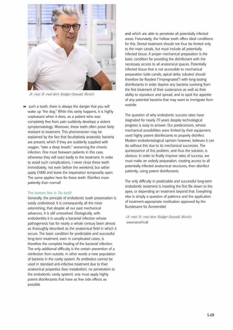
Dr. med. Dr. med dent. Rüdiger Osswald, Munich.
such a tooth, there is always the danger that you will wake up “the dog.” While this rarely happens, it is highly unpleasant when it does, as a patient who was completely free from pain suddenly develops a violent symptomatology. Moreover, these teeth often prove fairly resistant to treatment. This phenomenon may be explained by the fact that facultatively anaerobic bacteria are present, which if they are suddenly supplied with oxygen, “take a deep breath,” worsening the chronic infection. One must forewarn patients in this case, otherwise they will react badly to the treatment. In order to avoid such complications, I never close these teeth immediately, not even before the weekend, but rather apply ChKM and leave the trepanation temporarily open. The same applies here for these teeth: Disinfect more patiently than normal!
The bottom line is: Do both! Generally, the principle of endodontic tooth preservation is easily understood. It is consequently all the more astonishing, that despite all our past mechanical advances, it is still unresolved. Etiologically, with endodontitis it is usually a bacterial infection whose pathogenesis has for nearly a whole century been almost as thoroughly described as the anatomical field in which it occurs. The basic condition for predictable and successful long-term treatment, even in complicated cases, is therefore the complete healing of the bacterial infection. The only additional difficulty is the certain prevention of a reinfection from outside, in other words a new population of bacteria in the cavity system. As antibiotics cannot be used in standard anti-infective treatment due to their anatomical properties (low metabolism, no penetration to the endodontic cavity system), one must apply highly potent disinfectants that have as few side effects as possible
and which are able to penetrate all potentially infected areas. Fortunately, the hollow tooth offers ideal conditions for this. Dental treatment should not thus be limited only to the main canals, but must include all potentially infected tissue. A proper mechanical preparation is the basic condition for providing the disinfectant with the necessary access to all anatomical spaces. Potentially infected tissue that is not accessible to mechanical preparation (side canals, apical delta, tubules) should therefore be flooded (“impregnated”) with long-lasting disinfectants in order deprive any bacteria surviving from the first treatment of their sustenance as well as their ability to reproduce and spread, and to spoil the appetite of any potential bacteria that may want to immigrate from outside.
The question of why endodontic success rates have stagnated for nearly 70 years despite technological progress is easy to answer: Our predecessors, whose mechanical possibilities were limited by their equipment, used highly potent disinfectants to properly disinfect. Modern endodontological opinion however, believes it can do without this due to its mechanical successes. The quintessence of this problem, and thus the solution, is obvious: In order to finally improve rates of success, we must make an orderly preparation, creating access to all potentially infected anatomical structures, then disinfect patiently, using potent disinfectants.
The only difficulty in predictable and successful long-term endodontic treatment is inserting the first file down to the apex, or depending on treatment beyond that. Everything else is simply a question of patience and the application of treatment-appropriate medication approved by the Bundesamt für Arzneimittel.
-Dr. med. Dr. med dent. Rüdiger Osswald, Munich, www.tarzahn.de
5-09

Complicated gangrene The tooth is symptom free (dry gangrene) or exhibits pronounced clinical symptomatology, perio-endo lesions, high degree of mobility, pronounced apical osteitis, fistulas, broken-off instrument, clear buccal bone swelling, abscess, "sleeping dogs", etc.
� ChKM, cotton wool, (temporarily) open for one to two days, repeat severaltimes until the symptoms (e.g. bone swelling and mobility) decline, possibly apply antibiotic or make an incision if pain persists, or swelling or mobility does not improve.
� ChKM, cotton wool, Cavit for at least one week, orsometimes, if symptom-free, immediately.
� Depending on the size of the lesion treated and the complexity of therespective case, repeat several times.
� Iodoform paste, cotton wool, cement, for at least six, and at most 24 weeks, repeatseveral times in case of larger areas, with extreme cases confirm radiologically that the apical radiolucency is shrinking before applying the permanent filling
� Immobilization via thorough occlusal cutting until there is no occlusal contact. In thecase of high mobility, if possible splint via prosthetic ("that which is intended to heal must be carefully immobilized").
� With difficult cases in which the tooth will not be soothed, use the"magic paste" (see Part 4) (it can safely remain for up to three months)!
� If the patient should feel pain during one of the stages, go back a step and proceedmore thoroughly and with greater patience. After (nearly) each file and with each change in medication, pressure-free flushing with 3% H2O2.
� Should pus continue to empty from a “clean” tooth that has been sufficientlydisinfected, the cause for this lies in persisting (peri)apical osteitis. In this case, a prescription of Amoxicilin 1000 is recommendable (2 x 1 per day for at least five days), while chronic pain can be treated with Ibuprofen 600 alongside continued disinfection with ChKM or "magic paste," until the osteitis has become symptom free.
T H
E
I N D
I C
A T
I O
N -
S P
E C
I F I
C
T R
E A
T M
E N
T
O F
B A
C T
E R
I A
L
E N
D O
D O
N T
I T
I S
: T
H E
"T
I M
B U
K T
U -
P R
OT
O C
O L
“

List of literature The Indication-Specific Treatment of Bacterial Endodontitis The “Timbuktu Protocol” Section 5: Complicated Gangrene
1. Haapasalo, M., Endal, U., Zandi, H.,Coil, J.M.: Eradication of endodontic infection byinstrumentation and irrigation solutions. Endodontic Topics 10, 77 (2005).
2. Walkhoff, O.: Das Problem der dentalen Fokalinfektion und ihrer Bekämpfung durch diekonservierende Zahnheilkunde. Verlag von Gustav Fischer, Jena 1931.
3. Neelakantan, P., Subbarao, CV: An analysis of the antimicrobial activity of ten root canal sealers--aduration based in vitro evaluation. J Clin Pediatr Dent 33(2), (2008).
4. Susini, G., Pommel, L., About, I., Camps, J.: Lack of correlation between ex vivo apical dye penetration andpresence of apical radiolucencies. ORAL SURG ORAL MED ORAL PATHOL 3, 19 (2006).
5. Sjögren, U., Figdor, D., Persson, S., Sundqvist, G.: Influence of infection at the time of root filling on theoutcome of endodontic treatment of teeth with apical periodontitis. Int Endod J 30, 297 (1997).
6. Figdor, D.: Apical periodontitis: A very prevalent problem. Oral Surg Oral Med Oral Pathol 94, 651(2002).
7. Kojima, K., Inamoto, I.: Success rate of endodontic treatment of teeth with vital and nonvital pulps. Ametaanalysis. Oral Surg Oral Med Oral Pathol 95, 97 (2004).
8. Ng, Y., Mann, V., Rahbaran, S., Lewsey, J., Gulabivala, K.: Outcome of primary root canal treatment:systematic review of the literature - part 1. Effects of study characteristics on probability of success.Int Endod J 40, 921 (2007).
9. Ng, Y., Mann, V., Rahbaran, S., Lewsey, J., Gulabivala, K.: Outcome of primary root canal treatment:systematic review of the literature -- Part 2. Influence of clinical factors. Int Endod J 41, 6 (2008).
10. Wu, M., Dummer, P., Wesselink, P.: Consequences of and strategies to deal with residual post-treatment root canal infection. International Endodontic Journal, (2006).
11. Nair P. N. R. , H. S., Cano Victor, Vera Jorge: Microbial status of apical root canal system of humanmandibular first molars with primary apical periodontitis after ‘‘one-visit’’ endodontic treatment. ORALSURG ORAL MED ORAL PATHOL 99, (2005)
12. Lin S., P. O., Metzger Z., Tsesis I.: Residual bacteria in root apices removed by a diagonal root-endresection: a histopathological evaluation. J Endod 2008 May;34(5):511 34, 511 (2008).
13. Walkhoff, O.: Mein System der medikamentösen Behandlung schwerer Erkrankungen der Zahnpulpaund des Periodontiums. Verlag von Hermann Meuser, Berlin 1928.
14. Panitvisai, P., Parunnit, P., Sathorn, C., Messer, HH: Impact of a retained instrument on treatmentoutcome: a systematic review and meta-analysis. J Endod 36, 775 (2010).
15. McGuigan, M., Louca, C., Duncan, HF: Clinical decision-making after endodontic instrument fracture. BrDent J 214, 395 (2013).
16. Klimm, W.: Endodontolgie. Lehrbuch für Studium und Beruf 2. Auflage, (2010).
17. Stoll, R., Betke, K., Stachniss, V.: The influence of different factors on the survival of root canalfillings: a 10-year retrospective study. J Endod 31, 783 (2005).

List of instruments and drugs used:
A Mechanics: 1. Giromatic .......................... mechanical treatment (Micromega)2. Giro Files .......................... mechanical treatment (Micromega)3. Hedstrom files ................ mechanical treatment by hand4. Past Injekt ........................ obturation, iso 35 or 40 (Micromega)5. Hand spreader ............... to find obliterated root canals, initial channel expansion, revision6. Rotary NiTi files.............. revisions, therapeutic via falsa7. Trepan ............................... for the revision of the first straight part of root canals (Oral-Tronics)
B Disinfection, obturation: 1. H2O2, 3% ........................... rinsing solution after each instrument2. Ledermix paste .............. interappointment dressing for vital teeth3. Prof. Dr. Walkhoff's
ChKM-Solution ............... interappointment dressing (Adolf Haupt, Würzburg) 4. Ca(OH)2 .............................. interappointment dressing5. Endomethasone ............ obturation (Septodent)6. “Magic paste” ................. mixture of equal parts of N2 and Ledermix7. Standardized
gutta-percha points ..... obturation in single cone technique 8. Provis ................................. temporary closure9. Prof. Dr. Walkhoff's
Jodoformpaste ............... interappointment dressing (Adolf Haupt, Würzburg)