A Cerebral Central Pattern Generator in Aplysia and Its Connections ...
16
A Cerebral Central Pattern Generator in Aplysia and Its Connections with Buccal Feeding Circuitry Ray Perrins and Klaudiusz R. Weiss Department of Physiology and Biophysics, Mount Sinai School of Medicine, Mount Sinai Medical Center, New York, New York 10029-6574 Different feeding-related behaviors in Aplysia require substan- tial variations in the coordination of movements of two separate body parts, the lips and buccal mass. The central pattern generators (CPGs) and motoneurons that control buccal mass movements reside largely in the buccal ganglion. It was previ- ously thought that control of the cerebral neuronal circuitry and motoneurons that generate lip movements was coordinated directly by feedback from buccal interneurons. Here, we de- scribe cerebral lip motoneuron C15, which drives rhythmic activity in the isolated cerebral ganglion. Other lip motoneurons are active during this program, so we define it as a cerebral motor program (CMP). The C15 in each cerebral hemiganglion drives the CMP in ipsilateral neurons only, suggesting there are independent CPGs in each hemiganglion. The cerebral and buccal CPGs interact at several points. For example, cerebral- to-buccal interneurons (CBIs), which can drive the buccal CPG, receive excitatory input when the cerebral CPG is active. Like- wise, C15, which can drive the cerebral CPG, is excited when the buccal CPG is active. This excitation is simultaneous in both C15s, coupling the activity in the two hemiganglionic cerebral CPGs. Therefore, there are independent cerebral and buccal CPGs, which can produce distinct rhythms, but which interact at several points. Furthermore, the connections be- tween the cerebral and buccal CPGs alter during different forms of motor program. We suggest that such alterations in the interactions between these CPGs might contribute to the gen- eration of the various forms of coordination of lip and buccal mass movements that are necessary during different feeding- related behaviors. Key words: Aplysia; feeding; central pattern generator; motor program; command neuron; coupled oscillators Most animals need to alter the coordination of different body parts to produce a variety of behaviors. For example, the coordi- nation of different limbs changes during various locomotor gaits. It has been proposed, in several such systems, that there is series of individual central pattern generators (CPGs), one for each limb or joint (vertebrates: Grillner and Wallen, 1985; Rossignol et al., 1993; invertebrates: Mulloney et al., 1993; Ryckebusch and Lau- rent, 1994). These CPGs could interact in distinct ways to produce the different motor patterns needed to generate different gaits or even separate behaviors such as scratching. It should be noted that this is a different situation than that for chains of oscillators, which are coupled to produce undulatory swimming behaviors, as has been proposed in the leech (Friesen and Pearce, 1993) and lamprey (Sigvardt, 1993). In these systems, individual oscillators are largely identical, whereas in the examples for walking de- scribed above, each limb or joint CPG may have quite different intrinsic properties. Most progress in understanding the mechanisms of rearrange- ment of oscillatory networks has been accomplished in the crus- tacean stomatogastric nervous system (for review, see Dickinson and Moulins, 1992). In that system, four CPGs control body parts that are responsible for separate components of the ingestive behavior. Thus, although interactions do occur, the CPGs always produce rhythms with distinct cycle periods. In Aplysia, two body parts, the lips and radula, must be coordinated on a cycle-by-cycle basis during various feeding-related behaviors. In this paper, we show that, as postulated for locomotion, separate CPGs control each body part and that the interactions between these CPGs alter during different motor programs. The consumatory phase of feeding in Aplysia consists of rhyth- mic movements of the radula, which are coordinated with appro- priate opening and closing of the lips (Morton and Chiel, 1993a). The intrinsic buccal mass muscles that control the movement of the radula are innervated by motoneurons in the buccal ganglion (Cohen et al., 1978; Church and Lloyd, 1994). Firing of these motoneurons during feeding-related behaviors is controlled by CPGs, the neuronal circuitry for which appears to reside largely in the buccal ganglion (Weiss et al., 1982; Susswein and Byrne, 1988; Kirk, 1989). Motoneurons innervating lip muscles are located in the cerebral ganglion (Chiel et al., 1986; Rosen et al., 1989). It was previously considered likely that behaviorally appropriate output from these cerebral motoneurons was coordinated from the buc- cal ganglion. Each cerebral motoneuron was thought to be di- rectly activated by a number of buccal-to-cerebral interneurons (BCIs) (Chiel et al., 1988; Teyke et al., 1993; Rosen et al., 1991a). Here, we present evidence that each cerebral hemiganglion contains a CPG that can produce a cerebral motor program (CMP) in isolated cerebral ganglia. The cerebral CPGs provide an alternative means of activating lip motoneurons to the direct control by BCIs. We characterize neuron C15, which drives the CMP, describe basic features of the CMP, including synaptic drive to identified neurons, and demonstrate that different forms of Received June 10, 1996; revised Aug. 2, 1996; accepted Aug. 9, 1996. This work was supported by Human Frontiers Science Program Grant LT-561/95 and National Institutes of Health Grants GM 32009 and MH 50235. We thank Drs. E. C. Cropper and C. G. Evans for comments on earlier versions of this manuscript. Correspondence should be addressed to Ray Perrins, Department of Physiology and Biophysics, Mount Sinai School of Medicine, Mount Sinai Medical Center, One Gustave L. Levy Place, New York, NY 10029-6574. Copyright q 1996 Society for Neuroscience 0270-6474/96/167030-16$05.00/0 The Journal of Neuroscience, November 1, 1996, 16(21):7030 –7045
Transcript of A Cerebral Central Pattern Generator in Aplysia and Its Connections ...

A Cerebral Central Pattern Generator in Aplysia and ItsConnections with Buccal Feeding Circuitry
Ray Perrins and Klaudiusz R. Weiss
Department of Physiology and Biophysics, Mount Sinai School of Medicine, Mount Sinai Medical Center, New York,New York 10029-6574
Different feeding-related behaviors in Aplysia require substan-tial variations in the coordination of movements of two separatebody parts, the lips and buccal mass. The central patterngenerators (CPGs) and motoneurons that control buccal massmovements reside largely in the buccal ganglion. It was previ-ously thought that control of the cerebral neuronal circuitry andmotoneurons that generate lip movements was coordinateddirectly by feedback from buccal interneurons. Here, we de-scribe cerebral lip motoneuron C15, which drives rhythmicactivity in the isolated cerebral ganglion. Other lip motoneuronsare active during this program, so we define it as a cerebralmotor program (CMP). The C15 in each cerebral hemigangliondrives the CMP in ipsilateral neurons only, suggesting there areindependent CPGs in each hemiganglion. The cerebral andbuccal CPGs interact at several points. For example, cerebral-to-buccal interneurons (CBIs), which can drive the buccal CPG,
receive excitatory input when the cerebral CPG is active. Like-wise, C15, which can drive the cerebral CPG, is excited whenthe buccal CPG is active. This excitation is simultaneous inboth C15s, coupling the activity in the two hemiganglioniccerebral CPGs. Therefore, there are independent cerebral andbuccal CPGs, which can produce distinct rhythms, but whichinteract at several points. Furthermore, the connections be-tween the cerebral and buccal CPGs alter during different formsof motor program. We suggest that such alterations in theinteractions between these CPGs might contribute to the gen-eration of the various forms of coordination of lip and buccalmass movements that are necessary during different feeding-related behaviors.
Key words: Aplysia; feeding; central pattern generator; motorprogram; command neuron; coupled oscillators
Most animals need to alter the coordination of different bodyparts to produce a variety of behaviors. For example, the coordi-nation of different limbs changes during various locomotor gaits.It has been proposed, in several such systems, that there is seriesof individual central pattern generators (CPGs), one for each limbor joint (vertebrates: Grillner and Wallen, 1985; Rossignol et al.,1993; invertebrates: Mulloney et al., 1993; Ryckebusch and Lau-rent, 1994). These CPGs could interact in distinct ways to producethe different motor patterns needed to generate different gaits oreven separate behaviors such as scratching. It should be noted thatthis is a different situation than that for chains of oscillators, whichare coupled to produce undulatory swimming behaviors, as hasbeen proposed in the leech (Friesen and Pearce, 1993) andlamprey (Sigvardt, 1993). In these systems, individual oscillatorsare largely identical, whereas in the examples for walking de-scribed above, each limb or joint CPG may have quite differentintrinsic properties.Most progress in understanding the mechanisms of rearrange-
ment of oscillatory networks has been accomplished in the crus-tacean stomatogastric nervous system (for review, see Dickinsonand Moulins, 1992). In that system, four CPGs control body partsthat are responsible for separate components of the ingestivebehavior. Thus, although interactions do occur, the CPGs always
produce rhythms with distinct cycle periods. In Aplysia, two bodyparts, the lips and radula, must be coordinated on a cycle-by-cyclebasis during various feeding-related behaviors. In this paper, weshow that, as postulated for locomotion, separate CPGs controleach body part and that the interactions between these CPGs alterduring different motor programs.The consumatory phase of feeding in Aplysia consists of rhyth-
mic movements of the radula, which are coordinated with appro-priate opening and closing of the lips (Morton and Chiel, 1993a).The intrinsic buccal mass muscles that control the movement ofthe radula are innervated by motoneurons in the buccal ganglion(Cohen et al., 1978; Church and Lloyd, 1994). Firing of thesemotoneurons during feeding-related behaviors is controlled byCPGs, the neuronal circuitry for which appears to reside largely inthe buccal ganglion (Weiss et al., 1982; Susswein and Byrne, 1988;Kirk, 1989). Motoneurons innervating lip muscles are located inthe cerebral ganglion (Chiel et al., 1986; Rosen et al., 1989). It waspreviously considered likely that behaviorally appropriate outputfrom these cerebral motoneurons was coordinated from the buc-cal ganglion. Each cerebral motoneuron was thought to be di-rectly activated by a number of buccal-to-cerebral interneurons(BCIs) (Chiel et al., 1988; Teyke et al., 1993; Rosen et al., 1991a).Here, we present evidence that each cerebral hemiganglion
contains a CPG that can produce a cerebral motor program(CMP) in isolated cerebral ganglia. The cerebral CPGs provide analternative means of activating lip motoneurons to the directcontrol by BCIs. We characterize neuron C15, which drives theCMP, describe basic features of the CMP, including synaptic driveto identified neurons, and demonstrate that different forms of
Received June 10, 1996; revised Aug. 2, 1996; accepted Aug. 9, 1996.This work was supported by Human Frontiers Science Program Grant LT-561/95
and National Institutes of Health Grants GM 32009 and MH 50235. We thank Drs.E. C. Cropper and C. G. Evans for comments on earlier versions of this manuscript.Correspondence should be addressed to Ray Perrins, Department of Physiology
and Biophysics, Mount Sinai School of Medicine, Mount Sinai Medical Center, OneGustave L. Levy Place, New York, NY 10029-6574.Copyright q 1996 Society for Neuroscience 0270-6474/96/167030-16$05.00/0
The Journal of Neuroscience, November 1, 1996, 16(21):7030–7045
behaviorally relevant motor programs are accompanied bychanges in the interactions between cerebral and buccal CPGs.
MATERIALS AND METHODSExperiments were performed at 158C on Aplysia californica weighing100–300 gm. Animals were anaesthetized by an injection of isotonicMgCl2 (50% of body weight) into the body cavity. The cerebral ganglionwas dissected out in isolation for some experiments and removed with thebuccal ganglion attached for others (see Results for details). The gangliawere pinned onto a SYLGARD-coated dish at room temperature inartificial sea water (ASW). Cerebral ganglia were pinned ventral-surface-up and buccal ganglia rostral-surface-up. The connective tissueoverlying the uppermost surface of the ganglia was surgically removed.The cerebral–pedal connective was rotated through 1808 and repinned toallow access to the lateral area of the E-cluster. Simultaneous intracel-lular recordings were made from up to four neurons using single ordouble-barreled microelectrodes filled with 2 M potassium acetate andbeveled to a resistance ranging from 5 to 10 MV, depending on the sizeof the cell to be penetrated. In preparations where the buccal ganglionwas attached, buccal motor output was monitored using a polyethylenesuction electrode placed over buccal nerve 2 (bn2) (see Morton andChiel, 1993a) and connected to an AC amplifier. A recording or stimu-lating suction electrode was placed over the end of the severed cerebral-to-buccal connective (CBC) in experiments involving isolated cerebralganglia.To help identify previously described cells and to reveal the morphology of
new ones, microelectrodes, beveled to a resistance of 10–15 MV, were filledwith a 3% solution of 5(6)-carboxyfluorescin in 0.1 M potassium citrate,titrated to pH 8.0 with KOH (see Rao et al., 1986). At the end of eachexperiment, previously described cells were identified on the basis of theirmorphology and known synaptic connections. In some experiments, polysyn-aptic pathways were suppressed using solutions containing elevated levels ofdivalent cations (with both Ca21 and Mg21 at three times the normalconcentration) to raise neuronal firing thresholds.To determine possible peripheral functions of C15, experiments were
performed on preparations in which the cerebral ganglion was removedwith the lips and anterior tentacles still attached by their peripheralnerves (cf. Rosen et al., 1979). The ganglion was pinned on an elevatedSYLGARD platform, ventral-surface-uppermost, and desheathed as de-scribed above. The head structures were pinned, ventral-surface-uppermost, to the bottom of the SYLGARD-coated dish in such a way asto reveal the inner portions of the lips and perioral zone (Rosen et al.,1982). The anterior aorta was cannulated and perfused with ASW.Salines, all at pH 7.6, were composed as follows. ASW: NaCl, 460 mM;
KCl, 10 mM; CaCl2, 11 mM; MgCl2, 55 mM; NaHCO3, 5 mM. High divalentsaline (33 Mg21, 33 Ca21): NaCl, 460 mM; KCl, 10 mM; CaCl2, 33 mM;MgCl2, 165 mM; NaHCO3, 5 mM. For experiments in which chemicaltransmission was blocked, the Ca21 was omitted and replaced with 10 mMCo21. Experiments were performed at least six times, unless otherwisestated. Figures quoted are means 6 SE.
RESULTSMorphology and synaptic connections of C15We have discovered a neuron, C15, which was uniquely identifiedon the basis of its physiological and morphological properties.There was a single C15 in each half of the cerebral ganglion. It wasa medium-sized oval neuron (;100–150 mm in diameter) situatedmedially on the ventro-lateral surface of the E-cluster (Jahan-Parwar and Fredman, 1976). The approximate positions of C15and other cerebral neurons used in this study are shown schemat-ically in Figure 1A. Injections of 5(6)-carboxyfluorescin into C15revealed two peripheral axons, leaving the upper labial (ULAB)and anterior tentacular (AT) nerves (Fig. 1B). Dendritic arbori-sations were present in the E-cluster at the base of the CBC andin the M-cluster (Ono and McCaman, 1980). Processes werelimited to the ipsilateral half of the ganglion.In preparations in which the head structures were left con-
nected to the cerebral ganglion, firing C15 at frequencies above 8Hz produced a radial contraction in the tissue immediately adja-cent to the anterior perioral zone of the ipsilateral inner lips. This
resulted in an opening of the anterior portion of the jaws (n 5 5).This contraction was observed before the occurrence of largeIPSPs in C12, which would indicate the start CMP (see below).Firing C15 at these frequencies also readily produced the contrac-tion while the whole preparation was bathed in solution contain-ing 33 normal Ca21 and Mg21 (to suppress polysynaptic path-ways), conditions under which the CMP was completely abolished.This means that the contraction was likely to be a direct effect offiring C15, which can therefore be described as a probable lipmotoneuron. Mechanical or chemical (seaweed; Laver, Vegatrading company, NY) stimuli applied to the lips, rhinophores, ortentacles had no effect on the membrane potential of C15, so wehave no evidence for any sensory functions.Two neighboring E-cluster neurons, C16 and C17, which are
smaller and generally more rostral than C15, were also character-ized in this study. C16 and C17 had physiological properties andsynaptic inputs (see below) which were indistinguishable fromC15. These neurons produced radial contractions in the tissue ofthe ipsilateral inner lips that persisted in high divalent solutions,like C15, so are also probable lip motoneurons. While C15 openedthe anterior part of the jaws, C16 opened the medial region, andC17 the posterior. Firing all of these motoneurons together in thesemi-intact preparation resulted in full opening of the ipsilateraljaws along their entire length. The motoneurons could also bedistinguished from each other by the ability of C15 to reliablydrive the CMP and, more important, by their morphology, re-vealed by injection of 5(6)-carboxyfluorescin. Not only were C16and C17 generally smaller than C15, but they sent axons to theperiphery via different combinations of nerves. C16 had axons inthe AT and lower labial (LLAB) nerves and C17 had a single axonin the LLAB (Fig. 1C,D). These three motoneurons were electri-cally coupled to each other. Either a hyperpolarizing or a depo-larizing current injected into C15, C16, or C17 resulted in achange of the same polarity but reduced amplitude in the mem-brane potential of the other two neurons (Fig. 2A). The couplingratios (the voltage change in the postsynaptic neuron divided bythe voltage change in the neuron into which current was injected)were between 0.1 and 0.2. Apparent coupling was higher whencurrent was injected into C15 (Fig. 2A), possibly because of itslarger size. Because C2 produced an IPSP in these neurons (seebelow), C15, C16, and C17 probably correspond to the Ea clusterof neurons of Chiel et al. (1986). These were described as inhib-itory followers of C2, putative motoneurons for the lips that werein approximately the same position in the E-cluster.C15 received chemical input from several cerebral and buccal
neurons. The histaminergic mechanoafferent C2, which may playan important role in several feeding-related behaviors (Chiel etal., 1986), inhibited C15 (Fig. 2B) as well as C16 and C17. C15also received input from several buccal-to-cerebral interneurons(BCIs: B18, B19, and B24), which are responsible for directfeedback to cerebral neurons during BMPs (Chiel et al., 1988;Rosen et al., 1990; Rosen et al., 1991a; Teyke et al., 1993). Spikesin B18 were followed one-for-one by fast EPSPs in C15, whereasfiring in B19 produced a rapid inhibition in C15 followed by a slowexcitation and B24 produced just an inhibitory response (Fig.2C–E). All of these responses persisted in salines containing 33normal Ca21 and Mg21. Inputs from B18, B19, and B24 onto C16and C17 were similar to those onto C15. There appeared to be nomonosynaptic connections onto C15, C16, and C17 from anotherBCI, B20 (Teyke et al., 1993), although they all received excitatoryinput during the BMP driven by that cell.
Perrins and Weiss • Interacting Central Pattern Generators in Aplysia J. Neurosci., November 1, 1996, 16(21):7030–7045 7031

C15 can drive a motor program in the isolatedcerebral ganglionAt its resting potential (244.4 6 3.8 mV, n 5 15) C15 usuallyreceived no large spontaneous synaptic inputs and was silent (e.g.,Figs. 3A, 4, 8C) or weakly tonically active (e.g., Figs. 3B, 8A,B).Some low-amplitude spontaneous input was present, because theresting membrane potential showed less short-term variations inthe presence of 33 Ca21, 33 Mg21, which presumably reducedpresynaptic spiking activity (Fig. 2D,E). When C15 was fired atfrequencies above 11–12 Hz, rhythmic synaptic activity with acycle period of between 9 and 50 sec was recorded in a variety ofE- and M-cluster neurons (Figs. 3, 4, 8). In 25 out of 30 prepara-tions, C15 produced repeating cycles of the program. In theremaining cases, only a single cycle was initiated, despite contin-ued high-frequency firing of C15. Fast PSPs observed in someneurons (see below) were not one-for-one with spikes in C15 and
they were completely abolished by 33 Ca21, 33 Mg21. Thisindicates that the responses detailed below were not monosynap-tic from C15, but arose through a polysynaptic pathway, after theactivation of a cerebral CPG. The rhythmic activity could beinduced in the isolated cerebral ganglion and, because it incorpo-rated firing of lip motoneurons (see below) at frequencies thatproduce lip movements, it can be defined as a cerebral motorprogram (CMP). The CMP stopped within 100 msec, after cessa-tion of current injection in C15, even if this occurred in the middleof a cycle. Spontaneous CMPs were never observed. C15 couldreliably drive the CMP in preparations in which the cerebralganglion had been isolated for up to 36 hr. C16 and C17 couldoccasionally drive a single cycle of the CMP (4 of 25 prepara-tions), but never repeating cycles, unless both were depolarizedtogether, and interactions of this sort were not thoroughlyinvestigated.
Figure 1. Position and morphology of ce-rebral motoneurons C15, C16, and C17. A,Schematic diagram of the ventral surfaceof a cerebral hemiganglion, showing theapproximate positions of neurons in theE- and M-clusters used in this study. Filledcells indicate lip motoneurons. B, Drawingof the soma and central processes of C15filled by intracellular injection of 5(6)-carboxyfluorescin and viewed with a fluo-rescence microscope. C15 had arborisa-tions in the E- and M-clusters andperipheral axons in the ULAB and ATnerves. C, Morphology of C16 was similarto that of C15, except it had axons in theAT and LLAB nerves. D, Morphology ofC17, which had a single axon in the LLAB.Arrowheads indicate nerves in which thereare peripheral axons. ULAB, Upper labialnerve; AT, anterior tentacular nerve;LLAB, lower labial nerve; CBC, cerebral–buccal connective; CPC, cerebral–pedalconnective; CPLC, cerebral–pleuralconnective.
7032 J. Neurosci., November 1, 1996, 16(21):7030–7045 Perrins and Weiss • Interacting Central Pattern Generators in Aplysia

To characterize more fully the CMP, we investigated the inputreceived by a variety of identified cerebral neurons. Lip motoneu-rons C11 and C12 were active during the CMP (Fig. 3A). Firing inC11 was caused by barrages of EPSPs, whereas firing in C12relied, in part at least, on postinhibitory rebound from a barrageof IPSPs, because the cell could also fire a burst of spikes at theend of a negative current pulse. Lip motoneurons C16 and C17received large, fast EPSPs that drove high-frequency firing (Fig.3B). Because of the electrical coupling of these neurons to C15,they were also tonically depolarized throughout the period ofcurrent injection in C15. C15 itself also received rhythmic input(open bar in Fig. 3B), which caused higher-frequency spiking(15–20 Hz) during those periods. In most records, the exact natureof this input was obscured by the current-induced spikes but, incases where the CMP continued for a few tens of millisecondsafter the current pulse, the input could be seen to consist of fastEPSPs, similar to those seen in C16 and C17.In addition to motoneurons, we also recorded from a sample of
previously identified sensory and modulatory cerebral neurons
that participate in feeding motor programs (Chiel et al., 1986).The E-cluster neuron C4 can modulate contractions of extrinsicbuccal mass muscles produced by cerebral motoneurons (Chiel etal., 1986). During the CMP, C4 received large EPSPs that droveit above threshold (Fig. 4A). C5, which is neighboring C4 butwhose function is unclear, received relatively small EPSPs, one-for-one with those in C4, but these had little influence on its firingrate (Fig. 4A). The giant serotonergic metacerebral cell (MCC),which exerts modulatory influences both centrally and peripher-ally during feeding (Weiss et al., 1978; Rosen et al., 1989), re-ceived rhythmic weak, slow excitation but did not fire (Fig. 4B).The histaminergic mechanosensory neuron C2, which has a pow-erful influence over many neurons involved in the cerebral feedingmotor circuitry (Chiel et al., 1986), received fast IPSPs (Fig. 4B).Pairwise recordings revealed that the fast PSPs described above
occur synchronously in all neurons, suggesting a common source(see expanded time-base records in Figs. 3, 4A). Occasionally,there were also fast EPSPs in the MCC, associated with the peakof the slow depolarization, but these were not one-for-one with
Figure 2. Synaptic connections ontoC15. A, Lip motoneurons C15, C16, andC17 were electrically coupled to eachother. Negative current injected into anyof the three cells (solid bars) produced asmaller hyperpolarization in the othertwo. B, Firing in the histaminergic mech-anoafferent neuron C2 produced slow in-hibition in C15. C–E, Spikes in BCIs B18,B19, and B24 produced, respectively, fastone-for-one EPSPs, an I/EPSP, and anIPSP in C15. These responses all per-sisted in a solution containing high con-centrations of divalent cations, which sup-presses polysynaptic pathways. Scale bar:vertical5 40 mV except C15 in C–E5 15mV; horizontal 5 1 sec for C, 2 sec for A,10 sec for B, D, and E.
Perrins and Weiss • Interacting Central Pattern Generators in Aplysia J. Neurosci., November 1, 1996, 16(21):7030–7045 7033

the PSPs in the other cells (Fig. 4B). Only cells in the ipsilateralhalf of the ganglion appear to receive synaptic input during theCMP, because no rhythmic activity could be recorded contralat-erally (Fig. 11A). There were only two discernible phases duringthe CMP, one being characterized by the presence of fast PSPs inthe neurons described above and the other by firing in C12 and noobservable input to any other neurons. The CMP, therefore,appears to be a less complex rhythm than the usually multi-phase BMPs.
Input to buccal motoneurons during the CMPIn preparations with the buccal ganglion attached, a CMP thatappeared identical to that produced in the isolated cerebral gan-glion could still be driven by C15. Several identified cerebral-to-buccal interneurons (CBIs) fire during the CMP (see below) andare known to make synapses onto buccal motoneurons (Rosen etal., 1991b), so one might expect feedforward synaptic inputs fromthese neurons. Spiking of cerebral-to-buccal neurons was alsomonitored in isolated cerebral ganglia by an extracellular suction
electrode placed on the CBC. An increase in activity recorded inthis electrode was always observed during the phase of the CMPin which fast PSPs occurred in C2, C4, C5, C11, C12, C16, andC17. This might be explained, in part, by the increase in spikingactivity observed in CBI-2, CBI-3, and CBI-4 (described below).However, there was also a series of spikes in the CBC that wereone-for-one with the fast PSPs in C12 (Fig. 5A). Firing in theidentified CBIs occurred at a later phase than these spikes andwas never one-for-one with the individual fast PSPs seen in otherneurons (Figs. 8, 9B2,C2). This means there exists an as yetunidentified cerebral-to-buccal neuron that is responsible for thisactivity. Intracellular recordings from the buccal ganglion re-vealed that a number of neurons in the ventral motoneuroncluster received fast IPSPs during the CMP that were one-for-onewith the fast PSPs in the cerebral ganglion (Fig. 5B). This suggeststhat the unidentified cerebral-to-buccal neuron makes synapseswithin the buccal ganglion, providing another source of input tobuccal neurons, in addition to those arising via identified CBIs.
Figure 3. Input to cerebral lip motoneurons during the CMP in isolated cerebral ganglia. The CMP was driven by injecting constant depolarizing currentinto C15 (solid bars). A, M-cluster lip motoneurons C11 and C12 were active in alternation. C11 received EPSPs and C12 IPSPs. B, E-cluster lipmotoneurons C16 and C17 both received EPSPs during the CMP and fired at high frequency. C16 and C17 were electrically coupled to C15 (and eachother; see Fig. 2A) and so were also tonically depolarized throughout the current pulse. Arrows indicate points on the slow record from which the fastertime-base records to the right were taken. These show that the PSPs in all neurons were one-for-one with PSPs in the others (examples joined by dashedlines), suggesting a common source. C15 also received phasic excitatory input during the CMP (e.g., open bar in B) that was largely masked by thecurrent-induced spiking but was represented by a rise in the firing frequency and uneven spike amplitudes.
7034 J. Neurosci., November 1, 1996, 16(21):7030–7045 Perrins and Weiss • Interacting Central Pattern Generators in Aplysia

The contribution of C15 to the CMPWe have shown that C15 is sufficient to generate the CMP. Toassess more fully the contribution of C15 to the operation of thecerebral CPG, we investigated if C15 firing was also necessary forthe CMP (Kupfermann and Weiss, 1978). We therefore searchedfor other ways of driving the CMP to examine this possibility. InPleurobranchaea, it has been shown that high-frequency stimula-tion of the CBC can drive a rhythm in the isolated cerebralganglion (Davis et al., 1973; Cohan and Mpitsos, 1983), so weattempted the equivalent experiment in Aplysia. We found thatstimulating the CBC at frequencies above 10 Hz resulted inrhythmic input to cerebral neurons (Fig. 6). Because lip motoneu-rons C11, C15, C16, and C17 were active during this rhythm, thiscan also be classified as a CMP. All of the recorded neurons hadthe same category of input during both C15- and CBC-drivenCMPs. Thus, C4, C5, C11, C15, C16, and C17 all received fastEPSPs, whereas C2 and C12 received fast IPSPs. The cycle periodof the CBC-driven CMP was 20–60 sec, which overlaps the valuesfor the C15-driven rhythm. As with the C15-driven CMP, thesource of these PSPs appeared to be an unidentified cerebral-to-
buccal neuron, because the PSPs followed CBC stimulation with afixed latency, regardless of stimulation intensity or frequency (Fig.7A). Identified CBIs were ruled out as candidates because eitherno antidromic spikes were observed at the stimulus intensity used(CBI-2 and CBI-4) or blocking these spikes by hyperpolarizationhad no effect on the rhythm (CBI-1 and CBI-3). Because of thesesimilarities, the CMPs driven by C15 depolarization or CBCstimulation appear to be very closely related and are likely to beproduced by the same CPG. To test whether spiking in C15 isnecessary for the occurrence of the CMP, we hyperpolarized itduring the CBC-driven CMP to a level at which it no longerspiked. Under these conditions, the CMP could still be driven,suggesting that spiking in C15 is not necessary for the rhythm (n56; Fig. 6B).Using CBC stimulation, we were also able to investigate the
nature of the possible connection between C15 and the unidenti-fied cerebral-to-buccal neuron. For these experiments, the CBCwas stimulated at low frequency (1–2 Hz, insufficient to induce theCMP) at an intensity just suprathreshold for the PSPs in C15, C12,etc. Under these conditions, hyperpolarizing C15 increased the
Figure 4. Input to modulatory and sensory neurons during the C15-induced CMP in isolated cerebral ganglia. A, Modulatory neuron C4 received largeEPSPs that drove it above threshold during the CMP, whereas its neighbor C5 received much smaller EPSPs that were one-for-one with those in C4 (seerecord to the right, showing input at an expanded time-base). These EPSPs had little effect on the firing rate of C5. B, The sensory mechanoafferent C2received fast IPSPs during the CMP. The MCC, which has both central and peripheral modulatory roles, was slowly depolarized. There were also a fewfaster potentials in the MCC associated with the peak of some of the cyclical depolarizations, which were not one-for-one with the PSPs in other neurons(see record to the right). Arrows indicate points on the slow record from which the faster time-base records to the right were taken.
Perrins and Weiss • Interacting Central Pattern Generators in Aplysia J. Neurosci., November 1, 1996, 16(21):7030–7045 7035

amplitude of the EPSP in that cell, without affecting the ampli-tude of PSPs in other cells (Fig. 7B1). However, we found that byfurther hyperpolarizing C15, we were able to block the PSPs inC15 as well as the PSPs in other neurons (Fig. 7B2). This suggeststhat C15 is electrically coupled to the unidentified cerebral-to-buccal neuron so that the hyperpolarization in C15 is transferredelectrotonically, sufficiently hyperpolarizing the terminals of theother neuron to prevent transmitter release. It should be notedthat a chemical synapse from the cerebral-to-buccal neuron ontoC15 also exists, producing the EPSPs during the CMP, and that achemical synapse in the other direction cannot be ruled out yet.The electrical connection between C15 and the cerebral-to-buccalneuron means that although spiking in C15 is not necessary for theCMP, a certain level of membrane potential is necessary. Thus, ifC15 is strongly hyperpolarized (to presumably nonphysiologicallevels), it blocks the PSPs that follow CBC stimulation and,therefore, no program can be generated.
Connections between cerebral and buccal CPGsCerebral motoneurons receive input during BMPs (Rosen et al.,1991b) (see Figs. 10, 11), and we have shown above that buccalmotoneurons receive input during the CMP (Fig. 5). We alsowished to characterize possible interactions between the cerebraland buccal CPGs themselves. To this end, we investigatedwhether neurons that are important during the operation of onetype of program (either the CMP or various BMPs) received inputduring the other program. In Aplysia, we found the following. (1)Four identified cerebral-to-buccal interneurons (CBIs) are impor-tant in the control of BMPs (Rosen et al., 1991b) (see Fig. 10). Wetherefore investigated input received by these neurons during theCMP. (2) C15 is important in the control of the CMP (because itis sufficient to drive it and receives excitatory input during theprogram). We therefore investigated input received by this neu-ron during BMPs.
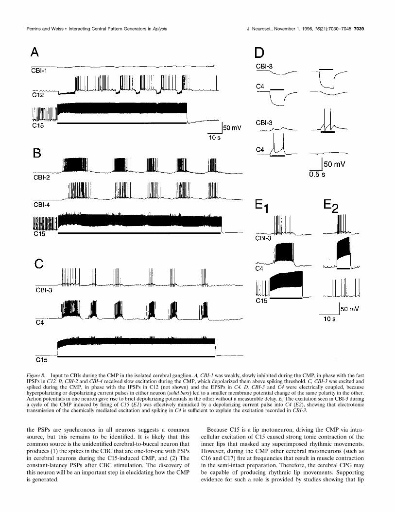
Inputs to CBIs during the CMPCBI-1 received weak, slow inhibition during the CMP (Fig. 8A),which was not studied further. CBI-2 and CBI-4 both receivedslow excitation, which was usually sufficient to produce spiking(Fig. 8B, 9B,C). Because firing C15 produced this excitation via apolysynaptic pathway (because the CMP was blocked in highdivalent solutions), a detailed examination of the excitation inCBI-2 and CBI-4 was not attempted. However, it was apparentthat in both cases the amplitude of the excitation was decreased byhyperpolarization of the CBIs (see Fig. 9B,C). CBI-3 was alsoexcited during the CMP (Fig. 8C). We found that CBI-3 wasstrongly electrically coupled to C4. Injecting either a positive or anegative current into either CBI-3 or C4 altered the membranepotential of the other (Fig. 8D), the coupling ratio being 0.20–0.25. Spikes in one cell produced transient depolarizations of theother with zero latency. Bathing the ganglion in ASW with 10 mMCo21 had no effect on these potentials, so there appears to be nochemical connection. The excitation of CBI-3 during the CMPcould be effectively mimicked by positive current injection into C4(Fig. 8E). This suggests that the excitation of CBI-3 during theCMP was largely attributable to electrotonic transfer of the chem-ical EPSPs and spiking in C4 described above. The input to all ofthe CBIs occurred during the same phase as the fast PSPs in thecerebral neurons described previously, although the firing inCBI-2 and CBI-4 was somewhat delayed and lasted longer, pre-sumably because of the slow nature of the excitation.In preparations with cerebral and buccal ganglia connected, the
same inputs were observed for each of the CBIs during the CMP.Inducing the CMP by exciting C15 most often had little effect onbuccal motor output, monitored via the extracellular recordingelectrode on bn2 (Figs. 9B,C, 14). However, in a significantnumber of preparations (5 of 23) single cycles of a BMP (definedas strong, multiphasic activity in bn2 and high-frequency firing inB4) were associated with the first cycle of the CMP (Fig. 9A). Inonly one case did consecutive cycles of the CMP produce repeat-ing cycles of a BMP. Both CBI-2 and CBI-4 can be excited beyondthreshold during the CMP. Because spiking in either CBI-2 orCBI-4 can drive BMPs (Rosen et al., 1991b) (see Fig. 10), this isa possible pathway for the generation of BMPs. This was tested byaltering the membrane potential of CBI-2 and CBI-4 in prepara-tions with the buccal ganglion attached. In the six preparationsinvestigated in this manner, BMPs were not induced by C15stimulation with all other neurons at their resting potentials.However, a full cycle of a BMP could be generated by slightlydepolarizing CBI-2 or CBI-4, thus increasing the frequency atwhich they fired during the CMP (Fig. 9B,C). These results arenot direct evidence that the CMP occasionally generates BMPs viaexcitation of CBI-2 and CBI-4 (or the combined action of both),but do show that this pathway is feasible.
Input to C15 during BMPsIn cerebral–buccal ganglia preparations, we also investigated theinput to C15 during BMPs evoked by a variety of means. Duringthe ingestive-like BMP driven by positive current injection intoCBI-2 (cf. Rosen et al., 1991b), C15 received fast EPSPs thatdrove spiking at high frequencies (maximum frequency sustainedover 1 sec 5 24.5 6 1.3 Hz, n 5 6; Figs. 10A, 11B, 12A). C15 wasalso excited, although more weakly (maximum firing frequency,14.3 6 2.3 Hz, n 5 5) during egestive-like BMPs (Fig. 10B,C).These were driven by a brief burst of high-frequency (10 Hz)electrical shocks to the radula nerve or by constant stimulation ofthe esophageal nerve at 2–3 Hz (Susswein and Byrne, 1988). C15
Figure 5. Feedback to the buccal ganglion during the CMP. A, A time-expanded record of the start of one cycle of a CMP. A series of extracel-lularly recorded spikes in the CBC were one-for-one with the IPSPs in C12(examples joined by lines). Because these were not attributable to spikingin any of the identified CBIs (see Results), these are the result of activityin an as yet uncharacterized cerebral-to-buccal neuron. There was also ageneral increase in the frequency of spikes in the CBC (examples atarrowheads), possibly because of spiking in CBI-2, 3, and 4 (Fig. 8). B, Atime-expanded record of the start of one cycle of a CMP. FacilitatingIPSPs in an unidentified neuron (b MN ) in the ventral motoneuron clusterof the buccal ganglion were one-for-one with IPSPs in C12, suggesting acommon source, probably the unidentified cerebral-to-buccal neuron. Inboth cases, the CMP was driven by constant current injected into C15 (notshown). Scale bar 5 40 mV for C12, 10 mV for b MN, and 250 msec forA, 500 msec for B.
7036 J. Neurosci., November 1, 1996, 16(21):7030–7045 Perrins and Weiss • Interacting Central Pattern Generators in Aplysia

was also excited during BMPs driven by both CBI-1 (maximumfiring frequency of C15 5 19.4 6 2.7 Hz, n 5 5; Fig. 13A) andCBI-4 (maximum firing frequency 5 16.0 6 1.5 Hz, n 5 6; Figs.10D, 13B). During BMPs initiated by any means, C15 was alsooccasionally inhibited between bursts (arrowheads, Figs. 10D,11B). The CMP was strictly limited to one-half of the cerebralganglion (Fig. 11A), but the general timing of excitatory inputs toboth right and left C15s was highly synchronized during BMPs(Fig. 11B), although individual EPSPs were not alwaysone-for-one.Although qualitatively similar (consisting of fast EPSPs), the
input to C15 was not quantitatively the same for each variety ofBMP. The firing rate of C15 was significantly higher ( p , 0.01)during the BMP induced by CBI-2 (mean firing rate measuredover the entire burst was 19.3 6 0.7 Hz, n 5 8) than that duringCBI-1 or CBI-4 induced BMPs (9.9 6 1.6 Hz, n 5 5 and 9.5 6 0.9Hz, n 5 6, respectively). Both of these firing rates were again
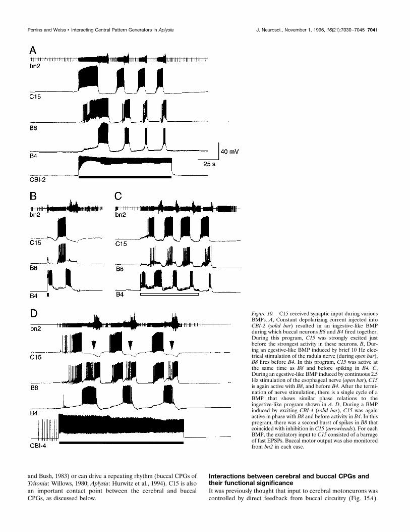
significantly higher ( p , 0.01) than during egestive-like BMPs(4.3 6 1.0 Hz, n 5 6). Note that the maximum firing ratesdescribed above were also different between the various BMPs.C15 was active during protraction for each type of BMP. Thus,
during ingestive BMPs (Morton and Chiel, 1993b), such as thatdriven by CBI-2, buccal motoneurons B8 and B4 are active at thesame time (Rosen et al., 1991b; Church and Lloyd, 1994). Duringthis program, C15 was active before B8/B4 (Fig. 10A), duringprotraction, and at the same time as “p-group” of neurons ofChurch and Lloyd (1994). During egestive-like motor programs,B8 and B4 are active out of phase (Morton and Chiel, 1993b;Church and Lloyd, 1994). In this program, C15 fires at the sametime as B8 and stops before firing in B4 (Fig. 10B,C). This is alsoduring protraction, but in this case at the same time as “c-group”neurons of Church and Lloyd (1994). C16 and C17 receivedsimilar input and were active at similar frequencies and with thesame timing as C15. The timing of the activity in these lip
Figure 6. A CMP can be driven by high-frequency stimulation of the CBC. A, C15, C4, and C12 all receive rhythmic input during CBC stimulation (openbar). The faster time-base records to the right show examples of the input to each neuron during the periods indicated by the numbered arrowheads onthe slow record. The brief downward deflections represent CBC stimulus artifacts. B, In another preparation, stimulation of the CBC again resulted inrhythmic input, this time recorded in C15, C4, and C16/C17. In this case, C15 was hyperpolarized by 11 mV, which prevented any spiking during the middlethree cycles of the CMP, showing that spiking in C15 was not necessary for this CMP. Note that this CMP had a similar cycle period to the CMP drivenby C15 and that all of the neurons received similar inputs in both programs (compare with Figs. 3, 4).
Perrins and Weiss • Interacting Central Pattern Generators in Aplysia J. Neurosci., November 1, 1996, 16(21):7030–7045 7037

motoneurons relative to buccal motoneurons is consistent withthe timing of jaw opening compared to radula movements duringthe two behaviors. During ingestion, the jaws are open while theradula is protracting and open, whereas during egestion the jawsare open while the radula is protracting and closed (Morton andChiel, 1993a). It is possible that the BMP driven by CBI-4 under-lies a swallowing behavior, so the second burst of spikes in B8,which coincides with inhibition of C15, during this motor program(Fig. 10D) may occur during hyper-retraction of the radula.
The interactions between the cerebral and buccalCPGs change during different feeding-relatedmotor programsCerebral neurons receive input during BMPs partly as a result ofdirect, monosynaptic feedback from buccal-to-cerebral neurons,via axons in the CBC (Chiel et al., 1988; Teyke et al., 1993).However, during BMPs, C15 fired at frequencies that could drivethe CMP. Because the cycle periods of BMPs were close to thosefor the CMP, C15 only fired at high frequencies long enough todrive part or all of a single CMP cycle for each BMP cycle.Nevertheless, this may be an important means of amplifying othersynaptic inputs to some cerebral neurons during BMPs. We there-fore investigated the effect of altering the membrane potential ofC15 on input to cerebral neurons during BMPs. We generallyused the large IPSPs observed in C12 during the CMP as aconvenient monitor, but the results were also confirmed using thelarge EPSPs in C4.For BMPs induced by CBI-2, large IPSPs were always observed
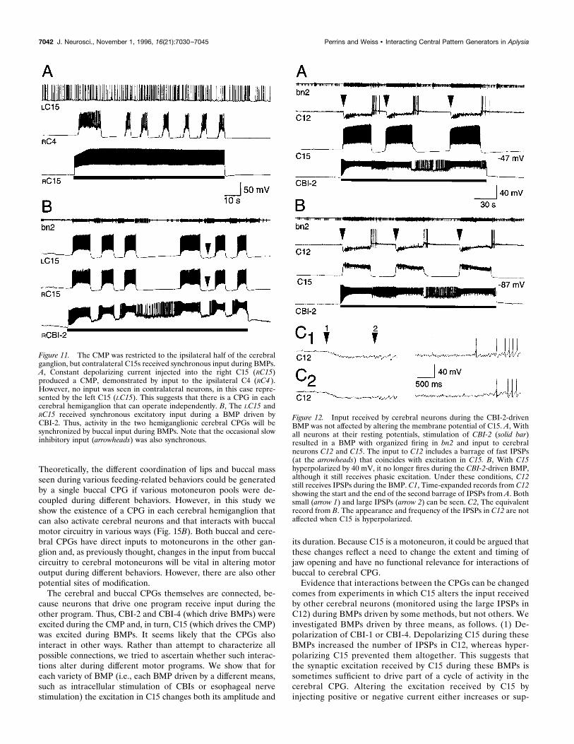
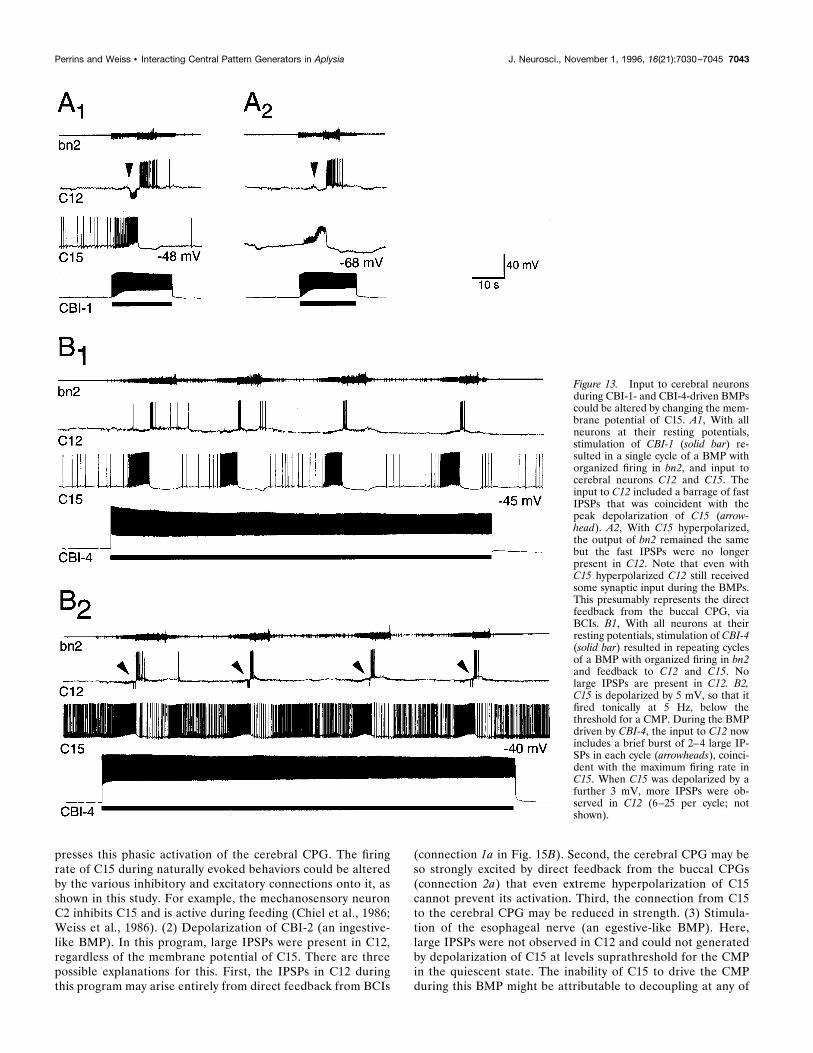
in C12. These IPSPs were in phase with the excitation in C15,suggesting that the feedback loop through C15 to the cerebralCPG could contribute to their formation. If this were the case,then hyperpolarizing C15 might prevent the activation of thecerebral CPG, and so block some of these IPSPs. However, noreduction in the frequency of these IPSPs was observed even after
quite extreme (and probably nonphysiological) hyperpolarizationof C15 (Fig. 12).In BMPs driven by both CBI-1 and CBI-4, large IPSPs in C12
were only sometimes observed (4 of 10 preparations), but whenpresent were again in phase with the peak excitation in C15. Inthese types of BMP, however, the presence of IPSPs in C12 wascritically dependent on the membrane potential of C15 (Fig.13A,B). If IPSPs were present, they could be blocked by hyper-polarizing C15 (n 5 4; Fig. 13A). Conversely, if no IPSPs werepresent, they appeared if C15 was depolarized by 4–10 mV, belowthe threshold for driving the CMP in the quiescent state (n 5 6;Fig. 13B). This suggests that the IPSPs in C12 during these BMPsresult from activity in the cerebral CPG, driven at least partly bythe phasic excitation in C15.A third situation was observed for the egestive-like BMP driven
by stimulation of the esophageal nerve. In this BMP, no largeIPSPs were observed in C12 at any phase of the rhythm. Addi-tionally, large IPSPs could not be induced by strong depolariza-tion in C15 during the peak of its synaptic excitation (Fig. 14).During the egestive-like BMP, C12 received PSPs not correlatedwith the current pulse in C15, presumably attributable to directinput from BCIs. These PSPs caused a drop of ;15% in the inputresistance of C12 measured by constant hyperpolarizing currentpulses. This relatively small change in the input resistance of C12is unlikely to reduce the IPSPs during the CMP below detectableamplitude. For comparison, the decrease in input resistance inC12 caused by the IPSPs of the CMP itself was as much as 50%.Even current levels well above the threshold for driving the CMPin the quiescent state could not drive the CMP during this BMP.Thus, not only does C15 fire at a lower frequency during theegestive-like BMP, but it is rendered incapable of drivingthe CMP.
DISCUSSIONA separate cerebral CPGOur findings indicate that there is a CPG within the cerebralganglion of Aplysia. In most other molluscan feeding systems,there is little evidence for separate cerebral CPGs, possibly be-cause studies have concentrated on buccal motor circuitry. InLymnaea, rhythmic synaptic inputs were recorded in some neu-rons in isolated cerebral ganglia (McCrohan, 1984), but the natureof this “oscillator” was not investigated further. In Pleurobran-chaea, cerebral interneurons that drive buccal feeding rhythms[the phasic paracerebral (PCp) cells] also drive rhythmic motoroutput in isolated cerebral ganglia (Davis et al., 1984). The cere-bral rhythm in Pleurobranchaea could only be obtained for aboutan hour after CBC transection. In Aplysia, however, the CMPcould be activated in cerebral ganglia 36 hr after CBC transection,showing that the cerebral CPG does not require short- or long-term feedback from the buccal ganglion to maintain its rhythm-generating ability.It should be noted that there are two separate cerebral
CPGs, one in each hemiganglion, and each can be activeindependently. This feature may be important in producingasymmetrical lip movements during feeding behaviors if theexposure of seaweed to the lips is unilateral. Once the seaweedis positioned centrally and ingestion has begun, activity in thetwo cerebral CPGs will be synchronized because of simulta-neous excitation of both C15s by the buccal CPG (Fig. 11),resulting in symmetrical lip movements.The fast PSPs observed in cerebral neurons during the CMP do
not represent monosynaptic connections from C15. The fact that
Figure 7. Properties of the PSPs evoked in cerebral neurons in responseto CBC stimulation. A, The latency of the PSPs (in this case the EPSP inC15) was not affected by increasing the stimulation intensity (top threetraces) or frequency (bottom two traces), indicating that the PSPs wereprobably attributable to a direct, monosynaptic connection from acerebral-to-buccal neuron. The timing of CBC stimulation is indicated bythe downward artifact at the arrowhead. B1, The EPSPs in C15 wereclassical chemical EPSPs, which increased in amplitude with increasingnegative current (current injected at each arrowhead). B2, HyperpolarizingC15 (during solid bar) beyond a threshold level completely abolished theEPSPs in C15, as well as the PSPs recorded in other cerebral neurons, inthis case C11 and C12. This suggests that C15 may be electrically coupledto the cerebral-to-buccal neuron responsible for the PSPs (see Results fordetails).
7038 J. Neurosci., November 1, 1996, 16(21):7030–7045 Perrins and Weiss • Interacting Central Pattern Generators in Aplysia
the PSPs are synchronous in all neurons suggests a commonsource, but this remains to be identified. It is likely that thiscommon source is the unidentified cerebral-to-buccal neuron thatproduces (1) the spikes in the CBC that are one-for-one with PSPsin cerebral neurons during the C15-induced CMP, and (2) Theconstant-latency PSPs after CBC stimulation. The discovery ofthis neuron will be an important step in elucidating how the CMPis generated.
Because C15 is a lip motoneuron, driving the CMP via intra-cellular excitation of C15 caused strong tonic contraction of theinner lips that masked any superimposed rhythmic movements.However, during the CMP other cerebral motoneurons (such asC16 and C17) fire at frequencies that result in muscle contractionin the semi-intact preparation. Therefore, the cerebral CPG maybe capable of producing rhythmic lip movements. Supportingevidence for such a role is provided by studies showing that lip
Figure 8. Input to CBIs during the CMP in the isolated cerebral ganglion. A, CBI-1 was weakly, slowly inhibited during the CMP, in phase with the fastIPSPs in C12. B, CBI-2 and CBI-4 received slow excitation during the CMP, which depolarized them above spiking threshold. C, CBI-3 was excited andspiked during the CMP, in phase with the IPSPs in C12 (not shown) and the EPSPs in C4. D, CBI-3 and C4 were electrically coupled, becausehyperpolarizing or depolarizing current pulses in either neuron (solid bars) led to a smaller membrane potential change of the same polarity in the other.Action potentials in one neuron gave rise to brief depolarizing potentials in the other without a measurable delay. E, The excitation seen in CBI-3 duringa cycle of the CMP induced by firing of C15 (E1) was effectively mimicked by a depolarizing current pulse into C4 (E2), showing that electrotonictransmission of the chemically mediated excitation and spiking in C4 is sufficient to explain the excitation recorded in CBI-3.
Perrins and Weiss • Interacting Central Pattern Generators in Aplysia J. Neurosci., November 1, 1996, 16(21):7030–7045 7039

movements can occur after CBC transection (Kupfermann, 1974),or in the absence of ingestive movements of the buccal mass(during feeding: Hurwitz et al., 1996; during egg laying: Arch andSmock, 1977). Even if no behavioral correlate for the CMP isdiscovered, the changing interactions of cerebral and buccal CPGsare likely to play a role in shaping the coordination of lips andradula, as discussed below.
The functional significance of C15C15, C16, and C17 cause contraction of the inner lips, resulting injaw opening. These motoneurons are active at phases of ingestiveand egestive BMPs when the jaws are opening and fire at a high
enough frequency during these BMPs to cause muscle contrac-tion, so are likely to contribute to jaw opening during feedingbehaviors. The higher firing rate of C15 during ingestive ratherthan egestive motor programs agrees with behavioral observationsthat the lips fully open during ingestion, but remain mostly closedduring egestion (Morton and Chiel, 1993a).C15 is a motoneuron capable of driving a CPG. There is
evidence that motoneurons supply excitation to the vertebratespinal locomotor pattern generator (Perrins and Roberts, 1995)and in several invertebrate systems motoneurons supply feedbackto CPGs (Kristan and Calabrese, 1976; Heitler, 1978; Simmers
Figure 9. C15 initiated single cyclesof a BMP, possibly via excitation ofCBI-2 and CBI-4. A, Constant currentinjected into C15 (solid bar) resulted ina single cycle of a BMP (gray bar),consisting of strong multiphasic motoroutput in bn2 and firing in buccal neu-ron B4. This is followed by repeatingcycles of a CMP (open bar) with inputto C12, but only weak output in bn2and no firing in B4. B, With CBI-2hyperpolarized (B1) or at its restingpotential (B2), a 10 sec pulse of depo-larizing current into C15 (solid bars)resulted in a single cycle of a CMPwith slow excitation in CBI-2, fast IP-SPs in C12 and no output in bn2. B3,When CBI-2 was depolarized by 4 mV,it fired at high frequency during theCMP and a single cycle of a BMP wasevoked, represented by multiphasicoutput in bn2 and feedback to CBI-2,C12, and C15. C, With CBI-4 hyperpo-larized (C1) or at its resting potential(C2), a 10 sec pulse of depolarizingcurrent into C15 (solid bars) resultedin a single cycle of a CMP with slowexcitation in CBI-4, fast IPSPs in C12,and weak activity in bn2. C3, WhenCBI-4 was depolarized by 5 mV, itfired at high frequency during theCMP and a single cycle of a BMP wasevoked, represented by multiphasicoutput in bn2 and feedback to C12 andC15. Membrane potential of CBI-2and CBI-4 is indicated at the right sideof each trace.
7040 J. Neurosci., November 1, 1996, 16(21):7030–7045 Perrins and Weiss • Interacting Central Pattern Generators in Aplysia
and Bush, 1983) or can drive a repeating rhythm (buccal CPGs ofTritonia: Willows, 1980; Aplysia: Hurwitz et al., 1994). C15 is alsoan important contact point between the cerebral and buccalCPGs, as discussed below.
Interactions between cerebral and buccal CPGs andtheir functional significanceIt was previously thought that input to cerebral motoneurons wascontrolled by direct feedback from buccal circuitry (Fig. 15A).
Figure 10. C15 received synaptic input during variousBMPs. A, Constant depolarizing current injected intoCBI-2 (solid bar) resulted in an ingestive-like BMPduring which buccal neurons B8 and B4 fired together.During this program, C15 was strongly excited justbefore the strongest activity in these neurons. B, Dur-ing an egestive-like BMP induced by brief 10 Hz elec-trical stimulation of the radula nerve (during open bar),B8 fires before B4. In this program, C15 was active atthe same time as B8 and before spiking in B4. C,During an egestive-like BMP induced by continuous 2.5Hz stimulation of the esophageal nerve (open bar), C15is again active with B8, and before B4. After the termi-nation of nerve stimulation, there is a single cycle of aBMP that shows similar phase relations to theingestive-like program shown in A. D, During a BMPinduced by exciting CBI-4 (solid bar), C15 was againactive in phase with B8 and before activity in B4. In thisprogram, there was a second burst of spikes in B8 thatcoincided with inhibition in C15 (arrowheads). For eachBMP, the excitatory input to C15 consisted of a barrageof fast EPSPs. Buccal motor output was also monitoredfrom bn2 in each case.
Perrins and Weiss • Interacting Central Pattern Generators in Aplysia J. Neurosci., November 1, 1996, 16(21):7030–7045 7041
Theoretically, the different coordination of lips and buccal massseen during various feeding-related behaviors could be generatedby a single buccal CPG if various motoneuron pools were de-coupled during different behaviors. However, in this study weshow the existence of a CPG in each cerebral hemiganglion thatcan also activate cerebral neurons and that interacts with buccalmotor circuitry in various ways (Fig. 15B). Both buccal and cere-bral CPGs have direct inputs to motoneurons in the other gan-glion and, as previously thought, changes in the input from buccalcircuitry to cerebral motoneurons will be vital in altering motoroutput during different behaviors. However, there are also otherpotential sites of modification.The cerebral and buccal CPGs themselves are connected, be-
cause neurons that drive one program receive input during theother program. Thus, CBI-2 and CBI-4 (which drive BMPs) wereexcited during the CMP and, in turn, C15 (which drives the CMP)was excited during BMPs. It seems likely that the CPGs alsointeract in other ways. Rather than attempt to characterize allpossible connections, we tried to ascertain whether such interac-tions alter during different motor programs. We show that foreach variety of BMP (i.e., each BMP driven by a different means,such as intracellular stimulation of CBIs or esophageal nervestimulation) the excitation in C15 changes both its amplitude and
its duration. Because C15 is a motoneuron, it could be argued thatthese changes reflect a need to change the extent and timing ofjaw opening and have no functional relevance for interactions ofbuccal to cerebral CPG.Evidence that interactions between the CPGs can be changed
comes from experiments in which C15 alters the input receivedby other cerebral neurons (monitored using the large IPSPs inC12) during BMPs driven by some methods, but not others. Weinvestigated BMPs driven by three means, as follows. (1) De-polarization of CBI-1 or CBI-4. Depolarizing C15 during theseBMPs increased the number of IPSPs in C12, whereas hyper-polarizing C15 prevented them altogether. This suggests thatthe synaptic excitation received by C15 during these BMPs issometimes sufficient to drive part of a cycle of activity in thecerebral CPG. Altering the excitation received by C15 byinjecting positive or negative current either increases or sup-
Figure 11. The CMP was restricted to the ipsilateral half of the cerebralganglion, but contralateral C15s received synchronous input during BMPs.A, Constant depolarizing current injected into the right C15 (RC15)produced a CMP, demonstrated by input to the ipsilateral C4 (RC4 ).However, no input was seen in contralateral neurons, in this case repre-sented by the left C15 (LC15). This suggests that there is a CPG in eachcerebral hemiganglion that can operate independently. B, The LC15 andRC15 received synchronous excitatory input during a BMP driven byCBI-2. Thus, activity in the two hemiganglionic cerebral CPGs will besynchronized by buccal input during BMPs. Note that the occasional slowinhibitory input (arrowheads) was also synchronous.
Figure 12. Input received by cerebral neurons during the CBI-2-drivenBMP was not affected by altering the membrane potential of C15. A, Withall neurons at their resting potentials, stimulation of CBI-2 (solid bar)resulted in a BMP with organized firing in bn2 and input to cerebralneurons C12 and C15. The input to C12 includes a barrage of fast IPSPs(at the arrowheads) that coincides with excitation in C15. B, With C15hyperpolarized by 40 mV, it no longer fires during the CBI-2-driven BMP,although it still receives phasic excitation. Under these conditions, C12still receives IPSPs during the BMP. C1, Time-expanded records from C12showing the start and the end of the second barrage of IPSPs from A. Bothsmall (arrow 1) and large IPSPs (arrow 2) can be seen. C2, The equivalentrecord from B. The appearance and frequency of the IPSPs in C12 are notaffected when C15 is hyperpolarized.
7042 J. Neurosci., November 1, 1996, 16(21):7030–7045 Perrins and Weiss • Interacting Central Pattern Generators in Aplysia
presses this phasic activation of the cerebral CPG. The firingrate of C15 during naturally evoked behaviors could be alteredby the various inhibitory and excitatory connections onto it, asshown in this study. For example, the mechanosensory neuronC2 inhibits C15 and is active during feeding (Chiel et al., 1986;Weiss et al., 1986). (2) Depolarization of CBI-2 (an ingestive-like BMP). In this program, large IPSPs were present in C12,regardless of the membrane potential of C15. There are threepossible explanations for this. First, the IPSPs in C12 duringthis program may arise entirely from direct feedback from BCIs
(connection 1a in Fig. 15B). Second, the cerebral CPG may beso strongly excited by direct feedback from the buccal CPGs(connection 2a) that even extreme hyperpolarization of C15cannot prevent its activation. Third, the connection from C15to the cerebral CPG may be reduced in strength. (3) Stimula-tion of the esophageal nerve (an egestive-like BMP). Here,large IPSPs were not observed in C12 and could not generatedby depolarization of C15 at levels suprathreshold for the CMPin the quiescent state. The inability of C15 to drive the CMPduring this BMP might be attributable to decoupling at any of
Figure 13. Input to cerebral neuronsduring CBI-1- and CBI-4-driven BMPscould be altered by changing the mem-brane potential of C15. A1, With allneurons at their resting potentials,stimulation of CBI-1 (solid bar) re-sulted in a single cycle of a BMP withorganized firing in bn2, and input tocerebral neurons C12 and C15. Theinput to C12 included a barrage of fastIPSPs that was coincident with thepeak depolarization of C15 (arrow-head). A2, With C15 hyperpolarized,the output of bn2 remained the samebut the fast IPSPs were no longerpresent in C12. Note that even withC15 hyperpolarized C12 still receivedsome synaptic input during the BMPs.This presumably represents the directfeedback from the buccal CPG, viaBCIs. B1, With all neurons at theirresting potentials, stimulation of CBI-4(solid bar) resulted in repeating cyclesof a BMP with organized firing in bn2and feedback to C12 and C15. Nolarge IPSPs are present in C12. B2,C15 is depolarized by 5 mV, so that itfired tonically at 5 Hz, below thethreshold for a CMP. During the BMPdriven by CBI-4, the input to C12 nowincludes a brief burst of 2–4 large IP-SPs in each cycle (arrowheads), coinci-dent with the maximum firing rate inC15. When C15 was depolarized by afurther 3 mV, more IPSPs were ob-served in C12 (6–25 per cycle; notshown).
Perrins and Weiss • Interacting Central Pattern Generators in Aplysia J. Neurosci., November 1, 1996, 16(21):7030–7045 7043

several sites. For example, the rhythm-generating ability of thecerebral CPG might be suppressed, the excitatory connectionfrom C15 to the cerebral CPG might be reduced, or thesynaptic outputs of the cerebral CPG might be inhibited.How might activity in the cerebral CPGs contribute to spe-
cific aspects of feeding behavior? A possible role in generatingasymmetrical lip movements was addressed earlier. Addition-ally, we can speculate how specific cerebral neurons may beinfluenced during protraction, the time when C15 receivesexcitation, thus possibly activating the CMP. For example, themechanoafferent C2 is inhibited during the CMP and appearsto be least active during protraction in the semi-intact prepa-ration (Weiss et al., 1986). The CMP, therefore, might contrib-ute to a relative enhancement of proprioceptive input from thelips and peri-oral zone during retraction, when seaweed isbeing ingested.In conclusion, there are distinct buccal and cerebral CPGs,
these CPGs interact at several points, and the interactions be-tween these CPGs vary during different motor programs. Al-though the mechanisms underlying the changes in the interactionsare not yet known, such variation does exist. Most obviously, C15can drive the cerebral CPG during CBI-1 and CBI-4 driven BMPsbut cannot during egestive-like BMPs. These results lend supportto two hypotheses: first, that there is often a separate CPG foreach individual body part; second, that the interconnections be-tween such CPGs alter during different motor programs. Futurestudies will need to determine whether such changes represent amechanism for altering the coordination of separate body parts ona cycle-by-cycle basis.
REFERENCESArch S, Smock T (1977) Egg-laying behavior in Aplysia californica. BehavBiol 19:45–54.
Chiel HJ, Weiss KR, Kupferman I (1986) An identified histaminergicneuron modulates feeding motor circuitry in Aplysia. J Neurosci6:2427–2450.
Chiel HJ, Kupferman I, Weiss KR (1988) An identified histaminergicneuron can modulate the outputs of buccal-cerebral interneurons inAplysia via presynaptic inhibition. J Neurosci 8:49–63.
Church PJ, Lloyd PE (1994) Activity of multiple identified motor neu-rons recorded intracellularly during evoked feeding-like motor pro-grams in Aplysia. J Neurophysiol 72:1794–1809.
Cohan CS, Mpitsos GJ (1983) The generation of rhythmic activity in adistributed motor system. J Exp Biol 102:25–42.
Cohen JL, Weiss KR, Kupferman I (1978) Motor control of buccalmuscles in Aplysia. J Neurophysiol 41:157–180.
Davis WJ, Siegler MVS, Mpitsos GJ (1973) Distributed neuronal oscil-lators and efference copy in the feeding system of Pleurobranchaea.J Neurophysiol 36:258–274.
Davis WJ, Kovac MP, Croll RP, Matera EM (1984) Brain oscillatorsunderlying rhythmic cerebral and buccal motor output in the mollusc,Pleurobranchaea californica. J Exp Biol 110:1–15.
Dickinson PS, Moulins M (1992) Interactions and combinations betweendifferent networks in the stomatogastric nervous system. In: Dynamic
Figure 14. C15 could not drive a CMP during the egestive-like BMPinduced by esophageal nerve stimulation. In the quiescent state, inject-ing C15 with a 10 sec pulse of either 8 or 4 nA depolarizing current(filled bars) initiated a cycle of the CMP, shown by the barrage of IPSPsin C12 (arrowheads 1 and 2). A BMP was then driven by 2 Hzesophageal nerve stimulation (during open bar). C15 was excited duringthis program, at the same time as B8, and just before B4 (compare Fig.11, and this synaptic drive is visible just before and after the currentpulse). Depolarizing current (8 nA, previously suprathreshold for theCMP) injected into C15 at the phase in which it received synapticexcitation could not drive a CMP, shown by the lack of large IPSPs inC12 (arrowhead 3). Soon after the end of the BMP, C15 could againdrive the CMP (4 nA, arrowhead 4 ). Figure 15. Summary diagram of the interactions between buccal and
cerebral CPGs. A, Previously, it was thought that cerebral motoneuronsthat innervate the lips were controlled during feeding-related behaviors bydirect feedback from one or more buccal CPGs (1). B, We now proposethat there is a CPG in each cerebral hemiganglion that interact in variableways with the buccal CPGs (2a, 2b). Each CPG also makes direct connec-tions onto motoneurons in the other ganglion (1a, 1b). In both ganglia,arrows also indicate that there is feedback into the CPGs from somemotoneurons (buccal: B31/32, Hurwitz et al., 1994; cerebral: C15, thisstudy). The situation in B allows greater flexibility than in A, because theoutput of the whole system can be altered by changing the strength or typeof interaction at any of these points. Note that in both parts other knownspecific cerebral influences over buccal circuitry, such as the CBIs andMCC, are omitted for clarity.
7044 J. Neurosci., November 1, 1996, 16(21):7030–7045 Perrins and Weiss • Interacting Central Pattern Generators in Aplysia

biological networks. (Harris-Warrick RM, Marder E, Selverston AI,Moulins M, eds), pp 139–160. Cambridge: MIT.
Friesen WO, Pearce RA (1993) Mechanisms of intersegmental coordi-nation in leech locomotion. Semin Neurosci 5:41–47.
Grillner S, Wallen P (1985) Central pattern generators, with specialreference to vertebrates. Annu Rev Neurosci 8:233–261.
Heitler WJ (1978) Coupled motoneurones are part of the crayfish swim-meret central oscillator. Nature 275:231–234.
Hurwitz I, Goldstein RS, Susswein AJ (1994) Compartmentalization ofpattern-initiation and motor functions in the B31 and B32 neurons ofthe buccal ganglia of Aplysia californica. J Neurophysiol 71:1514–1527.
Hurwitz I, Neustadter D, Morton DW, Chiel HJ, Susswein AJ (1996)Activity patterns of the B31/B32 pattern initiators innervating the I2muscle of the buccal mass during normal feeding movements in Aplysiacalifornica. J Neurophysiol 175:1309–1326.
Jahan-Parwar B, Fredman SM (1976) Cerebral ganglion of Aplysia: cellularorganization and origin of nerves. Comp Biochem Physiol 54A:347–357.
Kirk MD (1989) Premotor neurons in the feeding system of Aplysiacalifornica. J Neurobiol 20:497–512.
Kristan Jr WB, Calabrese RL (1976) Rhythmic swimming activity inneurons of the isolated nerve cord of the leech. J Exp Biol 65:643–668.
Kupfermann I (1974) Dissociation of the appetitive and consummatoryphases of feeding behavior in Aplysia: a lesion study. Behav Biol 10:89–97.
Kupfermann I, Weiss KR (1978) The command neuron concept. BehavBrain Sci 1:3–39.
McCrohan CR (1984) Initiation of feeding motor output by an identifiedinterneurone in the snail Lymnaea stagnalis. J Exp Biol 113:351–366.
Morton DW, Chiel HJ (1993a) In vivo buccal nerve activity that distin-guishes ingestion from rejection can be used to predict behavioraltransitions in Aplysia. J Comp Physiol [A] 172:17–32.
Morton DW, Chiel HJ (1993b) The timing of activity in motor neuronsthat produce radula movements distinguishes ingestion from rejectionin Aplysia. J Comp Physiol [A] 173:519–536.
Mulloney B, Murchison D, Chrachri A (1993) Modular organization ofpattern-generating circuits in a segmental motor system: the swim-merets of crayfish. Semin Neurosci 5:49–57.
Ono JK, McCaman RE (1980) Identification of additional histaminergicneurons in Aplysia: improvements of single cell isolation techniques forin tandem physiological and chemical studies. Neuroscience 5:835–840.
Perrins R, Roberts A (1995) Cholinergic contribution to excitation in aspinal locomotor central pattern generator in Xenopus embryos. J Neu-rophysiol 73:1013–1019.
Rao G, Barnes CA, McNaughton BL (1986) Intracellular fluorescentstaining with carboxyfluorescein: a rapid and reliable method for quan-tifying dye-coupling in mammalian central nervous system. J NeurosciMethods 16:251–263.
Rosen SC, Weiss KR, Kupferman I (1979) Response properties andsynaptic connections of mechanoafferent neurons in cerebral ganglionof Aplysia. J Neurophysiol 42:954–974.
Rosen SC, Weiss KR, Cohen JL, Kupfermann I (1982) Interganglioniccerebral-buccal mechanoafferents of Aplysia: receptive fields and syn-aptic connections to different classes of neurons involved in feedingbehavior. J Neurophysiol 48:271–288.
Rosen SC, Weiss KR, Goldstein RS, Kupferman I (1989) The role ofmodulatory neuron in feeding and satiation in Aplysia: effects of lesion-ing the sertonergic metacerebral cells. J Neurosci 9:1562–1578.
Rosen SC, Halvorsen EL, Cropper EC, Miller MW, Weiss KR, Kupfer-man I (1990) Neuronal control of coordinated lip movements duringfeeding in Aplysia. Soc Neurosci Abstr 16:1227.
Rosen SC, Weiss KR, Kupferman I (1991a) Command of feeding inAplysia: implication of a network of identified interganglionic inter-neurons in the buccal and cerebral ganglia. Soc Neurosci Abstr17:595.6.
Rosen SC, Teyke T, Miller MW, Weiss KR, Kupferman I (1991b) Iden-tification and characterization of cerebral-to-buccal interneurons impli-cated in the control of motor programs associated with feeding inAplysia. J Neurosci 11:3630–3655.
Rossignol S, Saltiel M-C, Perreault T, Drew K, Pearson K, Belanger M(1993) Intralimb and interlimb coordination in the cat during real andfictive rhythmic motor programs. Semin Neurosci 5:67–75.
Ryckebusch S, Laurent G (1994) Interactions between segmental legcentral pattern generators during fictive rhythms in the locust. J Neu-rophysiol 72:2771–2785.
Sigvardt KA (1993) Intersegmental coordination in the lamprey centralpattern generator for locomotion. Semin Neurosci 5:3–15.
Simmers AJ, Bush BMH (1983) Central nervous mechanisms controllingrhythmic burst generation in the ventilatory motoneurones of Carcinusmaenus. J Comp Physiol [A] 150:1–21.
Susswein AJ, Byrne JH (1988) Identification and characterisation of neu-rons initiating patterned neural activity in the buccal ganglia of Aplysia.J Neurosci 8:2049–2061.
Teyke T, Rosen SC, Weiss KR, Kupferman I (1993) Dopaminergic neu-ron B20 generates rhythmic neuronal activity in the feeding motorcircuitry of Aplysia. Brain Res 630:226–237.
Weiss KR, Cohen JL, Kupfermann I (1978) Modulatory control of buc-cal musculature by a serotonergic neuron (metacerebral cell) in Aplysia.J Neurophysiol 41:181–203.
Weiss KR, Koch UT, Koester J, Rosen SC, Kupferman I (1982) The roleof arousal in modulating feeding behaviour of Aplysia: neural andbehavioral studies. In: The neural basis of feeding and reward (HoebelBG, Novin D, eds), pp 25–57. Brunswick, ME: Haer Institute.
Weiss KR, Chiel HJ, Koch U, Kupferman I (1986) Activity of an iden-tified histaminergic neuron, and its possible role in arousal of feedingbehavior in semi-intact Aplysia. J Neurosci 6:2403–2415.
Willows AOD (1980) Physiological basis of feeding behavior in Tritoniadiomedea. II. Neuronal mechanisms. J Neurophysiol 44:849–861.
Perrins and Weiss • Interacting Central Pattern Generators in Aplysia J. Neurosci., November 1, 1996, 16(21):●–● 7045