A Case Study of Early Phase Purification Process ...
30
www.validated.com PSG–091010 A Case Study of Early Phase Purification Process Development for an Anti-Cancer Minibody 1 Pete Gagnon, 2 Chia-Wei Cheung and 2 Paul Yazaki 1 Validated Biosystems, San Clemente, CA. 2 Division of Cancer Therapeutics and Tumor Immunology, Beckman Research Institute, City of Hope, Duarte, CA 5th International Symposium on Hydroxyapatite, Rottach-Egern, Germany, October 11-14, 2009
Transcript of A Case Study of Early Phase Purification Process ...
www.validated.comPSG–091010
A Case Study of Early PhasePurification Process Development
for an Anti-Cancer Minibody
1Pete Gagnon, 2Chia-Wei Cheung and 2Paul Yazaki1Validated Biosystems, San Clemente, CA. 2Division of Cancer
Therapeutics and Tumor Immunology, BeckmanResearch Institute, City of Hope, Duarte, CA
5th International Symposium on Hydroxyapatite, Rottach-Egern, Germany, October 11-14, 2009

www.validated.comPSG–091010
Purification of minibodies
The structural similarity of minibodies to antibodies invites theexpectation that their purification might be similarly well definedand simple to develop.
VL: light greenVH: dark greenCDRs: whiteCH3: purple
GS18 linker: yellowHinge and GS10 linker: cyanDisulfide bonds: red

www.validated.comPSG–091010
Purification of minibodies
But appearances can be deceiving. The principal tool for purificationof IgG – the one that enables a simple platform approach to processdevelopment – is not applicable to minibodies.
Protein A, Fc binding sites
Protein A, Fab binding sites

www.validated.comPSG–091010
Purification of minibodies
Minibody behavior differs from IgG with non-affinity methods as well.
Cation ExchangeCIM™ SO3 Monolith
A: 20 mM MES, pH 6.0
B: A + 1 M NaCl
Dilute sample 1:9 with A
Equilibrate column with A
Load sample
Wash A
Elute: 30 CVLG to 50% B
Clean with B
-----------------
MiniB eluted as a trailing
shoulder on the BSA peak.
IgGs elute much later.
Anion ExchangeCIM QA Monolith
A: 20 mM Tris, pH 8.0
B: A + 1 M NaCl
Dilute sample 1:9 with A
Equilibrate column with A
Load sample
Wash A
Elute: 30 CVLG to 50% B
Clean with B
-----------------
MiniB eluted as a leading
shoulder on the BSA peak.
IgGs elute much earlier.
HydroxyapatiteCHT™ Type I, 40 µm
A: 10 mM NaPO4, pH 7.0
B: 500 mM NaPO4, pH 7.0
Dilute sample 1:1 with A
Equilibrate column with A
Load sample
Wash A
Elute: 30 CVLG to 50% B
Clean with B
-----------------
MiniB co-eluted with BSA.
IgGs elute much later, often
at purity greater than 90%.

www.validated.comPSG–091010
BSA removal from the minibody
Shared features of minibodies and BSA.
pI ~5.1, ~80 KDa pI ~5.2, ~70 KDa
Ribbon model of albumin modified from S. Fujiwara and T. Amisaki, 2008, Biophys. J., 94(1): 95-103
Albumin removal is further complicated by the fact that it spontaneously forms homo-polymers, and stable complexes with a variety of fatty acids, metals, and other smallmolecules, plus disulfide bonded hybrids with a variety of proteins, all of which increaseits chemical heterogeneity. Thus albumin elutes across a much broader zone than well-defined proteins, and is more likely to co-elute with product over a wide range ofseparation methods and conditions.

www.validated.comPSG–091010
BSA removal from the minibody
Luck is what happens when preparation meets opportunity. –Lucius Annaeus Seneca

www.validated.comPSG–091010
BSA removal from the minibody
Hydroxyapatite, phosphate gradient elution
CHT™ type I, 40 µm, 300 cm/hr.Equilibrate: 20 mM KPO4, pH 6.5Load: Mini cell culture supernatantWash: 20 mM KPO4, pH 6.5Elute: 20 CVLG to 250 KPO4, pH 6.5Step to 300 mM KPO4, pH 6.5Step to 500 mM KPO4, pH 6.5Cyan peak represents minibody
minitrfbsa
Virtually no separation from BSA or transferrin.

www.validated.comPSG–091010
BSA removal from the minibody
Hydroxyapatite, NaCl gradient
CHT type I, 40 µm, 300 cm/hr.Equilibrate: 20 mM NaPO4, pH 6.5Inj: mini cell culture supernatantWash: 20 mM NaPO4, pH 6.5Elute: 30 CVLG to 20 mM NaPO4,1.0 M NaCl, pH 6.5Cyan peak represents minibody
minitrfbsa
Near-total elimination of BSA and transferrin under initialscreening conditions! Trf elutes mostly in the wash. BSAelutes mostly in the cleaning step.

www.validated.comPSG–091010
Hydroxyapatite with NaCl gradients
Removal of high molecular weight species, IgG vs minibody
IgGCHT type I, 40 µm, 300 cm/hr.Equilibrate: 10 mM NaPO4, pH 7.0Inj: protein A-purified MabWash: 10 mM NaPO4, pH 7.0Elute: 30 CVLG to 10 mM NaPO4,1.0 M NaCl, pH 7.0MinibodyCHT type I, 40 µm, 300 cm/hr.Equilibrate: 10 mM NaPO4, pH 7.0Inj: MMC/AX-purified minibodyWash: 10 mM NaPO4, pH 7.0Elute: 10 CVLG to 10 mM NaPO4,1.0 M NaCl, pH 7.0Tetrabody is believed to form by non-covalent association of “Fab” regions.
IgG profile modified from Gagnon, P., Beam, K., 2009, AntibodyAggregate Removal with Hydroxyapatite, Current PharmaceuticalBiotechnology, 10(4) 440-446, with permission.

www.validated.comPSG–091010
Hydroxyapatite with NaCl gradients
Why did minibody behavior mimic IgG in NaCl gradients but not inphosphate gradients?
The results suggest, as with IgG, that minibodies participate in weakcalcium affinity interactions with hydroxyapatite. A low concentrationof phosphate (10 mM) largely eliminates these interactions, leavingthe minibody bound principally by phosphoryl cation exchange,which can then be eluted with the NaCl gradient.
BSA, which participates in strong calcium affinity interactions withhydroxyapatite, requires ~50 mM phosphate to elute. At 10 mMphosphate its retention is thereby protected from NaCl and itremains bound, along with DNA, endotoxin, and viruses.
www.validated.comPSG–091010
Hydroxyapatite with NaCl gradients
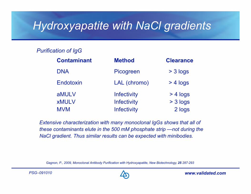
Purification of IgG
> 4 logs > 3 logs 2 logs
Infectivity Infectivity Infectivity
aMULV xMULV MVM
> 4 logs LAL (chromo) Endotoxin
> 3 logs Picogreen DNA
Clearance Method Contaminant
Gagnon, P., 2009, Monoclonal Antibody Purification with Hydroxyapatite, New Biotechnology, 25 287-293
Extensive characterization with many monoclonal IgGs shows that all ofthese contaminants elute in the 500 mM phosphate strip —not during theNaCl gradient. Thus similar results can be expected with minibodies.

www.validated.comPSG–091010
Capture with hydroxyapatite?
Despite excellent purification, minibody breakthrough bindingcapacity was only ~12 mL of supe (600 µg minibody) per mL ofhydroxyapatite, mostly due to competition from stronger-bindingcontaminants – chiefly albumin. Not a candidate for capture.
Capture with other methods?Cation exchange: ~15 mL supe (750 µg) per mL of resin, mostlydue to competition from contaminants – chiefly albumin. Purity ofthe eluted minibody was less than 20%.Anion exchange: < 2 mL supe (100 µg) per mL of resin, due toweak minibody binding and competition from albumin. Purity <10%.

www.validated.comPSG–091010
Minibody capture with Capto MMC
5-6 times greater capacity than cation exchange andmore than twice the purity!BSA was enriched on the leading side of the peak,minibody on the trailing side.
Capto™ MMC, 1 mL HiTrap™200 cm/hr (1 mL/min)Sample prep: dilute mini supe 1:1 with50 mM MES, pH 6.0Equilibrate: 20 mM Hepes, pH 7.0Inj: 140 mL sampleWash: 20 mM Hepes, pH 7.0Elute: Step to 20 mM Tris, 60 mMNaCl, pH 8.0
minitrfbsa
www.validated.comPSG–091010
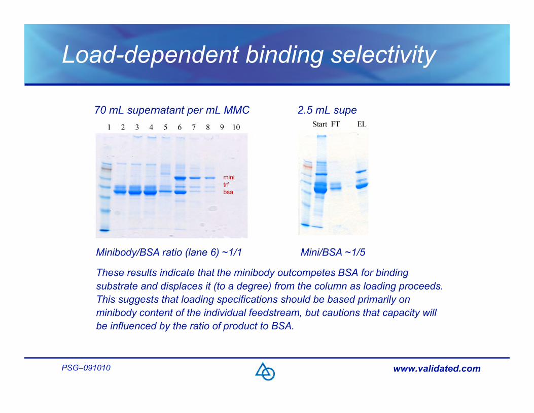
Load-dependent binding selectivity
70 mL supernatant per mL MMC 2.5 mL supe
Minibody/BSA ratio (lane 6) ~1/1 Mini/BSA ~1/5
These results indicate that the minibody outcompetes BSA for bindingsubstrate and displaces it (to a degree) from the column as loading proceeds.This suggests that loading specifications should be based primarily onminibody content of the individual feedstream, but cautions that capacity willbe influenced by the ratio of product to BSA.
minitrfbsa

www.validated.comPSG–091010
Load-dependent elution selectivity
Using a 20 mL load (on a 1 mL MMC column), we developed washconditions to selectively remove most of the leading albuminshoulder, with the goal of improving purity and reproducibility of theMMC step.
But, when the load was increased to 70 mL, more than half of theminibody eluted in the wash.
www.validated.comPSG–091010
Managing load-dependent capture
Load-dependent performance is undesirable because it predictspoor reproducibility of product recovery and purity as a function ofvariations among cell culture production methods, potentially evenamong different production lots within a single production method.It is also undesirable because it increases the burden onpurification process development:
It renders preliminary process modeling with 1 mL columnsinaccurate – but larger scale modeling requires proportion-ately greater sample volumes, which may not be availableduring early development stages.Variability at the capture step must be absorbed by subsequent purification steps, which means that more workwill be required to screen, identify, and optimize those steps.

www.validated.comPSG–091010
Managing load-dependent capture
Choose the life [chromatography method] that is most useful,and habit will make it the most agreeable.
–Sir Francis Bacon

www.validated.comPSG–091010
Managing load-dependent capture
gggggggg
Accommodations at the HA stepWe evaluated the effect of phosphate concentration on binding selectivity.Results suggested that highest capacity would be achieved at 5 mM* but thecolumn could be washed at 25 mM phosphate to remove contaminants boundby moderate calcium affinity. Best elution selectivity was achieved with achloride gradient at 10 mM phosphate.
*Capacity would be higher in the absence of phosphate, but 5 mM is required to maintain the stability of hydroxyapatite.

www.validated.comPSG–091010
Managing load-dependent capture
Accommodations at the HA stepCHT type I, 40 µm, 300 cm/hr.Sample prep: dilute AX FT 1:1 with 10mM NaPO4, pH 7.0 (final 5 mM PO4)Equilibrate: 30 mM NaPO4, pH 7.0InjectWash1: 30 mM NaPO4, pH 7.0Wash2: 10 mM NaPO4, pH 7.0Elute: 10 CVLG to 10 mM NaPO4,1.0 M NaCl, pH 7.0Clean: 500 mM NaPO4, pH 7.0Dashed lines mark buffer changes.The red arrow marks the peak thateluted when 30 mM phosphateresumed after sample loading. Someminibody was lost in this peak, so theequilibration and first wash weresubsequently reduced to 25 mM.

www.validated.comPSG–091010
Managing load-dependent capture
We chose anion exchange as a third chromatography step becauseof its established reputation in the regulatory community for removalof DNA, endotoxin, and virus.We evaluated both flow-through and bind-elute mode, but chosebind-elute mode because it removed more contaminants and wefelt that this added extra insurance against process variation due toload-associated variability at the capture step.We chose a high capacity porous particle-based anion exchangerbecause the high load of acidic contaminants (chiefly BSA) wouldoverwhelm the comparatively low protein-binding capacity ofmembrane-based anion exchangers.
www.validated.comPSG–091010
Process sequence and continuity
Anion exchange as a last step was impractical because theminibody eluted from hydroxyapatite at about 800 mM NaCl, whichwould have required insertion of a diafiltration step.Instead, we modified MMC elution conditions for high pH and lowconductivity so that the sample could be loaded onto the anionexchanger with minimal dilution.Following MMC with anion exchange also minimized thecontaminant load going into the hydroxyapatite step.Diafiltration was unnecessary because hydroxyapatite was able totolerate the NaCl concentration from elution of the anion exchanger,as long as the sample was equilibrated to no higher than 5 mMphosphate.

www.validated.comPSG–091010
The current process
A
CaptureCapto MMC
Dilute filtered supernatant
1:1 with 50 mM MES, pH 6
EQ: 50 mM MES, pH 6
Load
Wash: 50 mM MES, pH 6
Elute: Step to 20 mM Tris,
75 mM NaCl, pH 8.5
Clean: 2 M guanidine, pH 5
Sanitize: 1 M NaOH
IntermediateUNOsphere™ Q
Dilute MMC eluate 1:2 with
20 mM Tris, pH 8.5
EQ: 20 mM Tris, pH 8.5
Load
Wash: 20 mM Tris, pH 8.5
Elute: 10 CVLG* to 20 mM
Tris, 225 mM NaCl, pH 8.5
Clean: 1 M NaCl, pH 8.5
Sanitize: 1 M NaOH
PolishingCHT Type I, 40 µm
To AX eluate, add NaPO4 to
final concentration of 5 mM
EQ: 25 mM NaPO4, pH 7
Load
Wash1: 25 mM NaPO4, pH 7
Wash2: 10 mM NaPO4, pH 7
Elute: 10 CVLG* to 10 mM
NaPO4, 1 M NaCl, pH 7
Clean: 500 mM NaPO4, pH 7
Sanitize: 1 M NaOH
-----------------
Note that the minibody elutes
in phosphate buffered saline
at pH 7.0
www.validated.comPSG–091010
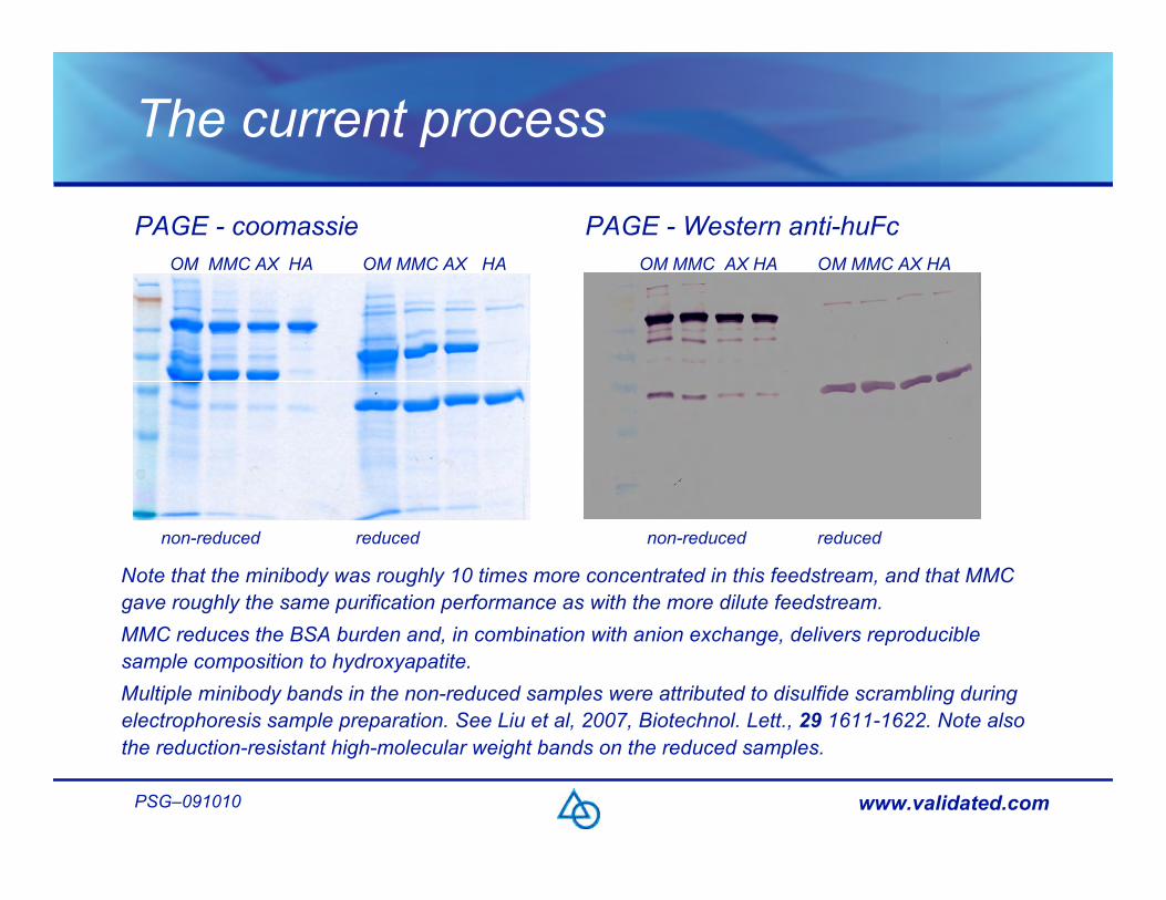
The current process
PAGE - coomassie PAGE - Western anti-huFc
non-reduced reduced non-reduced reduced
OM MMC AX HA OM MMC AX HA OM MMC AX HA OM MMC AX HA
Note that the minibody was roughly 10 times more concentrated in this feedstream, and that MMCgave roughly the same purification performance as with the more dilute feedstream.MMC reduces the BSA burden and, in combination with anion exchange, delivers reproduciblesample composition to hydroxyapatite.Multiple minibody bands in the non-reduced samples were attributed to disulfide scrambling duringelectrophoresis sample preparation. See Liu et al, 2007, Biotechnol. Lett., 29 1611-1622. Note alsothe reduction-resistant high-molecular weight bands on the reduced samples.

www.validated.comPSG–091010
The current process
Analytical size exclusion documenting HA fractionation of minibody and tetrabody Peak 1 (main) Peak 2
Superdex™ 75, HR 10/30Peak 2 was populated dominantly bytetrabody (retention time 16.57 min.)The small peak at 14.26 minutes mayindicate larger aggregates. With IgG,fragments usually elute on the leadingside of IgG so the small peaks elutingfrom SEC after 20 minutes are likelynot minibody-derived.The relatively large proportion ofminibody in peak 2 invites concernabout product loss, but peak 2 containsonly about 15% the UV absorbance ofthe main peak (slide 9), and minibodyonly about 40% of that, so actualproduct loss is probably less than 6%.

www.validated.comPSG–091010
The current process
Recovery
Stage µg/mL mL mg recoveryOM 462 19.7 9.1 100%MMC elution 362 22 8.0 88%MMC strip 189 5.5 1.0 11%Q elution 249 21 5.2 57%Q strip 117 7.4 0.9 10%HA elution 1 361 11 4.0 44%HA elution 2 120 11 1.3 14%*HA strip 76 3.8 0.3 3%**
*Dominantly Tetrabody. **Higher aggregates.

www.validated.comPSG–091010
Conclusions
This presentation, along with a rapidly growing number of others,marks the ascent of multimodal (mixed mode) methods in the fieldof process chromatography. Method development is morecomplicated than single-mode methods, but worth the investment.
www.validated.comPSG–091010
Conclusions
As shown by MMC, mixed modes can provide an effective capturealternative in the absence of a convenient bioaffinity method.
An important advantage of MMC is that it avoids the problem ofbioaffinity ligand leakage. This makes it unnecessary to developpurification methods to remove leachate, or develop analyticalmethods to measure leachate; and it suspends concerns aboutpotential adjuvancy or immunogenicity of bioaffinity leachates.
MMC also makes an ideal precursor to hydroxyapatite because itremoves the majority of contaminants that bind more strongly thanthe minibody (chiefly BSA and DNA), thereby increasing capacity forthe product. It also removes cell culture components that mightinteract directly with hydroxyapatite (chelating agents and metal ions).

www.validated.comPSG–091010
Conclusions
Hydroxyapatite demonstrates that mixed modes can offer uniqueselectivities, especially for removal of contaminants that are highlysimilar to the product, such as aggregates, fragments – and in thiscase, albumin.
Hydroxyapatite’s well-documented capabilities for removal of DNA,endotoxin, and virus add extra assurance, in combination with anionexchange, that specifications for reducing these contaminants willbe achieved with ease.
Economical regulatory-compliant purification of this minibody mightnot be possible without mixed modes like MMC and hydroxyapatite.

www.validated.comPSG–091010
Acknowledgements
Thanks to Mark Sherman at City of Hope for ribbon models of IgG andminibody. Thanks also to BIA Separations, Bio-Rad Laboratories, andGE Healthcare for providing chromatography media to develop thispurification process. Some parts of this research were supported byNCI grant CA43904.
Copies of this presentation can be downloaded at www.validated.com
www.validated.comPSG–091010
Disclaimer
If you ask me anything I don’t know, I’m not going to answer. –Yogi Berra