A CASE OF TORSION OF A PEDUNCULATED · PDF fileA CASE OF TORSION OF A PEDUNCULATED SUBSEROSAL...
4
WWW.KJOG.ORG 647 A CASE OF TORSION OF A PEDUNCULATED SUBSEROSAL UTERINE LEIOMYOMA ACCOMPANIED WITH ACUTE GASTROENTERITIS: A POSSIBLE DIAGNOSTIC PITFALL Ji Hyun Han, MD, Eun Young Jung, MD, Sun Young Jeon, MD, Han Young Park, MD, Nan-Hee Jeong, MD, Bo Yon Lee, MD Department of Obstetrics and Gynecology, KyungHee University College of Medicine, Seoul, Korea A literature search on acute abdomen caused by a leiomyoma does not yield many specific reports of myoma-related complications or the presenting features. We describe a case of torsion of a pedunculated subserosal uterine leiomyoma in a 25-year-old virgin female who noticed lower abdominal pain with severe diarrhea, which was assumed to be typical acute gastroenteritis undoubtedly. Awareness of the condition and a high index of suspicion are keys to early diagnosis and prompt intervention. A description of unique clinical features and radiologic evaluation is discussed. Keywords: Leiomyoma; Uterus; Torsion; Abdominal pain; Gastroenteritis Received: 2011. 4.28. Revised: 2011. 6.23. Accepted: 2011. 8.17. Corresponding author: Nan-Hee Jeong, MD Department of Obstetrics and Gynecology, KyungHee University College of Medicine, 1 Hoegi-dong, Dongdaemun-gu, Seoul 130- 701, Korea Tel: +82-2-958-8314 Fax: +82-2-958-8328 E-mail: [email protected] Acute abdominal pain is one of the most common presenting symptoms of emergency department patients. The classic guide- lines in surgical medicine are that abdominal pain with diarrhea suggests a diagnosis of gastroenteritis [1]. However, a substantial number of these patients need emergency surgical intervention. In one retrospective study which evaluated emergency patients with abdominal pain and diarrhea, 9% of such patients underwent emergency surgery [2]. Of note, torsion of a pedunculated subse- rosal uterine leiomyoma can also present with acute abdominal pain and diarrhea. A literature search on this topic does not yield many specific reports of leiomyoma-related complications or the presenting features [3]. Herein, we describe one case of torsion of a pedun- culated subserosal uterine leiomyoma in a woman who noticed lower abdominal pain with severe diarrhea, which was assumed to be typical acute gastroenteritis. Case Report A 25-year-old virgin sought evaluation in our emergency de- partment with acute lower abdominal pain and diarrhea on 21 January 2010. The abdominal pain developed 2 days ago, and in- creased in intensity over the past few hours. She had 10 episodes CASE REPORT Korean J Obstet Gynecol 2011;54(10):647-650 http://dx.doi.org/10.5468/KJOG.2011.54.10.647 pISSN 2233-5188 · eISSN 2233-5196 of watery diarrhea per day. Prior to presenting to our emergency room, she received conservative treatment at a local clinic for presumptive acute gastroenteritis; however, the symptoms did not improve, even though she was taking probiotics and analgesics. Contrast-enhanced abdominal computerized tomography scan revealed a non-enhanced pelvic mass connected to the posterior aspect of the uterine corpus with a distinct border, measuring 6.7 × 5.9 × 6.3 cm, with a small amount of ascites. Others includ- ing both ovaries were unremarkable (Fig. 1). She was referred to our emergency department. On physical examination, the temper- ature was 37.8˚C, and there was direct and rebound tenderness in is is an Open Access article distributed under the terms of the Creative Commons Attribution Non-Commercial License (http://creativecommons.org/licenses/ by-nc/3.0/) which permits unrestricted non-commercial use, distribution, and reproduction in any medium, provided the original work is properly cited. Copyright © 2011. Korean Society of Obstetrics and Gynecology
Transcript of A CASE OF TORSION OF A PEDUNCULATED · PDF fileA CASE OF TORSION OF A PEDUNCULATED SUBSEROSAL...

WWW.KJOG.ORG 647
A CASE OF TORSION OF A PEDUNCULATED SUBSEROSAL UTERINE LEIOMYOMA ACCOMPANIED WITH ACUTE GASTROENTERITIS: A POSSIBLE DIAGNOSTIC PITFALLJi Hyun Han, MD, Eun Young Jung, MD, Sun Young Jeon, MD, Han Young Park, MD, Nan-Hee Jeong, MD, Bo Yon Lee, MDDepartment of Obstetrics and Gynecology, KyungHee University College of Medicine, Seoul, Korea
A literature search on acute abdomen caused by a leiomyoma does not yield many specifi c reports of myoma-related complications or the presenting features. We describe a case of torsion of a pedunculated subserosal uterine leiomyoma in a 25-year-old virgin female who noticed lower abdominal pain with severe diarrhea, which was assumed to be typical acute gastroenteritis undoubtedly. Awareness of the condition and a high index of suspicion are keys to early diagnosis and prompt intervention. A description of unique clinical features and radiologic evaluation is discussed.
Keywords: Leiomyoma; Uterus; Torsion; Abdominal pain; Gastroenteritis
Received: 2011. 4.28. Revised: 2011. 6.23. Accepted: 2011. 8.17.Corresponding author: Nan-Hee Jeong, MDDepartment of Obstetrics and Gynecology, KyungHee University College of Medicine, 1 Hoegi-dong, Dongdaemun-gu, Seoul 130-701, KoreaTel: +82-2-958-8314 Fax: +82-2-958-8328E-mail: [email protected]
Acute abdominal pain is one of the most common presenting symptoms of emergency department patients. The classic guide-lines in surgical medicine are that abdominal pain with diarrhea suggests a diagnosis of gastroenteritis [1]. However, a substantial number of these patients need emergency surgical intervention. In one retrospective study which evaluated emergency patients with abdominal pain and diarrhea, 9% of such patients underwent emergency surgery [2]. Of note, torsion of a pedunculated subse-rosal uterine leiomyoma can also present with acute abdominal pain and diarrhea. A literature search on this topic does not yield many specific reports of leiomyoma-related complications or the presenting features [3]. Herein, we describe one case of torsion of a pedun-culated subserosal uterine leiomyoma in a woman who noticed lower abdominal pain with severe diarrhea, which was assumed to be typical acute gastroenteritis.
Case Report
A 25-year-old virgin sought evaluation in our emergency de-partment with acute lower abdominal pain and diarrhea on 21 January 2010. The abdominal pain developed 2 days ago, and in-creased in intensity over the past few hours. She had 10 episodes
CASE REPORTKorean J Obstet Gynecol 2011;54(10):647-650http://dx.doi.org/10.5468/KJOG.2011.54.10.647pISSN 2233-5188 · eISSN 2233-5196
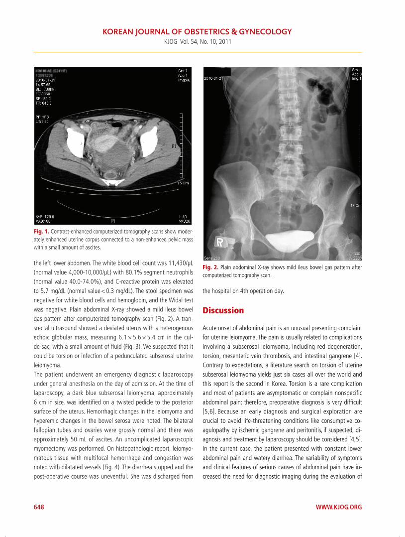
of watery diarrhea per day. Prior to presenting to our emergency room, she received conservative treatment at a local clinic for presumptive acute gastroenteritis; however, the symptoms did not improve, even though she was taking probiotics and analgesics. Contrast-enhanced abdominal computerized tomography scan revealed a non-enhanced pelvic mass connected to the posterior aspect of the uterine corpus with a distinct border, measuring 6.7 × 5.9 × 6.3 cm, with a small amount of ascites. Others includ-ing both ovaries were unremarkable (Fig. 1). She was referred to our emergency department. On physical examination, the temper-ature was 37.8˚C, and there was direct and rebound tenderness in
Th is is an Open Access article distributed under the terms of the Creative Commons Attribution Non-Commercial License (http://creativecommons.org/licenses/by-nc/3.0/) which permits unrestricted non-commercial use, distribution, and reproduction in any medium, provided the original work is properly cited.
Copyright © 2011. Korean Society of Obstetrics and Gynecology
WWW.KJOG.ORG648
KJOG Vol. 54, No. 10, 2011
the left lower abdomen. The white blood cell count was 11,430/μL (normal value 4,000-10,000/μL) with 80.1% segment neutrophils (normal value 40.0-74.0%), and C-reactive protein was elevated to 5.7 mg/dL (normal value < 0.3 mg/dL). The stool specimen was negative for white blood cells and hemoglobin, and the Widal test was negative. Plain abdominal X-ray showed a mild ileus bowel gas pattern after computerized tomography scan (Fig. 2). A tran-srectal ultrasound showed a deviated uterus with a heterogenous echoic globular mass, measuring 6.1 × 5.6 × 5.4 cm in the cul-de-sac, with a small amount of fl uid (Fig. 3). We suspected that it could be torsion or infection of a pedunculated subserosal uterine leiomyoma. The patient underwent an emergency diagnostic laparoscopy under general anesthesia on the day of admission. At the time of laparoscopy, a dark blue subserosal leiomyoma, approximately 6 cm in size, was identifi ed on a twisted pedicle to the posterior surface of the uterus. Hemorrhagic changes in the leiomyoma and hyperemic changes in the bowel serosa were noted. The bilateral fallopian tubes and ovaries were grossly normal and there was approximately 50 mL of ascites. An uncomplicated laparoscopic myomectomy was performed. On histopathologic report, leiomyo-matous tissue with multifocal hemorrhage and congestion was noted with dilatated vessels (Fig. 4). The diarrhea stopped and the post-operative course was uneventful. She was discharged from
the hospital on 4th operation day.
Discussion
Acute onset of abdominal pain is an unusual presenting complaint for uterine leiomyoma. The pain is usually related to complications involving a subserosal leiomyoma, including red degeneration, torsion, mesenteric vein thrombosis, and intestinal gangrene [4]. Contrary to expectations, a literature search on torsion of uterine subserosal leiomyoma yields just six cases all over the world and this report is the second in Korea. Torsion is a rare complication and most of patients are asymptomatic or complain nonspecific abdominal pain; therefore, preoperative diagnosis is very diffi cult [5,6]. Because an early diagnosis and surgical exploration are crucial to avoid life-threatening conditions like consumptive co-agulopathy by ischemic gangrene and peritonitis, if suspected, di-agnosis and treatment by laparoscopy should be considered [4,5]. In the current case, the patient presented with constant lower abdominal pain and watery diarrhea. The variability of symptoms and clinical features of serious causes of abdominal pain have in-creased the need for diagnostic imaging during the evaluation of
Fig. 1. Contrast-enhanced computerized tomography scans show moder-ately enhanced uterine corpus connected to a non-enhanced pelvic mass with a small amount of ascites.
Fig. 2. Plain abdominal X-ray shows mild ileus bowel gas pattern after computerized tomography scan.

WWW.KJOG.ORG 649
Ji Hyun Han, et al. Torsion of a pedunculated subserosal uterine leiomyoma
these patients. Transvaginal ultrasound may identify a lesion lateral to the uterus, and color Doppler on the twisted pedicle could imply torsion of a leiomyoma in some cases. Finding an abrupt cutoff or even twist-ing of the pedicle on color Doppler may suggest the diagnosis, but those patterns are not regularly depicted [6]. Doppler ultrasound can demonstrate normal adnexal and uterine arterial waveforms, but these do not exclude torsion [6]. The ultrasound of this patient showed a solid uterine mass with heterogenous echogenicity, which may mean the beginning of necrotic degeneration [7]. Re-grettably, color Doppler ultrasound could not be performed due to emergency department equipment settings at that time. However, the fi ndings on abdominal computerized tomography scan clari-fi ed the anatomic relationship and led to accurate diagnosis. Roy et al. [6] proposed a diagnosis of acute torsion of subserosal leio-myomas when normal ovaries and contrast enhancement of the uterine portion connected to the mass are present on abdominal computerized tomography scan. An abdominal computerized to-mography scan of this patient was obtained 1 day after onset; the entire lesion showed no enhancement, which indicates complete interruption of blood fl ow and infarction [8]. The continuity of the mass with the uterus indicated that the origin of the mass was the uterine corpus. Together, the moderately contrast-enhanced uterine corpus with a non-enhanced mass suggested torsion of a pedunculated subserosal uterine leiomyoma. In this case, obstruction of the arterial supply produced hemor-rhagic change and congestion with dilated vessels, which are typical pathologic features of torsion. Two days after acute torsion, hemorrhagic infarction of the involved leiomyoma may be fol-
lowed by necrosis, then peritonitis [8]. These changes could cause acute severe diarrhea, which resolved following surgical removal by emergency laparoscopy. We presented a patient with acute tor-sion of a pedunculated subserosal uterine leiomyoma causing ab-dominal pain and severe diarrhea. If women with acute diarrhea do not improve by empirical treatment, a surgical abdomen such as acute torsion of a subserosal leiomyoma should be considered. To facilitate prompt diagnosis and management, imaging tech-niques like ultrasound and an abdominal computerized tomogra-phy scan are essential. Once the diagnosis is made or suspected, surgical exploration is indicated and excision of the lesion can be suffi cient.
References
1. King RE, Wightman JM. Abdominal pain. In: Mark JA, editor. Rosen’s emergency medicine: concepts and clinical practice. 6th ed. Philadelphia (PA): Msby; 2006. p.209-20.
2. Chen EH, Shofer FS, Dean AJ, Hollander JE, Robey JL, Sease KL, et al. Derivation of a clinical prediction rule for evaluating patients with abdominal pain and diarrhea. Am J Emerg Med 2008;26:450-3.
3. Gaym A, Tilahun S. Torsion of pedunculated subserous myoma: a rare cause of acute abdomen. Ethiop Med J 2007;45:203-7.
4. Gupta S, Manyonda IT. Acute complications of fi broids. Best Pract Res Clin Obstet Gynaecol 2009;23:609-17.
5. Park SH, Park JS, Hong SY, Kim JH, Oh HK, Choi YS. A case of
Fig. 3. Transrectal ultrasound shows a solid mass with heterogenous echogenicity behind the uterine corpus.
Fig. 4. Leiomyomatous tissues show multifocal hemorrhage and conges-tion with dilatated vessels, which is typical pathologic features of torsion (H&E, ×100).
WWW.KJOG.ORG650
KJOG Vol. 54, No. 10, 2011
torsion of a subserosal leiomyoma. Korean J Obstet Gynecol 2009;52:970-3.
6. Roy C, Bierry G, El Ghali S, Buy X, Rossini A. Acute tor-sion of uterine leiomyoma: CT features. Abdom Imaging 2005;30:120-3.
7. Potter AW, Chandrasekhar CA. US and CT evaluation of acute
pelvic pain of gynecologic origin in nonpregnant premeno-pausal patients. Radiographics 2008;28:1645-59.
8. Marcotte-Bloch C, Novellas S, Buratti MS, Caramella T, Cheval-lier P, Bruneton JN. Torsion of a uterine leiomyoma: MRI fea-tures. Clin Imaging 2007;31:360-2.
급성 위장관염을 동반한 장막하 자궁근종의 염전 1예
경희대학교 의과대학 경희의료원 산부인과
한지현, 정은영, 전선영, 박한영, 정난희, 이보연
자궁근종을 가지고 있는 환자가 급성 복통을 호소할 때 고려해야 할 근종과 관련된 합병증이나 이에 따른 임상적 소견에 대한 문헌 보고
가 매우 드물다. 저자들은 자궁근종이 염전되어 나타난 비전형적이고 독특한 임상적 특징을 경험하게 되어 이를 소개하려 한다. 25세 성
경험이 없는 여성이 하복부 통증을 동반한 심한 설사를 주소로 내원하였다. 먼저 방문했던 지역 병원에서 전형적인 급성 위장관염으로 추
정되어 보존적 치료를 받았으나 증상의 호전이 없어 복부 전산화단층촬영을 시행하게 되었고 골반내 종괴가 확인되어 전원되었다. 본원에
서 시행한 경직장초음파에서 자궁과 접해 있는 골반내 종괴가 확인되었으며 이에 자궁근종에 의한 합병증을 의심하여 진단적 복강경술을
시행하였고 장막하 자궁근종이 염전된 것을 확인한 후 수술적 제거를 시행하였다. 이처럼 위장관염의 전형적인 증상을 동반한 급성 복통
을 호소하는 여성 환자가 보존적 치료에도 호전이 없다면 자궁근종의 염전을 초기에 반드시 의심해 보아야 한다. 이 경우 영상학적 검사
가 근종과 함께 관련된 합병증을 예측하는 데에 도움이 될 것이며 진단적 복강경술을 시행한다면 정확한 조기 진단과 치료적 중재로서 합
병증을 줄일 수 있을 것이다.
중심단어: 근종, 자궁, 염전, 복통, 위장관염