A case of orf... · 2020-01-30 · erythema multiforme. However, the disease is usually...
3
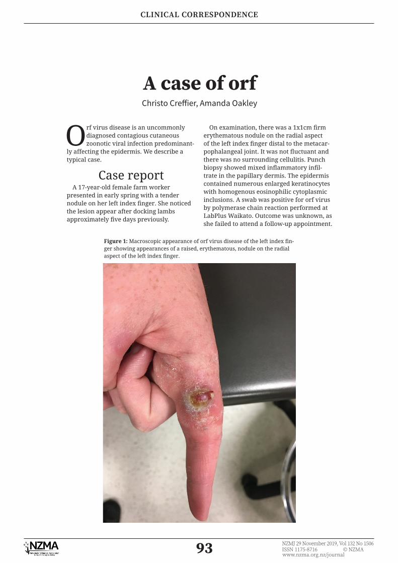
93 NZMJ 29 November 2019, Vol 132 No 1506 ISSN 1175-8716 © NZMA www.nzma.org.nz/journal A case of orf Christo Creffier, Amanda Oakley O rf virus disease is an uncommonly diagnosed contagious cutaneous zoonotic viral infection predominant- ly affecting the epidermis. We describe a typical case. Case report A 17-year-old female farm worker presented in early spring with a tender nodule on her left index finger. She noticed the lesion appear after docking lambs approximately five days previously. On examination, there was a 1x1cm firm erythematous nodule on the radial aspect of the left index finger distal to the metacar- pophalangeal joint. It was not fluctuant and there was no surrounding cellulitis. Punch biopsy showed mixed inflammatory infil- trate in the papillary dermis. The epidermis contained numerous enlarged keratinocytes with homogenous eosinophilic cytoplasmic inclusions. A swab was positive for orf virus by polymerase chain reaction performed at LabPlus Waikato. Outcome was unknown, as she failed to attend a follow-up appointment. Figure 1: Macroscopic appearance of orf virus disease of the left index fin- ger showing appearances of a raised, erythematous, nodule on the radial aspect of the left index finger. CLINICAL CORRESPONDENCE
Transcript of A case of orf... · 2020-01-30 · erythema multiforme. However, the disease is usually...
93 NZMJ 29 November 2019, Vol 132 No 1506ISSN 1175-8716 © NZMAwww.nzma.org.nz/journal
A case of orfChristo Cre� ier, Amanda Oakley
Orf virus disease is an uncommonly diagnosed contagious cutaneous zoonotic viral infection predominant-
ly affecting the epidermis. We describe a typical case.
Case reportA 17-year-old female farm worker
presented in early spring with a tender nodule on her left index fi nger. She noticed the lesion appear after docking lambs approximately fi ve days previously.
On examination, there was a 1x1cm fi rm erythematous nodule on the radial aspect of the left index fi nger distal to the metacar-pophalangeal joint. It was not fl uctuant and there was no surrounding cellulitis. Punch biopsy showed mixed infl ammatory infi l-trate in the papillary dermis. The epidermis contained numerous enlarged keratinocytes with homogenous eosinophilic cytoplasmic inclusions. A swab was positive for orf virus by polymerase chain reaction performed at LabPlus Waikato. Outcome was unknown, as she failed to attend a follow-up appointment.
Figure 1: Macroscopic appearance of orf virus disease of the left index fi n-ger showing appearances of a raised, erythematous, nodule on the radial aspect of the left index fi nger.
CLINICAL CORRESPONDENCE

94 NZMJ 29 November 2019, Vol 132 No 1506ISSN 1175-8716 © NZMAwww.nzma.org.nz/journal
Figure 2: Histological appearance of orf virus disease with direct view of pale cytoplasmic nuclear evacuation demonstrated by arrow A. There is also some intracytoplasmic and intranucleur inclusion typical of the virus as demonstrated by arrow B.
Figure 3: Histological appearance of orf virus disease with evidence of pale cytoplasmic nuclear evacuation as seen by arrow A.
CLINICAL CORRESPONDENCE

95 NZMJ 29 November 2019, Vol 132 No 1506ISSN 1175-8716 © NZMAwww.nzma.org.nz/journal
Competing interests:Nil.
Author information:Christo Creffi er, Waikato District Health Board, Hamilton;
Amanda Oakley, Dermatology, Waikato District Health Board, Hamilton.Corresponding author:
Dr Christo Creffi er, Urology, Waikato District Health Board, Hamilton.christocreffi [email protected]
URL:http://www.nzma.org.nz/journal/read-the-journal/all-issues/2010-2019/2019/vol-132-no-1506-
29-november-2019/8064
DiscussionOrf is uncommon and is mainly diagnosed
in rural communities in spring and early summer. Farmers, their families, shearers, slaughterers and butchers are at the greatest risk of developing orf if they are in close contact with sheep and goats (and rarely, other farm animals), untreated wool or the vehicles or buildings in which the animals have been housed. Only nine cases of orf were documented in New Zealand over the last 10 years but as the disease is well known in the rural community, it is likely that many incidences of the infection do not present to medical attention.1
Parapox virus infection is highly prevalent among sheep in New Zealand and can be diffi cult to eradicate once it enters a fl ock. A live vaccine is available to farms carrying the disease but, as immunity only lasts a few months, reinfection is common.2 Current management includes quarantine of the affected sheep and goats, with care made to avoid harsh environments to reduce the risk of cuts and abrasions.
The causative agent is an epithelio-tropic DNA parapoxvirus.3 Sheep and goats develop a ‘scabby mouth’ with a ‘contagious pustular dermatitis’ around the mouth, nose and teats, which is transmitted to other
animals via direct contact through cuts and abrasions. The developing symptoms vary depending on the location of the infection, but generally orf is painful and can lead to anorexia and starvation.4 The virus invades a damaged epidermis and replicates in follicular epithelium with a typical viral response involving of CD4+ helper cells and CD8+ cytotoxic T cells, antibodies and interferons.3
In humans, orf presents as a reddish-blue, targetoid, fl at-topped, blood-tinged, 2–5cm nodule, which is on the hand in 95% of cases; other sites include forearms and face.4 Immunocompetent humans may also develop a mild fever, lymphangitis and lymphadenopathy. Complications include secondary bacterial infection and reactive erythema multiforme. However, the disease is usually self-limiting, resolving in six weeks without scarring.
Orf virus disease is not notifi able to the Medical Offi cer of Health, as it is a relatively minor infection and is not transmitted from human to human.5 Orf virus disease may be under diagnosed and under-reported given the large population of sheep and goats in New Zealand. While orf is self-limiting, the secondary effects of bacterial infection are important to recognise, especially for clini-cians who work in a rural setting.
REFERENCES:1. Ministry of Health NZ
[Internet]. Ministry of Health NZ. 2018 [cited 23 December 2018]. Available from: http://www.health.govt.nz/
2. Scabby mouth. Fact Sheet. Beef + Lamb New Zealand, February 2007. Available from: http://beefl ambnz.com/knowledge-hub/PDF/scabby-mouth
3. McKeever D, McEwan Jenkinson D, Hutchison
G, Reid H. Studies of the pathogenesis of orf virus infection in sheep. Journal of Comparative Pathology. 1988; 99(3):317–328. Haig DM, Mercer AA. Ovine Diseases, Orf. Veterinary Research, 1998; 29(3–4):311–326.
4. Spickler AR. Contagious Ecthyma [Internet]. 2015 [cited 23 December 2018]. Available from: http://www.cfsph.iastate.
edu/Factsheets/pdfs/contagious_ecthyma.pdf
5. List of diseases notifi able by health practitioners and laboratories to the Medical Offi cer of Health, January 2017. Avail-able from http://www.health.govt.nz/system/fi les/documents/pages/schedule-of-notifi able-dis-eases-updated-jan2017.pdf
CLINICAL CORRESPONDENCE