A Case of Multicentric Gliomatosis Cerebri Diagnosed with ...e-jnc.org/upload/pdf/jnc-2-2-66.pdf ·...
4
CASE REPORT J Neurocrit Care 2009;2:66-69 ISSN 2005-0348 A Case of Multicentric Gliomatosis Cerebri Diagnosed with Brain Biopsy Dong-Gun Kim, MD and Hyun Sook Kim, MD Department of Neurology, Bundang CHA Medical Center, CHA University College of Medicine, Seongnam, Korea Background: Gliomatosis cerebri (GC) is a rare subtype of the CNS glioma and about 300 cases of GC are reported hitherto. Among those cases, multicentric GC was exceedingly uncommon. Case Report: A 63-year-old man was admitted with dizziness, nausea, and vomiting. Brain magnetic resonance image showed multiple, non-contiguous brain lesions in corpus callosum, right thalamus, midbrain and frontal and temporal cortices. The 14-3-3 protein was identified temporarily in this patient. Brain biopsy was performed to confirm the diagnosis and anaplastic astrocytoma was found at the right frontal cortex. Conclusion: This is the case report of pathologically confirmed GC with multicentric brain lesions. J Neurocrit Care 2009;2:66-69 KEY WORDS: Gliomatosis cerebri·Multicentric. 66 Copyright ⓒ 2009 The Korean Neurocritical Care Society Introduction Glioma is a type of primary central nervous system (CNS) tumor that arises from glial cells. Although gliomatosis cerebri (GC) is a subtype of the CNS glioma, it is distinct from other tumors of glial origin and classified as neuroepithelial tumor of uncertain origin. 1 Primary GC shows diffuse infiltration of neoplastic glial cells through more than two cerebral lobes or the infratentorial structures in the absence of any identifiable tumor mass. 2 Because neoplastic cells infiltrate the cerebral lobes with relatively mild destruction of the architecture of nor- mal surrounding tissues, clinical symptoms of GC might be very subtle in contrast to its radiological findings. 3,4 The GC has non-specific clinical presentations such as headache, sei- zure, memory impairment, confusion, unsteady gait, nausea, dizziness and vomiting. 3,4 GC is a rare disease and multicen- tric GC was exceedingly uncommon among hundreds of re- ported cases with GC. 5 We report a case of non-contiguous GC confirmed with the brain biopsy. Case A 63-year-old man was admitted with dizziness, nausea, and vomiting for 2 weeks. He was a 40-pack-year smoker and did not have any past surgical or medical history. Initial vital signs were stable and within normal limit, and physical exam was also normal. Neurological examination showed mild dys- arthria and bilateral sway on tandem gait. Other neurological exam including extraocular movement was normal. Routine laboratory tests including CBC, LFT, TFT, and autoimmune studies showed normal results. Tumor markers (AFP, CEA, PSA, CA19-9) and paraneoplastic autoantibody test (Hu anti- body, Ri antibody, Yo antibody, antinuclear antibody) were all negative. Brain magnetic resonance image (MRI) showed multiple scattered lesions in corpus callosum, right thalamus, right midbrain, and right frontal and temporal cortices (Fig. 1A). Contrast enhanced brain MRI and MR spectrography (MRS) was carried out at three weeks after the onset of symp- toms. Same lesions were found with iso-intensity signal on T1 weighted images and were not enhanced (Fig. 1B). On MRS, choline (Cho)/creatinine (Cr) and choline/N-acetylaspartate (NAA) ratio was not significantly different between bilateral temporal cortices. Routine CSF study and microbiologic mark- ers were all normal, but, 14-3-3 protein was positive. On elec- troencephalogram (EEG), posterior dominant rhythm was well regulated with 8 Hz activity, and there was no epilepti- form discharge, periodic complexes or focal slowing. Neuro- psychological (NP) test demonstrated preserved higher corti- cal functions except naming (Korean Boston naming test: 8 percentile). Neither dizziness nor dysarthria was improved significantly Address for correspondence: Hyun Sook Kim, MD Department of Neurology, Bundang CHA Medical Center, CHA University College of Medicine, 351 Yatap- dong, Bundang- gu, Seong- nam 463-712, Korea Tel: +82-31-780-5483, Fax: +82-31-780-5198 E-mail: [email protected] online © ML Comm
Transcript of A Case of Multicentric Gliomatosis Cerebri Diagnosed with ...e-jnc.org/upload/pdf/jnc-2-2-66.pdf ·...

CASE REPORTJ Neurocrit Care 2009;2:66-69 ISSN 2005-0348
A Case of Multicentric Gliomatosis Cerebri Diagnosed with Brain Biopsy
Dong-Gun Kim, MD and Hyun Sook Kim, MDDepartment of Neurology, Bundang CHA Medical Center, CHA University College of Medicine, Seongnam, Korea
Background: Gliomatosis cerebri (GC) is a rare subtype of the CNS glioma and about 300 cases of GC are reported hitherto. Among
those cases, multicentric GC was exceedingly uncommon. Case Report: A 63-year-old man was admitted with dizziness, nausea,
and vomiting. Brain magnetic resonance image showed multiple, non-contiguous brain lesions in corpus callosum, right thalamus,
midbrain and frontal and temporal cortices. The 14-3-3 protein was identified temporarily in this patient. Brain biopsy was performed
to confirm the diagnosis and anaplastic astrocytoma was found at the right frontal cortex. Conclusion: This is the case report of
pathologically confirmed GC with multicentric brain lesions. J Neurocrit Care 2009;2:66-69
KEY WORDS: Gliomatosis cerebri·Multicentric.
66 Copyright ⓒ 2009 The Korean Neurocritical Care Society
Introduction
Glioma is a type of primary central nervous system (CNS) tumor that arises from glial cells. Although gliomatosis cerebri (GC) is a subtype of the CNS glioma, it is distinct from other tumors of glial origin and classified as neuroepithelial tumor of uncertain origin.1 Primary GC shows diffuse infiltration of neoplastic glial cells through more than two cerebral lobes or the infratentorial structures in the absence of any identifiable tumor mass.2 Because neoplastic cells infiltrate the cerebral lobes with relatively mild destruction of the architecture of nor-mal surrounding tissues, clinical symptoms of GC might be very subtle in contrast to its radiological findings.3,4 The GC has non-specific clinical presentations such as headache, sei-zure, memory impairment, confusion, unsteady gait, nausea, dizziness and vomiting.3,4 GC is a rare disease and multicen-tric GC was exceedingly uncommon among hundreds of re-ported cases with GC.5 We report a case of non-contiguous GC confirmed with the brain biopsy.
Case
A 63-year-old man was admitted with dizziness, nausea,
and vomiting for 2 weeks. He was a 40-pack-year smoker and did not have any past surgical or medical history. Initial vital signs were stable and within normal limit, and physical exam was also normal. Neurological examination showed mild dys-arthria and bilateral sway on tandem gait. Other neurological exam including extraocular movement was normal. Routine laboratory tests including CBC, LFT, TFT, and autoimmune studies showed normal results. Tumor markers (AFP, CEA, PSA, CA19-9) and paraneoplastic autoantibody test (Hu anti-body, Ri antibody, Yo antibody, antinuclear antibody) were all negative. Brain magnetic resonance image (MRI) showed multiple scattered lesions in corpus callosum, right thalamus, right midbrain, and right frontal and temporal cortices (Fig. 1A). Contrast enhanced brain MRI and MR spectrography (MRS) was carried out at three weeks after the onset of symp-toms. Same lesions were found with iso-intensity signal on T1 weighted images and were not enhanced (Fig. 1B). On MRS, choline (Cho)/creatinine (Cr) and choline/N-acetylaspartate (NAA) ratio was not significantly different between bilateral temporal cortices. Routine CSF study and microbiologic mark-ers were all normal, but, 14-3-3 protein was positive. On elec-troencephalogram (EEG), posterior dominant rhythm was well regulated with 8 Hz activity, and there was no epilepti-form discharge, periodic complexes or focal slowing. Neuro-psychological (NP) test demonstrated preserved higher corti-cal functions except naming (Korean Boston naming test: 8 percentile).
Neither dizziness nor dysarthria was improved significantly
Address for correspondence: Hyun Sook Kim, MD Department of Neurology, Bundang CHA Medical Center, CHA University College of Medicine, 351 Yatap-dong, Bundang-gu, Seong-nam 463-712, Korea Tel: +82-31-780-5483, Fax: +82-31-780-5198E-mail: [email protected]
online © ML Comm
Multicentric Gliomatosis with Brain Biopsy ▌ DG Kim, et al.
67
A
B
C
D
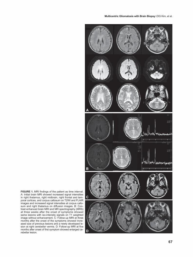
FIGURE 1. MRI findings of the patient as time interval. A: Initial brain MRI showed increased signal intensities in right thalamus, right midbrain, right frontal and tem-poral cortices, and corpus callosum on T2WI and FLAIR images and increased signal intensities at corpus callo-sum and right thalamus on diffusion images. B: Con-trast enhanced brain MRI and MR spectrography (MRS) at three weeks after the onset of symptoms showed same lesions with iso-intensity signals on T1 weighted image without enhancement. C: Follow-up MRI at three months after the onset of the symptoms showed incre-ased size of previous lesions and a newly developed le-sion at right cerebellar vermis. D: Follow-up MRI at five months after onset of first symptom showed enlarged ce-rebellar lesion.

J Neurocrit Care ▌ 2009;2:66-69
68
with steroid pulse therapy. Instead, nausea and vomiting dis-appeared with digestives and GI motility drugs for following two months. After then, dizziness, nausea and vomiting dis-turbed the daily activities of the patient again. Follow-up MRI at three months after the onset of the symptoms showed incre-ased size of previous lesions and a newly developed lesion at right cerebellar vermis (Fig. 1C). Brain and whole body pos-itron emission tomography (PET) was performed to rule out the underlying malignancy. However, it showed no abnormal glucose metabolism except decreased glucose metabolism at bilateral frontal and parietal lobes (Fig. 2). Follow-up EEG was normal and 14-3-3 protein of CSF became negative for two times. IgG index was 0.51 and oligoclonal band was neg-ative. Gait and truncal ataxia became prominent at 5months
after the onset of first symptoms and follow-up MRI showed enlarged cerebellar lesion (Fig. 1D). We performed the brain biopsy on right frontal cortex and anaplastic astrocytoma was found (Fig. 3). After the biopsy, mental status of the patient became comatose without additional focal neurological defi-cit. Further brain imaging and anti-cancer treatment were re-fused by the family members. The patient died at three weeks after the biopsy.
Discussion
Clinical symptoms or radiological features of primary GC are non-specific and patients are often misdiagnosed with other neurological disease such as CNS inflammatory disease, arteriopathies or leukoencephalopathies.5 MRI using contrast media generally provides important information for the diag-nosis of brain tumor and contrast enhancement means the breakdown of blood-brain barrier, which is often associated with high grade tumor. However, it was reported that about 80% of patients with GC showed no contrast enhancing le-sions on T1WI despite its malignancy.5 It was also true in our patient; initial symptoms of the patient were non-specific and there was no mass lesion with prominent contrast enhance-ment. Subsequently, the next non-invasive diagnostic meth-ods could be MRS, in which malignant GC demonstrates el-evated ratio of Choline/NAA6 and FDG-PET scan, in which high grade brain tumors generally show increased glucose metabolism.7 However, in our case, the ratio of Choline/NAA was not elevated on MRS and there was no difference of glu-cose metabolism in the abnormal lesion on PET scan. Fur-thermore, unlike other GC, our case has multicentric brain le-sions; diffuse infiltrations at corpus callosum, right thalamus, midbrain and distant focal lesions of frontal and temporal cor-tices. Those results made it difficult to diagnose properly with-out the brain biopsy.
Multiple gliomas are well-recognized but uncommon enti-ty. They are grouped in two categories: multifocal and multi-centric gliomas. Multifocal gliomas disseminate along an es-
FIGURE 2. Brain PET scan showed mildly decreased glucose metabolism on bilateral frontoparietal cortex. Bilateral BG, thala-mus and cerebellum showed symmetric glucose metabolism.
A B
FIGURE 3. Photomicrography of the biopsy specimen from the right frontal cortex. A: The tumor cells are diffusely infiltrative with high cellu-larity and produce little destruction of preexisting parenchyma. H&E st-ain, ×40. B: It composed of fibrillary glial cells that resemble astrocytes with nuclear atypia. Mitoses are ge-nerally infrequent and vascular pro-liferation and necrosis is absent. H&E stain, ×200.

Multicentric Gliomatosis with Brain Biopsy ▌ DG Kim, et al.
69
tablished route, spreading through commissural pathways, CSF channels, blood or by local extension through satellite forma-tions; multicentric gliomas are separated lesions that cannot be attributed to one of the above pathways.8,9 There are two dif-ferent hypotheses regarding widespread nature of GC, i.e., 1) GC arises from simultaneous neoplastic transformation of cells in different regions of the brain (oligoclonal origin hypo-thesis) by stimulation of specific intrinsic or extrinsic10,11 carci-nogenic influences or 2) GC arises from a single cone of cells and then spreads widely (monoclonal origin hypothesis).4 Al-though the MRI of our case was compatible with multicen-tric glioma/oligoclonal origin hypothesis, PET findings sug-gested the involvement of bilateral frontal and parietal cor-tices. It is possible that there were underlying neoplastic cell infiltration without MRI signal changes.
Despite the 14-3-3 protein assay has proved useful in the diagnosis of Creutzfeldt-Jakob disease (CJD) with average sensitivity and specificity of 92%,12,13 it can be found in the pa-tients with astrocytoma, herpes simplex encephalitis, hypoxic brain damage, atypical encephalitis, intracerebral metastases of a bronchial carcinoma, metabolic encephalopathy, and pro-gressive dementia of unknown cause.14 Then, it is recommend-ed to perform the test twice and if contradictory, a third test must be done to establish a final result.13 In this case, the path-ology of anaplastic astrocytoma made the false positive result in 14-3-3 protein assay and third test established the negative result.
GC showed fatal outcomes and overall median survival was 14.5 months in the 296 cases of GC with treatment.5 Ra-diotherapy was reported to improve clinical symptoms, but it usually had minimal effect on patients’ survival. However, re-cent randomized controlled trial performed in patients with glioblastoma has showed a survival benefit with concomitant and adjuvant temozolomide added to conventional radiother-apy. In this study, the 5 year-survival rate was 10% in the pa-tients with temozolomide plus radiotherapy versus 2% in the patients with radiotherapy alone.15 This result can be applied in patients with GC.
Our case was compatible with the GC in that multiple lobe invasions were confirmed with radiological findings and ana-plastic astrocytoma was confirmed with the brain biopsy. Al-
though many studied other than biopsy might be helpful in part, we suggest that pathological confirm should be required for the diagnosis of GC.
REFERENCES1. Kleihues P, Louis DN, Scheithauer BW, Rorke LB, Reifenberger G,
Burger PC, et al. The WHO classification of tumors of the nervous system. J Neuropathol Exp Neurol 2002;61:215-25.
2. Lantos PL, Bruner JM. Gliomatosis cerebri in tumours of the nervous system. In: Kleihues P, Cavence WK (eds). Pathology and Genetics of Tumours of the nervous system. Lyon: IARC Press. 2000;92-3.
3. del Carpio-O’Donovan R, Korah I, Salazar A, Melancon D. Glioma-tosis cerebri. Radiology 1996;198:831-5.
4. Vates GE, Chang S, Lamborn KR, Prados M, Berger MS. Gliomato-sis cerebri: a review of 22 cases. Neurosurgery 2003;53:261-71.
5. Taillibert S, Chodkiewicz C, Laigle-Donadey F, Napolitano M, Carta-lat-Carel S, Sanson M. Gliomatosis cerebri: a review of 296 cases from the ANOCEF database and the literature. J Neurooncol 2006;76: 201-5.
6. Bendszus M, Warmuth-Metz W, Klein R, Burger R, Schichor C, Tonn JC, et al. MR spectroscopy in gliomatosis cerebri. AJNR Am J Neuro-radiol 2000;21:375-80.
7. Padma MV, Said S, Jacobs M, Hwang DR, Dunigan K, Satter M, et al. Prediction of pathology and survival by FGD PET in gliomas. J Neu-rooncol 2003;49:227-37.
8. Zamponi N, Rychlichi F, Ducati A, Regnicolo L, Salvolini U, Ricciuti RA. Multicentric glioma with unusual clinical presentation. Childs Nerv Syst 2001;17:101-5.
9. Faria AV, Azevedo GC, Zanardi VA, Ghizoni E, Queiroz LS. Dissem-ination patterns of pilocytic astrocytoma. Clin Neurol Neurosurg 2006; 108:568-72.
10. Bussone G, Sinatra MG, Boiardi A, Lazzaroni M, Mariani C, Allegran-za A. A case of glioblastoma with multiple centers above and below the tentorium. J Neurol 1979;221:187-92.
11. Kato T, Aida T, Abe H, Ogata A, Nakamura N, Nagashima K, et al. Cli-nicopathological study of multiple gliomas--report of three cases. Neu-rol Med Chir (Tokyo) 1990;30:604-9.
12. Hsich G, Kenney K, Gibbs CJ, Lee KH, Harrington MG. The 14-3-3 brain protein in cerebrospinal fluid as a marker for transmissible spongiform encephalopathies. N Engl J Med 1996;335:924-30.
13. Van Everbroeck B, Boons J, Cras P. Cerebrospinal fluid biomarkers in Creutzfeldt-Jakob disease. Clin Neurol Neurosurg 2005;107:355-60.
14. Zerr I, Bodemer M, Gefeller O, Otto M, Poser S, Wiltfang J, et al. De-tection of 14-3-3 protein in the cerebrospinal fluid supports the diag-nosis of Creutzfeldt-Jakob disease. Ann Neurol 1998;43:32-40.
15. Stupp R, Hegi ME, Mason WP, van den Bent MJ, Taphoorn MJ, Jan-zer RC, et al. Effects of radiotherapy with concomitant and adjuvant temozolomide versus radiotherapy alone on survival in glioblastoma in a randomised phase III study: 5-year analysis of the EORTC-NCIC trial. Lancet Oncol 2009;10:459-66.