A Case of Elephantiasis Nostras Verrucosa - KoreaMed · Elephantiasis nostras verrucosa (ENV) is a...
4
326 Ann Dermatol Received March 13, 2009, Accepted for publication April 2, 2009 Reprint request to: Choong-Rim Haw, M.D., Department of Derma- tology, School of Medicine, Kyunghee University, 1, Hoegi-dong, Dongdaemun-gu, Seoul 130-702, Korea. Tel: 82-2-958-8510, Fax: 82-2-969-6538, E-mail: [email protected] Ann Dermatol Vol. 21, No. 3, 2009 CASE REPORT A Case of Elephantiasis Nostras Verrucosa Yun-Seok Yang, M.D., Jae-Jun Ahn, M.D., Sik Haw, M.D., Min-Kyung Shin, M.D., Choong-Rim Haw, M.D. Department of Dermatology, School of Medicine, Kyunghee University, Seoul, Korea Elephantiasis nostras verrucosa (ENV) is a rare clinical condition associated with chronic non-filarial lymphedema caused by bacterial or non-infectious lymphatic obstruction. A variety of etiologies, including infection, tumor obstruc- tion, trauma, radiation, chronic venous stasis, congestive heart failure, and obesity, can lead to chronic lymphatic obstruction and edema. Mossy papules, plaques, and cobblestone-like nodules are clinically impressive features of ENV, but biopsy reveals only moderately abnormal findings such as pseudoepitheliomatous hyperplasia, dilated lymphatic spaces, fibrous tissue hyperplasia, and chronic inflammation. We present a case of ENV in a 67-year-old man with a 10-year history of multiple nodules and verrucous plaques on both feet. Microbiology ruled out a filarial infection. Nodule biopsy revealed pseudoepitheli- omatous hyperplasia, marked dermal fibrosis, and a chronic inflammatory infiltrate. No evidence of carcinoma was identified. Both venous stasis and recurrent cellulitis could contribute to the dermal fibrotic changes of the lesions. However, before the recurrent cellulitis, he did not have any nodular lesions on his feet despite a 10-year history of venous disease. Therefore, this case suggests that venous stasis alone cannot produce the fibrotic nodular lesions of ENV. (Ann Dermatol 21(3) 326∼329, 2009) -Keywords- Cellulitis, Elephantiasis, Elephantiasis nostras verrucosa, Venous stasis INTRODUCTION In 1969, Castellani 1 classified elephantiasis into four subtypes: 1) elephantiasis tropica - due to filariasis; 2) ele- phantiasis nostras - due to bacterial infection; 3) elephan- tiasis symptomatica - due to mycotic, syphilitic, tuber- culoid, neoplastic, or traumatic causes of lymphatic ob- struction; and 4) elephantiasis congenita - associated with inherited disorders such as Milroy’s disease. Elephantiasis nostras verrucosa (ENV) was originally defined as a condition resulting from lymphatic blockage caused by recurrent bacterial infection and also called lymphangitis recurrens elephantogenica. However, many recent papers have included neoplastic, traumatic, and other non- infectious causes of lymphatic obstruction in the definition of ENV 2-4 . ENV is a rare disease, and only one case of elephantiasis nostras has been described in the Korean dermatologic literature 5 . Herein, we report a case of ENV due to re- current cellulitis and chronic venous stasis and offer suggestions concerning its possible pathogenesis. CASE REPORT A 67-year-old man presented with a chief complaint of slowly enlarging multiple hard papules and plaques on both feet over the past 10 years. Ten years ago, he had been in the habit of drinking raw deer blood as a health food, and after that suffered from toxic erythema with some erosion on both feet. Subsequently, he was hospi- talized several times for recurrent cellulitis of both feet. He first noticed venous symptoms such as heaviness of the legs, swelling, and mild pain while standing 20 years prior to presentation at our hospital but had declined any interventions that were recommended at other hospitals. Examination of the feet revealed confluent pinkish or brownish verrucous papules and plaques with a moss- covered appearance surrounding the peripheries of both
Transcript of A Case of Elephantiasis Nostras Verrucosa - KoreaMed · Elephantiasis nostras verrucosa (ENV) is a...
326 Ann Dermatol
Received March 13, 2009, Accepted for publication April 2, 2009
Reprint request to: Choong-Rim Haw, M.D., Department of Derma-tology, School of Medicine, Kyunghee University, 1, Hoegi-dong, Dongdaemun-gu, Seoul 130-702, Korea. Tel: 82-2-958-8510, Fax: 82-2-969-6538, E-mail: [email protected]
Ann Dermatol Vol. 21, No. 3, 2009
CASE REPORT
A Case of Elephantiasis Nostras Verrucosa
Yun-Seok Yang, M.D., Jae-Jun Ahn, M.D., Sik Haw, M.D., Min-Kyung Shin, M.D., Choong-Rim Haw, M.D.
Department of Dermatology, School of Medicine, Kyunghee University, Seoul, Korea
Elephantiasis nostras verrucosa (ENV) is a rare clinical condition associated with chronic non-filarial lymphedema caused by bacterial or non-infectious lymphatic obstruction. A variety of etiologies, including infection, tumor obstruc-tion, trauma, radiation, chronic venous stasis, congestive heart failure, and obesity, can lead to chronic lymphatic obstruction and edema. Mossy papules, plaques, and cobblestone-like nodules are clinically impressive features of ENV, but biopsy reveals only moderately abnormal findings such as pseudoepitheliomatous hyperplasia, dilated lymphatic spaces, fibrous tissue hyperplasia, and chronic inflammation. We present a case of ENV in a 67-year-old man with a 10-year history of multiple nodules and verrucous plaques on both feet. Microbiology ruled out a filarial infection. Nodule biopsy revealed pseudoepitheli-omatous hyperplasia, marked dermal fibrosis, and a chronic inflammatory infiltrate. No evidence of carcinoma was identified. Both venous stasis and recurrent cellulitis could contribute to the dermal fibrotic changes of the lesions. However, before the recurrent cellulitis, he did not have any nodular lesions on his feet despite a 10-year history of venous disease. Therefore, this case suggests that venous stasis alone cannot produce the fibrotic nodular lesions of ENV. (Ann Dermatol 21(3) 326∼329, 2009)
-Keywords-Cellulitis, Elephantiasis, Elephantiasis nostras verrucosa, Venous stasis
INTRODUCTION
In 1969, Castellani1 classified elephantiasis into four subtypes: 1) elephantiasis tropica - due to filariasis; 2) ele-phantiasis nostras - due to bacterial infection; 3) elephan-tiasis symptomatica - due to mycotic, syphilitic, tuber-culoid, neoplastic, or traumatic causes of lymphatic ob-struction; and 4) elephantiasis congenita - associated with inherited disorders such as Milroy’s disease. Elephantiasis nostras verrucosa (ENV) was originally defined as a condition resulting from lymphatic blockage caused by recurrent bacterial infection and also called lymphangitis recurrens elephantogenica. However, many recent papers have included neoplastic, traumatic, and other non- infectious causes of lymphatic obstruction in the definition of ENV2-4.ENV is a rare disease, and only one case of elephantiasis nostras has been described in the Korean dermatologic literature5. Herein, we report a case of ENV due to re-current cellulitis and chronic venous stasis and offer suggestions concerning its possible pathogenesis.
CASE REPORT
A 67-year-old man presented with a chief complaint of slowly enlarging multiple hard papules and plaques on both feet over the past 10 years. Ten years ago, he had been in the habit of drinking raw deer blood as a health food, and after that suffered from toxic erythema with some erosion on both feet. Subsequently, he was hospi-talized several times for recurrent cellulitis of both feet. He first noticed venous symptoms such as heaviness of the legs, swelling, and mild pain while standing 20 years prior to presentation at our hospital but had declined any interventions that were recommended at other hospitals. Examination of the feet revealed confluent pinkish or brownish verrucous papules and plaques with a moss- covered appearance surrounding the peripheries of both
A Case of Elephantiasis Nostras Verrucosa
Vol. 21, No. 3, 2009 327
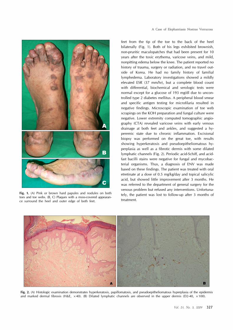
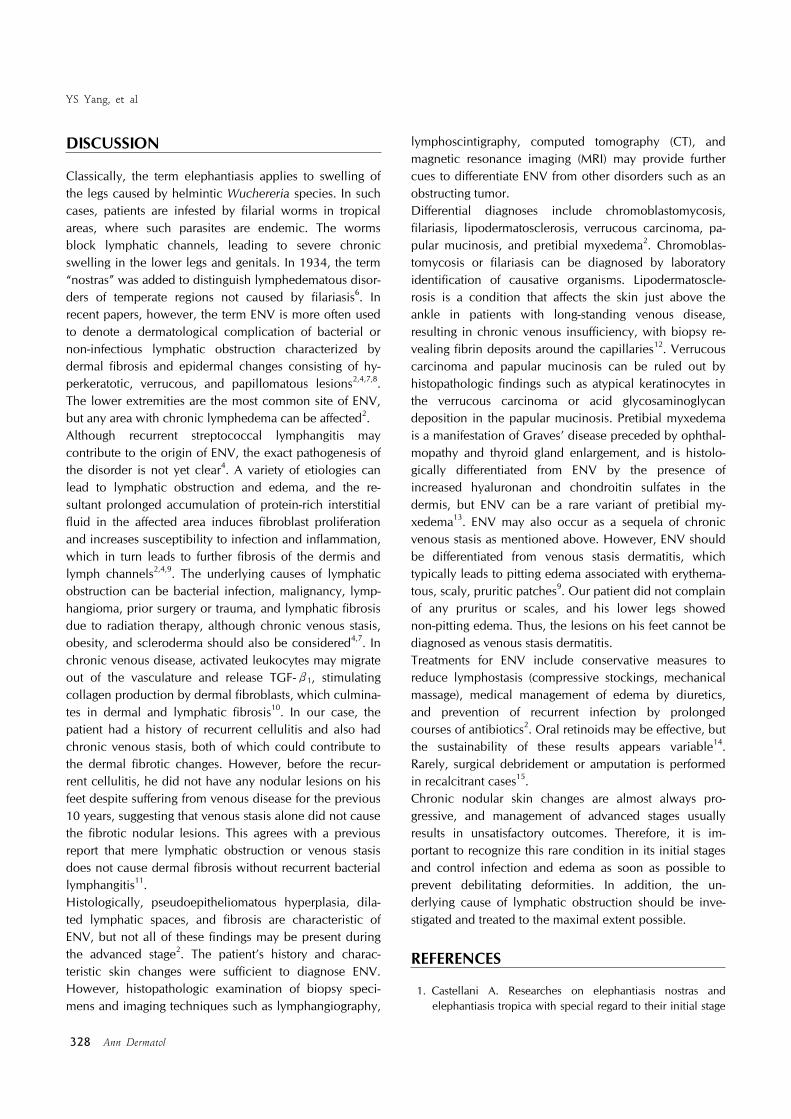
Fig. 1. (A) Pink or brown hard papules and nodules on bothtoes and toe webs. (B, C) Plaques with a moss-covered appearan-ce surround the heel and outer edge of both feet.
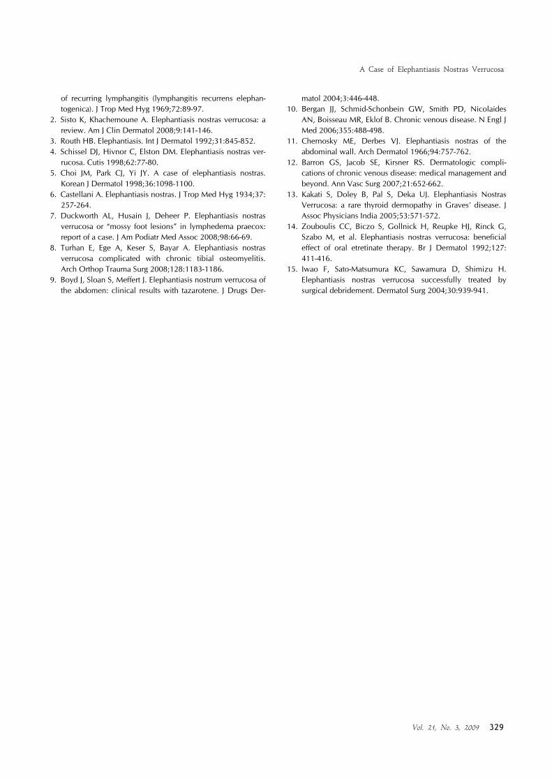
Fig. 2. (A) Histologic examination demonstrates hyperkeratosis, papillomatosis, and pseudoepitheliomatous hyperplasia of the epidermisand marked dermal fibrosis (H&E, ×40). (B) Dilated lymphatic channels are observed in the upper dermis (D2-40, ×100).
feet from the tip of the toe to the back of the heel bilaterally (Fig. 1). Both of his legs exhibited brownish, non-pruritic maculopatches that had been present for 10 years after the toxic erythema, varicose veins, and mild, nonpitting edema below the knee. The patient reported no history of trauma, surgery or radiation, and no travel out-side of Korea. He had no family history of familial lymphedema. Laboratory investigations showed a mildly elevated ESR (37 mm/hr), but a complete blood count with differential, biochemical and serologic tests were normal except for a glucose of 193 mg/dl due to uncon-trolled type 2 diabetes mellitus. A peripheral blood smear and specific antigen testing for microfilaria resulted in negative findings. Microscopic examination of toe web scrapings on the KOH preparation and fungal culture were negative. Lower extremity computed tomographic angio-graphy (CTA) revealed varicose veins with early venous drainage at both feet and ankles, and suggested a hy-peremic state due to chronic inflammation. Excisional biopsy was performed on the great toe, with results showing hyperkeratosis and pseudoepitheliomatous hy-perplasia as well as a fibrotic dermis with some dilated lymphatic channels (Fig. 2). Periodic acid-Schiff, and acid- fast bacilli stains were negative for fungal and mycobac-terial organisms. Thus, a diagnosis of ENV was made based on these findings. The patient was treated with oral etretinate at a dose of 0.5 mg/kg/day and topical salicylic acid, but showed little improvement after 3 months. He was referred to the department of general surgery for the venous problem but refused any interventions. Unfortuna-tely, the patient was lost to follow-up after 3 months of treatment.
YS Yang, et al
328 Ann Dermatol
DISCUSSION
Classically, the term elephantiasis applies to swelling of the legs caused by helmintic Wuchereria species. In such cases, patients are infested by filarial worms in tropical areas, where such parasites are endemic. The worms block lymphatic channels, leading to severe chronic swelling in the lower legs and genitals. In 1934, the term “nostras” was added to distinguish lymphedematous disor-ders of temperate regions not caused by filariasis6. In recent papers, however, the term ENV is more often used to denote a dermatological complication of bacterial or non-infectious lymphatic obstruction characterized by dermal fibrosis and epidermal changes consisting of hy-perkeratotic, verrucous, and papillomatous lesions2,4,7,8. The lower extremities are the most common site of ENV, but any area with chronic lymphedema can be affected2.Although recurrent streptococcal lymphangitis may contribute to the origin of ENV, the exact pathogenesis of the disorder is not yet clear4. A variety of etiologies can lead to lymphatic obstruction and edema, and the re-sultant prolonged accumulation of protein-rich interstitial fluid in the affected area induces fibroblast proliferation and increases susceptibility to infection and inflammation, which in turn leads to further fibrosis of the dermis and lymph channels2,4,9. The underlying causes of lymphatic obstruction can be bacterial infection, malignancy, lymp-hangioma, prior surgery or trauma, and lymphatic fibrosis due to radiation therapy, although chronic venous stasis, obesity, and scleroderma should also be considered4,7. In chronic venous disease, activated leukocytes may migrate out of the vasculature and release TGF-β1, stimulating collagen production by dermal fibroblasts, which culmina-tes in dermal and lymphatic fibrosis10. In our case, the patient had a history of recurrent cellulitis and also had chronic venous stasis, both of which could contribute to the dermal fibrotic changes. However, before the recur-rent cellulitis, he did not have any nodular lesions on his feet despite suffering from venous disease for the previous 10 years, suggesting that venous stasis alone did not cause the fibrotic nodular lesions. This agrees with a previous report that mere lymphatic obstruction or venous stasis does not cause dermal fibrosis without recurrent bacterial lymphangitis11.Histologically, pseudoepitheliomatous hyperplasia, dila-ted lymphatic spaces, and fibrosis are characteristic of ENV, but not all of these findings may be present during the advanced stage2. The patient’s history and charac-teristic skin changes were sufficient to diagnose ENV. However, histopathologic examination of biopsy speci-mens and imaging techniques such as lymphangiography,
lymphoscintigraphy, computed tomography (CT), and magnetic resonance imaging (MRI) may provide further cues to differentiate ENV from other disorders such as an obstructing tumor.Differential diagnoses include chromoblastomycosis, filariasis, lipodermatosclerosis, verrucous carcinoma, pa-pular mucinosis, and pretibial myxedema2. Chromoblas-tomycosis or filariasis can be diagnosed by laboratory identification of causative organisms. Lipodermatoscle-rosis is a condition that affects the skin just above the ankle in patients with long-standing venous disease, resulting in chronic venous insufficiency, with biopsy re-vealing fibrin deposits around the capillaries12. Verrucous carcinoma and papular mucinosis can be ruled out by histopathologic findings such as atypical keratinocytes in the verrucous carcinoma or acid glycosaminoglycan deposition in the papular mucinosis. Pretibial myxedema is a manifestation of Graves’ disease preceded by ophthal-mopathy and thyroid gland enlargement, and is histolo-gically differentiated from ENV by the presence of increased hyaluronan and chondroitin sulfates in the dermis, but ENV can be a rare variant of pretibial my-xedema13. ENV may also occur as a sequela of chronic venous stasis as mentioned above. However, ENV should be differentiated from venous stasis dermatitis, which typically leads to pitting edema associated with erythema-tous, scaly, pruritic patches9. Our patient did not complain of any pruritus or scales, and his lower legs showed non-pitting edema. Thus, the lesions on his feet cannot be diagnosed as venous stasis dermatitis.Treatments for ENV include conservative measures to reduce lymphostasis (compressive stockings, mechanical massage), medical management of edema by diuretics, and prevention of recurrent infection by prolonged courses of antibiotics2. Oral retinoids may be effective, but the sustainability of these results appears variable14. Rarely, surgical debridement or amputation is performed in recalcitrant cases15.Chronic nodular skin changes are almost always pro-gressive, and management of advanced stages usually results in unsatisfactory outcomes. Therefore, it is im-portant to recognize this rare condition in its initial stages and control infection and edema as soon as possible to prevent debilitating deformities. In addition, the un-derlying cause of lymphatic obstruction should be inve-stigated and treated to the maximal extent possible.
REFERENCES
1. Castellani A. Researches on elephantiasis nostras and elephantiasis tropica with special regard to their initial stage
A Case of Elephantiasis Nostras Verrucosa
Vol. 21, No. 3, 2009 329
of recurring lymphangitis (lymphangitis recurrens elephan-togenica). J Trop Med Hyg 1969;72:89-97.
2. Sisto K, Khachemoune A. Elephantiasis nostras verrucosa: a review. Am J Clin Dermatol 2008;9:141-146.
3. Routh HB. Elephantiasis. Int J Dermatol 1992;31:845-852.4. Schissel DJ, Hivnor C, Elston DM. Elephantiasis nostras ver-
rucosa. Cutis 1998;62:77-80.5. Choi JM, Park CJ, Yi JY. A case of elephantiasis nostras.
Korean J Dermatol 1998;36:1098-1100.6. Castellani A. Elephantiasis nostras. J Trop Med Hyg 1934;37:
257-264.7. Duckworth AL, Husain J, Deheer P. Elephantiasis nostras
verrucosa or “mossy foot lesions” in lymphedema praecox: report of a case. J Am Podiatr Med Assoc 2008;98:66-69.
8. Turhan E, Ege A, Keser S, Bayar A. Elephantiasis nostras verrucosa complicated with chronic tibial osteomyelitis. Arch Orthop Trauma Surg 2008;128:1183-1186.
9. Boyd J, Sloan S, Meffert J. Elephantiasis nostrum verrucosa of the abdomen: clinical results with tazarotene. J Drugs Der-
matol 2004;3:446-448.10. Bergan JJ, Schmid-Schonbein GW, Smith PD, Nicolaides
AN, Boisseau MR, Eklof B. Chronic venous disease. N Engl J Med 2006;355:488-498.
11. Chernosky ME, Derbes VJ. Elephantiasis nostras of the abdominal wall. Arch Dermatol 1966;94:757-762.
12. Barron GS, Jacob SE, Kirsner RS. Dermatologic compli-cations of chronic venous disease: medical management and beyond. Ann Vasc Surg 2007;21:652-662.
13. Kakati S, Doley B, Pal S, Deka UJ. Elephantiasis Nostras Verrucosa: a rare thyroid dermopathy in Graves' disease. J Assoc Physicians India 2005;53:571-572.
14. Zouboulis CC, Biczo S, Gollnick H, Reupke HJ, Rinck G, Szabo M, et al. Elephantiasis nostras verrucosa: beneficial effect of oral etretinate therapy. Br J Dermatol 1992;127: 411-416.
15. Iwao F, Sato-Matsumura KC, Sawamura D, Shimizu H. Elephantiasis nostras verrucosa successfully treated by surgical debridement. Dermatol Surg 2004;30:939-941.