A Case of Bilateral Coronary to Pulmonary Artery Fistulas ... · combined with valvular heart...
4
331 CASE REPORT Korean Circ J 2008;38:331-334 Print ISSN 1738-5520 / On-line ISSN 1738-5555 Copyright ⓒ 2008 The Korean Society of Cardiology A Case of Bilateral Coronary to Pulmonary Artery Fistulas Associated With Severe Aortic Regurgitation Seung-Jae Lee, MD, Sung-Ho Her, MD, Seung-Won Jin, MD, Jong-Min Lee, MD, Hee-Jeoung Yoon, MD, Hee-Yeon Lee, MD, Hee-Yeon Kim, MD, Gun-Min Kim, MD and Cheol-Hong Park, MD Division of Cardiology, College of Medicine, The Catholic University of Korea, Daejeon, Korea ABSTRACT Coronary artery fistula (CAF) is a rare form of congenital anomalies of the coronary arteries, and this is usually discovered by chance during coronary angiography. However, this type of fistula can cause important coronary morbidity and mortality leading to angina, syncope, congestive heart failure, myocardial infarction and sudden death. Bilateral CAFs are even rarer, and especially when combined with valvular heart disease. The coincidence of CAF with aortic regurgitation is relatively rare and this might sometimes cause myocardial ischemia. We pre- sent here a case of bilateral coronary-pulmonary artery fistulas that arose from the first diagonal branch of the left anterior descending artery and the conal branch of the right coronary artery combined with severe aortic regurgitation, and this all caused myocardial ischemia. (Korean Circ J 2008;38:331-334) KEY WORDS: Arteriovenous fistula; Coronary vessels; Aortic regurgitation. Introduction Congenital anomalies of the coronary arteries are rare and they have been discovered in 0.67% of the patients who undergo coronary arteriography and in 0.8% of patients at autopsy. 1)2) Although rare, they can cause important coronary morbidity and mortality lead- ing to angina, syncope, congestive heart failure, myo- cardial infarction and sudden death. 3) Coronary artery fistula (CAF) is an abnormal com- munication between an epicardial coronary artery and the cardiac chambers, the major vessels (the vena cavae, sub-pulmonary veins and pulmonary artery) or other structures. 4)5) Congenital CAFs joining into the pulmo- nary artery are rare cardiac anomalies. CAFs arising from two coronary arteries are even rarer, especially when they are combined with valvular heart disease. 6-9) Furthermore, a few cases of coronary-pulmonary artery fistula combined with aortic regurgitation have been previously reported. 6)9) To the best of our knowledge, the present report may be the first Korean case of 55-year-old woman with severe aortic regurgitation and she also had bilateral coronary-pulmonary artery fistulas. Case A fifty-five year old female patient was admitted with a squeezing type chest pain of 5 days duration, and this symptom was not effort related. She did not have any atherosclerotic risk factors or any significant past medical history. There was no family history of aortic, collagen, vascular or congenital heart disease. She had a regular pulse rate of 71 beats/min, a blood pressure of 140/30 mmHg and a respiratory rate 16 breaths/min. On physical examination, cardiac auscultation re- vealed 4 to 6 grade diastolic murmurs at the aortic and pulmonary areas radiating to the upper back. Her elecrocardiography demonstrated left ventricular hypertrophy by the voltage. No ST segment and T wave changes were apparent. The blood chemistries, including the cardiac enzyme and lipid profiles, were within normal limits. On chest X-ray, cardiomegaly was noted (cardiothoracic ratio: 0.6). Both transthoracic echocardiography and transesopha- geal echocardiography relatively revealed a mildly dilated left ventricle and good regional wall motion with a left ventricular ejection fraction of 65%, but she had se- Received: November 9, 2007 Revision Received: January 22, 2008 Accepted: February 27, 2008 Correspondence: Sung-Ho Her, MD, Division of Cardiolo gy, Colle ge of Medicine, The Catholic University of Korea, Daejeon St. Mary’ s Hospital, 520- 2 Daeheung-dong, Jung-gu, Daejeon 301-723, Korea Tel: 82-42-220-9504, Fax: 82-42-253-9505 E-mail: [email protected]
Transcript of A Case of Bilateral Coronary to Pulmonary Artery Fistulas ... · combined with valvular heart...
331
CASE REPORT
Korean Circ J 2008;38:331-334 Print ISSN 1738-5520 / On-line ISSN 1738-5555
Copyright ⓒ 2008 The Korean Society of Cardiology
A Case of Bilateral Coronary to Pulmonary Artery Fistulas Associated With Severe Aortic Regurgitation Seung-Jae Lee, MD, Sung-Ho Her, MD, Seung-Won Jin, MD, Jong-Min Lee, MD, Hee-Jeoung Yoon, MD, Hee-Yeon Lee, MD, Hee-Yeon Kim, MD, Gun-Min Kim, MD and Cheol-Hong Park, MD Division of Cardiology, College of Medicine, The Catholic University of Korea, Daejeon, Korea ABSTRACT
Coronary artery fistula (CAF) is a rare form of congenital anomalies of the coronary arteries, and this is usually discovered by chance during coronary angiography. However, this type of fistula can cause important coronary morbidity and mortality leading to angina, syncope, congestive heart failure, myocardial infarction and sudden death. Bilateral CAFs are even rarer, and especially when combined with valvular heart disease. The coincidence of CAF with aortic regurgitation is relatively rare and this might sometimes cause myocardial ischemia. We pre-sent here a case of bilateral coronary-pulmonary artery fistulas that arose from the first diagonal branch of the left anterior descending artery and the conal branch of the right coronary artery combined with severe aortic regurgitation, and this all caused myocardial ischemia. (Korean Circ J 2008;38:331-334) KEY WORDS: Arteriovenous fistula; Coronary vessels; Aortic regurgitation.
Introduction
Congenital anomalies of the coronary arteries are
rare and they have been discovered in 0.67% of the patients who undergo coronary arteriography and in 0.8% of patients at autopsy.1)2) Although rare, they can cause important coronary morbidity and mortality lead-ing to angina, syncope, congestive heart failure, myo-cardial infarction and sudden death.3)
Coronary artery fistula (CAF) is an abnormal com-munication between an epicardial coronary artery and the cardiac chambers, the major vessels (the vena cavae, sub-pulmonary veins and pulmonary artery) or other structures.4)5) Congenital CAFs joining into the pulmo-nary artery are rare cardiac anomalies. CAFs arising from two coronary arteries are even rarer, especially when they are combined with valvular heart disease.6-9) Furthermore, a few cases of coronary-pulmonary artery fistula combined with aortic regurgitation have been previously reported.6)9)
To the best of our knowledge, the present report
may be the first Korean case of 55-year-old woman with severe aortic regurgitation and she also had bilateral coronary-pulmonary artery fistulas.
Case
A fifty-five year old female patient was admitted with a squeezing type chest pain of 5 days duration, and this symptom was not effort related. She did not have any atherosclerotic risk factors or any significant past medical history. There was no family history of aortic, collagen, vascular or congenital heart disease.
She had a regular pulse rate of 71 beats/min, a blood pressure of 140/30 mmHg and a respiratory rate 16 breaths/min.
On physical examination, cardiac auscultation re-vealed 4 to 6 grade diastolic murmurs at the aortic and pulmonary areas radiating to the upper back.
Her elecrocardiography demonstrated left ventricular hypertrophy by the voltage. No ST segment and T wave changes were apparent.
The blood chemistries, including the cardiac enzyme and lipid profiles, were within normal limits. On chest X-ray, cardiomegaly was noted (cardiothoracic ratio: 0.6). Both transthoracic echocardiography and transesopha-geal echocardiography relatively revealed a mildly dilated left ventricle and good regional wall motion with a left ventricular ejection fraction of 65%, but she had se-
Received: November 9, 2007
Revision Received: January 22, 2008 Accepted: February 27, 2008
Correspondence: Sung-Ho Her, MD, Division of Cardiology, College of
Medicine, The Catholic University of Korea, Daejeon St. Mary’s Hospital, 520-
2 Daeheung-dong, Jung-gu, Daejeon 301-723, Korea
Tel: 82-42-220-9504, Fax: 82-42-253-9505
E-mail: [email protected]
332·Bilateral Coronary AV Fistulas With Severe AR
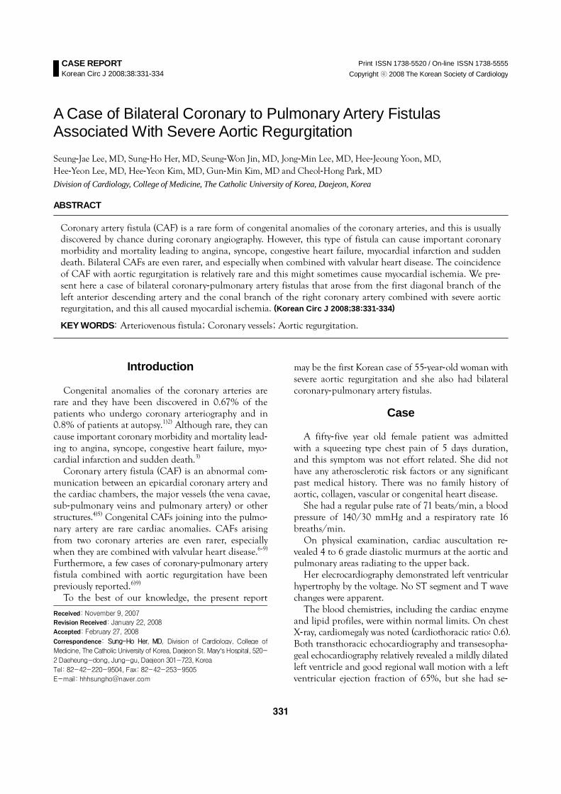
vere aortic regurgitation with incapacitated, minimally calcified cusps of the aortic valve (Fig. 1). The thallium 201 myocardial perfusion scintigraphy was normal.
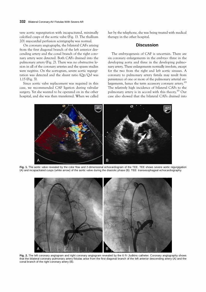
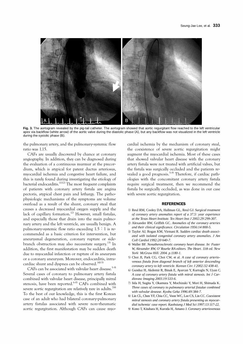
On coronary angiography, the bilateral CAFs arising from the first diagonal branch of the left anterior des-cending artery and the conal branch of the right coro-nary artery were detected. Both CAFs drained into the pulmonary artery (Fig. 2). There was no obstructive le-sion in all of the coronary arteries and the spasm studies were negitive. On the aortogram, severe aortic regurgi-tation was detected and the shunt ratio (Qp/Qs) was 1.15 (Fig. 3).
Since aortic valve replacement was required in this case, we recommended CAF ligation during valvular surgery. Yet she wanted to be operated on in the other hospital, and she was then transferred. When we called
her by the telephone, she was being treated with medical therapy in the other hospital.
Discussion
The embryogenesis of CAF is uncertain. There are six coronary enlargements in the embryo: three in the developing aorta and three in the developing pulmo-nary artery. These enlargements normally involute, except for the two from the right and left aortic sinuses. A coronary to pulmonary artery fistula may result from persistence of one or more of the pulmonary arterial en-largements, hence the term accessory coronary artery.10) The relatively high incidence of bilateral CAFs to the pulmonary artery is in accord with this theory.10) Our case also showed that the bilateral CAFs drained into
A B
Fig. 2. The left coronary angiogram and right coronary angiogram revealed by the 6 Fr Judkins catheter. Coronary angiography shows that the bilateral coronary pulmonary artery fistulas arise from the first diagonal branch of the left anterior descending artery (A) and the conal branch of the right coronary artery (B).
A B
Fig. 1. The aortic valve revealed by the color flow and 2-dimensional echocardiogram of the TEE. TEE shows severe aortic regurgigation(A) and incapacitated cusps (white arrow) of the aortic valve during the diastolic phase (B). TEE: transesophageal echocardiography.
Seung-Jae Lee, et al.·333
the pulmonary artery, and the pulmonary-systemic flow ratio was 1.15.
CAFs are usually discovered by chance at coronary angiography. In addition, they can be diagnosed during the evaluation of a continuous murmur at the precor-dium, which is atypical for patent ductus arteriosus, myocardial ischemia and congestive heart failure, and this is rarely found during investigating the etiology of bacterial endocarditis.11)12) The most frequent complaints of patients with coronary artery fistula are angina pectoris, atypical chest pain and lethargy. The patho-physiologic mechanisms of the symptoms are volume overload as a result of the shunt, coronary steal that causes a decreased myocardial oxygen supply and the lack of capillary formation.12) However, small fistulas, and especially those that drain into the main pulmo-nary artery and the left ventricle, are usually benign. A pulmonary-systemic flow ratio exceeding 1.5:1 is re-commended as a basic criterion for intervention, but aneurysmal degeneration, coronary rupture or side-branch obstruction may also necessitate surgery.13) In addition, the first manifestation may be sudden death due to myocardial infarction or rupture of its aneurysm or a coronary aneurysm. Moreover, endocarditis, intra-cardiac shunt and dyspnea can be observed.14)15)
CAFs can be associated with valvular heart disease.5-9) Several cases of coronary to pulmonary artery fistula combined with valvular heart disease, principally mitral stenosis, have been reported.6-8) CAFs combined with severe aortic regurgitation are relatively rare in adults.7)9) To the best of our knowledge, this is the first Korean case of an adult who had bilateral coronary-pulmonary artery fistulas associated with severe non-rheumatic aortic regurgitation. Although CAFs can cause myo-
cardial ischemia by the mechanism of coronary steal, the coexistence of severe aortic regurgitation might augment the myocardial ischemia. Most of these cases that showed valvular heart disease with the coronary artery fistula were not treated with artificial valves, but the fistula was surgically occluded and the patients re-vealed a good prognosis.5-9) Therefore, if cardiac path-ologies with the concomitant coronary artery fistula require surgical treatment, then we recommend the fistula be surgically occluded, as was done in our case with severe aortic regurgitation.
REFERENCES 1) Reul RM, Cooley DA, Hallman GL, Reul GJ. Surgical treatment
of coronary artery anomalies report of a 37.5: year experience at the Texas Heart Institute. Tex Heart Inst J 2002;29:299-307.
2) Alexander RW, Griffith GC. Anomalies of the coronary arteries and their clinical significance. Circulation 1956;14:800-5.
3) Taylor AJ, Rogan KM, Virmani R. Sudden cardiac death associ-ated with isolated congenital coronary artery anomalies. J Am Coll Cardiol 1992;20:640-7.
4) Waller BF. Nonatherosclerotic coronary heart disease. In: Fuster W, Alexander RW, O’ Rourke RA editors. The Heart. 11th ed. New York: McGraw Hill; 2004. p.1180-1.
5) Choi JI, Park CG, Choi CW, et al. A case of coronary arterio-venous fistula from diagonal branch of left anterior descending coronary artery to left ventricle. Korean Circ J 2002;32:438-41.
6) Gunduz H, Akdemir R, Binak E, Ayarcan Y, Kurtoglu N, Uyan C. A case of coronary artery fistula with mitral stenosis. Int J Car-diovasc Imaging 2003;19:533-6.
7) Iida H, Sugita Y, Okamura Y, Mochizuki Y, Mori H, Shimada K. Three cases of coronary to pulmonary arterial fistulae combined with valvular desease. Kyobu Geka 1996;49:384-7.
8) Lin CL, Chen YF, Chiu CC, Voon WC, Lee CS, Lin CC. Coexistent mitral stenosis and coronary artery fistula presenting as myocar-dial ischemia: case report. Kaohsiung J Med Sci 1997;13:117-22.
9) Kono T, Kitahara H, Kuroda H, Amano J. Coronary arteriovenous
A B
Fig. 3. The aortogram revealed by the pig-tail catheter. The aortogram showed that aortic regurgitant flow reached to the left ventricularapex via backflow (white arrow) of the aortic valve during the diastolic phase (A), but any backflow was not visualized in the left ventricle during the systolic phase (B).

334·Bilateral Coronary AV Fistulas With Severe AR
fistula with annuloaortic ectasia and aortic regurgitation. Jpn J Thorac Cardiovasc Surg 2001;49:135-7.
10) Perloff JK. Congenital coronary artery fistula. In: Perloff JK editor. Clinical Recognition of Congenital Heart Disease. 5th ed. Philadelphia: Saunders; 2003. p. 443-5.
11) Kim MS, Ahn YK, Bae Y, et al. Clinical charateristics of coronary arteriovenous fistula in Korean adults. Korean Circ J 1997;27: 900-6.
12) Sapin P, Frantz E, Jain A, Nichols TC, Dehmer GJ. Coronary artery fistula: an abnormality affecting all age groups. Medicine
1990;69:101-13. 13) Angelini P, Velasco JA, Flamm S. Coronary anomalies: inci-
dence, pathophysiology, and clinical relevance. Circulation 2002; 105:2449-54.
14) Rajs J, Brodin LA, Hertfeld I, Larsen FF. Death related to coronary artery fistula after rupture of an aneurysm to the coronary sinus. Am J Forensic Med Pathol 2001;22:58-61.
15) Hobbs RE, Millit HD, Raghavan PV, Moodie DS, Sheldon WC. Coronary artery fistula: a 10 year review. Cleve Clin Q 1982;49: 191-7.