Chronic Lymphocytic Leukemia ARTICLE SF3B1-mutated chronic ...

A CADASILA CADASIL--mutated Notch 3 receptor exhibits mutated Notch 3 receptor exhibits impaired intracellularimpaired intracellular trafficking and maturationtrafficking and maturation
but normal but normal ligandligand--induced signalinginduced signalingHelena Karlström, Paul Beatus, Karin Dannaeus, Gavin Chapman, Urban Lendahl, and Johan Lundkvist
Department of Cell and Molecular Biology, Medical Nobel Institute, Karolinska Institute, Stockholm, Sweden
PNAS; December 24,2002; Vol.99; no.26
ARZU SANDIKCIMolecular and Cellular Biology Master Program‘Molecular Mechanisms in Health and Disease’
WS 04/0522.01.2005

OUTLINE
• Notch Signaling• CADASIL• Aim and the Approach• Results• Discussion

NOTCH SIGNALING
• Notch Receptor:
Extracellular IntracellularTransmembrane
Single transmembrane receptors
Large number of tandemly organized EGF repeats in extracellular domain
Missense mutations removing or inserting cysteine residues in the EGF repeats, cause CADASIL

NOTCH SIGNALING
Notch Signaling is a system for cell-cell communication
Notch receptor undergoes a series of proteolyticprocessing events, which are a prerequisite for signaling
Golgi apparatus*S1 cleavage Extracellular side
Furin like convertase
Cell surface*S2 cleavage Extracellular sideclose to the plasma membrane
Cell surface*S3 cleavage Within the plasma membrane
γ-secretase
Ligand interaction
IC domain Nucleus

CADASILCADASIL CADASIL
(Cerebral Autosomal Dominant Arteriopathy with Subcortical Infarcts and Leukoencephalopathy)
• vascular stroke and dementia syndrome • has a late onset*• characterized by degeneration of vascular smooth
muscle cells and multiple small infarcts in the white and deep gray matter of the brain.
• autosomal dominant • mutations in the human Notch 3 gene • migraine, multiple infarcts and dementia• MRI is required for diagnosis

CADASIL
Normal Diseased

AIM OF THE PAPERTo find out ;• At what level a CADASIL mutation affects Notch 3
receptor function• Whether the mutations in EGF repeats primarily
affect receptor trafficking, maturation and/or signaling
• How CADASIL-mutated Notch 3 receptors lead to degeneration of vascular smooth muscle cells
• Whether this is a consequence of altered receptor trafficking, maturation, and/or signaling

APPROACH
Address the cellular mechanism underlying CADASIL
• By comparing *intracellular trafficking, *localization and *signalingfrom wild type mNotch 3 and the CADASIL-causing mutant mNotch 3 receptor in cultured human embryonic kidney (HEK) 293 cells
OUTLINE
• Notch Signaling• CADASIL• Aim and the Approach• Results• Discussion

Does the mutation have an effect on S1 cleavage?
The amount of S1 processed receptor was reduced for mutated Notch 3
Less EC (210kDa) was expressed in favor of the full length mNotch 3 receptor (280kDa)
Western blot analysis – Antibodies against EC

Is the amount of receptor reaching the plasmamembrane altered?
Cell surface biotinylation streptavidin pull down assay western blot
The relative amount of cell surface expression differed between wild typeNotch 3 and mutated Notch 3
Both wild type and the mutant receptors reach the cell surface exclusivelyin the S1 processed form
T: 5% of Total protein extract
B: 95% of Total protein extract; subject to streptavidin pull down
Wild type
Mutant

Can full length Notch 3 receptor respond to knownNotch ligands?
Co-culture with 3T3 cells expressing either Jagged1 or Delta1 ligandsLuciferase gene readout
??? Similar levels of receptor activation were observed for the wild type and the mutant receptor expressing cells

Signaling capacity, in more sensitive way:Insertion of GVP signaling domain into IC
Only signals from engineered receptor, not from endogenous Notch receptors arerecorded.
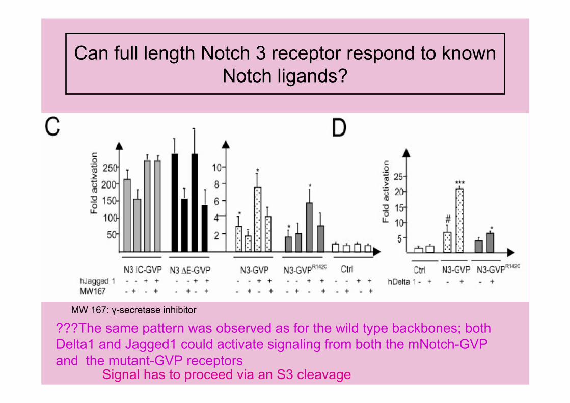
Can full length Notch 3 receptor respond to knownNotch ligands?
Signal has to proceed via an S3 cleavage
MW 167: γ-secretase inhibitor
???The same pattern was observed as for the wild type backbones; bothDelta1 and Jagged1 could activate signaling from both the mNotch-GVP and the mutant-GVP receptors

Are mutated Notch 3 receptors more prone tointracellular aggregation?
A large intracellular aggregate is formed in a majority (75%) of the R142C-GVP expressing cells; compared to only 25% of the Notch 3-GVP expressingcells.
Problems in transporting receptors through the ER; impaired ER transportresults in ER stress
Immunocytochemistry: Antibodies against GVP domain (α Gal4 antibody)
Brefeldin A treatment impairs vesiculartransport between ER and Golgi

What is the intracellular localization of the aggregates?
Calnexin: ER CTR433: Golgi apparatus p58: intermediate compart.
The aggregates did not co-localize any of the structures, but often located in the vicinity of the Golgi complex
Immunocytochemistry; double staining with markers for ER, Golgi andthe intermediate compartment

Are these intracellular aggregates Aggresomes?
Agresomes are cytoplasmic structures formed in response to ER stress. They are high MW complexes that contain ubiquitin, localize to thecentrosomes and are caged with vimentin.
The mNotch 3 containing aggregates were often localized close tocentrosomes but were not caged with vimentin and did not contain ubiquitin.
The introduction of the mutation increases the propensity to form cytoplasmicaggregates containing mNotch 3

• For the mutant receptors: – S1 cleavage is reduced– Less receptors are found at the cell surface– ? Can respond to ligand induction as wild type
receptors– Form intracellular aggregates
CONCLUSION

• The reduction in S1 cleavage;1. Mutant receptors are less potent for the
cleavage enzyme, furin like convertase2. Transport of mutated receptor through ER is
impaired or slowed down fewer receptorsbecoming avaible for S1 cleavage
Results in fewer receptors at the cellsurface only S1 processed receptorappears at the plasma membrane
DISCUSSION
• ER stress may play role in the lateonset of the disease
• Since there is no alteration of thesignaling from the receptor…
DISCUSSION

References from internet:
• http://www.neurocast.com/site/content/sessions_12_2000.asp
• http://ghr.nlm.nih.gov/gene=notch3• http://ghr.nlm.nih.gov/condition=cadasil• http://www.geneclinics.org/profiles/cadasil/• http://www.thedoctorsdoctor.com/diseases
/cadasil.htm#pathogenesis

THANK YOU FOR YOUR ATTENTION