9.14 - Brain Structure and its Origins Spring 2005 ......MIT 9.14 Class 5 Sketch of a pre-mammalian...
54
A sketch of the central nervous system and its origins G. Schneider 2005 Part 3: Specializations in the evolving CNS; introduction to connection patterns MIT 9.14 Class 5 Sketch of a pre-mammalian brain; some specializations and some basic pathways 9.14 - Brain Structure and its Origins Spring 2005 Massachusetts Institute of Technology Instructor: Professor Gerald Schneider
Transcript of 9.14 - Brain Structure and its Origins Spring 2005 ......MIT 9.14 Class 5 Sketch of a pre-mammalian...

A sketch of the central nervous system and its origins
G. Schneider 2005Part 3: Specializations in the evolving CNS;
introduction to connection patterns
MIT 9.14 Class 5Sketch of a pre-mammalian brain;
some specializations and some basic pathways
9.14 - Brain Structure and its OriginsSpring 2005Massachusetts Institute of TechnologyInstructor: Professor Gerald Schneider

Keep in mind:Basics of behavior enabling survival
• The most basic (multipurpose) actions of individual organisms from amoebae to human: – Approach/avoid – Orient towards/away– Explore/forage/seek
• Each evolutionary advance had to incorporate these multipurpose actions, needed for various goal-directed activities.

Two Terms:
• "notochord" vs. "notocord" – (either one is accepted by me, but 1st is proper)
• "spinal chord" vs. "spinal cord" – (only the latter is proper)

Ontogeny & phylogeny re: Evolution
• Can development tell us something about evolution?• Does "ontogeny recapitulate phylogeny"?
– Early in development, the human embryo looks nearly the same as a pig embryo as well as the same as a monkey or ape embryo. At later stages, the divergence from pig is more evident, while the similarity to monkeys and (finally) apes remains.
– It appears that the earlier the stage of embryonic development, the greater the resemblance of different species, excluding the very early (gastrula) stages.
– Ernst Haeckel presented an oversimplfied drawing of this, which became very well known.
• The next slides illustrate this. The first shows the similarities first noticed by Karl Ernst von Baer (1828). The second is Haeckel’s drawing.

Figure by MIT OCW.
from Romanes, 1901.“Ontogeny recapitulates phylogeny”

“The Developmental Hourglass” (Ernst Haeckel)
Figure by MIT OCW.
Early Gastrula Stages
"Phylotypic" Stage
Adults

Review of Evolution

The outline of the high-priority behavioral demands:
• One purpose is to call your attention to the organization of behavior as seen throughout the chordate phylum,
• focusing first on pre-mammalian vertebrates,• then on the mammals.
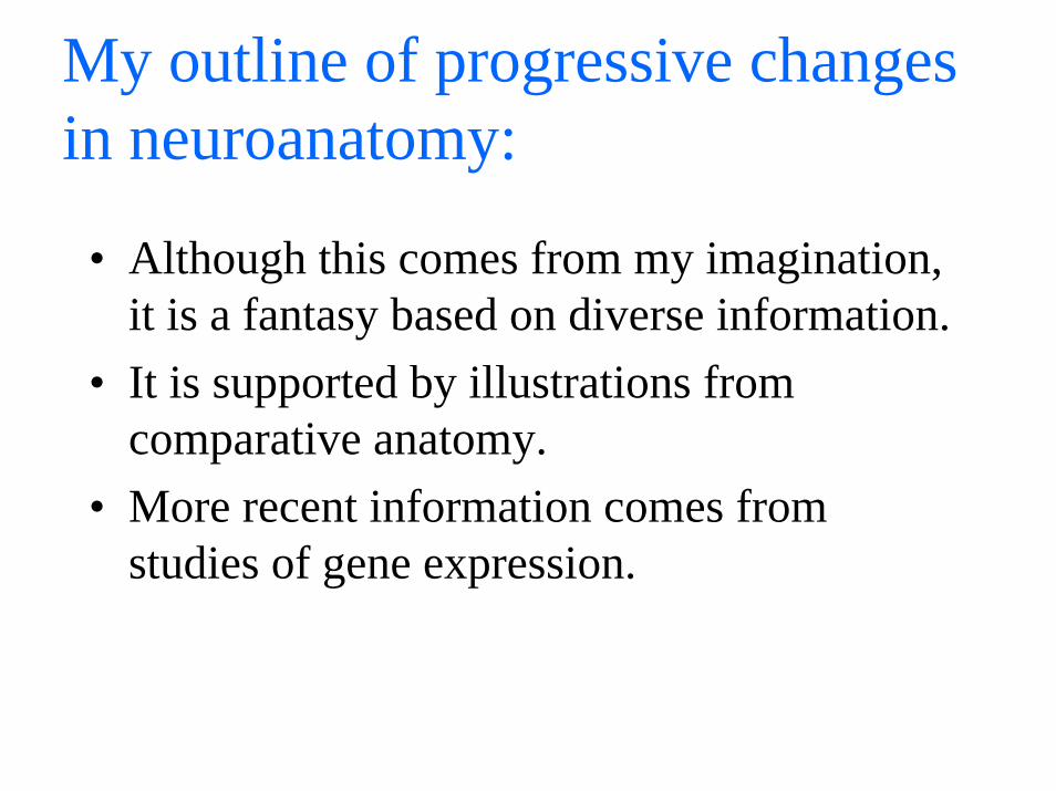
My outline of progressive changes in neuroanatomy:
• Although this comes from my imagination, it is a fantasy based on diverse information.
• It is supported by illustrations from comparative anatomy.
• More recent information comes from studies of gene expression.

Comments:
• Note the contrast between roles of somatosensoryand distance receptor inputs in shaping brain evolution. – The most primitive distance receptor (not including
receptors for light-dark detection) was probably olfaction: Recall the description of the early expansion of the forebrain in pre-mammalian vertebrates.
– Later expansions of the forebrain involved other distance receptors, especially visual and auditory.

Other sensory specializations have evolved, for example:• Electro-reception in some fish Cerebellar expansion
in Mormyrid fish• Infra-red detectors in the pit vipers specialized
trigeminal nerve (5th cranial nerve) inputs• Echo-location in bats and porpoises specialized
auditory system• High visual acuity and learned object recognition in
primates expansion of visual cortical areas• Vibrissal sensitivity in rodents specialized
somatosensory system

The cerebellar valvula of Mormyrids
Figure by MIT OCW.
ValvulaCerebellum
Telencephalon Diencephalon Medulla
ElectrosensoryLateral Line Lobe
1 mm

Figure by MIT OCW.
Sensory (Infrared) pit
Maxillary Branch
Mandibular Branch
Ophthalmic Branch
Rattlesnake trigeminal nerve: innervation of a specialized distance sense (Butler & Hodos)

Midbrain roof: the colliculi in four mammals
Figure by MIT OCW.
ECHOLOCATING BAT DOLPHIN
IBEX TARSIER
2 mm 1 cm
5 cm1 mm
{ {
{ {
IC
IC
SC
SC
IC
IC
SC
SC

Visual areas, owl monkey neocortex
Figure by MIT OCW.
VISUAL AREAS, OWL MONKEY NEOCORTEX
V1
V2M
LATERAL VIEW
MT

Three successive tangential sections of rat hemisphere, Nissl stain, showing the barrel fields
Specimen slides removed due to copyright reasons.

Other behavioral specializations
• The hand in apes, raccoons and, especially, humans (with high tactile acuity & fine motor control) expansions of somatic sensory-motor cortex and of cerebellar hemispheres
• Prehensile tail in spider monkey expanded tail representations in sensory & motor neocortex
• Language in humans polymodal association cortex of parietal, temporal and frontal lobes, with hemispheric specialization
• Elaborate social organization with planning and problem solving expanded prefrontal neocortical areas in primates

Somatosensory neocortex, hand area of two species
Figure by MIT OCW.
Abanerjee
Oval
Abanerjee
Oval
Abanerjee
Oval
Abanerjee
Oval
Abanerjee
Oval
Abanerjee
Oval
Abanerjee
Oval
Abanerjee
Oval
Abanerjee
Oval
Abanerjee
Oval
Abanerjee
Oval
Abanerjee
Oval
Abanerjee
Oval
Abanerjee
Oval
Abanerjee
Oval
Abanerjee
Oval
Abanerjee
Oval
Abanerjee
Oval
Abanerjee
Oval
Abanerjee
Oval
Abanerjee
Oval
Abanerjee
Oval
Abanerjee
Oval

Schematic outline of connections in vertebrate brains
topics
• Basic subdivisions, neuron types• Sensory channels of conduction• Overview of cephalic structures • Neocortex and its elaboration in mammals

Basic subdivisions, neuron types
• Introduced with schematic brain diagrams: Shmoo 1 and Shmoo 2
• We will identify basic structures and learn the definitions.

What is a “Shmoo” ?• It is useful to invent an imaginary creature that represents
animals ancestral to mammals. We will do this in a manner similar to one used in lectures by W.J.H. Nauta at MIT (1960s, early 1970s).
• Remember that we have already discussed amphioxus-like creatures that we think represent the earliest chordates.
• Later we will illustrate the mammalian additions to the ancestral brain.
• The name comes from a light-hearted student, noticing the resemblance of Nauta’s diagrams to a comic strip creature invented by the cartoonist Al Capp.

So lighten up, and Bring Back Al Capp’s Shmoo !
Scan of news article, "Bring Back the Schmoo!" removed due to copyright reasons.

Remember the Shmoo…• Because it loves all course 9 students, instinctively and
without fail, no matter what.• Because its devotion is completely reliable, arising from the
instincts buried deep within its limbic system.• Because its love and devotion to course 9 students can
never be overridden by higher cognitive functions, since it has none of those. (It has no neocortex.)
• Because it will gladly die by dissecting itself to teach you neuroanatomy.
• Because it never judges you for forgetting your math, physics, or subject requirements (it cannot handle numbers), and it never pretends that language is for anything but expressing feelings.

More on the Shmoo brain
• It originated as a pedagogical device of W.J.H. Nauta, to illustrate some basic principles of the anatomical organization of the brain of animals suggested to be ancestral to mammals.
• “Shmoo 1” could represent a generalized primitive amphibianbrain, based on the work of C.J. Herrick and more recent neuroanatomists. If we want to be more accurate concerning evolution, it could represent an early Cynodont of the Jurassic period, ancestral to the mammals (see Allman ch 5).
• Relevance to mammals, including human: This is the “old chassis”upon which new parts were built in evolution. Little is really discarded. (You still have that ancestral brain in the core of your human brain!) Note also that “old” is a relative term. The amphibian brain is very advanced in comparison to more primitive chordates.
• Later we will discuss in more detail some of the evolutionary transitions that gave rise to major changes in the CNS.

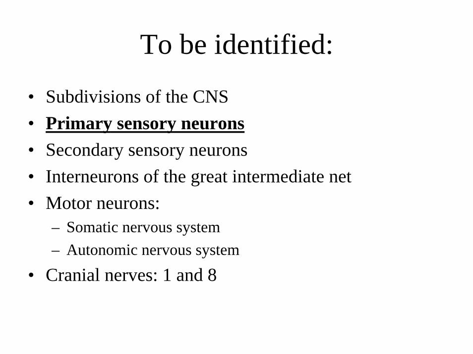
To be identified:
• Subdivisions of the CNS• Primary sensory neurons• Secondary sensory neurons• Interneurons of the great intermediate net• Motor neurons:
– Somatic nervous system– Autonomic nervous system
• Cranial nerves: 1 and 8
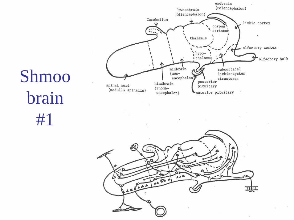
Shmoobrain
#1

Figure by MIT OCW.
The vertebrate brain archetype

A MORE REALISTIC VIEW:
The thickening embryonic neural tube in mammals
To be identified:
• Subdivisions of the CNS• Primary sensory neurons• Secondary sensory neurons• Interneurons of the great intermediate net• Motor neurons:
– Somatic nervous system– Autonomic nervous system
• Cranial nerves: 1 and 8

Primary somatosensory neurons in an animal series
1) Find one like this in nd
Sensory cell of the earthworm
Sensory cell of a mollusc
Sensory cell of a lower fish
Sensory cell of amphibian, reptile, bird, or mammal
2) thma
the Shmoo brain (amammals).
Find one like this in e Shmoo brain (and mmals)
3) Find one like this in the Shmoo brain (and mammals)
Figure by MIT OCW. Based on p.388 of Recollections of my life, by S. Ramon Cajal. MIT Press: Cambridge MA, 1989.

To be identified:
• Subdivisions of the CNS• Primary sensory neurons• Secondary sensory neurons• Interneurons of the great intermediate net• Motor neurons:
– Somatic nervous system– Autonomic nervous system
• Cranial nerves: 1 and 8


Sensory channels of conduction:
• Local reflex• Lemniscal 1• Lemniscal 2 (cerebellar)
• Terms: segmental, suprasegmental, dermatome, myotome, reticular formation, thalamus, decussation, cranial nerves.


How do we define “local” in “local reflex”?
• In the periphery: the dermatome• In the CNS: the segment

Dermatome map
Figure by MIT OCW.
C6
C3
C3
C5T4
T4
T5
T5
T6
T6
T7
T7
T8
T8
T9
T9
T10
T10
L1
L1
L2
L2
L3
L3
L4
L5
T1
T2
T2
C8C7
T11
T11
T12
T12
C2
C2
C4
C4
S3
S4
L5
T2
T3
T3
C4
T2
T1C6
C7
C8S3
S5
S4
S2
L3
L4L5
S1

Dermatome map, quadruped position, & spinal segments
Figure by MIT OCW.

How were the dermatomes mapped?
• Hypersensitive regions, resulting from irritation of single spinal roots, e.g., from a herniated intervertebral disk.
• Remaining sensibility, after severance of adjacent spinal nerves or dorsal roots.
• Cf. “myotome”

Sensory channels of conduction:• Local reflex• Lemniscal 1
– 1a (spinoreticular)– 1b (spinothalamic)
• Lemniscal 2 (spinocerebellar)
• Terms: segmental, suprasegmental, dermatome, myotome, reticular formation, thalamus, decussation, cranial nerves.

The oldest lemniscus: Spinoreticular
• A rostral extension of “propriospinal” fibers • Bilateral projections (largely ipsilateral)• Functions (most likely):
– Sensory modulation of autonomic activity (e.g., heart rate, blood pressure, breathing rate and volume)
– Autonomic and defensive behavioral responses to pain inputs– Temperature regulation– Sexual
• This is the least studied of the somatosensory pathways

Not included in the above:A primitive somatosensory modality in hagfishes, resembling tasteJ Comp Neurol. 1998, 392(2):135-163. Schreiner organs: a new craniatechemosensory modality in hagfishes, by CB Braun (University of California at San Diego, La Jolla).
From the abstract:These sensory organs are found throughout the epidermis. They are found also
at high densities in the prenasal sinus, nasopharyngeal duct, and pharynx, and at lower densities in the oral and velar chambers.
Schreiner organs are innervated by all sensory trigeminal rami, the glossopharyngeal/vagal nerve, and cutaneous rami of spinal nerves, but not by the facial nerve. The central projections of these rami form a continuous tract in the trigeminal sensory zone and the dorsolateral funiculus of the spinal cord.
This sensory modality of hagfishes has no direct homolog in vertebrates and appears to be a specialization of hagfishes, perhaps derived from the primitive somatosensory system of the earliest craniates.

Comparison of a cephalochordate (top: Amphioxus) and two jawless vertebrates:(middle) Hagfish(bottom) Lamprey
Amphioxus (Branchiostoma)
1 cm
5 cm
Hagfish (Myxine)
Lamprey (Petromyzon)
5 cm
o
o
t
t
d
d
m
m
o = olfactory Bulb
t = telencephalond = diencephalonm = mesencephalon
Figure by MIT OCW.

Vertebrates
Jawed Vertebrates
Cartilaginous FishesBony Vertebrates
Lobe-Finned VertebratesRay-Finned Fishes
Amph
ioxu
sHa
gfish
esLa
mpr
eys
Shar
ks, R
ays
Ratfi
shes
Amni
otes
Salam
ande
rsFr
ogs,T
oads
Coela
cant
hLu
ngfis
hes
Poly
pter
usSt
urgeo
nsGa
rsTe
leosts
Amia
Plac
ental
sM
arsu
pials
Mon
otre
mes
Liza
rds,
Snak
esTu
rtles
Bird
s
Croc
odile
s
Osteo
glos
som
orph
sEe
ls
Herri
ngs
Pike
sOs
tario
phys
ans
Neot
eleos
tsSa
lmon
SauropsidsMammals
Amniotes Teleosts
CLADOGRAM OF VERTEBRATES
Tetrapods
Figure by MIT OCW.

Notes on Amphioxus:brain and somatosensory inputs
• Gene studies indicate that this little creature is not brainless as once believed.
• Recent studies have indicated inputs from the body surface that enter the CNS and distribute to both sides (like inputs to the spinoreticular pathways of vertebrates).
• (Earlier, we have noted evidence for visual inputs to a forebrain.)

Early evolution of a crossed pathway
• We call the “paleolemniscus” of the spinal cord the “spinothalamic tract” – although most axons never reach the diencephalon, as they terminate in the hindbrain and midbrain.
• Why do the axons decussate? – The evolutionary origins of crossed
representations will be considered when we study the visual system.
– My suggestion/hypothesis concerning this topic was introduced in the previous class.

The “paleolemniscus” or “spinothalamic” tract
(Shmoo 1, side view)

The thickening embryonic neural tube
–top view and
sections

“Spinothalamic tract”
Note terms: decussation,reticular formation,thalamus (in ‘tweenbrain),endbrain.
Remember:
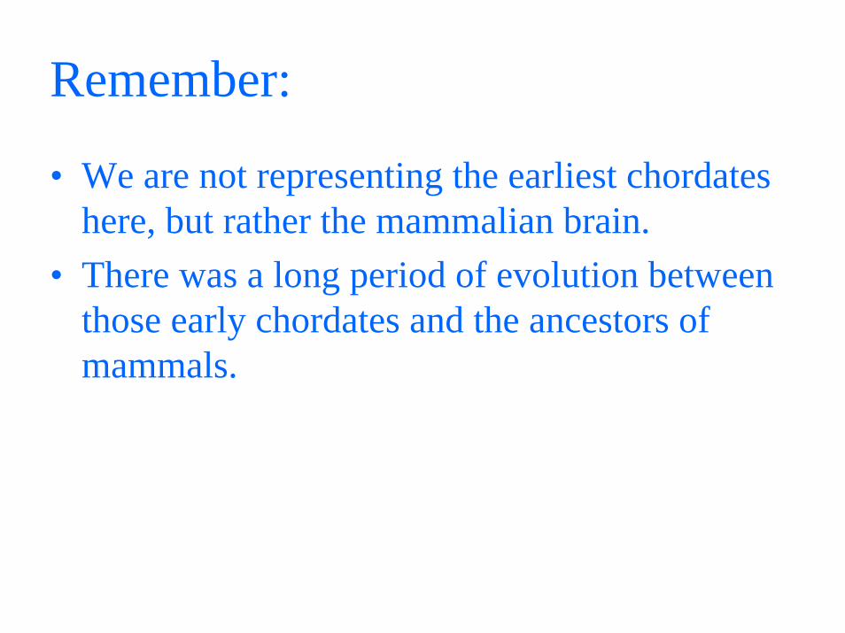
• We are not representing the earliest chordates here, but rather the mammalian brain.
• There was a long period of evolution between those early chordates and the ancestors of mammals.

On primitive somatosensory functions
• What can vertebrate animals do in the absence of somatosensory pathways to the forebrain?
• …or in the absence of all crossed ascending pathways (from the cord)?

Intro to the “cerebellar channel”, another kind of lemniscus
• A problem of multiple sensory pathways for control of the same outputs: Timing
• This problem increased with increasing body size, and increased role of head receptors in control of movements.
• It increased further with need for precise coordination of larger numbers of muscles, especially the distal muscles of the limbs.
• Cerebellum probably evolved to deal with this problem: adjustments of relative timing which could be varied according to feedback.
• See the more specific but related suggestion by Allman(2000), p. 77-78: stabilizing the retinal image by comparing eye velocity with head velocity.


Notes on evolution of cerebellum• The basic cell types and circuitry are highly conserved in
evolution– Layering becomes more regular– New inhibitory cell types in tetrapods– Deep nuclei as the output structures appear in tetrapods
• The size and form vary considerably– Huge expansion in the weakly electric fishes, especially in the
hyperfolded cerebellum of mormyrids– Dramatic lateral expansion and foliation as locomotor and
manipulatory abilities increased in land animals: the growth of the cerebellar hemispheres and the lateral deep nuclei
• Functions– Sensory: in animals with lateral line receptors and electroreceptors– Motor: the coordinated timing of outputs; changes with experience
(learning)– Cognitive: the coordinated timing of representations in the internal
model of the external world?

Selected References
Slide 5: Figure by MIT OCW, © MIT 2006. Based on: Darwin and after Darwin. An exposition of the Darwinian theory and a discussion of post-Darwinian questions, by George John Romanes. Published/Created: Chicago, The Open court publishing company, 1892-97. Slide 6: Figure by MIT OCW. © MIT 2006. Based on: Striedter, Georg F. Principles of Brain Evolution. Sunderland, MA: Sinauer Associates, 2005, p.78. ISBN: 0878938206. Slide 12: Figure by MIT OCW. © MIT 2006. Based on: Striedter, Georg F. Principles of Brain Evolution. Sunderland, MA: Sinauer Associates, 2005, p. 156. ISBN: 0878938206. Slide 13: Figure by MIT OCW. © MIT 2006. Based on:Butler, Ann B., and William Hodos. Comparative Vertebrate Neuroanatomy: Evolution and Adaptation. New York, NY: Wiley-Liss, 1996. ISBN: 0471888893. Slide 14: Figure by MIT OCW. © MIT 2006. Based on:Striedter, Georg F. Principles of Brain Evolution. Sunderland, MA: Sinauer Associates, 2005, p.166. ISBN: 0878938206. Slide 15: Figure by MIT OCW. © MIT 2006. Based on: Allman, John Morgan. Evolving Brains. New York, NY: Scientific American Library: Distributed by W. H. Freeman and Co., 1999, p. 38. ISBN: 0716750767. Slide 18: Figure by MIT OCW. © MIT 2006. Slide 27: Figure by MIT OCW. © MIT 2006. Based on: Striedter, Georg F. Principles of Brain Evolution. Sunderland,MA: Sinauer Associates, 2005, p. 66. ISBN: 0878938206. Slide 28: Drawing by Gerald Schneider. © G.E. Schneider 2006. Slide 30: Figure by MIT OCW. © MIT 2006. Based on: Cajal, S. Ramón y. Histology of the Nervous System of Man and Vertebrates. 2 vols. Translated from the French by Neely Swanson and Larry W. Swanson. New York, NY: Oxford University Press, 1995. ISBN: 0195074017. Slide 36: Figure by MIT OCW. © MIT 2006. Slide 37: Figure by MIT OCW. © MIT 2006. Slide 42: Figure by MIT OCW. © MIT 2006. Slide 43: Figure by MIT OCW. © MIT 2006. Based on Striedter, Georg F. Principles of Brain Evolution. Sunderland, MA: Sinauer Associates, 2005, p. 54. ISBN: 08789382 Slide 47: Drawing by Gerald Schneider. © G.E. Schneider 2006. Slide 48: Drawing by Gerald Schneider. © G.E. Schneider 2006. Principles of Brain Evolution. Sunderland, MA: Sinauer Associates, 2005, p. 54. ISBN: