90Y-Microspheres Radioembolization for Selective - NUCLEUS
32
Pr Francesco Giammarile CHLS Lyon Faculté de Lyon Sud « Aut tace aut loquere meliora silentio » 90 Y-Microspheres Radioembolization for Selective Internal Radiation Therapy of Primary and Metastatic Cancer in the Liver
Transcript of 90Y-Microspheres Radioembolization for Selective - NUCLEUS

Pr Francesco Giammarile
CHLS Lyon
Faculté de Lyon Sud
« Aut tace aut loquere meliora silentio »
90Y-Microspheres
Radioembolization for Selective
Internal Radiation Therapy of
Primary and Metastatic Cancer
in the Liver

Concept of Selective Internal Radiation Therapy (aka Radioembolization; Hepatic Artery Brachytherapy; Microbrachytherapy )
• To selectively target a very high radiation dose to all
tumours within the liver, regardless of their cell of origin
or location, while at the same time maintaining a low
radiation dose to the normal liver tissue
• Infusion via hepatic artery, using differential blood supply
to liver tumours thereby preferentially targeting tumours
• Uses 90Yttrium-labelled microspheres
– Half life: 64.1 hours
– β- decay, Emean = 0.93 MeV; Emax = 2.28 MeV
– Penetrates tissue, mean 2.5 mm; max 11 mm
– Achieves doses of 100–1,000+ Gy to tumour
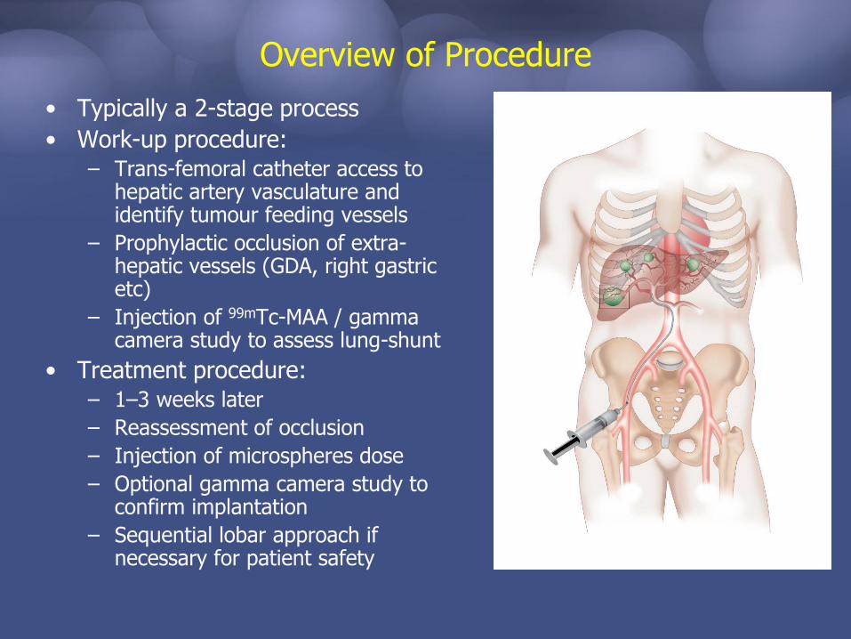
Overview of Procedure
• Typically a 2-stage process
• Work-up procedure: – Trans-femoral catheter access to
hepatic artery vasculature and identify tumour feeding vessels
– Prophylactic occlusion of extra-hepatic vessels (GDA, right gastric etc)
– Injection of 99mTc-MAA / gamma camera study to assess lung-shunt
• Treatment procedure: – 1–3 weeks later
– Reassessment of occlusion
– Injection of microspheres dose
– Optional gamma camera study to confirm implantation
– Sequential lobar approach if necessary for patient safety

90Y-microspheres
*Resin microspheres (SIR-Spheres® , SirTex)
• 90Y is within the spheres
• particle size: 20-60 µm; activity per particle: 50 Bq
• about 20-40 million particles per administration
• uniform dose biodistribution
*Glass microspheres (TheraSphere®, MDS Nordion)
• 90Y is an integral constituent of the glass matrix
• particle size: 20-30 µm; activity per particle: 2500 Bq
• about 1-4 million particles per administration
• less embolic effect on microvessels and potential influence of
gravity on biodistribution
Medical devices (not radiopharmaceutical !)

Characteristics SIR-spheres® TheraSphere®
Material Resin Glass
Particle size (mm) 20-60 20-30
Nb of spheres per vial
(range in million)
40-80 1.2-8
Specific gravity Low High
Embolic effect Moderate Mild
Activity per sphere (Bq) 40-70 2,500
Activity available (GBq) 3 3, 5, 7, 10, 15, 20
Handling for dispensing Required Not required
Splitting one vial for two
or more patients
Possible Not possible
Radiation precautions Urine contamination (?) None
From Salem, J Vasc Interv Radiol 2006 (modified)
90Y-microspheres

• Predominant liver-dominant or liver-only disease
• No extra-hepatic manifestation
• Life expectancy >12 weeks
• ECOG performance status 0–2
• Failed 1st and 2nd line of chemotherapy
• Unresectable disease (evaluated by GI specialists)
• Preferable tumour types:
– Liver metastases of colorectal cancer (CRC)
– Hepatocellular carcinoma (HCC)
Indications

Exclusion Criteria for Radioembolisation (relative contra-indications unless stated)
• Pregnancy [absolute]
• Ascites or other clinical signs of liver failure on physical exam [absolute]
• Abnormal organ or bone marrow function as determined by: – total bilirubin level >2.0 mg/dL (>34 μmol/L) in absence of reversible cause
– serum albumin <3.0 g/dL
– AST (SGOT)/ALT (SGPT) >5 x institutional ULN
– creatinine >2.5 mg/dL
– platelets <60,000/μL; leukocytes <2,500/μL; absolute neutrophil <1,500/μL
• Previous external radiation therapy to the liver
• Excessive tumour burden with limited hepatic reserve
• Abnormal blood circulation due to anatomic variations resulting in back-flow to the stomach, pancreas or bowel, compromised portal vein, unless selective/super-selective delivery is performed
Following work-up procedure:
• >30 Gy exposure to the lungs (pre-therapeutic scan) [absolute]
• Non-correctable shunting to the GI tract [absolute]
Kennedy A et al. Recommendations for radioembolization of hepatic malignancies using yttrium-90 microsphere brachytherapy: A consensus panel report from REBOC. Int J Radiation Oncology Biol Phys 2007; 68: 13–23.

• Contra-indications:
– Previous external beam radiation therapy to the major volume of the liver
– Ascites or clinical liver failure
– Abnormal vascular anatomy (significant reflux determined by angiogram)
– Lung shunting of the hepatic artery blood flow greater than 20% (determined by intra-arterial 99mTc MAA scintigraphy)
– Disseminated extra-hepatic malignant disease
• Under debate
– Capecitabine (within previous or subsequent 2 months only ?)
– Main portal vein thrombosis
• Special warnings
– Excessive radiation to the normal liver parenchyma: radiation hepatitis
– Inadvertent delivery to
• gastrointestinal tract or pancreas: acute abdominal pain, acute pancreatitis or peptic ulceration
• gall bladder: cholecystitis
– High levels of radiation and/or excessive shunting to the lung: radiation pneumonitis
– Antiangiogenetics (induce complications during angiography)
Specific Contraindications :
SIR-spheres®

• Contra-indications:
– Severe liver dysfunction or pulmonary insufficiency
– Deposition to the gastrointestinal tract that may not be corrected by
angiographic techniques
– Shunting of blood to the lungs that could result in delivery of > 610.5 MBq of
90Y to the lungs
• Special warnings (pre-treatment high risk factors):
– Infiltrative tumour type
– “Bulk disease” (tumour volume > 70% of the target liver volume, or multiple
tumour nodules)
– AST or ALT > 5 times ULN
– Bilirubin > 1 time ULN
– Tumour volume > 50% combined with an albumin < 3 g/dl
– Antiangiogenetics (induce complications during angiography)
Specific Contraindications :
Therasphere®

• Usual arteriography precautions
• Nursing personnel must be trained in radiation safety.
• Ensure minimum radiation exposure
• No drug interaction have been reported to date (however, discontinue
the radiation sensitizers)
• Prophylactic administration of antiulcer medication (for 2 weeks) and
steroids (for 5–7 days)
• Avoid pregnancy for at least 4 months following treatment
• Critical person for radiation protection = interventional radiologist
=> 40 treatments per year gives an extra dose of 1.9 mSv
Precautions

• Pre-therapy evaluation of serum liver enzymes, blood cell count,
coagulation and creatinin
Patients with adequate hepatic reserve and good functional status will
maximize the therapeutic effect with minimal risk to normal liver
parenchyma
• Visceral angiography performed by high-speed multi slice CT:
– to visualise hepatic vascular organisation and map tumour-perfusing vessels
– to assess portal vain patency (verify the absence of portal venous thrombosis)
– to place coils (embolize collateral vessels)
Pretherapeutic Procedures
• CT for determination of tumor load
• FDG-PET for exclusion of extrahepatic
manifestations and evaluation of
hepatic disease (since the aim is palliative, a
small extrahepatic tumour burden is not a
contraindication)
combined PET/CT
preferable

• Intra-arterial (via the hepatic artery) administration of 99mTc-
Albumin Macro-Aggregates (+/- perchlorate to prevent
unspecific extrahepatic free technetium):
• to assess arteriovenous liver/lung shunting
• to determine the optimal therapeutic dose-activity
- Preferable < 10%
- Dose reduction if shunt between 10% and 20%:
10%-15% shunt volume: calculated dose minus 20%
15%-20% shunt volume: calculated dose minus 40%
[99mTc]MAA Scintigraphy

• Delineation of lung borders by the help of a flood phantom
RVL LDR
RVL RVL
shunt ~ 4% 57Co flood
phantom
[99mTc]MAA Planar Scintigraphy

[99mTc]MAA SPECT-CT
• More sensitive than planar or SPECT images for
detecting extrahepatic accumulations
Gallbladder
=> prevention of
acute cholecystis
Stomach => pretreatment coiling of a
gastric branch
First experience of hepatic radioembolization using microspheres labelled with yttrium-90
(TheraSphere): practical aspects concerning its implementation
Etienne Garin et al. Eur J Nucl Med Mol Imaging (2010) 37:453–461

• < 25% liver replacement: 2 GBq
• 25% to 50% liver replacement: 2.5 GBq
• > 50% liver replacement: 3 GBq
Dosimetry • BSA Model:
– Recommended formula for most patients
– Dose reduction used if increased lung shunt; some centres also adopt
in low volume disease or for highly pre-treated patients
• Partition Model: – Only useful if the tumour can be localized in a discrete area and drawn
as an Area of Interest on SPECT
• Empiric Model: – Dose adjustment according to the estimated tumour load in the liver:
– Not recommended (increased risk of radiation-induced side effects)
Dose (Gy) = activity (GBq) x 49670 mass (g)
Dose (GBq) = BSA (m2) – 0.2 + volume tumour volume tumour + liver

Administration
Before treatment:
Coiling of the gastroduodenal artery (and optionally the
right gastric artery/pancreaticoduodenal branches) to avoid
deposition in the duodenum, stomach or pancreas
Treatment:
Administration of microspheres via a percutaneous
transfemoral hepatic artery catheter to each hepatic lobe
separately via the right and left hepatic artery
Pulsatil gentle infusion (no pressure) to strictly avoid
back-flow using a specially designed manufacturer’s device.

Side Effects
AE Incidence Characteristics Prevention/Action
Fever >50% mild (grade 1*) onset on none normally
Tx day for up to 1 week required
Abdominal ~50% acute onset during Tx may require narcotic pain ~10% grade 3–4 self-limiting; normally <24 h > oral analgesia
Nausea ~40% highest in Tx-experienced; prophylactic anti-emetics <5% grade 3–4 normally <24 h
Fatigue ~40% onset in 1st month post- adequate nutrition & <5% grade 3–4 SIRT, normally subsides hydration; within 2 weeks prophylactic oral steroids
Abnormal ~20–40% particularly in combination none normally LFTs 1–6% grade 3–4 with chemo or HCC/cirrhosis; required transient; resolves in days (ALT, AST), weeks (bilirubin) or months (alb.)
* CTC 2.0

Potential Complications
SAE Incidence Characteristics Prevention/action
Radiation ~5–10% non-target administration; meticulous technique &
gastritis or 1–2% grade 3–4 immediate, severe occlusion of GI arteries; duodenitis unremitting pain prophylactic PPI for 1 mo
Radiation <1% non-target administration; meticulous technique & pancreatitis immediate, severe occlusion of GI arteries unremitting pain
Radiation <1% non-target administration; various technical cholecystitis right upper quadrant pain approaches; may require cholecystectomy
Radiation- <1% excess radiation to normal liver; appropriate Induced Liver onset typically 30–90 d post-SIRT; dosimetry (BSA); Disease (RILD) permanently elevated LFTs, portal dose reduction in hypertension, eventual fibrosis patients with reduced hepatic reserve
Radiation no cases since excessive radiation routine lung-shunt study; pneumonitis lung-shunt study delivery to lung tissue maintain <30 Gy to lung

• Common side effects:
– Fatigue
– Abdominal pain
– Nausea
– Fever
– Transitory elevation of transaminases
• Possible severe adverse events (2-8%):
– Chronic abdominal pain
– Nontarget irradiation and/or attenuated irradiation in adjacent structures:
• radiation-induced gastric disease (gastritis, gastrointestinal ulceration, upper gastrointestinal bleeding, pancreatitis)
• radiation-induced lung disease (pneumonitis, right pleural effusion)
• radiation-induced liver disease (hepatitis, hepatic fibrosis and portal hypertension, cholecystitis)
Side Effects

Biodistribution evaluation
Lhommel et al,
EJNMMI 2009
90Y Half-life: 64.1 h
β- decay, Emax = 2.28 MeV
Internal pair-production, 0.000032 per decay

Bremsstrahlung images
Bremsstrahlung spectra from Y-90 in water phantom
0
0.5
1
1.5
2
2.5
0 100 200 300 400 500
Enegry [keV]
Inte
nsit
y
HE collimator
LEAP collimator
Whithout collimator

PET/CT images
• 90Y PET/CT (one or two frames of 30 minute)
– Confirmation of microspheres distribution
– Estimation of treatment efficacy ?
Internal Pair Production of 90Y Permits Hepatic Localization of Microspheres Using Routine PET: Proof of Concept
Vanessa L. Gates, Abdulredha A.H. Esmail, Karen Marshall, Stewart Spies, and Riad Salem
JNM, january 2011

• At 30 days and at 2- to 3-month in order to assess:
– treatment-emergent side effects
– tumour response
• Parameters to assess the result of treatment:
– determination of tumour load, volume and serum tumour
markers (AFP, CEA)
Follow-up

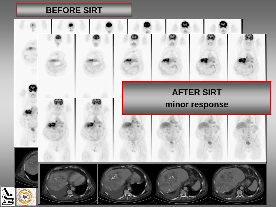
BEFORE SIRT
AFTER SIRT
good response
BEFORE SIRT
AFTER SIRT
minor response

Consultation – Tumour Board Medical Oncology Radiation Oncology Surgical Oncology Nuclear Medicine
Interventional Radiology Consensus for liver-directed therapy with 90Y-microspheres
Pre- Treatment Screening Evaluations
Abdominal CT and/or MRI
Hepatic angiogram, protective embolization of extra-hepatic vessels
and 99mTc-MAA scan
Safe delivery achievable Liver-predominant disease
Bremsstrahlung (gamma) scan Post-implant QA documentation
Delivery of 90Y-microspheres to the planned treatment volume
Vessel mapping Tumour mapping
Team reviews imaging, proposed dose, planned
tumour volume, and optimal catheter placement
for radioembolization
1–2 weeks
1–2 weeks
1 week
same day
REBOC Consensus Guidelines. Kennedy et al . Int J Radiation
Oncology Biol Phys 2007; 68: 13–23

Where do microspheres fit as a liver treatment?
• EU approval for treatment of unresectable liver tumours
• Loco-regional therapy: no impact on extra-hepatic disease
• Used in liver-only or liver-dominant disease: – in combination with systemic chemotherapy for…
• mCRC (at 1st-line, 2nd-line etc)
• in trials with systemic chemo for mNET and pancreatic cancer (at 1st-line, 2nd-line)
– as monotherapy for…
• mCRC and other metastatic disease (salvage in chemo-refractory)
• HCC (at 1st-line, 2nd-line)
• mNET (at 1st-line, 2nd-line etc)
• Cholangiocarcinoma (at 2nd-line etc)
• Studies suggest earlier use provides greater benefits
• Potential for down-staging to resection or ablation

SIR-Spheres microspheres + FOLFOX4 in mCRC: CT Response
Patient 2: Baseline CT scan pre-SIRT
Patient 2: CT scan 6 months post-SIRT
Sharma RA et al. Annals of Oncology 2006; 17 (Sup 6): vi78 Abstract P-191. Data on file; Sirtex Medical Limited.

SIR-Spheres microspheres + FOLFOX4 in mCRC: CT Response
Patient 3: Baseline CT scan pre-SIRT
Patient 3: CT scan 6 months post-SIRT; patient was subsequently resected
Sharma RA et al. Annals of Oncology 2006; 17 (Sup 6): vi78 Abstract P-191. Data on file; Sirtex Medical Limited.

SIR-Spheres + irinotecan in >2nd-line mCRC: CT Response
Baseline CT scan pre-SIRT
Scan 3 months after implantation
Data on file. Sirtex Medical Limited. MAR-PPP-0005 rev. 1

Conclusion
• 90Y-microspheres with delivery into the hepatic artery is a
promising new local therapy
• Data on comparing this treatment alone or in combination with
other modalities, particularly systemic or local chemotherapy
must be awaited
• However, response rates are sufficiently high to make it likely
that this treatment will have useful impact on the management
and survival of patients
• The treatment seems to be less appropriate as a stand alone
modality but should rather be considered in conjunction with
modern chemotherapy or other local treatment modalities

• Benefits of the treatments (survival gain, progression free survival and quality of life) assessed by prospective studies
• Minimum / optimal time interval between repeated treatments
• Comparison between radioactive ligands
• Therapeutic potential of 188Re-Lipiodol
• Role of combined therapy
Issues requiring further clarification