80: ' # '7& *#8 & 9 - InTechcdn.intechopen.com/pdfs-wm/21371.pdf23 Multipotent Dental Stem Cells: An...
24
3,350+ OPEN ACCESS BOOKS 108,000+ INTERNATIONAL AUTHORS AND EDITORS 115+ MILLION DOWNLOADS BOOKS DELIVERED TO 151 COUNTRIES AUTHORS AMONG TOP 1% MOST CITED SCIENTIST 12.2% AUTHORS AND EDITORS FROM TOP 500 UNIVERSITIES Selection of our books indexed in the Book Citation Index in Web of Science™ Core Collection (BKCI) Chapter from the book Embryonic Stem Cells - Differentiation and Pluripotent Alternatives Downloaded from: http://www.intechopen.com/books/embryonic-stem-cells- differentiation-and-pluripotent-alternatives PUBLISHED BY World's largest Science, Technology & Medicine Open Access book publisher Interested in publishing with IntechOpen? Contact us at [email protected]
Transcript of 80: ' # '7& *#8 & 9 - InTechcdn.intechopen.com/pdfs-wm/21371.pdf23 Multipotent Dental Stem Cells: An...
3,350+OPEN ACCESS BOOKS
108,000+INTERNATIONAL
AUTHORS AND EDITORS115+ MILLION
DOWNLOADS
BOOKSDELIVERED TO
151 COUNTRIES
AUTHORS AMONG
TOP 1%MOST CITED SCIENTIST
12.2%AUTHORS AND EDITORS
FROM TOP 500 UNIVERSITIES
Selection of our books indexed in theBook Citation Index in Web of Science™
Core Collection (BKCI)
Chapter from the book Embryonic Stem Cells - Differentiation and PluripotentAlternativesDownloaded from: http://www.intechopen.com/books/embryonic-stem-cells -differentiation-and-pluripotent-alternatives
PUBLISHED BY
World's largest Science,Technology & Medicine
Open Access book publisher
Interested in publishing with IntechOpen?Contact us at [email protected]

23
Multipotent Dental Stem Cells: An Alternative Adult Derived Stem
Cell Source for Regenerative Medicine
Tammy Laberge1,2 and Herman S. Cheung1,2 1Department of Biomedical Engineering, College of Engineering, University of Miami
2Geriatrics Research, Education and Clinical Center, Miami VA Healthcare System USA
1. Introduction
The pluripotent nature of embryonic stem cells (ESCs) makes them amenable for regenerative therapies because they can differentiate into cells that form all tissue types within the body (Zandstra and Nagy, 2001). The potential drawbacks to the use of ESCs for cellular therapies include the obvious ethical dilemmas of obtaining ESCs, the potential of cancer or tumor formation and the risk of immunogenic rejection (Wobus and Boheler, 2005). Therefore adult stem cell sources with multipotent and pluripotent potential have been sought as an alternative for ESCs including mesenchymal stem cell (MSCs) and tissue-derived specific stem cells. Interestingly, the isolation of a population of dental stem cells derived ectodermally from the neural crest (NC) have been shown to be multipotent and give rise to multifarious cell types that result in the development of many of the body’s organs or tissues (Huang et al., 2009a; Huang et al., 2009b). Stem cells extracted from dental tissues including dental pulp, periodontal ligament, apical papilla and dental follicle precursor cells have an expansive differentiation potential with respect to mesodermal and ectodermal lineages. Currently there are six types of dental stem cells that are well characterized and described both in vitro and in vivo (Gronthos et al., 2000; Huang et al., 2009a; Karaöz et al., 2010; Miura et al., 2003; Morsczeck et al., 2005; Seo et al., 2004; Sonoyama et al., 2006). Some dental stem cells lines have been shown to express ESC markers Oct4, Nanog, Sox2 and Klf4 and NC markers p75, Sox10, Slug and Nestin suggesting that dental stem cells may be able to become many of the same tissues as ESCs (Huang et al., 2009a). Further, dental stem cells have been shown to differentiate into neurogenic, adipogenic, cardiomyogenic, chondrogenic, myogenic and osteogenic lineages (Huang et al., 2009a; Karaöz et al., 2010; Miura et al., 2003; Seo et al., 2004; Sonoyama et al., 2006; Zhang et al., 2006). Since dental stem cells have been shown to differentiate into a multitude of cell types, their potential for use in tissue regeneration may be boundless. We are currently using dental stem cells to investigate the mechanisms of mechanotransduction elicited during dynamic cyclic compression for chondrogenesis. Our long term goal is to develop technology and protocols utilizing dental stem cells and biomechanical force for reparative medicine and tissue regeneration of cartilage. This review
www.intechopen.com
Embryonic Stem Cells – Differentiation and Pluripotent Alternatives 452
will discuss the most current findings in tissue engineering with respect to dental stem cells both for whole tooth regeneration and potential use in future stem cell therapies.
2. Characteristics and sources of stem cells
2.1 What is a stem cell?
The general properties that define a stem cells are: 1. Stem cells are cells that are clonogenic and have the ability for self-renewal; 2. Stem cells are unspecialized cells that when correctly stimulated have the ability to differentiate into specialized cell types (Blau et al., 2001; Bongso and Fong, 2009). There are two broader categories of stem cells: embryonic stem cells (ESCs) and adult stem cells. Embryonic stem cells are derived from the blastocyst stage of a developing embryo (Fortier, 2005; Thomson et al., 1998) and are capable of forming all three germ layers (ectoderm, endoderm, and mesoderm)(Bongso and Fong, 2009). Harvesting ESCs requires the destruction of the embryo (Lanzendorf et al., 2001) which leads to ethical dilemmas when obtaining these cells. The use of adult stem cells avoids these ethical issues. Adult stem cells have been obtained from multiple tissues including bone marrow (Pittenger et al., 1999), adipose tissue (Zuk et al., 2001), muscle (Deasy et al., 2001), umbilical cord tissue (Schugar et al., 2009), intestine (Wong, 2004), and skin (Blanpain et al., 2004). While most of this book focuses on the embryonically derived stem cells, this chapter focuses on the adult or postnatal stem cells with special emphasis on those derived from dental tissues.
2.2 Stem cell potency
Stem cell potency refers to the ability of the cell to differentiate into specific tissue type(s). Totipotent is defined as the ability to differentiate into any of the cell types of the entire organism, both embryonic and adult cell types (Dannan, 2009; Mummery et al., 2011; Smith, 2006). An example of a totipotent cell is a fertilized egg cell because it is able to differentiate into embryonic, extra-embryonic (ie. placenta) and adult tissues of the entire organism (Alison et al., 2002; Dannan, 2009; Mummery et al., 2011). Pluripotent stem cells have the potential to differentiate into all cell lineages including the three germ layers: ectoderm, mesoderm, or endoderm but not the extra-embryonic tissues (Alison et al., 2002; Dannan, 2009; Fortier, 2005; Mummery et al., 2011; Smith, 2006). ESCs are considered the gold standard of stem cells because of their pluripotency and their ability to be maintained indefinitely in culture (Thomson et al., 1998). Pluripotency has also been demonstrated in adult stem cells including bone marrow mesenchymal stem cells (BMMSCs) (Jiang et al., 2002). Multipotent stem cells have the ability to differentiate into multiple cell lineages that can form more than one tissue type (Alison et al., 2002; Mummery et al., 2011; Smith, 2006). Mesenchymal stem cells (MSCs) derived from adipose tissue are an example of a multipotent stem cell and are able to differentiate into multiple tissues of the mesodermal lineage including bone, fat and cartilage (Zuk et al., 2002).
2.3 Bone marrow mesenchymal stem cells
Stem cells obtained from bone marrow are a major source of adult MSCs and are widely studied. Bone marrow stromal cells can be harvested from bone marrow by mechanical disruption, but the cell suspension will contain both hematopoietic stem cells and BMMSCs (Bianco et al., 2001). BMMSCs are isolated as colony forming unit-fibroblasts (CFU-Fs) from the bone marrow cell suspension. In order to separate the two types of stem cells, the cell
www.intechopen.com
Multipotent Dental Stem Cells: An Alternative Adult Derived Stem Cell Source for Regenerative Medicine 453
suspension is cultured in vitro at low density. A small number of BMMSCs will adhere to the plate and begin to form colonies while the non-adherent hematopoietic cells are then removed by repeat washings (Bianco et al., 2001; Chamberlain et al., 2007). BMMSCs isolated in this manner are capable of 20–25 passages in vitro without significant changes to the cell phenotype (Bianco et al., 2001; Conget and Minguell, 1999). Gronthos et al. (2003) further showed that BMMSCs could be isolated from bone marrow aspirates by determining which CFU-F colonies were highly reactive to the antibody STRO-1 (STRO-1Bright) and also reactive to the antibody VCAM-1 (VCAM-1+). These new studies isolated BMMSCs in bone marrow aspirates based on STRO-1Bright/ VCAM-1+ cell surface markers by florescence activated cell sorting (FACS) and were capable of proliferating up to 40 population doublings (Gronthos et al., 2003). BMMSCs show a great level of plasticity and have shown the potential to differentiate into multiple tissue types in vitro including muscle, adipose, cartilage, bone, connective tissue, neurons and endothelial cells (Gronthos et al., 2003; Pittenger et al., 1999; Woodbury et al., 2000; Young and Black, 2004). Interestingly, when transplanted into immunodeficient mice, BMMSCs undergo osteogenic differentiation in vivo and form bone (Kuznetsov et al., 1997). In 2006, the minimum criteria to define human multipotent mesenchymal stromal cells was established as: Cells that are plastic adherent in standard culture; Cells that have the ability to differentiate in vitro into osteoblasts, adipocytes and chondroblasts; Cells that express the cell surface markers CD73, CD90 and CD105 in 95% of the cell population as determined by flow cytometry and lack the expression ( ≤ 2% positive) of CD14, CD34, CD45 or CD11b, CD79a or CD19 and HLA class II (Dominici et al., 2006).
2.4 Dental stem cells
Dental stem cells are an alternative source of adult stem cells that are easily accessible by tooth extraction with a local anesthetic or when a primary tooth is replaced. This section discusses where dental stem cells arise during tooth formation and the types of tissue they form. We also characterize the many types of the dental stem cells utilized in research today and compare the utility of dental stem cells versus BMMSCs. There are six types of human dental stem cells that have been well described in the literature: 1. Dental pulp stem cells (DPSCs) (Gronthos et al., 2000); 2. Stem cells isolated from human exfoliated deciduous teeth (SHEDs) (Miura et al., 2003); 3. Stem cells derived from human natal dental pulp (hNDPs) (Karaöz et al., 2010); 4. Periodontal ligament stem cells (PDLSCs) (Seo et al., 2004); 5. Stem cells isolated from the apical papilla (SCAPs) (Sonoyama et al., 2008); 6. Stem cells isolated from dental follicle precursor cells (DFPCs) (Morsczeck et al., 2005). Within the body, MSCs have been localized to perivascular niches (Crisan et al., 2009; Kolf et al., 2007) and recent studies have also shown that dental stem cells are also localized to perivascular niches within the tooth structure (Chen et al., 2006; Shi and Gronthos, 2003). Dental stem cells arise from dental mesenchyme which has early interaction with the neural crest during normal tooth development (Huang et al., 2009b). Therefore, dental stem cells may display characteristics of both mesoderm and ectoderm due to their ectomesenchymal origins (Huang et al., 2009b).
2.4.1 Mammalian tooth formation
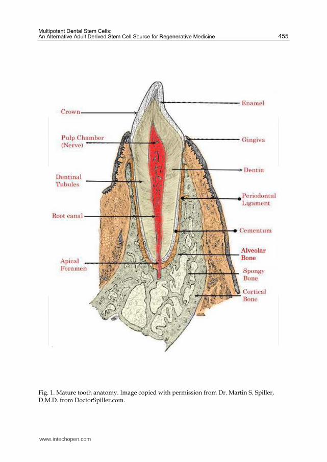
A mature tooth is comprised externally of hard structures of enamel, dentin and cementum and internally possesses a soft dental pulp (Figure 1). Tooth formation or odontogenesis is a
www.intechopen.com

Embryonic Stem Cells – Differentiation and Pluripotent Alternatives 454
complex process involving multiple tooth-associated cell types. Odontogenesis occurs as a tooth bud is formed from an aggregation of embryonic cells. These cells have ectodermal and ectomesodermal origins from the first branchial arch and the neural crest respectively (Ten Cate, 1998; Tucker and Sharpe, 2004). Tooth development has three stages. 1. The bud stage, where epithelial cells begin to proliferate into ectomesenchyme and condense in the jaw forming the tooth bud. 2. The cap stage, where ectomesenchmyal cells aggregate and begin to surround and enclose the epithelial cells which invaginate further into the mesenchyme and form the dental follicle, the enamel organ or cap and the dental papilla (Slatter, 2002; Ten Cate, 1998; Tucker and Sharpe, 2004). The dental follicle is of ectomesodermal origins and forms a sac surrounding the developing tooth that supports the tooth prior to eruption. The enamel organ is of ectodermal origins and eventually forms the enamel, whereas the dental papilla is of mesodermal origins and eventually forms the primary dentin and the pulp. 3. The bell stage, where the tooth undergoes extensive differentiation with the epithelial cells differentiating into ameloblasts and mesenchymal cells differentiating into odontoblasts. After the bell stage, the hard structures are formed with ameloblasts forming enamel while odontoblasts form dentin (Figure 1). Secondary dentin aids in root formation. Later in tooth development further differentiation of the dental follicle occurs with the formation of cementoblasts, fibroblasts and osteoblasts to form the cementum, the periodontal ligament and bone respectively (Figure 1).
2.4.2 Sources of dental stem cells
2.4.2.1 Dental Pulp Tissue
The soft dental pulp is located in the middle of the tooth surrounded by the harder
structures of the tooth including dentin, cementum and enamel (Figure 1). The dental pulp
contains a mix of cell types including fibroblasts which form the extracellular matrix and
collagen and odontoblasts that form reparative dentin (Gronthos et al., 2002; Liu et al., 2006).
The dental pulp region also contains nerve fibers and blood vessels and is accessible to
external stimuli through the apical foramen (Figure1). Three types of stem cells have been
identified from dental pulp tissue: DPSCs, SHEDs and hNDPs. DPSCs are present in the
pulp of the adult tooth, whereas SHEDs are only present in the pulp of primary teeth or
“baby teeth.” Lastly, hNDPs are a unique type of dental pulp stem cells isolated only from
the pulp of newborn teeth. Very few newborns are born with teeth, approximately one in
every two to three thousand births (Leung and Robson, 2006), so hNDPs are very rare.
2.4.2.1.1 Dental Pulp Stem Cells
DPSCs are a heterogeneous population of cells that were first isolated by Gronthos et al. (2000) and exhibited some characteristics of BMMSCs, including the production of fibroblast-like cells that were clonogenic and had a high proliferation rate. Interestingly, DPSCs had a higher proliferation rate than BMMSCs (Gronthos et al., 2000) . DPSCs also had a similar protein expression pattern to BMMSCs in vitro including vascular adhesion molecule 1, alkaline phosphatase, collagen I, collagen III, osteonectin, osteopontin, osteocalcin, bone sialoprotein, ┙-smooth muscle actin, fibroblast growth factor 2 and the cell surface marker CD 146 (Gronthos et al., 2000). Immunohistochemistry staining further showed that like BMMSCs, primary cultures of DPSCs did not stain for the cell surface markers CD14, CD34, and CD45 or other markers including MyoD, neurofilament, collagen II , and peroxisome-proliferator activated receptor ┛-2 (Gronthos et al., 2000).
www.intechopen.com
Multipotent Dental Stem Cells: An Alternative Adult Derived Stem Cell Source for Regenerative Medicine 455
Fig. 1. Mature tooth anatomy. Image copied with permission from Dr. Martin S. Spiller, D.M.D. from DoctorSpiller.com.
www.intechopen.com

Embryonic Stem Cells – Differentiation and Pluripotent Alternatives 456
Recently FACS has been used to sort DPSCs based on cell surface markers which found that in addition to the markers identified above, DPSCs expressed the following: CD9, CD10, CD13, CD29, CD44, CD49d, CD59, CD73, CD90, CD105, CD106, CD166 and STRO-1(Lindroos et al., 2008; Nam and Lee, 2009). Further, DPSC did not express CD14, CD 31, CD 45 (Nam and Lee, 2009) (Summarized in Table 1). When cultured under osteogenic conditions DPSCs were capable of forming calcified
deposits sparsely throughout the culture; these results were unlike BMMSCs which formed
sheets of calcium deposits (Gronthos et al., 2000). In vivo transplantation of DPSCs into
immunocompromised mice resulted in the production of a dentin-pulp-like complex with a
collagen matrix containing blood vessels and lined with odontoblasts (Gronthos et al., 2000)
suggesting that DPSCs are multipotent. Further studies also found DPSCs to be multipotent,
capable of differentiating into myoblasts, osteoblasts, odontoblast-like cells, chondrocytes,
adipocytes and neural cells (Gronthos et al., 2002; Liu et al., 2006; Pierdomenico et al., 2005;
Zhang et al., 2006).
Letter name
BMMSCs DPSCs SHEDs hNDPs PDLSCs SCAPs DFPCs
CD9 + + + + CD 10 + - + + CD 13 + + + + + + + CD 14 - - - - - CD 18 - CD 19 - CD 24 - - + CD 29 + + + + + CD 34 - - - - - - - CD 44 + + + + + + + CD 45 - - - - - - - CD 53 + CD 59 + + + CD73 + + + + + + + CD 90 + + + + + + +
CD 105 + + + + + + CD 106 + + + + + + CD 146 + + + + + + + CD 150 - CD 166 + + + + + STRO-1 + + + + + +
Table 1. Cell surface markers expressed in dental stem cells compared to bone marrow
mesenchymal stem cells as determined by flow cytometry. Table adapted from Karaöz et al.
2011, Rodriguez-Lozano et al. 2011, Huang, G.T. et al. 2009, Nam and Lee 2009, Lindroos et
al. 2008 and Shi et al. 2005. BMMSCs, bone marrow mesenchymal stem cells; DPSCs, dental
pulp stem cells; SHEDs, stem cells from human exfoliated deciduous teeth; SCAPs, stem
cells from apical papilla; DFPCs, dental follicle precursor cells; hNDPs, stem cells derived
from human natal dental pulp; + = marker present; - = marker absent.
www.intechopen.com
Multipotent Dental Stem Cells: An Alternative Adult Derived Stem Cell Source for Regenerative Medicine 457
2.4.2.1.2 Stem Cells from Human Exfoliated Deciduous Teeth
SHEDs are found in the pulp of the naturally exfoliated deciduous teeth or “baby teeth.” When the permanent tooth erupts from the gums the deciduous tooth in displaced. SHED cells were first isolated by Miura et al. (2003) from the remnant pulp in the crown of human deciduous incisors of children 7-8 years old. Similar to DPSCs, SHEDs met the criteria to be defined as a stem cell population as they were highly proliferative, capable of self-renewal and had the ability to differentiate into multiple cell types (Miura et al., 2003). SHEDs also had a fibroblast-like morphology similar to DPSCs. However, SHEDs were capable of a greater number of population doublings and had a higher proliferation rate than both BMMSCs and DPSCs (Miura et al., 2003). SHEDs have also been isolated and identified as immature dental pulp stem cells (IDPSCs) (Kerkis et al., 2006) and found to express embryonic stem cell markers Oct-4 (POU transcription factor), Nanog, stage specific embryonic antigens (SSEA-3, SSEA-4), and tumorigenic recognition antigens (TRA-1-60, TRA-1-81). SHEDs have also been shown to express neural stem cell markers SRY (sex determining region Y)-box 2 (Sox-2), nestin, and ATP-binding cassette, subfamily G, member 2 (ABCg2)(Morsczeck et al., 2010). SHEDs were further characterized using FACS as having the following cell surface markers: CD13, CD29, CD31, CD44, CD73, CD90, CD105, CD146, CD166, and STRO-1 and similar to BMMSCs and DPSCs, SHEDs did not express CD14, CD34 or CD45 (Kerkis et al., 2006; Morsczeck et al., 2010; Pivoriuunas et al., 2010; Shi et al., 2005; Wang et al., 2010) (Summarized in Table 1). A distinguishing feature of SHEDs not demonstrated for DPSCs is SHEDs formed sphere-like clusters when cultured in neuronal differentiation media (Miura et al., 2003). While Miura et al. (2003) demonstrated that SHEDs could differentiate into neural cells, adipocytes and odontoblasts, Kerkis et al. (2006) showed that SHEDs also had chondrogenic and myogenic potential. SHEDs were shown to express chondrogenic markers Sox-9, type II collagen and type X collagen when cultured for 14 days with bone morphogenic protein 2 (BMP2), a chondrogenic signaling protein in the TGF┚ family (Koyama et al., 2009). Interestingly, Koyama et al. (2009) did not find any expression of the chondrogenic markers in their untreated populations of SHED cultures. When SHED were transplanted into immunocompromised mice they exhibited an osteoinductive capacity in vivo but were not able to regenerate the dentin-pulp-like complex that DPSCs cells were able to form (Miura et al., 2003). Kerkis et al. (2006) also showed that when SHEDs were transplanted into immunocompromised mice via intraperitoneal injection they engrafted into the lungs, liver, spleen, brain and kidney and the tissue formed by the SHEDs was indistinguishable from the host tissue for liver, spleen, brain and kidney (Kerkis et al., 2006).
2.4.2.1.3 Stem Cells Derived from Human Natal Dental Pulp
Natal teeth are deciduous teeth that arise in newborns that are smaller than primary teeth and have little or no root development (Leung and Robson, 2006). Karaöz et al. (2010) isolated and characterized hNDPs from the remnant pulp of natal teeth. A small number of hNDPs adhered to plastic in culture and displayed a fibroblast-like spindle shaped morphology that eventually became flattened in later passages (Karaöz et al., 2010). Similar to DPSCs, hNDPs had a higher proliferation rate the BMMSCs, were clonogenic, and had the ability to differentiate into multiple cell types, satisfying the criteria to be classified as a stem cell population.
www.intechopen.com

Embryonic Stem Cells – Differentiation and Pluripotent Alternatives 458
Using flow-cytometry Karaöz et al. (2010) showed that like BMMSCs, hNDPs expressed CD13, CD44, CD73, CD90, CD146, and CD166 but did not express CD14, CD31 or CD45 (Summarized in Table 1). Stem cells derived from human natal dental pulp expressed many of the same cell surface markers seen in DPSCs and SHEDs (Table 1). Cultures of NDPs with chondrogenic, osteogenic, adipogenic, myogenic and neurogenic media expressed the appropriate differentiation markers associated with their culture media. Further, the multipotent nature of hNDPs was demonstrated by their differentiation in vitro into chondroblasts, osteoblasts, adipocytes, myoblasts and neuro-glial-like cells respectively (Karaöz et al., 2010). Interestingly, hNDPs expressed detectable levels of the embryonic stem cell markers Rex-1, Oct4 and Nanog as well as the transcription factors Sox-2 and FoxD3 suggesting that these cells display some of the characteristics for pluripotency (Karaöz et al., 2010).
2.4.2.2 Periodontal Ligament Stem Cells
The periodontal ligament is the part of the tooth derived from the neural crest and made of soft connective tissue that resides between the cementum and the alveolar bone of the jaw (Figure 1). It is responsible for anchoring and supporting the tooth within the tooth socket. The periodontal ligament is composed of a heterogeneous population of cells containing fibroblasts, osteoblasts and cementoblasts (Bartold et al., 2000; Gay et al., 2007; Lekic et al., 2001; Seo et al., 2004; Shimono et al., 2003). Early studies have suggested that the PDL tissue had regenerative or repair abilities when an injury was incurred by the periodontal tissue (Reviewed in Bartold et al. (2005) and Shimono et al. (2003)). Periodontal ligament stem cells were first isolated by Seo et al. (2004) from impacted third molar adult teeth. Immunohistochemical staining of PDLSCs stained positive for early mesenchymal stem cell markers STRO-1 and CD146 suggesting that these cells has similar stem cell characteristics to BMMSCs (Seo et al., 2004). PDLSCs demonstrated other characteristics of BMMSCs and DPSCs including fibroblastic-like cell morphology that adhered to plastic and formed clonogenic cell clusters with the ability to differentiate into multiple cell types (Seo et al., 2004). PDLSCs had a higher proliferation rate than BMMSCs, but similar rate to DPSCs after 24 hours in culture (Seo et al., 2004). Multipotent human PDLSCs cells have been characterized using FACS sorting and were shown to express the following cell surface markers: CD9, CD10, CD13, CD29, CD44, CD59, CD73, CD90, CD105, CD106, CD146, CD166 and STRO-1 (Feng et al., 2010; Lindroos et al., 2008; Shi et al., 2005; Wada et al., 2009). Like BMMSCs, DPSCs and SHEDs, PDLSCs do not express CD14, CD31 or CD45 (Shi et al., 2005)(Summarized in Table 1). PDLSCs have also been shown to express the embryonic stem cell markers Oct4, Sox-2, Nanog and Klf-4 and neural crest markers Nestin, Slug, p75 and Sox-10 (Huang et al., 2009a). Like BMMSCs and DPSCs, PDLSC’s formed calcium deposits when cultured in osteogenic media, however, unlike BMMSCs and DPSCs these deposits were sparsely distributed though out the culture (Gay et al., 2007; Seo et al., 2004). Increased protein expression of osteoblastic/cementoblastic markers alkaline phosphatase, bone sialoprotein, matrix extracellular protein, osteocalcin and TGF┚ receptor 1 was observed after osteogenic induction (Seo et al., 2004). PDLSCs were also capable of differentiation into adipocytes as demonstrated by the formation of oil red O positive droplets and the upregulation of adipocyte specific transcripts after 21-25 days of culture in adipogenic inducing media (Gay et al., 2007; Seo et al., 2004). Gay et al. (2007) showed that PDLSCs could undergo chondrogenic differentiation in vitro after 21 days culture.
www.intechopen.com
Multipotent Dental Stem Cells: An Alternative Adult Derived Stem Cell Source for Regenerative Medicine 459
When transplanted into immunocompromised mice PDLSCs formed a cementum/PDL-like structure with attached collagen fibers (Seo et al., 2004). However, despite the expression of osteogenic / cementoblastic markers in vitro (Gay et al., 2007; Seo et al., 2004), PDLSCs were unable to form dentin or bone in vivo (Seo et al., 2004). By implanting PDLSCs into immunocompromised rats with periodontal defects, Seo et al. (2004) were able to show that PDLSCs were capable of periodontal tissue repair.
2.4.2.3 Stem Cells from the Apical Papilla
SCAPs were first isolated by Sonoyama et al. (2006) from impacted wisdom teeth of adults aged 18-20. The apical papilla is a part of the soft tissue found at the apices of the immature permanent tooth that eventually becomes the pulp tissue in the mature tooth (Huang et al., 2009b; Sonoyama et al., 2006; Sonoyama et al., 2008). Histological characterization of the apical papilla by Sonoyama et al. (2008) showed that the apical papilla is separate from the pulp canal and apical cell rich zone of the immature tooth. SCAPs expressed the early mesenchymal stem cell markers STRO-1 and CD146 suggesting
that these cells were a stem cell population. Further characterization of this cell population
showed that SCAPs formed adherent fibroblastic cell cultures that were clonogenic and
capable of over 70 population doublings with the ability to transform into odontoblastic/
osteoblastic, adipogenic, chondrogenic and neural cell types (Abe et al., 2007; Sonoyama et
al., 2006; Sonoyama et al., 2008). Sonoyama et al. (2006) also showed that SCAPs had a
greater proliferation rate and population doubling than DPSCs isolated from the same tooth.
SCAPs were also distinct from DPSCs with respect to expression levels of survivin,
telomerase and the cell surface marker CD24, all which are thought to be associated with
cell proliferation (Sonoyama et al., 2006). Analysis of cell surface markers by flow cytometry showed that SCAPs expressed CD13, CD24, CD29, CD73, CD90, CD105, CD106, CD146, CD166 and STRO-1 but did not express CD14, CD18, CD34, CD45, or CD150 (Abe et al., 2007; Sonoyama et al., 2006) (Summarized in Table1). In vitro culture of SCAPs, DPSCs and BMMSCs showed that SCAPs and DPSCs had similar osteo/dentinogenic potential to BMMSCs, but had a weaker response to adipogenic differentiation than BMMSCs (Sonoyama et al., 2008). After neural induction, immunostaining showed that SCAPs expressed the following neuronal markers: ┚III tubulin, glial fibrillary acid protein, glutamic acid decarboxylase, nestin, neuronal nuclear antigen, neuronal filament M, neuron-specific enolase and 2′, 3′-cyclic nucleotide 3′-phosphodiesterase (Abe et al., 2007; Sonoyama et al., 2008). When transplanted into immunocompromised mice SCAPs underwent in vivo differentiation into odontoblasts which regenerated the dentin-pulp-like structure and connective tissue (Sonoyama et al., 2006).
2.4.2.4 Dental Follicle Progenitor Cells
As described above, the dental follicle is the ectomesodermal tissue surrounding the
developing tooth that leads to the formation of cementoblasts, periodontal ligament and
osteoblasts. Morsczeck et al. (2005) isolated human DFPCs from the dental follicle area of
impacted wisdom teeth and noted that a small number had stem cell characteristics. DFPCs
formed clonogenic, fibroblastic-like colonies in culture that adhered to plastic (Morsczeck et
al., 2005). Like SHEDs, DFPCs expressed neural stem cell associated markers Sox-2, nestin,
and ABCg2 (Morsczeck et al., 2010).
www.intechopen.com
Embryonic Stem Cells – Differentiation and Pluripotent Alternatives 460
Interestingly, multipotent DFPCs have been reported in mice and rats that are capable of
undergoing osteogenic, adipogenic, chondrogenic and neurogenic differentiation (Luan et
al., 2006; Yao et al., 2008). However, only osteogenic differentiation has been demonstrated
consistently for human DFPCs in vitro (Honda et al.; Kémoun et al., 2007; Lindroos et al.,
2008; Morsczeck et al., 2005). For human DFPCs neural induction has also been
demonstrated by Morsczeck et al. (2010) but conflicting results for adipogenic and
chondrogenic differentiation have been observed (Honda et al.; Kémoun et al., 2007;
Lindroos et al., 2008).
Immunohistochemistry and FACS sorting have shown that DFPCs express the following cell
surface markers: CD9, CD10, CD13, CD29, CD44, CD53, CD59, CD73, CD90, CD105, CD106,
CD146, CD166 and STRO-1 but do not express CD34 or CD45 (Lindroos et al., 2008;
Morsczeck et al., 2010; Yagyuu et al., 2010).
When Morsczeck et al. (2005) transplanted human DFPCs into severe combined immunodeficiency (SCID) mice they saw an increase in bone sialoprotein, osteocalcin and collagen I expression in vivo but did not see any evidence of cementum or bone formation. However, transplantation of mouse DFPCs into SCID mice demonstrated that DFPCs were capable of regenerating the PDL in vivo (Yokoi et al., 2007). Recently, when cryopreserved DFPCs were transplanted into immunocompromised rats, a mineralized tissue structure was formed in vivo containing cementocyte/osteocyte cells, but the exact identity of the tissue type could not be determined as dentin, cementum or bone (Yagyuu et al., 2010).
3. Tissue engineering
3.1 Dental stem cells in tissue engineering and regenerative medicine
When tissues become damaged or non-functional tissue engineering is used to replace,
repair or restore damaged tissue in the body (Levenberg and Langer, 2004). Tissue
engineering and regenerative medicine requires an abundant cell source capable of
differentiation into the required tissue. Therefore, stem cells with their ability to self-renew,
proliferate and differentiate make an ideal cell source for this type of tissue repair and
replacement (Barrilleaux et al., 2006).
Whole tooth regeneration is the goal of many researchers and much of tissue engineering
involving dental stem cells is used to reconstruct or repair damaged and diseased dental
tissue (Dannan, 2009; Huang et al., 2009b; Shi et al., 2005; Sonoyama et al., 2006; Yen and
Sharpe, 2008; Yokoi et al., 2007). When a patient’s dental pulp cavity becomes infected or
diseased, often the entire pulp is removed and replaced with a filling. Due to the ability of
DPSCs to form the dentine-pulp-like complex in vivo, it has been suggested that this may
soon be an option for regenerative therapy of teeth (Caton et al., 2010). SHEDs also have
shown potential for regenerating the dental-pulp-like tissue in vivo when transplanted into
immunocompromised mice (Cordeiro et al., 2008) and therefore may be useful for future
regenerative endodontic procedures. Further, Seo et al. (2004) showed that PDLs
participated in periodontal tissue repair and formed a PDL/cementum-like complex when
transplanted into immunocompromised mice suggesting that we will soon be able to
regenerate tissues surrounding the teeth. Unfortunately one of the challenges remaining for
whole tooth regeneration is that we are currently unable to regenerate human enamel
(Mitsiadis and Papagerakis, 2011).
www.intechopen.com

Multipotent Dental Stem Cells: An Alternative Adult Derived Stem Cell Source for Regenerative Medicine 461
Two significant advancements in the area of whole tooth engineering are the ability to generate dental tissue structures in vitro and the ability to deliver these dental stem cells in vivo (Cordeiro et al., 2008; Yen and Sharpe, 2008). An important development in tissue engineering is the use of hydroxyapatite/ tricalcium phosphate (HA/TCP) particles and other carrier particles that allow dental stem cells cultured in vitro and delivered in vivo (Caton et al., 2010; Sharma et al., 2010). Also important to for dental tissue engineering is developing appropriate biodegradable scaffolds that can be seeded with stem cells for use in transplants and that provide the correct 3D space for differentiation (Caton et al., 2010; Dannan, 2009; Huang, 2009; Sharma et al., 2010; Yen and Sharpe, 2008). Scaffolds are made from both synthetic polymers like polylactic acid (PLA), polyglycolic acid (PGA), polylacticco-glycolic acid (PLGA), and polycaprolactone (PCL)) or natural polymers like collagen, fibrin, polysaccharides and alginates (Sharma et al., 2010). PGA fibers have been shown to be useful for engineering dental pulp-like tissue (Bohl et al., 1998). Dental stem cells are also used to repair or supplement other types of tissues including
bone, heart and neuronal tissue (d'Aquino et al., 2009; Gandia et al., 2008; Huang et al.,
2009b; Wang et al., 2010). The potential of stem cells to regenerate bone tissue was
demonstrated in a study by d’Aquino et al. (2007). These researchers showed that in vivo
transplantation of human DPSCs in woven bone chips or polymer scaffolds into
immunocompromised rats resulted in adult bone formation complete with de novo
synthesis of blood vessels (d'Aquino et al., 2007). Potential treatment with DPSCs has also
been tested using cardiac tissue in rats that have been subjected to a myocardial infarction.
After transplanting DPSCs into the site of infarction via injection, there was a decrease in the
size of the infarct and increased vessel formation near the infarct (Gandia et al., 2008).
Interestingly, SHED cells have been used by researchers to produce dopaminergic neuron
cells (Wang et al., 2010) to alleviate the effects of Parkinson’s disease in rats. Wang et al.
(2010) used a two-step induction protocol to stimulate SHED cells to form neurospheres
which were then treated with a neurogenic cocktail to stimulate their differentiation into
dopaminergic neurons. The formation of neurons by SHED cells suggests that dental cells
may become an invaluable resource for neurodegenerative disease therapies. Another
suggested application of stem cell therapy for SHEDs is for the treatment of wound healing
(Nishino et al., 2011). Using a mouse model, SHEDs were transplanted into an excisional
wound and were found to accelerate healing after 5 days when compared to control
(Nishino et al., 2011). The potential use of dental stem cells has become even more viable for
tissue regeneration and other therapies with the recent advances in cryopreservation. These
advances allow proliferation and long term storage of these cells for future cell therapy
treatments while maintaining their differentiation potential (Ding et al., 2010; Papaccio et al.,
2006; Seo et al., 2005; Woods et al., 2009; Zhang et al., 2006).
3.2 Dental stem cells and dynamic compression
Our lab is exploring the area of cellular based tissue engineering and in particular the effects
of dynamic cyclic compression on chondrogenesis in two types of human dental stem cells,
PDLSCs and SHEDs.
Earlier work in our lab on biomechanical force has shown that short intervals of cyclic compression cause rabbit BMMSCs to up-regulate TGF┚ (Huang et al., 2005). TGF┚3 has been shown to induce chondrogenic differentiation of BMMSCs in vitro (Barry et al., 2001) and the extracellular signal-related kinase (ERK) 1/2 signal transduction pathway of
www.intechopen.com
Embryonic Stem Cells – Differentiation and Pluripotent Alternatives 462
mitogen activated protein kinases (MAPKs) has been implicated in this process (Lee et al., 2004). The application of dynamic mechanical compression has been shown to induce chondrogenic differentiation of stem cells via an autocrine signaling pathway (Huang et al., 2005). Interestingly, just two hours of cyclic compression applied to BMMSCs stimulated TGF┚ gene expression and the expression of both of its receptors (Huang et al., 2005). This stimulation in turn resulted in an up-regulation of the early response genes c-Fos and c-Jun as well as chondrogenic specific genes Sox-9, aggrecan and collagen type II (Huang et al., 2005). Our lab has developed a line of adult dental stem cells derived from the PDL that are
multipotent and express some ESC markers (Huang et al., 2009a). Using in vitro cultures in
chondrogenic media we were able to show that after two weeks in culture with TGF┚3,
PDLSCs increased expression of the chondrogenic markers collagen II and aggrecan (Huang
et al., 2009a). Further, we see that PDLSCs express chondrogenic markers when subject to
dynamic cyclic compression in a custom built bioreactor. After applying 15% strain at 1
Hertz for four hours we see a two to three fold increase in PDLSCs chondrogenic gene
expression of Sox-9 and aggrecan as well as a 50% increase in ERK1/2 activity (Fritz, 2009).
These results suggest that human PDLSCs, like BMMSCs, subject to dynamic cyclic
compression require the ERK1/2 signaling pathway for chondrogenic expression (Fritz,
2009).
Recently we examined the effects of shorter durations of dynamic cyclic compression on
SHEDs. As in previous experiments (Fritz, 2009; Pelaez et al., 2009), fibrin gel constructs
were cast into 1.5-mm deep and 8-mm diameter Teflon molds set on top of a clean
microscope slide. We loaded 1 X 10⁷ SHEDs into 85 µL fibrin gel mixture containing 5 U/mL
thrombin in PBS and 40 mg/mL fibrinogen in high-glucose DMEM. The fibrin gel constructs
were allowed to solidify for one hour before removing from the Teflon mold and then
placed in fibrin gel media containing high-glucose DMEM, 1% penicillin/streptomycin and
1× ITS supplement (BD Biosciences, San Jose, CA) and 0.0875 IU/mL aprotinin from bovine
lung (Sigma-Aldrich, St. Louis, MO) for 24 hours in a water-jacketed incubator at 37°C and
5% CO2. The compression chambers were loaded with the fibrin gel constructs and 650 µL of
fibrin gel media. Fibrin gel constructs were then subjected them to 1Hertz dynamic cyclic
compression with 15% strain in a custom built bioreactor placed in a water-jacketed
incubator at 37°C and 5% CO2. Twelve samples for each treatment were subjected to 0
minutes, 15 minutes, 30 minutes, 60 minutes and 240 minutes of compression. Fibrin gel
constructs were removed from the bioreactor and flash frozen in liquid nitrogen.
Messenger RNA expression was determined using methods similar to Fritz (2009) and
Pelaez et al. (2009). Briefely, fibrin gel constructs were homogenized using a IKA Ultra
Turrax® T8 Homogenizer in 1 mL TRIzol (Invitrogen, Carlsbad, CA) and RNA was
extracted according to manufacturers recommended protocol. Purified RNA concentration
was quantified on a NanoDrop ND-1000 spectrophotometer. Reverse transcription of
mRNA was performed using MultiScribe™ Reverse Transcriptase (Applied Biosciences,
Foster City, CA) according to the manufacturer’s suggested protocol. Quantitative real-time
PCR was performed in a MxPro 3005P machine (Stratagene) using SYBR® Green PCR
Master Mix (Applied Biosciences, Foster City, CA) according to manufacturer’s suggested
protocols. All samples were run in triplicate. GADPH was used as a reference gene. Fold
changes of the chondrogenic genes (Sox-9, c-fos, and TGF┚3) were calculated from the log-
transformed CT values and expressed relative to the No Compression treatment group using
www.intechopen.com
Multipotent Dental Stem Cells: An Alternative Adult Derived Stem Cell Source for Regenerative Medicine 463
a modification of the delta-delta CT method (Livak and Schmittgen, 2001; Vandesompele et
al., 2002).
Protein was extracted from the fibrin gel scaffolds and levels of p-ERK assessed using the
methods described in Fritz (2009). Briefly, fibrin gel constructs were homogenized in 1 mL
of RIPA cell lysis buffer 2 (Enzo Life Sciences Int’l, Inc., Plymouth Meeting, PA) plus 0.5
μL/mL protease inhibitor cocktail (Sigma-Aldrich, St. Louis, MO) and 1 mM
phenymethanesulfonyl fluoride (Sigma-Aldrich, St. Louis, MO). The homogenate was kept
on ice for 40 minutes and vortexed every 10 minutes. The homogenate centrifuged and the
remaining supernatant was saved for protein analyses. The level of p-ERK 1/2 in each
sample was determined using [pThr202/Tyr204]Erk1/2 EIA kit (Enzo Life Sciences Int’l, Inc.,
Plymouth Meeting, PA). Manufacturer suggested protocols were performed for the EIA
assays and all samples were analyzed in duplicates.
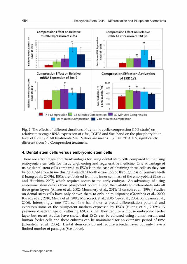
All data are reported as mean ± S.E.M. (N=number of samples). 0 minutes, 15 minutes, 30 minutes, 60 minutes and 240 minutes of compression were compared level of p-ERK 1/2 and for mRNA expression of Sox-9, c-fos, and TGF┚3. Significant differences were determined by using a One-way ANOVA in Sigma Stat 3.00 (SPSS Inc.). The effects of dynamic compression on chondrogenesis and ERK 1/2 signal transduction
was observed (Figure 2). As expected, we saw an increase in the levels of the early response
gene c-fos after 60 minutes of dynamic compression (Figure 2). Surprisingly, we did not see
any change in the other early response gene c-jun (Data not shown) as seen in BMMSCs
described above. We also saw an increase in the gene expression of both TGF┚3 and Sox-9
after 30 minutes of dynamic compression which lasted for at least 60 minutes of
compression (Figure 2). Unlike the PDLSCs response to dynamic compression, SHEDs did
not express Sox-9 after four hours. Further, we noticed and increase in the phosphorylation
of ERK 1/2 as early as 15 minutes which was sustained for at least 60 minutes, but was no
longer elevated after 4 hours (Figure 2). SHEDs clearly have a different response to dynamic compression than PDLSCs. Like PDLSCs we see an early rise in the phosphorylation of ERK 1/2 suggesting that this signal transduction pathway is responding to compressive forces in SHEDS. The rise in TGF┚3 likely triggers the chondrogenic differentiation. Similar to PDLSCs, we see an increase in Sox-9, a transcription factor for chondrocyte differentiation, suggesting that SHEDs are indeed beginning to undergo chondrogenic differentiation. However, unlike PDLSCs, we did not see any aggrecan expression in our experiment which arises later in chondrogenic differentiation. Therefore we can suggest that SHEDs, like PDLSCs, respond to compressive force by undergoing chondrogenic differentiation and this is likely mediated though the ERK 1/2 signal transduction pathway. However, although chondrogenic differentiation is triggered within 30 minutes by dynamic compression, it is not completed during these short time intervals. This may be due to the decrease in TGF┚3 gene expression after 4 hours, which may be required to maintain chondrogenic differentiation in the dynamically compressed constructs as there was no supplementation of TGF┚3 in the media. Interestingly, the control samples did not show any gene expression for aggrecan but these samples were maintained in media without any supplemental TGF┚3. Koyama et al. (2009) did not show any chondrogenic gene expression in control cultures of SHEDs, but felt it may be due to the fact that some of their cultures were infected. We would suggest repeating that experiment to determine if SHEDs cultures express any of the chondrogenic markers without TGF┚3 supplementation.
www.intechopen.com
Embryonic Stem Cells – Differentiation and Pluripotent Alternatives 464
Fig. 2. The effects of different durations of dynamic cyclic compression (15% strain) on relative messenger RNA expression of c-fos, TGF┚3 and Sox-9 and on the phosphorylation level of ERK 1/2. All treatments N=6. Values are means ± S.E.M.; *P < 0.05, significantly different from No Compression treatment.
4. Dental stem cells versus embryonic stem cells
There are advantages and disadvantages for using dental stem cells compared to the using embryonic stem cells for tissue engineering and regenerative medicine. One advantage of using dental stem cells compared to ESCs is in the ease of obtaining these cells as they can be obtained from tissue during a standard tooth extraction or through loss of primary teeth (Huang et al., 2009b). ESCs are obtained from the inner cell mass of the embryoblast (Biswas and Hutchins, 2007) which requires access to the early embryo. An advantage of using embryonic stem cells is their pluripotent potential and their ability to differentiate into all three germ layers (Alison et al., 2002; Mummery et al., 2011; Thomson et al., 1998). Studies on dental stem cells have only shown them to only be multipotent (Gronthos et al., 2000; Karaöz et al., 2010; Miura et al., 2003; Morsczeck et al., 2005; Seo et al., 2004; Sonoyama et al., 2006). Interestingly, one PDL cell line has shown a broad differentiation potential and expresses some of the pluripotent markers expressed by ESCs (Huang et al., 2009a). A previous disadvantage of culturing ESCs is that they require a mouse embryonic feeder layer but recent studies have shown that ESCs can be cultured using human serum and human feeder cells and these cultures can be maintained for an extensive period of time (Ellerström et al., 2006). Dental stem cells do not require a feeder layer but only have a limited number of passages (See above).
www.intechopen.com
Multipotent Dental Stem Cells: An Alternative Adult Derived Stem Cell Source for Regenerative Medicine 465
Many of the problems encountered with stem cells delivery and scaffold choice are the same
for both dental stem cells and ESCs. The use of dental stem cells or ESCs for tissue
engineering or regenerative repair often produce similar results and many of the same types
of problems arise. Currently whole tooth regeneration is not possible using ESCs or dental
stem cells. Similar to dental stem cells, human ESCs have been used to form osteoblasts both
in vitro and in vivo and had the capacity to form mineralized tissue (Bielby et al., 2004).
Like DPSCs, ESCs have been used to repair cardiac function in infarcted myocardium.
Human ESCs were injected into an infarcted mouse myocardium and were shown to
improve cardiac function after four weeks but this improvement was not maintained after
three months (van Laake et al., 2008). ESCs have also been used to explore neuronal
regeneration for patients affected with Parkinson disease. Using the monkey as a primate
model, ESCs have been used to generate dopaminergic neurons to aid in the relief of
Parkinson disease (Takagi et al., 2005).
5. Concluding remarks
This review shows that dental stem cells are a viable alternative to embryonic stem cells for
regenerative medicine. Dental stem cells are easily obtainable from the dental pulp of teeth
and from other dental tissues which are often discarded as waste. Further, dental stem cells,
like BMMSCs, form clonogenic, highly proliferative, multipotent cell populations in vitro
and maintain their differentiation potential in vivo. Dental stem cells also show potential for
cell therapy with respect to whole tooth regeneration. More work needs to be done to
optimize the use of dental stem cells for use in cell therapies of other tissue types in the
future including bone and cartilage formation. The potential of dental stem cells as an
alternative choice to embryonic stem cells seems realistic for future stem cell therapies and
regenerative medicine.
6. References
Abe, S., S. Yamaguchi, and T. Amagasa. 2007. Multilineage Cells from Apical Pulp of
Human Tooth with Immature Apex. Oral Science International. 4:45-58.
Alison, M.R., R. Poulsom, S. Forbes, and N.A. Wright. 2002. An introduction to stem cells. J
Pathol. 197:419-423.
Barrilleaux, B., D.G. Phinney, D.J. Prockop, and K.C. O'Connor. 2006. Review: ex vivo
engineering of living tissues with adult stem cells. Tissue Eng. 12:3007-3019.
Barry, F., R.E. Boynton, B. Liu, and J.M. Murphy. 2001. Chondrogenic Differentiation of
Mesenchymal Stem Cells from Bone Marrow: Differentiation-Dependent Gene
Expression of Matrix Components. Experimental Cell Research. 268:189-200.
Bartold, P.M., C.A.G. McCulloch, A.S. Narayanan, and S. Pitaru. 2000. Tissue engineering: a
new paradigm for periodontal regeneration based on molecular and cell biology.
Periodontology 2000. 24:253-269.
Bianco, P., M. Riminucci, S. Gronthos, and P.G. Robey. 2001. Bone marrow stromal stem
cells: nature, biology, and potential applications. Stem Cells. 19:180-192.
www.intechopen.com

Embryonic Stem Cells – Differentiation and Pluripotent Alternatives 466
Bielby, R.C., A.R. Boccaccini, J.M. Polak, and L.D. Buttery. 2004. In vitro differentiation and
in vivo mineralization of osteogenic cells derived from human embryonic stem
cells. Tissue Eng. 10:1518-1525.
Biswas, A., and R. Hutchins. 2007. Embryonic stem cells. Stem Cells Dev. 16:213-222.
Blanpain, C., W.E. Lowry, A. Geoghegan, L. Polak, and E. Fuchs. 2004. Self-Renewal,
Multipotency, and the Existence of Two Cell Populations within an Epithelial Stem
Cell Niche. Cell. 118:635-648.
Blau, H.M., T.R. Brazelton, and J.M. Weimann. 2001. The Evolving Concept of a Stem Cell:
Entity or Function? Cell. 105:829-841.
Bohl, K.S., J. Shon, B. Rutherford, and D.J. Mooney. 1998. Role of synthetic extracellular
matrix in development of engineered dental pulp. Journal of Biomaterials Science,
Polymer Edition. 9:749-764.
Bongso, A., and C.-Y. Fong. 2009. Human Embryonic Stem Cells: Their Nature, Properties,
and Uses. In Trends in Stem Cell Biology and Technology. H. Baharvand, editor.
Humana Press. 1-17.
Caton, J., N. Bostanci, E. Remboutsika, C. De Bari, and T.A. Mitsiadis. 2010. Future dentistry:
cell therapy meets tooth and periodontal repair and regeneration. J Cell Mol Med.
Chamberlain, G., J. Fox, B. Ashton, and J. Middleton. 2007. Concise Review: Mesenchymal
Stem Cells: Their Phenotype, Differentiation Capacity, Immunological Features,
and Potential for Homing. Stem Cells. 25:2739-2749.
Chen, S.C., V. Marino, S. Gronthos, and P.M. Bartold. 2006. Location of putative stem cells in
human periodontal ligament. Journal of Periodontal Research. 41:547-553.
Conget, P.A., and J.J. Minguell. 1999. Phenotypical and functional properties of human bone
marrow mesenchymal progenitor cells. J Cell Physiol. 181:67-73.
Cordeiro, M.M., Z. Dong, T. Kaneko, Z. Zhang, M. Miyazawa, S. Shi, A.J. Smith, and J.E.
Nör. 2008. Dental Pulp Tissue Engineering with Stem Cells from Exfoliated
Deciduous Teeth. Journal of Endodontics. 34:962-969.
Crisan, M., C.-W. Chen, M. Corselli, G. Andriolo, L. Lazzari, and B. Péault. 2009.
Perivascular Multipotent Progenitor Cells in Human Organs. Annals of the New
York Academy of Sciences. 1176:118-123.
d'Aquino, R., A. De Rosa, G. Laino, F. Caruso, L. Guida, R. Rullo, V. Checchi, L. Laino, V.
Tirino, and G. Papaccio. 2009. Human dental pulp stem cells: from biology to
clinical applications. J Exp Zool B Mol Dev Evol. 312B:408-415.
d'Aquino, R., A. Graziano, M. Sampaolesi, G. Laino, G. Pirozzi, A. De Rosa, and G. Papaccio.
2007. Human postnatal dental pulp cells co-differentiate into osteoblasts and
endotheliocytes: a pivotal synergy leading to adult bone tissue formation. Cell
Death Differ. 14:1162-1171.
Dannan, A. 2009. Dental-derived stem cells and whole tooth regeneration: an overview.
Journal of Clinical Medicine and Research. 1:63-71.
Deasy, B.M., R.J. Jankowski, and J. Huard. 2001. Muscle-derived stem cells: characterization
and potential for cell-mediated therapy. Blood Cells Mol Dis. 27:924-933.
Ding, G., W. Wang, Y. Liu, Y. An, C. Zhang, S. Shi, and S. Wang. 2010. Effect of
cryopreservation on biological and immunological properties of stem cells from
apical papilla. J Cell Physiol. 223:415-422.
www.intechopen.com

Multipotent Dental Stem Cells: An Alternative Adult Derived Stem Cell Source for Regenerative Medicine 467
Dominici, M., K. Le Blanc, I. Mueller, I. Slaper-Cortenbach, F. Marini, D. Krause, R. Deans,
A. Keating, D. Prockop, and E. Horwitz. 2006. Minimal criteria for defining
multipotent mesenchymal stromal cells. The International Society for Cellular
Therapy position statement. Cytotherapy. 8:315-317.
Ellerström, C., R. Strehl, K. Moya, K. Andersson, C. Bergh, K. Lundin, J. Hyllner, and H.
Semb. 2006. Derivation of a Xeno-Free Human Embryonic Stem Cell Line. Stem
Cells. 24:2170-2176.
Feng, F., K. Akiyama, Y. Liu, T. Yamaza, T.M. Wang, J.H. Chen, B.B. Wang, G.T.J. Huang, S.
Wang, and S. Shi. 2010. Utility of PDL progenitors for in vivo tissue regeneration: a
report of 3 cases. Oral Diseases. 16:20-28.
Fortier, L.A. 2005. Stem cells: classifications, controversies, and clinical applications. Vet
Surg. 34:415-423.
Fritz, J.R. 2009. The Chondrogenesis of PDLs by Dynamic Unconfined Compression is
Dependent on p42/44 and not p38 or JNK. In Biomedical Engineering. Vol. Masters
of Science. University of Miami, Coral Gables, Fl. 47.
Gandia, C., A. Armiñan, J.M. García-Verdugo, E. Lledó, A. Ruiz, M.D. Miñana, J. Sanchez-
Torrijos, R. Payá, V. Mirabet, F. Carbonell-Uberos, M. Llop, J.A. Montero, and P.
Sepúlveda. 2008. Human Dental Pulp Stem Cells Improve Left Ventricular
Function, Induce Angiogenesis, and Reduce Infarct Size in Rats with Acute
Myocardial Infarction. Stem Cells. 26:638-645.
Gay, I.C., S. Chen, and M. MacDougall. 2007. Isolation and characterization of multipotent
human periodontal ligament stem cells. Orthod Craniofac Res. 10:149-160.
Gronthos, S., J. Brahim, W. Li, L.W. Fisher, N. Cherman, A. Boyde, P. DenBesten, P.G.
Robey, and S. Shi. 2002. Stem cell properties of human dental pulp stem cells. J
Dent Res. 81:531-535.
Gronthos, S., M. Mankani, J. Brahim, P.G. Robey, and S. Shi. 2000. Postnatal human dental
pulp stem cells (DPSCs) in vitro and in vivo. Proc Natl Acad Sci U S A. 97:13625-
13630.
Gronthos, S., A.C. Zannettino, S.J. Hay, S. Shi, S.E. Graves, A. Kortesidis, and P.J. Simmons.
2003. Molecular and cellular characterisation of highly purified stromal stem cells
derived from human bone marrow. J Cell Sci. 116:1827-1835.
Honda, M.J., M. Imaizumi, H. Suzuki, S. Ohshima, S. Tsuchiya, and K. Satomura. Stem cells
isolated from human dental follicles have osteogenic potential. Oral Surgery, Oral
Medicine, Oral Pathology, Oral Radiology, and Endodontology. In Press, Corrected
Proof.
Huang, C.Y., D. Pelaez, J. Dominguez-Bendala, F. Garcia-Godoy, and H.S. Cheung. 2009a.
Plasticity of stem cells derived from adult periodontal ligament. Regen Med. 4:809-
821.
Huang, C.Y., P.M. Reuben, and H.S. Cheung. 2005. Temporal expression patterns and
corresponding protein inductions of early responsive genes in rabbit bone marrow-
derived mesenchymal stem cells under cyclic compressive loading. Stem Cells.
23:1113-1121.
Huang, G.T. 2009. Pulp and dentin tissue engineering and regeneration: current progress.
Regen Med. 4:697-707.
www.intechopen.com
Embryonic Stem Cells – Differentiation and Pluripotent Alternatives 468
Huang, G.T., S. Gronthos, and S. Shi. 2009b. Mesenchymal stem cells derived from dental
tissues vs. those from other sources: their biology and role in regenerative
medicine. J Dent Res. 88:792-806.
Jiang, Y., B.N. Jahagirdar, R.L. Reinhardt, R.E. Schwartz, C.D. Keene, X.R. Ortiz-Gonzalez,
M. Reyes, T. Lenvik, T. Lund, M. Blackstad, J. Du, S. Aldrich, A. Lisberg, W.C. Low,
D.A. Largaespada, and C.M. Verfaillie. 2002. Pluripotency of mesenchymal stem
cells derived from adult marrow. Nature. 418:41-49.
Karaöz, E., B. Doğan, A. Aksoy, G. Gacar, S. Akyüz, S. Ayhan, Z. Genç, S. Yürüker, G.
Duruksu, P. Demircan, and A. Sarıboyacı. 2010. Isolation and in vitro
characterisation of dental pulp stem cells from natal teeth. Histochemistry and Cell
Biology. 133:95-112.
Kémoun, P., S. Laurencin-Dalicieux, J. Rue, J.-C. Farges, I. Gennero, F. Conte-Auriol, F.
Briand-Mesange, M. Gadelorge, H. Arzate, A. Narayanan, G. Brunel, and J.-P.
Salles. 2007. Human dental follicle cells acquire cementoblast features under
stimulation by BMP-2/-7 and enamel matrix derivatives (EMD) in vitro. Cell and
Tissue Research. 329:283-294.
Kerkis, I., A. Kerkis, D. Dozortsev, G.C. Stukart-Parsons, S.M. Gomes Massironi, L.V.
Pereira, A.I. Caplan, and H.F. Cerruti. 2006. Isolation and characterization of a
population of immature dental pulp stem cells expressing OCT-4 and other
embryonic stem cell markers. Cells Tissues Organs. 184:105-116.
Kolf, C.M., E. Cho, and R.S. Tuan. 2007. Mesenchymal stromal cells. Biology of adult
mesenchymal stem cells: regulation of niche, self-renewal and differentiation.
Arthritis Res Ther. 9:204.
Koyama, N., Y. Okubo, K. Nakao, and K. Bessho. 2009. Evaluation of pluripotency in human
dental pulp cells. J Oral Maxillofac Surg. 67:501-506.
Kuznetsov, S.A., P.H. Krebsbach, K. Satomura, J. Kerr, M. Riminucci, D. Benayahu, and P.G.
Robey. 1997. Single-colony derived strains of human marrow stromal fibroblasts
form bone after transplantation in vivo. J Bone Miner Res. 12:1335-1347.
Lanzendorf, S.E., C.A. Boyd, D.L. Wright, S. Muasher, S. Oehninger, and G.D. Hodgen. 2001.
Use of human gametes obtained from anonymous donors for the production of
human embryonic stem cell lines. Fertil Steril. 76:132-137.
Lee, J.W., Y.H. Kim, S.H. Kim, S.H. Han, and S.B. Hahn. 2004. Chondrogenic differentiation
of mesenchymal stem cells and its clinical applications. Yonsei Med J. 45 Suppl:41-
47.
Lekic, P., J. Rojas, C. Birek, H. Tenenbaum, and C.A.G. McCulloch. 2001. Phenotypic
comparison of periodontal ligament cells in vivo and in vitro. Journal of Periodontal
Research. 36:71-79.
Leung, A.K., and W.L. Robson. 2006. Natal teeth: a review. J Natl Med Assoc. 98:226-228.
Levenberg, S., and R. Langer. 2004. Advances in Tissue Engineering. In Current Topics in
Developmental Biology. Vol. Volume 61. P.S. Gerald, editor. Academic Press. 113-
134.
Lindroos, B., K. Maenpaa, T. Ylikomi, H. Oja, R. Suuronen, and S. Miettinen. 2008.
Characterisation of human dental stem cells and buccal mucosa fibroblasts. Biochem
Biophys Res Commun. 368:329-335.
www.intechopen.com

Multipotent Dental Stem Cells: An Alternative Adult Derived Stem Cell Source for Regenerative Medicine 469
Liu, H., S. Gronthos, and S. Shi. 2006. Dental pulp stem cells. Methods Enzymol. 419:99-113.
Livak, K.J., and T.D. Schmittgen. 2001. Analysis of Relative Gene Expression Data Using
Real-Time Quantitative PCR and the 2−ΔΔCT Method. Methods. 25:402-408.
Luan, X., Y. Ito, S. Dangaria, and T.G. Diekwisch. 2006. Dental follicle progenitor cell
heterogeneity in the developing mouse periodontium. Stem Cells Dev. 15:595-608.
Mitsiadis, T.A., and P. Papagerakis. 2011. Regenerated teeth: the future of tooth
replacement? Regenerative Medicine. 6:135-139.
Miura, M., S. Gronthos, M. Zhao, B. Lu, L.W. Fisher, P.G. Robey, and S. Shi. 2003. SHED:
stem cells from human exfoliated deciduous teeth. Proc Natl Acad Sci U S A.
100:5807-5812.
Morsczeck, C., W. Gotz, J. Schierholz, F. Zeilhofer, U. Kuhn, C. Mohl, C. Sippel, and K.H.
Hoffmann. 2005. Isolation of precursor cells (PCs) from human dental follicle of
wisdom teeth. Matrix Biol. 24:155-165.
Morsczeck, C., F. Völlner, M. Saugspier, C. Brandl, T. Reichert, O. Driemel, and G. Schmalz.
2010. Comparison of human dental follicle cells (DFCs) and stem cells from human
exfoliated deciduous teeth (SHED) after neural differentiation in vitro. Clinical Oral
Investigations. 14:433-440.
Mummery, C., I. Wilmut, A. van de Stolpe, and B.A.J. Roelen. 2011. Stem Cells: Scientific
Facts and Fiction. Academic Press, London, England. 324 pp.
Nam, H., and G. Lee. 2009. Identification of novel epithelial stem cell-like cells in human
deciduous dental pulp. Biochem Biophys Res Commun. 386:135-139.
Nishino, Y., Y. Yamada, K. Ebisawa, S. Nakamura, K. Okabe, E. Umemura, K. Hara, and M.
Ueda. 2011. Stem cells from human exfoliated deciduous teeth (SHED) enhance
wound healing and the possibility of novel cell therapy. Cytotherapy. 13:598-605.
Papaccio, G., A. Graziano, R. d'Aquino, M.F. Graziano, G. Pirozzi, D. Menditti, A. De Rosa,
F. Carinci, and G. Laino. 2006. Long-term cryopreservation of dental pulp stem
cells (SBP-DPSCs) and their differentiated osteoblasts: a cell source for tissue
repair. J Cell Physiol. 208:319-325.
Pelaez, D., C.Y. Huang, and H.S. Cheung. 2009. Cyclic compression maintains viability and
induces chondrogenesis of human mesenchymal stem cells in fibrin gel scaffolds.
Stem Cells Dev. 18:93-102.
Pierdomenico, L., L. Bonsi, M. Calvitti, D. Rondelli, M. Arpinati, G. Chirumbolo, E.
Becchetti, C. Marchionni, F. Alviano, V. Fossati, N. Staffolani, M. Franchina, A.
Grossi, and G.P. Bagnara. 2005. Multipotent mesenchymal stem cells with
immunosuppressive activity can be easily isolated from dental pulp.
Transplantation. 80:836-842.
Pittenger, M.F., A.M. Mackay, S.C. Beck, R.K. Jaiswal, R. Douglas, J.D. Mosca, M.A.
Moorman, D.W. Simonetti, S. Craig, and D.R. Marshak. 1999. Multilineage
potential of adult human mesenchymal stem cells. Science. 284:143-147.
Pivoriuunas, A., A. Surovas, V. Borutinskaite, D. Matuzeviccius, G. Treigyte, J. Savickiene,
V. Tunaitis, R. Aldonyte, A. Jarmalavicciuute, K. Suriakaite, E. Liutkeviccius, A.
Venalis, D. Navakauskas, R. Navakauskiene, and K.E. Magnusson. 2010. Proteomic
analysis of stromal cells derived from the dental pulp of human exfoliated
deciduous teeth. Stem Cells Dev. 19:1081-1093.
www.intechopen.com

Embryonic Stem Cells – Differentiation and Pluripotent Alternatives 470
Rodriguez-Lozano, F.J., C. Bueno, C.L. Insausti, L. Meseguer, M.C. Ramirez, M. Blanquer, N.
Marin, S. Martinez, and J.M. Moraleda. 2011. Mesenchymal stem cells derived from
dental tissues. Int Endod J.
Schugar, R.C., S.M. Chirieleison, K.E. Wescoe, B.T. Schmidt, Y. Askew, J.J. Nance, J.M.
Evron, B. Peault, and B.M. Deasy. 2009. High harvest yield, high expansion, and
phenotype stability of CD146 mesenchymal stromal cells from whole primitive
human umbilical cord tissue. J Biomed Biotechnol. 2009:789526.
Seo, B.M., M. Miura, S. Gronthos, P.M. Bartold, S. Batouli, J. Brahim, M. Young, P.G. Robey,
C.Y. Wang, and S. Shi. 2004. Investigation of multipotent postnatal stem cells from
human periodontal ligament. Lancet. 364:149-155.
Seo, B.M., M. Miura, W. Sonoyama, C. Coppe, R. Stanyon, and S. Shi. 2005. Recovery of stem
cells from cryopreserved periodontal ligament. J Dent Res. 84:907-912.
Sharma, S., V. Sikri, N. Sharma, and V. Sharma. 2010. Regeneration of tooth pulp and dentin
: trends and advances. Annals of Neurosciences. 17:31-43.
Shi, S., P.M. Bartold, M. Miura, B.M. Seo, P.G. Robey, and S. Gronthos. 2005. The efficacy of
mesenchymal stem cells to regenerate and repair dental structures. Orthod Craniofac
Res. 8:191-199.
Shi, S., and S. Gronthos. 2003. Perivascular niche of postnatal mesenchymal stem cells in
human bone marrow and dental pulp. J Bone Miner Res. 18:696-704.
Shimono, M., T. Ishikawa, H. Ishikawa, H. Matsuzaki, S. Hashimoto, T. Muramatsu, K.
Shima, K.-I. Matsuzaka, and T. Inoue. 2003. Regulatory mechanisms of periodontal
regeneration. Microscopy Research and Technique. 60:491-502.
Slatter, D.H. 2002. Textbook of small animal surgery. Vol. 2. Saunders, Philadelphia, P.A.
3070.
Smith, A. 2006. A glossary for stem-cell biology. Nature. 441:1060-1060.
Sonoyama, W., Y. Liu, D. Fang, T. Yamaza, B.M. Seo, C. Zhang, H. Liu, S. Gronthos, C.Y.
Wang, S. Wang, and S. Shi. 2006. Mesenchymal stem cell-mediated functional tooth
regeneration in swine. PLoS One. 1:e79.
Sonoyama, W., Y. Liu, T. Yamaza, R.S. Tuan, S. Wang, S. Shi, and G.T. Huang. 2008.
Characterization of the apical papilla and its residing stem cells from human
immature permanent teeth: a pilot study. J Endod. 34:166-171.
Takagi, Y., J. Takahashi, H. Saiki, A. Morizane, T. Hayashi, Y. Kishi, H. Fukuda, Y. Okamoto,
M. Koyanagi, M. Ideguchi, H. Hayashi, T. Imazato, H. Kawasaki, H. Suemori, S.
Omachi, H. Iida, N. Itoh, N. Nakatsuji, Y. Sasai, and N. Hashimoto. 2005.
Dopaminergic neurons generated from monkey embryonic stem cells function in a
Parkinson primate model. J Clin Invest. 115:102-109.
Ten Cate, A.R. 1998. Oral histology: development, structure, and function. Mosby, St. Louis.
528 pp.
Thomson, J.A., J. Itskovitz-Eldor, S.S. Shapiro, M.A. Waknitz, J.J. Swiergiel, V.S. Marshall,
and J.M. Jones. 1998. Embryonic stem cell lines derived from human blastocysts.
Science. 282:1145-1147.
Tucker, A., and P. Sharpe. 2004. The cutting-edge of mammalian development; how the
embryo makes teeth. Nat Rev Genet. 5:499-508.
www.intechopen.com

Multipotent Dental Stem Cells: An Alternative Adult Derived Stem Cell Source for Regenerative Medicine 471
van Laake, L.W., R. Passier, P.A. Doevendans, and C.L. Mummery. 2008. Human Embryonic
Stem Cell–Derived Cardiomyocytes and Cardiac Repair in Rodents. Circulation
Research. 102:1008-1010.
Vandesompele, J., K. De Preter, F. Pattyn, B. Poppe, N. Van Roy, A. De Paepe, and F.
Speleman. 2002. Accurate normalization of real-time quantitative RT-PCR data by
geometric averaging of multiple internal control genes. Genome biology.
3:RESEARCH0034.
Wada, N., D. Menicanin, S. Shi, P.M. Bartold, and S. Gronthos. 2009. Immunomodulatory
properties of human periodontal ligament stem cells. J Cell Physiol. 219:667-676.
Wang, J., X. Wang, Z. Sun, H. Yang, S. Shi, and S. Wang. 2010. Stem cells from human-
exfoliated deciduous teeth can differentiate into dopaminergic neuron-like cells.
Stem Cells Dev. 19:1375-1383.
Wobus, A.M., and K.R. Boheler. 2005. Embryonic Stem Cells: Prospects for Developmental
Biology and Cell Therapy. Physiological Reviews. 85:635-678.
Wong, M.H. 2004. Regulation of Intestinal Stem Cells. J Investig Dermatol Symp Proc. 9:224-
228.
Woodbury, D., E.J. Schwarz, D.J. Prockop, and I.B. Black. 2000. Adult rat and human bone
marrow stromal cells differentiate into neurons. Journal of Neuroscience Research.
61:364-370.
Woods, E.J., B.C. Perry, J.J. Hockema, L. Larson, D. Zhou, and W.S. Goebel. 2009. Optimized
cryopreservation method for human dental pulp-derived stem cells and their
tissues of origin for banking and clinical use. Cryobiology. 59:150-157.
Yagyuu, T., E. Ikeda, H. Ohgushi, M. Tadokoro, M. Hirose, M. Maeda, K. Inagake, and T.
Kirita. 2010. Hard tissue-forming potential of stem/progenitor cells in human
dental follicle and dental papilla. Arch Oral Biol. 55:68-76.
Yao, S., F. Pan, V. Prpic, and G.E. Wise. 2008. Differentiation of stem cells in the dental
follicle. J Dent Res. 87:767-771.
Yen, A.H., and P.T. Sharpe. 2008. Stem cells and tooth tissue engineering. Cell Tissue Res.
331:359-372.
Yokoi, T., M. Saito, T. Kiyono, S. Iseki, K. Kosaka, E. Nishida, T. Tsubakimoto, H. Harada, K.
Eto, T. Noguchi, and T. Teranaka. 2007. Establishment of immortalized dental
follicle cells for generating periodontal ligament in vivo. Cell and Tissue Research.
327:301-311.
Young, H.E., and A.C. Black, Jr. 2004. Adult stem cells. Anat Rec A Discov Mol Cell Evol Biol.
276:75-102.
Zandstra, P.W., and A. Nagy. 2001. Stem cell bioengineering. Annu Rev Biomed Eng. 3:275-
305.
Zhang, W., X.F. Walboomers, S. Shi, M. Fan, and J.A. Jansen. 2006. Multilineage
differentiation potential of stem cells derived from human dental pulp after
cryopreservation. Tissue Eng. 12:2813-2823.
Zuk, P.A., M. Zhu, P. Ashjian, D.A. De Ugarte, J.I. Huang, H. Mizuno, Z.C. Alfonso, J.K.
Fraser, P. Benhaim, and M.H. Hedrick. 2002. Human adipose tissue is a source of
multipotent stem cells. Mol Biol Cell. 13:4279-4295.
www.intechopen.com

Embryonic Stem Cells – Differentiation and Pluripotent Alternatives 472
Zuk, P.A., M. Zhu, H. Mizuno, J. Huang, J.W. Futrell, A.J. Katz, P. Benhaim, H.P. Lorenz,
and M.H. Hedrick. 2001. Multilineage cells from human adipose tissue:
implications for cell-based therapies. Tissue Eng. 7:211-228.
www.intechopen.com

Embryonic Stem Cells - Differentiation and Pluripotent AlternativesEdited by Prof. Michael S. Kallos
ISBN 978-953-307-632-4Hard cover, 506 pagesPublisher InTechPublished online 12, October, 2011Published in print edition October, 2011
InTech EuropeUniversity Campus STeP Ri Slavka Krautzeka 83/A 51000 Rijeka, Croatia Phone: +385 (51) 770 447 Fax: +385 (51) 686 166www.intechopen.com
InTech ChinaUnit 405, Office Block, Hotel Equatorial Shanghai No.65, Yan An Road (West), Shanghai, 200040, China
Phone: +86-21-62489820 Fax: +86-21-62489821
The ultimate clinical implementation of embryonic stem cells will require methods and protocols to turn theseunspecialized cells into the fully functioning cell types found in a wide variety of tissues and organs. In order toachieve this, it is necessary to clearly understand the signals and cues that direct embryonic stem celldifferentiation. This book provides a snapshot of current research on the differentiation of embryonic stem cellsto a wide variety of cell types, including neural, cardiac, endothelial, osteogenic, and hepatic cells. In addition,induced pluripotent stem cells and other pluripotent stem cell sources are described. The book will serve as avaluable resource for engineers, scientists, and clinicians as well as students in a wide range of disciplines.
How to referenceIn order to correctly reference this scholarly work, feel free to copy and paste the following:
Tammy Laberge and Herman S. Cheung (2011). Multipotent Dental Stem Cells: An Alternative Adult DerivedStem Cell Source for Regenerative Medicine, Embryonic Stem Cells - Differentiation and PluripotentAlternatives, Prof. Michael S. Kallos (Ed.), ISBN: 978-953-307-632-4, InTech, Available from:http://www.intechopen.com/books/embryonic-stem-cells-differentiation-and-pluripotent-alternatives/multipotent-dental-stem-cells-an-alternative-adult-derived-stem-cell-source-for-regenerative-medicin