8 Fetal Considerations in the Critically Ill Gravida
28
123 Critical Care Obstetrics, Sixth Edition. Edited by Jeffrey P. Phelan, Luis D. Pacheco, Michael R. Foley, George R. Saade, Gary A. Dildy, and Michael A. Belfort. © 2019 John Wiley & Sons Ltd. Published 2019 by John Wiley & Sons Ltd. Introduction Unlike any other medical or surgical specialty, obstetrics deals with the simultaneous management of two – and sometimes more – individuals. Under all circumstances, the obstetrician must delicately balance the impact of each treatment decision on the pregnant woman and her fetus, seeking, when possible, to minimize the risks of harm to each person. Throughout this text, the primary focus has been on the critically ill obstetric patient and, secondarily, her fetus. Although the fetal effects of those illnesses were reviewed in part, the goal of this chapter is to highlight, especially for the non‐obstetric clinician, the important clinical fetal considerations encountered when caring for these complicated pregnancies. To achieve that objective, this chapter reviews: (1) current techniques for assessing fetal well‐being, (2) fetal assess- ment in the intensive care unit (ICU), (3) fetal considera- tions in several maternal medical and surgical conditions, (4) the contemporary management of the gravida who is brain‐dead or in a persistent vegetative state, and (5) the role of perimortem cesarean delivery in contemporary obstetrics. Detection of fetal distress in the critically ill obstetric patient Almost a half century ago, Hon and Quilligan [1] dem- onstrated the relationship between certain fetal heart rate (FHR) patterns and fetal condition by using con- tinuous electronic FHR monitoring in laboring patients. Since then, continuous electronic FHR monitoring has become a universally accepted method of assessing fetal well‐being during labor [2,3] with the goal of permitting the clinician to identify those fetuses at a greater likeli- hood of intrapartum fetal death [4] and to intervene when certain FHR abnormalities are present. In addition to the intrapartum assessment of fetal well‐being, the fetal monitor has been used to assess fetal health before labor [5] and to attempt to identify those fetuses at risk for intrauterine death. Once that fetus is identified, the maternal‐fetal unit is moved from outpatient to inpatient care. Once in labor and delivery, continuous fetal monitoring is used to determine whether continued expectant management or delivery by induction of labor or cesarean is the next form of intervention. It is this area of fetal monitoring, antepar- tum rather than intrapartum fetal assessment, that is used more frequently in the arena of the critically ill gravida. In keeping with those core principles, the focus of this chapter will be on applications of fetal monitor- ing to assess fetal status both in the ICU setting and intrapartum during labor. Although the presence of a reassuring FHR tracing is virtually always associated with a well‐perfused and oxygenated fetus [5,6], an “abnormal tracing” is not nec- essarily predictive of an adverse fetal outcome. While it was anticipated that the detection of abnormal FHR pat- terns during labor and expeditious delivery of such fetuses would impact the subsequent development of cerebral palsy, this expectation has not been realized because the number of fetuses injured during labor was highly overestimated and the number of fetuses injured before labor was highly underestimated [7]. However, with the ubiquitous use of electronic FHR monitoring during labor and a rise in the cesarean delivery rate for the past two decades from 5% to over 25%, a decline 8 Fetal Considerations in the Critically Ill Gravida Jeffrey P. Phelan Childbirth Injury Prevention Foundation, Glendora, CA, USA San Gabriel Valley Perinatal Medical Group, Inc., West Covina, CA Department of Obstetrics and Gynecology, Citrus Valley Medical Center, West Covina, CA, USA
Transcript of 8 Fetal Considerations in the Critically Ill Gravida

123
Critical Care Obstetrics, Sixth Edition. Edited by Jeffrey P. Phelan, Luis D. Pacheco, Michael R. Foley, George R. Saade, Gary A. Dildy, and Michael A. Belfort. © 2019 John Wiley & Sons Ltd. Published 2019 by John Wiley & Sons Ltd.
Introduction
Unlike any other medical or surgical specialty, obstetrics deals with the simultaneous management of two – and sometimes more – individuals. Under all circumstances, the obstetrician must delicately balance the impact of each treatment decision on the pregnant woman and her fetus, seeking, when possible, to minimize the risks of harm to each person. Throughout this text, the primary focus has been on the critically ill obstetric patient and, secondarily, her fetus. Although the fetal effects of those illnesses were reviewed in part, the goal of this chapter is to highlight, especially for the non‐obstetric clinician, the important clinical fetal considerations encountered when caring for these complicated pregnancies. To achieve that objective, this chapter reviews: (1) current techniques for assessing fetal well‐being, (2) fetal assess-ment in the intensive care unit (ICU), (3) fetal considera-tions in several maternal medical and surgical conditions, (4) the contemporary management of the gravida who is brain‐dead or in a persistent vegetative state, and (5) the role of perimortem cesarean delivery in contemporary obstetrics.
Detection of fetal distress in the critically ill obstetric patient
Almost a half century ago, Hon and Quilligan [1] dem-onstrated the relationship between certain fetal heart rate (FHR) patterns and fetal condition by using con-tinuous electronic FHR monitoring in laboring patients. Since then, continuous electronic FHR monitoring has become a universally accepted method of assessing fetal
well‐being during labor [2,3] with the goal of permitting the clinician to identify those fetuses at a greater likeli-hood of intrapartum fetal death [4] and to intervene when certain FHR abnormalities are present.
In addition to the intrapartum assessment of fetal well‐being, the fetal monitor has been used to assess fetal health before labor [5] and to attempt to identify those fetuses at risk for intrauterine death. Once that fetus is identified, the maternal‐fetal unit is moved from outpatient to inpatient care. Once in labor and delivery, continuous fetal monitoring is used to determine whether continued expectant management or delivery by induction of labor or cesarean is the next form of intervention. It is this area of fetal monitoring, antepar-tum rather than intrapartum fetal assessment, that is used more frequently in the arena of the critically ill gravida. In keeping with those core principles, the focus of this chapter will be on applications of fetal monitor-ing to assess fetal status both in the ICU setting and intrapartum during labor.
Although the presence of a reassuring FHR tracing is virtually always associated with a well‐perfused and oxygenated fetus [5,6], an “abnormal tracing” is not nec-essarily predictive of an adverse fetal outcome. While it was anticipated that the detection of abnormal FHR pat-terns during labor and expeditious delivery of such fetuses would impact the subsequent development of cerebral palsy, this expectation has not been realized because the number of fetuses injured during labor was highly overestimated and the number of fetuses injured before labor was highly underestimated [7]. However, with the ubiquitous use of electronic FHR monitoring during labor and a rise in the cesarean delivery rate for the past two decades from 5% to over 25%, a decline
8
Fetal Considerations in the Critically Ill GravidaJeffrey P. Phelan
Childbirth Injury Prevention Foundation, Glendora, CA, USASan Gabriel Valley Perinatal Medical Group, Inc., West Covina, CADepartment of Obstetrics and Gynecology, Citrus Valley Medical Center, West Covina, CA, USA

Fetal Considerations in the Critically Ill Gravida124
in the rate of asphyxia‐induced cerebral palsy among singleton term infants has been observed [8,9]. For example, Smith and associates [9] documented a 56% decline over two decades in the incidence of hypoxic ischemic encephalopathy (HIE) among singleton term infants. During this time, the incidence of HIE dropped from 1 per 8000 to 1 per 12,500 births.
While the specific entity of cerebral palsy is, in most cases, unrelated to the events associated with labor and delivery, it is more often related to prenatal developmen-tal events, infection, or complications of prematurity. Nevertheless, the basic physiologic observations relating to specific FHR patterns remain, for the most part, valid. The critically ill mother will necessarily shunt blood from the splanchnic bed (including the uterus) in response to shock. Because of this and the fact that the fetus operates on the steep portion of the oxyhemoglobin dissociation curve, any degree of maternal hypoxia or hypoperfusion may first be manifested as an abnormality of the FHR. In this sense, the late second‐ and third‐tri-mester fetus serves as a physiologic oximeter and cardiac output computer. Observation of FHR changes, thus, may assist or alert the clinician to subtle degrees of phys-iologic instability, which would be unimportant in a non‐pregnant adult but may have potentially detrimental effects to the fetus [10].
The next few pages present an overview of FHR pat-terns pertinent to the critically ill gravida. Interpretations of FHR patterns, like all diagnostic tests, depend on the index population, and consequently, certain of these observations may not be applicable to the laboring but otherwise well mother. For a more detailed description of antepartum and intrapartum FHR tracings associated with fetal brain injury, the reader is referred to the classic descriptions by Phelan and Ahn [11], Phelan and Kim [12], Phelan [13], and Phelan and associates [14].
Of note, FHR interpretation was changed into a cat-egory system [15] in 2008 (Table 8.1). As demonstrated in Table 8.1, a Category 1 strip includes a normal base-line rate, moderate variability, the absence of late and variable decelerations, and early decelerations, and accelerations may be present or absent. A Category 1 strip is consistent with a healthy fetus and a lack of metabolic acidosis. In contrast, a Category 3 FHR strip is the polar opposite of Category 1, and is indicative of a fetus in significant potential fetal jeopardy. This means, according to the “consensus,” that the fetus with a Category 3 tracing, if uncorrected, is at a higher probability of metabolic acidosis and the potential for fetal neurologic injury. A Category 2 strip is some-where in between 1 and 3. In other words, a Category 2 FHR tracing includes everything that is not Category 1 or Category 3. However, it is important to remember the following:
1) Not all fetuses require systemic metabolic acidosis to become neurologically impaired.
2) No one pH can determine whether a fetus will become permanently neurologically impaired [16].
3) The key issue with a Category 2 FHR tracing is that the recruitment of two or more FHR abnormalities, in all likelihood, increases the risk of an adverse outcome.
With that as a backdrop, we will now break down the numerous FHR characteristics into their separate parts for purposes of understanding.
Table 8.1 Category system for interpretation of intrapartum fetal heart rate tracings.
Category ICategory I fetal heart rate (FHR) tracings include all of the following:
● Baseline rate: 110–160 beats per minute (bpm) ● Baseline FHR variability: Moderate ● Late or variable decelerations: Absent ● Early decelerations: Present or absent ● Accelerations: Present or absent
Category IICategory II FHR tracings include all FHR tracings not categorized as Category I or Category III. Category II tracings may represent an appreciable fraction of those encountered in clinical care. Examples of Category II FHR tracings include any of the following:Baseline rate
● Bradycardia not accompanied by absent baseline variability ● Tachycardia
Baseline FHR variability ● Minimal baseline variability ● Absent baseline variability not accompanied by recurrent
decelerations ● Marked baseline variability
Accelerations ● Absence of induced accelerations after fetal stimulation
Periodic or episodic decelerations ● Recurrent variable decelerations accompanied by minimal or
moderate baseline variability ● Prolonged deceleration ≥2 min but ≤10 min ● Recurrent late decelerations with moderate baseline
variability ● Variable decelerations with other characteristics, such as slow
return to baseline, “overshoots,” or “shoulders”Category IIICategory III FHR tracings include either:
● Absent baseline FHR variability and any of the following: – Recurrent late decelerations – Recurrent variable decelerations – Bradycardia
Sinusoidal pattern
Source: Ref. [15].

Detection of fetal distress in the critically ill obstetric patient 125
Baseline fetal heart rate
The baseline FHR is the intrinsic heart rate of the fetus. The baseline FHR is determined by approximating the mean FHR rounded to increments of 5 beats per minute (bpm) during a 10 min window, excluding accelerations and decelerations and periods of marked variability (>25 bpm). A normal baseline FHR is between 110 bpm and 160 bpm. A baseline FHR below 110 bpm is termed a bradycardia, and a baseline FHR in excess of 160 bpm is termed a FHR tachycardia [15].
A persistent slow FHR or an intrinsic bradycardia
Bradycardia is defined as the intrinsic heart rate of the fetus of less than 110 bpm, as opposed to a sudden, rapid, and sustained deterioration of the FHR from a previously normal or tachycardic rate that lasts until delivery, or, under today’s parlance, a FHR bradycardia. As such, a persistent slow FHR may be associated with an underlying congenital fetal abnormality, such as a structural defect of the fetal heart. In addition, congeni-tal bradyarrhythmias may involve fetal heart block sec-ondary to a prior maternal infection, a structural defect of the fetal heart, or systemic lupus erythematosus with anti‐Ro/SSA antibodies [17]. In these circumstances, the FHR bradycardia is not usually a threat to the fetus. But, alternative methods of fetal assessment, such as the fetal biophysical profile (BPP) [18], are necessary in this select group of patients to assure fetal well‐being before and during labor. Given the inherent difficulties in pro-viding continuous fetal monitoring and assuring fetal well‐being in fetuses with a bradyarrhythmia, cesarean delivery may well represent the preferred route of deliv-ery for these patients. Obviously, the decision to pro-ceed directly to a cesarean will depend on the overall clinical circumstances and appropriate patient informed consent.
A sudden rapid and sustained deterioration of the fetal heart rate
Prolonged FHR deceleration is distinctly different from a bradycardia. In the former, the fetal monitor strip is typi-cally reactive with a normal or tachycardic baseline rate; but, due to a sentinel hypoxic event, such as those depicted in Table 8.2, the FHR suddenly drops and remains at a lower level unresponsive to remedial meas-ures and/or terbutaline therapy. In the critical care set-ting, a sudden, rapid, and sustained deterioration of the FHR or a prolonged FHR deceleration may arise from a partial or complete abruption in cases of markedly and persistently elevated maternal blood pressures or an aggressive lowering of maternal BP with antihypertensive
agents [19]. This type of FHR pattern may also herald a sudden maternal hypoxic event, such as amniotic fluid embolus syndrome [20], acute respiratory insufficiency, or an eclamptic seizure [19,21]. Prolonged FHR decelera-tions have also been associated with maternal operative procedures such as cardiopulmonary bypass with inade-quate maternal flow rates [22,23], and brain surgery dur-ing hypothermia [24].
In a patient with a prior normal baseline FHR, the abrupt occurrence and persistence of a fetal heart rate of less than 110 bpm for an extended period of time unre-sponsive to remedial measures and/or terbutaline ther-apy constitute an obstetric emergency. Under these circumstances, and assuming the pregnant woman is hemodynamically and clinically stable and the fetus is potentially viable, these patients should be managed as if the fetus has had a cardiac arrest and be delivered as rap-idly as it is technically feasible in keeping with the level of the institution in question.
Tachycardia
Fetal tachycardia is defined as a baseline FHR of 160 bpm or greater lasting 10 min or longer. Most com-monly, this type of baseline FHR abnormality can be associated with prematurity, maternal pyrexia, or cho-rioamnionitis. In addition, betamimetic administra-tion, hyperthyroidism, or fetal cardiac arrhythmias may also be responsible. The clinical observation of a FHR tachycardia, in and of itself, is probably not an ominous finding but probably reflects a normal physiologic adjustment to an underlying maternal or fetal condi-tion. Although operative intervention is infrequently required, a search for the underlying basis for the FHR tachycardia and a reanalysis of the admission FHR pat-tern may be helpful.
For example, a patient with a previously reactive FHR pattern with a normal baseline rate (Figure 8.1) who develops the Hon pattern of intrapartum asphyxia or ischemia [11] that is characterized by a substantial rise in the baseline rate – often to a level of tachycardia
Table 8.2 Examples of sentinel hypoxic events associated with a sudden, rapid, and sustained deterioration of the fetal heart rate that can be unresponsive to remedial measures and/or terbutaline lasting until delivery from a previously reactive fetal heart rate.
Umbilical cord prolapseUterine rupturePlacental abruptionMaternal arrest (e.g., AFE syndrome)Fetal exsanguination
AFE, Amniotic fluid embolus.

Figure 8.1 Admission FHR of this term pregnancy with spontaneously ruptured membranes exhibits a baseline rate of around 120 bpm and numerous FHR accelerations or a reactive FHR pattern. This FHR pattern would now be characterized as a Category 1 fetal monitor strip.
Figure 8.2 Some time later, the fetus exhibits an FHR tachycardia around 160 bpm, repetitive FHR decelerations, and nonreactivity. Under current characterizations, this FHR pattern would be considered a Category 2 fetal monitor strip.
Detection of fetal distress in the critically ill obstetric patient 127
(Figures 8.2 and 8.3) in association with an inability to accelerate or nonreactivity, repetitive FHR decelerations, and usually a loss of FHR variability – flags fetal brain injury [25], and the fetus is at risk for hypoxic ischemic brain injury [11–13]. In this clinical setting, assessment of the usual causes of FHR tachycardia should be under-taken. If the mother does not have a fever to account for the change in fetal status, assessment of fetal acid–base status with scalp or acoustic stimulation [6,12] or deliv-ery as soon as it is practical, in keeping with the capabil-ity of the hospital, should be considered. If the gravida has a fever, she should be cultured, and treated with anti-biotics and antipyretics. If the FHR pattern does not return to normal (i.e., the same FHR pattern the fetus had on admission – normal baseline FHR and reactive) within approximately an hour of the initiation of medical therapy and regardless of whether the FHR variability is average [11–13,26], the patient should be delivered as expeditiously as possible.
Baseline fetal heart rate variability
Fetal heart rate variability (FHRV) is determined in a 10 min window, excluding accelerations and decelerations. Baseline FHRV is defined as fluctuations in the baseline FHR that are irregular in amplitude and frequency. The fluctuations are visually quantitated as the amplitude of the peak‐to‐trough in BPM. In other words, FHRV is the beat‐to‐beat variation in the FHR resulting from the continuous interaction of the parasympathetic and sympathetic nervous systems on the fetal heart [15].
Since the last edition of this textbook, the two approaches to define FHRV [11,14,15,27] have been con-solidated under the National Institutes of Child Health and Human Development (NICHD) [15,27] umbrella to classify FHRV. The NICHD approach subclassifies FHRV into four categories:
1) Undetectable or absent FHRV2) Minimal (more than absent but ≤5 bpm)
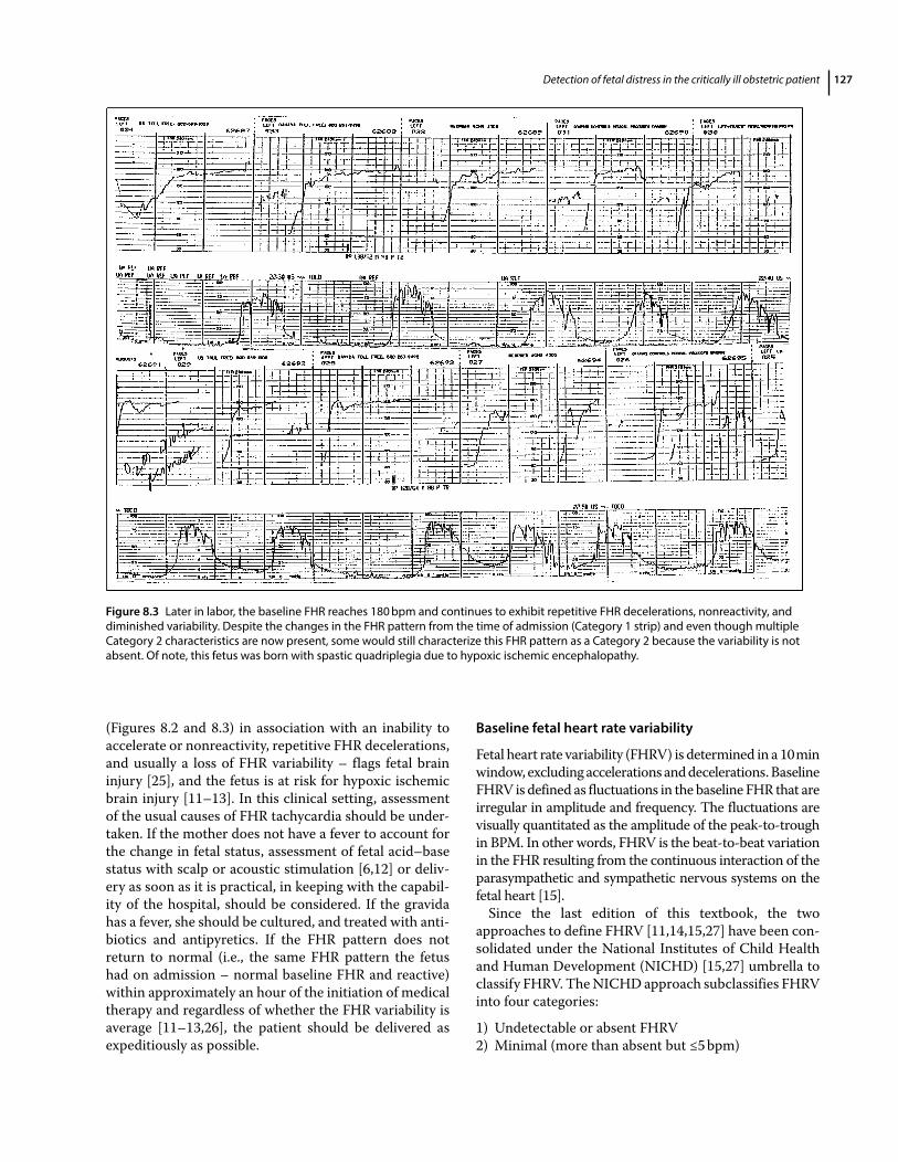
Figure 8.3 Later in labor, the baseline FHR reaches 180 bpm and continues to exhibit repetitive FHR decelerations, nonreactivity, and diminished variability. Despite the changes in the FHR pattern from the time of admission (Category 1 strip) and even though multiple Category 2 characteristics are now present, some would still characterize this FHR pattern as a Category 2 because the variability is not absent. Of note, this fetus was born with spastic quadriplegia due to hypoxic ischemic encephalopathy.

Fetal Considerations in the Critically Ill Gravida128
3) Moderate (6–25 bpm)4) Marked (>25 bpm).
A loss of FHRV, in and of itself, may not necessarily be an ominous observation. In most cases, the loss of FHRV can represent normal fetal physiologic adjustments to a number of medications, illicit substances, or simply behavioral state changes such as 1 F to 4 F [28]. For exam-ple, narcotic administration [29] or magnesium sulfate infusion [30] can alter FHRV by inducing a change in the behavioral state of the fetus to one of a sleep state or behavioral state 1 F. Clinically, the change in FHRV to minimal or absent appears to be clinically significant in cases of the Hon pattern of intrapartum asphyxia [11–13]. As observed herein (Figures 8.1–8.3), the FHR pattern was first reactive and exhibited a normal baseline rate or a Category 1 FHR tracing. Subsequently, the FHR pattern changed. Then, minimal FHRV was associated with a loss of FHR reactivity (the ability of the fetus to accelerate), a substantial rise in the baseline FHR to the level of a FHR tachycardia, and repetitive (more than recurrent) FHR decelerations. Under these circum-stances, the potential for fetal asphyxia is increased. Additionally, the presence of loss of FHRV [26] in the setting of the Hon pattern of intrapartum asphyxia has been associated with significantly higher rates of neona-tal cerebral edema.
Sinusoidal fetal heart rate pattern
A sinusoidal FHR pattern is a specific FHR pattern that has a visually apparent, smooth, sine wave–like undulat-ing pattern in the baseline FHR. It is further defined as a persistent regular sine wave variation of the baseline FHR that has a cycle frequency of 3–5 cycles per minute for 20 min or longer [15]. The degree of oscillation cor-relates with fetal outcome [31]. For instance, infants with oscillations of 25 bpm or more have a significantly greater perinatal mortality rate than do infants whose oscillations are less than 25 bpm (67% vs. 1%). A favora-ble fetal outcome also is associated with the presence of FHR accelerations and/or nonpersistent sinusoidal FHR pattern.
The key to the management of a persistent sinusoidal FHR pattern is recognition and prompt evaluation. Once a sinusoidal FHR pattern is recognized, a timely clinical evaluation of the patient and a search for the underlying cause should be considered. Nonpersistent or an inter-mittent sinusoidal FHR pattern is commonly related to maternal narcotic administration [32]. In the absence of maternal narcotic administration, the sudden appear-ance of a persistent sinusoidal FHR pattern and a lack of FHR accelerations do suggest the potential for fetal ane-mia and fetal‐maternal hemorrhage.
Fetal anemia may be associated with a number of obstetric conditions such as placental abruption or pre-via, fetal‐maternal hemorrhage, vasa previa, Rh sensiti-zation, and non‐immune hydrops [32]. If, for example, a persistent sinusoidal FHR pattern is observed in a patient who recently has been involved in a motor vehicle acci-dent or a victim of domestic violence, placental abrup-tion is one consideration. Evidence of an abruption or other forms of fetal hemorrhage may also be suggested by a positive Kleihauer–Betke (K‐B) test for fetal red blood cells (RBCs) in the maternal circulation. Finally, as suggested by Katz and associates [31], a persistent sinu-soidal FHR pattern in the absence of accelerations is a sign of potential fetal compromise. In this latter circum-stance, a K‐B test with either delivery or some form of fetal acid–base assessment with scalp or acoustic stimu-lation should be considered [33,34]. Often, patients with a persistent sinusoidal FHR pattern will have a history of reduced fetal activity, usually a stair‐step reduction over several days [35], and, occasionally, an abnormal K‐B test [34,36].
Periodic changes or FHR changes in response to uterine contractions
The focus of this section is on periodic FHR changes that occur in response to uterine contractions, such as FHR accelerations and variable and late decelerations. FHR decelerations, in and of themselves, are not associ-ated with an increased risk of perinatal morbidity and mortality. To be associated with adverse fetal outcome (i.e., cerebral palsy due to hypoxic ischemic encepha-lopathy), FHR decelerations should be repetitive and in association with usually diminished FHR variability, a rising baseline rate to a level of FHR tachycardia, and a nonreactive FHR pattern [11,14]. To understand these periodic changes, the reader is encouraged to review the NICHD and CIPF approaches to the interpretation of periodic FHR decelerations. The CIPF approach is based on the criteria established in the 1960s and 1970s and published in Corometric’s Teaching Program around 1974 [37] for FHR interpretation. Each of these periodic changes will be discussed separately here to assist the reader in their understanding of FHR patterns during labor.
Accelerations
An FHR acceleration is a visually apparent abrupt increase in the FHR above baseline. An abrupt increase is defined as an increase from the onset of the acceleration to the peak in less than 30 s. To be called an acceleration, the peak must

Periodic changes or FFHR changes in response to uterine contractions 129
15 bpm or higher and the acceleration must last 15 s or longer from the onset to the return [15]. In preterm fetuses at <32 weeks gestation, accelerations are defined as having a peak of 10 bpm or higher and a duration of 10 s or longer. Of note, a FHR acceleration lasting longer than 10 min is considered a baseline change.
FHR accelerations can occur spontaneously or in rela-tion to uterine activity, fetal body movement, or fetal breathing. Whenever spontaneous or induced FHR accelerations are present, a healthy and non‐acidotic fetus is probably present. This is true, regardless of whether otherwise “worrisome” features of the FHR tracing are present [5,6,38]. The presence of FHR accel-erations is the basis to assess fetal well‐being both before and during labor [5,6].
The presence of FHR accelerations is a sign of fetal well‐being with a low probability of fetal compromise [5], brain damage [39], or death within several days to a week of fetal surveillance testing [5]. This observation persists irrespective of whether the acceleration is spon-taneous or induced [5]. In contrast, the findings of a per-sistent nonreactive FHR pattern lasting longer than 120 min from admission to the hospital or the physician’s office is a sign of preexisting compromise due to a pread-mission to the hospital or pre‐NST fetal brain injury [14], structural [40] or chromosomal abnormality [41], fetal infection due to cytomegalovirus or toxoplasmosis [42], or maternal substance abuse.
Briefly, the clinical approach to assessing fetal health begins with monitoring the baseline FHR for a reasona-ble period to determine the presence of FHR accelera-tions or reactivity. In using an outpatient approach such as the NST, the goal is to identify the fetus at risk of death in utero. In this circumstance, a certain number of accel-erations are required within a 10 or 20 min window to satisfy the criteria for a reactive NST. In contrast, in the patient in the hospital or ICU, the criteria for reactivity can be less because surgical intervention is readily available.
If the NST is considered nonreactive after a 40 min monitoring period, several options are available to the clinician. These include but are not limited to the follow-ing: to continue fetal monitoring, or to perform a con-traction stress test [42], fetal biophysical profile (BPP) [43,44], or some form of fetal stimulation. If, after acous-tic stimulation, the fetus has a persistent nonreactive pattern, a contraction stress test [42] or the BPP [18,44] can be used to evaluate fetal status.
In the critical care setting, the BPP (Table 8.3) is the easiest approach to use after fetal monitoring. Since the introduction of the BPP, this technique has been modi-fied to include the amniotic fluid index to estimate the amniotic fluid volume [45,46]. Based on the work of Phelan and associates [5,45,46], an amniotic fluid index
(AFI) of ≤5.0 cm is considered oligohydramnios. Consequently, if a patient has an AFI ≤5.0 cm, her BPP score for that component will be 0. Additional compo-nents of the BPP include fetal breathing movements, fetal limb movements, fetal tone, and reactivity on an NST. Based on the presence or absence of each compo-nent, the patient receives 0 or 2 points.
A BPP score of 8 or 10 is considered normal. In patients whose score is 6, the test is considered equivocal or sus-picious. In such patients, a repeat BPP is recommended in 12–24 h. If the patient is considered to be at term, she should be evaluated for delivery [44]. The patient with a biophysical profile score of 0, 2, or 4 is considered for delivery; but this BPP score does not mandate a cesarean. A trial of labor is reasonable whenever the cervix is favorable for induction, the amniotic fluid volume is nor-mal (AFI >5.0 cm), and the fetus is not growth impaired. In the preterm fetus with a BPP score of 4 or less, the subsequent clinical management does not mandate delivery but does require an evaluation and a balancing of the risks of prematurity with those of continued intra-uterine existence. If delivery is determined to be the best course of action under the circumstances, and with proper informed consent, the options of induction of labor and cesarean are available.
Variable deceleration
Variable FHR decelerations have a variable or non‐ uniform shape and bear no consistent relationship to a uterine contraction. A variable deceleration is a visually apparent abrupt decrease in the FHR. An abrupt decrease is defined as a decrease from the onset of the deceleration to the beginning of the FHR nadir of 30 s or longer. The decrease in FHR is calculated from the onset of the decel-eration to the nadir of the deceleration. This decrease in the FHR is 15 bpm or more, lasting less than 2 min in
Table 8.3 Fetal biophysical profile (BPP) components required over a 30‐min period.a
Componentsb Normal result Score
Non‐stress test Reactive 2Fetal breathing Duration ≥1 min 2Fetal movement ≥3 movements 2Fetal tone Flexion and extension of limb 2Amniotic fluid volume Amniotic fluid index >5.0 cm 2Maximum score 10
aThis represents one approach to the BPP.bComponents of the BPP include the modification for determining the amniotic fluid volume using the amniotic fluid index.Source: Refs. [44–46].
Fetal Considerations in the Critically Ill Gravida130
duration [15]. Of note, NICHD suggests that variable decelerations be observed over successive contractions because the onset, depth, and duration of variable decel-erations commonly vary with successive contractions.
Umbilical cord compression leading to an increased fetal BP and baroreceptor response is felt to be the most likely etiology. Umbilical cord compression is more likely to occur in circumstances of nuchal cords, knots, cord pro-lapse [47], or a diminished amniotic fluid volume [48,49].
To simplify intrapartum management, investigators such as Kubli et al. [50] and Krebs et al. [51] have attempted to classify variable decelerations. For exam-ple, Kubli and associates [50] used the depth of the decel-eration to determine the fetal risk status and have correlated fetal outcome with mild, moderate, or severe variable decelerations. Kubli’s criteria, however, are cumbersome and do not reliably lend themselves to easy clinical use. In contrast, Krebs et al.’s [51] criteria rely on the visual characteristics of the variable decelerations rather than on the degree or amplitude of the FHR decel-eration. If the variable deceleration did not maintain the usual characteristics of a variable FHR deceleration, such as a variable deceleration with a late component or a slow return to the baseline, a rising baseline rate or tach-ycardia after the deceleration, or biphasic decelerations (Figures 8.2 and 8.3 illustrate a biphasic or “w” decelera-tion), Krebs termed these FHR decelerations as atypical variable decelerations. In addition, Krebs was able to demonstrate that when repetitive, atypical, variable decelerations are present over a prolonged period of time in a patient with a previously normal FHR tracing, the risk of low Apgars was increased. However, the pres-ence of nonrepetitive atypical variables, in and of them-selves, are clinically insignificant.
However, these atypical features in the circumstance of a Hon pattern of intrapartum asphyxia [11–13,52] can be associated with fetal brain injury. As demonstrated in Figures 8.1–8.3, when persistent, atypical variable FHR decelerations occur in association with a substantial rise in the baseline FHR to a level of tachycardia, in the absence of FHR accelerations or nonreactivity and with or without a loss of FHRV (Figures 8.1–8.3), expeditious delivery should be considered.
Late decelerations
A late deceleration is a visually apparent, usually symmet-rical gradual decrease and return of the FHR associated with a uterine contraction. The gradual FHR decrease is defined as from the onset of the FHR nadir of 30 s or longer. The decrease in FHR is calculated from the onset to the nadir of the deceleration. Additionally, the decelera-tion is delayed in timing, with the nadir of the deceleration occurring after the peak of the contraction. This usually
means that the onset, nadir, and recovery of the decelera-tion occur after the beginning, peak, and ending of the contraction. In general, classic late decelerations are a uni-form deceleration pattern with onset at the peak of the uterine contraction, the nadir in heart rate at the offset of the uterine contraction, and a delayed return to baseline after the contraction has ended [37].
In the past, late decelerations were considered clini-cally significant when they were repetitive (i.e., occurring with each contraction of similar magnitude) and associ-ated with a substantial rise in baseline FHR, a loss of reactivity, with or without a loss of FHRV [11–14]. At the same time, nonpersistent or intermittent late decelera-tions were probably variables and, consequently, appeared to have no bearing on fetal outcome [53]. Now, the definition has been broadened. Rather than repeti-tive late decelerations, late decelerations need only be recurrent if they occur in 50% or more of the contrac-tions in a 20 min segment [15]. Decelerations less fre-quent than 50% of the contractions in a 20 min window are termed intermittent [15]. In fact, Nelson and associ-ates [53] found that 99.7% of late decelerations observed on a fetal monitor strip were associated with a favorable fetal outcome.
Whenever a patient with a reactive admission FHR pattern develops recurrent late decelerations in associa-tion with a fetal tachycardia and a loss of reactivity, tradi-tional maneuvers of intrauterine resuscitation such as maternal repositioning, oxygen administration, and increased intravenous fluids are warranted. If this pat-tern persists, assessment of the fetal ability to accelerate its heart rate [5,6] or delivery should be considered.
In the critical care setting, late decelerations are fre-quently reversed in a number of clinical circumstances, such as diabetic ketoacidosis [54,55], sickle cell crisis [56], acute hypovolemia, or anaphylaxis [57,58]. With correction of the underlying maternal metabolic and hemodynamic abnormality, the FHR abnormality usually will resolve, and operative intervention is often unneces-sary. Persistence of the FHR pattern after maternal meta-bolic recovery, however, may suggest an underlying fetal diabetic cardiomyopathy [59] or preexisting fetal com-promise [11–13,52] and should, when accompanied by the aforementioned additional signs of fetal compro-mise, lead to assessment for fetal reactivity or delivery.
Overview of periodic changes
In summary, the NICHD changes [15] are as follows:
1) The NICHD criteria broadened the definition of a late deceleration to include a deceleration with its onset at any time during the contraction as opposed to at the

To important FFHRR” patterns 131
peak of the contraction. Additionally, the nadir or the lowest point of a late deceleration can occur after the peak of the contraction rather than at the offset of the contraction [27].
2) To determine whether a variable deceleration is pre-sent, the NICHD approach requires the practitioner to review successive contractions but does not appear to impose a similar requirement for late or early decelerations [15].
3) Recurrent FHR decelerations are persistent decelera-tions with more than 50% of contractions in any 20 min segment [27]. This definition is broader than the previous requirement of “repetitive” FHR decel-erations, or decelerations that occur with each and every contraction.
4) The characterization of variable decelerations is pat-terned after those of Kubli [50], which is based on the depth and duration of the deceleration (“the big, the bad and the ugly”). This contrasts with the approach described by Krebs and associates [51]. With the lat-ter approach, an atypical deceleration is defined as one that has lost its normal characteristics such as the loss of the primary and secondary accelerations asso-ciated with a typical or normal variable.
5) The key component to management of FHR patterns is to recognize the presence of changes in the FHR from admission to the hospital or the doctor’s office until the time the FHR is currently under evaluation. As exemplified by the Childbirth Injury Prevention Foundation [CIPF] approach [13,14], the questions to be asked are: Has the status of the fetus changed over time, and has the fetal risk of an adverse out-come increased since admission to the hospital? This risk of asphyxia appears to increase with the pres-ence of prolonged FHR decelerations and/or the development of a Hon pattern of intrapartum asphyxia [13,14].
Two important “FHR” patterns
The prolonged FHR deceleration
As previously noted in this chapter, a prolonged FHR deceleration is considered present when there is a visu-ally apparent decrease in the FHR from baseline that is 15 bpm or more, lasting 2 min or longer but less than 10 min [15]. According to the NICHD [15], cases with a “single prolonged FHR deceleration” are placed in Category 2. When the FHR pattern is placed in Category 2, a clinical evaluation is required and a plan [60] is established. Unanswered in the category approach to FHR interpretation is the circumstance of “two or more prolonged FHR decelerations.”
Under the circumstance of two or more prolonged FHR decelerations in a term infant, the focus is on the fetal risk for “acute” asphyxia [13]. With this approach, the issue is whether the asphyxia and resultant fetal brain injury were reasonably foreseeable. Foreseeability begs the question as to whether the nurse or physician were on notice of, in this case, a crash in the FHR or a sudden, rapid, and sustained deterioration of the FHR that could potentially last until delivery. In this circum-stance, the focus is on the presence, if any, of prolonged FHR decelerations and the risk of a FHR bradycardia. As noted by NICHD [15], a “single prolonged deceleration” places the tracing into Category 2. As such, a Category 2 tracing requires clinical evaluation. If there are two or more prolonged FHR decelerations and there is subse-quent crash in the FHR, has the subsequent crash become reasonably foreseeable and hence preventable? In other words, has the FHR with the presence of two or more prolonged FHR decelerations become a Category 3 FHR tracing?
Recording the maternal heart rate: A confounding variable
Since the development of the external fetal monitor, the maternal heart rate (MHR) rather than the FHR has been infrequently picked up by the fetal monitor. When this happens, the features of the MHR intrapartum may pre-vent proper interpretation of the FHR [61–63]. At the same time, the “true” status of the fetus may go unrecog-nized for an extended period of time until it is too late to intervene and to potentially prevent fetal brain injury or death. Many clinicians and nurses understand that the electronic fetal monitor can sometimes misrepresent the MHR as the FHR. As such, one has to be mindful of the circumstances in which this MHR/FHR pattern can emerge and recognize it in a timely manner.
Typically, the MHR/FHR pattern occurs in the second stage of labor, with an external fetal monitor, and in the presence of maternal tachycardia [61–63]. During this stage of labor, maternal efforts to deliver the fetus dra-matically increase. In response, the MHR escalates to a level of tachycardia (>100 bpm) due to her increased car-diac work brought on by maternal pushing. Under these circumstances, the external fetal monitor picks up the faster MHR and loses or fails to detect the FHR. This tends to happen when the FHR slows for whatever rea-son and the fetal monitor shifts to the faster MHR. The picking up of the MHR as opposed to the FHR appears to occur more frequently in the second stage of labor and usually during maternal pushing. It is during this time that the woman develops a tachycardia from the work of labor. Almost simultaneously, the MHR accelerates with every push during a contraction. Over time, the baseline

Fetal Considerations in the Critically Ill Gravida132
MHR continues to rise in response to the increase in car-diac work. This rise in MHR also provides an indirect indicator of the presence of maternal exhaustion and a justification for the use of operative vaginal delivery to deliver the fetus.
Figure 8.4A illustrates the recording of the apparent “FHR” immediately prior to the delivery of a stillborn infant. As noted in Figure 8.4A, the baseline rate is at or around 160 bpm. After delivery of the stillbirth, the external fetal monitor was left on for a period of time. Figure 8.4B illustrates that the heart rate is at or around 160–170 bpm, in keeping with the pulse rate of the mother postpartum. While the entire tracing is not shown, this case illustrates two things:
1) As illustrated by the MHR at or around 160 bpm, the pregnant woman exerts considerable cardiac work during the second stage of labor.
2) When the MHR becomes tachycardic during the sec-ond stage of labor in a woman on an external monitor, the external monitor may pick up the MHR, as it did in this case, rather than the FHR.
When the MHR is picked up by an external fetal monitor during the second stage of labor, the FHR pat-tern can appear normal or demonstrate a Category 1
FHR tracing. The accelerations observed during this period of time are related to accelerations of the MHR in response to maternal pushing during a contraction. As noted by Sherman and associates [61], “the absence of decelerations in the second stage of labor and marked accelerations coinciding with uterine contractions [does] suggest the MHR rather than the FHR is being recorded [61]. Along these same lines, VanVeen and associates [63] concluded that second‐stage tracings that show repetitive accelerations with contractions should be considered MHR until proven otherwise. An example of this FHR pattern is illustrated in Figure 8.5A and 8.5B. Figure 8.5A picks up the labor as the mother is in the second stage of labor. The fetal monitor strip demonstrates evidence of maternal pushing with each contraction and the presence of “accelerations” during those contractions. Figure 8.5B is a continuation of Figure 8.5A. Here, there are accelerations with each contraction and maternal pushing. Throughout this window of time, the fetal monitor strip appears to be a Category 1. In the lower portion of Figure 8.5B, one can see that the fetus was crowning and that the strip ends around 07:45. Two minutes later, the fetus was born at 07:47 with an umbilical artery pH of 6.82 and a base deficit of 17. The child was later diagnosed with spastic
(a) (b)
Figure 8.4 (A) A recording of the apparent “FHR” immediately prior to the delivery of a stillborn infant. (B) The heart rate is around 160–170 bpm, in keeping with the pulse rate of the mother postpartum.

To important FFHRR” patterns 133
(a)
(b)
Figure 8.5 (A,B) Second stage of labor.

Fetal Considerations in the Critically Ill Gravida134
quadriplegic cerebral palsy. This case illustrates the following:
1) During the second stage of labor, the MHR may be recorded rather than the FHR may be recorded.
2) When the MHE is being recorded, accelerations that coincide with each contraction are usually seen.
3) To potentially avoid MHR/FHR confusion, simulta-neous recording of the MHR should be considered. In the absence of the simultaneous recording of the MHR, the MHR should be correlated with the FHR.
Fetal acid–base assessment
Even though many clinicians continue to focus on fetal acid–base status as an indicator of fetal neurologic out-come, fetal acid–base assessment continues to have min-imal to no role in the contemporary practice of obstetrics. In the past, fetal acid–base status was thought to be a valuable adjunct for the assessment of fetal health during labor. This practice stemmed from the work of Saling [64]. In that work, Saling found that infants with a pH of less than 7.2 were more likely to be delivered physiologi-cally depressed. Conversely, a normal fetal outcome was more likely to be associated with a non‐acidotic fetus (pH ≥7.20) [65]. Even at the peak of its popularity, fetal scalp blood sampling was used in a limited number of pregnancies (~3%) [66]. Notwithstanding, Goodwin and associates [67] concluded in 1994 that fetal scalp blood sampling “has been virtually eliminated without an increase in the cesarean rate for fetal distress or an increase in indicators of perinatal asphyxia. [Its contin-ued role] in clinical practice is questioned.”
A profound metabolic acidemia or mixed acidemia at birth, as reflected by an umbilical artery pH of less than 7.00 and a base deficit of 12 or greater, although often a direct result of a sentinel hypoxic event, usually reflects the impact of a slow heart rate (<100 bpm) at the time of birth [68] and seems to be a poor predictor of long‐term neurologic impairment [69]. For example, Myers [70] demonstrated that animals whose blood pH was main-tained at 7.1 showed no hypoxic brain injury, and that fetuses who had a pH of less than 7.00 could survive sev-eral hours before they died. Thus, the initial abnormal pH that surrounds a given birth may not be, in and of itself, indicative of an intrapartum injury [14].
If the clinical circumstances suggest the need for fetal acid–base assessment and the clinician is concerned about fetal status, the clinician should look alternatively for the presence of FHR accelerations. In key studies, Phelan [5], Skupski and colleagues [6], and NICHD [15] have demonstrated (with labor stimulation tests such as scalp or acoustic stimulation) that FHR accelerations
were associated with a significantly greater likelihood of normal fetal acid–base status and a favorable fetal out-come. If the fetus fails to respond to sound or scalp stim-ulation, delivery should be considered.
As with fetal scalp blood sampling, umbilical cord blood gas data do not appear to be useful in predicting long‐term neurologic impairment. It is interesting to note that of 314 infants with severe umbilical artery acidosis identified in the world literature, 27 (8.6%) children were subsequently found to have permanent brain damage [69]. In the Fee study [71], for example, minor develop-mental delays or mild tone abnormalities were noted at the time of hospital discharge in 9 of 110 (8%) singleton term infants. When 108 of these infants were seen on long‐term follow‐up, all were considered neurologically normal, and none of these infants, which included a neo-nate with an umbilical artery pH of 6.57 at birth, demon-strated major motor or cognitive abnormality. In contrast, the neonatal outcomes for 113 infants in the Goodwin study [67] were known. Of these, 98 (87%) had normal outcomes. In the remaining 15 infants with known out-comes, five neonates died and 10 infants were brain dam-aged. Of interest, Dennis and colleagues [72] commented in their series of patients that “the very acidotic children did not perform worse than [the non‐acidotic children]. Thus, the finding of severe fetal acidosis on an umbilical artery cord gas does not appear to be linked to subse-quent neurologic deficits.”
In contrast, the absence of severe acidosis does not ensure a favorable neurologic outcome. For example, Korst and associates [73,74] had previously shown that neonates with sufficient intrapartum asphyxia to pro-duce persistent brain injury did not have to sustain severe acidosis (umbilical arterial pH ≤7.00). When her two studies are combined, 42 (60%) fetuses did not have severe acidosis, and all were neurologically impaired. Of 94 infants with reported permanent brain damage, Dennis and associates [72] also noted that children with-out acidosis appeared to fare worse than acidotic chil-dren. Thus, it appears that factors other than the presence of severe acidosis are probably responsible for fetal brain injury.
It is interesting to note that severe acidosis may not be a proper endpoint to study intrapartum asphyxia [75] or to define whether a fetus has sustained intrapartum brain damage [76–78]. These findings suggest that the pathophysiologic mechanisms responsible for fetal brain damage appear to operate independently of central fetal acid–base status and to be more likely related to the ade-quacy of cerebral perfusion and the presence of neuro-cellular acidemia [14].
Severe acidosis, rather than fetal brain damage, contin-ues to be used as an endpoint in the study of intrapartum asphyxia [78] and to define whether a fetus has sustained

FFHR patterns in the brainndamaged infant 135
intrapartum brain damage [76–78]. This alleged clinical relationship remains a puzzlement when you consider that “there is no pH value that separates cleanly those babies who have experienced intrapartum injury from those who have not – no prognosis can be made or refuted on the basis of a single laboratory study” [16]. The lack of a consistent relationship between the pres-ence or absence of fetal acidosis suggests that the patho-physiologic mechanisms that are responsible for fetal brain damage seem more likely to be related to the ade-quacy of cerebral perfusion [14] in that fetus rather than the mere presence of metabolic acidosis. Thus, as has happened with fetal scalp blood sampling, the use of umbilical cord blood gases to define or time fetal brain damage or the quality of care may not have a role in the contemporary or future practice of obstetrics.
FHR patterns in the brain‐damaged infant
Term infants found to be brain damaged do not manifest a uniform FHR pattern [11–14,52]. However, these fetuses do manifest distinct FHR patterns intrapartum that can be easily categorized and identified based on the admission FHR pattern and subsequent changes in the baseline rate.
Reactive admission test and subsequent fetal brain damage
When a pregnant woman is admitted to hospital, the overwhelming number of obstetric patients will have a reactive or Category 1 FHR pattern. Of these, more than 98% will go through labor uneventfully, and most will deliver vaginally. In the few patients (typically 1–2%) that develop intrapartum “fetal distress” [79,80], the charac-teristic “fetal distress” is usually, but not always, acute, usually precipitated by a sentinel hypoxic event and manifested by a sudden, rapid, and sustained deteriora-tion of the FHR that is unresponsive to remedial meas-ures and/or terbutaline and lasts until delivery. Of these, an even smaller number of fetuses will ultimately experi-ence a central nervous system (CNS) injury. So, while unusual, fetal brain injury in the fetus with a reactive fetal admission test may arise, in the absence of trauma, as a result of a sudden, rapid, and sustained deterioration of the FHR or a Hon pattern of intrapartum asphyxia.
Acute fetal brain injury
In this group (Table 8.1), the FHR pattern is reactive or Category 1 on admission. This FHR pattern may be fol-lowed by a sudden, rapid, and sustained deterioration of the FHR or bradycardia that lasts until the time of
delivery. In the circumstances of a bradycardia, there is typically a sentinel hypoxic event associated with it. If the sentinel hypoxic event is associated with an abrup-tion and/or a uterine rupture, the resultant bradycardia is usually unresponsive to remedial measures and/or subcutaneous or intravenous terbutaline. In contrast, the presence of a bradycardia unrelated to an abruption or uterine rupture will usually respond to terbutaline ther-apy. As with rapid delivery of the fetus in this situation, the goal of terbutaline is to resuscitate the fetus to avoid any brain injury.
As many of you know, the window to fetal brain injury is relative short in this situation, and the window to brain injury is no different than if one of us coded. For exam-ple, a fetus who has a sudden, rapid, and sustained dete-rioration of the FHR or bradycardia that is unresponsive to remedial measures and/or terbutaline and lasts for a prolonged period of time typically has an injury to the basal ganglia or the deep gray matter. Injury to this area of the brain, the deep gray matter, such as the basal gan-glia, gives rise to athetoid or dyskinetic cerebral palsy [14,81]. In this circumstance, the fetal brain injury is the result of a sudden reduction of fetal cardiac output and blood pressure or “cerebral hypotension due to an inef-fective or non‐functional cardiac pump.” That is not to say that the fetus cannot have injury to both the deep gray matter and the cerebral hemisphere, or white matter with this specific FHR pattern. Whether both areas of the fetal brain are affected often depends on the five fac-tors illustrated in Table 8.4. Fetal brain injuries that arise from this FHR pattern are associated with an array of hypoxic sentinel events (Table 8.2), such as uterine rup-ture, placental abruption, and cord prolapse. Given the acute nature of this FHR pattern, limited time is available to preserve normal brain function.
Timing of fetal neurologic injury in this specific FHR group is a function of multiple factors (Table 8.4). Each variable plays a role in determining the length of time required to sustain fetal brain damage. For example, the admission FHR pattern provides an indicator of fetal sta-tus before the catastrophic event. If, for example, the
Table 8.4 Five factors useful in determining the susceptibility of a fetus to fetal brain injury under the circumstances of a sudden, rapid, and sustained deterioration of the fetal heart rate (FHR) from a previously reactive FHR.
Prior FHR patternFetal growth patternDegree of intrafetal shuntingDuration of the FHR decelerationIntactness of the placenta
Source: Refs. [13].
Fetal Considerations in the Critically Ill Gravida136
FHR pattern is reactive with a normal baseline rate or a Category 1 FHR tracing and a sudden prolonged FHR deceleration occurs, the window to fetal brain injury will be longer than in the patient with a tachycardic baseline [82]. As with the baseline rate, the other variables also play a role. However, it is not within the scope of this chapter to detail this information. The reader is referred to the work of Phelan and associates [14]. In general, our experience [11–14] would suggest an even shorter time to neurologic injury of less than 16 min whenever the placenta has completely separated. If the placenta remains intact, a longer period of time appears to be available before the onset of CNS injury. Thus, the intact-ness of the placenta plays an important role in determin-ing long‐term fetal outcome.
Hon pattern of asphyxia
The Hon pattern of intrapartum asphyxia (Figures 8.1–8.3) is uniquely different because the asphyxia evolves over a longer period of time [11–14,52]. This FHR pat-tern begins with a reactive FHR pattern or a Category 1 FHR tracing on admission to the hospital. Subsequently during labor, the fetus develops a nonreactive FHR pat-tern or loses its ability to accelerate its heart rate [11–14,37]. As the labor continues, a rise in the baseline FHR to a level of tachycardia develops in association with a reduction in FHR variability. If uncorrected, a substantial rise in baseline heart rate from admission (135 ± 10 bpm) to a mean maximum (186 ± 15 bpm) baseline heart rate is seen [11]. The maximum FHR ranged from 155 bpm to 220 bpm. This constituted a 39 ± 13% mean percentage rise in baseline heart rate from admission and ranged from 17 to 82% [11]. This rise in baseline FHR is usually not accompanied by maternal pyrexia. When a substan-tial rise in baseline FHR is encountered, the FHR pattern is also associated with repetitive FHR decelerations but not necessarily late decelerations and usually a change to minimal FHR variability [11–14,52]. If the condition remains uncorrected, the variability becomes absent. “If labor continues to progress and the fetus nears death, the slopes become progressively less steep until the FHR does not return to its baseline rate and ultimately termi-nates in a profound bradycardia” [83] or a “stairsteps‐to‐death” (or heaven) FHR pattern [11,12].
Once a FHR tachycardia begins in association with the fetal inability to accelerate its heart rate at least 15 bpm for 15 s from the time the FHR leaves baseline until it returns, repetitive FHR decelerations, and usually a loss of FHR variability, the subsequent FHR pattern [11] does one of the following: (1) the FHR pattern remains tachycardic and/or continues to rise until the fetus is delivered; (2) the fetus develops a sudden, rapid, and sus-tained deterioration of the FHR that lasts until delivery; or
(3) the fetus initiates a stairsteps‐to‐death pattern or a progressive bradycardia. Of particular clinical relevance is that all patients manifested a substantial rise in their baseline heart rates, lost their ability to generate FHR accelerations, became nonreactive, and exhibited repeti-tive FHR decelerations. Of note, the repetitive FHR decelerations were not necessarily late decelerations and were frequently variable decelerations [11–13,78].
In the Hon FHR group, FHR variability appeared to be a predictor of neonatal cerebral edema [11]. For example, many brain‐damaged fetuses exhibited average FHR var-iability at the time of their deliveries [11]. In the neonatal period, brain‐damaged fetuses that had the Hon pattern of intrapartum asphyxia with average FHR variability had significantly less cerebral edema [26]. Kim’s cerebral edema [26] findings suggest that the use of “diminished,” which included those fetuses with minimal and absent FHR variability, as an endpoint for the Hon pattern of intrapartum asphyxia to decide the timing of operative intervention is probably unreasonable. This means that the fetal brain may well be injured before the loss of FHR variability.
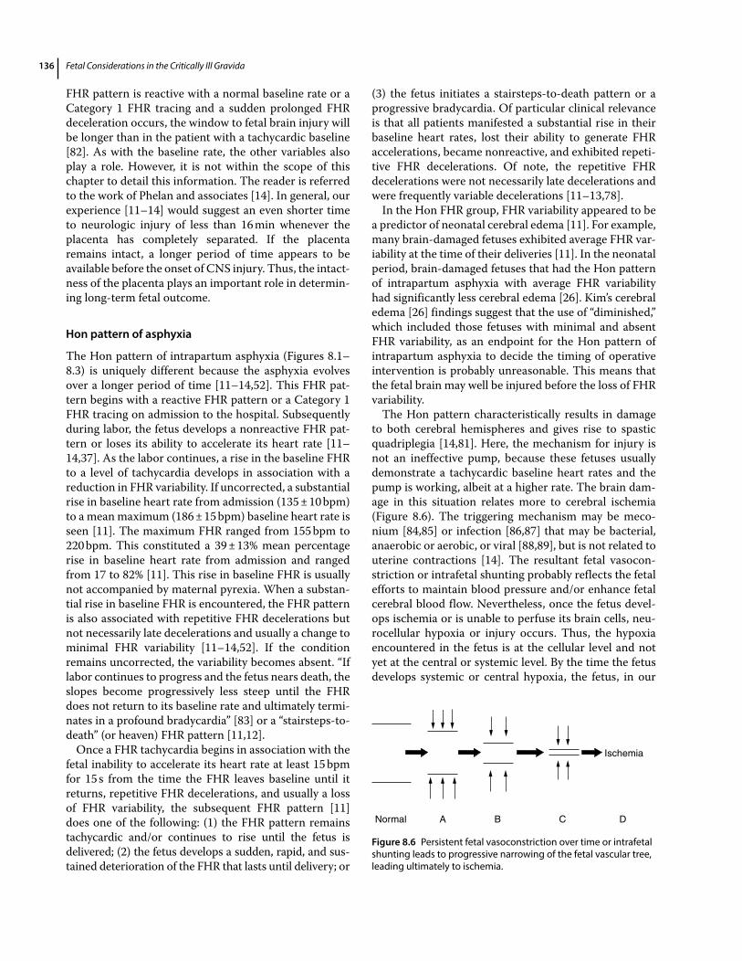
The Hon pattern characteristically results in damage to both cerebral hemispheres and gives rise to spastic quadriplegia [14,81]. Here, the mechanism for injury is not an ineffective pump, because these fetuses usually demonstrate a tachycardic baseline heart rates and the pump is working, albeit at a higher rate. The brain dam-age in this situation relates more to cerebral ischemia (Figure 8.6). The triggering mechanism may be meco-nium [84,85] or infection [86,87] that may be bacterial, anaerobic or aerobic, or viral [88,89], but is not related to uterine contractions [14]. The resultant fetal vasocon-striction or intrafetal shunting probably reflects the fetal efforts to maintain blood pressure and/or enhance fetal cerebral blood flow. Nevertheless, once the fetus devel-ops ischemia or is unable to perfuse its brain cells, neu-rocellular hypoxia or injury occurs. Thus, the hypoxia encountered in the fetus is at the cellular level and not yet at the central or systemic level. By the time the fetus develops systemic or central hypoxia, the fetus, in our
Normal A B C D
Ischemia
Figure 8.6 Persistent fetal vasoconstriction over time or intrafetal shunting leads to progressive narrowing of the fetal vascular tree, leading ultimately to ischemia.

Fetal monitoring made simple during labor 137
opinion, has already been brain injured and is probably near death [12,14]. Thus, cerebral perfusion deficits due to intrafetal and intracerebral shunting rather than fetal systemic hypoxia are most likely responsible for the fetal brain injury [90].
This means, for example, that a fetus that develops the Hon pattern of intrapartum asphyxia would appear to move to ischemia or from point C to point D (Figure 8.6). During this transition, a progressive and substantial rise in FHR is observed in an effort to pre-serve cerebral perfusion and neurocellular oxygenation. During this period, fetal systemic oxygenation and oxy-gen saturation are maintained. In our opinion [11], only after progressive and prolonged ischemia and brain injury do central fetal oxygen saturations begin to fall. These observations would be in keeping with American College of Obstetricians and Gynecologists (ACOG) recommendations on the intrapartum management of a Category 2 FHR tracing with the presence of FHR tachycardia, minimal variability, and no accelerations. Under these circumstances, one cannot reliably exclude fetal academia [60].
Additionally, it is important to emphasize that the pat-tern of fetal brain injury may change depending on the circumstances that gave rise to the delivery of the fetus. For example, and as previously discussed, this FHR pat-tern characteristically results in cerebral palsy of the spastic quadriplegic type due to cerebral hemispheric injury. If, however, the FHR pattern moves from a Hon pattern followed by a sudden, rapid, and sustained dete-rioration of the FHR that lasts until delivery, the pattern of brain damage becomes more global and involves not only the cerebral hemispheres but also the deep gray matter. As such, the fetuses with this latter FHR pattern have a more severe injury and shorter life expectancies.
The persistent nonreactive FHR pattern
The persistent nonreactive FHR pattern from admission to the hospital or a non‐stress test accounted for 45% of the FHR patterns observed in a population of 300 brain‐damaged babies [11] and 33% of an updated population of 423 singleton term brain‐damaged children [13,14]. This population is typically, but not always, character-ized by the presence of reduced fetal activity before admission to the hospital, male fetuses, old meconium, meconium sequelae such as meconium aspiration syn-drome and persistent pulmonary hypertension, and oli-gohydramnios [90]. Along with these observations, these fetuses usually but not always have elevated nucle-ated red blood cell counts [91,92], prolonged NRBC clearance times [91], low initial platelet counts [93], sig-nificant multi‐organ system dysfunction [73,74,91], delayed onset of seizures from birth [94,95], and cortical
or hemispheric brain injuries [13,14]. The typical FHR pattern is nonreactive with a fixed baseline rate that normally does not change from admission until delivery [13,14] in association with diminished or average variability.
When looking at the admission FHR pattern, the per-sistent nonreactive FHR pattern group can be divided into three phases. These three phases, in our opinion, represent a post‐CNS insult compensatory response in the fetus. Moreover, this FHR pattern, in our opinion, does not represent ongoing asphyxia or worsening of the CNS injury [11–14]. For a fetus to have ongoing fetal asphyxia, a FHR pattern similar to the Hon pattern of intrapartum asphyxia would have to be seen. There, a progressive and substantial rise in baseline heart rate in association with repetitive FHR decelerations is observed in response to ongoing fetal asphyxia (Figures 8.1–8.3). In contrast, the FHR baseline in the nonreactive group usually but not always remains fixed. Infrequently, a FHR tachycardia is seen; however, the rise in baseline rate is usually insubstantial. Thus, the phase of recovery appears to equate with the length of time from the fetal CNS insult. Thus, phase I would appear to be closer to the time of the insult, and phase III would appear to be more distant in time from the injury‐producing event [12].
The persistent nonreactive FHR pattern is not, in our opinion, a sign of ongoing fetal asphyxia but rather rep-resents a static encephalopathy [11–14]. This means that earlier intervention in the form of a cesarean on admis-sion to the hospital would not, in our opinion, substan-tially alter fetal outcome.
Fetal monitoring made simple during labor
In light of the lessons learned from the children damaged in utero before and during labor, current fetal monitor-ing interpretation will need to change to reflect and include the significance of the initial fetal monitoring period. When a patient presents to labor and delivery, the initial fetal assessment should include an initial fetal monitoring period to assess reactivity (the presence of FHR accelerations) and to ascertain from the patient the quality and quantity of fetal movement. In the patient with a reactive FHR pattern and normal fetal movement, the key to clinical management before and during labor is to follow the baseline fetal heart rate.
This means that the physician and nurse will need to watch for persistent elevations of the baseline rate to a level of tachycardia or higher or look for the potential for the baseline rate to fall suddenly. To assist with the iden-tification of the Hon pattern, medical and nursing per-sonnel should try to compare the current tracing with

Fetal Considerations in the Critically Ill Gravida138
the one obtained on admission. If the characteristics of the Hon pattern of intrapartum asphyxia develop, subse-quent clinical management will depend on whether the gravida is febrile and as outlined earlier in this chapter. In the nonreactive group, clinical management is to first evaluate the maternal and fetal status with respect to the etiology of the FHR pattern. These causes include, but are not limited to, the following: maternal substance abuse, fetal‐maternal hemorrhage, fetal anomaly, and the potential for a fetal chromosomal abnormality. During this period of maternal and fetal evaluation, continuous fetal monitoring is used, if technically feasible, to assess fetal status. In addition, fetal stimulation tests, a contrac-tion stress test, or a biophysical profile may be used to further determine fetal status. Once fetal status is clari-fied in the nonreactive group, the subsequent manage-ment with respect to the route of delivery in the term or near‐term pregnancy will depend on the discussion with the family and the clinical findings.
Maternal and surgical conditions
Anaphylaxis
Anaphylaxis is an acute allergic reaction to food inges-tion or drugs. The reported incidence of anaphylaxis during pregnancy has been reported to be 3–5 cases per 10,000 births [58]. It is generally associated with rapid onset of pruritus and urticaria and may result in respira-tory distress, edema, vascular collapse, and shock. Anaphylaxis during pregnancy has been linked with the use of misoprostol [96]; after laminaria insertion; the administration of antibiotics, such as ampicillin, cefazo-lin, penicillin, iron, ranitidine, and snake antivenom; insect stings, primarily bees and wasps; local anesthetics; and general anesthesia [58]. When anaphylaxis occurs during pregnancy, the typical fetal heart rate response is repetitive late deceleration [57] or a bradycardia [58].
When an anaphylactic reaction occurs during preg-nancy, the accompanying maternal physiologic changes may result in the manifestation of fetal distress, as noted earlier in this chapter. For example, in a case described by Klein and associates [57], a woman at 29 weeks’ gesta-tion presented with an acute allergic reaction after eating shellfish. On admission, she had evidence of regular uterine contractions and repetitive, severe late decelera-tions. The “fetal distress” was believed to be the result of maternal hypotension and relative hypovolemia, which accompanied the allergic reaction. Prompt treatment of the patient with intravenous fluids and ephedrine cor-rected the FHR abnormality. Subsequently, the patient delivered a healthy male infant at term with normal Apgar scores.
As suggested by these investigators, acute maternal allergic reactions can pose a threat to the fetus, and treat-ment directed at the underlying cause often remedies the accompanying fetal distress. As such, treatment is directed toward maternal cardiorespiratory support with the goal of maternal stabilization. Maternal stability should be re‐evaluated in the presence of a persistent fetal heart rate tachycardia or bradycardia, or other abnormal FHR patterns. The persistence of these FHR patterns suggests the need for additional maternal hemodynamic support or oxygenation. This means that intervention is delayed until the mother is sufficiently stabilized to be able to withstand a cesarean. Although Schoen [96] provides a clinical algorithm for the acute management of anaphylaxis during pregnancy, Schoen suggests that “if there is not rapid improvement in the maternal clinical condition, the [clinician should] move to immediate delivery” [96]. According to Gei [58], “poor neonatal outcomes have been experienced after an emer-gency cesarean prior to maternal stabilization.” The issue comes down to Schoen’s clinical meaning of “not rapid improvement.” As discussed later in this chapter, the cor-nerstone of the clinical management of these compli-cated clinical conditions is that maternal health trumps fetal health.
Once maternal stabilization has been achieved, the clinical focus should be shifted toward the status of the fetus. Generally, to afford the fetus a wider margin of safety, efforts should be directed at maintaining maternal systolic BP above 90 mmHg. In addition, oxygen should be administered to correct maternal hypoxia; in the absence of maternal hypovolemia, a maternal PaO2 in excess of 60–70 mmHg will assure adequate fetal oxy-genation [57,58].
In summary, the obstetrician should be prepared for anaphylaxis in the office as well as the hospital setting [97] and incorporate this type of emergency into their obstetrical drills [98]
Eclampsia
Maternal seizures are a well‐known but infrequent sequel of preeclampsia [19]. Although the maternal hemodynamic findings in patients with eclampsia are similar to those with severe preeclampsia [99], mater-nal convulsions require prompt attention to potentially prevent harm to both mother and fetus [19]. During a seizure, the fetal response usually is manifested as an abrupt, prolonged FHR deceleration [21,100]. During the seizure, which generally lasts less than 1–2 min [21], transient maternal hypoxia and uterine artery vasos-pasm occur and combine to produce a decline in uter-ine blood flow. In addition, uterine activity increases secondary to the release of norepinephrine, resulting in
Maternal and surgical conditions 139
additional reduction in uteroplacental perfusion. Ultimately, the reduction of uteroplacental perfusion causes the FHR deceleration. Such a deceleration may last up to 10 min after the termination of the convul-sions and the correction of maternal hypoxemia [19,21]. Following the seizure and recovery from the FHR decel-eration, a loss of FHRV and a compensatory rise in baseline FHR are characteristically seen. Transient late decelerations are not uncommon but usually resolve once maternal metabolic recovery is complete. During this recovery period, it is reasonably believed to be ben-eficial for the fetus to permit recovery in utero from convulsion induced hypoxia and hypercarbia [19]. During this time, the patient should not be rushed to an emergency cesarean based on the FHR changes associ-ated with an eclamptic seizure [19]. This is especially true if the patient is unstable.
The cornerstone of patient management during an eclamptic seizure is to maintain adequate maternal oxy-genation and to administer appropriate anticonvulsants. After a convulsion occurs, an adequate airway should be maintained and oxygen administered. To optimize uter-oplacental perfusion, the mother is repositioned onto her side. Anticonvulsant therapy with intravenous mag-nesium sulfate [19,101–103] to prevent seizure recur-rence is recommended. In spite of adequate magnesium sulfate therapy, adjunctive anticonvulsant therapy occa-sionally may be necessary in about 10% of patients [19,21,101].
In the event of persistent FHR decelerations, intrauter-ine resuscitation with a betamimetic [104] or additional magnesium sulfate [105] may be helpful in relieving eclampsia‐induced uterine hypertonus. Continuous electronic fetal monitoring should be used to follow the fetal condition. After the mother has been stabilized, and if the fetus continues to show signs of a FHR bradycardia and/or repetitive late decelerations after a reasonable period of recovery, delivery should be considered.
Disseminated intravascular coagulopathy
Disseminated intravascular coagulopathy (DIC) occurs in a variety of obstetric conditions, such as abruptio pla-centae, amniotic fluid embolus syndrome, severe preec-lampsia and eclampsia, and the dead fetus syndrome. The pathophysiology of this condition is discussed in greater detail in Chapter 31.
Infrequently, DIC may be advanced to a point of overt bleeding [106]. Under these circumstances, laboratory abnormalities accompany the clinical evidence of con-sumptive coagulopathy. In the rare circumstance of overt “fetal distress” and a clinically apparent maternal coagu-lopathy, obstetric management requires prompt replace-ment of deficient coagulation components before
attempting to deliver the distressed fetus. This frequently requires balancing the interests of the pregnant woman with those of her unborn child.
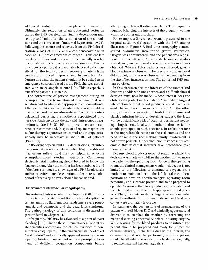
For example, a 34‐year‐old woman presented to the hospital at 33 weeks’ gestation with the FHR tracing illustrated in Figure 8.7. Real‐time sonography demon-strated asymmetric intrauterine growth restriction. Oxygen was administered, and the patient was reposi-tioned on her left side. Appropriate laboratory studies were drawn, and informed consent for a cesarean was obtained. When a Foley catheter was inserted, grossly bloody urine was observed. The previously drawn blood did not clot, and she was observed to be bleeding from the site of her intravenous line. The abnormal FHR pat-tern persisted.
In this circumstance, the interests of the mother and fetus are at odds with one another, and a difficult clinical decision must now be made. Whose interest does the obstetrician protect in this instance? Immediate surgical intervention without blood products would have less-ened the mother’s chances of survival. On the other hand, if the clinician waits for fresh frozen plasma and platelet infusion before undertaking surgery, the fetus will be at significant risk of death or permanent neuro-logic impairment. Ideally, the mother and/or her family should participate in such decisions. In reality, because of the unpredictable nature of these dilemmas and the need for rapid decision making, family involvement is not always possible. Under such circumstances, it is axi-omatic that maternal interests take precedence over those of the fetus.
Because blood products were not readily available, the decision was made to stabilize the mother and to move the patient to the operating room. Once in the operating room, the clinical management would include, but is not limited to, the following: to continue to oxygenate the mother; to maintain her in the left lateral recumbent position; to have an anesthesiologist, operating room personnel, and surgeons present; and to be prepared to operate. As soon as the blood products are available, and the fetus is alive, transfuse with appropriate blood prod-ucts. Then, the clinician should begin the cesarean under general anesthesia. In this case, maternal and fetal out-comes were ultimately favorable.
In summary, the cornerstone of management of the patient with full‐blown DIC and clinically apparent fetal distress is to stabilize the mother by correcting the maternal clotting abnormality before initiating surgery. While waiting for the blood products to be infused, the patient should be prepared and ready for immediate cesarean delivery. If the fetus dies in the interim, the cesarean should not be performed, and the patient should be afforded the opportunity to deliver vaginally, to reduce maternal hemorrhagic risks.

Fetal Considerations in the Critically Ill Gravida140
The burn victim
Although burn victims are uncommonly encountered in high‐risk obstetric units, the pregnant burn patient is suf-ficiently complex to require a team approach to enhance maternal and perinatal survival [107,108]. In most cases, this will require maternal–fetal transfer to a facility skilled to handle burn patients. Transfer will depend primarily on the severity of the burn and the stability of the preg-nant woman and her fetus. For greater detail and discus-sion on the clinical management of various types of thermal injuries, the reader is referred to Chapter 53.
As an overview, the first step in the management of the pregnant burn patient is to determine the depth and size of the burn. The depth of a burn may be partial or full thickness. A full‐thickness burn, formerly called a
third‐degree burn, is the most severe and involves total destruction of the skin. As a result, regeneration of the epithelial surface is not technically feasible.
The second element of burn management is to deter-mine the percentage of body surface area involved (Table 8.5). The percentage of maternal total body sur-face area covered by the burn is linked to maternal and perinatal outcome. The more severe the maternal burn, the higher the maternal and perinatal mortality is [107,108]. The risk of mortality becomes significant whenever 40% or more of the maternal total body surface area is burned [107].
The subsequent clinical management of the pregnant burn patient will depend on the patient’s burn phase (e.g., acute, convalescent, or remote) or burn period [109] (e.g., resuscitation, postresuscitation, inflammation/infection,
Figure 8.7 The FHR pattern from a 33‐week fetus with asymmetric intrauterine growth impairment whose mother presented with clinical disseminated intravascular coagulation.

Maternal and surgical conditions 141
or rehabilitation). Each phase has unique problems. For example, the acute phase is characterized by premature labor, electrolyte and fluid disturbances, maternal cardio-pulmonary instability, and the potential for fetal compro-mise. In contrast, the convalescent and remote periods are unique for their problems of sepsis and abdominal scarring, respectively. Because the potential for fetal compromise is greatest during the window of time imme-diately following the burn, the focus in this chapter is on acute‐phase burn patients.
In the acute phase of a severe burn, the primary mater-nal focus centers on stabilization [108]. Here, electrolyte disturbances due to transudation of fluid and altered renal function mandate close attention to the maternal intravascular volume and prompt and aggressive fluid resuscitation. To estimate fluid resuscitation require-ments, multiple formulas are available and include but are not limited to the following formulas: Evans, Third Military Medical University, and Ruijins. The latter two formulas are used primarily in China and other Asian countries. At the same time, these patients are also potentially compromised from airway injury and/or smoke inhalation, and ventilator support may be neces-sary to maintain cardiopulmonary stability. Additionally, a high index of suspicion for venous thrombosis and sep-sis with early and aggressive treatment should be consid-ered. Given the complexities of these patients, invasive hemodynamic monitoring may be necessary. Because most of these patients will be in an ICU, appropriate medical consultation and intensive nursing care for the mother and fetus are essential.
Assessing fetal well‐being in the burn patient may be difficult. The ability to determine fetal status with ultra-sound or fetal monitoring will depend on the size and location of the burn. If, for example, the burn involves the maternal abdominal wall, alternative methods of fetal assessment, such as fetal kick counts (alone or in response to acoustic stimulation) [28] or a modified BPP [18,43,44] using vaginal ultrasound, may be necessary. Whenever abdominal burns are present, a sterile transducer cover for the ultrasound device, fetal monitor, or doptone should be used to reduce the risk of infection. In the
absence of a maternal abdominal burn, continuous elec-tronic fetal monitoring can generally be used. Because of such monitoring difficulties and the direct relationship between the size of the maternal burn and perinatal out-come (see Figure 8.8), Matthews [110] and Shi [107] have recommended immediate cesarean delivery (assuming maternal stability) in any pregnant burn patient with a potentially viable fetus and a burn that involves 50% or more of the maternal body surface area. In contrast, Guo [108] recommends early delivery if the pregnancy is in the third trimester. As a reminder, burn patients with electrolyte disturbances may exhibit alterations in fetal status similar to those of a patient in sickle cell crisis [56] or diabetic ketoacidosis [54,55]. Once the maternal elec-trolyte disturbance is corrected, fetal status may return to normal and intervention often can be avoided.
Fetal considerations specific to cardiac bypass proce-dures and electrical shock are discussed in Chapters 17 and 53.
Maternal brain death or persistent vegetative state
With the advent of artificial life‐support systems, pro-longed viability of the brain‐dead pregnant woman [111–122] or one in a persistent vegetative state (PVS) [123–137] is no longer unusual in a perinatal unit. As a consequence, an increasing number of obstetric patients on artificial life support will be encountered in the medi-cal community. In addition to maternal somatic survival to prolong fetal life, the recent clinical focus has also been on the role of the brain‐dead gravida as a potential organ donor [138–140]. “Where one life ends and another begins” has been captured in the articles by Ecker [139] and Esmaeilzadeh [140]. As one can readily see, maternal brain death and persistent vegetative state
Table 8.5 Classification of burn patients based on the percentage of body surface area involved.
Classification Body surface area (%)
Minor <10Major
Moderate 10–19Severe 20–39Critical ≥40
Maternal deaths
100
80
60
Inci
denc
e
40
20
0<40 >8050
Body surface area involved (%)
Perinatal deaths
Figure 8.8 Estimated maternal and perinatal mortality rates following maternal burn injuries according to the amount of body surface area involved.

Fetal Considerations in the Critically Ill Gravida142
pose an array of medical, legal, and ethical dilemmas for the obstetric healthcare provider [118,136,141–145].
In each case of maternal brain death or PVS, multiple questions need to be addressed depending on the role, if any, of continued somatic survival. When first con-fronted by the clinical circumstances of confirmed maternal brain death or PVS, the focus shifts to that of the fetus. If the fetus is alive, the question arises as to whether extraordinary care for the brain‐dead patient should be initiated to preserve the life of her unborn child, and if so, at what gestational age? If artificial life support is elected to permit further maturation of the fetus, how should the pregnancy be managed, and when and under what circumstances should the fetus be deliv-ered? When should maternal life support be terminated? Is consent required to maintain the pregnancy? If so, from whom should consent be obtained? Such questions barely touch the surface of the complexities associated with these cases. But it is clearly not within the scope of this chapter to deal with the ethical, moral, and legal issues related to the obstetric care of the brain‐dead gravida or the gravida with PVS. Rather, the emphasis is on the clinical management of these patients when a decision has been made to maintain somatic support for the benefit of the unborn child.
The key distinction between brain death and PVS is that in PVS, the brainstem is usually but not always functioning normally. In the initial phases, it is arguably difficult to separate the two entities. With time, the dis-tinction becomes clearer. For example, a PVS patient could appear to be awake, be capable of swallowing, and have normal respiratory control, but have no purpose-ful interactions. PVS patients are “truly unconscious because, although they are wakeful, they lack aware-ness” [136]. Nevertheless, the clinical management of the brain‐dead or PVS gravida is similar initially.
To date, more than 13 cases of maternal brain death [111–122] and more than 17 cases of PVS [123–137] during pregnancy have been reported (Tables 8.6 and 8.7). In this update, recent cases have not been added to the tables or references unless the case was clinically necessary because the care of these complicated preg-nancies has changed little since the fifth edition. In gen-eral, PVS patients require less somatic support than do brain‐dead pregnant women but can require a similar degree of medical management. The review by Bush and associates [136] illustrates the key differences between these two groups. When compared with the brain‐dead group, the PVS population is more likely to demonstrate the following [136]:
Table 8.6 Perinatal outcome in 13 reported cases of maternal brain death during pregnancy.
Gestation age (weeks)
Reference YearBrain death Delivery Indication for delivery
Mode of delivery
Apgar score at 5 min
Birth Weight (grams)
Dillon 1 [116] 1982 25 26 Fetal distress Cesarean 8 850Dillon 2 1982 18 19 Life support
TerminatedSVD NA NA
Heikkinen [117] 1985 21 31 Maternal hypotension Cesarean 7 1600Field [118] 1988 22 31 Growth impaired
Maternal sepsisCesarean 8 1440
Bernstein [119] 1989 15 32 Fetal distress Cesarean 9 1555Wuermeling [120] 1994 14 NA NA SVD NA NAIriye [121] 1995 30 30 Maternal hypotension
FHR decelerationsCesarean 8 1610
Vives [122] 1996 27 27 Fetal distressMaternal Hypotension
Cesarean 10 1150
Catanzarite [123] 1997 25 29 Chorioamnionitis Cesarean 7 1315Lewis [124] 1997 25 31 Fetal Lung Maturity Cesarean NA NASpike [125] 1999 16 31 Maternal Hypotension Cesarean 8 1440Souza [126] 2006 25 28 Oligohydramnios
Growth ImpairedCesarean 10 815
Hussein [12] 2006 26 28 Oligohydramnios Cesarean NA 1285
FHR, Fetal heart rate; NA, not available; SVD, spontaneous vaginal delivery.

Maternal and surgical conditions 143
1) Longer time interval between maternal brain injury and delivery
2) Heavier birth weights at delivery3) Delivery at a more advanced gestational age.
It is important to note that these differences may be more a reflection of the severity of the maternal condition in the brain‐dead gravida [136]. Moreover, prolonged “maternal survival” is related to the ability to maintain euthermia, to have spontaneous respirations, and to have a functioning cardiovascular system [136].
Therefore, it is easy to see that for optimal care of such patients and fetuses, a cooperative effort among various healthcare providers is essential. The goal is to maintain maternal somatic survival until the fetus is viable and reasonably mature. To achieve this goal, a number of maternal and fetal considerations must be addressed to enhance fetal outcome (Table 8.8) [113].
As demonstrated in Table 8.8, Field and associates [113] have tried to capture the complexities associated with the medical management of these patients. Maternal medical management involves the regulation of most, if not all, maternal bodily functions. For example, the loss of the
pneumotaxic center in the pons, which is responsi-ble for cyclic respirations, and the medullary center, which is responsible for spontaneous respirations, make mechanical ventilation mandatory. Ventilation, under
Table 8.7 Perinatal outcome in 17 reported cases of persistent vegetative state during pregnancy.
Gestation age (weeks)
Reference Year PVS Delivery Indication for deliveryMode of delivery
Apgar score at 5 min
Birth Weight (grams)
Lucas [123] 1976 6 mo 8 mo None SVD‐breech NA 1760Sampson [124] 1979 6 34 Premature labor Forceps 5 1640BenAderet [125] 1984 17 35 Premature rupture of membranes Cesarean 9 2450Hill [126] 1985 14 34 Fetal lung maturity Cesarean 9 1600Diamond [127] 1986 22 34 Contraction stress test Cesarean 5 2835Landye [128] 1987 5 mo 37 Vacuum 9 2530Koh [129] 1993 13 37 Failed VBAC Cesarean 9 3680Webb [130] 1996 14 31 Abruption Cesarean 7 2240Wong [131] 1997 22 33 Chorioamnionitis Cesarean 9 2150Finerty‐1 [132] 1999 12 NA NA Cesarean NA NAFinerty‐2 [132] 1999 17 33 NA SVD NA NAAyorinde [133] 2000 12 35 Premature labor SVD 10 2200Feldman [134] 2000 15 31 Seizures/hypertension Cesarean 9 1506Sim [135] 2001 4 33 Premature rupture of membranes Cesarean 6 1680Bush [136] 2003 15 24 FHR bradycardia Cesarean 1 740Chiossi‐1 [137] 2006 10 34 Hypotension
Fetal lung maturityCesarean 9 2680
Chiossi‐2 [137] 2006 19 31 Abnormal FHR patternBiophysical profile 6/10
Cesarean 7 1701
FHR, Fetal heart rate; mo, months; NA, not available; PVS, persistent vegetative state; SVD, spontaneous vaginal delivery.
Table 8.8 Medical and obstetric considerations in providing artificial life support to the brain‐dead gravida.
Maternal considerationsMechanical ventilationCardiovascular supportTemperature labilityHyperalimentationPanhypopituitarismInfection surveillanceProphylactic anticoagulationFetal considerationsFetal surveillanceUltrasonographySteroidsTiming of delivery

Fetal Considerations in the Critically Ill Gravida144
these circumstances, is similar to that for the non‐preg-nant patient. In contrast to the non‐pregnant patient, the desirable gas concentrations are stricter due to the pres-ence of the fetus. As such, the maternal PaCO2 should be kept between 30 and 35 mmHg [141] and the maternal PaO2 greater than 60–70 mmHg to avoid deleterious effects on uteroplacental perfusion.
Maternal hypotension occurs frequently in these patients and may be due to a combination of factors, including hypothermia, hypoxia, and panhypopituita-rism. Maintenance of maternal BP can often be achieved with the infusion of low‐dose dopamine, which elevates BP without affecting renal or splanchnic blood flow. Along with vasopressors to support the maternal blood pressure and organ perfusion, the patient should be kept, when possible, in the lateral recumbent position to main-tain uteroplacental blood flow. At the same time, care should be exercised to avoid decubitus ulcers.
With maternal brain death, the thermoregulatory center located in the ventromedian nucleus of the hypothalamus does not function, and maternal body temperature cannot be maintained normally. As a result, maternal hypother-mia is the rule. Maintenance of maternal euthermia is important and usually can be accomplished through the use of warming blankets and the administration of warm, inspired, humidified air.
Maternal pyrexia suggests an infectious process and the need for a thorough septic workup. Thus, infection surveillance for, and the treatment of, infectious compli-cations is helpful to prolong maternal somatic survival [141]. If the maternal temperature remains elevated for a protracted period, cooling blankets may be necessary to avoid potentially deleterious effects on the fetus [145].
Nutritional support, usually in the form of enteral or parenteral hyperalimentation, is required for maternal maintenance and fetal growth and development (see Chapter 15). Because of poor maternal gastric motility, parenteral rather than enteral hyperalimentation is often preferred [113] to maintain a positive nitrogen balance. The use of hyperalimentation during pregnancy does not appear to have deleterious effects on the fetus [146]. As a rule, the amount of hyperalimentation should be in keeping with the caloric requirements for that gesta-tional age of the pregnancy and be sufficient to avoid maternal hyperglycemia.
In such patients, panhypopituitarism frequently occurs. As a result, a variety of hypoendocrinopathies, such as diabetes insipidus, secondary adrenal insuffi-ciency, and hypothyroidism, may develop, each mandat-ing therapy to maintain the pregnancy. Treatment of these conditions requires the use of vasopressin, corti-costeroids, and thyroid replacement, respectively.
Because of the hypercoagulable state of pregnancy and the immobility of the brain‐dead gravida, these patients
also are at an increased risk for thromboembolism. Therefore, to minimize the potential for deep venous thrombosis or pulmonary embolus, heparin prophylaxis (5000–7500 units twice or three times a day) and/or intermittent pneumatic calf compression are recom-mended [147].
By artificially supporting the maternal physiologic system, the intrauterine environment can be theoreti-cally maintained to allow for adequate fetal growth and development (Table 8.8). Obstetric management should focus on monitoring fetal growth with frequent ultra-sound evaluations, antepartum FHR assessment, and the administration of corticosteroids between 24 and 34 weeks of gestation to enhance fetal lung maturation [113,148]. For stimulation of fetal lung maturity, betamethasone or dexamethasone is recommended. Repeated steroid injections in subsequent weeks are not recommended due to the concern over the effect of repeated steroid injections on fetal brain growth [148]. However, “a single repeat course of antenatal corticos-teroids should be considered in women who are less than 34 0/7 weeks of gestation who have an imminent risk of preterm delivery within the next 7 days, and whose prior course of antenatal corticosteroids was administered more than 14 days previously” [148].
Another obstetric concern is the development of pre-mature labor. Here, tocolytic therapy has been used suc-cessfully [118,149]. Catanzarite et al. [123] described the use of a magnesium sulfate infusion and indomethacin to control uterine contractions, allowing prolongation of the pregnancy for 25 days. Other agents available for tocolysis include betamimetics, calcium‐channel block-ers, and oxytocin antagonists. The hemodynamic effects of betamimetics and calcium‐channel‐blocking agents may make these drugs less than ideal choices in these settings, in which maternal hemodynamic instability is common [118].
The timing of delivery is based on the deterioration of maternal or fetal status or the presence of fetal lung maturity. Classical cesarean is the procedure of choice [113,141] and is the least traumatic procedure for the fetus. To assure immediate cesarean capability, a cesar-ean pack and neonatal resuscitation equipment should be immediately available in the ICU.
Perimortem cesarean delivery
For centuries, perimortem cesarean delivery has been described as an attempt to preserve the life of the unborn child when the pregnant woman dies [151]. The first description of a perimortem cesarean was by Pliny the Elder in 237 AD. This delivery related to that of Scipio Africanus. Over a thousand years later, in 1280, the

Perimortem cesarean delivery 145
Catholic Church at the Council of Cologne decreed that a perimortem cesarean delivery must be performed to permit the unborn child to be baptized and to undergo a proper burial. Failure to perform the delivery con-stituted a punishable offense. Moreover, perimortem cesarean was mandated specifically in those women whose pregnancies were advanced beyond 6 months. To date, there have been 307 cases of perimortem cesarean delivery reported in the English literature [151,152]. Of these cesareans, there have been 222 surviving infants [151,152].
Since Weber’s monumental review of the subject in 1971, the causes of maternal death leading to a perimor-tem cesarean delivery have not changed substantially [151,152] but are more reflective of contemporary obstetric care [130,131]. These include traumatic events, pulmonary embolism from amniotic fluid, clot or air, acute respiratory or cardiac failure, and sepsis. In the case of a sudden, unanticipated maternal arrest, the tim-ing of cesarean delivery becomes the quintessential ele-ment [151,152].
If a pregnant woman does sustain a cardiopulmonary arrest, cardiopulmonary resuscitation (CPR) should be initiated immediately (Chapter 11). Optimal perfor-mance of CPR in the non‐pregnant patient results in a cardiac output less than one‐third of normal [152]. In the pregnant woman at term, CPR, under optimal cir-cumstances, produces a cardiac output around 10% of normal. To optimize maternal cardiac output, the patient should be placed in the supine position. Dextrorotation of the uterus and compression of the major vessels of the uterus may impede venous return and may further compromise this effort. Lateral uterine displacement may help to remedy this problem, but CPR in this position is extremely awkward. Ultimately, a cesarean may be necessary to alleviate this impedance to CPR.
If maternal and fetal outcomes are to be optimized, the timing of the cesarean delivery is critical. According to Katz and associates [151] in 1986 and reaffirmed in 2005 [152], the theory behind a perimortem cesarean is that if CPR fails to produce a pulse within 4 min, a cesarean delivery should be begun and the baby delivered within 5 min of maternal cardiac arrest. Once the baby is deliv-ered, maternal CPR should continue because many women will have “sudden and profound improvement” [152] after evacuation of the uterus. Hence, the “4‐ minute rule” came into effect and had been adopted by the American Heart Association when maternal CPR has been ineffective [153–155]. Thus, the standard ABCs of cardiopulmonary resuscitation (airway, breathing, and circulation) have been expanded to include D (delivery).
As demonstrated in Table 8.9, fetal survival is linked consistently with the interval between maternal arrest
and delivery. It is clear from the available data [154,156] that the longer the time interval from maternal death to the delivery of the fetus, the greater is the likelihood of permanent neurologic impairment of the fetus. Ideally, the fetus should be delivered within 5 min of maternal arrest. Within that 5 min window rests the greatest likeli-hood of delivering a child who will be neurologically nor-mal (Table 8.9). However, the potential exists for a favorable fetal outcome beyond 15 min of maternal car-diac arrest, and therefore, delivery should not be with-held even if beyond 5 min, if the fetus is still alive [151,152]. Recently, Benson and associates [157] have challenged the “Katz rule” [151,152]. In their review of 53 perimortem cesareans, Benson found that the fetal/neo-natal injury‐free interval was longer than 5 min. These investigators found the injury‐free window to be around 9–10 min from arrest to delivery interval. Analysis of misses and near misses will have to be evaluated in the future to determine the optimal interval.
While the timing of cesarean delivery is a major deter-minant of subsequent fetal outcome, the gestational age of the fetus also is an important consideration. The prob-ability of survival is related directly to the neonatal birth weight and gestational age [154–156,158]. At what gesta-tional age should a perimortem cesarean delivery be con-sidered? Is there a lower limit? It becomes obvious immediately that there are no clear answers to these questions. As a general rule, intervention appears pru-dent whenever the fetus is potentially viable or is “capa-ble of a meaningful existence outside the mother’s womb” [159]. According to ACOG [148], the gray zone rests between 23 and 24 weeks’ gestation. But, this threshold is continually pushed to earlier gestational ages in keep-ing with the advances in obstetrical and neonatal care. Ideally, criteria for intervention in such circumstances should be formulated with the aid of an institution’s cur-rent neonatal survival statistics and guidance from its bioethics committee. In light of the continual techno-logic advances in neonatology, care must be taken to periodically review these criteria because the gestational age and weight criteria may be lowered in the future [154–156,158,159].
Table 8.9 Perimortem cesarean delivery with the outcome of surviving infants from the time of maternal death until delivery.
Time interval (min)
Surviving infants (no.)
Intact neurologic status of survivors (%)
0–5 9 8 (89%)6–15 5 2 (60%)>15 7 4 (57%)
Source: Refs. [150–152].

Fetal Considerations in the Critically Ill Gravida146
When maternal death is an anticipated event, is informed consent necessary? For instance, patients hos-pitalized with terminal cancer, class IV cardiac disease, pulmonary hypertension, or previous myocardial infarc-tion are at an increased risk of death during pregnancy. Although these cases are infrequent, it seems reasonable
to prepare for such an eventuality. Decisions regarding intervention should be made in advance with the patient and family. When intervention has been agreed to, one consideration is to have a cesarean delivery pack and neonatal resuscitation equipment immediately available in the ICU.
References
1 Hon EH, Quilligan EJ. Electronic evaluation of the fetal heart rate. Clin Obstet Gynecol. 1968;11:145–155.
2 Paul RH, Gauthier RJ, Quilligan EJ. Clinical fetal monitoring: The usage and relationship to trends in cesarean delivery and perinatal mortality. Acta Obstet Gynecol Scand. 1980;59:289–295.
3 Shenker L, Post RC, Seiler JS. Routine electronic monitoring of fetal heart rate and uterine activity during labor. Obstet Gynecol. 1980;46:185–189.
4 Yeh SY, Diaz F, Paul RH. Ten year experience in fetal monitoring at Los Angeles County/University of Southern California Medical Center. Am J Obstet Gynecol. 1982;143:496–500.
5 Phelan JP. Labor admission test. Clin Perinatol. 1994;21(4):879–885.
6 Skupski DW, Rosenberg CR, Eglinton GS. Intrapartum fetal stimulation tests: A meta‐analysis. Obstet Gynecol. 2002;99:129–134.
7 Perkins, RP. Perspectives on perinatal brain damage. Obstet Gynecol. 1987;69:807–819.
8 Rosen M, Dickinson MG. The incidence of cerebral palsy. Am J Obstet Gynecol. 1992;167:417–423.
9 Smith J, Wells L, Dodd K. The continuing fall in incidence of hypoxic‐ischemic encephalopathy in term infants. Br J Obstet Gynecol. 2000;107:461–466.
10 Clark SL. Shock in the pregnant patient. Semin Perinatol. 1990;14:52–58.
11 Phelan JP, Ahn MO. Fetal heart rate observations in 300 term brain‐damaged infants. J Matern Fetal Invest. 1998;8:1–5.
12 Phelan JP, Kim JO. Fetal heart rate observations in the brain‐damaged infant. Semin Perinatol. 2000;24:221–229.
13 Phelan JP. Perinatal risk management: Obstetric methods to prevent birth asphyxia. Clin Perinatol. 2005;32:1–17.
14 Phelan JP, Korst LM, Martin GI. Causation – fetal brain injury and uterine rupture. Clin Perinatol. 2007;34(3):409–438.
15 Macones, GA, Hankins GD, Spong CY, et al. The 2008 National Institutes of Child Health and Human Development workshop report on electronic fetal monitoring: Update on definitions, interpretation, and research guidelines. Obstet Gynecol. 2008;112:661–666.
16 Schifrin BS. The CTG and the timing and mechanism of fetal neurologic injuries. Best Prac Res. 2004;18:43–56.
17 Lockshin MD, Bonfa E, Elkon D, Druzin ML. Neonatal lupus risk to newborns of mother with systemic lupus erythematosus. Arthritis Rheum. 1988;31:697–701.
18 Manning FA, Platt LD, Sipos L. Antepartum fetal evaluation: Development of a fetal biophysical profile. Am J Obstet Gynecol. 1980;136:787–795.
19 Sibai BM. Diagnosis, prevention, and management of eclampsia. Obstet Gynecol. 2005;105:402–410.
20 Clark SL, Hankins GD, Dudley DA, et al. Amniotic fluid embolism: Analysis of the National Registry. Am J Obstet Gynecol. 1995;172:1158–1167.
21 Paul RH, Koh KS, Bernstein SG. Change in fetal heart rate: Uterine contraction patterns associated with eclampsia. Am J Obstet Gynecol. 1978;130:165–169.
22 Koh KD, Friesen RM, Livingstone RA, et al. Fetal monitoring during maternal cardiac surgery with cardiopulmonary bypass. Can Med Assoc J. 1975;112:1102–1106.
23 Korsten HHM, van Zundert AAJ, Moou PNM, et al. Emergency aortic valve replacement in the 24th week of pregnancy. Acta Anaestesiol Belg. 1989;40:201–205.
24 Strange K, Halldin M. Hypothermia in pregnancy. Anesthesiology 1983;58:460–465.
25 Bates B. Hon fetal heart rate pattern flags brain damage. Ob Gyn News. 2005;40(10):1–2.
26 Kim JO, Martin G, Kirkendall C, Phelan JP. Intrapartum fetal heart rate variability and subsequent neonatal cerebral edema. Obstet Gynecol. 2000;95:75S.
27 National Institute of Child Health and Human Development Research Planning Workshop. Electronic fetal heart rate monitoring: Research guidelines for interpretation. Am J Obstet Gynecol. 1997;177:1385–1390.
28 Smith CV. Vibroacoustic stimulation for risk assessment. Clin Perinatol. 1994;21:797–808.
29 Petrie RH, Yeh SY, Maurata Y, et al. Effect of drugs on fetal heart rate variability. Am J Obstet Gynecol. 1978;130:294–299.
30 Babakania A, Niebyl R. The effect of magnesium sulfate on fetal heart rate variability. Obstet Gynecol. 1978;51(Suppl.):2S–4S.

HReferences 147
31 Katz M, Meizner I, Shani N, et al. Clinical significance of sinusoidal fetal heart rate pattern. Br J Obstet Gynaecol. 1984;149:97–100.
32 Modanlou HD, Freeman RK. Sinusoidal fetal heart rate pattern: Its definition and clinical significance. Am J Obstet Gynecol. 1982;142:1033–1038.
33 Theard FC, Penny LL, Otterson WN. Sinusoidal fetal heart rate: Ominous or benign? J Reprod Med. 1984;29:265–268.
34 Kirkendall C, Romo M, Phelan JP. Fetomaternal hemorrhage in fetal brain injury. Am J Obstet Gynecol. 2001;185(6):S153.
35 Heise RH, van Winter JT, Ogburn PL Jr. Identification of acute transplacental hemorrhage in a low‐risk patient as a result of daily counting of fetal movements. Mayo Clin Proc. 1993;68:892–894.
36 Kosasa TS, Ebesugawa I, Nakayama RT, Hale R. Massive fetomaternal hemorrhage preceded by decreased fetal movement and a nonreactive fetal heart rate pattern. Obstet Gynecol. 1993;82:711–714.
37 Corometrics Inc. Corometrics Teaching Program. Wallingford, CT: GE Medical Systems; 1974.
38 Shaw K, Clark SL. Reliability of intrapartum fetal heart rate monitoring in the postterm fetus with meconium passage. Obstet Gynecol. 1988;72:886–889.
39 Ahn MO, Korst LM, Phelan JP. Normal fetal heart rate patterns in the brain‐damaged infant: A failure of intrapartum fetal monitoring? J Matern Fetal Invest. 1998;8:58–60.
40 Garite TJ, Linzey EM, Freeman RK, et al. Fetal heart rate patterns and fetal distress in fetuses with congenital anomalies. Obstet Gynecol. 1979;53:716–720.
41 Slomka C, Phelan JP. Pregnancy outcome in the gravida with a nonreactive nonstress test and positive contraction stress test. Am J Obstet Gynecol. 1981;139:11–15.
42 Phelan JP, Smith CV. Antepartum fetal assessment: The contraction stress test. In: Hill A, Volpe JJ, editors. Fetal neurology. New York: Raven; 1989; p. 75–90.
43 Manning FA, Morrison I, Harman CR, Menticoglou SM. The abnormal fetal biophysical profile score V: Predictive accuracy according to score composition. Am J Obstet Gynecol. 1990;162:918–927.
44 Phelan JP. Antepartum fetal assessment: Newer techniques. Semin Perinatol. 1988;12:57–65.
45 Phelan JP, Smith CV, Broussard P, Small M. Amniotic fluid volume assessment using the four quadrant technique at 36–42 weeks’ gestation. J Reprod Med. 1987;32:540–543.
46 Phelan JP, Ahn MO, Smith CV, et al. Amniotic fluid index measurements during pregnancy. J Reprod Med. 1987;32:601–604.
47 Phelan JP, Lewis PE. Fetal heart rate decelerations during a nonstress test. Obstet Gynecol. 1981;57:228–232.
48 Gabbe SG, Ettinger RB, Freeman RK, et al. Umbilical cord compression with amniotomy: Laboratory observations. Am J Obstet Gynecol. 1976;126:353–355.
49 Phelan JP. The postdate pregnancy: An overview. Clin Obstet Gynecol. 1989;32(2):221–227.
50 Kubli FW, Hon EH, Hhazin AF, et al. Observations in heart rate and pH in the human fetus during labor. Am J Obstet Gynecol. 1969;104:1190–1206.
51 Krebs HB, Petres RE, Dunn LH, et al. Intrapartum fetal heart rate monitoring: VII. Atypical variable decelerations. Am J Obstet Gynecol. 1983;145:297–305.
52 Greenberg J, Economy K, Mark A, et al. In search of “true” birth asphyxia: Labor characteristics associated with the asphyxiated term infant. Am J Obstet Gynecol. 2001;185:S94.
53 Nelson KB, Dambrosia JM, Ting TY, Grether JK. Uncertain value of electronic fetal monitoring in predicting cerebral palsy. N Engl J Med. 1996;334: 613–618.
54 LoBue C, Goodlin RC. Treatment of fetal distress during diabetic ketoacidosis. J Reprod Med. 1978;20:101–104.
55 Rhodes RW, Ogburn PL. Treatment of severe diabetic ketoacidosis in the early third trimester in a patient with fetal distress. J Reprod Med. 1984;29:621–625.
56 Cruz AC, Spellacy WN, Jarrell M. Fetal heart rate tracing during sickle cell crisis: A cause for transient late decelerations. Obstet Gynecol. 1979;54:647–649.
57 Klein VR, Harris AP, Abraham RA, Niebyl JR. Fetal distress during a maternal systemic allergic reaction. Obstet Gynecol. 1984;64(Suppl.):15S–17S.
58 Gei AF, Pacheco LD, Vanhook JW, et al. The use of continuous infusion of epinephrine for anaphylactic shock. Obstet Gynecol. 2003;102:1332–1335.
59 Sheehan PQ, Rowland TW, Shah BL, et al. Maternal diabetic control and hypertrophic cardiomyopathy in infants of diabetic mothers. Clin Pediatr. 1986;25:226–230.
60 American College of Obstetricians and Gynecologists (ACOG). Management of intrapartum fetal heart rate tracings. ACOG Practice Bulletin No. 116. Washington, DC: ACOG; 2015.
61 Sherman DJ, Frenkel E, Kurzwell Y, et al. Characteristics of maternal heart rate patterns during labor and delivery. Obstet Gynecol. 2002;99:542–527.
62 Schifrin BS, Harwell R, Rubinstein T, et al. Maternal heart rate pattern: A confounding factor in intrapartum fetal surveillance. Prenat Neonate Med. 2001;6:75–82.
63 VanVeen TR, Belfort MA. Maternal heart rate patterns differ in the 1st and 2nd stages of labor. Am J Obstet Gynecol. 2009;201:S170.

Fetal Considerations in the Critically Ill Gravida148
64 Saling E. Technik der endoskopischen microbluentnahme am feten. Geburtshilfe Frauenheikd. 1964;24:464–467.
65 Saling E, Schneider D. Biochemical supervision of the fetus during labor. J Obstet Gynaecol Br Cwlth. 1967;74:799–803.
66 Clark SL, Paul RH. Intrapartum fetal surveillance: The role of fetal scalp sampling. Am J Obstet Gynecol. 1985;153:717–720.
67 Goodwin TM, Milner‐Masterson L, Paul RH. Elimination of fetal scalp blood sampling on a large clinical service. Obstet Gynecol. 1994;83:971–974.
68 Phelan JP, Kirkendall C, Korst L, Martin G. In cases of fetal brain injury, a slow heart rate at birth is an indicator of severe acidosis. Am J Obstet Gynecol. 2003;189(6):S184.
69 Kirkendall C, Phelan JP. Severe acidosis at birth and normal neurological outcome. Prenat Neonat Med. 2001;6:267–270.
70 Myers RE. Two patterns of perinatal brain damage and their conditions of occurrence. Am J Obstet Gynecol. 1972;112:246–277.
71 Fee SC, Malee K, Deddish R, et al. Severe acidosis and subsequent neurologic status. Am J Obstet Gynecol. 1990;162:802–806.
72 Dennis J, Johnson A, Mutch L, et al. Acid‐base status at birth and neurodevelopmental outcome at four and one‐half years. Am J Obstet Gynecol. 1989;161:213–220.
73 Korst LM, Phelan JP, Ahn MO, Martin GI. Can persistent brain injury resulting from intrapartum asphyxia be predicted by current criteria? Prenat Neonat Med. 1997;2:286–293.
74 Korst L, Phelan JP, Wang YM, et al. Acute fetal asphyxia and permanent brain injury: A retrospective analysis of current indicators. J Matern Fetal Med. 1999;8:101–106.
75 Garite TJ, Dildy GA, McNamara H, et al. A multicenter controlled trial of fetal pulse oximetry in the intrapartum management of non‐reassuring fetal heart rate patterns. Am J Obstet Gynecol. 2000;183:1049–1058.
76 American College of Obstetricians and Gynecologists (ACOG). Fetal and Neonatal Injury. Technical Bulletin No. 163. Washington, DC: ACOG; 1992.
77 MacLennan A. A template for defining a causal relation between acute intrapartum events and cerebral palsy: International consensus statement. BMJ. 1999;319:1054–1059.
78 American College of Obstetricians and Gynecologists (ACOG) and American Academy of Pediatrics (AAP). Neonatal Encephalopathy Committee Opinion. Washington, DC: ACOG and AAP; 2003.
79 Krebs HB, Petres RE, Dunn LH, et al. Intrapartum fetal heart rate monitoring: VI. Prognostic significance of accelerations. Am J Obstet Gynecol. 1982;142:297–305.
80 Ingemarsson I, Arulkumaran S, Paul RH, et al. Admission test: A screening test for fetal distress in labor. Obstet Gynecol. 1986;68:800–806.
81 Pasternak JF, Gorey MT. The syndrome of acute near‐total intrauterine asphyxia in the term infant. Pediatr Neurol. 1998;18:391–398.
82 Leung A, Leung EK, Paul RH. Uterine rupture after previous cesarean delivery: Maternal and fetal consequences. Am J Obstet Gynecol. 1993;169:945–950.
83 Hon EH, Lee ST. Electronic evaluation of the fetal heart rate: VIII. Patterns preceding fetal death, further observations. Am J Obstet Gynecol. 1963;87:814–826.
84 Altschuler G, Hyde S. Meconium induced vasoconstriction: A potential cause of cerebral and other fetal hypoperfusion and of poor pregnancy outcome. J Child Neurol. 1989;4:1337–1342.
85 Altschuler G, Arizawa M, Molnar‐Nadasy G. Meconium‐induced umbilical cord vascular necrosis and ulceration: A potential link between the placenta and poor pregnancy outcome. Obstet Gynecol. 1992;79:760–766.
86 Yoon BH, Kim CJ, Jun JK. Amniotic fluid interleukin 6: A sensitive test for antenatal diagnosis of acute inflammatory lesions of preterm placenta and prediction of perinatal morbidity. Am J Obstet Gynecol. 1995;172:960–970.
87 Perlman JM, Risser R, Broyles RS. Bilateral cystic leukomalacia in the premature infant: Associated risk factors. Pediatrics. 1996;97:822–827.
88 Gibson CS, Maclennan AH, Goldwater PN, et al. Neurotropic viruses and cerebral palsy: Population based case‐control study. BMJ. 2006;332:76–80.
89 Euscher E, Davis J, Holzman J, Nuovo GJ. Coxsackie virus infection of the placenta associated with neurodevelopmental delays in the newborn. Obstet Gynecol. 2001;98:1019–1026.
90 Visser GHA, deVries LS, Groeneudaul F. How bad is a low pH at birth? Prenat Neonat Med. 2001;6:265–266.
91 Phelan JP, Martin GI, Korst LM. Birth asphyxia and cerebral palsy. Clin Perinatol. 2005;32:61–76.
92 Blackwell SC, Refuerzo JS, Wolfe HM, et al. The relationship between nucleated red blood cell counts and early‐onset neonatal seizures. Am J Obstet Gynecol. 2000;182:1452–1457.
93 Korst LM, Phelan JP, Wang YM, Ahn MO. Neonatal platelet counts in fetal brain injury. Am J Perinatol. 1999;16:79–83.
94 Ahn MO, Korst LM, Phelan JP, Martin GI. Does the onset of neonatal seizures correlate with the timing of fetal neurologic injury. Clin Pediatr. 1998;37:673–676.
95 Kirkendall C, Ahn MO, Martin G, et al. The brain injured baby, neonatal seizures, and the intrapartum fetal heart rate pattern: Is there a relationship? Am J Obstet Gynecol. 2000;182:S184.

HReferences 149
96 Schoen, C, Campbell, S, Maratas, A. et al. Anaphylaxis to buccal misoprostol for labor induction. Obstet Gynecol. 2014;124:466–468.
97 Yankowitz, J. Brace for the case: Be prepared for anaphylaxis. Obstet Gynecol. 2004;103:1043–1044.
98 American College of Obstetricians and Gynecologists (ACOG). Preparing for clinical emergencies obstetrics and gynecology Committee Opinion No. 590. Obstet Gynecol. 2014;123:722–725.
99 Clark SL, Divon M, Phelan JP. Preeclampsia/eclampsia: Hemodynamic and neurologic correlations. Obstet Gynecol. 1985;66:337–340.
100 Boehm FH, Growdon JH. The effect of eclamptic convulsions of the fetal heart rate. Am J Obstet Gynecol. 1974;120:851–853.
101 Pritchard JA, Cunningham FG, Pritchard SA. The Parkland Memorial Hospital protocol for treatment of eclampsia: Evaluation of 245 cases. Am J Obstet Gynecol. 1984;148:951–963.
102 Lucas MJ, Leveno KJ, Cunningham FG. A comparison of magnesium sulfate with phenytoin for the prevention of eclampsia. N Engl J Med. 1995;333:201–205.
103 Naidu S, Payne AJ, Moodley J, et al. Randomised study assessing the effect of phenytoin and magnesium sulfate on maternal cerebral circulation in eclampsia using transcranial doppler ultrasound. Br J Obstet Gynaecol. 1996;103:111–116.
104 Barrett JM. Fetal resuscitation with terbutaline during eclampsia‐induced uterine hypertonus. Am J Obstet Gynecol. 1984;150:895.
105 Reece E, Chervenak F, Romero R, Hobbins J. Magnesium sulfate in the management of acute intrapartum fetal distress. Am J Obstet Gynecol. 1984;148:104–106.
106 Porter TF, Clark SL, Dildy GA, et al. Isolated disseminated intravascular coagulation and amniotic fluid embolism. Am J Obstet Gynecol. 1996;174:486.
107 Shi, Y., Zhang, X., Huang, BG., et al. Severe burn injury in late pregnancy: A case report and literature analysis. Burns Trauma. 2015;3:2.
108 Guo SS, Greenspoon JS, Kahn AM. Management of burn injuries during pregnancy. Burns. 2001;27:394–397.
109 Demling RH. Management of the burn patient. In: Grenvik A, Holbrook PR, Shoemaker WC, editors. Textbook of critical care. 3rd ed. Philadelphia: W.B. Saunders; 1995; p. 1499.
110 Matthews RN. Obstetric implications of burns in pregnancy. Br J Obstet Gynaecol. 1982;89:603–609.
111 Dillon WP, Lee RV, Tronolone MJ, et al. Life support and maternal brain death during pregnancy. JAMA. 1982;248:1089–1091.
112 Heikkinen JE, Rinne RI, Alahuhta SM, et al. Life support of 10 weeks with successful fetal outcome after fatal maternal brain damage. BMJ. Clin Res Ed. 1985;290:1237–1238.
113 Field DR, Gates EA, Creasy RK, et al. Maternal brain death during pregnancy: Medical and ethical issues. JAMA. 1988;260:816–822.
114 Bernstein IM, Watson M, Simmons GM, et al. Maternal brain death and prolonged fetal survival. Obstet Gynecol. 1989;74:434–437.
115 Wuermeling H. Brain‐death and pregnancy. Forens Sci Int. 1994;68:243–245.
116 Iriye BK, Asrat T, Adashek JA, Carr MH. Intraventricular hemorrhage and maternal brain death associated with antepartum cocaine abuse. Br J Obstet Gynaecol. 1995;102:68–69.
117 Vives A, Carmona F, Zabala E, et al. Maternal brain death during pregnancy. Int J Gynecol Obstet. 1996;52:67–69.
118 Catanzarite VA, Willms DC, Holdy KE, et al. Brain death during pregnancy: Tocolytic therapy and aggressive maternal support on behalf of the fetus. Am J Perinatol. 1997;14:431–434.
119 Lewis DD, Vidovich RR. Organ recovery following childbirth in a brain‐dead mother: A case report. J Transpl Coord. 1997;7:103–105.
120 Spike J. Brain death, pregnancy, and posthumous motherhood. J Clin Ethics. 1999;10:57–65.
121 Souza JP, Cecatta JG, Amaral E, et al. The prolongation of somatic support in a pregnant woman with brain death: A case report. J Reprod Health. 2006;3:3.
122 Hussein IY, Govenden V, Grant JM, Said MR. Prolongation of pregnancy in a woman who sustained brain death at 26 weeks of gestation. Br J Obstet Gynaecol. 2006;113:120–122.
123 Lucas B. Pregnant car crash victim. Nurs Times. 1976;72:451–453.
124 Sampson MB, Peterson LP. Post‐traumatic coma during pregnancy. Obstet Gynecol. 1979;53:25–35.
125 BenAderet N, Cohen I, Abramowicz JS. Traumatic coma during pregnancy with persistent vegetative state. Case report. Br J Obstet Gynecol. 1984;91:939–941.
126 Hill LM. Management of maternal vegetative state during pregnancy. Mayo Clin Proc. 1985;60:469–472.
127 Diamond MP, Boehm FH, Allen G. Long term management of pregnancy in a comatose patient. J Tenn Med Assoc. 1986;79:557–566.
128 Landye ST. Successful enteral nutrition support of a pregnant comatose patient: A case study. J Am Diet Assoc. 1988;88:718–720.
129 Koh ML, Lipkin EW. Nutrition support of a pregnant comatose patient via percutaneous endoscopic gastrostomy. J Parenter Enteral Nutr. 1993;17:384–387.

Fetal Considerations in the Critically Ill Gravida150
130 Webb GW, Huddleston JF. Management of the pregnant woman who sustains severe brain damage. Clin Perinatol. 1996;23:453–464.
131 Wong M, Apodaca CC. Nutrition management in a pregnant comatose patient. NCP Bull. 1997;12: 63–67.
132 Finerty JJ, Chisolm CA, Chapple H, et al. Cerebral arterio‐venous malformation in pregnancy: Presentation and neurologic, obstetric, and ethical significance. Am J Obstet Gynecol. 1999;181: 296–303.
133 Ayorinde BT, Scudamore I, Buggy DJ. Anaesthetic management of a pregnant patient in a persistent vegetative state. Br J Anaesth. 2000;85:478–491.
134 Feldman DM, Borgida AF, Rodis JF, Campbell WA. Irreversible maternal brain injury during pregnancy: A case report and review of the literature. Obstet Gynecol. Surv 2000;55:708–714.
135 Sim KH. Maternal persistent vegetative state with successful fetal outcome. J Korean Med. 2001;16:669–672.
136 Bush MC, Nagy S, Berkowitz RC, Gaddipati S. Pregnancy in a persistent vegetative state: Case report, comparison to brain death, and review of literature. Obstet Gynecol Surv. 2003;50:738–748.
137 Chiossi G, Novic K, Celebreeze JU, Thomas RL. Successful neonatal outcome in 2 cases of maternal persistent vegetative state treated in a labor and delivery suite. Am J Obstet Gynecol. 2006;195:316–322.
138 Mejia R, Badarriotti G, DeDiego B, et al. Brain death in a pregnant woman and fetus survival. Medicina [B. Aires]. 2008;68:447–452.
139 Ecker, J.L. Death in pregnancy: An American tragedy. N Engl J Med. 2014;370:10.
140 Esmaeilzadeh M, Dictus C, Kayvanpour E, et al. One life ends, another begins: Management of a brain‐dead pregnant mother. A systematic review. BMC Med. 2010;18:74.
141 Mallampalli A, Powner DJ, Gardner MO. Cardiopulmonary resuscitation and somatic support of the pregnant patient. Crit Care Clin. 2004;20:747–761.
142 Gdanski E, Schenker G. Management of pregnancy in women with circulatory and brain death. Prenat Neonat Med. 1998;3:327–333.
143 Black PM. Brain death. N Engl J Med. 1978;229(7):338–344, and 299(8):393–401.
144 Bernat JL, Culver CM, Gert B. On the definition and criterion of death. Ann Intern Med. 1981;94: 389–394.
145 Edwards MJ, Wanner RA. Extremes of temperature. In: Wilson JG, Graser FC, editors. Handbook of teratology, vol. 1. New York: Plenum; 1977; p. 421.
146 Smith CV, Rufleth P, Phelan JP, et al. Longterm enteral hyperalimentation in the pregnant woman with insulin dependent diabetes. Am J Obstet Gynecol. 1981;141:180–183.
147 Clark‐Pearson DL, Synan IS, Dodge R, et al. A randomized trial of low‐dose heparin and intermittent pneumatic calf compression for the prevention of deep venous thrombosis after gynecologic oncology surgery. Am J Obstet Gynecol. 1993;168:1146–1154.
148 American College of Obstetricians and Gynecologists (ACOG). Antenatal corticosteroids therapy for fetal maturation. Committee Opinion No. 677. October. Washington, DC: ACOG; 2016.
149 Powner DJ, Bernstein J. Extended somatic support for pregnant women after brain death. Crit Care Med. 2003;31:1241–1249.
150 Weber CE. Postmortem cesarean section: review of the literature and case reports. Am J Obstet Gynecol. 1971;110:158–165.
151 Katz VL, Dotters DJ, Droegemueller W. Perimortem cesarean delivery. Obstet Gynecol. 1986;68:571–576.
152 Katz VL, Balderston K, DeFreest M. Perimortem cesarean delivery: were our assumptions correct? Am J Obstet Gynecol. 2005;192:1916–1921.
153 Jeejeebhoy FM, Zelop C, Lipman S, et al. Cardiac arrest in pregnancy: A scientific statement from the American Heart Association. Circulation. 2015;132:1747–1773.
154 Hussain N, Galal M, Ehrenkranz RA, et al. Pre‐discharge outcomes of 22–27 weeks gestational age infants born at tertiary care centers in Connecticut: Implications for perinatal management. Conn Med. 1998;62:131–137.
155 Draper ES, Manktelow B, Field DJ, James D. Prediction of survival for preterm births by weight and gestational age: Retrospective population based study. BMJ. 1999;319:1093–1097.
156 Copper RL, Goldenberg RL, Creasy RK, et al. A multicenter study of preterm birth weight and gestational age specific neonatal mortality. Am J Obstet Gynecol. 1993;168:78–84.
157 Benson MD, Padovano A, Bourjeily G, Zhou Y. Perimortem cesarean delivery: Injury‐free survival as a function of arrest‐to delivery interval time. Obstet Gynecol. 2014;123:137S.
158 Aschner JL, Walsh MC. Long‐term outcomes: What should the focus be? Clin Perinatol. 2007;34:205–217.
159 Roe v. Wade, 410 U.S. 113, 93 S. Ct. 705 (1973).