7.2 Muscles and movement - Pearson Education 7 Run for your life 143 HSW Properties of skeletal...
10
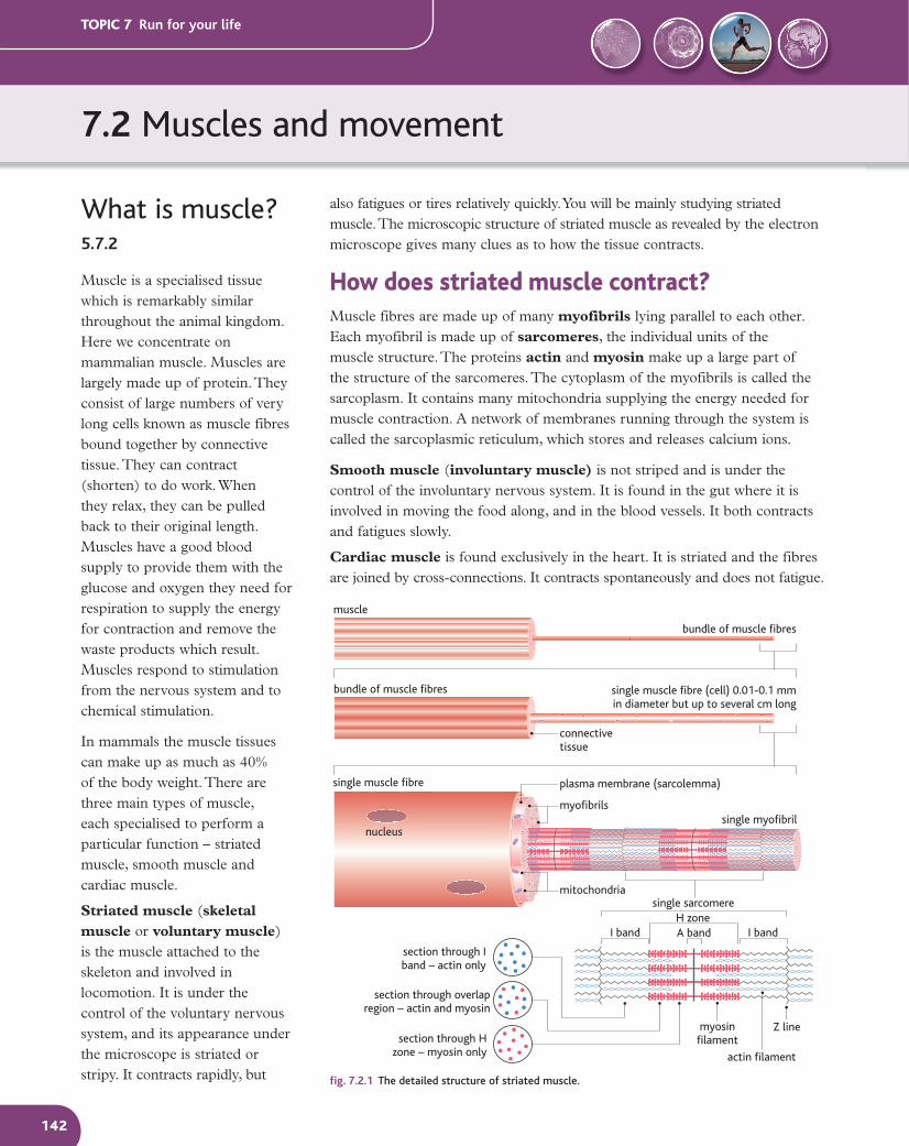
7.2 Muscles and movement TOPIC 7 Run for your life 142 What is muscle? 5.7.2 Muscle is a specialised tissue which is remarkably similar throughout the animal kingdom. Here we concentrate on mammalian muscle. Muscles are largely made up of protein. They consist of large numbers of very long cells known as muscle fibres bound together by connective tissue. They can contract (shorten) to do work. When they relax, they can be pulled back to their original length. Muscles have a good blood supply to provide them with the glucose and oxygen they need for respiration to supply the energy for contraction and remove the waste products which result. Muscles respond to stimulation from the nervous system and to chemical stimulation. In mammals the muscle tissues can make up as much as 40% of the body weight. There are three main types of muscle, each specialised to perform a particular function – striated muscle, smooth muscle and cardiac muscle. Striated muscle (skeletal muscle or voluntary muscle) is the muscle attached to the skeleton and involved in locomotion. It is under the control of the voluntary nervous system, and its appearance under the microscope is striated or stripy. It contracts rapidly, but muscle bundle of muscle fibres bundle of muscle fibres connective tissue plasma membrane (sarcolemma) single muscle fibre (cell) 0.01-0.1 mm in diameter but up to several cm long single sarcomere nucleus mitochondria myofibrils single myofibril single muscle fibre H zone I band I band A band Z line actin filament myosin filament section through I band – actin only section through H zone – myosin only section through overlap region – actin and myosin fig. 7.2.1 The detailed structure of striated muscle. also fatigues or tires relatively quickly. You will be mainly studying striated muscle. The microscopic structure of striated muscle as revealed by the electron microscope gives many clues as to how the tissue contracts. How does striated muscle contract? Muscle fibres are made up of many myofibrils lying parallel to each other. Each myofibril is made up of sarcomeres, the individual units of the muscle structure. The proteins actin and myosin make up a large part of the structure of the sarcomeres. The cytoplasm of the myofibrils is called the sarcoplasm. It contains many mitochondria supplying the energy needed for muscle contraction. A network of membranes running through the system is called the sarcoplasmic reticulum, which stores and releases calcium ions. Smooth muscle (involuntary muscle) is not striped and is under the control of the involuntary nervous system. It is found in the gut where it is involved in moving the food along, and in the blood vessels. It both contracts and fatigues slowly. Cardiac muscle is found exclusively in the heart. It is striated and the fibres are joined by cross-connections. It contracts spontaneously and does not fatigue.
Transcript of 7.2 Muscles and movement - Pearson Education 7 Run for your life 143 HSW Properties of skeletal...
7.2 Muscles and movement
TOPIC 7 Run for your life
142
What is muscle?5.7.2
Muscle is a specialised tissue which is remarkably similar throughout the animal kingdom. Here we concentrate on mammalian muscle. Muscles are largely made up of protein. They consist of large numbers of very long cells known as muscle fibres bound together by connective tissue. They can contract (shorten) to do work. When they relax, they can be pulled back to their original length. Muscles have a good blood supply to provide them with the glucose and oxygen they need for respiration to supply the energy for contraction and remove the waste products which result. Muscles respond to stimulation from the nervous system and to chemical stimulation.
In mammals the muscle tissues can make up as much as 40% of the body weight. There are three main types of muscle, each specialised to perform a particular function – striated muscle, smooth muscle and cardiac muscle.
Striated muscle (skeletal muscle or voluntary muscle) is the muscle attached to the skeleton and involved in locomotion. It is under the control of the voluntary nervous system, and its appearance under the microscope is striated or stripy. It contracts rapidly, but
muscle
bundle of muscle fibres
bundle of muscle fibres
connectivetissue
plasma membrane (sarcolemma)
single muscle fibre (cell) 0.01-0.1 mmin diameter but up to several cm long
single sarcomere
nucleus
mitochondria
myofibrilssingle myofibril
single muscle fibre
H zoneI band I bandA band
Z line
actin filament
myosinfilament
section through Iband – actin only
section through Hzone – myosin only
section through overlapregion – actin and myosin
fig. 7.2.1 The detailed structure of striated muscle.
also fatigues or tires relatively quickly. You will be mainly studying striated muscle. The microscopic structure of striated muscle as revealed by the electron microscope gives many clues as to how the tissue contracts.
How does striated muscle contract?Muscle fibres are made up of many myofibrils lying parallel to each other. Each myofibril is made up of sarcomeres, the individual units of the muscle structure. The proteins actin and myosin make up a large part of the structure of the sarcomeres. The cytoplasm of the myofibrils is called the sarcoplasm. It contains many mitochondria supplying the energy needed for muscle contraction. A network of membranes running through the system is called the sarcoplasmic reticulum, which stores and releases calcium ions.
Smooth muscle (involuntary muscle) is not striped and is under the control of the involuntary nervous system. It is found in the gut where it is involved in moving the food along, and in the blood vessels. It both contracts and fatigues slowly.
Cardiac muscle is found exclusively in the heart. It is striated and the fibres are joined by cross-connections. It contracts spontaneously and does not fatigue.
M12_BIOL_SB_A2_6027_C7_2.indd 142 3/4/09 08:44:29
TOPIC 7 Run for your life
143
HSW Properties of skeletal muscle
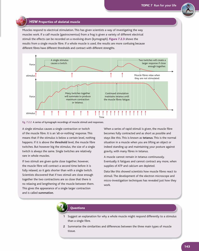
Muscles respond to electrical stimulation. This has given scientists a way of investigating the way
muscles work. If a calf muscle (gastrocnemius) from a frog is given a variety of different electrical
stimuli the effects can be recorded on a revolving drum (kymograph). Figure 7.2.3 shows the
results from a single muscle fibre. If a whole muscle is used, the results are more confusing because
different fibres have different thresholds and contract with different strengths.
Time
Two twitches will create alarger response if close
enough together.
Muscle fibres relax whenthey are not stimulated.
Continued stimulationmaintains tetanus until
the muscle fibres fatigue.
Many twitches togetherwill summate to produce
maximum contractionor tetanus.
A single stimuluscauses a twitch.
Force
Force
stimulus
stimulus
fig. 7.2.2 A series of kymograph recordings of muscle stimuli and responses.
1 Suggest an explanation for why a whole muscle might respond differently to a stimulus
than a single fibre.
2 Summarise the similarities and differences between the three main types of muscle
tissue.
Questions
A single stimulus causes a single contraction or twitch
of the muscle fibre. It is an ‘all-or-nothing’ response. This
means that if the stimulus is below a certain level, nothing
happens. If it is above the threshold level, the muscle fibre
twitches. But however big the stimulus, the size of a single
twitch is always the same. Single twitches are relatively
rare in whole muscles.
If two stimuli are given quite close together, however,
the muscle fibre will contract a second time before it is
fully relaxed, so it gets shorter than with a single twitch.
Scientists discovered that if two stimuli are close enough
together the two contractions are so close that there is
no relaxing and lengthening of the muscle between them.
This gives the appearance of a single larger contraction
and is called summation.
When a series of rapid stimuli is given, the muscle fibre
becomes fully contracted and as short as possible and
stays like this. This is known as tetanus. This is the normal
situation in a muscle when you are lifting an object or
indeed standing up and maintaining your posture against
gravity, with many fibres in tetanus.
A muscle cannot remain in tetanus continuously.
Eventually it fatigues and cannot contract any more, when
supplies of ATP and calcium are depleted.
Data like this showed scientists how muscle fibres react to
stimuli. The development of the electron microscope and
micro-investigation techniques has revealed just how they
work.
M12_BIOL_SB_A2_6027_C7_2.indd 143 3/4/09 08:44:39
Different types of muscle fibre 5.7.2
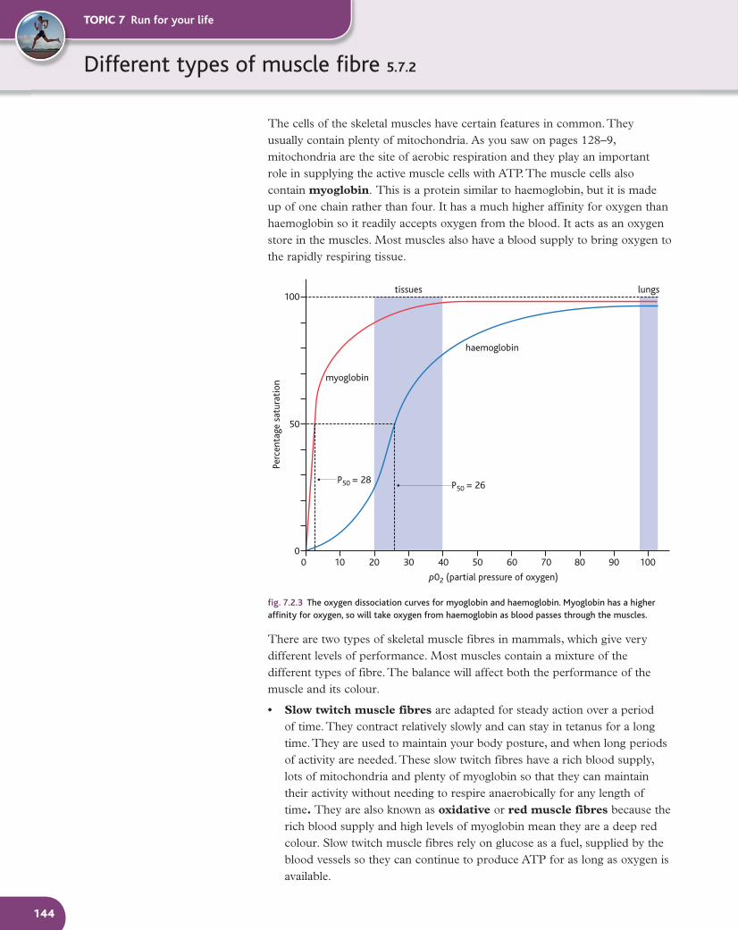
The cells of the skeletal muscles have certain features in common. They usually contain plenty of mitochondria. As you saw on pages 128–9, mitochondria are the site of aerobic respiration and they play an important role in supplying the active muscle cells with ATP. The muscle cells also contain myoglobin. This is a protein similar to haemoglobin, but it is made up of one chain rather than four. It has a much higher affinity for oxygen than haemoglobin so it readily accepts oxygen from the blood. It acts as an oxygen store in the muscles. Most muscles also have a blood supply to bring oxygen to the rapidly respiring tissue.
100 20 30 40 50 60 70 80 90 1000
50
100tissues lungs
haemoglobin
P50 = 28 P50 = 26
p02 (partial pressure of oxygen)
myoglobin
Perc
enta
ge s
atur
atio
n
fig. 7.2.3 The oxygen dissociation curves for myoglobin and haemoglobin. Myoglobin has a higher affinity for oxygen, so will take oxygen from haemoglobin as blood passes through the muscles.
There are two types of skeletal muscle fibres in mammals, which give very different levels of performance. Most muscles contain a mixture of the different types of fibre. The balance will affect both the performance of the muscle and its colour.
• Slow twitch muscle fibres are adapted for steady action over a period of time. They contract relatively slowly and can stay in tetanus for a long time. They are used to maintain your body posture, and when long periods of activity are needed. These slow twitch fibres have a rich blood supply, lots of mitochondria and plenty of myoglobin so that they can maintain their activity without needing to respire anaerobically for any length of time. They are also known as oxidative or red muscle fibres because the rich blood supply and high levels of myoglobin mean they are a deep red colour. Slow twitch muscle fibres rely on glucose as a fuel, supplied by the blood vessels so they can continue to produce ATP for as long as oxygen is available.
TOPIC 7 Run for your life
144
M12_BIOL_SB_A2_6027_C7_2.indd 144 3/4/09 08:44:44
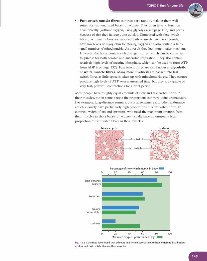
• Fast twitch muscle fibres contract very rapidly, making them well suited for sudden, rapid bursts of activity. They often have to function anaerobically (without oxygen, using glycolysis, see page 132) and partly because of this they fatigue quite quickly. Compared with slow twitch fibres, fast twitch fibres are supplied with relatively few blood vessels, have low levels of myoglobin for storing oxygen and also contain a fairly small number of mitochondria. As a result they look much paler in colour. However, the fibres contain rich glycogen stores, which can be converted to glucose for both aerobic and anaerobic respiration. They also contain relatively high levels of creatine phosphate, which can be used to form ATP from ADP (see page 132). Fast twitch fibres are also known as glycolytic or white muscle fibres. Many more myofibrils are packed into fast twitch fibres as little space is taken up with mitochondria, etc. They cannot produce high levels of ATP over a sustained time, but they are capable of very fast, powerful contractions for a brief period.
Most people have roughly equal amounts of slow and fast twitch fibres in their muscles, but in some people the proportions can vary quite dramatically. For example, long-distance runners, cyclists, swimmers and other endurance athletes usually have particularly high proportions of slow twitch fibres. In contrast, weightlifters and sprinters, who need the maximum strength from their muscles in short bursts of activity, usually have an unusually high proportion of fast twitch fibres in their muscles.
fig. 7.2.4 Scientists have found that athletes in different sports tend to have different distributions of slow and fast twitch fibres in their muscles.
TOPIC 7 Run for your life
145
0 20 40 60 80 100
0 20 40 60 80 100
distance cyclist sprinter
slow twitch
fast twitch
Percentage of slow twitch muscle in body
Maximum oxygen uptake/mlmin−1kg−1
long-distancerunners
swimmers
trainednon-athletes
sprinters
M12_BIOL_SB_A2_6027_C7_2.indd 145 3/4/09 08:44:51
TOPIC 7 Run for your life
146
So why does this difference occur? Part of the answer is training. The number of muscle fibres you possess does not change, but the size and type of the fibre can alter in response to exercise. So if you practise sprinting, you will develop more fast twitch fibres, while if you do endurance training then slow twitch muscle fibres will increase in number.
There are also a variety of different genes which affect the basic components of our muscles, which can then be enhanced with further training. So while most of us have about 50% of each type of muscle fibre, some people have around 75% fast twitch and others have 75% slow twitch. These differences are bound to make a difference to sporting potential. So for example, someone born with a high proportion of fast twitch fibres may well be a good sprinter but is unlikely to make a top-class marathon runner, while someone with more than average slow twitch fibres is unlikely to be a successful weightlifter but may well make an endurance athlete.
Scientists have also discovered ‘superfast’ twitch fibres, which contract even more quickly and strongly than usual. When the muscle structure of Colin Jackson, a former world 110 m hurdle record holder, was analysed, not only did he have 75% fast twitch fibres in his leg muscles, but 25% of those were superfast fibres – something which must have helped him in his sporting career.
HSW Explaining the difference
Currently there is a great deal of research into the
genetics of sporting performance. However, it is an area
which is fraught with ethical difficulties and examples
of how the public perception of valid research may be
skewed by media presentation.
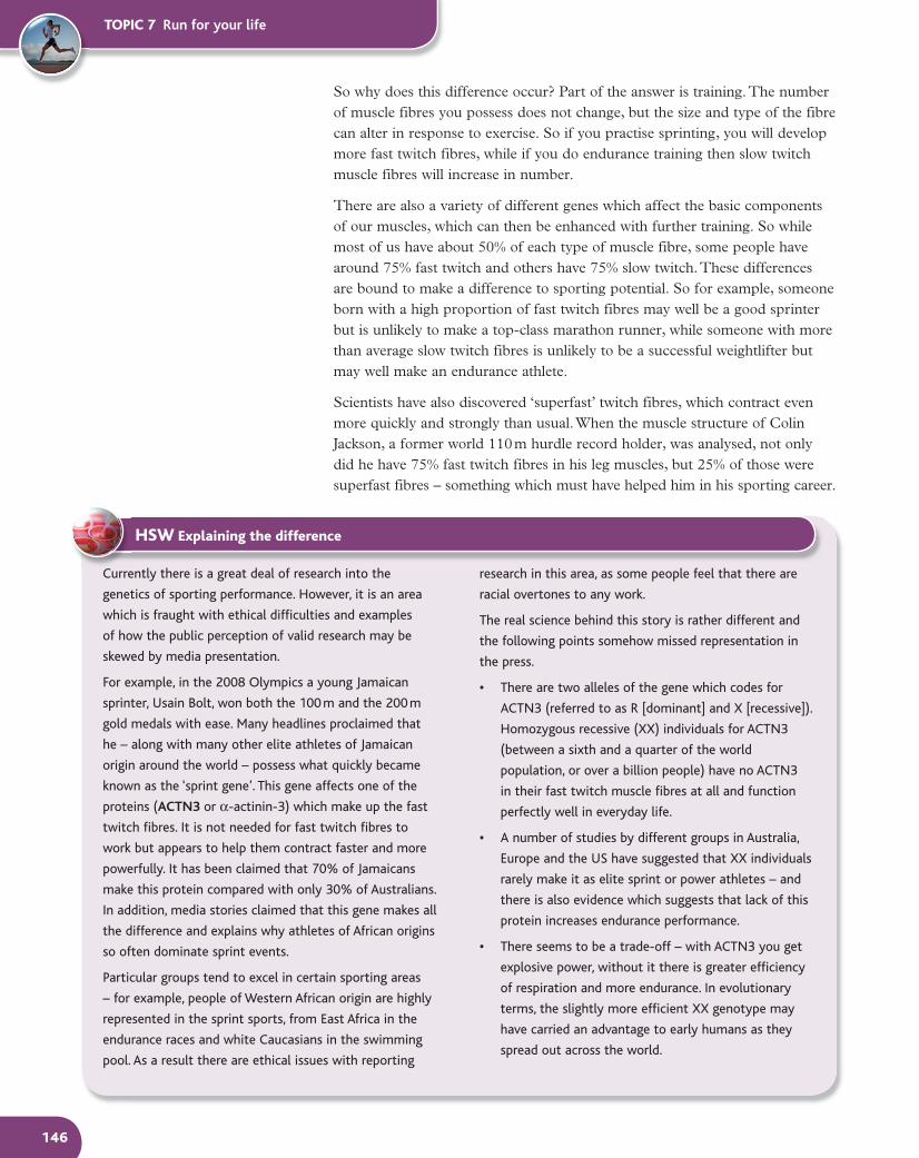
For example, in the 2008 Olympics a young Jamaican
sprinter, Usain Bolt, won both the 100 m and the 200 m
gold medals with ease. Many headlines proclaimed that
he – along with many other elite athletes of Jamaican
origin around the world – possess what quickly became
known as the ‘sprint gene’. This gene affects one of the
proteins (ACTN3 or α-actinin-3) which make up the fast
twitch fibres. It is not needed for fast twitch fibres to
work but appears to help them contract faster and more
powerfully. It has been claimed that 70% of Jamaicans
make this protein compared with only 30% of Australians.
In addition, media stories claimed that this gene makes all
the difference and explains why athletes of African origins
so often dominate sprint events.
Particular groups tend to excel in certain sporting areas
– for example, people of Western African origin are highly
represented in the sprint sports, from East Africa in the
endurance races and white Caucasians in the swimming
pool. As a result there are ethical issues with reporting
research in this area, as some people feel that there are
racial overtones to any work.
The real science behind this story is rather different and
the following points somehow missed representation in
the press.
• There are two alleles of the gene which codes for
ACTN3 (referred to as R [dominant] and X [recessive]).
Homozygous recessive (XX) individuals for ACTN3
(between a sixth and a quarter of the world
population, or over a billion people) have no ACTN3
in their fast twitch muscle fibres at all and function
perfectly well in everyday life.
• A number of studies by different groups in Australia,
Europe and the US have suggested that XX individuals
rarely make it as elite sprint or power athletes – and
there is also evidence which suggests that lack of this
protein increases endurance performance.
• There seems to be a trade-off – with ACTN3 you get
explosive power, without it there is greater efficiency
of respiration and more endurance. In evolutionary
terms, the slightly more efficient XX genotype may
have carried an advantage to early humans as they
spread out across the world.
M12_BIOL_SB_A2_6027_C7_2.indd 146 3/4/09 08:44:58
TOPIC 7 Run for your life
147
1 Describe the roles of mitochondria and myoglobin in muscle fibres.
2 Describe how mitochondria and myoglobin vary between fast and slow
twitch fibres.
3 Chickens are birds which spend much of their time walking around on the
ground. If they are startled or frightened, they will fly up almost vertically
to escape – but the flying doesn’t last long. When carving a chicken to
eat, the breast meat is pale and the leg meat is much darker. Explain these
observations in the light of your knowledge about different types of
muscle fibres.
4 Draw up a table to compare slow and fast twitch muscle fibres.
Questions
The team that has been behind much of the research into
the ACTN3 protein and its genetic inheritance, lead by
Katherine North, have extended their work in several ways.
They are looking at the inheritance of muscle protein genes
in human evolution and also, most excitingly, looking for
application of their work for children with genetic diseases
affecting the muscle protein such as muscular dystrophy.
fig. 7.2.5 Usain Bolt, the young Jamaican sprinter whose success in the 2008 Olympics brought the ‘sprint gene’ to greater public attention than ever before!
• The impact of the ‘sprint’ variant of ACTN3 on
performance is only to give 2–3% increase in speed,
not enough to have any effect on most people, but
enough to make a difference when performing at the
highest levels. This explains why almost all top sprint
and power athletes have the RR or RX variant. Only
one Olympic level sprint or power athlete investigated
– a Spanish short-distance hurdler – is XX!
• There are dozens – and may even be hundreds – of
genes that affect physical performance and the ACTN3
gene is just one of them.
• This gene cannot explain Jamaican dominance in
sprint sports. The 70/30 percentage figures given in
the media refer to the incidence of the homozygous
RR state in Jamaican athletes compared with
Australian athletes. However, when RX is considered
as well (and the heterozygote state also produces the
vital protein) the difference between the incidence
in the Jamaican population (98%) and the European
populations (82%) is much less striking. Both
populations have plenty of potential elite sprinters to
choose from. In fact around 5 billion people worldwide
have this protein – and we’re not all top athletes!
• The frequency of ACTN3 in Jamaicans is not unique.
Research has shown even higher incidences of
this variant (99% of people) in Kenya – a country
renowned for its middle and long-distance runners
rather than its sprinters!
• When the massively successful Usain Bolt ran in
the 2008 Olympics, everyone he was competing
against almost certainly had the same RX or RR
genetic combination, giving them the useful ACTN3
protein. The superiority of Bolt’s performance
cannot be dismissed as a simple genetic advantage.
Environmental factors such as training, hard work
and motivation along with many other advantageous
genes will have carried him to success.
SC
M12_BIOL_SB_A2_6027_C7_2.indd 147 3/4/09 08:45:17
How muscle contracts 5.7.3
TOPIC 7 Run for your life
148
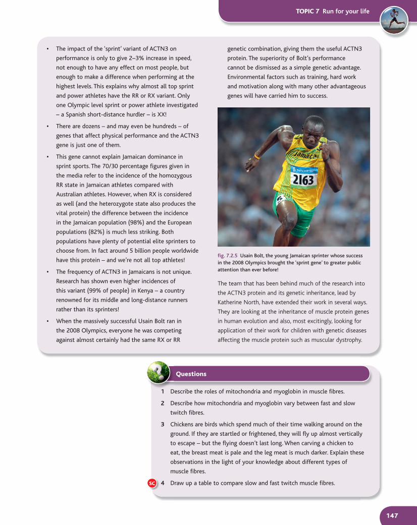
Observations of micrographs show that whether a muscle fibre is contracted or relaxed, the dark A bands remain the same length. However, the light I bands and the H zone get shorter when a muscle fibre contracts and return to normal length when it relaxes again. This suggests that the two types of filaments slide over each other during contraction – the basis of the sliding filament theory (fig. 7.2.6)
one sarcomere
contracted staterelaxed state
myosin actin
H zone
A band
I band
fig. 7.2.6 The sliding filament theory
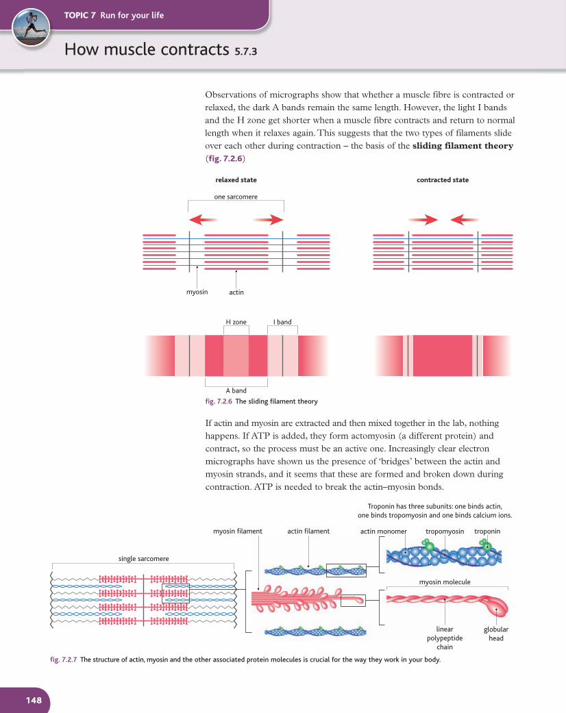
If actin and myosin are extracted and then mixed together in the lab, nothing happens. If ATP is added, they form actomyosin (a different protein) and contract, so the process must be an active one. Increasingly clear electron micrographs have shown us the presence of ‘bridges’ between the actin and myosin strands, and it seems that these are formed and broken down during contraction. ATP is needed to break the actin–myosin bonds.
single sarcomere
myosin filament actin filament actin monomer tropomyosin troponin
myosin molecule
linearpolypeptide
chain
globularhead
Troponin has three subunits: one binds actin,one binds tropomyosin and one binds calcium ions.
fig. 7.2.7 The structure of actin, myosin and the other associated protein molecules is crucial for the way they work in your body.
M12_BIOL_SB_A2_6027_C7_2.indd 148 3/4/09 08:45:26

TOPIC 7 Run for your life
149
tropomyosinactin filament
Calcium ions bind to thetroponin molecules, changing
their shape, so troponinmolecules pull on the
tropomyosin molecules they areattached to. This moves thetropomyosin away from the
myosin binding sites, exposingthem ready for action.
The myosin heads bind tothe actin, forming an
actomyosin bridge.
ADP and Pi are released fromthe myosin head. The myosin
changes shape – the headbends forward moving the actinfilament about 10 nm along the
myosin filament, shorteningthe sarcomere.
Free ATP binds to the head,causing another shape change in
the myosin, so the bindingof the head to the actin strandis broken. This activates ATPasein the myosin head, which also
needs calcium ions to work. TheATP is hydrolysed, providing the
energy to return the myosin head to its original position,
primed with ADP and Pi,ready to go again.
With continued stimulation,calcium ions remain in the
sarcoplasm and the cycle isrepeated. If not, calcium ions
are pumped back into thesarcoplasmic reticulum using
energy from ATP. The troponinand tropomyosin return totheir original positions and
the contraction is complete.The muscle fibre is relaxed.
The diagram shows the actinand myosin unit before
contraction starts. The myosinheads have ADP and Pi
bound closely to them as well.
troponin
ADP Pi
ADP
ADP
ADP
Pi
ADP Pi
ADP
ATP
Pi
Pi
Pi
Ca2+
Ca2+ myosin binding site
exposed myosinbinding sites
fig. 7.2.8 Muscles contract in response to a stimulation at a neuromuscular junction between a nerve cell and a muscle. This triggers the release of calcium ions (Ca2+) from the sarcoplasmic reticulum which then flood the sarcomeres.
1 Draw and label the appearance of a sarcomere as you
would expect it to look under an electron microscope:
a fully contracted
b fully relaxed.
2 Describe the role of calcium ions in the contraction of
skeletal muscles.
3 a Explain why the presence of ATP is so important
for the contraction of striated muscles.
b Suggest how this explains what happens when
rigor mortis sets in after death.
1 Draw and label the appearance of a sarcomere as you
Questions
Actin–myosin interactionsA sort of ratchet mechanism has been proposed for the contraction of the myofibrils. The shape of the myosin molecule enables it to attach to the actin to form cross-bridges of actomyosin. This changes the molecular shape and so pulls the actin filament across the myosin, increasing the interlocking region and shortening the sarcomere. The bridges then break and the process is repeated between 50 and 100 times per second. The combined effect of this happening in each sarcomere is a shortening of the whole myofibril. The shortening of many myofibrils together brings about the contraction of a muscle. For the ratchet mechanism to work, both calcium ions and ATP must be present.
To understand how the sliding filament theory explains the way in which muscles contract, it is important to understand the structure of actin and myosin. A myosin molecule is made up of two long polypeptide chains twisted together, each ending in a large, globular head which has ADP and inorganic phosphate molecules bound to it. In some circumstances the head can act as an ATPase enzyme. A myosin filament is made up of lots of these molecules bundled together, with their heads sticking out from the filament. An actin filament is made up of two chains of actin monomers joined together like beads on a necklace. The shape of the actin molecule produces myosin binding sites at regular intervals, where the globular heads of the myosin molecules can fit. However, wrapping around the double actin chains is another long chain protein molecule called tropomyosin. In a relaxed muscle, the tropomyosin chain covers up the myosin bonding sites. In turn the tropomyosin has molecules of another protein, troponin, attached regularly along the chain.
SC
M12_BIOL_SB_A2_6027_C7_2.indd 149 3/4/09 08:45:37

Tissues of the skeletal system 5.7.4
Imagine yourself moving – walking, running, jumping – and then try to imagine the variety of demands being made on your body. The properties of your skeletal tissues vary, as they are adapted to carry out these various functions effectively.
Bone is strong and hard, made up of bone cells embedded in a matrix of collagen and calcium salts. Bone is particularly strong under compression (squashing) forces. It not only needs to be strong and hard – it must be as light as possible to reduce the weight moved about.
Cartilage is a hard but flexible tissue made up of cells called chondrocytes within an organic matrix which consists of varying amounts of collagen fibrils. Cartilage is elastic and able to withstand compressive forces. It is a very good shock absorber and is frequently found between bones such as the vertebrae and in the joints. There are two main types of cartilage found in the skeleton:
• Hyaline cartilage is found at the ends of bones (and in the nose, air passageways and parts of the ear).
• White fibrous cartilage has bundles of densely packed collagen in the matrix. It has great tensile strength but is less flexible than the other forms of cartilage. It forms the intervertebral discs and is found between the bones in the joints.
Tendons are made up almost entirely of white fibrous tissue. This consists of bundles of collagen fibres and gives a tissue that is strong but relatively inelastic. This makes it ideal for joining muscles to bones. One end of the tendon is attached to a muscle and the other end attached either directly to a bone or to the fibrous cover of the bone. This makes a more secure attachment for muscles to bone and provides a little shock absorption if the joint is subjected to sudden stretch. However, if tendons stretched a lot, much of the work done by the muscles would be wasted as they would stretch the tendons without moving the bones!
Ligaments hold bones together and in the correct alignment, both around the joint as a capsule and within the joint itself. They need to be elastic to allow the bones of the joint to move when necessary. They are made of yellow elastic tissue which gives an ideal combination of strength with elasticity. Some ligament capsules are very loose, while others are very tight – different joints need
TOPIC 7 Run for your life
150
different properties. The differences in the properties of the ligaments result from the presence of varying amounts of collagen and even some white fibrous tissue in the mixture.
fig. 7.2.9 The tissues of the skeletal system – seen here at the knee – work together to hold the bones together, stop them wearing away and make movement possible.
Joints, muscles and movementThe joints are vital to allow movement and locomotion. Anyone who has experienced the pain of a dislocated joint will know that keeping the bones lined up correctly is essential to the working of a joint.
The ends of the bones at a joint are shaped to move smoothly over each other, and the way in which the two bones meet varies according to the type of movement required. The ball and socket joints found at the hip and shoulder give very free movement, whereas the hinge joints of the fingers and knees are much more restrictive.
The bones in a joint form two solid masses moving over each other while subjected to quite severe forces. If the joint consisted just of bone on bone, the ends would soon wear each other away. To prevent the bones from being eroded like this the joint is lined with a replacable layer of rubbery cartilage which allows the joint to articulate smoothly. The most mobile joints also produce a liquid lubricant known as synovial fluid which fills the joint cavity and ensures easy friction-free movement (see fig. 7.2.11).
Flexor and extensor muscles workantagonistically to operate the joint.
flexor muscle (biceps femoris)
femur
fibula
tibia
Ligaments attach bone to bone.
cartilage
patella (knee cap)
Tendons attach muscle to bone.
extensor muscle (quadriceps)
M12_BIOL_SB_A2_6027_C7_2.indd 150 3/4/09 08:45:43
TOPIC 7 Run for your life
151
fig. 7.2.10 The skeleton has several different types of joints which make different types of movement possible.
fig. 7.2.11 A synovial joint – the lubrication provided by the synovial fluid is vital for smooth, painfree movement in the most mobile joints.
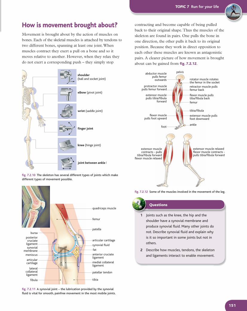
How is movement brought about?Movement is brought about by the action of muscles on bones. Each of the skeletal muscles is attached by tendons to two different bones, spanning at least one joint. When muscles contract they exert a pull on a bone and so it moves relative to another. However, when they relax they do not exert a corresponding push – they simply stop
quadriceps muscle
femur
patella
articular cartilage
synovial fluid
anterior cruciateligament
bursa
fibula
articularcartilage
lateralcollateralligament
meniscus
posteriorcruciate
ligamentsynovial
membrane fat
medial collateralligament
patellar tendon
tibia
fig. 7.2.12 Some of the muscles involved in the movement of the leg.
1 Joints such as the knee, the hip and the
shoulder have a synovial membrane and
produce synovial fluid. Many other joints do
not. Describe synovial fluid and explain why
is it so important in some joints but not in
others.
2 Describe how muscles, tendons, the skeleton
and ligaments interact to enable movement.
Questions
extensor musclecontracts - pulls
tibia/fibula forwardflexor muscle relaxed
extensor muscle relaxedflexor muscle contracts -pulls tibia/fibula forward
flexor musclepulls foot upward
abductor musclepulls femur
outwards rotator muscle rotatesthe femur in the socket
protractor musclepulls femur forward
retractor muscle pullsfemur back
extensor musclepulls tibia/fibula
forward
extensor muscle pullsfoot downward
flexor muscle pullstiba/fibula back
pelvis
femur
tibia/fibula
anklefoot
shoulder(ball and socket joint)
elbow (pivot joint)
wrist (saddle joint)
finger joint
knee (hinge joint)
joint between ankle bones
contracting and become capable of being pulled back to their original shape. Thus the muscles of the skeleton are found in pairs. One pulls the bone in one direction, the other pulls it back to its original position. Because they work in direct opposition to each other these muscles are known as antagonistic pairs. A clearer picture of how movement is brought about can be gained from fig. 7.2.12.
M12_BIOL_SB_A2_6027_C7_2.indd 151 3/4/09 08:46:19