6.S897 Machine Learning for Healthcare, Lecture 12 ...
68
Machine learning for Pathology Andrew H Beck MD PhD CEO @ PathAI 6.S897/HST.956: Machine Learning for Healthcare. MIT. March 19, 2019 1 © source unknown. All rights reserved. This content is excluded from our Creative Commons license. For more information, see https://ocw.mit.edu/help/faq-fair-use/
Transcript of 6.S897 Machine Learning for Healthcare, Lecture 12 ...

Machine learning for Pathology
Andrew H Beck MD PhD CEO @ PathAI
6.S897/HST.956: Machine Learning for Healthcare. MIT. March 19, 2019
1© source unknown. All rights reserved. This content is excluded from our Creative Commons license. For more information, see https://ocw.mit.edu/help/faq-fair-use/

Pathology
No Treatment
Minimal Treatment
Aggressive Treatment
Pathologic diagnosis is a central determinant of therapeutic decisions.
Pathology Diagnosis
Radiology Impression
Clinical Signs
Patient Symptoms
2
Proprietary & Confidential

TCGA:https://portal.gdc.cancer.gov
Courtesy of the NIH. Used with permission.
3

Courtesy of the NIH. Used with permission.
4
TCGA:https://portal.gdc.cancer.gov

Emergence of early computational approaches in Pathology (1981)
Baak et al. Lancet 1981 Courtesy of Elsevier, Inc., https://www.sciencedirect.com. Used with permission. 5

Artificial Neural Nets in Quantitative Pathology (1990)
“It is concluded that artificial neural networks, used in conjunction with other nonalgorithmic artificial intelligence techniques and traditional algorithmic processing, may provide useful software engineering tools for the development of systems in quantitative pathology.”
6

Proprietary & Confidential
Emergence of Digital Pathology (2000)
7

Extracting a rich quantitative feature set
© AAAS. All rights reserved. This content is excluded from our Creative Commons license. For more information, see https://ocw.mit.edu/help/faq-fair-use/
Beck ... Koller. Science Translational Medicine 2011 8

C-Path 5YS Score Significantly Associated with Overall Survival on Both Cohorts
© AAAS. All rights reserved. This content is excluded from our Creative Commons license. For more information, see https://ocw.mit.edu/help/faq-fair-use/
Beck ... Koller. Science Translational Medicine 2011 9
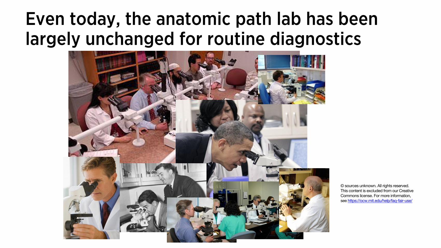
Even today, the anatomic path lab has been largely unchanged for routine diagnostics
10
© sources unknown. All rights reserved.This content is excluded from our Creative Commons license. For more information, see https://ocw.mit.edu/help/faq-fair-use/
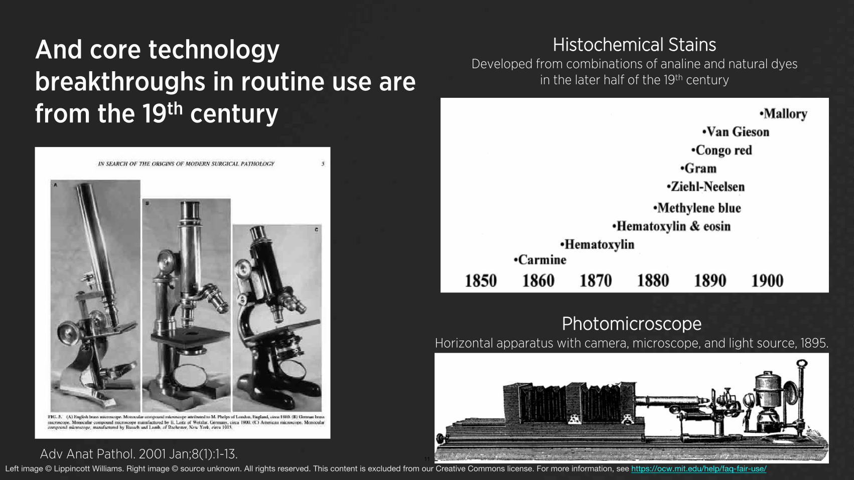
Histochemical Stains And core technology Developed from combinations of analine and natural dyes
in the later half of the 19th century breakthroughs in routine use are from the 19th century
Photomicroscope Horizontal apparatus with camera, microscope, and light source, 1895.
Adv Anat Pathol. 2001 Jan;8(1):1-13. 11
Left image © Lippincott Williams. Right image © source unknown. All rights reserved. This content is excluded from our Creative Commons license. For more information, see https://ocw.mit.edu/help/faq-fair-use/

Discordance among pathologists is common in interpretation of breast biopsies © AMA. All rights reserved. This content is excluded from our Creative Commons license. For more information, see https://ocw.mit.edu/help/faq-fair-use/
Pathologist Interpretation
Credit: Elmore et al. (JAMA 2015)
Pathologists in individual practice setting
Overall concordance rate of 75% on breast biopsies.
Inter-observer concordance rate of only 48% for a diagnosis of atypia.
Intra-observer concordance is only 79% overall and 53% for atypical lesions
12© Springer Nature. All rights reserved. This content is excluded from our Creative Commons license. For more information, see https://ocw.mit.edu/help/faq-fair-use/
• • •
•
Ref: Jackson SL … Elmore JG. Ann Surg Oncol. 2017 May;24(5):1234-1241.

Discordance among pathologists is common in interpretation of melanocytic neoplasms on skin biopsies
• 187 pathologists interpreted skin lesion biopsies, resulting in an overall discordance of 45%
• 118 pathologists read the same samples 8 months apart, and had an intraobserver discordance of 33%
Courtesy of Elmore, et al. Used under CC BY-NC. 13
BMJ 2017;357:j2813 | doi: 10.1136/bmj.j2813

30 Er
ror
Rat
e %
Massive advances in deep 24 learning for computer vision…
18
12
Human 6 Error Rate
0 2010 2011 2012 2013 2014 2015
ImageNet Performance over Time 14
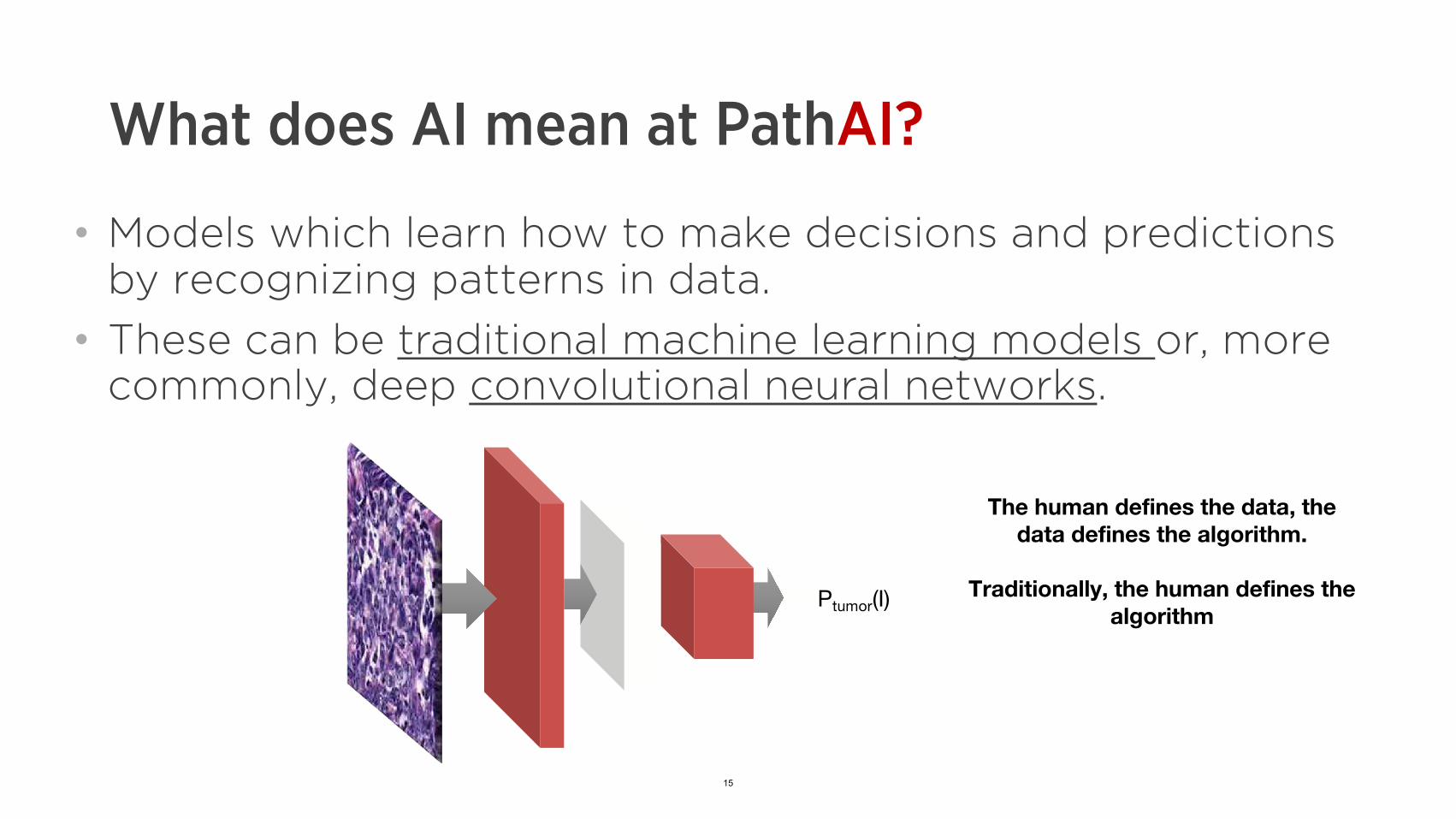
What does AI mean at PathAI?
• Models which learn how to make decisions and predictions by recognizing patterns in data.
• These can be traditional machine learning models or, more commonly, deep convolutional neural networks.
15
The human defines the data, the data defines the algorithm.
Traditionally, the human defines the Ptumor(I) algorithm

What can AI do for pathology?
A (somewhat) practical treatment
• Exhaustive – the model is tireless and is not distracted
• Quantitative – the model is reproducible and objective
• Efficient – massive parallelization for speedy processing
• Exploratory - learn relationships in a purely data-driven manner
16

What AI can’t do for pathology
Replace pathologists!
Proprietary & Confidential
17
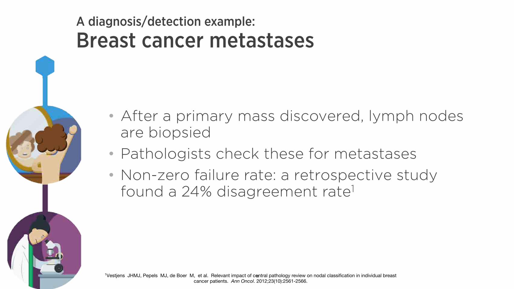
A diagnosis/detection example:
Breast cancer metastases
• After a primary mass discovered, lymph nodes are biopsied
• Pathologists check these for metastases • Non-zero failure rate: a retrospective study
found a 24% disagreement rate1
1Vestjens JHMJ, Pepels MJ, de Boer M, et al. Relevant impact of central pathology review 18 on nodal classification in individual breast cancer patients. Ann Oncol. 2012;23(10):2561-2566.
d
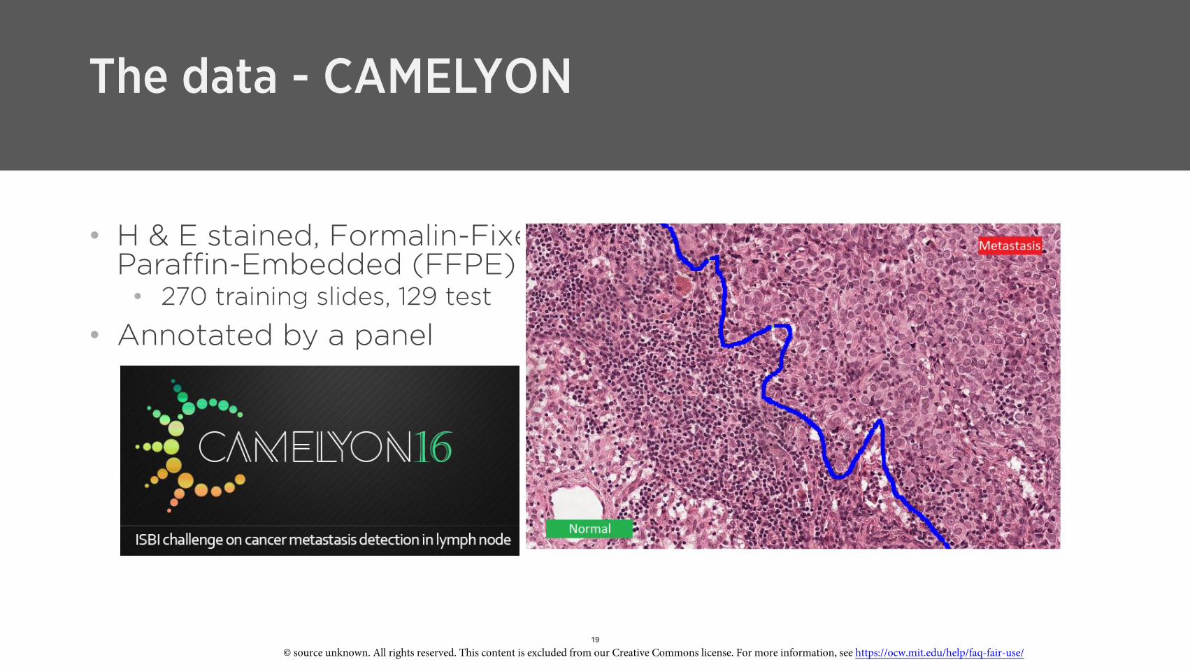
The data - CAMELYON
• H & E stained, Formalin-Fixe Paraffin-Embedded (FFPE) • 270 training slides, 129 test
• Annotated by a panel
19
© source unknown. All rights reserved. This content is excluded from our Creative Commons license. For more information, see https://ocw.mit.edu/help/faq-fair-use/

The data – Whole-Slide Images • WSIs are large –
~20,000-200,000 pixels on a side (“gigapixel”) • mm-cm imaged at 20x/40x
20
© source unknown. All rights reserved. This content is excluded from our Creative Commons license. For more information, see https://ocw.mit.edu/help/faq-fair-use/

Approach
• Standard image classification approach needs a twist for WSIs: sampling
Ptumor(I)
Proprietary & Confidential
21

Successfully applied deep learning approach to pathology Our team won the Camelyon challenge in 2016, demonstrating outstanding initial performance in pathology
TRA
INTE
ST
NO
RM
AL
TUM
OR
Whole Slide Image Training Data Deep Model
Whole Slide Image Image Patches Deep Model from Training Tumor Probability Map
© Wang, Beck, et al. All rights reserved. This content is excluded from our Creative Commons license. For more information, see https://ocw.mit.edu/help/faq-fair-use/ 22 26Wang, D., Khosla, A., … Beck, A.H., 2016. Deep learning for identifying metastatic breast cancer. arXiv preprint arXiv:1606.05718. JAMA. 2017 Dec 12;318(22):2199-2210.

Deep learning model outperforms human pathologists in the diagnosis of metastatic cancer
Error Rate (1-AUC)
Pathologists in competition 3.5%
Pathologists in clinical practice1 13 – 26%
Pathologists on micro-metastasis2 23 – 42%
Deep learning model 0.65%
1 n=12 2 Small tumors
References: Wang, Khosla, … Beck (2016) https://arxiv.org/abs/1606.05718 23 Camelyon16 (JAMA, 2017)

Proprietary & Confidential© 2016 PathAI - Confidential & proprietary.Do not distribute.
Pathologist + PathAI
24© source unknown. All rights reserved. This content is excluded from our Creative Commons license. For more information, see https://ocw.mit.edu/help/faq-fair-use/

Proprietary & Confidential© 2016 PathAI - Confidential & proprietary.Do not distribute.
Pathologist + PathAI
25© source unknown. All rights reserved. This content is excluded from our Creative Commons license. For more information, see https://ocw.mit.edu/help/faq-fair-use/

Proprietary & Confidential© 2016 PathAI - Confidential & proprietary.Do not distribute.
Pathologist + PathAI
26© source unknown. All rights reserved. This content is excluded from our Creative Commons license. For more information, see https://ocw.mit.edu/help/faq-fair-use/

Proprietary & Confidential© 2016 PathAI - Confidential & proprietary.Do not distribute.
Pathologist + PathAI
27© source unknown. All rights reserved. This content is excluded from our Creative Commons license. For more information, see https://ocw.mit.edu/help/faq-fair-use/

Proprietary & Confidential© 2016 PathAI - Confidential & proprietary.Do not distribute.
Pathologist + PathAI
28© source unknown. All rights reserved. This content is excluded from our Creative Commons license. For more information, see https://ocw.mit.edu/help/faq-fair-use/

Pathology Report
Patient: John Doe Diagnosis: Size:
pTNM staging: # of Pos LN: # of Neg LN:
Time per slide: 1 – 10 minutes Accuracy: ~85% Reproducibility: Low
Pathology Report Confirm
Patient: John Doe pTNM staging: pT2N1MX Diagnosis: Met. Cancer # of Pos LN: 1 Size: 2.3mm # of Neg LN: 4
Time per slide: 10– 60 seconds Accuracy: >99.5% Reproducibility: High Proprietary & Confidential
© source unknown. All rights reserved. This content is excluded from our Creative Commons license. For more information, see https://ocw.mit.edu/help/faq-fair-use/
29

Why is this a good application for AI?
• Exhaustive analysis is beneficial • Large volume
• Local image data necessary and sufficient • Interpretability: Heatmaps & simple
models provide insight into how the patient-level prediction was made
© source unknown. All rights reserved. This content is excluded from our Creative Commons license. For more information, see https://ocw.mit.edu/help/faq-fair-use/
• Required accuracy is high
30
Proprietary & Confidential
A predictive example:
Precision immunotherapy
• Some cancers express immune-inhibitory ligands,activating immune“checkpoints”
• “checkpointinhibitors” mask these signals,unleashing theimmune system
31
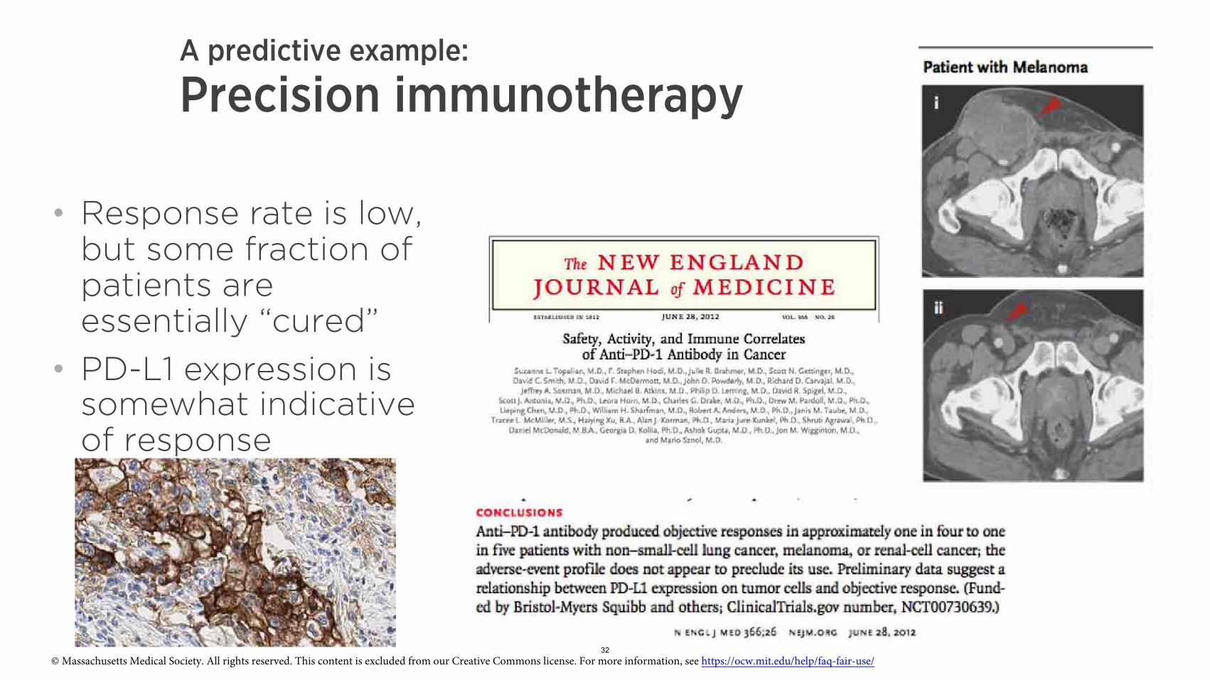
A predictive example:
Precision immunotherapy
• Response rate is low, but some fraction of patients are essentially “cured”
• PD-L1 expression is somewhat indicative of response
32© Massachusetts Medical Society. All rights reserved. This content is excluded from our Creative Commons license. For more information, see https://ocw.mit.edu/help/faq-fair-use/

Manual interpretation of PD-L1 IHC is highly variable
PDL1 manual IHC scores on immune cells are unreliable
© American Medical Association. All rights reserved. This content is excluded from our Creative Commons license. For more information, see https://ocw.mit.edu/help/faq-fair-use/
Rimm et al. (JAMA Oncol; 2017) 33

Manual scoring of PD-L1 is variable …and not always predictive
© Massachusetts Medical Society. All rights reserved. This content is excluded from our Creative Commons license. For more information, see https://ocw.mit.edu/help/faq-fair-use/
34

Can we do better?
• Deep learning is data hungry data
• Need 10s of thousands of precise cell annotations
35
First, we need the
Proprietary & Confidential

Board-certified training data
Working with pathologists around the country to generate high-quality annotations
>2.5M Annotations
36

Automatic and exhaustive regions of interest tumor and relevant stroma
© source unknown. All rights reserved. This content is excluded from our Creative Commons license. For more information, see https:// ocw.mit.edu/help/faq-fair-use/
37
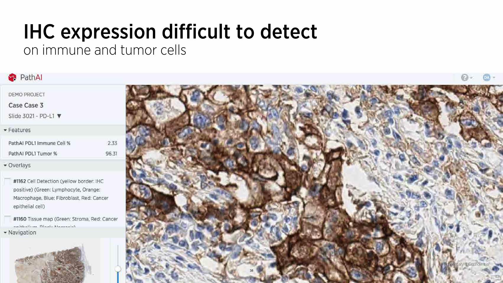
IHC expression difficult to detect on immune and tumor cells
38
Proprietary & Confidential
43

Exhaustive automated classification Cell type and cellular IHC positivity classification
Proprietary & Confidential
44 39
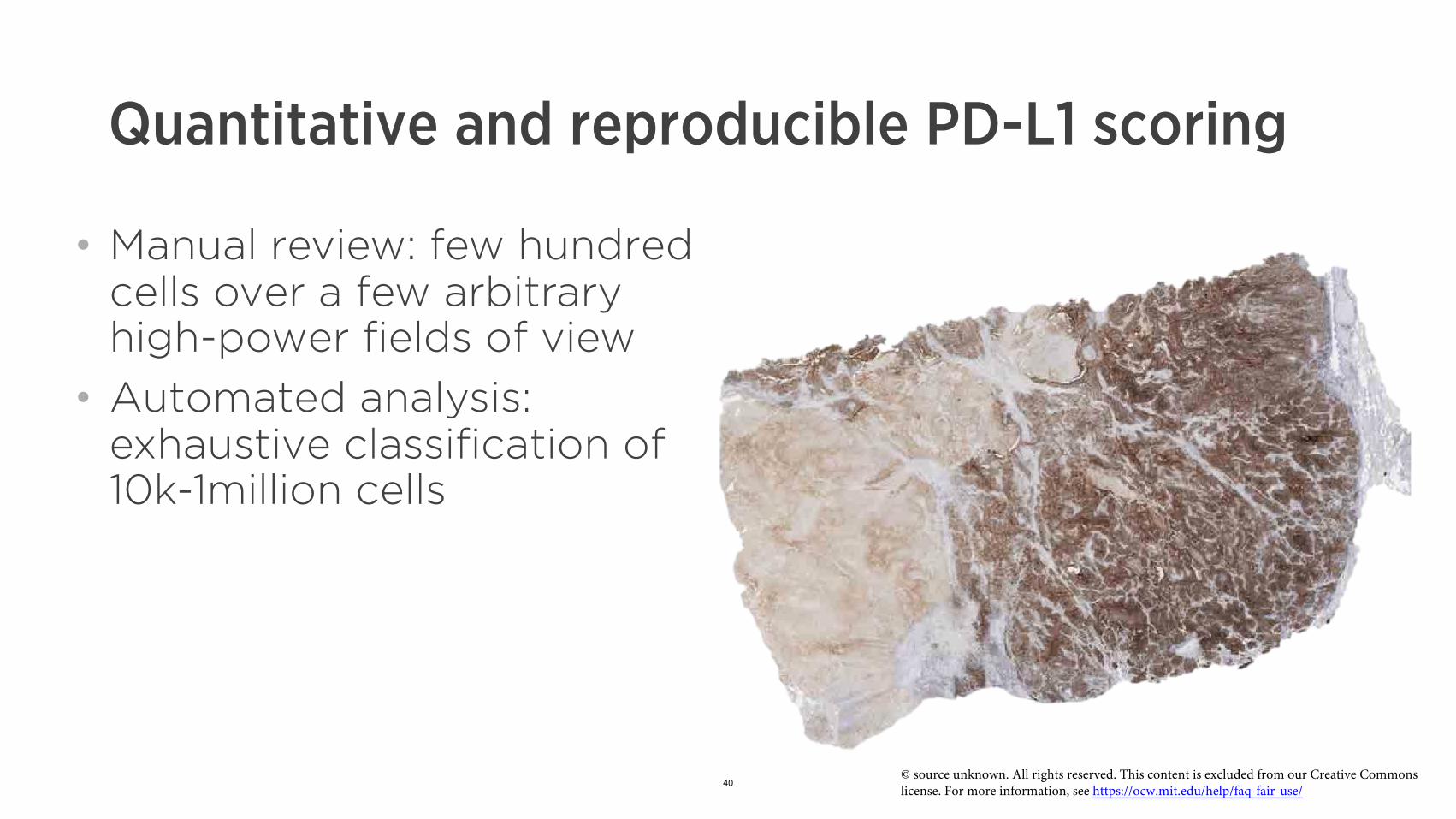
Quantitative and reproducible PD-L1 scoring
• Manual review: few hundred cells over a few arbitrary high-power fields of view
• Automated analysis: exhaustive classification of 10k-1million cells
40© source unknown. All rights reserved. This content is excluded from our Creative Commons license. For more information, see https://ocw.mit.edu/help/faq-fair-use/
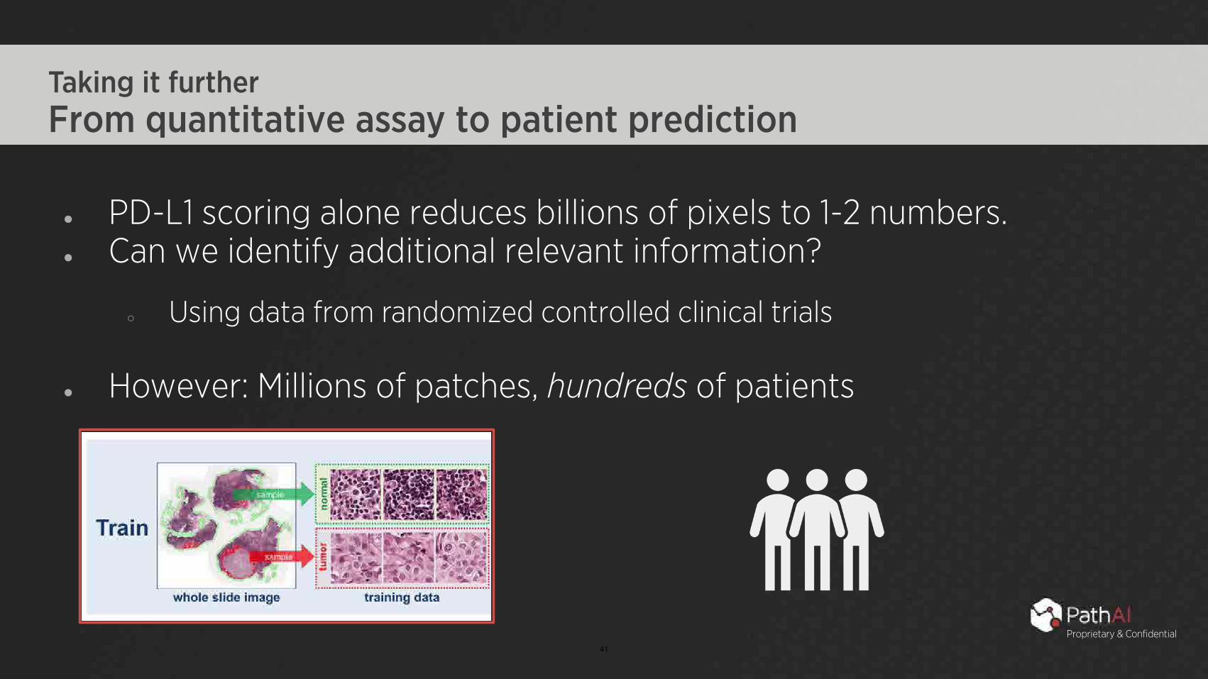
Taking it further From quantitative assay to patient prediction
● PD-L1 scoring alone reduces billions of pixels to 1-2 numbers. ● Can we identify additional relevant information?
○ Using data from randomized controlled clinical trials
● However: Millions of patches, hundreds of patients
41
Proprietary & Confidential

Proprietary & Confidential
Predictive features guided by biomedical priors © source unknown. All rights reserved. This content is excluded from our Creative Commons license. For more information, see https://ocw.mit.edu/help/faq-fair-use/H & E slide matching PD-L1 slide
42
Proprietary & Confidential
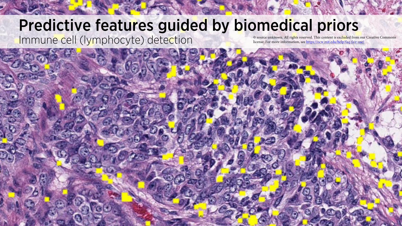
Predictive features guided by biomedical priors © source unknown. All rights reserved. This content is excluded from our Creative Commons license. For more information, see https://ocw.mit.edu/help/faq-fair-use/Immune cell (lymphocyte) detection
43

Proprietary & Confidential
Predictive features guided by biomedical priors© source unknown. All rights reserved. This content is excluded from our Creative Commons license. For moreCancer epithelium (red) and stroma (green) segmentation information, see https://ocw.mit.edu/help/faq-fair-use/
44

Proprietary & Confidential
Predictive features guided by biomedical priors © source unknown. All rights reserved. This content is excluded from our Creative Commons license. For more information, see https://ocw.mit.edu/help/faq-fair-use/Epithelial-stromal interface definition
45

Cell-type specific, tissue context-aware IHC-quantification
cell
license. For more information, see https://ocw.mit.edu/help/faq-fair-use/
Lymphocyte Macrophage
Cancer epithelial
© source unknown. All rights reserved. This content is excluded from our Creative Commons 46

Data-driven identification of pathological phenotypes associated with drug response
Total number of macrophages in epithelial/stroma interface (80um)
Total number of macrophages in epithelial/stroma interface (120um)
Total number of macrophages in invasive margin (250um)
Total number of lymphocytes in epithelial/stromal interface on H&E stain
Total number of plasma cells in epithelium on H&E stain
Total number of plasma cells in stroma on H&E stain
Tumor (epithelium + stroma) area on H&E stain
Total number of plasma cells in epithelial/stroma interface (40um)
Total number of plasma cells in epithelial/stroma interface (80um)
Area (mm2) of epithelial/stroma interface (80um) target positive cancer cells on target stain
Area (mm2) of epithelial PDL-1 positive macrophages on target stain
Necrosis area on target stain
Proportion of tumor infiltrating lymphocytes engaged by target positive macrophages
Stroma area on target stain
Tissue area on target stain 47

Multivariate models predictive of IO response • Low n, interpretability and measures of uncertainty valuable:
• No deep learning (gasp!) • Feature importance/selection in these models can provide disease insight
• Now we’re doing things pathologists can’t rather than automating / improving what they already can
Note: KM curves for illustration only
48

How do we know these features are correct? © source unknown. All rights reserved. This content is excluded from our Creative Commons license. For more information, see https://ocw.mit.edu/help/faq-fair-use/
Frames
Validation by exhaustive consensus
Proprietary & Confidential
49
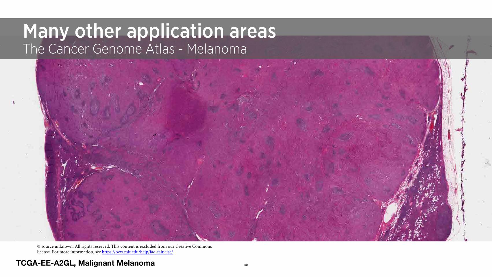
Many other application areas The Cancer Genome Atlas - Melanoma
© source unknown. All rights reserved. This content is excluded from our Creative Commons license. For more information, see https://ocw.mit.edu/help/faq-fair-use/
TCGA-EE-A2GL, Malignant Melanoma 50

Melanoma Tissue Map
Tumor Stroma Tumor Epithelium © source unknown. All rights reserved. This content is excluded from our Creative Commons license. For more information, see https://ocw.mit.edu/help/faq-fair-use/
TCGA-EE-A2GL, Malignant Melanoma 51

Melanoma Cell Map
Lymphocytes: Green Macrophages: Orange Plasma Cells: Blue Fibroblasts: Yellow Melanoma Cells: Red
© source unknown. All rights reserved. This content is excluded from our Creative Commons license. For more information, see https://ocw.mit.edu/help/faq-fair-use/
TCGA-EE-A2GL, Malignant Melanoma 52

Exhaustive analysis of cellular features in TCGA to enable data-driven identification of pathological predictors of survival in malignant melanoma
Pathological phenotypes with FDR < 5% for association with Increased area of stromal plasma cells associated with Progression Free Survival improved survival in melanoma
53

Gene Correlation REC8 0.57 GPR174 0.53 CD38 0.53 LAX1 0.53 TOX 0.53 AKAP5 0.53 C8orf80 0.52 JSRP1 0.52 IGJ 0.52 TNFRSF17 0.51 EAF2 0.51
Data-driven identification of transcriptional signature underlying stromal area of plasma cells in melanoma
Top-ranking transcripts associatedwith stromal area of plasma cells
Spea
rman
Cor
rela
tion
with
Str
omal
Pla
sma
Cel
l Are
a
Transcript Ranking 54

Stromal plasma cell area RNA signature strongly enriched for immune genes
Gene Set Name REACTOME_IMMUNE_SYSTEM REACTOME_ADAPTIVE_IMMUNE_SYSTEM PID_TCR_PATHWAY REACTOME_IMMUNOREGULATORY_INTERACTIONS_BETWEEN_A_ LYMPHOID_AND_A_NON_LYMPHOID_CELL KEGG_PRIMARY_IMMUNODEFICIENCY PID_IL12_2PATHWAY PID_CD8_TCR_PATHWAY KEGG_CELL_ADHESION_MOLECULES_CAMS KEGG_CYTOKINE_CYTOKINE_RECEPTOR_INTERACTION KEGG_INTESTINAL_IMMUNE_NETWORK_FOR_IGA_PRODUCTION REACTOME_TCR_SIGNALING REACTOME_PD1_SIGNALING
REACTOME_COSTIMULATION_BY_THE_CD28_FAMILY
FDR q-Description value
Genes involved in Adaptive Immune System Genes involved in Immune System 7.62E-57
6.02E-42
Genes involved in Immunoregulatory interactions between a Lymphoid and a non-Lymphoid cell
TCR signaling in naive CD4+ T cells 4.24E-30
6.07E-26
IL12-mediated signaling events Primary immunodeficiency 7.98E-24
9.27E-24
Cell adhesion molecules (CAMs) TCR signaling in naive CD8+ T cells 9.27E-24
3.00E-22
Intestinal immune network for IgA production Cytokine-cytokine receptor interaction 6.38E-22
3.37E-21
Genes involved in PD-1 signaling Genes involved in TCR signaling 3.24E-20
3.44E-19 Genes involved in Costimulation by the CD28 family 5.48E-19
55
© source unknown. All rights reserved. This content is excluded from our Creative Commons license. For more information, see https://ocw.mit.edu/help/faq-fair-use/
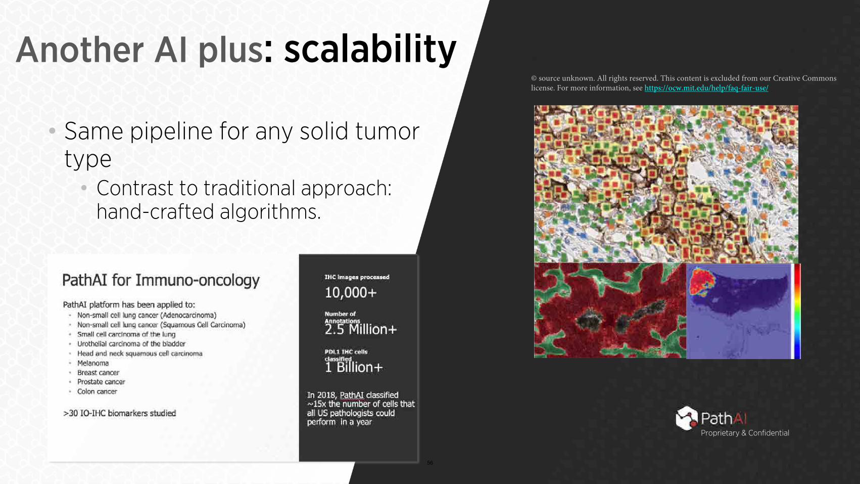
Another AI plus: scalability
• Same pipeline for any solid tumor type • Contrast to traditional approach:
hand-crafted algorithms.
56
Proprietary & Confidential

Extensive Slide Search & Data Standardization
57
Proprietary & Confidential

© source unknown. All rights reserved. This content is excluded from our Creative Commons license. For more information, see https://ocw.mit.edu/help/faq-fair-use/
Proprietary & Confidential
Automated quality control Folded /damaged
tissue Blurred areas
Debris
© source unknown. All rights reserved. This content is excluded from our Creative Commons license. For more information, see https://ocw.mit.edu/help/faq-fair-use/
58

Annotate, train and deploy task-specific models • Determined by partner needs
© source unknown. All rights reserved. This content is excluded from our Creative Commons license. For more information, see https://ocw.mit.edu/help/faq-fair-use/
Proprietary & Confidential 59
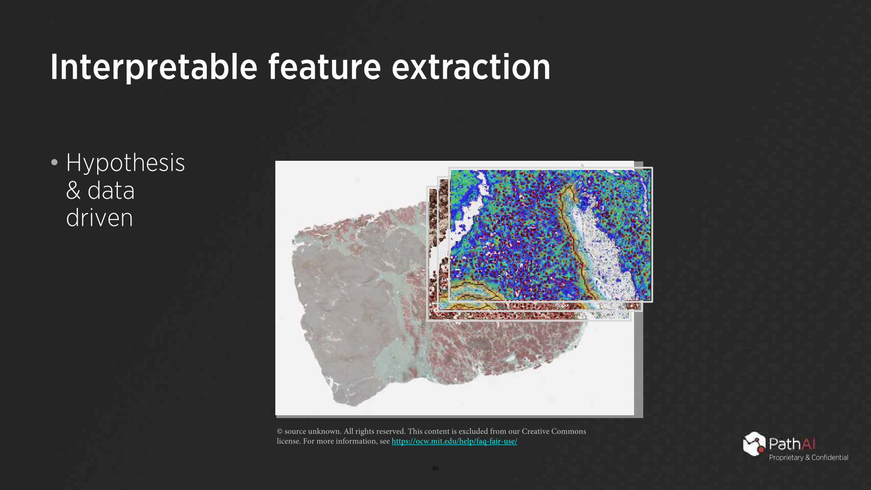
Interpretable feature extraction
• Hypothesis & data driven
© source unknown. All rights reserved. This content is excluded from our Creative Commons license. For more information, see https://ocw.mit.edu/help/faq-fair-use/
Proprietary & Confidential 60

Interactive Reports & Live Project Progress
Proprietary & Confidential
66 61

The PathAI Deep Learning Process
Transmit training data securely to the PathAI
cloud
Whole-Slide Images + Data
Over 200 relevant features extracted,
measured and analyzed
Deep Learning Feature Analysis Annotations
Network of board-certified pathologists
to provide ground truth consensus
Cell detection, tissue & region classification
Deep Learning Analysis Assay Validated
Identified features of significance reduced to
practice
We can execute process in 4 – 8 weeks for new assays
Assay Deployed
Analyze samples, quantified & visual results delivered
62
Proprietary & Confidential

AI in medicine Some closing thoughts • ML in the real world:
• Building the right dataset is 75% of the challenge
• Modern ML: engineering and empirical science • Rigorous validation is key
• Ideas and algorithms vs. teams and infrastructure
63
Proprietary & Confidential
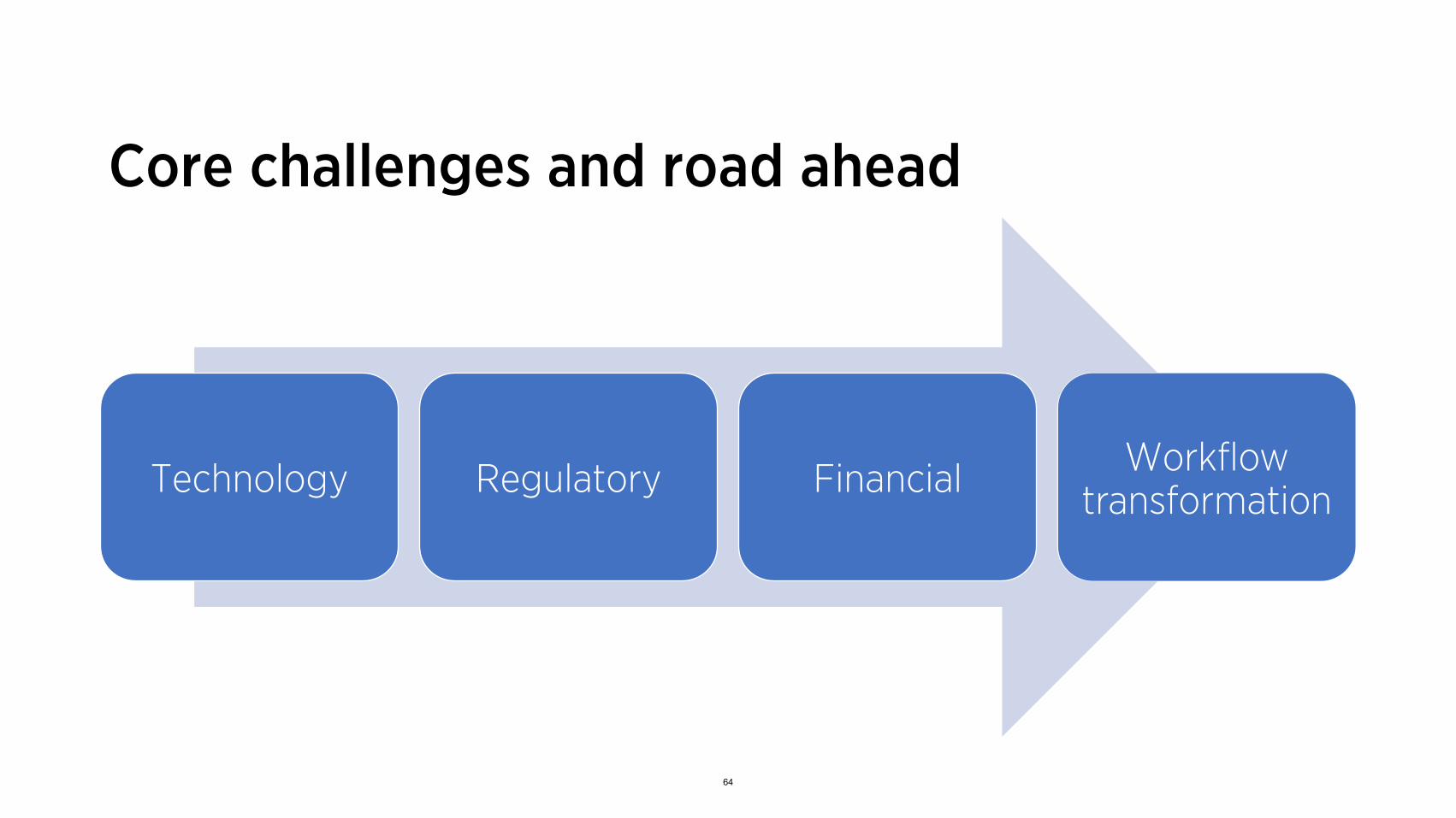
Core challenges and road ahead
Technology Regulatory Financial
64
Workflow transformation

Key Takeaways • Researchers have been working on AI for pathology for
~30 years • In the past 5 years, advances in: • Availability of digital data • Access to large-scale computing resources • Major algorithmic advances (e.g., Deep CNNs)
• AI works extremely well when these 3 factors are all available and fails when they are not
65

Key Takeaways • AI-powered pathology is broadly applicable across all image-
based tasks in pathology and enables integration with other structured data types (e.g., ‘Omics) • As AI and digital pathology are incorporated into clinical
workflow, they will offer significant operational and efficiency advantages • AI will drive improvements in the accuracy and predictiveness
of pathology leading to research advances and improved care for patients
66

“In the Future…” (1987)
• “Integrated information systems, patient care management by exception, decision support tools, and, in the future, "artificial intelligence" assists can all be expected to become staples of pathology practice, especially impacting those pathologists who choose to be responsive to the new practice milieu of medical information science.”
“Using the computer to optimize human performance in health care delivery. The pathologist as medical information specialist.”
(Arch Pathol Lab Med. 1987) 67

MIT OpenCourseWare https://ocw.mit.edu
6.S897 / HST.956 Machine Learning for Healthcare Spring 2019
For information about citing these materials or our Terms of Use, visit: https://ocw.mit.edu/terms
68