
6.1 Evolution and Roles of Senses
105
6.1 Evolution and Roles of Senses ▪ Sensory cells • Organisms must gather information about their environment and internal state • Sensory cells respond to specific stimuli • Transduction is the conversion of the energy of a stimulus into electrical energy • Linked to the opening and closing of gated ion channels • Sensory cells arose long before the evolution of the nervous system (e.g. paramecium respond to a variety of environmental stimuli)
Transcript of 6.1 Evolution and Roles of Senses

6.1 Evolution and Roles of Senses
▪ Sensory cells
• Organisms must gather information about their environment and internal state
• Sensory cells respond to specific stimuli
• Transduction is the conversion of the energy of a stimulus into electrical energy• Linked to the opening and closing of gated ion
channels
• Sensory cells arose long before the evolution of the nervous system (e.g. paramecium respond to a variety of environmental stimuli)

6.1 Evolution and Roles of Senses
▪ Sensors are categorized by the specific modalities to which they respond.
• Mechanoreceptors -- mechanical energy (e.g. touch and pressure)
• Chemoreceptors -- specific chemicals• Thermoreceptors -- heat and cold• Photoreceptors -- photic energy• Electroreceptors -- electric fields• Magnetoreceptors -- magnetic fields
• Nociceptors (pain receptors) respond to tissue damage; may be chemoreceptors or mechanoreceptors

6.1 Evolution and Roles of Senses
▪ Primary roles of receptor cells
• Interoreceptors detect information about internal body fluids crucial to homeostasis• In blood vessels and gut fluids
• Proprioceptors monitor body movementand position• In muscles, tendons and joints
• Exteroreceptors detect external stimuli• Somesthetic senses arise from body surface• Special senses -- highly localized, specialized
senses in distinct sensory organs

6.1 Evolution and Roles of Senses
▪ Perception is an animal’s interpretation of the external world
• Sensors detect a limited number of energy forms
• Stimuli are filtered during precortical processing
• Data are further manipulated by the cerebral cortex
• Optical illusions can make objects look smaller or larger than they are (ex. great bowerbird nests)

6.1 Evolution and Roles of Senses

6.2 Receptor Cell Physiology
▪ Doctrine of specific nerve energies
• Each type of receptor is specialized to respond to one type of stimulus (adequate stimulus)• Example: The adequate stimulus for
photoreceptors in the eye is light
• Even when activated by a different stimulus, the sensation is the one usually detected by that receptor type

6.2 Receptor Cell Physiology
▪ Receptor potential
• Stimulation of receptor opens gated Na+ channels• Inward flux of Na+ depolarizes the receptor
membrane• Receptor potential in receptor cells• Generator potential in afferent neurons
• Receptor potential is graded -- the greater the stimulus, the larger the receptor potential
• Receptor potential must be converted into action potentials for long-distance transmission
6.2 Receptor Cell Physiology
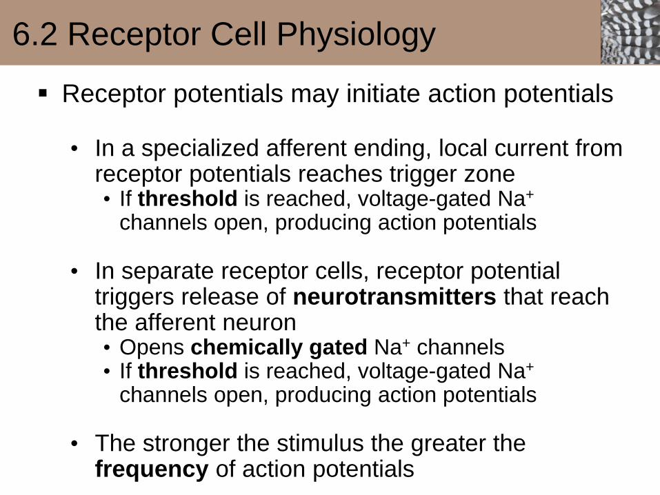
▪ Receptor potentials may initiate action potentials
• In a specialized afferent ending, local current from receptor potentials reaches trigger zone• If threshold is reached, voltage-gated Na+
channels open, producing action potentials
• In separate receptor cells, receptor potential triggers release of neurotransmitters that reach the afferent neuron• Opens chemically gated Na+ channels• If threshold is reached, voltage-gated Na+
channels open, producing action potentials
• The stronger the stimulus the greater the frequency of action potentials

6.2 Receptor Cell Physiology

6.2 Receptor Cell Physiology
6.2 Receptor Cell Physiology
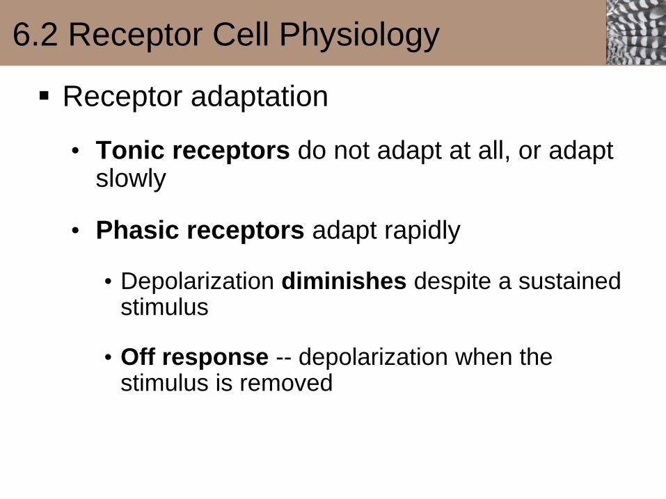
▪ Receptor adaptation
• Tonic receptors do not adapt at all, or adapt slowly
• Phasic receptors adapt rapidly
• Depolarization diminishes despite a sustained stimulus
• Off response -- depolarization when the stimulus is removed

6.2 Receptor Cell Physiology


http://www.boredpanda.com/new-crayfish-species-discovered-cherax-pulcher-christian-lukhaup-indonesia/


http://www.pkmedillus.com/portfolio/

https://kin450-neurophysiology.wikispaces.com/Muscle+Spindle

Therefore …… we are like crayfish in this regard

6.2 Receptor Cell Physiology
▪ Receptive fields
• Each sensory neuron responds to stimuli in a specific
area -- receptive field
• The smaller the receptive fields, the greater the
density of receptors• Smaller receptive fields produce greater acuity or
discriminative ability (e.g. fingertips)
• Amount of cortical representation on the sensory
homunculus corresponds with receptor density
• Strong signal in center of receptive field inhibits
pathways in fringe areas -- lateral inhibition

6.2 Receptor Cell Physiology

6.3 Mechanoreception: Touch, Pressure, and
Proprioception
▪ Touch and pressure mechanoreceptors in skin
• Pacinian corpuscle -- deep pressure
• Touch sensors -- highly sensitive, closer to skin surface
• Touch mechanoreceptors -- base of hairs or insect bristles

6.3 Mechanoreception: Touch, Pressure, and
Proprioception

Figure 6-8b p217
Sebaceous gland
Smooth
muscle
Keratinized
layerHair shaft
Living layer
Epidermis
Dermis
Hypodermis
Nerve fiberPacinian corpuscle Adipose cells
Hair follicle

6.3 Mechanoreception: Touch, Pressure, and
Proprioception
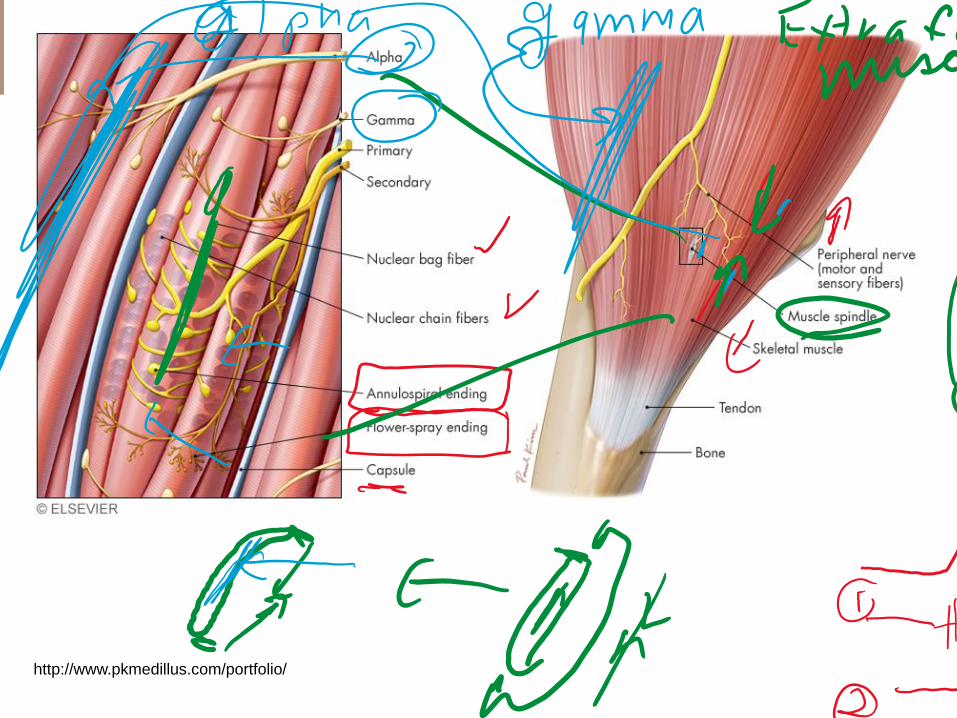
▪ Proprioceptors give information on body position and motion
• Stretch receptors include muscle spindles and Golgi tendon organs
• Statocysts are gravity receptors -- simplest organs of equilibrium
• Body movement tilts the statocyst• Statoliths move in direction of body movement,
bending sensory hairs• When sensory hairs are bent, mechanically gated
channels open and action potentials are generated

6.3 Mechanoreception: Touch, Pressure, and
Proprioception

6.3 Mechanoreception: Touch, Pressure, and
Proprioception
▪ Lateral line system in fishes
• Neuromast cells are arranged in a line along the length of the body
• Stereocilia are sensory transducers that protrude from sensory hair cells
• Can detect pressure waves set up by other fishes

6.3 Mechanoreception: Touch, Pressure, and
Proprioception

6.3 Mechanoreception: Touch, Pressure, and
Proprioception

6.3 Mechanoreception: Touch, Pressure, and
Proprioception
▪ Vestibular apparatus of vertebrate inner ears
• Semicircular canals detect rotational or angular acceleration or deceleration of the head• Receptive hair cells lie on a ridge in the ampulla• Semicircular canals are larger in primates that
swing in trees and flying vertebrates
• Otolith organs (utricle and saccule) provide information about the position of the head
• Signals from vestibular apparatus are carried through the vestibulocochlear nerve to the cerebellum and vestibular nuclei.

6.3 Mechanoreception: Touch, Pressure, and
Proprioception

Oval window
Saccule
Semicircular
canalsVestibular
nerveAuditory
nerve
Cochlea
Round window
Vestibular
apparatus
Utricle
Figure 6-12a p221
Endolymph
Perilymph
Ampulla

Hairs of hair cell;
kinocilium ( red )
and stereocilia ( blue )
Cupula
Hair cell
Support
cell
Vestibular
nerve fibers
Ridge in
ampulla
Figure 6-12b p221

Figure 6-12c p221
Kinocilium
Stereocilia

6.3 Mechanoreception: Touch, Pressure, and
Proprioception

6.3 Mechanoreception: Touch, Pressure, and
Proprioception

Otoliths
Gelatinous
layer
Hair cells
Supporting
cell
Sensory
nerve fiberFigure 6-14a p223
Kinocilium Stereocilia

6.3 Mechanoreception: Touch, Pressure, and
Proprioception

6.4 Mechanoreception: Ears and Hearing
▪ Sound travels as waves through a medium• Detected by mechanoreceptors• Ear is a complex organ of hearing
▪ Hearing in fishes• Lateral lines detect very-low-frequency sounds• Weberian apparatus -- transfers sound from gas
bladder to inner ear

6.4 Mechanoreception: Ears and Hearing
▪ External ear
• Vertebrates typically have two ears, allowing for localization of sound
• Tympanic membrane vibrates as sound hits• Insects have similar structures on their abdominal
segments or legs
• Amphibians and some reptiles have only a tympanic membrane
• Mammalian external ear consists of the pinna, external auditory meatus and tympanic membrane
• External ear is inconspicuous in birds

6.4 Mechanoreception: Ears and Hearing
▪ Middle ear
• Transfers vibrations of the tympanicmembrane to the inner ear
• Movable chain of three small bones (ossicles) in mammals• Evolved from jaw structures• Malleus is attached to the tympanic membrane• Incus is between the malleus and stapes• Stapes is attached to the oval window• Single ossicle (columella) in anuran amphibians,
reptiles and birds
• Reflex response of middle ear muscles tightens tympanic membrane during loud sound for protection

6.4 Mechanoreception: Ears and Hearing

Tympanic membrane
External
auditory
meatus
Middle ear
cavity
Round window
Stapes
at oval
window
Malleus Incus
Helicotrema
Cochlea
Organ of Corti (with hairs of
hair cells displayed on surface)
Tectorial membrane
Scala media (cochlear
duct)
Basilar membrane
Scala tympani
Scala vestibuli
Figure 6-19a p228
Vestibular membrane

Auditory nerve
Scala vestibuli
Basilar
membrane
Scala
media
(cochlear
duct)
Tectorial membrane
Scala tympani
Figure 6-19b p228
Vestibular membrane

6.4 Mechanoreception: Ears and Hearing
▪ Inner ear
• Cochlea is a coiled tubular system with three fluid-filled longitudinal compartments• Scala vestibuli (upper) -- contains perilymph• Scala media or cochlear duct (middle) -- contains
endolymph• Scala tympani (lower) -- contains perilymph
• Organ of Corti is the sense organ for hearing• On top of basilar membrane in the floor of the
cochlear duct• 15,000 hair cells arranged in four parallel rows• Inner row of hair cells transform cochlear fluid
vibration into action potentials

Figure 6-20a p230
Cochlear duct
Vestibular
membraneScala
vestibuli
Tectorial
membraneIncus
Malleus Oval
windowCochlea
Helicotrema 1Hairs
Perilymph
Organ
of CortiStapesPerilymph Basilar
membrane
Scala
tympani
Tympanic
membrane
Round
window
Fluid movement within the perilymph set up by vibration of the oval
window follows two pathways:

Basilar membranewith organ of Cortiand its hair cells
Fluid movements in thecochlea cause deflectionof the basilar membrane.
The stereocilia (hairs) from the hair cells of the basilar membranecontact the overlying tectorial membrane. These hairs are bentwhen the basilar membrane is deflected in relation to thestationary tectorial membrane. This bending of the inner haircells’ hairs opens mechanically gated channels, leading to ionmovements that result in a receptor potential.
Tectorialmembrane
Hair cell
Figure 6-19c p228
Outer hair
cells

6.4 Mechanoreception: Ears and Hearing

6.5 Chemoreception: Taste and Smell
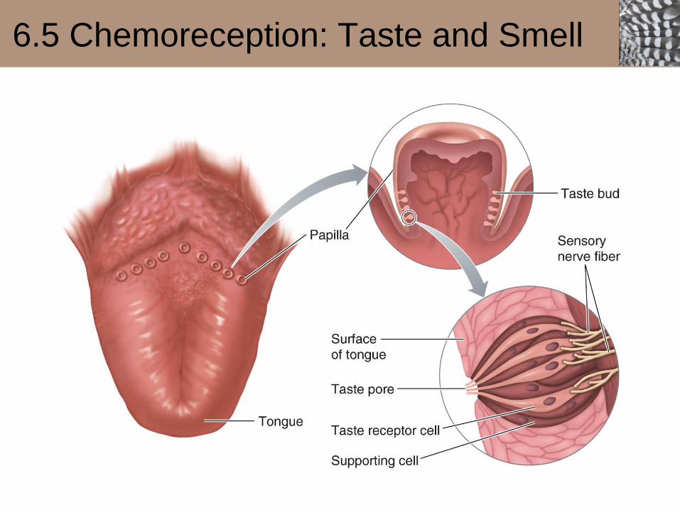
▪ Chemoreceptors for taste (gustatory) sensation
• Each mammalian taste bud has about 50 receptor cells, supporting cells and a taste pore• Only chemicals in solution can evoke taste
sensation
• Microvilli contain chemoreceptors
• Binding of tastant with receptor cell alters ion channels to produce a depolarizing receptor potential
• Action potentials are carried to the cortical gustatory area (parietal lobe), hypothalamusand limbic system
6.5 Chemoreception: Taste and Smell

6.5 Chemoreception: Taste and Smell
▪ Primary tastes
• Salty (sodium)• Direct entry of Na+ ions through channels in receptor
cell membrane
• Sour (acid)• H+ blocks K+ efflux from cell
• Sweet (sugar)• G-protein-coupled receptor stimulates cAMP or IP3
pathway
• Bitter (plant alkaloids)• Variety of G-protein-coupled receptor mechanisms
• Umami (savory)• Glutamate binds to G-protein-coupled receptor

6.5 Chemoreception: Taste and Smell

6.5 Chemoreception: Taste and Smell
▪ Chemoreceptors for olfactory (smell) sensation
• Olfactory mucosa in nasal fossae contains olfactory receptors, supporting cells and basal cells
• Olfactory afferent neurons are the only mammalian neurons that undergo cell division
• Each receptor responds to only one discrete component of an odor
• Odorant binds to G-protein-coupled receptor

6.5 Chemoreception: Taste and Smell

6.5 Chemoreception: Taste and Smell
▪ Olfactory processing
• Afferent fibers synapse on mitral cells in glomeruli of the olfactory bulb• Glomeruli serve as “smell files”, each detecting one
particular odor component
• Mitral cells refine smell signals and relay them to the brain• Subcortical route to primary olfactory cortex in
lower medial temporal lobe associated with memory and behavior
• Thalamic-cortical route permits conscious perception and fine discrimination of smell
• Cortex can distinguish 20,000 different scents from 1,000 or fewer different receptor proteins

6.5 Chemoreception: Taste and Smell

6.5 Chemoreception: Taste and Smell

6.5 Chemoreception: Taste and Smell
▪ Vomeronasal organ (VNO)
• In noses of mammals and reptiles
• Governs reproductive and social behaviors by reception of pheromones
• Pheromones are volatile chemical messengers released into the environment for intraspecies communication

6.6 Photoreception: Eyes and Vision
▪ Light sensing organs
• Eyespots• Less than 100 photoreceptor cells lining an open cup• Permits animal to locate a light source• Platyhelminthes, Cnidarians, and Echinoderms
• Pinhole eye• Size of cup aperture is reduced• Permits formation of an image
• Camera eye• Lens enhances light-gathering power• Many phyla, including vertebrates and cephalopods
• Compound eye• Densely packed units (ommatidia), each having its own
lens and photoreceptors• Arthropods

6.6 Photoreception: Eyes and Vision

Figure 6-27d p240

Figure 6-27e p240

6.6 Photoreception: Eyes and Vision

6.6 Photoreception: Eyes and Vision
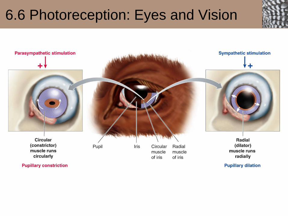
▪ The iris controls the amount of light entering the eye.
• Iris is a pigmented ring of smooth muscle
• Round central opening is the pupil
• Circular muscle constricts pupil in response to light
• Radial muscle increases pupil size in dim light
• Iris muscles are controlled by the autonomic nervous system• Parasympathetic fibers innervate circular muscle• Sympathetic fibers innervate radial muscle
6.6 Photoreception: Eyes and Vision

6.6 Photoreception: Eyes and Vision

6.6 Photoreception: Eyes and Vision
▪ Light is focused on the retina by adjusting the strength of the lens (accommodation)
• Convex surfaces of the cornea and lens determine the eye’s refractive ability
• Curvature of the lens is adjusted by the ciliarymuscle in mammals, birds and some reptiles
• In fish, the lens is moved back and forth to focus, due to the lens’ fixed focal length
• Some annelids alter the distance between the lens and photoreceptors by changing the fluid volumeof the optic chamber.

6.6 Photoreception: Eyes and Vision
6.6 Photoreception: Eyes and Vision
▪ Structure of the retina
• Three layers of neurons• Light must pass through the ganglion and bipolar
layers before reaching the photoreceptors
• A layer of reflecting material (tapetum lucidum)
enhances vision in dim light in some species
• Fovea is the point of greatest visual acuity
• Axons of ganglion cells form the optic nerve• Region where optic nerve exits the eye (optic disc)
is the blind spot

6.6 Photoreception: Eyes and Vision

6.6 Photoreception: Eyes and Vision

6.6 Photoreception: Eyes and Vision
▪ Photoreceptors
• Outer segment detects the light stimulus• Stacked, flattened, membranous discs
containing photo-pigment molecules• Rods and cones are named for their shapes
• Inner segment contains the metabolic machinery of the cell
• Synaptic terminal lies closest to the eye’s interior

6.6 Photoreception: Eyes and Vision

6.6 Photoreception: Eyes and Vision
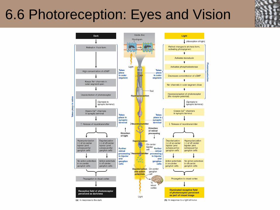
▪ Photoreceptors are electrically active in the
dark
• In the absence of light, cyclic GMP
concentration is high in photoreceptors
• Na+ channels are open ----> depolarization
• Ca2+ channels in synaptic terminal remain
open
• Glutamate is released

6.6 Photoreception: Eyes and Vision
▪ Phototransduction
• In the presence of light, a retinene molecule absorbs a
photon
• Retinine changes shape from cis to trans conformation
• Triggers enzymatic activity of opsin
• Activates a G protein called transducin
• Phosphodiesterase degrades cyclic GMP, causing Na+
channels to close
• Hyperpolarizing receptor potential reduces glutamate
release
6.6 Photoreception: Eyes and Vision

6.6 Photoreception: Eyes and Vision
▪ Rods
• 20 times more rods than cones in human eye• Most abundant in periphery of retina• High sensitivity to light• Rhodopsin absorbs all visible wavelengths with a
peak around 500 nm• Vision is in shades of gray
▪ Cones
• Most abundant in the macula/fovea regions• Lower sensitivity to light• Small receptive fields lead to highly detailed vision• Scotopsins respond to different wavelengths and
provide color vision (red, green, yellow, blue and ultraviolet)
• Primates have three cone types

6.6 Photoreception: Eyes and Vision


6.6 Photoreception: Eyes and Vision
▪ Dark adaptation
• Pupils dilate
• Photopigments broken down during light exposure
gradually regenerate
• Increased sensitivity of rods to light
• Night blindness is caused by dietary deficiency of
vitamin A (retinene is a derivative of vitamin A)
▪ Light adaptation
• Pupils constrict
• Rhodopsin rapidly breaks down
• Decreased sensitivity of rods to light

6.6 Photoreception: Eyes and Vision
▪ Visual pathways
• Image detected on the retina is upside down andbackward
• Light rays from left half of visual field fall on the right halfof the retina
• At the optic chiasm, fibers from medial half of each retina cross over, while fibers from the lateral half remain on the same side
• Information from each half of the visual field is brought together on the opposite side of the brain
• Optic tracts project to the lateral geniculate nucleus of the thalamus
• Fibers terminate in the visual cortex in the occipital lobe

6.6 Photoreception: Eyes and Vision

Figure 6-40a p255

6.6 Photoreception: Eyes and Vision
▪ Cephalopod eyes have cornea, lens, and retina
• Light-sensing cells are on top of neural cells,
receiving light directly from the lens
• Optic nerve exits from back side (no blind spot)
• Some cuttlefish see polarized light
▪ Compound eyes of arthropods consist of
multiple image-forming units (ommatidia)
• Lower visual acuity than vertebrate eyes
• Rhabdomeric photoreceptors use rhodopsin, but
depolarize in response to light

6.6 Photoreception: Eyes and Vision

Figure 6-42 p256
Eye formation in
cephalopods
Eye formation in
vertebrates
Epidermis
Eye placode Neural plate Retinal bulge
Lens foldLens anlagen
Optic vesicalRetinal anlagen
Brain
Migrating lens cellsLens placode
Retinal anlagen
Corneal fold Iris fold
Lens Optic cup
Cornea
Pigment epithelium
Iris
Lens
Retina
Photoreceptors
Optic nerve

6.6 Photoreception: Eyes and Vision

Figure 6-43a p257
Lateral
simple eye
Lateral
compound eye
Median
simple eye
(a) Limulus polyphemus

Figure 6-43b p257
Lens
Photoreceptors
Axon of
eccentric cell
(b) Compound eye

Figure 6-43c p257
Light
Lens Retinular cell
(photoreceptor cell)
Dendrite of eccentric
cell
Rhabdomere of
retinular cell
Eccentric cell
(c) Single ommatidium

http://www.scholarpedia.org/article/File:Photoreceptor_and_LM
C_responses.jpg
A compound eye is
characterized by a
variable number (a few
to thousands) of small
eyes, ommatidia.
DEPOLARIZATION
of sensory neuron
with light

6.7 Thermoreception
▪ Warm and cold thermoreceptorsrespond to changes in skin temperature
• Heat-gated and cold-gated ion channels• Used primarily for thermoregulation
▪ Infrared thermoreceptors
• Located in small pits in skin of pit vipers, pythons and boas
• Detect warm mammalian prey• Used in first strike to capture prey

6.7 Thermoreception

Figure 6-45a p259

Figure 6-45b p259
6.8 Nociception: Pain
▪ Categories of pain receptors (nociceptors)
• Mechanical nociceptors -- respond to cutting, crushing or pinching
• Thermal nociceptors -- respond to temperature extremes
• Polymodal nociceptors -- respond to all kinds of damaging stimuli, including chemical

6.8 Nociception: Pain
▪ Fast pain
• Initial pain response arises from mechanical or thermal nociceptors
• Easily localized • Transmitted rapidly over large, myelinated A-delta
fibers• Fast adapting
▪ Slow pain
• Dull, aching sensation arises from nociceptors activated by chemicals (e.g. bradykinin)
• Poorly localized• Transmitted more slowly by small, unmyelinated
C fibers• Slow adapting
6.8 Nociception: Pain
▪ Prostaglandins• Enhance nociceptor response• Synthesis is blocked by aspirin and other
analgesic drugs
▪ Substance P• Neurotransmitter that activates ascending pain
pathways
▪ Glutamate• Generates action potentials in dorsal horn
interneurons• Increases excitability of dorsal horn cells• Exaggerated sensitivity of an injured area to
stimuli

6.8 Nociception: Pain

6.8 Nociception: Pain
▪ Mammals have a built-in analgesic system.
• Regulated at the spinal cord level by neurons originating in periaqueductal gray matter in brainstem
• Suppress release of substance P by presynaptic inhibition
• Endogenous opioids (endorphins, enkephalins, dynorphin) bind to opiate receptors as a natural analgesic system
• Morphine produces analgesia through its action on opiate receptors

6.8 Nociception: Pain

6.9 Electroreception and Magnetoreception
▪ Passive electroreception
• Ampullary electroreceptors in fishes and some amphibians respond to low-frequency electric signals
• Used to locate prey (electrolocation)
▪ Active electroreception
• Electric organs emit electric organ discharges(EODs)
• Tuberous electoreceptors receive the feedback signal
• Used in electrolocation and electrocommunication• Electrosensory lateral line lobe (ELL) is organized
somatotopically

6.9 Electroreception and Magnetoreception

6.9 Electroreception and Magnetoreception
▪ Navigation by magnetic fields
• Many animals have an internal compass(e.g. migratory birds)
• Possible mechanisms of magnetoreception
• Magnetic induction -- sensitive electroreceptors of elasmobranches may detect magnetic fields
• Magnetic minerals -- magnetic crystals arranged in chains (magnetosomes) within the cell align with magnetic fields
• Magnetochemical reactions -- light absorption by cryptochromes (ancient photoreceptors) causes magnetically sensitive free-radical reactions