
document
4
Nature © Macmillan Publishers Ltd 1998 8 letters to nature 288 NATURE | VOL 391 | 15 JANUARY 1998 labelled h-SIE probe (59-CATTTCCCGTAAATC-39) 18 containing a protruding poly-G tail at both sense and antisense 59 ends. HGF-induced stimulation of transcriptional activities of the isolated c-fos (ref. 26) and waf-1 (ref. 27) promoters was measured by the Luciferase assay 23 . Synthesis of p62 c-fos and p21 waf-1 was measured by western blot with specific antibodies (gifts from C. Schneider). Cell ‘scattering’ in response to HGF (50 ng ml -1 ) was measured as described 1 . Micrographs (×1,000) were taken 6 h after stimulation. Stimulation of growth was assessed by 3 H-thymidine uptake 12 h after HGF stimulation of cells synchronized before electroporation by two cycles of thymidine (2 mM)- deoxycitidine (250 mM). Formation of branched tubular structures was mon- itored on cell monolayers, coated with a 0.5-mm layer of collagen G (2 mg ml -1 ) in the presence of HGF (10 ng ml -1 ) supplemented daily 1 . Phosphopeptides, decoys and electroporation. N-acetylated and C- amidated synthetic peptides, mimicking the consensus sequences for SH2- signal transducers in the HGF receptor or in the STAT family, were synthesized in their phosphorylated or non-phosphorylated forms by Fmoc chemistry, and purified by high-performance liquid chromatography (HPLC; .99%), as will be described elsewhere. For in vivo electroporation the phosphate group was substituted by the non-hydrolysable methylenphosphonate group. The h-SIE decoy (59-CATTTCCCGTAAATC-39) and its scrambled control 59- ACTCTTGCCAATTAC-39 were synthesized and used in the inhibition assay as described 28 . For electroporation, adherent cells were cultured onto 1-cm 2 indium-tin oxide conductive glass surfaces and electroporated in situ with a PBS solution containing either the competing peptides (1 mM) or the decoys (10 mM). The electric pulse was generated using the Epizap apparatus (Ask Science, Canada), following the technique described previously 29 . More than 90% of cells were permeabilized as tested by the uptake of Lucifer yellow, and viability was higher than 80% as judged by trypan blue exclusion. Received 4 August; accepted 6 October 1997 1. Medico, E. et al. The tyrosine kinase receptors Ron and Sea control ‘‘scattering’’ and morphogenesis of liver progenitor cells in vitro. Mol. Biol. Cell 7, 495–504 (1996). 2. Sachs, M. et al. Mitogenic and morphogenic activity of epithelial receptor tyrosine kinases. J. Cell Biol. 133, 1095–1107 (1996). 3. Ponzetto, C. et al. A multifunctional docking site mediates signalling and transformation by the Hepatocyte Growth Factor/Scatter Factor receptor family. Cell 77, 261–271 (1994). 4. Stocker, M., Gherardi, E., Perryman, M. & Gray, J. Scatter factor is a fibroblast-derived modulator of epithelial cell motility. Nature 327, 239–242 (1987). 5. Ridley, A. J., Comoglio, P. M. & Hall, A. Regulation of Scatter Factor/Hepatocyte Growth Factor responses by Ras, Rac, and Rho in MDCK cells. J. Cell Biol. 15, 1110–1122 (1995). 6. Royal, I. & Park, M. Hepatocyte Growth Factor-induced scatter of Madin–Darby canine kidney cells requires phosphatidylinositol 3-kinase. J. Biol. Chem. 270, 27780–27787 (1995). 7. Ponzetto, C. et al. Specific uncoupling of GRB2 from the Met receptor. J. Biol. Chem. 271, 14119– 14123 (1996). 8. Pawson, T. Protein modules and signalling networks. Nature 373, 573–580 (1995). 9. Pelicci, G. et al. The motogenic and mitogenic responses to HGF are amplified by the Shc adaptor protein. Oncogene 10, 1631–1638 (1995). 10. Weidner, K. M. et al. Interaction between Gab1 and the c-Met receptor tyrosine kinase is responsible for epithelial morphogenesis. Nature 384, 173–176 (1996). 11. Graziani, A., Gramaglia, D., Cantley, L. C. & Comoglio, P. M. The tyrosine-phosphorylated hepatocyte growth factor/scatter factor receptor associates with phosphatidylinositol 3-kinase. J. Biol. Chem. 266, 22087–22090 (1991). 12. Ponzetto, C. et al. A novel recognition motif for Phosphatidylinositol 3-kinase binding mediates its association with the Hepatocyte Growth Factor/Scatter Factor receptor. Mol. Cell Biol. 13, 4600–4608 (1993). 13. Graziani, A., Gramaglia, D., dalla Zonca, P. & Comoglio, P. M. Hepatocyte Growth Factor/Scatter Factor stimulates the Ras-guanine nucleotide exchanger. J. Biol. Chem. 268, 9165–9168 (1993). 14. Schindler, C. & Darnell, J. E. Jr Transcriptional responses to polypeptide ligands: the JAK-STAT pathway. Annu. Rev. Biochem. 64, 621–651 (1995). 15. Ihle,J. N. STATs: signal transducers and activators of transcription. Cell 84, 221–334 (1996). 16. Leaman, D. W. et al. Roles of JAKs in activation of STATs and stimulation of c-fos gene expression by Epidermal Growth Factor. Mol. Cell. Biol. 16, 369–375 (1996). 17. Silvennoinen, O., Ihle, J. N., Schlessinger, J. & Levy, D. E. Interferon-induced nuclear signalling by Jak protein tyrosine kinases. Nature 366, 583–585 (1993). 18. Wagner, B. J., Hayes, T. E., Hoban, C. J. & Cochran, B. H. The SIF binding element confers sis/PDGF inductibility onto the c-fos promoter. EMBO J. 9, 4477–4484 (1990). 19. Durbin, J. E., Hackenmiller, R., Simon, M. C. & Levy, D. E. Targeted disruption of the mouse Stat1 gene results in compromised innate immunity to viral disease. Cell 84, 443–450 (1996). 20. Liu, X. et al. Stat5a is mandatory for adult mammary gland development and lactogenesis. Genes Dev. 11, 179–186 (1967). 21. Takeda, K. et al. Targeted disruption of the mouse Stat3 gene leads to early embryonic lethality. Proc. Natl Acad. Sci. USA 94, 3801–3804 (1997). 22. Chin, Y. E. et al. Cell growth arrest and induction of cyclin-dependent kinase inhibitor p21 WAF1/CIP1 mediated by STAT1. Science 272, 719–722 (1996). 23. Boccaccio, C., Gaudino, G., Gambarotta, G., Galimi, F. & Comoglio, P. M. Hepatocyte Growth factor (HGF) receptor expression is inducible and is part of the delayed-early response to HGF. J. Biol. Chem. 269, 12846–12851 (1994). 24. Lucibello, F. C., Lowag, C., Neuberg, M. & Muller, R. Trans-repression of the mouse c-fos promoter: a novel mechanism of Fos-mediated trans-regulation. Cell 59, 999–1007 (1989). 25. Giordano, S., Ponzetto, C., Di Renzo, M. F., Cooper, C. S. & Comoglio, P. M. Tyrosine kinase receptor indistinguishable from the c-Met protein. Nature 339, 155–156 (1989). 26. Gilman, M. Z., Wilson, R. N. & Weinberg, R. A. Multiple protein binding sites in the 59-flanking region regulate c-fos expression. Mol. Cell. Biol. 6, 4305–4316 (1986). 27. El-Deiry, W. S. et al. WAF-1, a potential mediator of p53 tumor suppression. Cell 75, 817–825 (1993). 28. Gambarotta, G. et al. Ets up-regulates MET transcription. Oncogene 13, 1911–1917 (1996). 29. Raptis, L. H., Liu, S. K. W., Firth, K. L., Stiles, C. D. & Alberta, J. A. Electroporation of peptides into adherent cells in situ. Biotechniques 19, 104–114 (1995). 30. Comoglio, P. M. et al. Detection of phosphotyrosine containing proteins in the detergent insoluble fraction of RSV-transformed fibroblasts by azobenzene phosphonate antibodies. EMBO J. 3, 483–489 (1984). Acknowledgements. We thank P. Giordano (Pharmacia & Upjohn) for peptide synthesis; C. Ponzetto for discussions; A. Cignetto for secretarial help; and E. Wright for help with the manuscript. This work was supported by an AIRC grant to P.M.C. M.A. is recipient of a FIRCFellowship. Correspondence and requests for materials should be addressed to C. Boccaccio (e-mail: cboccaccio@hal. ircc.polito.it). DNA shuffling of a family of genes from diverse species accelerates directed evolution Andreas Crameri, Sun-Ai Raillard, Ericka Bermudez & Willem P. C. Stemmer Maxygen Inc., 3410 Central Expressway, Santa Clara, California 95051, USA ......................................................................................................................... DNA shuffling is a powerful process for directed evolution, which generates diversity by recombination 1,2 , combining useful muta- tions from individual genes. Libraries of chimaeric genes can be generated by random fragmentation of a pool of related genes, followed by reassembly of the fragments in a self-priming poly- merase reaction. Template switching causes crossovers in areas of sequence homology. Our previous studies used single genes and random point mutations as the source of diversity 3–6 . An alter- native source of diversity is naturally occurring homologous genes, which provide ‘functional diversity’. To evaluate whether natural diversity could accelerate the evolution process, we com- pared the efficiency of obtaining moxalactamase activity from four cephalosporinase genes evolved separately with that from a mixed pool of the four genes. A single cycle of shuffling yielded eightfold improvements from the four separately evolved genes, versus a 270- to 540-fold improvement from the four genes shuffled together, a 50-fold increase per cycle of shuffling. The best clone contained eight segments from three of the four genes as well as 33 amino-acid point mutations. Molecular breeding by shuffling can efficiently mix sequences from different species, unlike traditional breeding techniques. The power of family shuffling may arise from sparse sampling of a larger portion of sequence space. Reiterative cycles of shuffling followed by screening or selection has proved to be a useful approach for the evolution of single gene products with enhanced activity 3 , altered substrate specificity 4 or improved protein folding 5 and of entire operons with improved function 6 . When a single starting sequence is used, diversity origi- nates as random point mutations resulting from the polymerase reaction 1 . Because most point mutations are deleterious or neutral 7 , the random point mutation rate must be low 8 and the accumulation of beneficial mutations and the evolution of a desired function is relatively slow in such experiments. For example, the evolution of a fucosidase from a galactosidase required five rounds of shuffling and screening before a .10-fold improvement in activity was detected 4 . Naturally occurring homologous sequences are pre- enriched for ‘functional diversity’ because deleterious variants Figure 1 Phylogenetic tree of the four cephalosporinase genes. The numbers on the vertical bars indicate the percentage of DNA sequence similarity.
Transcript of document
Nature © Macmillan Publishers Ltd 1998
8
letters to nature
288 NATURE | VOL 391 | 15 JANUARY 1998
labelled h-SIE probe (59-CATTTCCCGTAAATC-39)18 containing a protrudingpoly-G tail at both sense and antisense 59 ends. HGF-induced stimulation oftranscriptional activities of the isolated c-fos (ref. 26) and waf-1 (ref. 27)promoters was measured by the Luciferase assay23. Synthesis of p62c-fos andp21waf-1 was measured by western blot with specific antibodies (gifts from C.Schneider). Cell ‘scattering’ in response to HGF (50 ng ml−1) was measured asdescribed1. Micrographs (×1,000) were taken 6 h after stimulation. Stimulationof growth was assessed by 3H-thymidine uptake 12 h after HGF stimulation ofcells synchronized before electroporation by two cycles of thymidine (2 mM)-deoxycitidine (250 mM). Formation of branched tubular structures was mon-itored on cell monolayers, coated with a 0.5-mm layer of collagen G (2 mg ml−1)in the presence of HGF (10 ng ml−1) supplemented daily1.Phosphopeptides, decoys and electroporation. N-acetylated and C-amidated synthetic peptides, mimicking the consensus sequences for SH2-signal transducers in the HGF receptor or in the STAT family, were synthesizedin their phosphorylated or non-phosphorylated forms by Fmoc chemistry, andpurified by high-performance liquid chromatography (HPLC; .99%), as willbe described elsewhere. For in vivo electroporation the phosphate group wassubstituted by the non-hydrolysable methylenphosphonate group. The h-SIEdecoy (59-CATTTCCCGTAAATC-39) and its scrambled control 59-ACTCTTGCCAATTAC-39 were synthesized and used in the inhibition assayas described28. For electroporation, adherent cells were cultured onto 1-cm2
indium-tin oxide conductive glass surfaces and electroporated in situ with aPBS solution containing either the competing peptides (1 mM) or the decoys(10 mM). The electric pulse was generated using the Epizap apparatus (AskScience, Canada), following the technique described previously29. More than90% of cells were permeabilized as tested by the uptake of Lucifer yellow, andviability was higher than 80% as judged by trypan blue exclusion.
Received 4 August; accepted 6 October 1997
1. Medico, E. et al. The tyrosine kinase receptors Ron and Sea control ‘‘scattering’’ and morphogenesis ofliver progenitor cells in vitro. Mol. Biol. Cell 7, 495–504 (1996).
2. Sachs, M. et al. Mitogenic and morphogenic activity of epithelial receptor tyrosine kinases. J. Cell Biol.133, 1095–1107 (1996).
3. Ponzetto, C. et al. A multifunctional docking site mediates signalling and transformation by theHepatocyte Growth Factor/Scatter Factor receptor family. Cell 77, 261–271 (1994).
4. Stocker, M., Gherardi, E., Perryman, M. & Gray, J. Scatter factor is a fibroblast-derived modulator ofepithelial cell motility. Nature 327, 239–242 (1987).
5. Ridley, A. J., Comoglio, P. M. & Hall, A. Regulation of Scatter Factor/Hepatocyte Growth Factorresponses by Ras, Rac, and Rho in MDCK cells. J. Cell Biol. 15, 1110–1122 (1995).
6. Royal, I. & Park, M. Hepatocyte Growth Factor-induced scatter of Madin–Darby canine kidney cellsrequires phosphatidylinositol 3-kinase. J. Biol. Chem. 270, 27780–27787 (1995).
7. Ponzetto, C. et al. Specific uncoupling of GRB2 from the Met receptor. J. Biol. Chem. 271, 14119–14123 (1996).
8. Pawson, T. Protein modules and signalling networks. Nature 373, 573–580 (1995).9. Pelicci, G. et al. The motogenic and mitogenic responses to HGF are amplified by the Shc adaptor
protein. Oncogene 10, 1631–1638 (1995).10. Weidner, K. M. et al. Interaction between Gab1 and the c-Met receptor tyrosine kinase is responsible
for epithelial morphogenesis. Nature 384, 173–176 (1996).11. Graziani, A., Gramaglia, D., Cantley, L. C. & Comoglio, P. M. The tyrosine-phosphorylated
hepatocyte growth factor/scatter factor receptor associates with phosphatidylinositol 3-kinase. J.Biol. Chem. 266, 22087–22090 (1991).
12. Ponzetto, C. et al. A novel recognition motif for Phosphatidylinositol 3-kinase binding mediates itsassociation with the Hepatocyte Growth Factor/Scatter Factor receptor. Mol. Cell Biol. 13, 4600–4608 (1993).
13. Graziani, A., Gramaglia, D., dalla Zonca, P. & Comoglio, P. M. Hepatocyte Growth Factor/ScatterFactor stimulates the Ras-guanine nucleotide exchanger. J. Biol. Chem. 268, 9165–9168 (1993).
14. Schindler, C. & Darnell, J. E. Jr Transcriptional responses to polypeptide ligands: the JAK-STATpathway. Annu. Rev. Biochem. 64, 621–651 (1995).
15. Ihle, J. N. STATs: signal transducers and activators of transcription. Cell 84, 221–334 (1996).16. Leaman, D. W. et al. Roles of JAKs in activation of STATs and stimulation of c-fos gene expression by
Epidermal Growth Factor. Mol. Cell. Biol. 16, 369–375 (1996).17. Silvennoinen, O., Ihle, J. N., Schlessinger, J. & Levy, D. E. Interferon-induced nuclear signalling by Jak
protein tyrosine kinases. Nature 366, 583–585 (1993).18. Wagner, B. J., Hayes, T. E., Hoban, C. J. & Cochran, B. H. The SIF binding element confers sis/PDGF
inductibility onto the c-fos promoter. EMBO J. 9, 4477–4484 (1990).19. Durbin, J. E., Hackenmiller, R., Simon, M. C. & Levy, D. E. Targeted disruption of the mouse Stat1
gene results in compromised innate immunity to viral disease. Cell 84, 443–450 (1996).20. Liu, X. et al. Stat5a is mandatory for adult mammary gland development and lactogenesis. Genes Dev.
11, 179–186 (1967).21. Takeda, K. et al. Targeted disruption of the mouse Stat3 gene leads to early embryonic lethality. Proc.
Natl Acad. Sci. USA 94, 3801–3804 (1997).22. Chin, Y. E. et al. Cell growth arrest and induction of cyclin-dependent kinase inhibitor p21WAF1/CIP1
mediated by STAT1. Science 272, 719–722 (1996).23. Boccaccio, C., Gaudino, G., Gambarotta, G., Galimi, F. & Comoglio, P. M. Hepatocyte Growth factor
(HGF) receptor expression is inducible and is part of the delayed-early response to HGF. J. Biol. Chem.269, 12846–12851 (1994).
24. Lucibello, F. C., Lowag, C., Neuberg, M. & Muller, R. Trans-repression of the mouse c-fos promoter: anovel mechanism of Fos-mediated trans-regulation. Cell 59, 999–1007 (1989).
25. Giordano, S., Ponzetto, C., Di Renzo, M. F., Cooper, C. S. & Comoglio, P. M. Tyrosine kinase receptorindistinguishable from the c-Met protein. Nature 339, 155–156 (1989).
26. Gilman, M. Z., Wilson, R. N. & Weinberg, R. A. Multiple protein binding sites in the 59-flankingregion regulate c-fos expression. Mol. Cell. Biol. 6, 4305–4316 (1986).
27. El-Deiry, W. S. et al. WAF-1, a potential mediator of p53 tumor suppression. Cell 75, 817–825 (1993).28. Gambarotta, G. et al. Ets up-regulates MET transcription. Oncogene 13, 1911–1917 (1996).29. Raptis, L. H., Liu, S. K. W., Firth, K. L., Stiles, C. D. & Alberta, J. A. Electroporation of peptides into
adherent cells in situ. Biotechniques 19, 104–114 (1995).30. Comoglio, P. M. et al. Detection of phosphotyrosine containing proteins in the detergent insoluble
fraction of RSV-transformed fibroblasts by azobenzene phosphonate antibodies. EMBO J. 3, 483–489(1984).
Acknowledgements. We thank P. Giordano (Pharmacia & Upjohn) for peptide synthesis; C. Ponzetto fordiscussions; A. Cignetto for secretarial help; and E. Wright for help with the manuscript. This work wassupported by an AIRC grant to P.M.C. M.A. is recipient of a FIRC Fellowship.
Correspondence and requests for materials should be addressed to C. Boccaccio (e-mail: [email protected]).
DNAshufflingof a familyofgenes fromdiversespeciesacceleratesdirectedevolutionAndreas Crameri, Sun-Ai Raillard, Ericka Bermudez& Willem P. C. Stemmer
Maxygen Inc., 3410 Central Expressway, Santa Clara, California 95051, USA. . . . . . . . . . . . . . . . . . . . . . . . . . . . . . . . . . . . . . . . . . . . . . . . . . . . . . . . . . . . . . . . . . . . . . . . . . . . . . . . . . . . . . . . . . . . . . . . . . . . . . . . . . . . . . . . . . . . . . . . .
DNA shuffling is a powerful process for directed evolution, whichgenerates diversity by recombination1,2, combining useful muta-tions from individual genes. Libraries of chimaeric genes can begenerated by random fragmentation of a pool of related genes,followed by reassembly of the fragments in a self-priming poly-merase reaction. Template switching causes crossovers in areas ofsequence homology. Our previous studies used single genes andrandom point mutations as the source of diversity3–6. An alter-native source of diversity is naturally occurring homologousgenes, which provide ‘functional diversity’. To evaluate whethernatural diversity could accelerate the evolution process, we com-pared the efficiency of obtaining moxalactamase activity fromfour cephalosporinase genes evolved separately with that from amixed pool of the four genes. A single cycle of shuffling yieldedeightfold improvements from the four separately evolved genes,versus a 270- to 540-fold improvement from the four genesshuffled together, a 50-fold increase per cycle of shuffling. Thebest clone contained eight segments from three of the four genesas well as 33 amino-acid point mutations. Molecular breeding byshuffling can efficiently mix sequences from different species,unlike traditional breeding techniques. The power of familyshuffling may arise from sparse sampling of a larger portion ofsequence space.
Reiterative cycles of shuffling followed by screening or selectionhas proved to be a useful approach for the evolution of single geneproducts with enhanced activity3, altered substrate specificity4 orimproved protein folding5 and of entire operons with improvedfunction6. When a single starting sequence is used, diversity origi-nates as random point mutations resulting from the polymerasereaction1. Because most point mutations are deleterious or neutral7,the random point mutation rate must be low8 and the accumulationof beneficial mutations and the evolution of a desired function isrelatively slow in such experiments. For example, the evolution of afucosidase from a galactosidase required five rounds of shufflingand screening before a .10-fold improvement in activity wasdetected4. Naturally occurring homologous sequences are pre-enriched for ‘functional diversity’ because deleterious variants
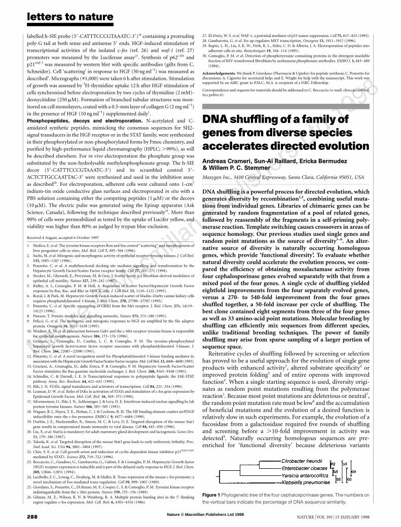
Figure 1 Phylogenetic tree of the four cephalosporinase genes. The numbers on
the vertical bars indicate the percentage of DNA sequence similarity.
Nature © Macmillan Publishers Ltd 1998
8
letters to nature
NATURE | VOL 391 | 15 JANUARY 1998 289
have been selected against over billions of years of evolution. Wetherefore wanted to determine whether shuffling of gene familieswould accelerate the evolution process.
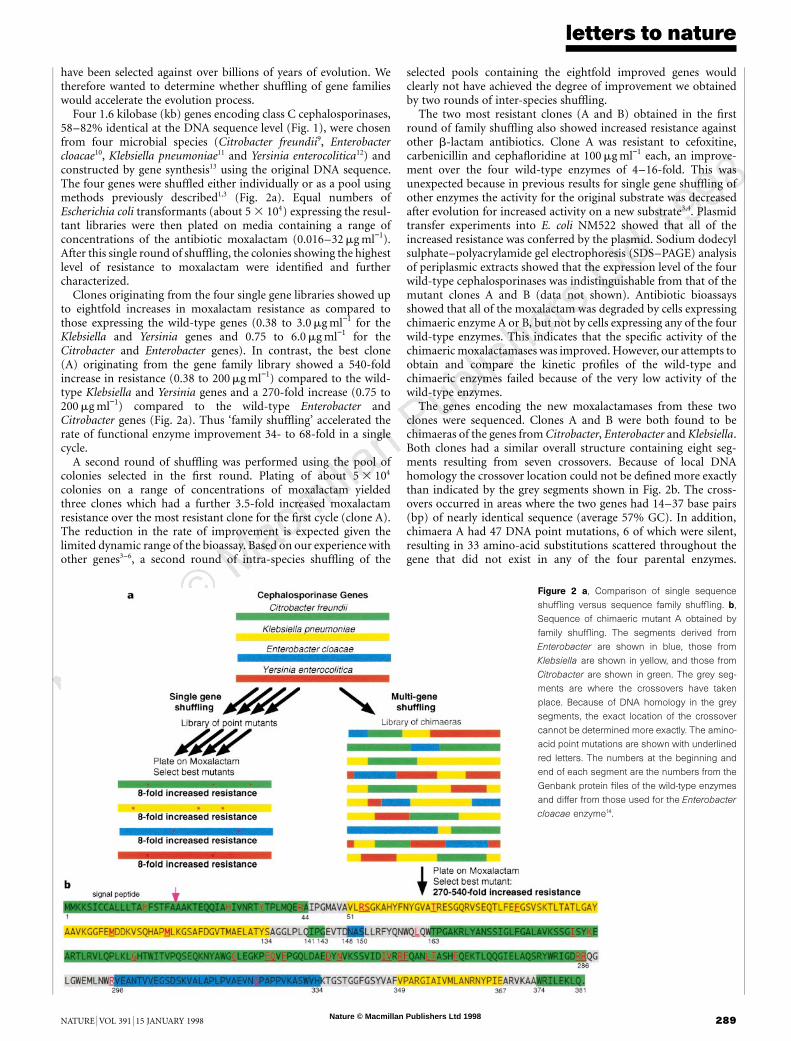
Four 1.6 kilobase (kb) genes encoding class C cephalosporinases,58–82% identical at the DNA sequence level (Fig. 1), were chosenfrom four microbial species (Citrobacter freundii9, Enterobactercloacae10, Klebsiella pneumoniae11 and Yersinia enterocolitica12) andconstructed by gene synthesis13 using the original DNA sequence.The four genes were shuffled either individually or as a pool usingmethods previously described1,3 (Fig. 2a). Equal numbers ofEscherichia coli transformants (about 5 3 104) expressing the resul-tant libraries were then plated on media containing a range ofconcentrations of the antibiotic moxalactam (0.016–32 mg ml−1).After this single round of shuffling, the colonies showing the highestlevel of resistance to moxalactam were identified and furthercharacterized.
Clones originating from the four single gene libraries showed upto eightfold increases in moxalactam resistance as compared tothose expressing the wild-type genes (0.38 to 3.0 mg ml−1 for theKlebsiella and Yersinia genes and 0.75 to 6.0 mg ml−1 for theCitrobacter and Enterobacter genes). In contrast, the best clone(A) originating from the gene family library showed a 540-foldincrease in resistance (0.38 to 200 mg ml−1) compared to the wild-type Klebsiella and Yersinia genes and a 270-fold increase (0.75 to200 mg ml−1) compared to the wild-type Enterobacter andCitrobacter genes (Fig. 2a). Thus ‘family shuffling’ accelerated therate of functional enzyme improvement 34- to 68-fold in a singlecycle.
A second round of shuffling was performed using the pool ofcolonies selected in the first round. Plating of about 5 3 104
colonies on a range of concentrations of moxalactam yieldedthree clones which had a further 3.5-fold increased moxalactamresistance over the most resistant clone for the first cycle (clone A).The reduction in the rate of improvement is expected given thelimited dynamic range of the bioassay. Based on our experience withother genes3–6, a second round of intra-species shuffling of the
selected pools containing the eightfold improved genes wouldclearly not have achieved the degree of improvement we obtainedby two rounds of inter-species shuffling.
The two most resistant clones (A and B) obtained in the firstround of family shuffling also showed increased resistance againstother b-lactam antibiotics. Clone A was resistant to cefoxitine,carbenicillin and cephafloridine at 100 mg ml−1 each, an improve-ment over the four wild-type enzymes of 4–16-fold. This wasunexpected because in previous results for single gene shuffling ofother enzymes the activity for the original substrate was decreasedafter evolution for increased activity on a new substrate3,4. Plasmidtransfer experiments into E. coli NM522 showed that all of theincreased resistance was conferred by the plasmid. Sodium dodecylsulphate–polyacrylamide gel electrophoresis (SDS–PAGE) analysisof periplasmic extracts showed that the expression level of the fourwild-type cephalosporinases was indistinguishable from that of themutant clones A and B (data not shown). Antibiotic bioassaysshowed that all of the moxalactam was degraded by cells expressingchimaeric enzyme A or B, but not by cells expressing any of the fourwild-type enzymes. This indicates that the specific activity of thechimaeric moxalactamases was improved. However, our attempts toobtain and compare the kinetic profiles of the wild-type andchimaeric enzymes failed because of the very low activity of thewild-type enzymes.
The genes encoding the new moxalactamases from these twoclones were sequenced. Clones A and B were both found to bechimaeras of the genes from Citrobacter, Enterobacter and Klebsiella.Both clones had a similar overall structure containing eight seg-ments resulting from seven crossovers. Because of local DNAhomology the crossover location could not be defined more exactlythan indicated by the grey segments shown in Fig. 2b. The cross-overs occurred in areas where the two genes had 14–37 base pairs(bp) of nearly identical sequence (average 57% GC). In addition,chimaera A had 47 DNA point mutations, 6 of which were silent,resulting in 33 amino-acid substitutions scattered throughout thegene that did not exist in any of the four parental enzymes.
Figure 2 a, Comparison of single sequence
shuffling versus sequence family shuffling. b,
Sequence of chimaeric mutant A obtained by
family shuffling. The segments derived from
Enterobacter are shown in blue, those from
Klebsiella are shown in yellow, and those from
Citrobacter are shown in green. The grey seg-
ments are where the crossovers have taken
place. Because of DNA homology in the grey
segments, the exact location of the crossover
cannot be determined more exactly. The amino-
acid point mutations are shown with underlined
red letters. The numbers at the beginning and
end of each segment are the numbers from the
Genbank protein files of the wild-type enzymes
and differ from those used for the Enterobacter
cloacae enzyme14.

Nature © Macmillan Publishers Ltd 1998
8
letters to nature
290 NATURE | VOL 391 | 15 JANUARY 1998
Chimaera B had 14 amino-acid substitutions, of which 12 wereidentical to those in chimaera A.
A model of the best chimaeric moxalactamase, chimaera A, wascreated from the known structure of the Enterobacter cloacae AmpCenzyme14 (Fig. 3). Although 37% of the amino acids (142 residues)of the chimaeric clone A differ from the Enterobacter cloacaeenzyme for which the crystal structure is known, after energyminimization the predicted structure of the a-chain backbone ofthe A chimaera remained nearly identical to the known structure(r.m.d.s. deviation of 0.766 A). This model shows that twocrossovers occurred in loop or random coil regions separatinga-helical and b-sheet structures, and one occurred inside the C-terminal a-helix. The remaining five crossovers could haveoccurred in loops, a-helices or b-sheets; therefore it is unclearwhether the crossovers preferentially occurred in loops separatingstructural elements. The conserved catalytic residues S64, K67,
Y150 and K315 of the Enterobacter enzyme14 were retained.Although shuffling of a single gene creates a library of genes that
differ by only a few point mutations1–6, the block-exchange natureof family shuffling creates chimaeras that differ in many positions.For example, in previous work a single b-lactamase gene wasshuffled for three cycles, yielding only four amino-acid mutations3,whereas a single cycle of family shuffling of the four cephalospor-inases resulted in a mutant enzyme which differs by 102 amino acidsfrom the Citrobacter enzyme, by 142 amino acids from the Enterobacterenzyme, by 181 amino acid from the Klebsiella enzyme and by 196amino acids from the Yersinia enzyme. The increased sequencediversity of the library members obtained by family shuffling resultsin a ‘sparse sampling’ of a much greater portion of sequence space15,the theoretical collection of all possible sequences of equal length,ordered by similarity (Fig. 4). Selection from ‘sparse libraries’ allowsrapid identification of the most promising areas within an extendedsequence landscape (a multidimensional graph of sequence spaceversus function)15. However, the sparseness also decreases the like-lihood of immediate location of the area’s best sequence, the localoptimum. Subsequent shuffling of the selected sequences allowsfurther exploration of the still vast intermittent sequence space at anincreased sampling density. Although the search algorithm remainsunchanged, the scale of the searched area decreases with each cycleuntil no further improvement occurs.
Because the species definition in eukaryotes is based on the abilityto exchange genetic information, the family shuffling that occurs innatural evolution and in classical breeding is generally restricted tothe diversity existing within a single species. DNA shuffling cansuccessfully mix genes from diverse species because single genes cantolerate much higher mutation densities16 than whole genomes. Thehigh degree of DNA homology that is required for traditionalbreeding of plants and animals is therefore not required for themolecular breeding of single genes and gene clusters. M. . . . . . . . . . . . . . . . . . . . . . . . . . . . . . . . . . . . . . . . . . . . . . . . . . . . . . . . . . . . . . . . . . . . . . . . . . . . . . . . . . . . . . . . . . . . . . . . . . . . . . . . . . . . . . . . . . . . . . . . .
Methods
Gene synthesis. All four 1.6 kb genes were separately assembled from 60-mersynthetic oligonucleotides in a single assembly reaction13. These genes werecloned into pBR322 downstream of the b-lactamase promoter and in place ofthe native b-lactamase gene. The DNA sequence and drug resistance of eachconstruct was confirmed.Library construction. The presence of chimaeric genes in the library made byfamily shuffling was confirmed by restriction fragment length polymorphism(RFLP) analysis of individual clones. Each of the eight clones analysed had anRFLP pattern distinct from each other as well as from those of the parentalsequences. The library thus contained a diverse array of chimaeric sequences.The second cycle of shuffling was performed on the pool of chimaeras selectedin the first round.Drug resistance. An equal number of transformants were plated on mediumcontaining moxalactam. The numbers were based on the number oftetracyclin-resistant colonies after transformation. The resistance of theselected clones to a variety of b-lactam antibiotics was measured in 96-wellplates in which each antibiotic was serially diluted (1 : 1) into culture mediumcontaining the test organism. Assays for all native and mutant clones were donein quadruplicate.Moxalactamase assays. The moxalactam degradation activity of whole cellswas measured by incubating an overnight culture of bacteria with 34 mg ml−1 ofmoxalactam in LB media. After incubation for 16 h the concentration ofmoxalactam in the sterilized supernatant was measured by bioassay on platesinoculated with E. coli NM522, using filter discs saturated with serial twofolddilutions of the supernatants. A moxalactam standard curve was used todetermine the fraction of moxolactam degraded by the host cell alone, the fourwild-type constructs and the two chimaeric mutants.Modelling. The deduced amino-acid sequence of chimaeric mutant A wasaligned with the wild-type Enterobacter sequence using HOMOLOGY (Biosym,San Diego) and the calculated coordinates were assigned to the chimaericprotein. The modelled structure was constructed using a three-step protocolthat involved an energy minimization to relieve steric hindrance, molecular
Figure 3 Computer model of evolved mutant A obtained bya single cycle of family
shuffling. The 142 amino-acid mutations were introduced into the Enterobacter
cloacae sequence, whose structure is known14, followed by energy minimization.
The predicted structure of the a-chain backbone of the chimaeric enzyme is
within an r.m.s. deviation of 0.766 A from the known native structure. The
segments derived from Enterobacter are shown in blue, those from Klebsiella
are shown in yellow, and those from Citrobacter are shown in green. The 33
amino-acid point mutations are shown in red.
Figure 4 Searching sequence space by family shuffling versus by single
sequence shuffling. Single sequence shuffling yields clones with a few point
mutations and the library members are typically 97–99% identical. Family
shuffling causes sequence block exchange which yields chimaeras that have
greater sequence divergence. At equal library size, the increased sequence
diversity of the chimaeric library results in sparse sampling of a much greater area
of sequence space, allowingmorepromisingareas to be found and subsequently
explored at increased sampling density.

Nature © Macmillan Publishers Ltd 1998
8
letters to nature
NATURE | VOL 391 | 15 JANUARY 1998 291
dynamics calculations to find the lowest energy conformation followed byanother energy minimization to provide the final structure.
Received 23 June; accepted 8 October 1997.
1. Stemmer, W. P. C. DNA shuffling by random fragmentation and reassembly: in vitro recombinationfor molecular evolution. Proc. Natl Acad. Sci. USA 91, 10747–10751 (1994).
2. Stemmer, W. P. C. Searching sequence space. Bio/Technology 13, 549–553 (1995).3. Stemmer, W. P. C. Rapid evolution of a protein in vitro by DNA shuffling. Nature 370, 389–391 (1994).4. Zhang, J., Dawes, G. & Stemmer, W. P. C. Evolution of a fucosidase from a galactosidase by DNA
shuffling and screening. Proc. Natl Acac. Sci. USA 94, 4504–4509 (1997).5. Crameri, A., Whitehorn, E., Tate, E. & Stemmer, W. P. C. Improved green fluorescent protein by
molecular evolution using DNA shuffling. Nature Biotech. 14, 315–319 (1996).6. Crameri, A., Dawes, G., Rodriguez, E., Silver, S. & Stemmer, W. P. C. Molecular Evolution of an
arsenate detoxification pathway by DNA shuffling. Nature Biotech. 15, 436–438 (1997).7. Moore, J. C. & Arnold, F. H. Directed evolution of a para-nitrobenzyl esterase for aqueous-organic
solvents. Nature Biotech. 14, 458–467 (1996).8. Zhao, H. & Arnold, F. H. Optimization of DNA shuffling for high fidelity recombination. Nucleic
Acids Res. 25, 1307–1308 (1997).9. Lindberg, F. & Normark, S. Sequence of the Citrobacter freundii OS60 chromosomal ampC b-
lactamase gene. Eur. J. Biochem. 156, 441–445 (1986).10. Galleni, M. et al. Sequence and comparative analysis of three Enterobacter cloacae ampC b-lactamase
genes and their products. Biochem. J. 250, 753–760 (1988).11. Leiza, M. G. et al. Gene sequence and biochemical characterization of FOX-1 from Klebsiella
pneumoniae, a new AmpC-type plasmid-mediated b-lactamase with two molecular variants. \ita-Antimicrob. Agents Chemother. 38, 2150–2157 (1994).
12. Seoane, A., Francia, M. V. & Garcia Lobo, J. M. Nucleotide sequence of the ampC-ampR region fromthe chromosome of Yersinia enterocolitica. Antimicrob. Agents Chemother. 36, 1049–1052 (1992).
13. Stemmer, W. P. C., Crameri, A., Ha, K. D., Brennan, T. M. & Heyneker, H. L. Single-step PCR assemblyof a gene and a whole plasmid from large numbers of oligonucleotides. Gene 164, 49–53 (1995).
14. Lobkovsky, E. et al. Evolution of enzyme activity: crystallographic structure at 2 A resolution ofcephalosporinase from the ampC gene of Enterobacter cloacae P99 and comparison with a class Apenicillinase. Proc. Natl Acad. Sci. USA 90, 11257–11261 (1993).
15. Kauffman, S. The Origins of Order (Oxford University Press, Oxford, 1993).16. Eigen, M. Steps Towards Life: a Perspective on Evolution (Oxford University Press, Oxford, 1992).
Acknowledgements. We thank G. Dawes, J. Kieft, S. DelCardayre and M. Tobin and R. Howard,C. Yanofsky, P. Schultz, F. Arnold and A. Kornberg for useful comments on the manuscript.
Correspondence should be addressed to W.P.C.S. (e-mail: [email protected]).
Abacterial antibiotic-resistancegene thatcomplements thehumanmultidrug-resistanceP-glycoproteingeneHendrik W. van Veen*, Richard Callaghan†,Loredana Soceneantu†, Alessandro Sardini‡,Wil N. Konings* & Christopher F. Higgins†
* Department of Microbiology, Groningen Biomolecular Sciences andBiotechnology Institute, University of Groningen, Kerklaan 30, 9751 NN Haren,The Netherlands† Imperial Cancer Research Fund Laboratories and Cancer Research CampaignDrug Resistance Group, Nuffield Department of Clinical Biochemistry,Institute of Molecular Medicine, John Radcliffe Hospital, University of Oxford,Oxford OX3 9DS, UK‡ Department of Physiology, King’s College London, The Strand,London WC2R 2LS, UK. . . . . . . . . . . . . . . . . . . . . . . . . . . . . . . . . . . . . . . . . . . . . . . . . . . . . . . . . . . . . . . . . . . . . . . . . . . . . . . . . . . . . . . . . . . . . . . . . . . . . . . . . . . . . . . . . . . . . . . . .
Bacteria have developed many fascinating antibiotic-resistancemechanisms1,2. A protein in Lactococcus lactis, LmrA, mediatesantibiotic resistance by extruding amphiphilic compounds fromthe inner leaflet of the cytoplasmic membrane3,4. Unlike otherknown bacterial multidrug-resistance proteins, LmrA is an ATP-binding cassette (ABC) transporter5. The human multidrug-resis-tance P-glycoprotein6, encoded by the MDR1 gene, is also an ABCtransporter, overexpression of which is one of the principal causesof resistance of human cancers to chemotherapy7,8. We expressedlmrA in human lung fibroblast cells. Surprisingly, LmrA wastargeted to the plasma membrane and conferred typical multi-drug resistance on these human cells. The pharmacologicalcharacteristics of LmrA and P-glycoprotein-expressing lung fibro-blasts were very similar, and the affinities of both proteins for
vinblastine and magnesium-ATP were indistinguishable. Blockersof P-glycoprotein-mediated multidrug resistance also inhibitedLmrA-dependent drug resistance. Kinetic analysis of drug dis-sociation from LmrA expressed in plasma membranes of insectcells revealed the presence of two allosterically linked drug-binding sites indistinguishable from those of P-glycoprotein.These findings have implications for the reversal of antibioticresistance in pathogenic microorganisms. Taken together, theydemonstrate that bacterial LmrA and human P-glycoprotein arefunctionally interchangeable and that this type of multidrug-resistance efflux pump is conserved from bacteria to man.
Using the polymerase chain reaction (PCR), the bacterial lmrAcoding sequence3 was cloned into a pCI-neo mammalian expressionvector under the control of the human cytomegalovirus immedi-ate–early enhancer/promoter region. A Kozak sequence9 was intro-duced at the ATG initiation codon of lmrA to enhance translationalefficiency. For control experiments, a transport-inactive LmrAprotein was generated in the same vector by introducing a lysine-to-methionine substitution at position 388 (K388M)10 in the WalkerA motif of the nucleotide-binding domain of the protein by site-directed mutagenesis. Hexa-histidine tags were also added to theamino termini of both the wild-type and K388M forms of LmrA.
Figure 1 Expression of LmrA in GM0637 human lung fibroblast cells. a, Western
blot of total cell protein (30 mg per lane) using the anti hexa-histidine tag antibody.
Control, wild type and mutant refer to mock-transfected cells, and cells
expressing the wild-type and K388M forms of LmrA, respectively. The migration
of molecular mass markers is indicated. Arrowhead indicates LmrA protein. b,
Distribution of wild-type LmrA expressed in fibroblast cells 48h after transfection.
The three frames show cross-sections through a cell, from top to bottom along
the z-axis. The fluorescence intensity is shown colour-coded ona scale from blue
(low) to white (high). Scale bar, 25 mm.