54259299 Anatomy of the Hepatobiliary System Maran
18
WELCOME TO SURGERY SYMPOSIUM BY S4 UNIT 26.04.2011
description
so much stuff rain on a liver
Transcript of 54259299 Anatomy of the Hepatobiliary System Maran

WELCOME TO SURGERY SYMPOSIUM
BY S4 UNIT
26.04.2011

TOPIC – OBSTRUCTIVE JAUNDICE
Anatomy – Nivash Maran. RPhysiology – Praveen. CEpidemiology & Pathogenesis – Clinical Features – Management – Recent Advances -

Anatomy of the Hepatobiliary System
Nivash Maran . R

Hepatobiliary SystemLiverGallbladderBile ductsPancreas

LIVERLiver is the largest of the
abdominal visceraLiver lies in the upper right
part of the abdominal cavity, occupying most of the right hypochondrium and epigastrium.
Liver weighs roughly about 2% of the adult body weight.
BLOOD SUPPLY:Dual blood supply – 80%
nutrient-rich blood from Portal Vein. 20% oxygen-rich blood from Hepatic Artery.

Liver weighs roughly about 2% of the adult body weight.
BLOOD SUPPLY:Dual blood supply – 80% nutrient-rich blood
from Portal Vein. 20% oxygen-rich blood from Hepatic Artery.

Classically liver is divided into left and right lobes by the main portal fissure which is also called the Cantlie’s Line.
Based on the hepatic blood supply, it can also be divided into right and left hemilivers.
According to Couinad classification, there are 8 functionally independent segments in the liver.
Each segment has its own vascular inflow, outflow and biliary drainage.

Bile is secreted by hepatocytes and flows through the canaliculus into the bile ductule.
The bile ductules join together to form larger intrahepatic bile ducts which in turn merge to form the right and left Hepatic Ducts.
CENTRAL VEIN

Gallbladder
The gallbladder is a flask-shaped organ attached to the common bile duct by the cystic duct.
It is 7 – 10 cm long with a capacity of up to 50 ml.
It usually lies in a shallow fossa in the liver parenchyma covered by peritoneum continued from the liver surface.

Gallbladder has 3 parts – Fundus, body and neck.
Widening of the neck to form the body is called the ‘Hartmann’s pouch’.
Blood supply is from the cystic artery which arises from the right hepatic artery.

Cystic duct drains the gallbladder into the common hepatic duct to form the common bile duct.
It is 3 – 4 cm long.Mucosa of cystic duct bears 5 – 12 crescentic
folds continuous with those in the neck of the gallbladder.

Bile DuctsCommon Hepatic Duct:
Right and Left hepatic ducts unite to form the common hepatic duct.
It is about 3 cm long.
Common Bile Duct:Cystic duct joins the common hepatic
duct and forms the common bile duct.
It is about 8 cm long.It has 4 parts:
1. Supraduodenal2. Retroduodenal3. Infraduodenal4. Intraduodenal


Levels of obstruction in the biliary tree

Pancreas
Pancreas is both exocrine and endocrine organ situated retroperitoneally behind the stomach.
It extends from the duodenum on the right side to the spleen on the left side.
Length is about 6 inches.

Parts of Pancreas:Pancreas has 4 parts –
HeadNeckBodyTail
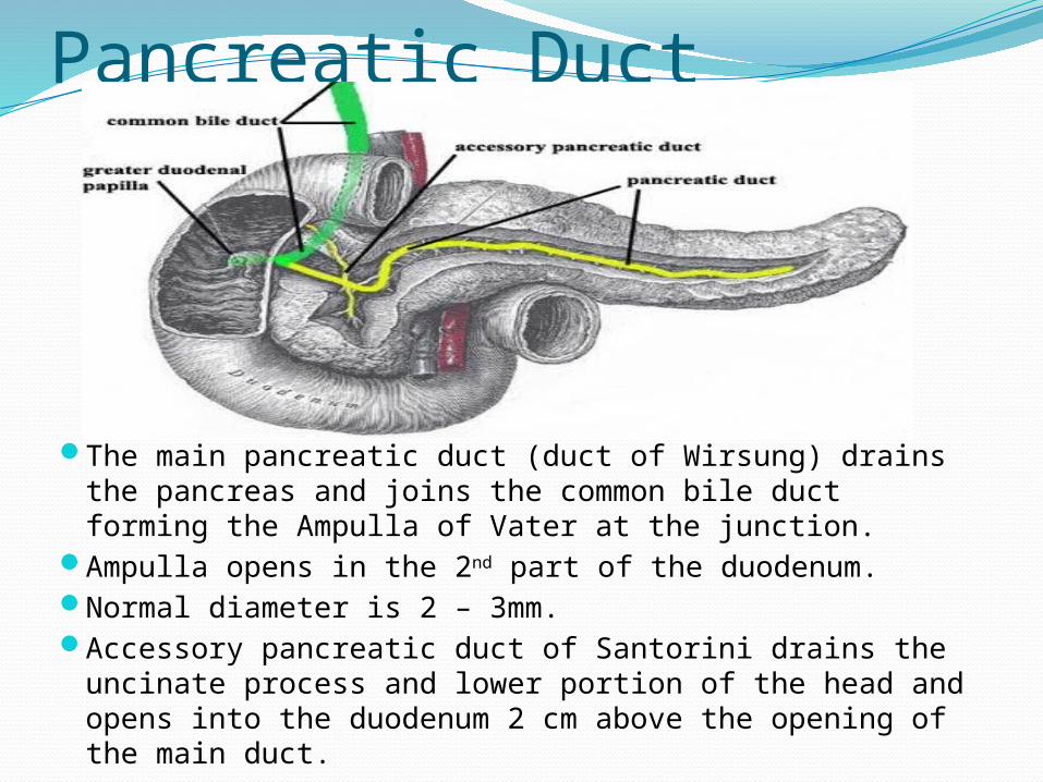
Pancreatic Duct
The main pancreatic duct (duct of Wirsung) drains the pancreas and joins the common bile duct forming the Ampulla of Vater at the junction.
Ampulla opens in the 2nd part of the duodenum.Normal diameter is 2 – 3mm.Accessory pancreatic duct of Santorini drains the
uncinate process and lower portion of the head and opens into the duodenum 2 cm above the opening of the main duct.