53cr
6
< Previous Next > Current Issue Available Issues Online First Lorem ipsum dolor sit amet, consectetuer adipiscing elit. Nunc augue metus, mollis vehicula, dapibus eget, convallis nec, massa. Vivamus volutpat! Vivamus ultricies congue nibh. Mauris purus sapien, pretium vitae, sodales id, mollis sit amet, libero. Morbi ac massa nec augue pulvinar pretium. Abstract INTRODUCTION MATERIALS AND METHODS RESULTS DISCUSSION CONCLUSIONS Acknowledgments REFERENCES Articles Citing this Article Google Scholar Search for Other Articles By Author Soo-Bum An Soo-Byung Park Yong-Il Kim Woo-Sung Son Search in: DEFINE_ME_WA PubMed Google Scholar Search < Previous Article Next Article > Volume 84, Issue 2 (March 2014) Add to Favorites Share Article Export Citations Track Citations Permissions Abstract PDF Article Citation: Soo-Bum An, Soo-Byung Park, Yong-Il Kim, and Woo-Sung Son (2014) Effect of post–orthognathic surgery condylar axis changes on condylar morphology as determined by 3-dimensional surface reconstruction. The Angle Orthodontist: March 2014, Vol. 84, No. 2, pp. 316-321. doi: http://dx.doi.org/10.2319/052113-387.1 Original Articles Effect of post–orthognathic surgery condylar axis changes on condylar morphology as determined by 3-dimensional surface reconstruction Soo-Bum An a* ; Soo-Byung Park b* ; Yong-Il Kim c ; Woo-Sung Son b a Postgraduate student, Department of Orthodontics, School of Dentistry, Pusan National University Dental Hospital, Yangsan, South Korea ???welcome.guest??? Sign In | Register Search within: All journals Go Advanced Search Volume 84, Issue 2 (March 2014) Journal Information Table of Contents Related Articles
-
Upload
dattatraya-parle -
Category
Documents
-
view
13 -
download
2
description
RESEARCH
Transcript of 53cr
< Previous Next >
Current Issue Available Issues
Online First
Lorem ipsum dolor sit amet,consectetuer adipiscing elit. Nuncaugue metus, mollis vehicula, dapibuseget, convallis nec, massa. Vivamusvolutpat! Vivamus ultricies congue nibh.Mauris purus sapien, pretium vitae,sodales id, mollis sit amet, libero.Morbi ac massa nec augue pulvinarpretium.
AbstractINTRODUCTIONMATERIALS AND METHODSRESULTSDISCUSSIONCONCLUSIONSAcknowledgmentsREFERENCES
Articles Citing this Article
Google Scholar
Search for Other Articles By Author
Soo-Bum An
Soo-Byung Park
Yong-Il Kim
Woo-Sung Son
Search in:
DEFINE_ME_WA
PubMed
Google Scholar
Search
< Previous Article Next Article >Volume 84, Issue 2 (March 2014)
Add to Favorites Share Article Export Citations Track Citations Permissions
Abstract PDFArticle Citation:Soo-Bum An, Soo-Byung Park, Yong-Il Kim, and Woo-Sung Son (2014) Effect of post–orthognathic surgery condylar axis changes oncondylar morphology as determined by 3-dimensional surface reconstruction. The Angle Orthodontist: March 2014, Vol. 84, No. 2,pp. 316-321.
doi: http://dx.doi.org/10.2319/052113-387.1
Original Articles
Effect of post–orthognathic surgery condylar axis changes on condylar morphology as determined by3-dimensional surface reconstruction
Soo-Bum Ana*; Soo-Byung Parkb*; Yong-Il Kimc; Woo-Sung Sonb
a Postgraduate student, Department of Orthodontics, School of Dentistry, Pusan National University Dental Hospital, Yangsan, SouthKorea
???welcome.guest???Sign In | Register
Search within:
All journals
Go
Advanced Search
Volume 84, Issue 2
(March 2014)
Journal Information
Table of Contents
Related Articles

Koreab Professor, Department of Orthodontics, School of Dentistry, Pusan National University Dental Hospital, Yangsan, South Koreac Assistant Professor, Department of Orthodontics, School of Dentistry, Dental Research Institute, Pusan National University DentalHospital, and Biomedical Research Institute, Pusan National University Hospital, Yangsan, South Korea
ABSTRACT
Objective: To evaluate the effect of postoperative condylar axis changes on mandibular condylar remodeling by comparingthe condylar head in three-dimensional (3D) surface reconstructions before and after surgery in skeletal Class IIIdeformities (one-jaw [mandibular setback] or two-jaw surgery), and also to determine the relationship between condylarinward rotation and condylar surface remodeling after orthognathic surgery.
Materials and Methods: A retrospective analysis was conducted of 30 patients with skeletal Class III deformities who hadreceived orthognathic surgery. Group 1 underwent one-jaw surgery (10 men, five women, age 22.4 ± 3.3 years), and group2 underwent two-jaw surgery (10 men, five women, age 22.3 ± 2.2 years). Sixty condyles were reconstructed andsuperimposed pre- and postoperatively to compare the changes of condylar surfaces. The relation between the condylaraxis change and the surface change using the Pearson correlation were investigated from the 3D image software.
Results: Condylar surface changes before and after the surgery were significant. The postoperative inward rotation of thecondyles was correlated with the average absolute deviation of the condyles, regardless of the surgery type (one- or 2-jawsurgery; r = .70, P < .05).
Conclusion: After orthognathic surgery, condylar surface changes occurred, and condylar inward rotation was closelyrelated to changes of condylar surface.
KEY WORDS: Condyle, Remodeling, Superimposition, CT
Received: May 2013; Accepted: June 2013 ;Published Online: August 14, 2013
Corresponding author: Dr Yong-Il Kim, Department of Orthodontics, School of Dentistry, Dental Research Institute, PusanNational University Dental Hospital, Gudeokro 305, Seogu, Busan 602-739, South Korea (e-mail: [email protected])
*Contributed equally to this article.
INTRODUCTION
Advancements in imaging technology have generalized the use of cone-beam computed tomography (CBCT) in the field of dentistry.CBCT convertes two-dimensional image data to three-dimensional (3D) images, effectively overcoming the limitations of the formermodality, particularly in orthodontic treatment and orthognathic surgery, greatly facilitating patient diagnosis, treatment planning, andtreatment-outcome assessment.1 Kau et al.,2 in comparing the many types of 3D imaging devices, asserted that 3D imaging ismost effectively used for examination and treatment relating to the oral and maxillofacial region. Bayram et al.3 reported that 3Dmodalities are necessary for quantification of mandibular condyle volumes. Recently, a number of studies employing pre- andpostoperative 3D images have been reported.
For patients with skeletal Class III deformities, corrective orthognathic surgery improves both oral functionality and related esthetics.However, one orthognathic-surgical procedure, sagittal split ramus osteotomy (SSRO), is known to induce postoperativeanteroposterior movement and condylar rotation.1 During surgery, the condylar axis rotates to maintain the intersegmental contactafter the distal segment moves anteriorly or posteriorly. Kim et al.4 reported that in patients who had undergone SSRO with rigidfixation, the condyle showed an inward rotation on the axial plane and was positioned posteriorly, on the basis of short- and long-term observation of condylar location. These movements reportedly do not have a negative impact on postsurgical stability.
Orthognathic surgery–induced changes of condylar location impart physical stress on the condylar surface through condylarremodeling of the temporomandibular joint (TMJ) structure, which is an adaptation to the new functional requirements.5 Thisfunctional stress can result in TMJ changes postoperatively. In some cases, however, severe condylar change is incurred,deteriorating skeletal stability, though the cause remains mostly unknown.6,7 Many other studies have reported on postoperativecondylar changes as well.8–10
For assessment of condylar remodeling, several CBCT-based analysis methods have already been introduced.11,12 Carvalho etal.13 evaluated 3D changes of location in the ramus, condylar, and mental region in patients who had received mandibularadvancement. Motta et al.14 superimposed a 3D CBCT model, executed mandibular advancement, and evaluated the patients'condyles. They stressed an individual variability in condylar displacement after surgery.13,14 A study by da Motta et al.15 used a 3DCBCT overlapping technique to evaluate mandibular anatomy and condylar location, proving its effectiveness. However, there arestill only a few postoperative condylar remodeling analyses of skeletal Class III patients using 3D-reconstructed images andcondylar superimpositions.3,13,14,16
The purpose of this study was to evaluate the effect of postoperative condylar axis changes on mandibular condylar remodeling bycomparing the condylar head in 3D surface reconstructions before and after surgery (condylar pre- and postoperative 3D surfacereconstructions). In addition, the purpose was to determine the relationship between condylar inward rotation and condylar surfaceremodeling after orthognathic surgery.
MATERIALS AND METHODS
This was a retrospective analysis of all patients who underwent mandibular setback SSRO with or without Le Fort I osteotomy forClass III malocclusion at the Department of Orthodontics, Pusan National University Dental Hospital, within the period beginningJanuary 2010 and ending December 2012. Rigid internal fixation was achieved in the maxilla and mandible by means of plates andscrews. The sample consisted of 30 adults: group 1 underwent mandibular setback SSRO with rigid fixation (10 males, fivefemales, age 22.4 ± 3.3 years), and group 2 underwent mandibular setback SSRO and Le Fort I osteotomy (10 males, five females,age 22.3 ± 2.2 years). Patients who had been diagnosed with any syndromes, facial trauma, or degenerative joint disease wereexcluded. The study was reviewed and approved by the Ethics Committee at Pusan National University Hospital (E-2012055).
Images were obtained using a CBCT scanner (DCT Pro; Vatech, Seoul, Korea) within an average of 1.0 month (range, 0.1–1.5 months) prior to surgery and 12.4 months (range, 10–19 months) after surgery. The CBCT was acquired with the subjectpatients in an upright position for maximum intercuspation and with the FH plane adjusted parallel to the floor. The maxillofacialregions were CBCT scanned according to the following parameters: 20 × 19-cm field of view, 90-kVp tube voltage, 4.0-mA tubecurrent, and 24-second scan time. On the basis of the CBCT data, 60 condyles total were evaluated pre- and postoperatively.
3D images of the proximal segments of the mandible were reconstructed and reformatted into the stereolithography (STL) data

3D images of the proximal segments of the mandible were reconstructed and reformatted into the stereolithography (STL) dataformat using 3D imaging software (Vworks 4.0; Cybermed Co, Seoul, South Korea). The segmented proximal segment imageswere then imported into 3D scan data processing software (Rapidform XOS3; Inus Technology Inc, Seoul, South Korea).
To investigate the surface remodeling of the condylar head, pre- and postoperative segmented images were superimposed overthree registration areas (condylar neck, mandibular notch, and posterior border of ramus).16 This function of Rapidform XOS3,designated as 3D surface-to-surface matching (best fit method), employs a least-mean-squared algorithm.17 According to thecoordination between the registration areas, the reconstructed images were automatically fitted. Since the procedure is fullyautomated, the influence of the observers' variability on the accuracy of measurements is completely eliminated. The superimposedcondylar heads were divided by a plane connecting the median and lateral poles. The surface displacements of the heads werecalculated using Rapidform XOS3 (Figure 1). In order to calculate the surface difference on the 3D-superimposed images, average
absolute deviation, the averages of the absolute value of the deviation between the pre- and postoperative 3D images, was firstobtained; second, average deviation representing the average values of deviation between the pre- and postoperative 3D imageswere taken.
View larger version(39K)
Figure 1.(A) Superimposed pre- and postsurgery ramus images. These are the absolute linearmeasurements. (B) Superimposed upper condylar head. These are the signed color mapmeasurements (red is bone formation, blue is bone resorption).
To evaluate the surface remodeling on the superimposed condylar heads, six areas (the anterolateral, anteromiddle, anteromedial,posterolateral, posteromiddle, and posteromedial regions) were investigated and classified into three categories (bone resorption,unchanged, bone formation; Figure 2).
View larger version(28K)
Figure 2.Six areas of condylar head: (A) Anterior surface, (B) Superior surface, and (C) Posterior surface.Ant-Lat indicates anterolateral; Ant-Mid, anteromiddle; Ant-Med, anteromedial; Post-Lat,posterolateral; Post-Mid, posteromiddle; Post-Med, posteromedial.
To evaluate the effect of postoperative condylar axis change on the condylar surface changes, the changes of the condylar axisangles (the axis passing through the medial and lateral pole of the condylar head) were measured from the midsagittal referenceplane (MSR; Figure 3).
View larger version(61K)
Figure 3.Axial condylar axis angle: angle between condylar axis and MSR (the plane perpendicular to theFH plane, passing through Na and Ba points) on the axial plane.
Statistical Analysis
The data were statistically analyzed using SPSS 18.0 for Windows (SPSS Inc, Chicago, Ill). The differences were considered to besignificant at P < .05. The postoperative extents of the condylar head changes were tested by sample t-test (P < .05). The one- andtwo-jaw surgery differences of condylar head changes were compared by independent t-test (P < .05). The condylar axis changeswere similarly compared, but by paired t-test. To analyze the correlation between the extents of condylar axis and condylar headchanges, the relevant Pearson correlation coefficients were calculated. The intraoperator error was obtained by intraclasscorrelation coefficient (ICC) and Cohen kappa index.
RESULTS
The intraoperator reliability for condylar axis changes was high according to the ICC (average, .993). The Cohen kappa index forremodeling signs also showed substantial agreement (average, .805). The mean of the average absolute deviation of condylarsurface change before and after surgery was 0.22 ± 0.03 mm, and the mean of the average deviation was 0.01 ± 0.09 mm. The pre-and postoperative images revealed a significant condylar surface change difference (P < .05), though not for the average deviation (P> .05).
In a comparison of the groups according to surgery (one-jaw vs two-jaw), there was no significance in either the average absolutedeviation or the average deviation (P > .05; Table 1).
View larger version(14K)
Table 1.
Condylar Surface Change (Preoperative to Postoperative)a
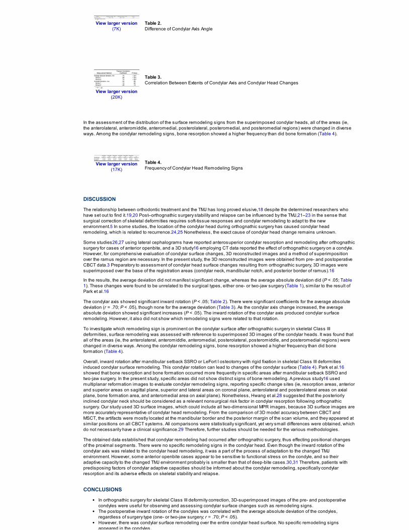
In order to uncover the relationship between the condylar axis changes and the condylar surface changes, a Pearson correlationanalysis was performed. It was found that after the surgery, the condylar axis changes showed significant changes with inwardrotation (P < .05; Table 2). There were significant coefficients for the average absolute deviation (r = .70; P < .05); however, therewere none for the average deviation (Table 3).
View larger version(7K)
Table 2.Difference of Condylar Axis Angle
View larger version(20K)
Table 3.Correlation Between Extents of Condylar Axis and Condylar Head Changes
In the assessment of the distribution of the surface remodeling signs from the superimposed condylar heads, all of the areas (ie,the anterolateral, anteromiddle, anteromedial, posterolateral, posteromedial, and posteromedial regions) were changed in diverseways. Among the condylar remodeling signs, bone resorption showed a higher frequency than did bone formation (Table 4).
View larger version(17K)
Table 4.Frequency of Condylar Head Remodeling Signs
DISCUSSION
The relationship between orthodontic treatment and the TMJ has long proved elusive,18 despite the determined researchers whohave set out to find it.19,20 Post–orthognathic surgery stability and relapse can be influenced by the TMJ,21–23 in the sense thatsurgical correction of skeletal deformities requires soft-tissue responses and condylar remodeling to adapt to the newenvironment.5 In some studies, the location of the condylar head during orthognathic surgery has caused condylar headremodeling, which is related to recurrence.24,25 Nonetheless, the exact cause of condylar head change remains unknown.
Some studies26,27 using lateral cephalograms have reported anterosuperior condylar resorption and remodeling after orthognathicsurgery for cases of anterior openbite, and a 3D study16 employing CT data reported the effect of orthognathic surgery on a condyle.However, for comprehensive evaluation of condylar surface changes, 3D reconstructed images and a method of superimpositionover the ramus region are necessary. In the present study, the 3D reconstructed images were obtained from pre- and postoperativeCBCT data.3 Preparatory to assessment of condylar head surface changes resulting from orthognathic surgery, 3D images weresuperimposed over the base of the registration areas (condylar neck, mandibular notch, and posterior border of ramus).16
In the results, the average deviation did not manifest significant change, whereas the average absolute deviation did (P < .05; Table 1). These changes were found to be unrelated to the surgical types, either one- or two-jaw surgery (Table 1), similar to the result ofPark et al.16
The condylar axis showed significant inward rotation (P < .05; Table 2). There were significant coefficients for the average absolutedeviation (r = .70; P < .05), though none for the average deviation (Table 3). As the condylar axis change increased, the averageabsolute deviation showed significant increases (P < .05). The inward rotation of the condylar axis produced condylar surfaceremodeling. However, it also did not show which remodeling signs were related to that rotation.
To investigate which remodeling sign is prominent on the condylar surface after orthognathic surgery in skeletal Class IIIdeformities, surface remodeling was assessed with reference to superimposed 3D images of the condylar heads. It was found thatall of the areas (ie, the anterolateral, anteromiddle, anteromedial, posterolateral, posteromiddle, and posteromedial regions) werechanged in diverse ways. Among the condylar remodeling signs, bone resorption showed a higher frequency than did boneformation (Table 4).
Overall, inward rotation after mandibular setback SSRO or LeFort I osteotomy with rigid fixation in skeletal Class III deformitiesinduced condylar surface remodeling. This condylar rotation can lead to changes of the condylar surface (Table 4). Park et al.16showed that bone resorption and bone formation occurred more frequently in specific areas after mandibular setback SSRO andtwo-jaw surgery. In the present study, specific areas did not show distinct signs of bone remodeling. A previous study16 usedmultiplanar reformation images to evaluate condylar remodeling signs, reporting specific change sites (ie, resorption areas, anteriorand superior areas on sagittal plane, superior and lateral areas on coronal plane, anterolateral and posterolateral areas on axialplane, bone formation area, and anteromedial area on axial plane). Nonetheless, Hwang et al.28 suggested that the posteriorlyinclined condylar neck should be considered as a relevant nonsurgical risk factor in condylar resorption following orthognathicsurgery. Our study used 3D surface images, which could include all two-dimensional MPR images, because 3D surface images aremore accurately representative of condylar head remodeling. From the comparison of 3D model accuracy between CBCT andMSCT, the artifacts were mostly located at the mandibular border and the posterior margin of the scan volume, and they appeared atsimilar positions on all CBCT systems. All comparisons were statistically significant, yet very small differences were obtained, whichdo not necessarily have a clinical significance.29 Therefore, further studies should be needed for the various methodologies.
The obtained data established that condylar remodeling had occurred after orthognathic surgery, thus effecting positional changesof the proximal segments. There were no specific remodeling signs in the condylar head. Even though the inward rotation of thecondylar axis was related to the condylar head remodeling, it was a part of the process of adaptation to the changed TMJenvironment. However, some anterior openbite cases appear to be sensitive to functional stress on the condyle, and so theiradaptive capacity to the changed TMJ environment probably is smaller than that of deep-bite cases.30,31 Therefore, patients withpredisposing factors of condylar adaptive capacities should be informed about the condylar remodeling, specifically condylarresorption and its adverse effects on skeletal stability and relapse.
CONCLUSIONS
In orthognathic surgery for skeletal Class III deformity correction, 3D-superimposed images of the pre- and postoperativecondyles were useful for observing and assessing condylar surface changes such as remodeling signs.The postoperative inward rotation of the condyles was correlated with the average absolute deviation of the condyles,regardless of surgery type (one- or two-jaw surgery; r = .70; P < .05).However, there was condylar surface remodeling over the entire condylar head surface. No specific remodeling signsappeared in the condyles.

appeared in the condyles.
ACKNOWLEDGMENTS
This study was supported by Dental Research Grant (PNUDH-DER 2013-04). Pusan National University Dental Hospital, Yangsan,South Korea.
REFERENCES
1. Jung YJ, Kim MJ, Baek SH. Hard and soft tissue changes after correction of mandibular prognathism and facial asymmetry bymandibular setback surgery: three-dimensional analysis using computerized tomography. Oral Surg Oral Med Oral Pathol OralRadiol Endod. 2009;107:763–771. [CrossRef] [Medline]
2. Kau CH, Richmond S, Incrapera A, English J, Xia JJ. Three-dimensional surface acquisition systems for the study of facialmorphology and their application to maxillofacial surgery. Int J Med Robotics Comput Assist Surg. 2007;3:97–110. [CrossRef][Medline]
3. Bayram M, Kayipmaz S, Sezgin OS, Küçük M. Volumetric analysis of the mandibular condyle using cone beam computedtomography. Eur J Radiol. 2012;81:1812–1816. [CrossRef] [Medline]
4. Kim YI, Jung YH, Cho BH, et al. The assessment of the short- and long-term changes in the condylar position following sagittal
split ramus osteotomy (SSRO) with rigid fixation. J Oral Rehabil. 2010;37:262–270. [CrossRef] [Medline]
5. Boulétreau P, Frey R, Breton P, Freidel M. Focus on the effect of orthognathic surgery on condylar remodeling. Rev StomatolChir Maxillofac. 2004;105:283–288. [CrossRef] [Medline]
6. De Clercq CA, Neyt LF, Mommaerts MY, Abeloos JV, De Mot BM. Condylar resorption in orthognathic surgery. Int J AdultOrthodon Orthognath Surg. 1994;9:233–240. [Medline]
7. Hwang SJ, Haers PE, Sailer HF. The role of a posteriorly inclined condylar neck in condylar resorption after orthognathicsurgery. J Craniomaxillofac Surg. 2000;28:85–90. [CrossRef] [Medline]
8. Ueki K, Moroi A, Sotobori M, et al. A hypothesis on the desired postoperative position of the condyle in orthognathic surgery: areview. Oral Surg Oral Med Oral Pathol Oral Radiol Endod. 2012;114:567–576. [CrossRef]
9. Draenert FG, Erbe C, Zenglein V, Kämmerer PW, Wriedt S, Al Nawas B. 3D analysis of condylar position after sagittal splitosteotomy of the mandible in mono- and bimaxillary orthognathic surgery: a methodology study in 18 patients. J Orofac Orthop.2010;71:421–429. [CrossRef] [Medline]
10. Fang B, Shen GF, Yang C, et al. Changes in condylar and joint disc positions after bilateral sagittal split ramus osteotomy forcorrection of mandibular prognathism. Int J Oral Maxillofac Surg. 2009;38:726–730. [CrossRef] [Medline]
11. Hwang HS, Hwang CH, Lee KH, Kang BC. Maxillofacial 3-dimensional image analysis for the diagnosis of facial asymmetry.Am J Orthod Dentofacial Orthop. 2006;130:779–785. [CrossRef] [Medline]
12. Park SH, Yu HS, Kim KD, Lee KJ, Baik HS. A proposal for a new analysis of craniofacial morphology by 3-dimensionalcomputed tomography. Am J Orthod Dentofacial Orthop. 2006;129:600.e23–600.e34. [CrossRef]
13. Carvalho F, Cevidanes LH, da Motta AT, Almeida MA, Phillips C. Three-dimensional assessment of mandibular advancement1 year after surgery. Am J Orthod Dentofacial Orthop. 2010;137:S53.e1–e12.
14. Motta AT, de Assis Ribeiro Carvalho F, Cevidanes LH, de Oliveira Almeida MA. Assessment of mandibular advancementsurgery with 3DCBCT models superimposition. Dental Press J Orthod. 2010;15:45e1–45e12.
15. da Motta AT, de Assis Ribeiro Carvalho F, Oliveira AE, Cevidanes LH, de Oliveira Almeida MA. Superimposition of 3D cone-beam CT models in orthognathic surgery. Dental Press J Orthod. 2010;15:39–41. [CrossRef]
16. Park SB, Kim YI, Cho BH, Jung YH, Hwang DS. Effect of bimaxillary surgery on adaptive condylar head remodeling: metricanalysis and image interpretation using cone-beam computed tomography volume superimposition. J Oral Maxillofac Surg.2012;70:1951–1959. [CrossRef] [Medline]
17. McDonagh S, Moss JP, Goodwin P, Lee RT. A prospective optical surface scanning and cephalometric assessment of theeffect of functional appliances on the soft tissues. Eur J Orthod. 2001;23:115–126. [CrossRef] [Medline]
18. Reynders RM. Orthodontics and temporomandibular disorders: a review of the literature (1966–1988). Am J OrthodDentofacial Orthop. 1990;97:463–471. [CrossRef] [Medline]
19. Richuse DJ. Counterpoint: preventing adverse effects on the temporomandibular joint through orthodontic treatment. Am JOrthod Dentofacial Orthop. 1987;91:500–506. [CrossRef] [Medline]
20. Owen AH. Unexpected TMJ response to functional jaw orthopedic therapy. Am J Orthod Dentofacial Orthop. 1988;94:338–349.[CrossRef] [Medline]
21. Reyneke JP, Ferretti C. Intraoperative diagnosis of condylar sag after bilateral sagittal split ramus osteotomy. Br J OralMaxillofac Surg. 2002;40:285–292. [CrossRef] [Medline]
22. Arnett GW. A redefinition of bilateral sagittal osteotomy (BSO) advancement relapse. Am J Orthod Dentofacial Orthop.1993;104:506–515. [CrossRef] [Medline]
23. Papadopoulou AK, Papachristou DJ, Chatzopoulos SA, et al. Load application induces changes in the expression level of Sox-9 FGFR-3 and VEGF in condylar chondrocytes. FEBS Lett. 2007;581:2041–2046. [CrossRef] [Medline]
24. Komori E, Aigase K, Sugisaki M, Tanabe H. Cause of early skeletal relapse after mandibular setback. Am J Orthod DentofacialOrthop. 1989;95:29–36. [CrossRef] [Medline]
25. Van Sickels JE, Larsen AJ, Thrash WJ. Relapse after rigid fixation of mandibular advancement. J Oral Maxillofac Surg.1986;44:698–702. [CrossRef] [Medline]
26. Hoppenreijs TJ, Freihofer HP, Stoelinga PJ, et al. Condylar remodeling and resorption after Le Fort I and bimaxillaryosteotomies in patients with anterior openbite: a clinical and radiological study. Int J Oral Maxillofac Surg. 1998;27:81–91.[CrossRef] [Medline]
27. Moore KE, Gooris PJ, Stoelinga PJ. The contributing role of condylar resorption to skeletal relapse following mandibularadvancement surgery: report of 5 cases. J Oral Maxillofac Surg. 1991;49:448–460. [CrossRef] [Medline]

advancement surgery: report of 5 cases. J Oral Maxillofac Surg. 1991;49:448–460. [CrossRef] [Medline]
28. Hwang SJ, Haers PE, Seifert B, Sailer HF. Non-surgical risk factors for condylar resorption after orthognathic surgery. JCraniomaxillofac Surg. 2004;32:103–111. [CrossRef] [Medline]
29. Liang X, Lambrichts I, Sun Y, et al. A comparative evaluation of cone beam computed tomography (CBCT) and multi-slice CT(MSCT). Part II: on 3D model accuracy. Eur J Radiol. 2010;75:270–274. [CrossRef] [Medline]
30. Kawamata A, Fujishita M, Nagahara K, Kanematu N, Niwa KI, Langlais RP. Three-dimensional condylar displacement aftermandibular osteotomy. Oral Surg Oral Med Oral Pathol Oral Radiol Endod. 1998;85:371–376. [CrossRef] [Medline]
31. O'Ryan F, Epker BN. Temporomandibular joint function and morphology: observation on the spectra of normalcy. Oral SurgOral Med Oral Pathol. 1984;58:272–279. [CrossRef] [Medline]