5. Giant Pyogenic Granuloma a case report - odermatol.com Pyogenic... · 12 GIANT PYOGENIC...
2
12 GIANT PYOGENIC GRANULOMA - A CASE REPORT GIANT PYOGENIC GRANULOMA – OPIS PRZYPADKU 1 Hassan Iffat, 1 Mashkoor Wani, 2 Hassan Zareena 1 Department of Dermatology, STD & Leprosy Govt. Medical College & Associated SMHS Hospital, Srinagar-Kashmir, India, [email protected] 2 Department of Gynaecology &Obstetrics, Govt. Medical College & Associated SMHS Hospital, Srinagar-Kashmir, India N Dermatol Online. 2011; 2(1): 12-13 Abstract Pyogenic granuloma (PG) is a relatively common benign vascular lesion of the skin and mucosa. The exact cause is not known but multiple factors have been implicated in the etiology of pyogenic granuloma. The PG typically evolves rapidly over a period of few weeks. It can mimic a number of malignant tumors making the histopathological examination of lesion necessary. Very large size PGs are very rare. We report this rare case of a giant pyogenic granuloma in a ninety three year old, otherwise healthy, female patient. Streszczenie Ziarniniak ropotwórczy (PG) jest stosunkowo częstą, lagodną zmianą naczyniową skóry i blony śluzowej. Dokladna przyczyna nie jest znana, ale istnieje wiele czynników, które zostaly zaangaŜowane w ich etiologii. PG zwykle rozwija się szybko w ciągu kilku tygodni. MoŜe naśladować wiele nowotworów zlośliwych, stąd konieczne jest badanie histopatologiczne zmiany. Bardzo duŜe rozmiary PG występują bardzo rzadko. Zglaszamy ten rzadki przypadek giant pyogenic granuloma u 93-letniej, ogólnie zdrowej pacjentki. Key words: pyogenic granuloma, giant tumor, tumor Slowa klucze: pyogenic granuloma, guz olbrzymi, guz Introduction Pyogenic granuloma (PG) is a benign localized exuberant mass composed of proliferating capillaries in loose stroma produced after injury or local infection. The term PG is a misnomer and lobular capillary hemangioma is the preferred term [1]. PG occurs on skin and mucosal surfaces of upper aero-digestive tract but has also been reported to occur in gut, burn scars and intravenously [2,3]. PG has no malignant potential but recurrence is quite common after excision [4]. Most reports suggest that PGs grow to maximum size of 2cm but large size lesions are also reported [5,6]. We report this unusual case of a large size PG in an elderly female patient. Case Report A 93 year old female patient presented with a three month history of a growing mass near mandibular angle on right side of the face. There was history of frequent bleeding from the lesion spontaneously and on trivial trauma. The lesion started three months back after an injury which the patient had herself inflicted to remove a small pigmented lesion at the same site. The lesion then gradually increased to the present size within a period of two months. There was no history suggestive of any systemic disease. Patient was not on long term use of any drug. On examination, there was a pedunculated mass protruding from the angle of mandible on right side of the face measuring 15x12x10 cm. The base was narrow and measured 6x3cm.It was not fixed to the underlying bone. The surface was pink to purplish gray in color and had crusts and old blood clots indicating bleeding episodes (fig. 1).The surrounding skin was normal. There was no regional or generalized lymphadenopathy. The general physical and systemic examination was normal. Radiographs of chest and ultrasonography of abdomen and pelvis was normal. Complete blood counts, liver function tests and kidney function tests were all in normal limits. HIV serology was negative. On the basis of history and clinical examination a provisional diagnosis of pyogenic granuloma was made. An excision of the mass with a margin of 15mm was performed and the defect was closed with a primary closure. The biopsy of the excised specimen was suggestive of a lobular capillary hemangioma (fig. 2). Prace Oryginalne / Original Articles © N Dermatol Online 1.2011
Transcript of 5. Giant Pyogenic Granuloma a case report - odermatol.com Pyogenic... · 12 GIANT PYOGENIC...
12
GIANT PYOGENIC GRANULOMA - A CASE REPORT GIANT PYOGENIC GRANULOMA – OPIS PRZYPADKU 1Hassan Iffat, 1Mashkoor Wani, 2Hassan Zareena
1Department of Dermatology, STD & Leprosy Govt. Medical College & Associated SMHS Hospital, Srinagar-Kashmir, India, [email protected]
2Department of Gynaecology &Obstetrics, Govt. Medical College & Associated SMHS Hospital, Srinagar-Kashmir, India
N Dermatol Online. 2011; 2(1): 12-13
Abstract Pyogenic granuloma (PG) is a relatively common benign vascular lesion of the skin and mucosa. The exact cause is not known but multiple factors have been implicated in the etiology of pyogenic granuloma. The PG typically evolves rapidly over a period of few weeks. It can mimic a number of malignant tumors making the histopathological examination of lesion necessary. Very large size PGs are very rare. We report this rare case of a giant pyogenic granuloma in a ninety three year old, otherwise healthy, female patient. Streszczenie Ziarniniak ropotwórczy (PG) jest stosunkowo częstą, łagodną zmianą naczyniową skóry i błony śluzowej. Dokładna przyczyna nie jest znana, ale istnieje wiele czynników, które zostały zaangaŜowane w ich etiologii. PG zwykle rozwija się szybko w ciągu kilku tygodni. MoŜe naśladować wiele nowotworów złośliwych, stąd konieczne jest badanie histopatologiczne zmiany. Bardzo duŜe rozmiary PG występują bardzo rzadko. Zgłaszamy ten rzadki przypadek giant pyogenic granuloma u 93-letniej, ogólnie zdrowej pacjentki. Key words: pyogenic granuloma, giant tumor, tumor Słowa klucze: pyogenic granuloma, guz olbrzymi, guz
Introduction
Pyogenic granuloma (PG) is a benign localized exuberant mass composed of proliferating capillaries in loose stroma produced after injury or local infection. The term PG is a misnomer and lobular capillary hemangioma is the preferred term [1]. PG occurs on skin and mucosal surfaces of upper aero-digestive tract but has also been reported to occur in gut, burn scars and intravenously [2,3]. PG has no malignant potential but recurrence is quite common after excision [4]. Most reports suggest that PGs grow to maximum size of 2cm but large size lesions are also reported [5,6].
We report this unusual case of a large size PG in an elderly female patient. Case Report
A 93 year old female patient presented with a three month history of a growing mass near mandibular angle on right side of the face. There was history of frequent bleeding from the lesion spontaneously and on trivial trauma. The lesion started three months back after an injury which the patient had herself inflicted to remove a small pigmented lesion at the same site. The lesion then gradually increased to the present size within
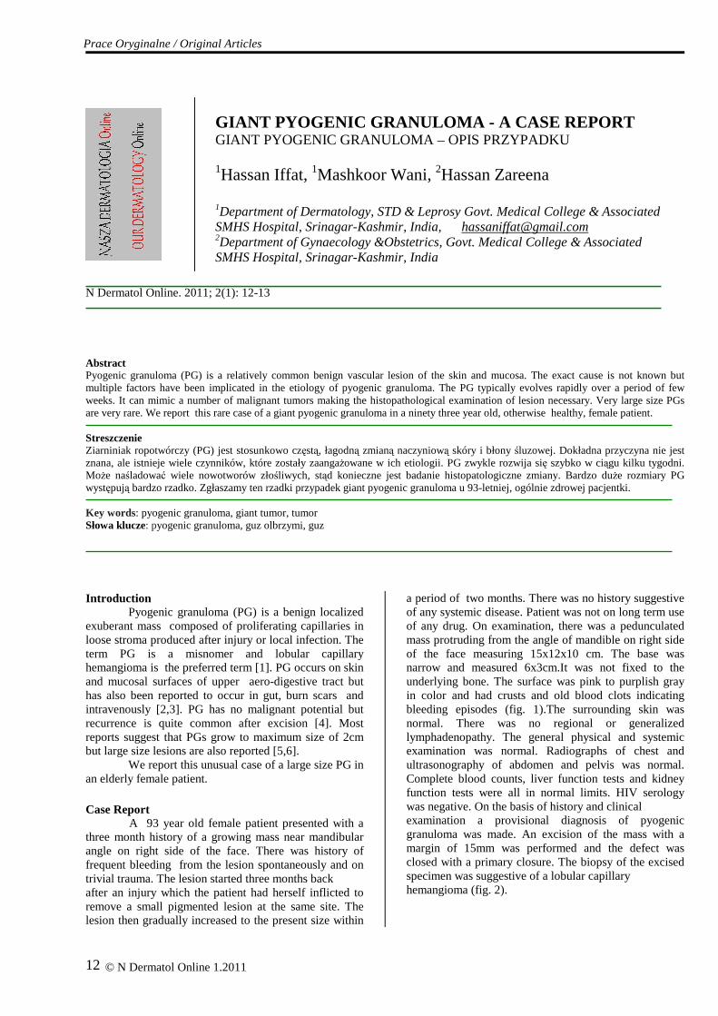
a period of two months. There was no history suggestive of any systemic disease. Patient was not on long term use of any drug. On examination, there was a pedunculated mass protruding from the angle of mandible on right side of the face measuring 15x12x10 cm. The base was narrow and measured 6x3cm.It was not fixed to the underlying bone. The surface was pink to purplish gray in color and had crusts and old blood clots indicating bleeding episodes (fig. 1).The surrounding skin was normal. There was no regional or generalized lymphadenopathy. The general physical and systemic examination was normal. Radiographs of chest and ultrasonography of abdomen and pelvis was normal. Complete blood counts, liver function tests and kidney function tests were all in normal limits. HIV serology was negative. On the basis of history and clinical examination a provisional diagnosis of pyogenic granuloma was made. An excision of the mass with a margin of 15mm was performed and the defect was closed with a primary closure. The biopsy of the excised specimen was suggestive of a lobular capillary hemangioma (fig. 2).
Prace Oryginalne / Original Articles
© N Dermatol Online 1.2011
13
Figure 1. Giant Pyogenic granuloma, left angle of mandible. Discussion
Pyogenic granulomas are benign, exophytic vascular tumors first described by Poncet and Dor in 1897. It is seen quite often in children and young adults but is unusual in elderly [7]. Although exact pathogenesis is not known, trauma, hormonal influences, inflammatory and infectious agents have all been hypothesized as possible factors in causation [8,9]. Because of similarity in clinical and histopathological findings with bacillary angiomatosis, some workers have suggested that PG may be caused by Bartonella spp. infection [10].We were unable to find any predisposing factor other than trauma in our patient. The usual size of PG is less than 2cm,but there are reports of giant pyogenic granulomas in immunocompromised patients [11]. Our patient, although elderly, did not show any signs of immunosupression. PG can mimic malignant diseases, therefore a biopsy is recommended. In one series 38 percent cases of clinical diagnosis of PG proved to be wrong [12]. In our case there was a possibility of malignant melanoma as there was a history of pigmented lesion at the site of origin but histopathology cleared the doubt.
Figure 2. Histopathology shows numerous vascular spaces lined by endothelial cells, surrounded by stroma mixed with inflammatory cells.
REFERENCES / PIŚMIENNICTWO :
1. Grosshans E: Editorial; Pyogenic granuloma: who are you? J Eur Acad Dermatol Venereol 2001; 15: 106-107. 2. Ghekiere O, Galant C, Vand de Berg B: Intravenous pyogenic granuloma or intra venous lobular capillary hemangioma. Skeltal Radiol 2005; 34: 343-346. 3. Blanchard S, Chelimsky G, Czinn S, Redline R, Splawsky J: Pyogenic granuloma of colon in children. J Pediatr Gastroenterol Nutr 2006; 43: 119-121. 4. Kapadia SP, Heffuer DK: Pitfalls in histopathological diagnosis of pyogenic granuloma. Eur Arch Otorhinolaryngiol 1992; 249: 195-200. 5. Choudary S, Mackinnon CA, Morissey GP, Tan ST: A case of giant nasal pyogenic granuloma gravidarum. J Craniofac Surg 2005; 16: 319-321. 6. Tursen U, Demirkan F, Ikizoglu G: Giant pyogenic granuloma on face with satallitosis responsive to systemic steroids. Clin Exp Dermatol 2004; 29: 40-41. 7. Knoth W, Ehlers G: On the problem of existence of telengiectatic pyogenic granuloma with special reference to its relations tohemangioma and hemangioendothelioma. Arch Klin Exp Dermatol 1962; 214: 394-414. 8. Patrice SJ, Wiss K, Mullikan JB: Pyogenic granuloma:a clinicopathological study of 178 cases .Pediatr Dermatol 1991; 8: 267-276. 9. Behne K, Robertson I, Weedon D: Disseminated lobular capillary hemangioma. Australas J Dermatol 2002; 43: 297-300. 10. Itin PH, Fluckiger R, Zbinden R, Frei R: Recurrent pyogenic granuloma with satallitosis:a localized variant of bacillary angiomatosis?. Dermatology 1994; 189: 409-412. 11.Yucel A, Ayden Y, Benlier E, Demirkesen C, Yildirim I: A giant pyogenic granuloma of thumb. Ann Plast Surg 2000; 45: 216-218. 12. Rowe L: Granuloma pyogenicum. AMA Arch Dermatol 1958; 78: 341-347.
© N Dermatol Online 1.2011