4th Lecture (NCM106 CA I) Care of Clients in Cellular Aberrations, ABC, Emergency and Disaster...
14
jcmendiola_Achievers 2013 Care of Clients in Cellular Aberrations, Acute Biologic Crisis (ABC), Emergency and Disaster Nursing (NCM106) Cellular Aberration I Cellular Aberration Basic structural and functional unit of an organism Cell Cycle - Is a coordinated sequence of events resulting in duplication of DNA and division into 2 daughter cell 4 Phases of the Cell Cycle 1. G1 / Gap Phase • Lasts from hours to days / longer • RNA and Protein synthesis occurs in preparation for DNA replication 2. S Phase / Synthesis Phase • Lasts from 10 – 20 hours • DNA replication in preparation for division 3. G2 / Gap 2 • Ranges from 2 – 10 hours • DNA synthesis while RNA and Protein synthesis continues 4. M Phase / Mitosis Phase • Lasts from 30 – 60 minutes • Cell division occurs • After mitosis the daughter cells enter the G1 Phase and begin the reproductive cycle again 5. G0 / Resting Phase • Is activity to reenter the cell cycle in response to various stimuli that signal for cell renewal CELL CYCLE Oncology Nursing - Field of specialty - Nurse must be equipped to support patient and family through a wide range of physical, emotional, social, cultural and spiritual crises - Provide realistic support to those receiving nursing care and use standards of practice and nursing process as basis of care Cellular Aberration - A group of disorders characterized by abnormal cell growth and the ability to metastasize with potential in killing the host - The term “cancer” refers to the group of diseases in which cells grow and spread unrestrained throughout the body - Derived from the Latin word ‘crab’ which means Cancer - Synonymous with neoplasm LOOKY HERE ☺ 1. Introduction on Cellular Aberration 2. Multistage Theory of Oncogenesis 3. Tumor Invasion and Metastasis 4. Primary Prevention and Control 5. Secondary Prevention and Early Detection 6. Staging 7. Chemotherapy
-
Upload
kamx-mohammed -
Category
Documents
-
view
68 -
download
2
description
:)
Transcript of 4th Lecture (NCM106 CA I) Care of Clients in Cellular Aberrations, ABC, Emergency and Disaster...

jcmendiola_Achievers 2013
Care of Clients in Cellular Aberrations,
Acute Biologic Crisis (ABC), Emergency and Disaster Nursing
(NCM106)
Cellular Aberration I
Cellular Aberration
� Basic structural and functional unit of an organism
Cell Cycle
- Is a coordinated sequence of events resulting in duplication of
DNA and division into 2 daughter cell
4 Phases of the Cell Cycle
1. G1 / Gap Phase
• Lasts from hours to days / longer
• RNA and Protein synthesis occurs in preparation for DNA replication
2. S Phase / Synthesis Phase
• Lasts from 10 – 20 hours
• DNA replication in preparation for division
3. G2 / Gap 2
• Ranges from 2 – 10 hours
• DNA synthesis while RNA and Protein synthesis continues
4. M Phase / Mitosis Phase
• Lasts from 30 – 60 minutes
• Cell division occurs
• After mitosis the daughter cells enter the G1 Phase and begin the reproductive cycle again
5. G0 / Resting Phase
• Is activity to reenter the cell cycle in response to various stimuli that signal for cell renewal
CELL CYCLE
Oncology Nursing
- Field of specialty
- Nurse must be equipped to support patient and
family through a wide range of physical, emotional,
social, cultural and spiritual crises
- Provide realistic support to those receiving nursing
care and use standards of practice and nursing
process as basis of care
Cellular Aberration
- A group of disorders characterized by abnormal cell growth and the ability to metastasize with potential in
killing the host
- The term “cancer” refers to the group of diseases in which cells grow and spread unrestrained throughout
the body
- Derived from the Latin word ‘crab’ which means Cancer
- Synonymous with neoplasm
LOOKY
HERE ☺
1. Introduction on Cellular
Aberration
2. Multistage Theory of
Oncogenesis
3. Tumor Invasion and Metastasis
4. Primary Prevention and Control
5. Secondary Prevention and Early
Detection
6. Staging
7. Chemotherapy

jcmendiola_Achievers 2013
INCIDENCE and EPIDEMICS
Male Female
Most Common Cause of Death Most Common Cause of Death
Prostate Cancer
(33%)
Lung Cancer
(31%)
Breast Cancer
(32%)
Lung Cancer
(27%)
Lung Cancer
(13%)
Prostate Cancer
(10%)
Lung Cancer
(12%)
Breast Cancer
(15%)
Colorectal Cancer
(10%)
Colorectal Cancer
(10%)
Colorectal Cancer
(11%)
Colorectal Cancer
(10%)
Bladder Cancer
(7%)
Pancreatic Cancer
(5%)
Endometrial Cancer
(6%)
Ovarian Cancer
(6%)
Cutaneous Melanoma
(5%)
Leukemia
(4%)
Non-Hodgkins Lymphoma
(4%)
Pancreatic Cancer
(6%)
TOP 5 Cancer Incidences by Site and Sex
Male Female
1. Prostate 1. Breast
2. Lungs 2. Lungs
3. Colon 3. Colon
4. Urinary Tract 4. Uterus
5. Leukemia 5. Leukemia and Lymphoma
Women Men
� Breast Cancer followed by lung
and colon and rectum
� High incidence of cancer of the lung and bladder
� Most common neoplasm aged 20 – 34; testicular Cancer
Etiologic Agent 1. Viruses and Bacteria
� “Oncogenic viruses”
� Prolonged / frequent viral infections may cause breakdown of the immune system / overwhelm the
immune system
2. Chemical Carcinogens
� Act by causing cellular mutation / alterations in cell enzymes and protein
� E.g. Industrial compounds – vinyl chloride, polycyclic aromatic hydrocarbons, fertilizers, weed
killers, dyes and drugs
3. Physical Agents
� Radiation – X-ray / radioactive isotopes and sunlight / UV Rays
� Physical Irritation/ trauma – Pipe smoking, multiple deliveries, ragged tooth, irritation of the
tongue, “overuse” of any organ / body part
4. Hormonal Agents
� Estrogen as replacement therapy ↑ incidence of vaginal and cervical adenocarcinoma
� Estrogen, diethylstilbestrol (DES)
5. Genetics and Familial Factors
� Oncogene � When exposed to carcinogens � Changes in the cell structure � Becomes
malignant
Predisposing Factors
1. Age – Older individuals exposed to carcinogens longer develop immune system alterations
2. Sex
• Women = Breast, Uterus, Cervix Cancer
• Men = Prostate, Lung Cancer
3. Occupation – E.g. Chemical factory worker, radiology department personnel
4. Hereditary – Greater risk with positive family history

jcmendiola_Achievers 2013
Urban Versus Rural Incidence
- Common among URBAN DWELLERS than RURAL RESIDENCES
(Greater exposure to carcinogens)
- Geographic Distribution
o Cancer in stomach – Japan
o Breast Cancer – US; due to environmental diet, ethnic customs and types of pollution
5. Psychological Stress
� Depression, grieving, anger, aggression, despair or life stresses decreases immune competence
(Affects hypothalamus and pituitary gland)
� Immunodeficiency may spurt the growth and proliferation of Cancer cells
6. Precancerous Lesions
o May undergo transfer into cancer lesion and tumor
o E.g. Pigmented moles, burn scars, senile keratosis, leukoplakia, benign polyps, adenoma of the
colon / stomach fibrocystic disease of the breast
7. Obesity
Studies have linked obesity to breast and colorectal Cancer
Factors to Consider
MR JUAN DELA CRUZ
Etiology - Carcinogens – The process of transferring a
normal cell into cancerous cell which
consists of 3 Stages
1) Initiation (Carcinogen)
2) Promotion, repeated exposure to promote agents (Carcinogen)
3) Progression (↑ Malignancy behavior)
D Drugs
E Educational Attainment
L Living Conditions
A Ask family History
C Culture
R Radiation Therapy
U Ur Activity
Z Zex
M Marital Status
R Race
J Job
U Ur Life Style
A Age
N Nutrition

jcmendiola_Achievers 2013
Multistage Theory of Oncogenesis
1. Cellular Transformation and Pre-agent Theory
� Conceptualize that normal cells may be transformed into cancer cells due to exposure to etiologic
agents
2. Failure of the Prime Resource Theory
� Advocates that all individuals possess cancer cells, however the cancer cells are recognized by the
immune system so the cancer cells undergo destruction
� Failure of the immune response system leads to inability to destroy the cancer cells
TERMS
1. Cell Proliferation
� Is the process whereby cells divide and bear offspring, it normally is regulated so that the number
of cells that are actively dividing is equal to the number of dying / being shed
2. Differentiation
� Is the process whereby proliferative cells are transformed into different and more specialized cell
types, as they proliferate it determines what a cells looks like, and how it functions, how long it
will live
3. Apoptosis
� It is the process of programmed death of unwanted cells
BENIGN GROWTH PATTERN
1. Hypertrophy
� ↑ In cell size resulting in an ↑ in organ size!
2. Hyperplasia
� A reversible ↑ in the number of cells in an organ or tissue in response to a specific growth stimulus
3. Metaplasia
� Conversion of one cell type to another cell type not usually found in the involved tissue
4. Dysplasia
� Characterized by abnormal changes in the size, shape, or organization of cells
� Reversible when stimulus is removed

jcmendiola_Achievers 2013
5. Anaplasia � Disorganized irregular cells that have no structure and have loss of differentiation, the result is
almost malignant
CLASSIFICATIONS OF TUMORS
1. Benign
� Are tumors designated by attaching the suffix ‘–oma’ to the cells of organ
� E.g. Fibroma, Chondroma,, Osteoma
2. Malignant
� Tumors that are capable of spreading by invasion and metastasis
� E.g. Fibrosarcoma, Chondrosarcoma
CATEGORIES OF MALIGNANT NEOPLASMS
1. Carcinogens – Growth from epithelial cells, usually solid tumors
2. Sarcoma – Arise from muscle, bone, fat and connective tissue, may be solid
3. Lymphoma – Arise from lymphoid tissues
4. Leukemia and Myeloma – Grows from blood forming organs
Nomenclature of Tumors
Tissue of Organ Benign Malignant
Connective tissue and derivatives Fibroma
Lipoma
Chondroma
Osteoma
Fibrosarcoma
Liposarcoma
Chondrosarcoma
Osteogenic Sarcoma
Blood Vessels Hemangioma Angiosarcoma
Lymphatic Vessels Lympangioma Lymphangiosarcoma
Brain Meningioma Invasive Meningioma
Hematopoietic Cells Leukemia
Lymphatics Malignant lymph***
Smooth Muscles Leiomyoma Leiomyosarcoma
Stratified Muscles Rhabdomyoma Rhabdomyomasarcoma
Epithelial Tumors
Stratified Squamous Squamous cell papilloma Squamous cell carcinoma
Basal Cells Basal Cells carcinoma
Liver Cells Liver cell adenoma Hepatocellular Carcinoma
Placental epithelium
(Trophoblast)
Hydatidiform Mole
Tumor Invasion and Metastasis
• Invasion
� Occurs when cancer cells infiltrate adjacent tissues surrounding the neoplasm
• Metastasis
� Occurs when malignant cells travel through the blood / lymph and invade other tissues and organs
to form a secondary tumor
� Types of Metastasis
Extension and Invasion
1. Lymphatic Spread
2. Seeding of body cavities and surfaces
3. Hematogenous spread
� Spread of cancer cells from a primary tumor to distant sites
� “Break away”
� Only malignant cells has the capability
� Lymph, blood, serosal seeding

jcmendiola_Achievers 2013
Comparison of the Characteristics of Benign and Malignant Neoplasms
Characteristics Benign Malignant
Speed of Growth Slow growth
Grows by expansion
Aggressive growth; rapid cell
division and growth
Mode of Growth Localizes and encapsulation Establishes new site malignant
lesion
Cellular Characteristics Well-differentiated Invade surrounding tissues
Metastasis It does not metastasize
No tissue damage
With poor cellular differentation
Prognosis Very good prognosis
Does not cause death, unless
localization affects vital functions
Malignant Cells – Mitosis
o Mitosis – Multiple daughter cells that may / may not resemble the parent, multiply mitotic spindles
1. Larger, grows more rapidly than normal cells
2. Cells not as cohesive, irregular pattern of expansion
3. Larger, more prominent nucleus
4. Lack characteristic pattern of organization of host cells
5. Anaplastic = Lack of differentiated cell characteristics specific function
Malignant Cells – Growth
1. Invade adjacent tissues
2. Proliferation in response to abnormal stimulus
3. Grow in adverse condition such as lack of nutrients
4. Do not exhibit contact-inhibition
5. Cell birth exceeds cell death
6. Loss of cell control as a result of cell membrane changes
7. Growth rate, erratic
8. Able to break off cells that migrate through blood stream / lympati** seed to distant sites and grow in other
sites
Malignant Cells – Function
1. Senseless, no useful purpose
2. Do not contribute to the well-being of the host, parasitic
3. If the cells function at all, they do not function normally may cause damage
Malignant Cells
1. Develop antigens completely different from a normal cell
2. Chromosomal aberrations occur as a cell matures
3. Has a more prominent and simplified metabolic enzyme pattern
4. Invasive and spreads
5. Grow in presence of necrosis and inflammatory cells such as lymphocytes and macrophages
6. Exhibit periods of latency that vary from tumor to tumor
7. Have own blood supply and suppository stoma (Angiogenesis factory 2 cm in diameter)
jcmendiola_Achievers 2013
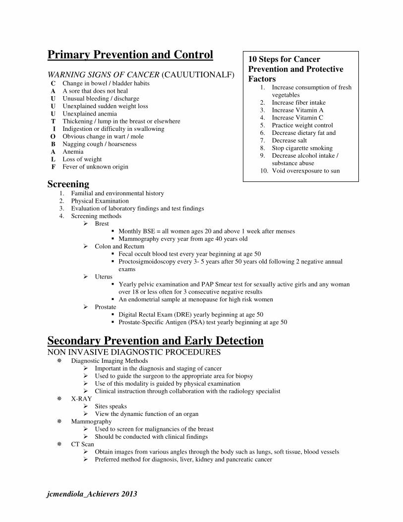
Primary Prevention and Control
WARNING SIGNS OF CANCER (CAUUUTIONALF)
C Change in bowel / bladder habits
A A sore that does not heal
U Unusual bleeding / discharge
U Unexplained sudden weight loss
U Unexplained anemia
T Thickening / lump in the breast or elsewhere
I Indigestion or difficulty in swallowing
O Obvious change in wart / mole
B Nagging cough / hoarseness
A Anemia
L Loss of weight
F Fever of unknown origin
Screening
1. Familial and environmental history
2. Physical Examination
3. Evaluation of laboratory findings and test findings
4. Screening methods
� Brest
� Monthly BSE = all women ages 20 and above 1 week after menses
� Mammography every year from age 40 years old
� Colon and Rectum
� Fecal occult blood test every year beginning at age 50
� Proctosigmoidoscopy every 3- 5 years after 50 years old following 2 negative annual
exams
� Uterus
� Yearly pelvic examination and PAP Smear test for sexually active girls and any woman
over 18 or less often for 3 consecutive negative results
� An endometrial sample at menopause for high risk women
� Prostate
� Digital Rectal Exam (DRE) yearly beginning at age 50
� Prostate-Specific Antigen (PSA) test yearly beginning at age 50
Secondary Prevention and Early Detection
NON INVASIVE DIAGNOSTIC PROCEDURES
� Diagnostic Imaging Methods
� Important in the diagnosis and staging of cancer
� Used to guide the surgeon to the appropriate area for biopsy
� Use of this modality is guided by physical examination
� Clinical instruction through collaboration with the radiology specialist
� X-RAY
� Sites speaks
� View the dynamic function of an organ
� Mammography
� Used to screen for malignancies of the breast
� Should be conducted with clinical findings
� CT Scan
� Obtain images from various angles through the body such as lungs, soft tissue, blood vessels
� Preferred method for diagnosis, liver, kidney and pancreatic cancer
10 Steps for Cancer
Prevention and Protective
Factors
1. Increase consumption of fresh
vegetables
2. Increase fiber intake
3. Increase Vitamin A
4. Increase Vitamin C
5. Practice weight control
6. Decrease dietary fat and
7. Decrease salt
8. Stop cigarette smoking
9. Decrease alcohol intake /
substance abuse
10. Void overexposure to sun

jcmendiola_Achievers 2013
� MRI
� Preferred imaging technique for soft tissue structures, hematologic imaging, vascular imaging and
avascular necrosis
� Not exposed to radiation
INVASIVE DIAGNOSTIC PROCEDURE
� Histologic / Cytologic Examination
o For malignant tissues to be identified by name, grade and stage
o Morphologic feature of the cells are examined
3 Basic Methods of Specimen Collection
1. Exfoliation from an epithelial surface (pap smear) or bronchial washing
2. Aspiration of fluid from body cavities or blood
3. Needle suction aspiration of solid tumor
� Direct Visualization
1. Sigmoidoscopy (Viewing the sigmoid colon by use of fiberoptic flexible sigmoidoscopes
2. Cystoscopy (Viewing the urethra and bladder)
3. Endoscopy (Viewing of the upper GIT)
4. Bronchoscopy (Inspection of the tracheobronchial tree
LABORATORY STUDIES
• Tumor Markers
� Biochemical substances synthesized and released by tumor cells
� May be protein products exerted by cancer cells, released in response to the presence of cancer
cells or other conditions � Used to aid in the diagnosis of cancer to determine recurrence or identify regression of a known malignancy
TUMOR MARKER DESCRIPTION
1. Oncofetal Antigen Present in fetal tissue normally suppressed after birth
2. Hormones Present in considerable amount
High levels in hormone-secreting malignancies
3. Isoenzymes Elevated levels can promote hyperplasia of the tissue (Prostate acid
phosphatase)
4. Tissue-Specific Protein Narrows down the type of malignancy that can be increased in
hyperplastic disorders
5. Prostate-Specific Antigen Useful in evaluating response to treatment, recurrent surgery / radiation
therapy
Elevated in prostate cancer, can be elevated in BPH in older men, should
be accompanied with DRE
6. S-100 Found in melanoma cells
Elevated means METASTATIC MELANOMA
7. Thyroglobulin Protein made by the thyroid gland
Removal of the entire gland with or without radiation therapy
Rise in thyroglobulin levels indicate cancer recurrence
8. Estrogen and Progesterone
Receptors
Once diagnosed, breast cancer tissue become tested for the presence of
E and P receptors
Provides an indication of the aggressiveness of the cancer and how
likely the cancer will respond to specific types of endocrine therapy
9. Ca 15 – 3 and Ca 27 – 29 Specific for BREAST CANCER
Found in the blood of an affected patient
Ca 27 – 29 test is MORE sensitive than Ca 15 – 3
10. Carcinoembryonic Antigen
(CEA)
and Ca 19 – 9
Elevated in ADVANCED COLORECTAL CANCER
↑ CEA level before surgery – POORER PROGNOSIS
11. Human Chorionic
Gonadotropin (HCG) and
Alpha-fetoprotein (AFP)
With germ cell ovarian tumors in men with non-seminomatous
TESTICULAR CANCER = Elevated HCG and AFP
Proportionately ↑ to the size of tumors
AFP levels may also be increased in CHRONIC HEPATITIS

jcmendiola_Achievers 2013
12. Beta-2-Microglobulin (B2M) Elevated in periods with multiple myeloma with chronic lymphocytic
leukemia, kidney disease
13. HER-2 / NEU Elevated in one-thirds of persons diagnosed with breast cancer
Laboratory Tests
- Complete Blood Count (CBC)
- Blood Chemistry
Serum electrolytes
ALT – Alanine Aminotransferase
AST – Aspartate Aminotransferase
LDH – For liver metastases
CEA – For colon cancer
STAGING
• Done during the pre-treatment phase
• After surgical resection
• Recurrence after disease free interval
STAGING – TUMOR
Tumor TNM Staging System
T0 No end of primary tumor
Tis Carcinoma in situ
T1, T2, T3, T4 Progressive increase in tumor size and involvement
Tx Tumor cannot be assessed
STAGES
Stage I The tumor is small, local, detected early
Stage II The tumor is somewhat larger and has started to spread to nearby lymph nodes
Stage III The tumor has spread to nearby lymph nodes
Stage IV Cancer has spread to other parts of the body and is generally in an advanced stage
STAGING – NODE
N0 Regional lymph nodes
N1, N2, N3 ↑ degree of demonstrable abnormality of regional lymph nodes
Nx Regional lymph nodes cannot be assessed clinically
STAGING – METASTASIS
M0 No evidence of distant metastasis
M1, M2, M3 Ascending degree of distant metastasis, including metastasis to different lymph nodes
GRADING
Gx Grade cannot be assessed
G1 Well differentiated
G2 Moderately well-differentiated
G3 and G4 Poorly to very poorly differentiated Poorer differentiation – poorer prognosis
Classification, Grading and Stages
� TNM Classification
T Extent of primary tumor
Tx Cannot be adequately assessed
T0 No evidence of primary tumor
Tis Tumor in situ 0 localized; no spread
T1 4 prognosis, increase in size
1.5 cm < 2: 6-9 cm
3:10-15 cm 4:15 cm >

jcmendiola_Achievers 2013
STAGES
0 Benign state
I Spread to nearby tissue
II 2 – 5 cm sometimes involve lymph
III Greater than 5 cm spread – advanced spread to connective tissue
IV Metastasis
Grading of Tumor
Grade I Well differentiated
Grade II Moderately well differentiated
Grade III Poorly differentiated
Grade IV Undifferentiated
CHEMOTHERAPY
A systematic mode of treatment that uses cytotoxins and chemicals to effectively CURE (Leukemia,
Lymphomas, some solid tumors)
� ↓ Tumor size
� Adjunct to surgery / radiation
� Prevent / treat suspected metastasis
Most effective when the tumor is small and cell replication is rapid
Individualized to the patient and is often prescribed according to the patient’s calculated body surface area
and type of cancer
Example:
� Acute Lymphocytic Leukemia (ALL)
� Uses DVPA
� Daunorubicin – Given days 1 – 3
� Vincristine – Given days 1, 8, 15 and 22
� Prednisone – Given days 11 – 28
� Asparaginase – Given days 17 – 28
� Given in cycles with rest periods (especially if with toxic effects) until disease goes to remission
Chemotherapy Cell Cycle
- Used to disrupt the cell cycle in various phases in specific protocols that are given over varying periods of
time
Cell Kill Hypothesis
1. Several doses of chemotherapy are necessary
2. Each exposure kills: 20% - 99% depending on dosage
3. Repeated exposure targets even those in G0 and leads to regression
4. 100% eradication of tumor cells – IMPOSSIBLE
5. But the goal is: To reduce the amount that can be destroyed by the immune system
Factors Crucial to the Rate of Normal / Malignant Tissues
1. Cell Cycle Timing: Amount of time required for cells to remove from one mitosis to the next
2. Growth Fraction: Ratio of dividing cells to resting cells, fraction of cycling cells in the entire cell
population
3. Rate of Cell Loss: Fracture of cell die or leaves
Route of Chemotherapy
1. Oral – Hodgkin’s Lymphoma, Leukemia (Maintenance phase), Lung Cancer
2. Intravenous – Leukemia,
3. Intra-arterial – Hepatic tumors, head and neck cancer
4. Intracavity – Ovarian cancer
New RESEARCH!
� Use of chemotherapy based on
CIRCARDIAN RHYTHMS
� E.g. Colon Cancer

jcmendiola_Achievers 2013
5. Intraperitoneal – Brain tumors
6. Intraventricular – Brain tumor
7. Intravesical – Bladder tumors
OBJECTIVES:
� To destroy all malignant tumor cells without excessive destruction of normal cells
� To control growth if cure is no longer possible
� Used as adjunct therapy
CONTRAINDICATIONS
� Infection: Anti-tumor drugs are immunosuppressive
� Recent surgery: Drugs may retard healing process
� Impaired renal / Hepatic Function: Drugs are nephrotoxic and hepatotoxic
� Recent Radiation Therapy: Immunosuppressive
� Pregnancy: Drugs may cause congenital defects
� Bone Marrow Depression: Drugs may aggravate the condition, WBC must be within normal levels
Safe Handling of Chemotherapeutic Agents
� Wear mask, gloves and back-closing gown
� Skin contact with drugs must be washed immediately with soap and water. Eye must be flushed
immediately with copious amount of water
� Sterile / Alcohol – Wet cotton pledgets should be used, wrapped around the neck of the ampule / vial
when breaking and withdrawing the drug
� Expel air bubbles or wet cotton
� Vent vials to reduce internal pressure after mixing
� Wipe external surface of syringe and IV bottles
� Avoid self-inoculation by needle stab
� Clearly label the hanging IV bottle with antineoplastic chemotherapy
� Contaminated needles and syringes must be disposed in a clearly marked special container “leak-proof” or
“puncture proof”
� Dispose half-empty ampules, vials, IV bottles by putting them into plastic bags sealed and then into
another plastic bag or box, clearly marked before placing for removal. Label as “Hazardous Wastes”
� Handwashing should be done before and after removal of gloves
� Trained personnel only should be involved in use of drugs
Effects of Chemotherapeutic Drugs
Tissues normally affected are:
1. Mucous Membranes
� Mouth, tongue, esophagus, stomach, intestine and rectum
� Results in anorexia, loss of taste, aversion to food, Erythema,
painful ulceration of GIT, NV, diarrhea
2. Hair Cells
� Alopecia
3. Bone Marrow Depression
� Affects: Granulocytes, lymphocytes, thrombocytes, erythrocytes
� Impaired ability to respond to infection, blood clot and severe
anemia
4. Organ
� Heart, lungs, bladder, kidney
� Due to specific agents
� E.g. Cardiac toxicity (Doxorubicin)
� Pneumocystis (Bleomycin)
Effects of CHEMO DRUGS
1. Combined medication
therapy is used to
enhance tumor cell
kill
2. Synergistic actions of
drugs will prevent the
development of drug
resistance
3. Combats resistance of
cells to
chemotherapeutic
agents

jcmendiola_Achievers 2013
Classification of Chemotherapeutic Drugs
Related to the cell cycle
1. Cycle Specific Agents
� They are specific to certain phases of the cell cycle
� Destroy cells that are actively reproducing
� Most affects there in the S Phase of interfering with DNA and RNA synthesis
� M Phase (Vinca / Plant Alkaloids: Halt spindle function)
2. Cycle Non-Specific Agents
� Act independently of the cell cycle place
� Usually have prolonged effects or cells leading to cell death and damage
Classifications of Drugs
1. Alkylating Agents
� Contains alkyl groups which binds to DNA and prevents replication and mitosis
� Cell Cycle non-Specific
� Effective against many types of cancer, including acute and chronic leukemia, solid tumors
� Common Side Effects
� Bone marrow suppression
� N/V
� Alopecia
� Sterility
� Cystic cyclophosphamide
� Stomatitis
� Renal Toxicity (Cisplastin)
� E.g.
� Bisulfiram (Bisulflex)
� Cyclophosphamide (Cytoxan)
� Chlorambucil (Leukeran)
� Cisplastin (Planitol-AQ)
� Nursing Implications:
� Maintain good hydration
� Administer anti-emetics prior to chemotherapy
� Monitor WBC, Uric Acid
� Assess for possible infection
� Discuss concerns for hair loss
2. Nitrosoureas
� Similar to the alkylating agent
� ONLY CHEMODRUG THAT CAN CROSS THE BLOOD BRAIN BARRIER (BBB)
[Important for Central Nervous System diseases]
� Side Effects:
� Delayed cumulative myelosuppression (In 3 – 5 weeks) especially thrombocytopenia;
N/V
� Nursing Implications:
� Maintain good hydration
� Administer anti-emetics prior to chemotherapy
� Monitor WBC, Uric Acid
� Assess for possible infection
� Discuss concerns for hair loss
3. Anti Metabolites
� Interferes with the biosynthesis of metabolism or nucleic acid needed for RNA and DNA synthesis
� Cell specific (Best in S Phase)
� Used to treat acute leukemia, breast cancer, head and neck cancer, lung cancer, and osteosarcoma
� Side Effects:
� Bone Marrow suppression (Anemia, leukopenia)

jcmendiola_Achievers 2013
� Stomatitis
� N/V
� Alopecia
� Hepatitis and renal dysfunction
� E.g.
� Methotrexate
� Lethal in high doses, must give antidote (Leucovorin) within 24 – 36 hours after
initiation of therapy
� 5-Flurouracil (5-FU)
� Cytarabine (Depocyt, Tarabine)
� 5-Azacytidine
� Side Effects
� N/V
� Diarrhea
� Bone Marrow suppression: Reaches NADIR in 1 – 2
weeks; with leukopenia being most severe
� Renal toxicity (Methotrexate)
� Hepatotoxicity
� Nursing Implications
� Monitor CBC, WBC, Uric acid
� Assess oral mucus membranes
� Assess for infection, bleeding
� Provide oral care
� Administer anti-emetics PRN
� Discuss concern for hair loss
� Evaluate hydration and nutritional status
4. Antitumor Antibiotics
� Inhibit RNA synthesis and bind DNA causing fragmentation; interfere with DNA repair
� These drugs bind to almost everything they contact and kill cells
� Main toxic effect is cardiac muscle toxicity (Limits the amount and duration of treatment)
� Side Effects are the same with other anti-Cancer drugs
� E.g.
� Doxorubicin (Adriamycin)
� Bleomycin (Blenoxane)
� Dactinomycin (Cosmegen)
� Nursing Implications
� Monitor ECG, CBC
� Assess for bleeding
� Assess for hydration and nutritional status
� Check for fever 36 hours after administration
� Administer anti-emetic PRN
5. Plant Alkaloids
� Two main Groups (From natural products)
1. Vinca Alkaloids – Mitosis phase, inhibit mitotic tubular formation (spindle); inhibit DNA
and protein synthesis
2. Etoposide (VP-16) or Mitotic Inhibitors – All phases; causes breaks in DNA and
metaphase arrest
� E.g.
1. Vincristine (Oncovin)
Vinblastin (Velban)
2. Etoposide (Toposar)
Teniposide (Venom)
� Side Effect:
� Hypotension (Too rapid IV administration), muscle weakness, areflexia, constipation,
N/V, alopecia
� Nursing Implications:
NADIR – Is the lowest level of
a red blood cell count while a
patient is undergoing
chemotherapy

jcmendiola_Achievers 2013
� Assess neuromuscular functions
� Monitor CBC, GI function
� Manage constipation
� Hydration
� Discuss concerns for hair loss
6. Hormonal Agents
� Alter the deviate / environment to depress / prevent cell proliferation
� Corticosteroids (e.g. Prednisone: Mostly used in CA therapy; G1 Phase)
� E.g.
� Androgen, estrogen, anti-androgens, anti-estrogens
� Side Effects
� N/V
� Hyperglycemia
� Hypertension
� Weight gain; gynecomastia
� Mood changes
� Cessation of menstruation
� Acne, alopecia
Nursing Interventions for Chemical Side Effects
• GI System = N/V, diarrhea, constipation
� Administer anti-emetics to relieve N/V
� Replace fluids and electrolyte losses, low fiber diet to relieve diarrhea
� ↑ fluid intake and fibers in diet to prevent / relieve constipation
• Integumentary System
� Pruritus; urticaria and systemic signs
� Provide good skin care
� Stomatitis
� Provide good oral care, avoid HOT and SPICY food
� Alopecia
� Reassure that it is temporary, wear wigs / hats
� Skin Pigmentation
� Inform that it is temporary
� Nail changes (Grow normally after chemotherapy)
• Hematopoietic System
� Anemia
� Frequent rest periods, eat foods high in Iron!
� Neutropenia
� Protect from infection
� Avoid people with infection
� Thrombocytopenia
� Protect from trauma
� Avoid ASA
• Genito-Urinary System
� Hemorrhagic Cystitis
� Provide 2 – 3 L of fluids per day
� Urine color changes
� Reassure that it is harmless