
4,6--Glucanotransferase, a Novel Enzyme That Structurally ... · Ariyanti Oetari,3 Johannis P....
10
APPLIED AND ENVIRONMENTAL MICROBIOLOGY, Nov. 2011, p. 8154–8163 Vol. 77, No. 22 0099-2240/11/$12.00 doi:10.1128/AEM.05735-11 Copyright © 2011, American Society for Microbiology. All Rights Reserved. 4,6--Glucanotransferase, a Novel Enzyme That Structurally and Functionally Provides an Evolutionary Link between Glycoside Hydrolase Enzyme Families 13 and 70 Slavko Kralj, 1 § Pieter Grijpstra, 1 Sander S. van Leeuwen, 1 Hans Leemhuis, 1 Justyna M. Dobruchowska, 1 Rachel M. van der Kaaij, 1 Amarila Malik, 2 Ariyanti Oetari, 3 Johannis P. Kamerling, 1 and Lubbert Dijkhuizen 1 * Department of Microbiology, Groningen Biomolecular Sciences and Biotechnology Institute (GBB), University of Groningen, Nijenborgh 7, 9747 AG Groningen, The Netherlands, 1 and Department of Pharmacy 2 and Department of Biology, 3 Faculty of Mathematics and Natural Sciences, University of Indonesia, Kampus UI Depok, Depok 16424, Indonesia Received 3 June 2011/Accepted 12 September 2011 Lactobacillus reuteri 121 uses the glucosyltransferase A (GTFA) enzyme to convert sucrose into large amounts of the -D-glucan reuteran, an exopolysaccharide. Upstream of gtfA lies another putative glucansucrase gene, designated gtfB. Previously, we have shown that the purified recombinant GTFB protein/enzyme is inactive with sucrose. Various homologs of gtfB are present in other Lactobacillus strains, including the L. reuteri type strain, DSM 20016, the genome sequence of which is available. Here we report that GTFB is a novel -glucanotrans- ferase enzyme with disproportionating (cleaving 134 and synthesizing 136 and 134 glycosidic linkages) and 136 polymerizing types of activity on maltotetraose and larger maltooligosaccharide substrates (in short, it is a 4,6--glucanotransferase). Characterization of the types of compounds synthesized from malto- heptaose by matrix-assisted laser desorption ionization–time of flight mass spectrometry (MALDI-TOF MS), methylation analysis, and 1-dimensional 1 H nuclear magnetic resonance (NMR) spectroscopy revealed that only linear products were made and that with increasing degrees of polymerization (DP), more 136 glycosidic linkages were introduced into the final products, ranging from 18% in the incubation mixture to 33% in an enriched fraction. In view of its primary structure, GTFB clearly is a member of the glycoside hydrolase 70 (GH70) family, comprising enzymes with a permuted (/) 8 barrel that use sucrose to synthesize -D-glucan polymers. The GTFB enzyme reaction and product specificities, however, are novel for the GH70 family, resembling those of the GH13 -amylase type of enzymes in using maltooligosaccharides as substrates but differing in introducing a series of 136 glycosidic linkages into linear oligosaccharide products. We conclude that GTFB represents a novel evolutionary intermediate between the GH13 and GH70 enzyme families, and we speculate about its origin. Glucansucrase (GS) (or glucosyltransferase [GTF]) enzymes (EC 2.4.1.5) of lactic acid bacteria (LAB) use sucrose to syn- thesize a diversity of -glucans with 136 (dextran; found mainly in Leuconostoc), 133 (mutan; found mainly in Strep- tococcus), alternating 133 and 136 (alternan; reported only in Leuconostoc mesenteroides), and 134 (reuteran; syn- thesized by GTFA and GTFO from Lactobacillus reuteri strains) glycosidic bonds (1, 14, 16, 23, 34). The first glycoside hydrolase 70 (GH70) family 3-dimen- sional (3D) structures, recently elucidated (9, 38), showed that the catalytic domains of GS enzymes possess a (/) 8 barrel structure similar to that of members of the GH13 family, con- firming earlier secondary-structure predictions (4, 21). The core of the proteins belonging to the GH13 family comprises 8 -sheets alternated with 8 -helices. In GS enzymes, however, this (/) 8 barrel structure is circularly permuted (21). Also, the four conserved regions (regions I to IV) identified in mem- bers of the -amylase family GH13 (31) are present in glucan- sucrases. However, as a consequence of the circular permuta- tion, region I occurs C-terminally to regions II to IV in glucansucrase enzymes. Upstream of the gtfA gene from L. reuteri 121 we identified a second glucansucrase-like gene (gtfB) (15, 16). However, after cloning and expression in Escherichia coli, this GTFB enzyme was inactive with sucrose (15). In parallel investiga- tions, we identified various genes encoding putative GTFB homologs in other Lactobacillus strains. This emergence of more GTFB homologs, indicating that they occur more widely, prompted us to investigate the activity, reaction specificity, and product specificity of the L. reuteri 121 GTFB enzyme in more detail. Here we show that the GTFB enzyme uses maltooligosac- charides (MOS), e.g., maltoheptaose (with 134 glycosidic linkages only), as substrates to synthesize oligosaccharides up to a degree of polymerization (DP) of at least 14. During this disproportionation/polymerization process, GTFB introduces 136 glycosidic linkages (18%) into the final mixture of prod- ucts. Furthermore, we show that incubation of GTFB with a large amylose type of donor substrate (amylose-V) and smaller saccharides (glucose, maltose) as acceptor substrates results in the synthesis of larger saccharides with both 136 and 134 * Corresponding author. Mailing address: Department of Microbi- ology, University of Groningen, Nijenborgh 7, 9747 AG Groningen, The Netherlands. Phone: 31-50-3632150. Fax: 31-50-3632154. E-mail: [email protected]. § Present address: Genencor, Archimedesweg 30, 2333 CN Leiden, The Netherlands. Published ahead of print on 23 September 2011. 8154 on October 12, 2019 by guest http://aem.asm.org/ Downloaded from
Transcript of 4,6--Glucanotransferase, a Novel Enzyme That Structurally ... · Ariyanti Oetari,3 Johannis P....

APPLIED AND ENVIRONMENTAL MICROBIOLOGY, Nov. 2011, p. 8154–8163 Vol. 77, No. 220099-2240/11/$12.00 doi:10.1128/AEM.05735-11Copyright © 2011, American Society for Microbiology. All Rights Reserved.
4,6-�-Glucanotransferase, a Novel Enzyme That Structurally andFunctionally Provides an Evolutionary Link between Glycoside
Hydrolase Enzyme Families 13 and 70�
Slavko Kralj,1§ Pieter Grijpstra,1 Sander S. van Leeuwen,1 Hans Leemhuis,1Justyna M. Dobruchowska,1 Rachel M. van der Kaaij,1 Amarila Malik,2
Ariyanti Oetari,3 Johannis P. Kamerling,1 and Lubbert Dijkhuizen1*Department of Microbiology, Groningen Biomolecular Sciences and Biotechnology Institute (GBB), University of Groningen,
Nijenborgh 7, 9747 AG Groningen, The Netherlands,1 and Department of Pharmacy2 and Department of Biology,3
Faculty of Mathematics and Natural Sciences, University of Indonesia, Kampus UI Depok, Depok 16424, Indonesia
Received 3 June 2011/Accepted 12 September 2011
Lactobacillus reuteri 121 uses the glucosyltransferase A (GTFA) enzyme to convert sucrose into large amountsof the �-D-glucan reuteran, an exopolysaccharide. Upstream of gtfA lies another putative glucansucrase gene,designated gtfB. Previously, we have shown that the purified recombinant GTFB protein/enzyme is inactive withsucrose. Various homologs of gtfB are present in other Lactobacillus strains, including the L. reuteri type strain,DSM 20016, the genome sequence of which is available. Here we report that GTFB is a novel �-glucanotrans-ferase enzyme with disproportionating (cleaving �134 and synthesizing �136 and �134 glycosidic linkages)and �136 polymerizing types of activity on maltotetraose and larger maltooligosaccharide substrates (inshort, it is a 4,6-�-glucanotransferase). Characterization of the types of compounds synthesized from malto-heptaose by matrix-assisted laser desorption ionization–time of flight mass spectrometry (MALDI-TOF MS),methylation analysis, and 1-dimensional 1H nuclear magnetic resonance (NMR) spectroscopy revealed thatonly linear products were made and that with increasing degrees of polymerization (DP), more �136glycosidic linkages were introduced into the final products, ranging from 18% in the incubation mixture to 33%in an enriched fraction. In view of its primary structure, GTFB clearly is a member of the glycoside hydrolase70 (GH70) family, comprising enzymes with a permuted (�/�)8 barrel that use sucrose to synthesize �-D-glucanpolymers. The GTFB enzyme reaction and product specificities, however, are novel for the GH70 family,resembling those of the GH13 �-amylase type of enzymes in using maltooligosaccharides as substrates butdiffering in introducing a series of �136 glycosidic linkages into linear oligosaccharide products. We concludethat GTFB represents a novel evolutionary intermediate between the GH13 and GH70 enzyme families, and wespeculate about its origin.
Glucansucrase (GS) (or glucosyltransferase [GTF]) enzymes(EC 2.4.1.5) of lactic acid bacteria (LAB) use sucrose to syn-thesize a diversity of �-glucans with �136 (dextran; foundmainly in Leuconostoc), �133 (mutan; found mainly in Strep-tococcus), alternating �133 and �136 (alternan; reportedonly in Leuconostoc mesenteroides), and �134 (reuteran; syn-thesized by GTFA and GTFO from Lactobacillus reuteristrains) glycosidic bonds (1, 14, 16, 23, 34).
The first glycoside hydrolase 70 (GH70) family 3-dimen-sional (3D) structures, recently elucidated (9, 38), showed thatthe catalytic domains of GS enzymes possess a (�/�)8 barrelstructure similar to that of members of the GH13 family, con-firming earlier secondary-structure predictions (4, 21). Thecore of the proteins belonging to the GH13 family comprises 8�-sheets alternated with 8 �-helices. In GS enzymes, however,this (�/�)8 barrel structure is circularly permuted (21). Also,the four conserved regions (regions I to IV) identified in mem-
bers of the �-amylase family GH13 (31) are present in glucan-sucrases. However, as a consequence of the circular permuta-tion, region I occurs C-terminally to regions II to IV inglucansucrase enzymes.
Upstream of the gtfA gene from L. reuteri 121 we identifieda second glucansucrase-like gene (gtfB) (15, 16). However,after cloning and expression in Escherichia coli, this GTFBenzyme was inactive with sucrose (15). In parallel investiga-tions, we identified various genes encoding putative GTFBhomologs in other Lactobacillus strains. This emergence ofmore GTFB homologs, indicating that they occur more widely,prompted us to investigate the activity, reaction specificity, andproduct specificity of the L. reuteri 121 GTFB enzyme in moredetail.
Here we show that the GTFB enzyme uses maltooligosac-charides (MOS), e.g., maltoheptaose (with �134 glycosidiclinkages only), as substrates to synthesize oligosaccharides upto a degree of polymerization (DP) of at least 14. During thisdisproportionation/polymerization process, GTFB introduces�136 glycosidic linkages (18%) into the final mixture of prod-ucts. Furthermore, we show that incubation of GTFB with alarge amylose type of donor substrate (amylose-V) and smallersaccharides (glucose, maltose) as acceptor substrates results inthe synthesis of larger saccharides with both �136 and �134
* Corresponding author. Mailing address: Department of Microbi-ology, University of Groningen, Nijenborgh 7, 9747 AG Groningen,The Netherlands. Phone: 31-50-3632150. Fax: 31-50-3632154. E-mail:[email protected].
§ Present address: Genencor, Archimedesweg 30, 2333 CN Leiden,The Netherlands.
� Published ahead of print on 23 September 2011.
8154
on October 12, 2019 by guest
http://aem.asm
.org/D
ownloaded from

glycosidic linkages. Finally, the biochemical characterization ofGTFB as a 4,6-�-glucanotransferase enzyme instead of a glu-cansucrase is discussed.
MATERIALS AND METHODS
Bacterial strains, plasmids, media, and growth conditions. Escherichia coliTOP10 (Invitrogen, Carlsbad, CA) was used as the host for cloning purposes.Plasmid pET15b (Novagen, Madison, WI) was used for expression of the (mu-tant) gtfB genes in E. coli BL21 Star (DE3) (Invitrogen, Taastrup, Denmark). E.coli strains were grown aerobically at 37°C in Luria-Bertani (LB) medium (2). E.coli strains containing recombinant plasmids were cultivated in LB medium with100 �g/ml ampicillin. Agar plates were made by adding 1.5% agar to the LBmedium.
Amino acid sequence alignment of GTFB from L. reuteri and phylogenetic-treeconstruction. Multiple amino acid sequence alignments of GTFB and knownglucansucrases and putative 4,6-�-glucanotransferases from lactic acid bacteriawere made with the ClustalW interface in MEGA, version 4 (www.megasoftware.net), with gap-opening and extension penalties of 10 and 0.2, respectively. Thesame program was used to construct a phylogenetic tree of Lactobacillus, Leu-conostoc, and Streptococcus glucansucrases and 4,6-�-glucanotransferases.Amino acid sequences were acquired from the CAZy (Carbohydrate-ActiveEnzymes) database (www.cazy.org). A bootstrap test of phylogeny was per-formed by the neighbor-joining method using 500 replicates.
Molecular techniques. General procedures for gene cloning, E. coli DNAtransformations, DNA manipulations, and agarose gel electrophoresis have beendescribed previously (27). Restriction endonuclease digestions and ligations withT4 DNA ligase were performed as recommended by the enzyme suppliers (NewEngland BioLabs, Beverly, MA; Roche Biochemicals, Basel, Switzerland). Prim-ers were obtained from Eurogentec, Seraing, Belgium. Sequencing was per-formed by GATC Biotech (Constance, Germany). DNA was amplified by PCRon a DNA Thermal Cycler, model PTC-200 (MJ Research, Waltham, MA) usingPwo DNA polymerase (Roche Biochemicals) or Expand High Fidelity polymer-ase (Fermentas, St. Leon-Rot, Germany). Plasmid DNA was isolated from E. coliusing a GenElute plasmid extraction kit (Sigma, St. Louis, MO).
Construction of plasmids. Appropriate primer pairs and template DNA wereused to create two different expression constructs with C-terminal His tags: onefor the complete GTFB protein (1,587 amino acids), constructed in three sepa-rate PCRs using the method previously described for L. reuteri 121 GTFA (seebelow) (16), and one for a variant lacking the N-terminal variable region ofGTFB (889 amino acids).
To facilitate future mutagenesis and nucleotide sequencing, gtfB was dividedand cloned in three parts. The first of the two PstI restriction sites present (at bp1385 and bp 1751) was altered using the megaprimer method (28) and primersBpstIfor (5�-GTAAGTCGTTACTCAGCAGATGCTAATGG-3�), containing amutated PstI restriction site (underlined; boldface letter represents a changedbase resulting in a silent mutation), and BpstIrev (5�-GGTCAGTAAATCCACCGTTATTAATTGG-3�). In a subsequent PCR, the amplified product (420 bp)was used as a (reverse) primer together with Bfor (5�-GCAATTGTCGACCATGGATACAAATACTGGTGATCAGCAAACTGAACAGG-3�), containingSalI (italicized) and NcoI (boldface) restriction sites. The resulting 1,700-bpproduct was digested with SalI and PstI and was ligated into the correspondingsites of pBluescript II SK(�), yielding pBSP1600. The amplified 420-bp productwas also used as a forward primer together with BrevBamHI (5�-GGACTGTTATCACTATTATTATTTCCGGCC-3�) 70 bp downstream of a BamHI restric-tion site. The resulting product (�1,500 bp) was digested with PstI and BamHIand was ligated into the corresponding sites of pBluescript II SK(�), yieldingpBPB1000. The third fragment was obtained using primers BforBamHI (5�-CGCTATGTAATTGAACAGAGTATTGCTGC-3�) 200 bp upstream of a BamHIrestriction site and BRevHis (5�-CCTCCTTTCTAGATCTATTAGTGATGGTGATGGTGATGGTTGTTAAAGTTTAATGAAATTGCAGTTGG-3�), con-taining XbaI (italicized) and BglI (boldface) restriction sites and a 6�His tag(underlined). The resulting 2,300-bp product was digested with BamHI and XbaIand was ligated into the corresponding sites of pBluescript II SK(�), yieldingpBBX2300. The complete gene was assembled as follows. pBPB1000 was di-gested with PstI and BamHI, and the resulting fragment was ligated intopBSP1600 restricted with the same restriction enzymes, yielding pBSB2600 (con-taining the first and second fragment). Subsequently, plasmid pBBX2300 wasdigested with BamHI and SacII (present on the plasmid; used instead of XbaI),and the fragment was ligated into pBSB2600, yielding pBSS4900, containing thefull-length gtfB gene. This plasmid was digested with NcoI and BglI, and the gtfB
gene was ligated into the NcoI and BamHI sites of pET15b, yielding pET15B-GTFB.
A 5�-truncated gtfB gene was constructed using primers GTFBcore (5�-GATGCATCCATGGGCAGCTCATGAGAAACTTGGTTGCAAAACCTAATA-3)(with the NcoI restriction site in boldface) and BrevBamHI, with pET15b-GTFBas the template. The resulting PCR product was digested with NcoI and BamHIand was ligated into the corresponding sites of pET15b-GTFB, yielding pET15b-GTFB-dN.
Site-directed mutagenesis of putative nucleophilic catalytic residues of GTFB.Plasmid pBBX2300 (see above) was used as the template for mutagenesis. TheQuikChange site-directed mutagenesis kit (Stratagene, La Jolla, CA) was used toconstruct the D1015N (putative nucleophile) mutant using primer 5�-GGTTTCCGAGTTAATGCTGCTGATA-3� (changed bases shown in boldface) and theappropriate complementary primer. After mutagenesis, the resulting fragmentwas digested with BamHI and SacII and ligated in the corresponding sites ofpet15b-GTFB, yielding pET15b-GTFB*D1015N.
Expression and purification of GTFB. An overnight culture of E. coli BL21Star (DE3) harboring (mutant) GTFB (15) was diluted 1/100. Cells were grownto an optical density at 600 nm (OD600) of 0.4 and were induced with 0.2 mMisopropyl-�-D-thiogalactopyranoside (IPTG); after 4 h of growth, cells wereharvested by centrifugation (10 min at 4°C and 10,000 � g). Cell extracts wereprepared by sonication, and the (mutant) GTFB proteins were purified by Ni2�-nitrilotriacetic acid (NTA) and anion-exchange chromatography, as describedpreviously for L. reuteri 121 GTFA (reuteransucrase) (18), with the followingmodification: for anion-exchange chromatography, a 1-ml HiTrap Q HP columnwas used (GE Healthcare, Uppsala, Sweden).
pH and temperature optima. The GTFB pH and temperature optima weredetermined by measuring qualitatively on thin-layer chromatography (TLC)plates (see below) the amounts of saccharides synthesized from 25 mM malto-tetraose after overnight incubation (data not shown).
Incubation of MOS and other saccharide substrates with GTFB. GTFB (90nM) was incubated separately overnight with 25 mM sucrose (Acros), raffinose(Sigma), turanose (Sigma), palatinose (Sigma), panose (Sigma), isomaltopen-taose, isomaltohexaose (Sigma), MOS with different degrees of polymerization(G2 to G7), or 0.25% amylose-V (Avebe, Foxhol, The Netherlands) in 25 mMsodium acetate buffer, pH 4.7, containing 1 mM CaCl2 at 37°C.
Donor and acceptor substrate studies. Purified GTFB (90 nM in 25 mMsodium acetate buffer, pH 4.7, containing 1 mM CaCl2) was incubated overnightat 37°C with the donor substrate (0.25% amylose-V) and 25 mM acceptorsubstrate (glucose [G1] or maltose [G2]).
TLC and high-performance anion-exchange chromatography (HPAEC). ForTLC analysis of saccharide product mixtures, 1 to 3 �l of the sample was appliedto a silica gel 60 F254 plate (Merck, Darmstadt, Germany), and after drying, theplate was run for 6 h in butanol-ethanol-H2O (5:5:3 [vol/vol/vol]). Then the platewas dried, sprayed with 50% H2SO4 in methanol, and left to develop for 10 minat 110°C.
For HPAEC analysis, appropriate dilutions of enzyme reaction mixtures weredissolved in 90% dimethyl sulfoxide. A commercial mixture of MOS (DP1 toDP7) and a debranched waxy maize starch solution containing a broad mixtureof oligosaccharides of known compositions were used as standards. Separationwas achieved on a CarboPac PA1 anion-exchange column (250 mm by 4 mm)coupled to a CarboPac1 guard column (both from Dionex, Amsterdam, TheNetherlands). Eluent A was 0.1 M NaOH; eluent B was 0.6 M sodium acetate in0.1 M NaOH; and the gradient used was eluent A (1 ml/min) at 95% (10 min),65% (10 min), 55% (30 min), 35% (4 min), 0% (7 min), and 95% (14 min).Detection was performed with an ED40 electrochemical detector (Dionex) withan Au working electrode and an Ag/AgCl reference electrode with a sensitivityof 300 nC. The pulse program used was as follows: �0.1 V (0 to 0.41 s), �2.0 V(0.41 to 0.43 s), �0.6 V (0.43 to 0.44 s), and �0.10 V (0.44 to 0.50 s); integrationtime, 0.20 to 0.40 s. Data were integrated using a TotalChrom (Perkin-Elmer)data integration system.
Production and analysis of saccharides from maltoheptaose incubation withGTFB. Purified GTFB (90 nM) was incubated for 7 days with 150 mM malto-heptaose (G7) (Sigma) under the conditions described under “Incubation ofMOS and other saccharide substrates with GTFB” above. The saccharides pro-duced were separated by treatment with 96% ethanol into two fractions (33): asupernatant fraction (O1) and a precipitate fraction (P2).
(i) Methylation analysis. Fractions O1 and P2 were permethylated usingmethyl iodide and sodium methylsulfinylmethanide in dimethyl sulfoxide at roomtemperature (15). After hydrolysis with 2 M trifluoroacetic acid (2 h, 120°C), thepartially methylated monosaccharide mixtures generated were reduced withNaBD4 (2 h, room temperature). The workup involved neutralization with aceticacid and removal of boric acid by coevaporation with methanol. Partially meth-
VOL. 77, 2011 GTFB, AN EVOLUTIONARY LINK BETWEEN GH13 AND GH70 8155
on October 12, 2019 by guest
http://aem.asm
.org/D
ownloaded from

ylated alditols were peracetylated with acetic anhydride-pyridine, 1:1 (vol/vol) (3h, 120°C), yielding mixtures of partially methylated alditol acetates, which wereanalyzed by gas-liquid chromatography–electron impact mass spectrometry(GLC-EI-MS).
(ii) NMR spectroscopy. One-dimensional 1H nuclear magnetic resonance(NMR) spectra of the incubation sample and fraction P2 were recorded on aBruker DRX 500 spectrometer (Bijvoet Center, Department of NMR Spectros-copy, Utrecht University) at a probe temperature of 300 K. Samples were ex-changed once with 99.9 atom% D2O, lyophilized, and dissolved in 650 �l D2O.1H chemical shifts () are expressed in parts per million by reference to internalacetone ( 2.225). The 1H NMR spectra were recorded with a spectral width of5 kHz in 16 kHz complex data sets and were zero filled to 32 kHz. A water-eliminated Fourier transform (WEFT) pulse sequence was applied to suppressthe HOD signal (7). When necessary, a fifth-order polynomial baseline correc-tion was applied. NMR data were processed using software originally developedby J. A. van Kuik (Bijvoet Center, Department of Bio-Organic Chemistry,Utrecht University).
(iii) MS. Matrix-assisted laser desorption ionization–time of flight mass spec-trometry (MALDI-TOF MS) experiments with the incubation sample and frac-tions O1 and P2 were performed on a Voyager DE Pro mass spectrometer(Applied Biosystems, Nieuwerkerk aan de IJssel, The Netherlands) equippedwith a nitrogen laser (wavelength, 337 nm; pulse width, 3 ns) (Bijvoet Center,Department of Bio-Organic Chemistry, Utrecht University). Positive-ion-modespectra were recorded in the reflector mode at an accelerating voltage of 24 kV,using an extraction delay of 90 ns, with a resolution of 5,000 to 9,000 (full widthat half-maximum intensity [FWHM]). The acquisition mass range was 500 to3,000 Da. Samples were prepared by mixing on the target 1 �l of aqueoussaccharide solutions with 1 �l 2,5-dihydroxybenzoic acid (10 mg/ml) in 40%aqueous acetonitrile as the matrix solution. GLC-EI-MS was performed on aFisons Instruments GC 8060/MD 800 system (Interscience BV, Breda, TheNetherlands) equipped with an AT-1 column (30 m by 0.25 mm; Alltech, Uden,The Netherlands) by using a temperature gradient of 140 to 240°C at 4°C/min(11) (Bijvoet Center, Department of Bio-Organic Chemistry, Utrecht Univer-sity).
RESULTS
Dendrogram and alignment of GTFB. L. reuteri 121 GTFB isthe first representative of a group of homologous enzymesidentified in different lactic acid bacteria. Genes encoding pu-tative GTFB homologs are present in several Lactobacillusstrains, e.g., in L. reuteri ML1, where fragments of a GTFBhomolog, gtfML4, were identified upstream of gtfML1, encod-ing a mutansucrase (15). A GTFB homolog could also beidentified in the recently elucidated genome sequence of L.reuteri DSM 20016 (73% identity and 85% similarity in 883amino acids). Finally, L. reuteri TMW1.106 also containsGTFB (GTF106B) and GTFA (GTF106A) homologs. TheGTF106B enzyme showed 92% identity and 95% similarity in1,383 amino acids with L. reuteri 121 GTFB. However, in con-trast to GTFB, GTF106B hydrolyzed sucrose slowly (apparentafter 27 h of incubation) (10). Screening of samples fromIndonesia also revealed the catalytic part of a putative GTFBhomolog in Weissella confusa MBF 8.1 (22). Phylogenetically,these GTFB enzymes cluster together but are also closely re-lated to glucansucrases acting on sucrose as a substrate (Fig. 1)(5, 34). Nevertheless, GTFB has no detectable activity withsucrose (15). In the present study, it is shown (see below) thatGTFB uses maltooligosaccharides (MOS) instead as donorand acceptor substrates and that on the basis of the analyticalfindings, GTFB has to be designated a (134)-�-D-glucan:(134),(136)-�-D-glucan �-glucanotransferase (in short, a 4,6-�-glucanotransferase).
Alignments of members of this novel group of GTFB-likeenzymes with typical glucansucrases showed similarities (forGTFB and GTFA of L. reuteri 121, 46% identity and 61%
similarity in 1,683 amino acids) but also clear and characteristicdifferences (Fig. 2). The three catalytic residues present(D1024, E1061, and D1133 in L. reuteri 121 GTFA [this num-bering is used throughout unless indicated otherwise]) in glu-cansucrases are also present in the group of 4,6-�-glucano-transferases (D1015, E1053, and D1125 [L. reuteri 121 GTFBnumbering]). Nevertheless, a large number of amino acid res-idues conserved in regions I, II, III, and IV of glucansucrasesare absent in the 4,6-�-glucanotransferase group of enzymes(Fig. 2). In region II (encompassing the putative nucleophilicresidue), the conserved residue V1026 (Pro in GTFA andGTFO) is replaced by Ala in the 4,6-�-glucanotransferases.Region III, the region downstream of the putative acid/basecatalyst E1061, is completely different in glucansucrases versus4,6-�-glucanotransferases. The conserved residue W1063 is re-placed by a Tyr residue in the 4,6-�-glucanotransferases (Fig.2). Also, region IV, with the transition state-stabilizing D1133residue, is very different in glucansucrases versus 4,6-�-glu-canotransferases. The GTFB homologs contain a gap immedi-ately upstream of the location of the Q1137 residue, and allhave a Lys residue instead of this Gln residue. In conservedregion I, the consensus sequence DWVPDQ, present in mostglucansucrases, differs from the D(I/L)VMNQ motif present inthe 4,6-�-glucanotransferases (Fig. 2).
Cloning and expression of the gtfB gene. Full-length GTFB,the N-terminally truncated version, and the putative nucleo-philic mutant D1015N GTFB were constructed and expressedsuccessfully in E. coli. Following their purification, both thefull-length enzyme (Fig. 3) and the N-terminally truncatedvariant (GTFB-N) (data not shown) showed clear activity onMOS G4 to G7. The GTFB-N variant was not expressed asefficiently as full-length GTFB, and therefore, all further ex-periments were performed using full-length GTFB. No back-ground activity was detected in E. coli itself, as was evidentfrom a control experiment with an empty pET15b plasmid,which demonstrated that after the His tag purification step, noactivity on MOS (maltose to maltoheptaose [G2 to G7]) wasdetected in the samples obtained (data not shown). The puri-fied full-length D1015N (putative) nucleophilic GTFB mutantalso showed no activity on MOS G2 to G7 (data not shown).
GTFB enzyme characteristics. The optimal temperature andpH for GTFB activity with maltotetraose (G4) as a substratewere 30 to 37°C and pH 4 to 5, respectively (data not shown).Assays with various combinations of temperatures and buffersyielded the highest activity levels at 37°C and pH 4.7; thesevalues were used in all subsequent assays.
GTFB donor substrates. GTFB was unable to use sucrose asa donor substrate (Fig. 3, TLC analysis) (15) and was alsoinactive with the sucrose analogs turanose and palatinose, withraffinose, with the DP5 and DP6 isomaltooligosaccharides(IMO), with panose, and with an oligosaccharide mixture ob-tained by hydrolysis of a partially purified reuteran (producedby the L. reuteri 121 GTFA enzyme) (data not shown). Clearactivity, however, was observed after relatively short incuba-tion times with MOS; i.e., G4 and larger MOS yielded a rangeof different oligosaccharide products (Fig. 3). With substratesof DP6 or larger, polymeric material as well as oligosaccha-rides started to accumulate (Fig. 3). Under the incubationconditions tested, virtually no activity was observed on maltose
8156 KRALJ ET AL. APPL. ENVIRON. MICROBIOL.
on October 12, 2019 by guest
http://aem.asm
.org/D
ownloaded from

or maltotriose (Fig. 3). Only low activity was observed withamylose-V, resulting mainly in G1 and G2 release (see below).
GTFB products from G6 and G7 accumulating in time. ByHPAEC analysis, the first clear reaction products detectableafter 1 h with G6 (slightly contaminated with G5) as the sub-strate were G1 (glucose) and G5 (maltopentaose) (Fig. 4A).Similarly, with G7 (slightly contaminated with oligosaccharidesof shorter retention times) as the substrate, the first saccharideproducts released were G1 (glucose) and G6 (maltohexaose)(Fig. 4B). Later on, incubations with G6 or G7 yielded peaks atretention times of MOS with lower DPs than that of the start-ing donor, but also peaks that did not fit with the MOS reten-tion times, especially products with longer retention times thanthat of the starting donor.
GTFB donor and acceptor substrate studies. As indicated byHPAEC analysis, incubation of amylose-V with GTFB yieldedsome glucose (G1) and maltose (G2) (compare Fig. 5A and B).When amylose-V as a donor substrate was incubated withGTFB and glucose as an acceptor substrate, a range of oligo-saccharides was synthesized (Fig. 5); also, larger amounts ofmaltose were detected than for the incubation with amylose-Valone (Fig. 5). Incubation of amylose-V with GTFB and malt-ose as the acceptor substrate yielded, among others, HPAEC
peaks at the positions of panose [Glc-(�136)-maltose] andmaltotriose (G3) (Fig. 5).
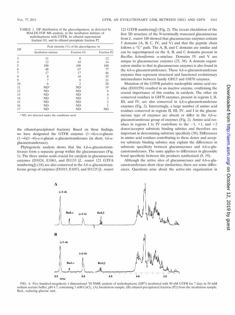
Characterization of the GTFB products with maltoheptaose(G7). MALDI-TOF MS analysis of the 150 mM G7 incubationsample showed visible [M � Na]� peaks in the region of DP3(m/z 527) up to DP10 (m/z 1,661), with DP5 as the major peak(100%), followed by DP6 (48%) (Table 1). It should be notedthat m/z values below 500 were not recorded due to matrix-related noise in that area. The 1-dimensional 1H NMR spec-trum (Fig. 6A) revealed, besides the anomeric signals at 5.42to 5.37 (indicative of �134 linkages, in the absence of �133linkages), 5.225 [reducing -(134)-�-D-Glcp unit and �-ano-mer of free glucose], 4.653 [reducing -(134)-�-D-Glcp unit;H-2 at 3.271], and 4.634 (�-anomer of free glucose; H-2 at 3.237), an additional broad anomeric peak at 5.00 to 4.95,indicative of �136 linkages (35, 37). The molar ratio of the�134-linked, �136-linked, and reducing unit glucose resi-dues was 60:18:22. Ethanol treatment of the G7 incubationsample yielded a supernatant fraction (O1) and a precipitatefraction (P2).
MALDI-TOF MS analysis of fraction O1 showed evidenceof the same DP range as that in the incubation sample, withDP5 as the major component(s) (100%), followed by DP6
FIG. 1. Unrooted phylogenetic tree of GTF proteins (glucansucrases and [putative] 4,6-�-glucanotransferases) from lactic acid bacteria.Alignments and dendrogram construction were carried out (using the catalytic cores only [for example, from “WYRP” to “WVPDQ” of L. reuteri121 GTFA]) with MEGA version 4, using the neighbor joining method. Bootstrap values (expressed as percentages) are given at the branchingpoints. The bar corresponds to a genetic distance of 0.1 substitution per position (10% amino acid sequence difference).
VOL. 77, 2011 GTFB, AN EVOLUTIONARY LINK BETWEEN GH13 AND GH70 8157
on October 12, 2019 by guest
http://aem.asm
.org/D
ownloaded from

(67%) (Table 1). Methylation (linkage) analysis of fraction O1showed the presence of 4-substituted, 6-substituted, and ter-minal glucose residues at a molar ratio of 63, 17, and 20%,indicating the presence of linear products only (no branchedglucose residues).
MALDI-TOF MS analysis of fraction P2 showed visible[M � Na]� peaks in the region of DP3 (m/z 527) up to DP16(m/z 2633), with DP5 as the major peak (100%), followed by
DP6 (77%). Compared with those in the incubation mixtureand fraction O1, the peak intensities of DP7 to DP11 glucoo-ligomers were significantly increased. Methylation (linkage)analysis of fraction P2 showed 4-substituted, 6-substituted, andterminal glucose residues at a molar ratio of 52, 35, and 13%,indicating the presence of linear products only. In the 1-di-mensional 1H NMR spectrum of fraction P2 (Fig. 6B), theareas of the anomeric signals at 5.42 to 5.37 (�134 linkages[36, 37]), 5.226 and 4.655 (� and � configuration, respec-tively, of the reducing glucose unit [37]), and 5.00 to 4.95(�136 linkages [35, 37]) occurred at a molar ratio of 55, 33,and 12%, indicating a significant increase in the percentage of�136-linked glucose residues over those in the incubationsample. This suggests that the �136 linkages are specificallyincluded in the higher-DP linear chains.
DISCUSSION
This paper reports the identification and characterization ofa new reaction specificity within the GH70 family, namely, thedisproportionation of maltooligosaccharides (MOS), also in-troducing �136 glycosidic linkages into the products formed.
L. reuteri 121 GTFB is the first member of this group of en-zymes that has been characterized in detail. Surprisingly, GTFBhas virtually no activity with sucrose. Incubation of L. reuteri 121GTFB with MOS of different DPs produced saccharides with�134 glycosidic linkages alone and saccharides that also con-tained �136 glycosidic linkages. Incubation of GTFB with amy-lose as a donor substrate and glucose or maltose as an acceptorsubstrate confirmed the ability of the enzyme to synthesize �134glycosidic linkages also. In particular, with maltose as an acceptor,
FIG. 2. Amino acid sequence (http://www.cazy.org) alignment of conserved regions (II, III, IV, and I) in the catalytic domains of (putative)4,6-�-glucanotransferase enzymes (A), DSRE (5) and DSRP, glucansucrase enzymes containing two catalytic domains (CD1 and CD2) (B), anddextran-, mutan-, alternan-, and reuteransucrase enzymes of lactic acid bacteria (C) (see also references 17 and 23). The seven strictly conservedamino acid residues (indicated by the numbers 1 to 7 above the sequences; underlined and lightly shaded in L. reuteri 121 GTFA and GTFB), withimportant contributions to the �1 and �1 subsites in glucansucrase enzymes, are also conserved in the 4,6-�-glucanotransferase enzymes. GTFBamino acid D1015 (the putative nucleophilic residue), targeted in this study, is shown in boldface. Dark shading indicates changes in conservedamino acid residues between 4,6-�-glucanotransferases and glucansucrases; the corresponding amino acid numbering is indicated. *, low activity.
FIG. 3. TLC analysis of the reaction products of 90 nM GTFBincubated for 13 h in 50 mM sodium acetate buffer, pH 4.7, containing1 mM CaCl2 with 25 mM sucrose or 25 mM maltooligosaccharides. St,standard; Suc, sucrose; G1, glucose; G2, maltose; G3, maltotriose; G4,maltotetraose; G5, maltopentaose; G6, maltohexaose; G7, maltohep-taose; Pol, polymer.
8158 KRALJ ET AL. APPL. ENVIRON. MICROBIOL.
on October 12, 2019 by guest
http://aem.asm
.org/D
ownloaded from

FIG. 4. HPAEC analysis of the reaction products of 90 nM GTFB incubated for 0 to 8 h in 50 mM sodium acetate buffer, pH 4.7, containing1 mM CaCl2 with 25 mM maltohexaose (A) or 25 mM maltoheptaose (B).
8159
on October 12, 2019 by guest
http://aem.asm
.org/D
ownloaded from

among other unidentified products, panose [glucosyl-(�136)-maltose] and G3 [glucosyl-(�134)-maltose] were detected. Pre-liminary investigation of the products generated when GTFB wasincubated with maltoheptaose (DP7) by methylation (linkage)analysis (GLC-EI-MS), 1H NMR analysis, and MALDI-TOF MS
revealed the presence of only linear oligosaccharides containing�134 and �136 glycosidic linkages. There was a range of glu-cooligosaccharides from DP3 to DP16, with an overall content ofaround 18% �136 glycosidic bonds, and the percentage of �136glycosidic linkages was higher in the higher-DP products (33% in
FIG. 5. HPAEC analysis of samples with 0.25% amylose-V (AMV) alone (donor substrate) or amylose-V with 25 mM glucose or 25 mMmaltose (acceptor substrates), either without the GTFB enzyme (A) or with GTFB (90 nM) (B) incubated overnight at 37°C in 25 mM sodiumacetate buffer, pH 4.7, containing 1 mM CaCl2. Pa, panose.
8160 KRALJ ET AL. APPL. ENVIRON. MICROBIOL.
on October 12, 2019 by guest
http://aem.asm
.org/D
ownloaded from
the ethanol-precipitated fraction). Based on these findings,we have designated the GTFB enzymes (134)-�-D-glucan:(134)(136)-�-D-glucan �-glucanotransferases (in short, 4,6-�-glucanotransferases).
Phylogenetic analysis shows that the 4,6-�-glucanotrans-ferases form a separate group within the glucansucrases (Fig.1). The three amino acids crucial for catalysis in glucansucraseenzymes (D1024, E1061, and D1133 [L. reuteri 121 GTFAnumbering]) (18) are also conserved in the 4,6-�-glucanotrans-ferase group of enzymes (D1015, E1053, and D1125 [L. reuteri
121 GTFB numbering]) (Fig. 2). The recent elucidation of thefirst 3D structure of the N-terminally truncated glucansucrasefrom L. reuteri 180 showed that glucansucrase enzymes contain5 domains (A, B, C, IV, and V) and that the peptide chainfollows a “U” path. The A, B, and C domains are similar andcan be superimposed on the A, B, and C domains present inBacillus licheniformis �-amylase. Domains IV and V areunique to glucansucrase enzymes (25, 38). A domain organi-zation similar to that in glucansucrase enzymes is also found inthe 4,6-�-glucanotransferases. These 4,6-�-glucanotransferaseenzymes thus represent structural and functional evolutionaryintermediates between family GH13 and GH70 enzymes.
Mutation of the GTFB putative nucleophile amino acid res-idue (D1015N) resulted in an inactive enzyme, confirming thecrucial importance of this residue in catalysis. The other sixconserved residues in GH70 enzymes, present in regions I, II,III, and IV, are also conserved in 4,6-�-glucanotransferaseenzymes (Fig. 2). Interestingly, a large number of amino acidresidues conserved in regions II, III, IV, and I in the glucan-sucrase type of enzymes are absent or differ in the 4,6-�-glucanotransferase group of enzymes (Fig. 2). Amino acid res-idues in regions I to IV contribute to the �1, �1, and �2donor/acceptor substrate binding subsites and therefore areimportant in determining substrate specificity (38). Differencesin amino acid residues contributing to these donor and accep-tor substrate binding subsites may explain the differences insubstrate specificity between glucansucrases and 4,6-�-glu-canotransferases. The same applies to differences in glycosidicbond specificity between the products synthesized (8, 19).
Although the active sites of glucansucrases and 4,6-�-glu-canotransferases show clear similarities, there are some differ-ences. Questions arise about the active-site organization in
TABLE 1. DP distribution of the glucooligomers, as detected byMALDI-TOF MS analysis, in the incubation mixture of
maltoheptaose with GTFB, its ethanol supernatantfraction O1, and its ethanol precipitate fraction P2
DPPeak intensity (%) of the glucooligomer in:
Incubation mixture Fraction O1 Fraction P2
3 11 8 124 12 10 145 100 100 1006 48 67 777 17 17 468 8 10 359 7 9 3110 6 2 2811 NDa ND 1912 ND ND 813 ND ND 414 ND ND 315 ND ND 216 ND ND 117 ND ND ND
a ND, not detected under the conditions used.
FIG. 6. Five hundred-megahertz 1-dimensional 1H NMR analysis of maltoheptaose (DP7) incubated with 90 nM GTFB for 7 days in 50 mMsodium acetate buffer, pH 4.7, containing 1 mM CaCl2. (A) Incubation sample; (B) ethanol-precipitated fraction (P2) from the incubation sample.Red., reducing glucose unit.
VOL. 77, 2011 GTFB, AN EVOLUTIONARY LINK BETWEEN GH13 AND GH70 8161
on October 12, 2019 by guest
http://aem.asm
.org/D
ownloaded from

4,6-�-glucanotransferases, in particular the number of donorsubstrate binding subsites present. Only one donor subsite ispresent in glucansucrase enzymes, whereas other amylolyticenzymes, such as amylases and amylomaltases, have more thanone donor subsite. In order to address this question, GTFBenzyme incubations with maltohexaose (G6) or maltoheptaose(G7) as the substrate were followed over time using HPAEC.The first reaction products detectable with G6 were G1 (glu-cose) and G5 (maltopentaose) (Fig. 4A). Similarly, with G7,the first products released were G1 (glucose) and G6 (malto-hexaose) (Fig. 4B). With both G6 and G7, other MOS of lowerDPs also accumulated, and at later times, unknown saccharidesof higher DPs, which, besides �134 glycosidic linkages, werelikely to contain other linkages (in fact, �136 glycosidic link-ages, as demonstrated by methylation and 1H NMR analysis),were also present. The data thus suggest that 4,6-�-glucano-transferases have an active-site architecture similar to that ofglucansucrases, both containing only a single donor substratebinding subsite (�1).
The enzymes from glycoside hydrolase families GH13,GH70, and GH77 together form clan GH-H (www.cazy.org).All GH-H members employ similar catalytic mechanisms in-volving a covalent glucosyl-enzyme intermediate and retentionof the �-anomeric configuration of the product upon hydrolysis(13, 32). Amylomaltases (also known as 4-�-glucanotrans-ferases) from GH77 are capable of synthesizing large cyclicglucans and disproportionating MOS. Notably, amylomaltasesalmost exclusively catalyze transglycosylation reactions andonly cleave and synthesize �134 glycosidic bonds (3). TheGH13 enzyme family, acting mainly on starch-like substratesand displaying a large variety of different reaction specific-ities, is the largest of the GH families at present, organizedin different subfamilies in the CAZy database (30). Amylo-sucrase is the only enzyme from family GH13 that usessucrose as a substrate to synthesize an amylose type of�-glucan polymer. Amylosucrase enzymes thus have theGH13 type of domain architecture and the GH70 type ofglucansucrase activity (24, 29).
All �-amylases have similar domain organizations (seeabove), even those of archaeal origin (20). Glucansucrases arefound only in Bacteria, suggesting that the precursor amylaseenzyme from which GH70 glucansucrases and present-day am-ylases have evolved had a similar domain organization. Inaddition, it is most likely that amylolytic activity, the mainactivity of �-amylases, emerged first, and that the �-glucano-transferase and sucrase types of activities evolved from theprecursor amylase. It appears less likely that a family GH77enzyme is an intermediate, since these proteins possess variousadditional domains (B1, partly present in �-amylase members;B2 and B3, unique to amylomaltases); in addition, they lack theC domain (26). Furthermore, GH77 enzymes do not possess�136 specificity, in contrast to GH13 and GH70 family mem-bers. The GTFB enzyme clearly is a glucansucrase type ofprotein with regard to its amino acid sequence and domainorganization but lacks the ability to act on sucrose. Surpris-ingly, GTFB acts on MOS, substrates used by various GH13(and GH77) family members, and is able to cleave �134glycosidic linkages and to synthesize �134 and �136 glyco-sidic linkages. GTFB from the GH70 family therefore providesa link between the GH13 �-amylase and GH70 glucansucrase
families. The latter use sucrose as a substrate to synthesizelinear as well as branched �-glucan polymers, differing in thetype of glycosidic linkages (reuteran, �134; dextran, �136;alternan, �133/�136; mutan, �133), the degree and type ofbranching, the length of the glucan chains, molecular mass, andthe conformation of the polymers (34).
Glucansucrase enzymes also use MOS as acceptor substrateswith sucrose as donor substrate. This results in the synthesis ofa range of oligosaccharides, e.g., a maltose extended with aseries of glucose units bound via �136 linkages in the case ofdextransucrase. In this case, however, the �134 linkages inMOS substrates are not cleaved; MOS are used only as accep-tor substrates (6, 12). This is a major difference from the GFTBenzyme, which fails to act on sucrose and instead uses MOS asdonor and acceptor substrates, cleaving �134 linkages andintroducing new �134 and �136 linkages. GTFB thus ap-pears to be an evolutionary precursor for the glucansucrasetype of enzymes with the GH70 domain architecture and theGH13 amylolytic activity (38).
Although 4,6-�-glucanotransferases and glucansucrase en-zymes are commonly arranged in tandem on the genome (10,15), the exact in vivo role of GTFB-like enzymes remainsunknown. They may scavenge and modify the oligosaccharidesformed by glucansucrase enzymes as substrates for the synthe-sis of larger saccharides, which are inaccessible to other mi-crobes. They may also play a role in the modification of theglucan synthesized by the glucansucrase enzyme of the micro-organism. Interestingly, the genome of L. reuteri DSM 20016contains only a GTFB-like enzyme and no glucansucrase en-zyme. This Lactobacillus strain may be the key to answeringquestions about the in vivo role of 4,6-�-glucanotransferaseenzymes.
Conclusions. Based on the enzymatic activity of GTFB, wepropose to give this group of enzymes a separate EC number,different from EC 2.4.1.5 (for common glucansucrase enzymes)or EC 2.4.1.140 (for alternansucrase). We propose to namethese enzymes (134)-�-D-glucan:(134),(136)-�-D-glucan�-glucanotransferase enzymes (in short 4,6-�-glucanotrans-ferases). We also propose to divide GH70 family enzymes intotwo subfamilies in the CAZy database, as has been done forGH13 enzymes (30), namely, glucansucrase enzymes acting onsucrose or on MOS.
The precise in vivo reaction and the physiological function ofthe GTFB type of enzymes remain to be determined. Clearly,this new group of �-glucanotransferase enzymes provides avaluable asset in the toolbox for saccharide synthesis. Thelinear oligosaccharides synthesized by GTFB containing �134and �136 glycosidic linkages may have interesting physico-chemical properties, which remain to be determined, and mayhave potentially new applications in the food, cosmetics,and/or pharmaceutical industries. In the future, we will char-acterize the saccharides synthesized by GTFB in more detail,as well as the characteristic properties of other members of thisenzyme group.
ACKNOWLEDGMENTS
We thank Peter Sanders (TNO Quality of Life) for anion-exchange(Dionex) analysis and Rolf Boelens (Bijvoet Center, Department ofNMR Spectroscopy, Utrecht University, Utrecht, The Netherlands)
8162 KRALJ ET AL. APPL. ENVIRON. MICROBIOL.
on October 12, 2019 by guest
http://aem.asm
.org/D
ownloaded from

for providing us with measuring time on the 500-MHz NMR instru-ment.
REFERENCES
1. Arguello-Morales, M. A., et al. 2000. Sequence analysis of the gene encodingalternansucrase, a sucrose glucosyltransferase from Leuconostoc mesen-teroides NRRL B-1355. FEMS Microbiol. Lett. 182:81–85.
2. Ausubel, F. M., et al. 1987. Current protocols in molecular biology. JohnWiley & Sons, Inc., New York, NY.
3. Barends, T. R. M., et al. 2007. Three-way stabilization of the covalentintermediate in amylomaltase, an �-amylase-like transglycosylase. J. Biol.Chem. 282:17242–17249.
4. Devulapalle, K. S., S. D. Goodman, Q. Gao, A. Hemsley, and G. Mooser.1997. Knowledge-based model of a glucosyltransferase from the oral bacte-rial group of mutans streptococci. Protein Sci. 6:2489–2493.
5. Fabre, E., et al. 2005. Role of the two catalytic domains of DSR-E dextran-sucrase and their involvement in the formation of highly �-1,2 brancheddextran. J. Bacteriol. 187:296–303.
6. Fu, D., and J. F. Robyt. 1991. Maltodextrin acceptor reactions of Streptococ-cus mutans 6715 glucosyltransferases. Carbohydr. Res. 217:201–211.
7. Hård, K., G. van Zadelhoff, P. Moonen, J. P. Kamerling, and J. F. G.Vliegenthart. 1992. The Asn-linked carbohydrate chains of human Tamm-Horsfall glycoprotein of one male. Novel sulfated and novel N-acetylgalac-tosamine-containing N-linked carbohydrate chains. Eur. J. Biochem. 209:895–915.
8. Hellmuth, H., et al. 2008. Engineering the glucansucrase GTFR enzymereaction and glycosidic bond specificity: toward tailor-made polymer andoligosaccharide products. Biochemistry 47:6678–6684.
9. Ito, K., et al. 2011. Crystal structure of glucansucrase from the dental cariespathogen Streptococcus mutans. J. Mol. Biol. 408:177–186.
10. Kaditzky, S. B., et al. 2008. Influence of pH on the formation of glucan byLactobacillus reuteri TMW 1.106 exerting a protective function against ex-treme pH values. Food Biotechnol. 22:398–418.
11. Kamerling, J. P., and J. F. G. Vliegenthart. 1989. Carbohydrates, p. 176–263.In A. M. Lawson (ed.), Clinbiochemistry: principles, methods, applications,vol. 1. mass spectrometry. Walter de Gruyter, Berlin, Germany.
12. Kang, H. K., J.-S. Oh, and D. Kim. 2009. Molecular characterization andexpression analysis of the glucansucrase DSRWC from Weissella cibariasynthesizing a �(136) glucan. FEMS Microbiol. Lett. 292:33–41.
13. Kelly, R. M., L. Dijkhuizen, and H. Leemhuis. 2009. Starch and �-glucanacting enzymes, modulating their properties by directed evolution. J. Bio-technol. 140:184–193.
14. Kralj, S., E. Stripling, P. Sanders, G. H. van Geel-Schutten, and L. Dijkhui-zen. 2005. Highly hydrolytic reuteransucrase from probiotic Lactobacillusreuteri strain ATCC 55730. Appl. Environ. Microbiol. 71:3942–3950.
15. Kralj, S., et al. 2004. Glucan synthesis in the genus Lactobacillus: isolationand characterization of glucansucrase genes, enzymes and glucan productsfrom six different strains. Microbiology 150:3681–3690.
16. Kralj, S., et al. 2002. Molecular characterization of a novel glucosyltrans-ferase from Lactobacillus reuteri strain 121 synthesizing a unique, highlybranched glucan with �-(134) and �-(136) glucosidic bonds. Appl. Envi-ron. Microbiol. 68:4283–4291.
17. Kralj, S., G. H. van Geel-Schutten, M. J. E. C. van der Maarel, and L.Dijkhuizen. 2003. Efficient screening methods for glucosyltransferase genesin Lactobacillus strains. Biocatal. Biotransform. 21:181–187.
18. Kralj, S., G. H. van Geel-Schutten, M. J. E. C. van der Maarel, and L.Dijkhuizen. 2004. Biochemical and molecular characterization of Lactoba-cillus reuteri 121 reuteransucrase. Microbiology 150:2099–2112.
19. Kralj, S., I. G. H. van Geel-Schutten, E. J. Faber, M. J. E. C. van der Maarel,
and L. Dijkhuizen. 2005. Rational transformation of Lactobacillus reuteri 121reuteransucrase into a dextransucrase. Biochemistry 44:9206–9216.
20. Linden, A., O. Mayans, W. Meyer-Klaucke, G. Antranikian, and M. Wil-manns. 2003. Differential regulation of a hyperthermophilic �-amylase witha novel (Ca,Zn) two-metal center by zinc. J. Biol. Chem. 278:9875–9884.
21. MacGregor, E. A., H. M. Jespersen, and B. Svensson. 1996. A circularlypermuted �-amylase-type �/�-barrel structure in glucan-synthesizing glu-cosyltransferases. FEBS Lett. 378:263–266.
22. Malik, A., M. Radji, S. Kralj, and L. Dijkhuizen. 2009. Screening of lacticacid bacteria from Indonesia reveals glucansucrase and fructansucrase genesin two different Weissella confusa strains from soya. FEMS Microbiol. Lett.300:131–138.
23. Monchois, V., R.-M. Willemot, and P. Monsan. 1999. Glucansucrases: mech-anism of action and structure-function relationships. FEMS Microbiol. Rev.23:131–151.
24. Moulis, C., et al. 2006. Understanding the polymerization mechanism ofglycoside-hydrolase family 70 glucansucrases. J. Biol. Chem. 281:31254–31267.
25. Pijning, T., et al. 2008. Biochemical and crystallographic characterizationof a glucansucrase from Lactobacillus reuteri 180. Biocatal. Biotransform.26:12–17.
26. Przylas, I., et al. 2000. Crystal structure of amylomaltase from Thermusaquaticus, a glycosyltransferase catalysing the production of large cyclic glu-cans. J. Mol. Biol. 296:873–886.
27. Sambrook, J., E. F. Fritsch, and T. Maniatis. 1989. Molecular cloning: alaboratory manual, 2nd ed. Cold Spring Harbor Laboratory Press, ColdSpring Harbor, NY.
28. Sarkar, G., and S. S. Sommer. 1990. The “megaprimer” method of site-directed mutagenesis. Biotechniques 8:404–407.
29. Skov, L. K., et al. 2001. Amylosucrase, a glucan-synthesizing enzyme fromthe �-amylase family. J. Biol. Chem. 276:25273–25278.
30. Stam, M. R., E. G. J. Danchin, C. Rancurel, P. M. Coutinho, and B. Hen-rissat. 2006. Dividing the large glycoside hydrolase family 13 into subfami-lies: towards improved functional annotations of �-amylase-related proteins.Protein Eng. Des. Sel. 19:555–562.
31. Svensson, B. 1994. Protein engineering in the �-amylase family: catalyticmechanism, substrate specificity, and stability. Plant Mol. Biol. 25:141–157.
32. Uitdehaag, J. C. M., et al. 1999. X-ray structures along the reaction pathwayof cyclodextrin glycosyltransferase elucidate catalysis in the �-amylase fam-ily. Nat. Struct. Biol. 6:432–436.
33. van Geel-Schutten, G. H., et al. 1999. Biochemical and structural character-ization of the glucan and fructan exopolysaccharides synthesized by theLactobacillus reuteri wild-type strain and by mutant strains. Appl. Environ.Microbiol. 65:3008–3014.
34. van Hijum, S. A. F. T., S. Kralj, L. K. Ozimek, L. Dijkhuizen, and I. G. vanGeel-Schutten. 2006. Structure-function relationships of glucansucrase andfructansucrase enzymes from lactic acid bacteria. Microbiol. Mol. Biol. Rev.70:157–176.
35. van Leeuwen, S. S., et al. 2008. Structural analysis of the �-D-glucan(EPS180) produced by the Lactobacillus reuteri strain 180 glucansucraseGTF180 enzyme. Carbohydr. Res. 343:1237–1250.
36. van Leeuwen, S. S., et al. 2008. Structural analysis of the �-D-glucan(EPS35-5) produced by the Lactobacillus reuteri strain 35-5 glucansucraseGTFA enzyme. Carbohydr. Res. 343:1251–1265.
37. van Leeuwen, S. S., B. R. Leeflang, G. J. Gerwig, and J. P. Kamerling. 2008.Development of a 1H NMR structural-reporter-group concept for the pri-mary structural characterisation of �-D-glucans. Carbohydr. Res. 343:1114–1119.
38. Vujicic-Zagar, A., et al. 2010. Crystal structure of a 117 kDa glucansucrasefragment provides insight into evolution and product specificity of GH70enzymes. Proc. Natl. Acad. Sci. U. S. A. 107:21406–21411.
VOL. 77, 2011 GTFB, AN EVOLUTIONARY LINK BETWEEN GH13 AND GH70 8163
on October 12, 2019 by guest
http://aem.asm
.org/D
ownloaded from


