CHAPTER244-87).pdf · 1.Introduction Erythropoiesis is the pathway that produces mature red blood...
44
* CHAPTER 2 Julie Vandekerckhove, Geneviève Courtois, Séverine Coulon, Jean-Antoine Ribeil, Olivier Hermine Regulation of erythropoiesis IRON2009_CAP.2(44-87):EBMT2008 4-12-2009 16:03 Pagina 44
-
Upload
duongthuan -
Category
Documents
-
view
214 -
download
1
Transcript of CHAPTER244-87).pdf · 1.Introduction Erythropoiesis is the pathway that produces mature red blood...

* CHAPTER 2
Julie Vandekerckhove, Geneviève Courtois,Séverine Coulon, Jean-Antoine Ribeil, Olivier Hermine
Regulation of erythropoiesis
IRON2009_CAP.2(44-87):EBMT2008 4-12-2009 16:03 Pagina 44

1. IntroductionErythropoiesis is the pathway that produces mature red blood cells fromhaematopoietic stem cells. During mammalian development, erythropoiesis occurssuccessively in the yolk sac, the foetal liver and the bone marrow. This cellular processis characterised by commitment and differentiation steps that restrict thedifferentiation potential and the proliferative capacity of the cells as they gothrough the erythroid-specific program of gene expression. Erythropoiesis isregulated by the combined effects of microenvironmental and growth factors thatpromote the survival, proliferation and/or differentiation of erythroid progenitors,and nuclear factors that regulate the transcription of genes involved in theestablishment of the erythroid phenotype. At the cellular and molecular levels,erythropoiesis is one of the best-studied haematopoietic lineages for the followingreasons: first, the different stages of erythropoiesis can all be defined by phenotypicmarkers; second, erythroid diseases are well-characterised and in many cases theirmolecular causes have now been found; third, terminal erythroid differentiationdepends on only one exogenous growth factor, erythropoietin; fourth, most if notall the transcription factors that regulate erythropoiesis are known. The understandingof this complex system may shed light on basic cellular biology and also thepathophysiology of various diseases including bone marrow failure, degenerativediseases and cancers.Many features differentiate primitive and foetal erythropoiesis from adult erythropoiesisboth at the cellular and molecular levels. This review will focus on the late stagesof adult erythropoiesis starting from the common erythro/megakaryocytic precursorand ending with the mature red blood cells.
2. Description of adult erythropoiesis at the cellular levelThe production of erythrocytes is the largest quantitative output of the haematopoieticsystem with an estimated production rate of 2x10¹¹ erythrocytes per day. Thefrequency of committed granulocyte-macrophage progenitors is actually three timesgreater than that of committed erythroid progenitors in the bone marrow but thisis counteracted by a high proliferative index in the late transitional stages oferythroid development (1, 2). The cell cycle time of pro-erythroblasts is estimatedto be 6-7 hours, which is unique in the adult under steady state conditions and moreclosely resembles that of embryonic cells. How cell differentiation and activation ofspecific gene expression programs are coordinated with cell growth remains to beclarified, despite a detailed knowledge of the network of transcription factors thatgoverns lineage gene expression (see (3) for review). Committed erythroid progenitorscan be detected by their ability to form erythroid colonies in in vitro culture in methyl
DISORDERS OF ERYTHROPOIESIS, ERYTHROCYTES AND IRON METABOLISM45
CHAPTER 2 • Regulation of erythropoiesis
IRON2009_CAP.2(44-87):EBMT2008 4-12-2009 16:03 Pagina 45

cellulose, and are called burst forming unit-erythroid (BFU-E) and colony formingunit-erythroid (CFU-E) (1, 4). BFU-Es are the most immature haematopoietic cellsthat are already committed to the erythroid lineage. BFU-Es represent only 0.03%of bone marrow haematopoietic cells and only 40% are cycling (2), whereas CFU-Esrepresent 0.3% of bone marrow haematopoietic cells and most of them are activelycycling (5). There is a continuous cellular process from the earliest BFU-Es to thelatest CFU-Es but the growth factor requirements for BFU-Es and CFU-Es are completelydifferent. BFU-Es require stem cell factor (SCF) (6) and many other haematopoieticgrowth factors for their growth and differentiation, whereas CFU-Es are highlydependent on erythropoietin (Epo) (5). CFU-E will differentiate into the firstmorphologically identifiable cell of the erythrocyte lineage, the pro-erythroblast, thendifferentiates successively into the basophilic erythroblast, the polychromatophilicerythroblast, and the acidophilic erythroblast, which is the last nucleated cell of themammalian erythrocyte lineage. Enucleation of the acidophilic erythroblast gives riseto the reticulocyte which matures finally into the red blood cell. This process occurswithin the erythroblastic blood island in the bone marrow, in which a macrophageis surrounded by erythroblasts at all stages of maturation (Figure 1).
3. Description of adult erythropoiesis at the transcriptional levelDuring erythroid differentiation, the expression pattern of erythroid-specific genesfollows a precise timing that is mainly regulated at the transcriptional level.Studies on the cis and trans acting factors that regulate the erythroid-specific genes,together with perturbations of erythropoiesis in leukaemia and inherited erythroiddiseases and in experimental animal models, have greatly increased our knowledgeof the transcriptional regulation of erythropoiesis. The most interesting findings ofthese studies are the importance of protein-protein interactions in the transcriptionalregulation of erythropoiesis and its relationship with leukaemogenesis (see (3) forreview).
3.1 Cis-acting sequences involved in the regulation of erythroid-specific genesFunctional analysis of erythroid and megakaryocytic specific genes has shown theimportance of a sequence, 5’ A/T GATA A/G 3’, now called the GATA motif, in thelineage specific expression of these genes (7). This sequence is associated with aGT or CACC-like sequence in erythroid specific genes, whereas it is associated withan Ets binding site in megakaryocytic specific genes (7). These two associationsare now considered as hallmarks of erythroid or megakaryocytic-specific regulatoryregions. Another motif, 5’ TGAC/GTCAGCA 3’, is also found in the core region oferythroid expressed gene promoters or enhancers and is necessary for efficient
THE HANDBOOK 2009 EDITION46
IRON2009_CAP.2(44-87):EBMT2008 4-12-2009 16:03 Pagina 46

erythroid-specific transcription of the genes that contain this motif (8). No othermotif has been repeatedly found in regulatory sequences of erythroid ormegakaryocytic specific genes. For each binding motif identified as recurrent in theregulatory sequences of erythroid or megakaryocytic genes, families of nuclear factorsthat recognise the motif have been characterised. Of importance, none of the trans-acting factors identified has an expression restricted to the erythroid or themegakaryocytic lineage indicating that the specificity of gene expression inerythropoiesis or megakaryopoiesis, as in the other haematopoietic lineages, isestablished by a specific combination of trans-acting factors rather than by lineagespecific factors.
3.2 Trans-acting factors that regulate the expression of erythroid-specific genesTrans-acting factors involved in erythropoiesis are shown in Table 1.
DISORDERS OF ERYTHROPOIESIS, ERYTHROCYTES AND IRON METABOLISM
CHAPTER 2 • Regulation of erythropoiesis
47
HSC CMP MEP
PlateletBFU-MK
PrimitiveBFU-E
Megacaryocyte
CFU-E Red bloodcell
MatureBFU-E
ReticulocyteProerythroblast
Erythroblast
CD34
CD33Epo-R
CD71Glycophorin A
HLA-DRC-Kit
BFU-Ep BFU-Em CFU-E Proebl
ErythroblastsGRBaso I Poly AcidoBaso II Ret
SCF
Epo
Figure 1: Erythropoiesis is the process by which multipotential haematopoieticstem cells differentiate into mature red blood cells
The requirement for erythropoietin starts at mature BFU-E and stops after the proerythroblasts stage.The expression is indicated of the different surface markers commonly used to characterise the differenterythroid precursors. BFU-Ep: burst forming unit- erythroid primitive. BFU-Em: burst forming unit-erythroidmature HSC: haematopoietic stem cell; CMP: common myeloid progenitor; MEP: megakaryocyteerythrocyte progenitor.
IRON2009_CAP.2(44-87):EBMT2008 4-12-2009 16:03 Pagina 47

3.2.1 GATA-1 and GATA-2The GATA family of transcription factors consists of six transcription factors, GATA-1 to -6, that can bind the consensus sequence 5’ A/T GATA A/G 3’. These proteinscontain two conserved zinc fingers motifs (ys-X2-Cys-X17-Cys-X2-Cys) specific to theGATA family. The carboxyl terminal zinc finger is responsible for binding to DNAwhereas the amino terminal zinc finger stabilises the DNA/GATA interaction. Outsideof the zinc finger regions, the conservation between GATA factors is low but eachfactor is conserved between species. As regards erythropoiesis, only GATA-1 and GATA-2 have been shown to play a role both in erythrocytic determination and interminal differentiation (see (9-11) for review).During erythropoiesis, there is sequential but overlapping expression of GATA-1 andGATA-2. GATA-2 is expressed early in the erythroid lineage and its expressiondecreases concomitantly with an increasing GATA-1 expression. Numerous studies
Table 1: Trans-acting factors involved in erythropoiesis
Trans-actingfactor
DNA-binding site Target genes Phenotype ofgeneinactivation
Human pathology
GATA-1 5’ A/T GATA A/G 3’ Globin, erythroidspecific membraneproteins, GATA-1,GATA-2
Anaemia andthrombocyto-penia
X-linked anaemia orthrombocytopenia,megakaryoblasticleukaemia of Down’ssyndrome
GATA-2 5’ A/T GATA A/G 3’ GATA-1, GATA-2 No proliferationof immatureprogenitors
TAL-1 5’ CAGATG 3’ Glycophorin A,p21
Anaemia, no HSC T-cell acuteleukaemia
EKLF 5’ CCNCACCC 3’ Globin Anaemia
p45NF-E2 5’ TGAC/GTCAGCA 3’ Enzymes of thehaem biosyntheticpathway, globin
Thrombocyto-penia
ZBP-89 5’ CCNCACCC 3’ Globin, erythroidspecific gene,GATA-1
Anaemia andthrombocyto-penia
Gfi-1B 5’ TAAATC(A/T)GCA 3’ p21, SOCS1-3,BclxL
Anaemia
DNA-binding sites and known target genes of the indicated transcription factors together with the phenotypeof their gene inactivation in mice and the human pathologies associated with gene mutations are shown.
THE HANDBOOK 2009 EDITION48
IRON2009_CAP.2(44-87):EBMT2008 4-12-2009 16:03 Pagina 48

have shown that GATA-2 is involved in the cellular proliferation of non-committedand committed erythroid progenitors, whereas GATA-1 is critical for terminalmaturation of erythroid cells. At the molecular level, cross talk between GATA-1 andGATA-2 occurs at the transcriptional level, as GATA-2 inhibits transcription of theGATA-1 gene and GATA-1 has the same effect on the GATA-2 gene. The regulatorynetwork between GATA-1 and GATA-2 during erythropoiesis results in the sequentialsubstitution of GATA-2 by GATA-1 in the regulatory regions of numerous erythroidspecific genes.The X-linked GATA-1 gene is the founding member of the GATA factor family. Duringadult haematopoiesis, its expression is restricted to the erythroid, megakaryocytic,eosinophil and mast cell lineages. Mice that lack GATA-1 die at day 10-12 ofanaemia with a blockade of erythroid differentiation at the pro-erythroblast stage.The cells die by apoptosis, indicating a role for GATA-1 in cell survival as well asmaturation. GATA-1 activity during erythropoiesis is highly regulated at thetranscriptional, translational and posttranslational levels. GATA-1 positively regulatesthe transcription of its gene through GATA sites located in the promoter andenhancer regions of the GATA-1 gene. GATA-1 possesses an alternative translationinitiation site located at methionine 84 and the GATA-1s (short isoform of GATA-1) protein produced is not fully functional, as it cannot drive erythroid differentiationwhen expressed at normal levels. GATA-1 activity and levels must be tightlyregulated to prevent continuous accumulation of GATA-1 in the cells, which mayultimately block erythroid differentiation (12).An important way by which GATA-1 activity is regulated is by acetylation. GATA-1acetylation by P300 (13) or CREB-binding protein seems to enhance the self-association of GATA-1 and thus its transcriptional activity. Likewise, mutation of themain site of acetylation eliminated the ability of GATA-1 to bind to all examinedcellular target sites in vivo, including genes that are normally activated andrepressed by GATA-1 during erythroid differentiation (14). On the other hand,acetylated GATA-1 is targeted for degradation via the ubiquitin/proteasome pathway.Acetylation positively signals ubiquitination, suggesting that activation byacetylation simultaneously marks GATA-1 for degradation. Acetylation alone is notenough to signal degradation, but MAPK phosphorylation of GATA-1 cooperates withacetylation for efficient ubiquitination (15).Biochemical studies have shown that GATA-1 is phosphorylated at six serine residuesin undifferentiated murine erythroleukaemia cells. Akt directly phosphorylatesGATA-1 at serine 310 in vitro and in vivo and enhances its activity in erythroid cells(16). GATA-1 Ser310 phosphorylation is not essential for overall erythroid geneexpression (major late erythroid genes) but it is absolutely required for a subsetof genes like the TIMP-1 gene (17). Finally, GATA-1 activity is in some circumstances
DISORDERS OF ERYTHROPOIESIS, ERYTHROCYTES AND IRON METABOLISM
CHAPTER 2 • Regulation of erythropoiesis
49
IRON2009_CAP.2(44-87):EBMT2008 4-12-2009 16:03 Pagina 49

regulated by a caspase-mediated cleavage during terminal erythroid differentiation(see below).GATA-1 transcriptional activity is also dependent on protein-protein interactions. Manyproteins have been shown to interact physically with GATA-1 including FOG-1, LMO-2, TAL-1, Gfi-1b, PU-1, ZBP-89 and CBP. While most of these interactions may havesome relevance to the transcriptional activity of GATA-1, we will focus on the GATA-1/FOG-1 interaction as it has numerous implications in the physiology of erythropoiesis(see (18) for review). FOG-1 (Friend of GATA-1) is a multitype zinc finger proteinthat specifically interacts with the amino zinc finger of GATA-1. FOG-1 is co-expressed with GATA-1 in the erythroid and megakaryocytic lineages and FOG-1-/-mice die in mid-embryonic gestation of severe anaemia with an arrest in erythroidmaturation that is similar to the one observed in GATA-1-/- mice. There is considerableevidence that the function of GATA-1 in erythropoiesis is linked to FOG-1. One ofthe most important observations was that a single amino-acid change in the GATA-1 amino zinc finger, which abolishes interaction with FOG-1, is lethal in mice dueto severe anaemia and is also associated with dyserythropoietic anaemia in patients.The different functions of GATA-1/FOG-1 are mediated by two distinct nuclearcomplexes. One, the GATA-1/FOG-1/MeCP1 complex, represses transcription whereasthe other, GATA-1/FOG-1, links GATA-1 to transcriptional activation. The existenceof these two different complexes with opposing transcriptional activities explainshow GATA-1 simultaneously activates and represses target genes during erythropoiesis.However, the crucial question as to how some genes harbouring GATA binding sitesare activated whereas others are repressed remains open (see (18) for review).
3.2.2 TAL-1, LMO-2 and LMO-4TAL-1 (SCL), a member of the basic helix-loop-helix (bHLH) family of transcriptionfactors, was first identified through its involvement in human T-cell acutelymphoblastic leukaemia (T-ALL), where it is found in up to 30% of cases. Studiesin mice have shown that TAL-1 is required for specification, but not maintenance,of haematopoietic stem cells and for maturation of the erythroid and megakaryocyticlineages (19, 20).At the molecular level, TAL-1 acts through both DNA-binding dependent andindependent mechanisms and can activate or repress transcription. TAL-1 canassemble a pentameric complex (21) containing the ubiquitously expressed E-proteins (E2A, HEB, E2-2), LMO-2, Ldb1 and GATA-1 in erythroid committed cellsand can activate the expression of terminally expressed erythroid specific genes,while in CD34+ haematopoietic progenitors, a GATA-2 variant of this TAL-1 complexactivates c-Kit transcription (22). Alternatively, the TAL-1/E2A heterodimers, whichare less potent activators than E2A homodimers or E2A/HEB heterodimers, can repress
THE HANDBOOK 2009 EDITION50
IRON2009_CAP.2(44-87):EBMT2008 4-12-2009 16:03 Pagina 50

E2A target genes. Analysis of the complexes nucleated by TAL-1 in erythroid cellshas shown that ETO2, a potent transcriptional repressor, is associated with thepentameric complex during the initial stages of erythropoiesis whereas ETO2 is nolonger present in the complex during terminal erythroid differentiation. Thus, thecomposition of transcription factor complexes varies during erythroid differentiation,resulting in differential transcriptional output. In early erythroid cells, the TAL-1complex associates with erythroid specific regulatory regions such as the GPApromoter or the HS-26 of the murine α-globin locus. This association might keepthe chromatin in an open state but transcription is repressed by ETO2. Later duringerythropoiesis, an elevation in TAL-1 and E2A or HEB levels changes the ratio ofTAL-1 to ETO2 in favour of TAL-1, which offsets the inhibitory activity of ETO2 andtriggers sustained expression of TAL-1 marked erythroid genes.LMO-2, a LIM domain nuclear factor, has also been identified through its involvementin rare cases of human T-ALL. LMO-2-/- and TAL-1-/- mice have identical phenotypesconsistent with the direct physical interaction between these two nuclear factorsduring erythropoiesis.A transcriptional complex comprising LMO-2, TAL1, E47, GATA-1, and LDB1 regulateserythroid genes. While TAL1 has been shown to induce erythroid differentiation, LMO2appears to suppress foetal erythropoiesis. In addition to LMO-2, the closely relatedLMO-4 gene is expressed in haematopoietic cells, but has unknown functions.LMO-2 and LMO-4 are expressed at the same level in erythroid colonies from mousebone marrow, implying a function in erythroid differentiation. However, whileLMO-2 induced erythroid differentiation, LMO-4 had no such effect. Interestingly,both LMO-2 and TAL1 were able to partially suppress myeloid differentiation,implying that they activate erythroid differentiation in uncommitted bone marrowprogenitors. Both LMO-2 and LMO-4 interacted strongly to LDB1, which was requiredfor their localisation to the nucleus (23).
3.2.3 EKLF (Erythroid Krüppel-like factor) and NFE-2The CACCC motif associated with the GATA motif in the regulatory regions of erythroidspecific genes is recognised by widely expressed nuclear factors of the SP1 familyand, in some genes, by the erythroid specific factor EKLF. EKLF binds with high affinityto the CACCC site found in the promoter of the human β-globin gene. Mutation ofthis sequence in humans is found associated with β-thalassaemia and targeteddisruption of the EKLF gene also results in β-thalassaemia in mice. Beside its rolein globin gene activation and the switch from foetal to adult globin gene expression,EKLF is required for the last steps of erythroid differentiation - but not proliferation- and directly regulates genes involved in haemoglobin metabolism and membrane
DISORDERS OF ERYTHROPOIESIS, ERYTHROCYTES AND IRON METABOLISM
CHAPTER 2 • Regulation of erythropoiesis
51
IRON2009_CAP.2(44-87):EBMT2008 4-12-2009 16:03 Pagina 51
stability. Despite these functions, EKLF is very different from GATA-1 or TAL-1 as itmay only be required for the regulation of a few erythroid-specific genes, thoughthese are of major importance for the function of red blood cells (see (24) for review).Besides the GATA and CACCC motifs, an AP1 like sequence that is recognised by theerythroid nuclear factor NF-E2 recurs in the regulatory regions of erythroid-specificgenes. NF-E2 is a member of the leucine-zipper family of transcriptional activatorsand recognises the consensus sequence TGCTGA(G/C)TCA located in the regulatorysequences of a number of erythroid-specific genes, including the locus control regions(LCRs) of both β and α-globin genes. The NF-E2 complex consists of a haematopoietic-specific subunit NF-E2p45, associated with a ubiquitous small Maf-protein subunitNF-E2p18 (also known as MafK). NF-E2p18, which does not contain a transactivationdomain, can bind DNA as a homodimer or heterodimer with other basic leucine zipperproteins. Consistent with its lack of transactivation domain, NF-E2p18 homodimerscan act as transcriptional repressors through NF-E2 DNA-binding sites. NF-E2p45 bindsDNA as an obligate heterodimer with p18 and contains a transactivation domainessential for target gene activation. The regulation of NF-E2 transcriptional activityduring erythropoiesis is unique as the levels of p18 and p45 proteins do notchange during erythroid differentiation, but NF-E2 DNA-binding and transcriptionalactivities markedly increase upon differentiation. This occurs through relocalisationof p18 from heterochromatic nuclear compartments in erythroid progenitors toeuchromatic compartments where p45 is located in erythroid cells expressing theNF-E2 target genes (25).The transcriptional factor E2f2, which regulates the transition from G1 to S phase,is a direct target of EKLF and EKLF-deficiency leads to cell cycle perturbation anddefective terminal erythroid differentiation (26).
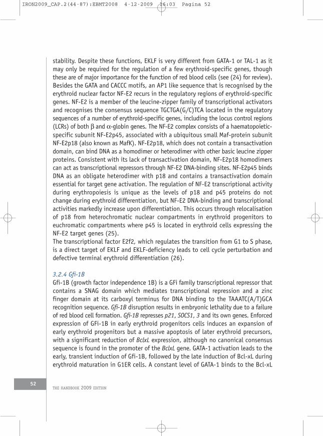
3.2.4 Gfi-1BGfi-1B (growth factor independence 1B) is a GFi family transcriptional repressor thatcontains a SNAG domain which mediates transcriptional repression and a zincfinger domain at its carboxyl terminus for DNA binding to the TAAATC(A/T)GCArecognition sequence. Gfi-1B disruption results in embryonic lethality due to a failureof red blood cell formation. Gfi-1B represses p21, SOCS1, 3 and its own genes. Enforcedexpression of GFi-1B in early erythroid progenitors cells induces an expansion ofearly erythroid progenitors but a massive apoptosis of later erythroid precursors,with a significant reduction of BclxL expression, although no canonical consensussequence is found in the promoter of the BclxL gene. GATA-1 activation leads to theearly, transient induction of Gfi-1B, followed by the late induction of Bcl-xL duringerythroid maturation in G1ER cells. A constant level of GATA-1 binds to the Bcl-xL
THE HANDBOOK 2009 EDITION52
IRON2009_CAP.2(44-87):EBMT2008 4-12-2009 16:03 Pagina 52

promoter throughout the entire induction period, while Gfi-1B is transientlyassociated with the promoter in the early phase. The sustained expression of Gfi-1B abolished GATA-1-induced Bcl-xL expression. GATA-1 binds to the noncanonicalGATT motif of the Bcl-xL promoter for trans-activation. Gfi-1B expressed at increasedlevels is recruited to the Bcl-xL promoter through its association with GATA-1,suppressing Bcl-xL transcription. Therefore, the down-regulation of Gfi-1B in thelate phase of erythroid maturation is necessary for Bcl-xL induction (27). Takentogether, Gfi-1B plays a complex role in erythropoiesis. In immature erythroidprogenitors, through the zinc finger domains, Gfi-1B would trans-activate target genesimplicated in cell proliferation for erythroblast expansion. At the onset ofdifferentiation, Gfi-1B would regulate genes that have to be repressed fordifferentiation induction.
3.2.5 ZBP-89 and Id2The Krüppel-type zinc finger transcription factor ZBP-89 is a component ofmultiprotein complexes involving GATA-1 and its essential cofactor FOG-1. GATA-1and ZBP-89 co-occupy cis-regulatory elements of certain erythroid-specific genes,including an enhancer of the GATA-1 gene itself. Loss-of-function studies inzebrafish and mice demonstrate an in vivo requirement for ZBP-89 in definitiveerythropoiesis but not primitive erythropoiesis, phenocopying aspects of FOG-1- andGATA-1-deficient animals. These findings identify ZBP-89 as being a novel transcriptionfactor involved in erythroid development and suggest that it serves a cooperativefunction with GATA-1 and/or FOG-1 in a developmental stage-specific manner (28).Inhibitors of DNA binding (Id) family members are key regulators of cellularproliferation and differentiation. These activities are related to the ability of Idproteins to antagonise E proteins and other transcription factors. Id2 intrinsicallyregulates erythroid development via interaction with different target proteins (29).Overexpression of Id2 enhances erythroid development, while decreased level of Id2impairs normal erythroid development. Id2 regulation is mediated via interactingwith PU.1 and modulating the activities of PU.1 and GATA-1.
4. Regulation of erythropoiesisAs discussed, the process of making red cells is orchestrated by a complex networkof transcription factors. Among these, GATA-1 plays a critical role by regulating genesinvolved not only in erythroid differentiation, but also in cell cycle and survival.This program of differentiation must be regulated positively and negatively to ensurea continuous but controlled production of red cells in order to provide oxygen ata physiologic level in peripheral tissues.
DISORDERS OF ERYTHROPOIESIS, ERYTHROCYTES AND IRON METABOLISM
CHAPTER 2 • Regulation of erythropoiesis
53
IRON2009_CAP.2(44-87):EBMT2008 4-12-2009 16:03 Pagina 53

4.1 Role of stromal cells in the regulation of erythropoiesisIn the adult, definitive erythropoiesis occurs essentially if not exclusively withinthe bone marrow microenvironment. At this level, stromal and immune cells playan important role by providing factors that include integrins and/or their ligandsand cytokines. These factors are capable of inducing proliferation and survival, whichallows the program of differentiation of erythroid progenitors to take place (forreview see (30, 31)). Erythroblastic islands, the specialised niches in whichprecursors proliferate, differentiate and enucleate, are composed of erythroblastssurrounding a central macrophage. Erythroid islands localise not only to regionsadjacent to bone marrow sinusoids but also to regions throughout the marrow.Erythroblasts express adhesion molecules that mediate both erythroblast/erythroblastand erythroblast/macrophage interactions. Historically, Emp (erythroblastmacrophage protein) was the first molecule identified that appeared capable offorming attachments via homophilic binding. Emp-null foetuses die with severeanaemia showing that Emp performs a critical role in erythropoiesis (32). Theinteractions VLA-4/ICAM-4 and VCAM-1/αV integrin between erythroblasts andcentral macrophage contribute to island integrity. An enhanced erythroblastproliferation related to contact with macrophages has been demonstrated whichoccurred at all Epo concentrations which resulted from decreased transit time inthe G0/G1 phase of cell cycle by a mechanism different from the anti-apoptoticeffect of Epo (33).Recent studies have shown a role of DNase II in erythroid maturation and,particularly, in the process of enucleation occurring at very late stages of erythroidmaturation. Mice deficient in DNase II die of severe anaemia and show a markeddecrease of circulating red blood cells associated with the presence in the circulationof definitive nucleated erythroblasts, a cell type normally present only in the bonemarrow and not in peripheral blood. Central macrophages present in erythroblasticislands may represent the source of DNase II and are then responsible for theengulfment of expelled nuclei through recognition of exposed nuclei phosphatidylserine as they do for apoptotic cells (34).Abnormalities in macrophage differentiation can lead to perturbations in thefunction of the erythroid island and affect erythroid differentiation. Rb protein, aregulator of macrophage differentiation is necessary for erythroid maturation.Target disruption of Rb gene in mice leads to embryonic death with anaemiacaused by failure of enucleation (35-37). Rb protein plays also a cell-intrinsic rolein erythropoiesis mediated by coupling the process of mitochondrial biogenesis toexit from the cell cycle. Its absence leads to ineffective erythropoiesis, with adifferentiation block at the transition from early to late erythroblasts (38).
THE HANDBOOK 2009 EDITION54
IRON2009_CAP.2(44-87):EBMT2008 4-12-2009 16:03 Pagina 54

4.2 Terminal maturation and enucleation
4.2.1 Role of the pro-apoptotic BH3-only-like factor NixErythroid cells undergoing terminal differentiation exhibit concurrent transcriptionalup-regulation of the anti-apoptotic Bcl-xL and the pro-apoptotic BH3-only-like factorNix. Nix null mice exhibit anaemia and erythroid hyperplasia. Cultured Nix nullerythroid cells are hypersensitive to Epo and resistant to apoptosis induced bycytokine deprivation. These results indicate that Nix is a negative regulator oferythropoiesis through modulated apoptosis (39). Nix is also required for theselective mitochondrial clearance during reticulocyte maturation, by triggeringincorporation of mitochondria into autophagosomes followed by maturation of theautophagosomes (40, 41).
4.2.2 Role of the Rac GTPasesRecent studies suggested that Rac GTPases and their effector protein mDia2 playsignificant roles in mouse foetal erythroblast enucleation by affecting the formationof the contractile actin ring in late-stage erythroblasts. Rac GTPases are in adynamic on-and-off state and either inhibition or excessive activation of RacGTPases leads to inhibition of enucleation (29).
4.3 Positive regulation of erythropoiesis
4.3.1 Positive regulation of erythropoiesis at the cellular levelTo ensure a constant production of red cells, various components are requiredincluding iron for haemoglobin synthesis, and folic acid and vitamin B12 for DNAsynthesis (review in (42)). Insulin growth factor 1 synergises with other cytokinesto increase proliferation of early erythroid progenitors and may coordinate theproduction of red cells during development and increase of body mass. Steroidhormones through their nuclear receptors may also enhance red cell production butnone of these molecules are involved in the fine regulation of erythropoiesis.However, androgen production may explain why haematocrit is higher in males thanin females (43). Similarly, glucocorticoids may contribute to the increase oferythroid production during stress erythropoiesis (44). There is also evidence for arole of the renin angiotensin system in the regulation of erythropoiesis. AngiotensinII stimulates the proliferation of normal erythroid progenitors and a lack ofangiotensin-converting enzyme may cause anaemia. In angiotensin-convertingenzyme knock out mice, there is moderate anaemia and Epo synthesis is blunted,suggesting that in vivo angiotensin II plays rather a critical role for the regulationof Epo production by the kidney (45) (see below), consistent with the therapeutic
DISORDERS OF ERYTHROPOIESIS, ERYTHROCYTES AND IRON METABOLISM
CHAPTER 2 • Regulation of erythropoiesis
55
IRON2009_CAP.2(44-87):EBMT2008 4-12-2009 16:03 Pagina 55

effect of angiotensin-converting enzyme inhibitors in secondary erythrocytosis afterrenal transplantation.Cytokines play a critical role in positive regulation of erythropoiesis. They act onerythroid progenitors and precursors at all stage of maturation (BFU-E, CFU-E,erythroid precursors) to prevent apoptosis, induce proliferation, and promote ordelay/inhibit differentiation. Delay in differentiation may play an important roleby allowing self-renewal and expansion of erythroid progenitors before terminalmaturation. As a result, production of red cells is increased. Cytokines are mainlyproduced by bone marrow stromal cells and endothelial cells, within the bone marrowenvironment, but also by immune cells including macrophages and lymphocytes andby organs outside the bone marrow, particularly the liver and the kidney whichrespectively produce thrombopoietin (TPO) and Epo.Several cytokines have been described as possessing burst-promoting activity, i.e.they act on early erythroid progenitors to increase of the number of BFU-E; theseinclude TPO, IL-3, IL-6, IL-8, IL-9, IL-11, GM-CSF (for review see (46)). However,none of them is critical for in vivo erythroid development. Genetic and biochemicalapproaches have demonstrated that, in contrast, SCF and Epo, which act on theirrespective receptors c-Kit and Epo-R, are absolutely required for erythroid cellproduction. Mice with mutations in the SCF gene (Steel (Sl) phenotype), or its receptorgene c-Kit (W phenotype) develop severe anaemia characterised by depressederythropoiesis (47). Epo and Epo-receptor (Epo-R) knockout mice die of failure offoetal liver erythrocyte generation. Therefore, we will focus in this review on theeffect of these two cytokines at the cellular and molecular levels and we will analysethe mechanisms by which they act synergistically.
a. SCF and c-KitSCF is a cytokine produced by stromal cells which interacts with its receptor (c-Kit)(48). C-Kit is expressed in the majority of CD34+ haematopoietic progenitors andits expression is maintained at high levels during the stages of differentiation fromBFU-E to CFU-E; at later stages of differentiation during the maturation of CFU-E,c-Kit expression progressively declines and disappears in polychromatophilic andorthochromatic erythroblasts. Thus, SCF exerts its effects on erythroid cells mainlyduring the early and late stages of differentiation of erythroid progenitors, and mayact also on immature erythroid precursors.Two isoforms of SCF are encoded as a result of mRNA splicing. One isoform is cleavedand released as a soluble biological form and another, which lacks the proteolyticcleavage site, remains principally membrane associated. Importantly, mutant micethat lack membrane associated SCF, but have the normal soluble form, are severelyanaemic. Although not fully understood this difference may be due to the kinetics
THE HANDBOOK 2009 EDITION56
IRON2009_CAP.2(44-87):EBMT2008 4-12-2009 16:03 Pagina 56

of c-Kit activation. C-Kit activation by membrane associated SCF results in a slowinternalisation and degradation and in an increase of activation when compared tothe activation induced by the soluble form.At the cellular level, SCF induces an increase of cell survival and proliferation of earlyerythroid progenitors and precursors towards CFU-E, and the proerythroblast stageof maturation. At the same time SCF delayed erythroid differentiation and maturation(49). This effect allows self-renewal of early erythroid progenitors and precursorsbefore they reach the stage where cells are no longer able to divide. At the laterstage of maturation its main role is to prevent apoptosis and to induce proliferation,in synergy with Epo.Currently, there is no evidence that SCF production is regulated by the need for redcells and oxygenation within the bone marrow. Its synthesis is probably moreconstitutive and although unproven, the level of available SCF may depend on itsconsumption by the amount of c-Kit protein expressed on erythroid precursors.In fact, it is now agreed that regulation of erythropoiesis occurs at the CFU-E leveldepending essentially on the level of available Epo.
b. Erythropoietin and erythropoietin receptorHaemoglobin in erythrocytes carries oxygen from the lungs and delivers it to othertissues. The number of circulating erythrocytes is the major determinant of tissueoxygenation.Although the daily production of erythrocytes is tightly regulated, loss of blood dueto bleeding or haemolysis leads to increased reticulocyte production within a fewdays. As the increased reticulocyte production normalises circulating erythrocytenumbers, the rate of new reticulocyte formation decreases to avoid reboundpolycythaemia. Epo is responsible for this finely tuned homeostatic control oferythrocyte numbers by tissue oxygenation.There is therefore an inverse relationship between erythropoiesis and oxygendelivery by the blood (Figure 2). The normal range of Epo serum concentrations isrelatively low (5-30 mU/mL) and in physiologic conditions, a linear decline inhaematocrit results in an exponential increase in serum Epo. The kidney is the majorsite of Epo production for definitive erythropoiesis. Epo is also produced in the adultliver, which is the main Epo producing organ during foetal erythropoiesis at thetime where erythropoiesis is located in foetal liver. The renal Epo-producing cellsare a subset of cortical interstitial cells located adjacent to the proximal tubules(see Chapter 1). The number of cells producing Epo increases exponentially withlinear decreases in haematocrit. In adult liver the Epo-producing cells are thehepatocytes and interstitial cells, and their number also increases dramatically as
DISORDERS OF ERYTHROPOIESIS, ERYTHROCYTES AND IRON METABOLISM
CHAPTER 2 • Regulation of erythropoiesis
57
IRON2009_CAP.2(44-87):EBMT2008 4-12-2009 16:03 Pagina 57

haematocrit decreases (for review see (50)). Hypoxia inducible factor (HIF)-1α isinduced by hypoxia and interacts with the promoter of the EPO gene to form atranscription factor complex that positively regulates EPO gene expression. Therapidity of HIF-1α induction by hypoxia and its almost immediate disappearanceafter relief of hypoxia is a consequence of its rapid ubiquitination by a complexinvolving the von Hippel-Lindau protein, and then degradation by the proteasome(see Chapter 1). The binding of HIF-1α by the von Hippel-Lindau protein isdependent on the hydroxylation of two specific prolines by a proline hydroxylase.Within the transcriptional complex, HIF-1α is hydroxylated on asparagine residuesby an asparaginase hydroxylase, which as a consequence inhibits HIF-1α bindingto other proteins of the transcriptional factor complex. Thus, normoxic cells havesufficient oxygen for the hydroxylations of HIF-1α that lead to its rapid degradationand inactivation, while hypoxic cells do not have sufficient oxygen for these
THE HANDBOOK 2009 EDITION58
Figure 2: Endocrine regulation of erythropoiesis
Pink erythroid progenitors are less sensitive to Epo than red ones, and die of apoptosis at lowconcentration of Epo. On the right side, correlation of Epo level with haematocrit (Hct).
No apoptosis
pO2
Kidney
pO2
Epo
HctApoptosis
Epo
Epo
IRON2009_CAP.2(44-87):EBMT2008 4-12-2009 16:03 Pagina 58

reactions, and the nonhydroxylated HIF-1α survives and mediates the transcriptionof the EPO gene. The reports that patients with loss of function mutations in VHLand more recently in proline hydroxylase may develop polycythaemia havedemonstrated in vivo the critical role of these proteins in the regulation of Eposynthesis (see Chapter 1).EPO gene targeting reveals that Epo is dispensable at earlier stage of erythropoiesisincluding BFU-E (51). Likewise, in vivo administration of Epo has little influence onthe number of BFU-E. In contrast, Epo administration dramatically increases the numberof CFU-E and erythroid precursors up to the stage of basophilic erythroblasts, mainlyby preventing their apoptosis. Erythroid progenitors and precursors exhibit a wide rangeof Epo sensitivities. This range of Epo sensitivities corresponds to the exponential rangeof serum Epo concentrations found in normal and anaemic states. At low Epoconcentration only hypersensitive progenitors and precursors will survive.Therefore, the regulation of apoptosis of erythroid progenitor and precursor cellsduring their stages of Epo dependence explain the rapid but tightly regulated controlof erythrocyte populations in response to hypoxia, hyperoxia, and anaemia withoutrequiring any effect of Epo on cell cycling or differentiation. In this model Epomodulates the rate of apoptosis of erythroid progenitors and precursors. Althoughthis model was proposed more than twenty years ago, the molecular mechanismsunderlying the Epo sensitivity of erythroid progenitors remain to be determined.It is possible that besides different intrinsic properties of erythroid progenitors, Eposensitivity may be related to heterogeneity in the microenvironment in whicherythropoiesis occurs.Thus, erythropoiesis is mainly regulated in an endocrine manner, the kidney beingthe Epo producing gland and the target being the bone marrow (Figure 2).
4.3.2 Positive regulation of erythropoiesis at the molecular level
a. SCF and c-KitThe binding of SCF to c-Kit results in dimerisation and autophosphorylation of thereceptor on several distinct and specific cytoplasmic tyrosine residues that serveas docking sites for various Src homology 2 domain-containing enzymes andadaptor proteins including phospholipase Cg (PLC-g), phosphatidylinositol 3-kinasep85 subunit (PI 3-kinase), Ras GTPase activating protein, SHP2 phosphatase, Srckinases, Grb2, Grb7, and Shc. Although the precise role of each pathway is not yetfully elucidated, all of them are involved in cell proliferation and survival. However,PI 3-kinase and Src family kinases appear to play an essential role in regulating growthand survival of erythroid cells. The PI 3-kinase pathway might be activated through
DISORDERS OF ERYTHROPOIESIS, ERYTHROCYTES AND IRON METABOLISM
CHAPTER 2 • Regulation of erythropoiesis
59
IRON2009_CAP.2(44-87):EBMT2008 4-12-2009 16:03 Pagina 59
both direct binding to specific tyrosine phosphorylated residues or through Src kinaseactivation. P85 knockout mice exhibit a reduction in BFU-E and CFU-E numbers (forreview see (52)).At the level of erythroid progenitors these pathways may act synergistically withEPO-R signalling as discussed below (for review see (53)).
b. Erythropoietin receptorAs discussed above, Epo is the crucial regulator of red blood-cell production anddelivers essential growth, differentiation and survival signals to erythroid progenitorsand precursors. Epo at the molecular level exerts its effect by stimulating Epo-R.The Epo-R belongs to the type I cytokine receptor family, characterised by a singletransmembrane domain and a cytoplasmic tail lacking a kinase domain (54). TheEpo-R is believed to exist in an unliganded dimeric state. On binding Epo, itundergoes a conformational change that activates the pre-bound, cytoplasmic,tyrosine kinase, Janus kinase 2 (JAK2). JAK2 is absolutely required for Epo-Ractivation and JAK2-knockout mice have a phenotype identical to Epo-R knockoutmice. A ligand-induced receptor homodimer conformational change leads to trans-phosphorylation and activation of JAK2. Activated JAK2 phosphorylates eight keytyrosine residues in the Epo-R cytoplasmic domain, thereby providing dockingsites for SH2 domain-containing downstream signalling molecules STAT5a/b, SHP1,SHP2, SHIP, p85a regulatory subunit of PI 3-kinase, Grb2, Lyn tyrosine kinase, andsuppressor of cytokine signalling (SOCS) (for review see (55)). In addition to thesesignalling molecules, other signalling molecules that bind Epo-R have also beenidentified, including tyrosine kinases Syk, Tec, PLC-γ, and adaptor proteins Shc, Cbl,Crkl, IRS-2, and Gab1/2, and the nucleotide exchange factors Sos and Vav. Thephysiologic roles of the signalling cascade initiated via the phosphorylation of thesetyrosines in Epo-R remain to be fully elucidated. Epo-R activates signal transducersand activators of transcription 5 (STAT5), Ras/mitogen-activated protein kinase(MAPK), and phosphoinositide-3 kinase (PI-3K)/Akt pathways as well as calciumentry and other signalling molecules. The role of these pathways in erythropoiesiswill be briefly reviewed (Figure 3).
› b1. JAK2/STAT5 and Bcl-XLEpo stimulation leads to a robust tyrosine phosphorylation of JAK2, which istime and dose dependent. JAK2 like all other members of the JAK kinases familyhas seven unique domains, termed JAK-homology (JH) domains. A catalyticallyactive tyrosine kinase domain (JH1) and adjacent pseudokinase domain (JH2)are located at the C terminus. The pseudokinase domain has the architecture
THE HANDBOOK 2009 EDITION60
IRON2009_CAP.2(44-87):EBMT2008 4-12-2009 16:03 Pagina 60

of a tyrosine kinase domain, but may have a negative regulatory role asrecently evidenced in the pathophysiology of polycythaemia vera (56). JAK2plays a pivotal role in mediating signals downstream of Epo.JAK2 can support steady-state erythropoiesis via a route independent of Epo-R tyrosine phosphorylation that, in part, may involve MEK 1,2, and ERK 1,2stimulation. Recent investigation of Epo-regulated survival genes shows thatthe “JAK2 only” axis induces down-modulation of pro-apoptotic Bim, Foxo3aand Trb2 (57). Moreover, the Epo “JAK2 only” pathway induces cell cycleregulation through induction of cell-cycle progression factors like G1 to Sphase transition 1 (Gspt1), nuclear protein 1 (Nupr1), Egr1, Ngfi-A bindingprotein 2 (Nab2) and Myc (58).Although in erythroid cells JAK2 is able to phosphorylate STAT1 (59) or STAT3(60) that may play a role in regulation of stress erythropoiesis, the mostimportant targets for steady state erythropoiesis are STAT5a/b isoforms. STAT5aand STAT5b are two isoforms of STAT5 that share 90% of homology and are oneof the seven members of the STAT family, which are characterised by their criticalrole in transducing cytokine signalling. These proteins are latent in the
DISORDERS OF ERYTHROPOIESIS, ERYTHROCYTES AND IRON METABOLISM
CHAPTER 2 • Regulation of erythropoiesis
61
AKT
PDK1 PDK2
P P
GAB
P
PP
P
p110α,β,δ
p85PI3K
IRSPI(4,5)P2PI(3,4)P2 PI(3,4,5)P3
SHIP-1 PTEN
P
FOXO3a
BAD
Fas-L Bim
P
P
Survival
MDM2
p53
P
Survival
GSK3
P
Proliferation
IKK
NF- κB
P
P
Survival
STAT5
STAT5P
P
Epo
Epo
Jak 2Jak 2PP
Jak2
RasSos
Grb2
MAPK
Raf-1
Survival
LynBTK
Survival
p27
BTG1
Survivin
GATA-1
Differentiation
Vav
Calciumentry
Proliferation
Differentiation
BAX
STAT5
Figure 3: EpoR and its role in cell survival, proliferation, and differentiation
IRON2009_CAP.2(44-87):EBMT2008 4-12-2009 16:03 Pagina 61

cytoplasm. They contain an SH2 domain allowing their docking to Y343phosphorylated Epo-R and in turn their activation by phosphorylation ontyrosine residues in the transactivation domain. STAT5 is also phosporylated onserine residues but the function of these is not yet elucidated andphosphorylation may have an inhibitory effect on STAT5 transactivation.Activated STAT5 homodimers translocate to the nucleus to bind DNA andenhance gene activity involved in cell survival, proliferation, and differentiation.Mice expressing truncated Epo-R-mutants that retain solely the ability toactivate STAT5 but lack all other tyrosines critical for activation of othersignalling pathways live normally (61). These animals display only mildabnormalities in recovery from erythropoietic stress. Additional mutation of theTyr 343 required for STAT5 activation, however, strongly affects stresserythropoiesis (62).STAT5 plays an important role in maintaining a high erythropoietic rate duringfoetal development and during stress responses in adult mice, as shown by studiesusing STAT5a-/- STAT5b-/- mice (63). STAT5a/b-deficient erythroid progenitorsexhibit high levels of apoptosis and are less sensitive to Epo. Indeed, a recentstudy implicated STAT5 in the stimulation of Pim1, Pim3, Irs2, Serphin-3G andTrb3, new anti-apoptotic effectors (57). Introduction of dominant negative STAT5into adult erythroid progenitors also induced cell cycle arrest. It has recentlybeen shown that STAT5 is implicated in cell cycle regulation through down-modulation of inhibitory cyclin G2, p27/Cdkn1b, B-cell leukaemia/lymphoma6 (Bcl6), and also through induction of cyclin D2. Furthermore, duringdifferentiation divisions of later stage erythroblasts, Epo modulation of cell cycledistribution suggests an influence of Epo in reprogramming toward differentiation(58). Expression of a constitutively activated STAT5a mutant is sufficient tocomplement the proliferation defect of Epo-R-/- JAK2-/- cells, enabling self-renewal and erythroid differentiation in the absence of an Epo signal (64).STAT5ab-/- mice show differentiation instead of renewal, causing accumulationof mature cell and gradual proliferation arrest. STAT5ab was additionallyrequired for Epo-induced terminal differentiation. Differentiating STAT5ab-/-
erythroblasts undergo apoptosis instead of erythroid maturation, due to theabsent induction of the anti-apoptotic protein Bcl-xL. This defect can be fullyrescued by exogenous Bcl-xL (65).The anti-apoptotic role of STAT5 in Epo-R signalling, although still controversial,is probably mediated in part through its direct induction of Bcl-xL in erythroidcells. The Bcl-xL promoter region contains binding sites for STAT5 and GATA-1that may act synergistically to positively regulate its activity (63). EmbryonicBcl-xL-knockout mice die around E12.5 because of brain defects and severe
THE HANDBOOK 2009 EDITION62
IRON2009_CAP.2(44-87):EBMT2008 4-12-2009 16:03 Pagina 62

anaemia, and disruption of Bcl-xL in the haematopoietic lineage results in severehaemolytic anaemia (66). Although both models generate normal numbers ofBFU-E, CFU-E and erythroblasts, defective maturation leads to anaemia.Exogenous expression of human Bcl-xL results in Epo-independent differentiationof wild-type murine erythroblasts, which indicates that expression of Bcl-xL alonecan drive erythroid differentiation without the need for activation of otherpathways. Therefore, Epo appears to be a permissive factor for the program ofdifferentiation, which is driven by the complex network of transcriptionalfactors. Interestingly, Bcl-xL besides its major role as an anti-apoptotic factorin erythropoiesis, Bcl-xL is involved in the regulation of haem synthesis (67)and a marked increase in Bcl-xL expression was observed during early stagesof the differentiation from a BFU-E to a CFU-E and from CFU-E to pro-erythroblasts. Bcl-xL levels increase during the early and intermediate stagesof erythroid maturation, reaching a peak level of expression when the majorityof erythroid cells had reached the stage of polychromatophilic erythroblasts.
› b2. PI 3-kinaseEpo also stimulates cell survival, proliferation and differentiation by activatingPI 3-kinase (p85 (regulatory subunit)/p110 (catalytic subunit)) through directrecruitment of the p85 to Epo-R phosphorylated tyrosines, and also indirectlythrough binding to Cbl, Gab1, Gab2, insulin receptor substrate (IRS)-2 and Vav(reviewed in (68)). It may also be activated by Ras. PI 3-kinase activation isnecessary but not sufficient to protect against apoptosis. Mice that do not expressthe p85 subunit of PI 3-kinase have markedly reduced erythropoiesis withreductions in the number of BFU-E and CFU-E precursors. By activating PI 3-kinase, Epo has an effect on the progression of the cell cycle through up-regulation of cyclin D3, E and A at the protein level, and c-Kit at the mRNAand protein level (69). Protein kinase B (PKB)/Akt, a serine/threonine kinaseis activated downstream of PI 3-kinase through the generation of secondmessenger 3’-phosphorylated inositol lipids (PIP3), and in this process PKCactivity is required (70). Many downstream substrates of PKB/Akt play animportant role in regulating proliferation and survival of erythroid progenitors.They include the forkhead Foxo3a/ FKHR-L1 (71, 72) transcription factors, theproaoptotic protein Bad, and the glycogen synthetase kinase-3 (GSK3) (73).PI 3-kinase may induce cell proliferation through the phosphorylation and therebyinhibition of transcriptional activity of Foxo3a. Among the genes transcriptionallyactivated by Foxo3a two play a critical role in erythroid cell proliferation, B celltranslocation gene 1 (BTG1) and p27 Kip1 cyclin-dependent kinase inhibitors(74), and Ccng2 (Cyclin G2).
DISORDERS OF ERYTHROPOIESIS, ERYTHROCYTES AND IRON METABOLISM
CHAPTER 2 • Regulation of erythropoiesis
63
IRON2009_CAP.2(44-87):EBMT2008 4-12-2009 16:03 Pagina 63

PKB/Akt promotes cell survival at various levels (for review see (75)). First, itphosphorylates the pro-apoptotic protein Bad. Bad sequestrates the Bcl-xLprotein in the cytoplasm. Upon Bad phosphorylation, Bcl-xL is released and thusallows maintenance of mitochondrial integrity. Second, upon phosphorylationby Akt, Foxo3a is exported from the nucleus to the cytoplasm where it issequestered by 14-3-3 proteins and thereby cannot activate genes involved incell death including Fas-L and Bim. Third, Akt activation is involved in thepositive regulation of Survivin, a member of the IAP family (inhibitor ofapoptosis family) that inhibits caspase activity. Fourth, by a mechanism notfully elucidated, Akt increases the transcription of survival genes through theactivation of NF-kB and CREB transcription factors. Finally, the glycogensynthetase kinase-3 (GSK3), which is involved in glycogen metabolism is alsoinhibited by Akt and the inhibition of its activity is able to protect erythroidcells from apoptosis upon Epo deprivation. Although its mechanism of actionis not yet fully known, PI 3-kinase may be also involved in cell differentiationand its activity, at least in some cell lines, is required for Glycophorin A andhaemoglobin synthesis. Akt mediates a signal downstream of Epo-R, distinctfrom its survival signal that supports differentiation of foetal liver erythroidprogenitor cells. Activated Akt complements Epo-R signalling and supportserythroid-cell differentiation in WT and JAK2-deficient foetal liver erythroidprogenitors (76). Akt may act by phosphorylation of GATA-1, which increasesits transcriptional activity (17). Moreover, the PI 3K/Akt signalling pathway hasbeen identified as a mediator of Epo-induced phosphorylation of GATA-1. Aktphosphorylates GATA-1 S310 in erythroid cells and enhances GATA-1transcriptional activity (16).
› b3. Ras and MAP kinase pathwayEpo activates the Ras pathway through several mechanisms. Grb2, a cytoplasmicadapter protein, can bind directly through its SH2 domain to the tyrosinephosphorylated Epo receptor, or indirectly through the tyrosine phosphataseSHP2 or SHC. All these mechanisms lead to translocation of SOS, a Ras guaninenucleotide exchange factor, to Ras, resulting in its activation. In addition, Vavproteins may also play the role of exchange proteins to activate Ras. Downstreamof Ras activation, Raf-1 is activated and in turn MAP kinases, and some earlygenes are activated including c-fos, c-myc, c-jun.Ras activation is essential for the differentiation, proliferation, and survival oferythroid progenitors in vitro. In mammals, although there are 3 homologousRas proteins, H-Ras, N-Ras, and K-Ras, only K-Ras is essential for erythropoiesis;it is particularly required for the differentiation of erythroid progenitor cells to
THE HANDBOOK 2009 EDITION64
IRON2009_CAP.2(44-87):EBMT2008 4-12-2009 16:03 Pagina 64

late basophilic erythroblasts and for the proliferation of haematopoieticprogenitors (77).Epo activates the catalytic activity of several MAP kinases, including extracellularregulated kinases 1/2 (Erk1/2), SAP kinase (SAPK)/Jun kinase (Jnk) and p38.Erk1/2 is thought to participate in mitogenesis, whereas the roles of SAPK/JNKand p38 are more complex and not fully elucidated. The ERK1/2 pathway inerythroid cells is involved in the early proliferative phases of erythropoiesis andin the inhibition of terminal erythroid differentiation (78). JNK is aserine/threonine kinase that phosphorylates Jun proteins and activates itstranscriptional activity within AP1 complexes. AP1 is implicated in the promotionof cell cycle activity, and in both positive and negative regulation of apoptosis.AP1 complexes and particularly those containing Jun-B have been implicatedin the induction of apoptosis observed when growth factors are removed. Incontrast, AP1 complexes containing activated c-jun have been implicated inthe repression of apoptosis in growth factor-dependent cells, and expressionof c-jun delays apoptosis induced in erythroid cells. JNK activity appears to bean important regulator of proliferation in immature, primary erythroid cells anderythroid cell lines but may not be required for the survival or proliferation ofCFU-E or pro-erythroblasts (79). P38 knock out mice are severely anaemic. Thisphenotype is due to a defect in the post-translational regulation of Epo mRNA(80). The p38 MAPK signal transduction pathway may play a critical role indifferentiation as evidenced in butyrate-induced erythroid differentiation of theK562 cell line, as well as in Epo-dependent differentiation of mouseerythroleukaemia SKT6 cells and of primary erythroblasts (81).
› b4. Lyn and Btk kinase pathwayOther kinases associated with Epo-R including Lck/Yes-related novel tyrosinekinase (Lyn) and Bruton’s tyrosine kinase (Btk) may play a role in Epo-Rphosphorylation and signal transduction (82). Lyn is a substrate for Eposignalling and has a role in erythroid differentiation. Evidence of its involvementhas emerged following the isolation of erythoid cell line J2E-NR, which is Lyn-deficient and has impaired erythroid differentiation (83). Erythroblasts from Lyn-deficient mice do not express GATA-1 and EKLF, and expression of STAT5a andSTAT5b are decreased. Impairment of erythroid differentiation and anaemia areobserved as evidenced by an increase of BFU-E and CFU-E number in spleen (84).Reduced phosphorylation of Lyn but not of JAK2 by CD45 inhibits erythoiddifferentiation of cord blood CD34+ (85). Lyn plays a positive role at both stagesof erythroid development, for expansion of early progenitors and survival of lateerythroid progenitors (86). Bruton tyrosine kinase, which is critical for B cell
DISORDERS OF ERYTHROPOIESIS, ERYTHROCYTES AND IRON METABOLISM
CHAPTER 2 • Regulation of erythropoiesis
65
IRON2009_CAP.2(44-87):EBMT2008 4-12-2009 16:03 Pagina 65
development is also involved in Epo-R signalling. Although neither patients withX linked agammaglobulinemia (Btk deficiency) nor Btk knockout mice show anyovert defect in erythropoiesis, Btk deficient erythroblast progenitors exhibit areduction in Epo-R and JAK2 phosphorylation and have enhanced erythroiddifferentiation and reduced capacity for self renewal. The mechanism of actionof Btk on Epo-R signalling remains to be determined, but probably occursdownstream of JAK2 and Lyn activation. Furthermore, Btk is involved in theprotection of erythroid progenitors and early precursor sensitivity to deathreceptor stimulation (87) (see below).
› b5. Regulation of intracellular calcium by erythropoietinActivation of Epo-R causes an increase in intracellular calcium. Specific tyrosineresidues in the Epo-R are required for this calcium influx. The transient receptorpotential channel proteins TRPC2 and TRPC6 are highly expressed in erythroidprogenitors and may play a role in calcium entry. In erythroid cells calciumconcentration increases more in the nucleus than in the cytoplasm after Epostimulation. Calcium may regulate cell proliferation and differentiation throughexpression of proto-oncogenes, phosphorylation of transcriptional factors, oractivation of calcineurin (55).
c. Synergy between c-Kit and erythropoietin receptor at the molecular levelEpo and SCF act synergistically to increase erythropoiesis by several molecularmechanisms. SCF sensitises cells to erythropoietin and therefore the requirementof Epo is lower for full completion of erythroid proliferation and survival. At thesame time, SCF slows down differentiation of erythroid progenitors, which may resultin expansion of erythroid cells. In vitro, whereas Epo alone is able to produce humanerythroid cell development and maturation from progenitor cells, a markedlyenhanced, synergistic proliferation and expansion of developing erythroid cells issupported by the combination of Epo and SCF. In cell lines, c-Kit and Epo-Rinteract physically via the box2 cytoplasmic domain of the Epo-R (88), but thisinteraction is not sufficient to induce erythroid formation in primary cells.Mechanistically, c-Kit stimulation by SCF does not activate Epo-R by inducing itsdimerisation, but by phosphorylating tyrosine residues in the cytoplasmic domainof Epo-R. It seems that Epo-R Y343, which binds STAT5a, is implicated in c-Kit co-signalling. On the other hand, tyrosine residues that bind Src kinases in the c-Kitreceptor appear to be sufficient for co-signalling with Epo-R (88).However, Epo and SCF may have distinct and sequential effects on erythroiddifferentiation and no joint effect of Epo and SCF is required to quantitatively describethe proliferation or death of erythroid cells during erythroid differentiation. Overall,
THE HANDBOOK 2009 EDITION66
IRON2009_CAP.2(44-87):EBMT2008 4-12-2009 16:03 Pagina 66
SCF promotes the early proliferation of primitive cells, while Epo primarily promotesthe survival of differentiating erythroid progenitors (89).At the molecular level, SCF and Epo synergistically enhance the magnitude and theduration of the MAP kinase (MAPK, ERK1/2) pathway, which may be important forboth proliferation and for reducing differentiation of erythroid cells (78). Prolongedactivation of MAPK might also play a role in preventing apoptosis. However, SCFand Epo may lead to activation of MAPK through different pathways and theirdownstream targets may be not identical.The synergistic effect between Epo and SCF on cell survival may also occur at otherslevels through distinct signalling mechanisms. For example, SCF through PI 3-kinaseactivation induces a robust activation of Akt and thereby phosphorylation of Badand its sequestration in the cytosol by 14-3-3 proteins, allowing the release of Bcl-xL in the mitochondrial membrane which protects the cell against apoptosis (seebelow). In this model of synergy, Bcl-xL expression is increased by the JAK/STATpathway activated by Epo-R, and its activity is increased by the c-Kit receptor.Likewise, STAT transactivation activity is increased by the activation of the PKA/CREBdependant pathway induced by c-Kit and the recruitment of CREB binding proteinCBP/p300 (90). Multiple other pathways that remain to be determined could alsobe involved in the major synergistic effect observed between SCF and Epo.Interestingly, pathways activated by c-Kit might be sufficiently divergent from thoseactivated by other cytokines (TPO, IL-3, GM-CSF, etc) to explain the lack ofredundancy with SCF in vivo.
4.4 Negative regulation of erythropoiesisAlthough not fully elucidated, the cellular regulation and the molecular signalsresponsible of positive regulation of erythropoiesis are well characterised. However,a negative control is clearly required at several levels to avoid overproduction oferythroid cells and polycythaemia, which may lead to hyperviscosity and ultimatelyto thrombosis.
4.4.1 Negative regulation of erythropoiesis at the cellular level
a. SCF and c-KitAs mentioned above there is no clear evidence that excess of erythroid progenitorsor red cell regulates SCF production by stromal cells. In contrast, Epo concentrationand Epo-R activation are the major parameters that determine red cell production.
b. Erythropoietin productionEpo production is determined at two levels. First, Epo production is well controlled
DISORDERS OF ERYTHROPOIESIS, ERYTHROCYTES AND IRON METABOLISM
CHAPTER 2 • Regulation of erythropoiesis
67
IRON2009_CAP.2(44-87):EBMT2008 4-12-2009 16:03 Pagina 67

by renal oxygenation (see above and Chapter 1). Hence, a sufficient number of redcells and adequate oxygenation results in decrease of Epo production by thekidney, through degradation of HIF by the proteasome (see Chapter 1). As aconsequence, the less Epo sensitive erythroid progenitors and precursors will dieof apoptosis. In addition to the reduction of Epo production, a down-modulationof Epo action occurs after Epo/Epo-R interaction through the endocytosis andsubsequent degradation of Epo and the Epo-R by the proteasome of erythroidprogenitors (see below).These multiple levels of down regulation contribute to the fine control of Epoconcentration in the blood, preventing polycythaemia after recovery from bleedingor red cell haemolysis.
c. Death receptors and their ligandsBeside oxygenation and Epo production, erythropoiesis is also regulated in aparacrine fashion within the bone marrow erythroblastic islands. In this system,mature erythroblasts inhibit survival and differentiation of immature erythroblaststhrough Fas/Fas-L interactions, with death ligands being expressed on matureerythroid cells and the death receptor being present on the surface of immatureerythroblasts (increased expression from BFU-E to CFU-E, to reach maximal level atthe stage of pro-erythroblasts and basophilic erythroblasts) (75, 91). The patternof expression of membrane bound Fas-L is different, Fas-L being expressed only inmature erythroblasts (orthochromatic erythroblasts). Sensitivity to Fas-L is increasedby cytokines that induce cell cycling of immature erythroid progenitors, includingSCF, IL-3 and TPO. In contrast, mature precursors are insensitive to Fas-L. Restingprogenitors are protected by elevated levels of c-FLIP, an inhibitor of Fas activation.Furthermore, Fas triggering induces an arrest of erythroid maturation through thecleavage of TAL-1 and GATA-1 by caspases (91, 92). In addition to directdepolarisation of the mitochondrial membrane (see below), these mechanisms mayaccount for the low, but significant, level of ineffective erythropoiesis associatedwith premature death of erythroid cells observed in normal subjects and also themaintenance of an available pool of erythroid progenitors. Interestingly, SCF or highdose of Epo are able to circumvent the apoptotic effect of Fas-L on erythroidprecursors, allowing production of red cells during stress erythropoiesis. A similarsystem is observed with another death receptor (TRAIL-R) and its ligand (TRAIL)(93). Therefore, the control of the production of mature red cells may be summarisedas follows (Figure 4). At low doses of Epo, cell die by apoptosis, at intermediatedoses, cell are arrested in their maturation or enter a program of apoptosis,depending on the number of mature erythroblasts in the bone marrow, and at highdoses of Epo, erythroid progenitors and precursors pursue their maturation
THE HANDBOOK 2009 EDITION68
IRON2009_CAP.2(44-87):EBMT2008 4-12-2009 16:03 Pagina 68
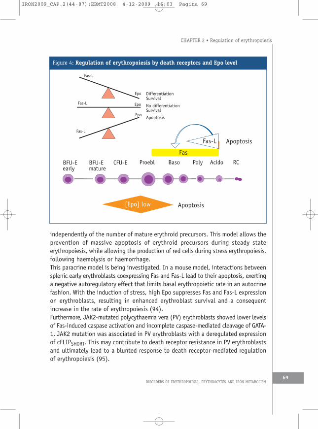
independently of the number of mature erythroid precursors. This model allows theprevention of massive apoptosis of erythroid precursors during steady stateerythropoiesis, while allowing the production of red cells during stress erythropoiesis,following haemolysis or haemorrhage.This paracrine model is being investigated. In a mouse model, interactions betweensplenic early erythroblasts coexpressing Fas and Fas-L lead to their apoptosis, exertinga negative autoregulatory effect that limits basal erythropoietic rate in an autocrinefashion. With the induction of stress, high Epo suppresses Fas and Fas-L expressionon erythroblasts, resulting in enhanced erythroblast survival and a consequentincrease in the rate of erythropoiesis (94).Furthermore, JAK2-mutated polycythaemia vera (PV) erythroblasts showed lower levelsof Fas-induced caspase activation and incomplete caspase-mediated cleavage of GATA-1. JAK2 mutation was associated in PV erythroblasts with a deregulated expressionof cFLIPSHORT. This may contribute to death receptor resistance in PV erythroblastsand ultimately lead to a blunted response to death receptor-mediated regulationof erythropoiesis (95).
DISORDERS OF ERYTHROPOIESIS, ERYTHROCYTES AND IRON METABOLISM
CHAPTER 2 • Regulation of erythropoiesis
69
BFU-Eearly
BFU-Emature
CFU-E Proebl RCBaso Poly Acido
[Epo] low
Fas-L
Apoptosis
Apoptosis
DifferentiationSurvival
Epo
Fas-L
Fas-L
Fas-L Epo
Epo
No differentiationSurvival
Apoptosis
Fas
Figure 4: Regulation of erythropoiesis by death receptors and Epo level
IRON2009_CAP.2(44-87):EBMT2008 4-12-2009 16:03 Pagina 69

d. Cytokines that negatively regulate erythropoiesisThere are a number of pathological conditions where erythropoiesis is inhibited. Thepathophysiologic mechanisms underlying this inhibition are reviewed in otherchapters of this book. We will briefly review the mechanism of action of theinflammatory cytokines that are overexpressed in anaemia of chronic disease (67).In the inflammatory process various cytokines including tumour necrosis factor (TNF)-α, interferon (INF)-γ and transforming growth factor (TGF)-β and chemokines suchas stromal derived factor (SDF)-1 are abnormally synthesised, and may impairerythropoiesis at several levels.For example, inflammatory cytokines are responsible for a blunted erythropoietinresponse by the kidney. The level of Epo in the serum is therefore not adjustedaccording to tissue oxygenation. They also decrease the availability of iron toerythroid progenitors by inducing synthesis of hepcidin (for review see (96)).Interestingly, it has been suggested that hepcidin may also act directly on erythroidprogenitors to decrease their threshold of sensitivity to Epo (97). Inflammatorycytokines can also directly act on erythroid progenitors.
› d1. Tumour necrosis factor-aTNF-α induces a decrease in mature erythroblasts and an increased rate ofapoptosis within the compartment of immature erythroblasts. It may actdirectly on the TNF receptor on immature erythroblasts and through theinduction of ceramide synthesis. In vivo, treatment with a blocking anti-TNF-α monoclonal antibody results in improvement in anaemia and in the proportionof apoptotic erythroblasts (98). TNF-α could play a role in modulating NF-kBactivity which positively regulates erythroid cell proliferation and survival andnegatively regulates cell maturation.
› d2. Interferon-gINF-γ plays a complex role in the regulation of erythropoiesis. It has noapoptotic effect by itself and may even exert a protective effect on apoptosisat the mature stage of erythroblastic differentiation.In contrast, by increasing expression of several death receptors and theirligands, including Fas-L/Fas and TRAIL as well as the recently characterisedprotein TWEAK and its receptor fibroblast growth factor-inducible 14 (Fn14),INF-γ contributes to indirectly inducing apoptosis of erythroid progenitors(99). Furthermore INF-γ induces the formation of ceramide and the blockadeof ceramide synthesis abolishes apoptosis (100).
THE HANDBOOK 2009 EDITION70
IRON2009_CAP.2(44-87):EBMT2008 4-12-2009 16:03 Pagina 70

› d3. Transforming growth factor-b1TGF-β1 is a powerful inhibitor of erythropoiesis. However, its mechanism of actionis different from other cytokines or death ligands. It has virtually no effect onapoptosis, but it markedly accelerates and increases erythroid differentiationto produce normal enucleated red cells even in the absence of macrophages.TGF-β1 inhibits cell proliferation by decreasing the cycle of immature erythroidcells and by accelerating maturation toward orthochromatic normoblasts thatare not in cycle. Therefore, TGF-β1 is a paradoxical inhibitor of erythropoiesisthat acts by blocking proliferation and accelerating differentiation of erythroidprogenitors (101).
› d4. Stromal derived factor-1Bone marrow stromal cells produce SDF-1, and haematopoietic cells express itsreceptor, CXC chemokine receptor 4. Low concentrations of SDF-1 promotehaematopoietic cell growth, but high levels decrease erythroid progenitorgrowth through up regulation of Fas-ligand production and subsequent erythroidcell apoptosis via the Fas-ligand/Fas pathway (102). The factors that modulatethis dual effect are not yet known.
4.4.2 Negative regulation of erythropoiesis at the molecular level
a. Negative regulation of c-Kit phosphorylationThe negative regulation of c-Kit signalling is mediated by various phosphatasesincluding SHP-1, SHP-2, and SHIP. SHP-1 is known to interact with phosphorylatedc-Kit at a specific tyrosine residue and dephosphorylate the receptor, and as suchinactivates all pathways downstream of c-Kit signalling. SHP-2 interacts with c-Kitat another tyrosine residue and influences the activation of the MAP kinase pathway(52). C-Kit is also a target for the adaptor protein Lnk (see below), which maycontribute to inactivation of c-Kit receptor (103).
b. Negative regulation of the erythropoietin receptor at the molecular levelBesides the control of Epo levels to avoid over stimulation, Epo-R activation is alsofinely regulated after its activation. Activation of the Epo-R after Epo binding isvery transient because of the rapid activation of strong down-regulation mechanismsthat quickly decrease Epo sensitivity of the cells. These down-regulation mechanismsinclude receptor internalisation, degradation and inhibition of tyrosinephosphorylation.
DISORDERS OF ERYTHROPOIESIS, ERYTHROCYTES AND IRON METABOLISM
CHAPTER 2 • Regulation of erythropoiesis
71
IRON2009_CAP.2(44-87):EBMT2008 4-12-2009 16:03 Pagina 71

› b1. Degradation of erythropoietin receptorThe Epo receptor is rapidly ubiquitinated after ligand stimulation and the C-terminal part of the Epo receptor is degraded by the proteasome. Then, the Epo-Epo-R complexes are rapidly internalised and targeted to the lysosomes fordegradation. Both ubiquitination and receptor degradation by the proteasomesoccur at the cell surface, and require JAK2 activation (104).
› b2. Inactivation of erythropoietin receptor by phosphatasesIn addition to its degradation, tyrosine phosphorylation in response to Epo istransient and returns to basal levels within 30 min of stimulation. Epo-R is rapidlydephosphorylated by several systems involving SOCS proteins and tyrosinephosphatases.Several phosphatases have been involved in this process, but SHP-1 phosphataseappears to play an essential role. SHP-1 binds via its SH2-domain to a specifictyrosine on the phosphorylated Epo-R. Epo-R lacking this binding site is unableto bind SHP-1 and to dephosphorylate and inactivate JAK2 (105). The lack ofexpression of SHP-1, or impaired binding of SHP-1 to the Epo-R, leads to Epohypersensitivity and erythrocytosis. These phenotypes have been described inhumans as a consequence of the expression of truncated or frame-shift mutationsof Epo-R that lack the C-terminal binding site for SHP-1 (106). Another mannerof desensitisation involves the regulation of metabolic intermediates of inositol.Phosphatidylinositol (3,4,5)-trisphosphate (PtdIns(3,4,5)P3) activates manycrucial signalling events. Several enzymes dephosphorylate PtdIns (3,4,5) P3,including phosphatase and tensin homolog (which dephosphorylate the 3phosphate) and SH2-inositol phosphatase, SHIP-1 and SHIP-2 (whichdephosphorylate the 5 phosphate) (107). Their precise role in erythropoiesisremains to be determined.
› b3. Inactivation of erythropoietin receptor by SOCS proteinsIn addition to phosphatases, the SOCS family of negative regulators of cytokinesignalling is also involved in the control of Epo-R activation (108). Expressionof SOCS genes is rapidly modulated in response to a variety of cytokines andthe SOCS family acts via a negative feedback loop to suppress cytokine-inducedsignal transduction. Three SOCS genes are associated with erythropoiesis,including SOCS-1, CIS, and SOCS-3. Expression of CIS is elevated upon activationof JAK2 and STAT5. SOCS-1 inhibits the activation of JAK2. SOCS-3 is tyrosinephosphorylated in response to Epo stimulation and also suppresses Eposignalling by associating with the Epo receptor and JAK2. Recruitment of CISto the Epo-R via the SH2 domain represses proliferation. SOCS genes are
THE HANDBOOK 2009 EDITION72
IRON2009_CAP.2(44-87):EBMT2008 4-12-2009 16:03 Pagina 72

differentially regulated in normal and transformed erythroid cells depending uponthe stage of maturation and may exert a complex regulation. Analysis oftransgenic and knockout mice indicates that SOCS proteins are not vital to theregulation of erythropoiesis.
› b4. Adaptor proteins, Lnk and modulation of erythropoietin receptor activationCytokine receptor signalling networks rely heavily on adaptor proteins, and theymay play a critical role in controlling receptor activation and threshold ofsensitivity to Epo. Lnk, a member of a newly discovered adaptor protein family,is involved in erythroid regulation. Lnk does not possess a kinase domain butcontains several protein-protein interaction domains including a proline-richamino-terminus, a pleckstrin homology (PH) domain, a Src homology 2 (SH2)domain, and a conserved tyrosine near the carboxyl-terminus. Lnk becomestyrosine-phosphorylated following Epo administration and inhibits Epo-inducedEpo-R phosphorylation and JAK2 activation as well as downstream pathwaysincluding STAT5, AKT and MAPK. The Lnk Src homology 2 (SH2) domain isessential for its inhibitory function. However, its mechanism of action remainsto be elucidated. Lnk may disrupt the binding of positive regulators of JAK2,recruit other JAK2 inhibitors, such as SOCS proteins or SHP-1. Finally, bindingof Lnk to JAK2 may cause a conformational change that keeps JAK2 in akinase-inactive state. Likewise, Lnk-deficient mice have an elevated number oferythroid progenitors, and exhibit superior recovery after erythropoietic stress.In vitro, CFU-E progenitors are hypersensitive to Epo, and Epo-R phosphorylation,JAK2 activation and Epo-induced signalling pathways, including STAT5, AKT, andMAPK are enhanced. Conversely, Lnk overexpression inhibits Epo-dependenterythroblast differentiation and induces apoptosis (103).Taken together, the efficiency of these processes provides explanation for theshort duration of intracellular signalling activated by Epo. In addition thenegative regulator of Epo-R signalling may play a fundamental role in the controlof the survival of erythroid progenitors and precursors. Indeed, as mentionedabove, erythroid progenitor cells possess a vast heterogeneity in their sensitivityto Epo. This intrinsic difference of erythroblasts in their sensitivity to Epo mightbe tightly regulated through negative signalling molecules associated with theEpo-R, providing another checkpoint in addition to Epo level for the regulationof erythropoiesis.
c. Molecular regulation of erythropoiesis by apoptosisAs mentioned above, during erythropoiesis not all erythroid cells survive, becausethey are particularly sensitive to apoptosis. The rate of apoptotic cells might be
DISORDERS OF ERYTHROPOIESIS, ERYTHROCYTES AND IRON METABOLISM
CHAPTER 2 • Regulation of erythropoiesis
73
IRON2009_CAP.2(44-87):EBMT2008 4-12-2009 16:03 Pagina 73
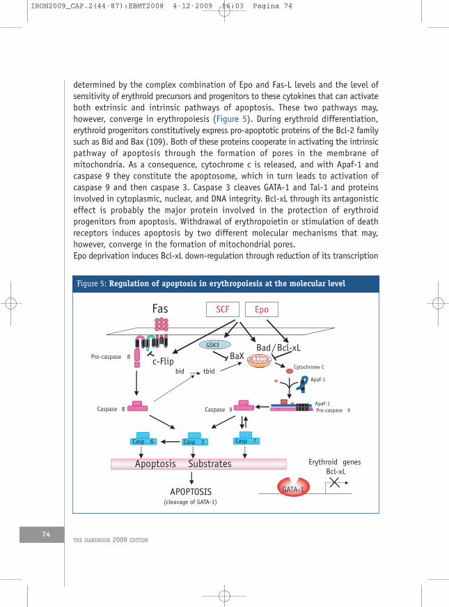
determined by the complex combination of Epo and Fas-L levels and the level ofsensitivity of erythroid precursors and progenitors to these cytokines that can activateboth extrinsic and intrinsic pathways of apoptosis. These two pathways may,however, converge in erythropoiesis (Figure 5). During erythroid differentiation,erythroid progenitors constitutively express pro-apoptotic proteins of the Bcl-2 familysuch as Bid and Bax (109). Both of these proteins cooperate in activating the intrinsicpathway of apoptosis through the formation of pores in the membrane ofmitochondria. As a consequence, cytochrome c is released, and with Apaf-1 andcaspase 9 they constitute the apoptosome, which in turn leads to activation ofcaspase 9 and then caspase 3. Caspase 3 cleaves GATA-1 and Tal-1 and proteinsinvolved in cytoplasmic, nuclear, and DNA integrity. Bcl-xL through its antagonisticeffect is probably the major protein involved in the protection of erythroidprogenitors from apoptosis. Withdrawal of erythropoietin or stimulation of deathreceptors induces apoptosis by two different molecular mechanisms that may,however, converge in the formation of mitochondrial pores.Epo deprivation induces Bcl-xL down-regulation through reduction of its transcription
THE HANDBOOK 2009 EDITION74
Pro-caspase 8
Caspase 8 Caspase 9
Casp 6 Casp 3 Casp 7
Cytochrome C
Apaf-1
Apaf-1Pro-caspase 9
Apoptosis Substrates
APOPTOSIS(cleavage of GATA-1)
SCFFas
bid tbid
Bad/Bcl-xL
Erythroid genesBcl-xL
GATA-1
Epo
BaXc-Flip
GSK3
Figure 5: Regulation of apoptosis in erythropoiesis at the molecular level
IRON2009_CAP.2(44-87):EBMT2008 4-12-2009 16:03 Pagina 74

as a consequence of decrease of STAT5 activation and GATA-1 cleavage. Furthermore,caspase 3 cleaves Bcl-xL. ERK1 and ERK2 activities may at this level control caspaseactivity and as such maintain expression of Bcl-xL.GSK3 activity is suppressed by its phosphorylation induced by activation of theerythropoietin receptor and c-Kit in human erythroid progenitor cells. Therefore,increased GSK3 activity induced by growth factor withdrawal may result in aconformational change of Bax that increases its apoptotic activity (73).The extrinsic pathway involves the death receptors (DR) at the plasma membranelevel. In response to their specific ligand (DR-Ligand), these trimeric receptors recruitthe adapter molecule FADD and either procaspase-8 or -10 in the death-inducingsignalling complex (DISC). These enzymes, either directly through caspase 8activation, or indirectly through cleavage and consequent activation of Bid,resulting in formation of pores in the mitochondria induce caspase 3 activation.Although not fully demonstrated, in erythroid cells the intrinsic pathway seems themost important since Epo and SCF are able to block apoptosis induced by Fas-Lthrough the maintenance of mitochondrial membrane integrity.Foxo3A alone can recapitulate all known elements of the apoptotic programnormally induced by cytokine withdrawal through the activation of the intrinsicpathway, but also through activation of the extrinsic pathway. Foxo3A is expressedin erythroid cells, its expression decreases progressively during erythroid maturation.In immature erythroid precursors, Foxo3A is rapidly phosphorylated by Epo througha PI 3-K-dependent pathway and is exported outside the nucleus and therefore cannotinduce the expression of the pro-apoptotic protein of the bcl2 family Bim and ofFas-L.SCF and Epo may protect CFU-E from Fas-induced apoptosis, the protective effectof SCF being significantly more effective than the effect elicited by Epo alone. Thisprotective effect of SCF involves the activation of Src-kinases, PI-3 kinase, andJAK/STAT5 activation, and MAP kinases. Activation of PKCε could also modulate thesensitivity of erythroid progenitors to death receptor by inducing anti-apoptoticproteins of the bcl2 family (100). The simultaneous addition of IFN-γ and Fas-L toCFU-E elicits a marked activation of caspase 8 and 3 with a consequent apoptoticresponse. This phenomenon is inhibited by SCF via a mechanism involving the upregulation of c-FLIP expression, an inhibitor of caspase 8 activation (111).
5. Role of caspases in maturation of erythroid cellsTaken together, in the regulation of erythropoiesis, caspases appear to play acritical role in the negative regulation of erythropoiesis. Both, low concentrationof Epo or excess of mature erythroblasts (through activation of Fas-L and TRAIL)
DISORDERS OF ERYTHROPOIESIS, ERYTHROCYTES AND IRON METABOLISM
CHAPTER 2 • Regulation of erythropoiesis
75
IRON2009_CAP.2(44-87):EBMT2008 4-12-2009 16:03 Pagina 75

induce caspase activation and, as a result, apoptosis of erythroid progenitors.However, as in other systems of differentiation with terminal enucleation, includingkeratinocytes and lens epithelial cells, or without enucleation such as megakaryocytesto proplatelets or monocytes to macrophages, caspases may also play a critical andpositive role in erythroid precursor maturation (112).The terminal differentiation of erythroid cells exhibits some similarities withapoptosis such as chromatin condensation and the degradation of nuclear components.A transient activation of several effector caspases is required for Epo-inducederythroid differentiation in both humans and mice. This transient caspase activationis not required for the process of enucleation itself, but occurs at earlier steps ofred blood cell formation (113-115). The process of enucleation itself could involvep53 in erythroid cells (116), and as mentioned above DNase II from centralmacrophages in the erythroblastic island. However, caspase inhibition arrests the invitro maturation of erythroid progenitors before nuclear and chromatin condensationat the transition between pro- and basophilic erythroblasts. Caspases are probablyactivated through a mitochondria-dependent pathway similar to that identified invarious forms of apoptosis. Release of cytochrome c from the mitochondria andactivation of caspase 9 and then caspases 3 and 6 are observed. Likewise, caspaseactivation in erythroid cells undergoing differentiation is negatively regulated by theRaf-1 kinase, which prevents premature differentiation of actively proliferatingprecursors (114). It has recently been demonstrated that a balance between Raf-1and Fas expression permits erythroid differentiation. Indeed, Raf-1 by activating theMEK/ERK cascade induces progenitor proliferation. In contrast, differentiation ismediated by Fas via the activation of both ASK1/JNK/p38 pathway and caspasescascade. The point of convergence between the two cascades is activated ERK, whichmaintains Raf-1 expression and inhibits Fas expression. In turn, Fas, once expressed,exerts negative feedback on ERK activation and Raf-1 expression (117, 118).Emphasizing the role of mitochondria in erythroid differentiation, Nix-/- mice exhibitincreased polychromatophils and abnormally shaped erythrocytes suggestive of amaturation defect. Nix deficiency caused anaemia, reticulocytosis and erythroid-myeloid hyperplasia (40). Those results show that pro-apoptotic proteins such as Fasor Nix are involved in erythroid differentiation. Further research is needed todetermine by which mechanism caspase 3 is activated during terminal erythropoiesis.Caspase 3 induces cleavage of the protein acinus responsible for chromatincondensation without DNA degradation, and caspase 6 cleaves proteins involved innuclear integrity such as lamin B (113), which may explain morphological changesin the nucleus occurring during erythroid differentiation. The identification ofother targets cleaved during erythroid differentiation remains an important questionto be elucidated. Interestingly, ICAD, a chaperone of CAD, is not cleaved by caspase
THE HANDBOOK 2009 EDITION76
IRON2009_CAP.2(44-87):EBMT2008 4-12-2009 16:03 Pagina 76

3 during erythroid differentiation. In another study on cell lines, ICAD is cleavedduring erythroid differentiation but CAD is down-regulated at the mRNA and proteinlevel. These changes provide a mechanism by which cells avoid DNA fragmentationwith activated caspase 3 (118).In addition, during erythroid differentiation, caspases are activated, but cells donot enter apoptosis as they do not cleave their DNA and do not expose phosphatidylserine at the cell surface. Although GATA-1 is cleaved by caspases in erythroid cellsundergoing apoptosis under Epo deprivation or death receptor stimulation, itremains uncleaved in erythroid cells undergoing terminal differentiation. Anexplanation for this apparent paradox came from the finding of a constitutiveexpression of HSP70 in erythroid cells. During differentiation but not duringapoptosis, the chaperone protein Hsp70 protects GATA-1 from caspase 3-mediatedproteolysis. At the onset of caspase activation, Hsp70 translocates and co-localisesand interacts with GATA-1 in the nucleus of erythroid precursors undergoing terminaldifferentiation. In contrast, Epo starvation induces the nuclear export of Hsp70 andthe cleavage of GATA-1 (Figure 6) (119). This model provides a potential explanationfor the lack of cell death when caspases are activated in erythroid cells undergoingterminal differentiation, although the crucial question as to how Hsp70 translocatesto the nucleus and remains nuclear localised under Epo is remains open. These resultsstrongly indicate that Hsp70 is another key anti-apoptotic protein in erythroid cells
DISORDERS OF ERYTHROPOIESIS, ERYTHROCYTES AND IRON METABOLISM
CHAPTER 2 • Regulation of erythropoiesis
77
APOPTOSISSURVIVAL and MATURATION
+Epo -Epo
<
GATA-1Hsp70Nucleus
Cytoplasm
Hsp70
<
GA TA-1
Casp 3
Casp 3
Hsp70
Casp 3
Casp 3
Figure 6: Hsp70 protects GATA-1 from caspase 3-mediated proteolysis
At the onset of caspase activation, Hsp70 co-localizes and interacts with GATA-1 in the nucleus of erythroidprecursors undergoing terminal differentiation. In contrast, Epo starvation induces the nuclear exportof Hsp70 and the cleavage of GATA-1.
IRON2009_CAP.2(44-87):EBMT2008 4-12-2009 16:03 Pagina 77

by protecting GATA-1 from caspase 3-mediated cleavage, allowing Bcl-xL expressionvia GATA-1 protection.Thus it appears that dysregulation of Hsp70 expression or localisation couldaccount for some congenital or acquired erythroid disorders. The clarification of themechanism responsible for Hsp70 nuclear localisation under Epo during terminalerythroid differentiation could shed light in the understanding of various acquiredand congenital erythroid pathologies and may serve as a new therapeutic target inthese disorders. Furthermore other chaperones including heat shock proteins HSP27and 90 are also expressed during erythroid differentiation and they may also playan important role in the regulation of erythropoiesis that remains to be clarified.
6. ConclusionsErythropoiesis is therefore a fascinating system of differentiation that is regulatedat several levels. Differentiation is under the control of a complex set of transcriptionalfactors, and growth factors play a critical role in preventing apoptosis and in inducingproliferation. In this system caspases play an essential role in controlling the rateof red cell maturation and as such erythropoiesis is an example of a system in whichregulation of apoptosis and differentiation share similar molecular pathways.Subtle differences in the set of cleaved proteins determine the fate of erythroidprogenitors and precursors. It will be a great challenge to elucidate at the molecularlevel the mechanisms underlying this fine regulation as demonstrated with HSP70in the protection of GATA-1. Understanding the complex role of stromal cells andthe molecules that they produce will certainly help to elucidate the regulation oferythropoiesis within its natural bone marrow environment.The understanding of all these processes will shed light on the pathophysiology ofanaemia associated with various erythroid disorders. All these mechanisms may betranslated beyond erythropoiesis and may be involved in neoplastic transformationof cells and in various degenerative disorders.
References1. Gregory CJ, Eaves AC. Three stages of erythropoietic progenitor cell differentiation
distinguished by a number of physical and biologic properties. Blood 1978; 51: 527-537.2. Iscove NN. The role of erythropoietin in regulation of population size and cell cycling
of early and late erythroid precursors in mouse bone marrow. Cell Tissue Kinet 1977; 10:323-334.
3. Cantor AB, Orkin SH. Transcriptional regulation of erythropoiesis: An affair involvingmultiple partners. Oncogene 2002; 21: 3368-3376.
4. Gregory CJ, Eaves AC. Human marrow cells capable of erythropoietic differentiation invitro: Definition of three erythroid colony responses. Blood 1977; 49: 855-864.
THE HANDBOOK 2009 EDITION78
IRON2009_CAP.2(44-87):EBMT2008 4-12-2009 16:03 Pagina 78

5. Iscove NN, Sieber F, Winterhalter KH. Erythroid colony formation in cultures of mouseand human bone marrow: Analysis of the requirement for erythropoietin by gel filtrationand affinity chromatography on agarose-concanavalin A. J Cell Physiol 1974; 83: 309-320.
6. Emerson SG, Sieff CA, Wang EA et al. Purification of fetal hematopoietic progenitors anddemonstration of recombinant multipotential colony-stimulating activity. J Clin Invest1985; 76: 1286-1290.
7. Orkin SH. GATA-binding transcription factors in hematopoietic cells. Blood 1992; 80: 575-581.
8. Raich N, Romeo PH. Erythroid regulatory elements. Stem Cells 1993; 11: 95-104.9. Ferreira R, Ohneda K, Yamamoto M, Philipsen S. GATA1 function, a paradigm for
transcription factors in hematopoiesis. Mol Cell Biol 2005; 25: 1215-1227.10. Crispino JD. GATA-1 in normal and malignant hematopoiesis. Semin Cell Dev Biol 2005;
16: 137-147.11. Shimizu R, Yamamoto M. Gene expression regulation and domain function of hematopoietic
GATA factors. Semin Cell Dev Biol 2005;16: 129-136.12. Orkin SH. Diversification of haematopoietic stem cells to specific lineages. Nat Rev Genet
2000; 1: 57-64.13. Boyes J, Byfield P, Nakatani Y, Ogryzko V. Regulation of activity of the transcription factor
GATA-1 by acetylation. Nature 1998; 396: 594-598.14. Lamonica JM, Vakoc CR, Blobel GA. Acetylation of GATA-1 is required for chromatin
occupancy. Blood 2006; 108: 3736-3738.15. Hernandez-Hernandez A, Ray P, Litos G et al. Acetylation and MAPK phosphorylation
cooperate to regulate the degradation of active GATA-1. EMBO J 2006; 25: 3264-3274.16. Zhao W, Kitidis C, Fleming MD et al. Erythropoietin stimulates phosphorylation and
activation of GATA-1 via the PI3-kinase/AKT signaling pathway. Blood 2006; 107: 907-915.17. Kadri Z, Maouche-Chretien L, Rooke HM et al. Phosphatidylinositol 3-kinase/Akt induced
by erythropoietin renders the erythroid differentiation factor GATA-1 competent for TIMP-1 gene transactivation. Mol Cell Biol 2005; 25: 7412-7422.
18. Cantor AB, Orkin SH. Coregulation of GATA factors by the Friend of GATA (FOG) familyof multitype zinc finger proteins. Semin Cell Dev Biol 2005; 16: 117-128.
19. Ravet E, Reynaud D, Titeux M et al. Characterization of DNA-binding-dependent and -independent functions of SCL/TAL1 during human erythropoiesis. Blood 2004; 103: 3326-3335.
20. Hall MA, Curtis DJ, Metcalf D et al. The critical regulator of embryonic hematopoiesis,SCL, is vital in the adult for megakaryopoiesis, erythropoiesis, and lineage choice in CFU-S12. Proc Natl Acad Sci USA 2003; 100: 992-997.
21. Wadman IA, Osada H, Grutz GG et al. The LIM-only protein Lmo2 is a bridging moleculeassembling an erythroid, DNA-binding complex which includes the TAL1, E47, GATA-1and Ldb1/NLI proteins. EMBO J 1997; 16: 3145-3157.
22. Lecuyer E, Herblot S, Saint-Denis M et al. The SCL complex regulates c-kit expressionin hematopoietic cells through functional interaction with Sp1. Blood 2002; 100: 2430-2440.
DISORDERS OF ERYTHROPOIESIS, ERYTHROCYTES AND IRON METABOLISM
CHAPTER 2 • Regulation of erythropoiesis
79
IRON2009_CAP.2(44-87):EBMT2008 4-12-2009 16:03 Pagina 79

23. Hansson A, Zetterblad J, van Duren C et al. The Lim-only protein LMO2 acts as apositive regulator of erythroid differentiation. Biochem Biophys Res Commun 2007; 364:675-681.
24. Bieker JJ. Probing the onset and regulation of erythroid cell-specific gene expression.Mt Sinai J Med 2005; 72: 333-338.
25. Shivdasani RA, Rosenblatt MF, Zucker-Franklin D et al. Transcription factor NF-E2 is requiredfor platelet formation independent of the actions of thrombopoietin/MGDF inmegakaryocyte development. Cell 1995; 81: 695-704.
26. Pilon AM, Arcasoy MO, Dressman HK et al. Failure of terminal erythroid differentiationin EKLF-deficient mice is associated with cell cycle perturbation and reduced expressionof E2F2. Mol Cell Biol 2008; 28: 7394-7401.
27. Kuo YY, Chang ZF. GATA-1 and Gfi-1B interplay to regulate Bcl-xL transcription. Mol CellBiol 2007; 27: 4261-4272.
28. Woo AJ, Moran TB, Schindler YL et al. Identification of ZBP-89 as a novel GATA-1-associated transcription factor involved in megakaryocytic and erythroid development.Mol Cell Biol 2008; 28: 2675-2689.
29. Ji M, Li H, Suh HC et al. Id2 intrinsically regulates lymphoid and erythroid developmentvia interaction with different target proteins. Blood 2008; 112: 1068-1077.
30. Hanspal M. Importance of cell-cell interactions in regulation of erythropoiesis. Curr OpinHematol 1997; 4: 142-147.
31. Kapur R, Cooper R, Zhang L, Williams DA. Cross-talk between alpha(4)beta(1)/alpha(5)beta(1) and c-Kit results in opposing effect on growth and survival ofhematopoietic cells via the activation of focal adhesion kinase, mitogen-activatedprotein kinase, and Akt signaling pathways. Blood 2001; 97: 1975-1981.
32. Soni S, Bala S, Gwynn B et al. Absence of erythroblast macrophage protein (Emp) leadsto failure of erythroblast nuclear extrusion. J Biol Chem 2006; 281: 20181-20189.
33. Rhodes MM, Kopsombut P, Bondurant MC et al. Adherence to macrophages in erythroblasticislands enhances erythroblast proliferation and increases erythrocyte production by adifferent mechanism than erythropoietin. Blood 2008; 111: 1700-1708.
34. Yoshida H, Kawane K, Koike M et al. Phosphatidylserine-dependent engulfment bymacrophages of nuclei from erythroid precursor cells. Nature 2005; 437: 754-758.
35. Clarke AR, Maandag ER, van Roon M et al. Requirement for a functional Rb-1 gene inmurine development. Nature 1992; 359: 328-330.
36. Jacks T, Fazeli A, Schmitt EM et al. Effects of an Rb mutation in the mouse. Nature 1992;359: 295-300.
37. Lee EY, Chang CY, Hu N et al. Mice deficient for Rb are nonviable and show defects inneurogenesis and haematopoiesis. Nature 1992; 359: 288-294.
38. Sankaran VG, Orkin SH, Walkley CR. Rb intrinsically promotes erythropoiesis by couplingcell cycle exit with mitochondrial biogenesis. Genes Dev 2008; 22: 463-475.
39. Diwan A, Koesters AG, Odley AM et al. Unrestrained erythroblast development in Nix-/-mice reveals a mechanism for apoptotic modulation of erythropoiesis. Proc Natl Acad SciUSA 2007; 104: 6794-6799.
40. Schweers RL, Zhang J, Randall MS et al. NIX is required for programmed mitochondrial
THE HANDBOOK 2009 EDITION80
IRON2009_CAP.2(44-87):EBMT2008 4-12-2009 16:03 Pagina 80

clearance during reticulocyte maturation. Proc Natl Acad Sci USA 2007; 104: 19500-19505.41. Sandoval H, Thiagarajan P, Dasgupta SK et al. Essential role for Nix in autophagic
maturation of erythroid cells. Nature 2008; 454: 232-235.42. Koury MJ, Ponka P. New insights into erythropoiesis: The roles of folate, vitamin B12,
and iron. Annu Rev Nutr 2004; 24: 105-131.43. Hero M, Wickman S, Hanhijarvi R et al. Pubertal upregulation of erythropoiesis in boys
is determined primarily by androgen. J Pediatr 2005; 146: 245-252.44. Bauer A, Tronche F, Wessely O et al. The glucocorticoid receptor is required for stress
erythropoiesis. Genes Dev 1999; 13: 2996-3002.45. Kato H, Ishida J, Imagawa S et al. Enhanced erythropoiesis mediated by activation of
the renin-angiotensin system via angiotensin II type 1a receptor. FASEB J 2005; 19: 2023-2025.
46. Birkmann J, Oez S, Smetak M et al. Effects of recombinant human thrombopoietin aloneand in combination with erythropoietin and early-acting cytokines on human mobilizedpurified CD34+ progenitor cells cultured in serum-depleted medium. Stem Cells 1997;15: 18-32.
47. Russell ES. Hereditary anemias of the mouse: A review for geneticists. Adv Genet 1979;20: 357-459.
48. Broudy VC. Stem cell factor and hematopoiesis. Blood 1997; 90: 1345-1364.49. Muta K, Krantz SB, Bondurant MC, Dai CH. Stem cell factor retards differentiation of normal
human erythroid progenitor cells while stimulating proliferation. Blood 1995; 86: 572-580.
50. Koury MJ, Koury ST, Kopsombut P, Bondurant MC. In vitro maturation of nascentreticulocytes to erythrocytes. Blood 2005; 105: 2168-2174.
51. Wu H, Liu X, Jaenisch R, Lodish HF. Generation of committed erythroid BFU-E and CFU-Eprogenitors does not require erythropoietin or the erythropoietin receptor. Cell 1995; 83:59-67.
52. Munugalavadla V, Kapur R. Role of c-Kit and erythropoietin receptor in erythropoiesis.Crit Rev Oncol Hematol 2005; 54: 63-75.
53. Arcasoy MO, Jiang X. Co-operative signalling mechanisms required for erythroid precursorexpansion in response to erythropoietin and stem cell factor. Br J Haematol 2005; 130:121-129.
54. Bazan JF. Structural design and molecular evolution of a cytokine receptor superfamily.Proc Natl Acad Sci USA 1990; 87: 6934-6938.
55. Richmond TD, Chohan M, Barber DL. Turning cells red: Signal transduction mediated byerythropoietin. Trends Cell Biol 2005; 15: 146-155.
56. Witthuhn BA, Quelle FW, Silvennoinen O et al. JAK2 associates with the erythropoietinreceptor and is tyrosine phosphorylated and activated following stimulation witherythropoietin. Cell 1993; 74: 227-236.
57. Sathyanarayana P, Dev A, Fang J et al. EPO receptor circuits for primary erythroblastsurvival. Blood 2008; 111: 5390-5399.
58. Fang J, Menon M, Kapelle W et al. EPO modulation of cell-cycle regulatory genes, andcell division, in primary bone marrow erythroblasts. Blood 2007; 110: 2361-2370.
DISORDERS OF ERYTHROPOIESIS, ERYTHROCYTES AND IRON METABOLISM
CHAPTER 2 • Regulation of erythropoiesis
81
IRON2009_CAP.2(44-87):EBMT2008 4-12-2009 16:03 Pagina 81

59. Halupa A, Bailey ML, Huang K et al. A novel role for STAT1 in regulating murineerythropoiesis: Deletion of STAT1 results in overall reduction of erythroid progenitorsand alters their distribution. Blood 2005; 105: 552-561.
60. Wierenga AT, Vogelzang I, Eggen BJ, Vellenga E. Erythropoietin-induced serine 727phosphorylation of STAT3 in erythroid cells is mediated by a MEK-, ERK-, and MSK1-dependent pathway. Exp Hematol 2003; 31: 398-405.
61. Zang H, Sato K, Nakajima H et al. The distal region and receptor tyrosines of the Eporeceptor are non-essential for in vivo erythropoiesis. EMBO J 2001; 20: 3156-3166.
62. Menon MP, Karur V, Bogacheva O et al. Signals for stress erythropoiesis are integratedvia an erythropoietin receptor-phosphotyrosine-343-Stat5 axis. J Clin Invest 2006;116: 683-694.
63. Socolovsky M, Fallon AE, Wang S et al. Fetal anemia and apoptosis of red cell progenitorsin Stat5a-/-5b-/- mice: A direct role for Stat5 in Bcl-X(L) induction. Cell 1999; 98: 181-191.
64. Grebien F, Kerenyi MA, Kovacic B et al. Stat5 activation enables erythropoiesis in theabsence of EpoR and Jak2. Blood 2008; 111: 4511-4522.
65. Dolznig H, Grebien F, Deiner EM et al. Erythroid progenitor renewal versus differentiation:Genetic evidence for cell autonomous, essential functions of EpoR, Stat5 and the GR.Oncogene 2006; 25: 2890-2900.
66. Rhodes MM, Kopsombut P, Bondurant MC et al. Bcl-x(L) prevents apoptosis of late-stageerythroblasts but does not mediate the antiapoptotic effect of erythropoietin. Blood 2005;106: 1857-1863.
67. Hafid-Medheb K, Augery-Bourget Y, Minatchy MN et al. Bcl-xL is required for hemesynthesis during the chemical induction of erythroid differentiation of murineerythroleukemia cells independently of its antiapoptotic function. Blood 2003; 101: 2575-2583.
68. Constantinescu SN, Ghaffari S, Lodish HF. The erythropoietin receptor: Structure, activationand intracellular signal transduction. Trends Endocrinol Metab 1999; 10: 18-23.
69. Sivertsen EA, Hystad ME, Gutzkow KB et al. PI3K/Akt-dependent Epo-induced signallingand target genes in human early erythroid progenitor cells. Br J Haematol 2006; 135:117-128.
70. von Lindern M, Parren-van Amelsvoort M, van Dijk T et al. Protein kinase C alpha controlserythropoietin receptor signaling. J Biol Chem 2000; 275: 34719-34727.
71. Brunet A, Bonni A, Zigmond MJ et al. Akt promotes cell survival by phosphorylating andinhibiting a Forkhead transcription factor. Cell 1999; 96: 857-868.
72. Kashii Y, Uchida M, Kirito K et al. A member of Forkhead family transcription factor,FKHRL1, is one of the downstream molecules of phosphatidylinositol 3-kinase-Aktactivation pathway in erythropoietin signal transduction. Blood 2000; 96: 941-949.
73. Somervaille TC, Linch DC, Khwaja A. Growth factor withdrawal from primary humanerythroid progenitors induces apoptosis through a pathway involving glycogen synthasekinase-3 and Bax. Blood 2001; 98: 1374-1381.
74. Bakker WJ, Blazquez-Domingo M, Kolbus A et al. FoxO3a regulates erythroid differentiation
THE HANDBOOK 2009 EDITION82
IRON2009_CAP.2(44-87):EBMT2008 4-12-2009 16:03 Pagina 82

and induces BTG1, an activator of protein arginine methyl transferase 1. J Cell Biol 2004;164: 175-184.
75. Testa U. Apoptotic mechanisms in the control of erythropoiesis. Leukemia 2004; 18: 1176-1199.
76. Ghaffari S, Kitidis C, Zhao W et al. AKT induces erythroid-cell maturation of JAK2-deficientfetal liver progenitor cells and is required for Epo regulation of erythroid-celldifferentiation. Blood 2006; 107: 1888-1891.
77. Khalaf WF, White H, Wenning MJ et al. K-Ras is essential for normal fetal livererythropoiesis. Blood 2005; 105: 3538-3541.
78. Sui X, Krantz SB, You M, Zhao Z. Synergistic activation of MAP kinase (ERK1/2) byerythropoietin and stem cell factor is essential for expanded erythropoiesis. Blood1998; 92: 1142-1149.
79. Jacobs-Helber SM, Sawyer ST. Jun N-terminal kinase promotes proliferation of immatureerythroid cells and erythropoietin-dependent cell lines. Blood 2004; 104: 696-703.
80. Tamura K, Sudo T, Senftleben U et al. Requirement for p38alpha in erythropoietinexpression: A role for stress kinases in erythropoiesis. Cell 2000; 102: 221-231.
81. Jacobs-Helber SM, Ryan JJ, Sawyer ST. JNK and p38 are activated by erythropoietin (EPO)but are not induced in apoptosis following EPO withdrawal in EPO-dependent HCD57 cells.Blood 2000; 96: 933-940.
82. Chin H, Arai A, Wakao H et al. Lyn physically associates with the erythropoietin receptorand may play a role in activation of the Stat5 pathway. Blood 1998; 91: 3734-3745.
83. Tilbrook PA, Ingley E, Williams JH et al. Lyn tyrosine kinase is essential for erythropoietin-induced differentiation of J2E erythroid cells. EMBO J 1997; 16: 1610-1619.
84. Ingley E, McCarthy DJ, Pore JR et al. Lyn deficiency reduces GATA-1, EKLF and STAT5,and induces extramedullary stress erythropoiesis. Oncogene 2005; 24: 336-343.
85. Harashima A, Suzuki M, Okochi A et al. CD45 tyrosine phosphatase inhibits erythroiddifferentiation of umbilical cord blood CD34+ cells associated with selective inactivationof Lyn. Blood 2002; 100: 4440-4445.
86. Karur VG, Lowell CA, Besmer P et al. Lyn kinase promotes erythroblast expansion andlate-stage development. Blood 2006; 108: 1524-1532.
87. Schmidt U, van den Akker E, Parren-van Amelsvoort M et al. Btk is required for an efficientresponse to erythropoietin and for SCF-controlled protection against TRAIL in erythroidprogenitors. J Exp Med 2004; 199: 785-795.
88. Hong L, Ramdas B, Chen J et al. KIT associated intracellular tyrosines play an essentialrole in EpoR co-signaling. Cell Signal 2008; 20: 1513-1520.
89. Wang W, Horner DN, Chen WL et al. Synergy between erythropoietin and stem cell factorduring erythropoiesis can be quantitatively described without co-signaling effects.Biotechnol Bioeng 2008; 99: 1261-1272.
90. Boer AK, Drayer AL, Vellenga E. Stem cell factor enhances erythropoietin-mediatedtransactivation of signal transducer and activator of transcription 5 (STAT5) via thePKA/CREB pathway. Exp Hematol 2003; 31: 512-520.
91. De Maria R, Testa U, Luchetti L et al. Apoptotic role of Fas/Fas ligand system in the
DISORDERS OF ERYTHROPOIESIS, ERYTHROCYTES AND IRON METABOLISM
CHAPTER 2 • Regulation of erythropoiesis
83
IRON2009_CAP.2(44-87):EBMT2008 4-12-2009 16:03 Pagina 83
regulation of erythropoiesis. Blood 1999; 93: 796-803.92. De Maria R, Zeuner A, Eramo A et al. Negative regulation of erythropoiesis by caspase-
mediated cleavage of GATA-1. Nature 1999; 401: 489-493.93. Zamai L, Secchiero P, Pierpaoli S et al. TNF-related apoptosis-inducing ligand (TRAIL)
as a negative regulator of normal human erythropoiesis. Blood 2000; 95: 3716-3724.94. Liu Y, Pop R, Sadegh C et al. Suppression of Fas-FasL coexpression by erythropoietin
mediates erythroblast expansion during the erythropoietic stress response in vivo.Blood 2006; 108: 123-133.
95. Zeuner A, Pedini F, Signore M et al. Increased death receptor resistance and FLIPshortexpression in polycythemia vera erythroid precursor cells. Blood 2006; 107: 3495-3502.
96. Roy CN, Andrews NC. Anemia of inflammation: The hepcidin link. Curr Opin Hematol 2005;12: 107-111.
97. Dallalio G, Law E, Means RT Jr. Hepcidin inhibits in vitro erythroid colony formation atreduced erythropoietin concentrations. Blood 2006; 107: 2702-2704.
98. Xiao W, Koizumi K, Nishio M et al. Tumor necrosis factor-alpha inhibits generation ofglycophorin A+ cells by CD34+ cells. Exp Hematol 2002; 30: 1238-1247.
99. Felli N, Pedini F, Zeuner A et al. Multiple members of the TNF superfamily contribute toIFN-gamma-mediated inhibition of erythropoiesis. J Immunol 2005; 175: 1464-1472.
100.Dallalio G, North M, Worden BD, Means RT Jr. Inhibition of human erythroid colonyformation by ceramide. Exp Hematol 1999; 27: 1133-1138.
101.Zermati Y, Varet B, Hermine O. TGF-beta1 drives and accelerates erythroid differentiationin the epo-dependent UT-7 cell line even in the absence of erythropoietin. Exp Hematol2000; 28: 256-266.
102.Gibellini D, Bassini A, Re MC et al. Stroma-derived factor 1alpha induces a selectiveinhibition of human erythroid development via the functional upregulation of Fas/CD95ligand. Br J Haematol 2000; 111: 432-440.
103.Tong W, Zhang J, Lodish HF. Lnk inhibits erythropoiesis and Epo-dependent JAK2activation and downstream signaling pathways. Blood 2005; 105: 4604-4612.
104.Walrafen P, Verdier F, Kadri Z et al. Both proteasomes and lysosomes degrade theactivated erythropoietin receptor. Blood 2005; 105: 600-608.
105.Klingmuller U, Lorenz U, Cantley LC et al. Specific recruitment of SH-PTP1 to theerythropoietin receptor causes inactivation of JAK2 and termination of proliferative signals.Cell 1995; 80: 729-738.
106.de la Chapelle A, Traskelin AL, Juvonen E. Truncated erythropoietin receptor causesdominantly inherited benign human erythrocytosis. Proc Natl Acad Sci USA 1993; 90:4495-4499.
107.Erneux C, Govaerts C, Communi D, Pesesse X. The diversity and possible functions of theinositol polyphosphate 5-phosphatases. Biochim Biophys Acta 1998; 1436: 185-199.
108.Sasaki A, Yasukawa H, Shouda T et al. CIS3/SOCS-3 suppresses erythropoietin (EPO)signaling by binding the EPO receptor and JAK2. J Biol Chem 2000; 275: 29338-29347.
109.Gregoli PA, Bondurant MC. The roles of Bcl-X(L) and apopain in the control oferythropoiesis by erythropoietin. Blood 1997; 90: 630-640.
THE HANDBOOK 2009 EDITION84
IRON2009_CAP.2(44-87):EBMT2008 4-12-2009 16:03 Pagina 84

110.Mirandola P, Gobbi G, Ponti C et al. PKCepsilon controls protection against TRAIL inerythroid progenitors. Blood 2006; 107: 508-513.
111.Chung IJ, Dai C, Krantz SB. Stem cell factor increases the expression of FLIP that inhibitsIFNgamma -induced apoptosis in human erythroid progenitor cells. Blood 2003; 101:1324-1328.
112.Launay S, Hermine O, Fontenay M et al. Vital functions for lethal caspases. Oncogene2005; 24: 5137-5148.
113.Zermati Y, Garrido C, Amsellem S et al. Caspase activation is required for terminal erythroiddifferentiation. J Exp Med 2001; 193: 247-254.
114.Kolbus A, Pilat S, Husak Z et al. Raf-1 antagonizes erythroid differentiation by restrainingcaspase activation. J Exp Med 2002; 196: 1347-1353.
115.Carlile GW, Smith DH, Wiedmann M. Caspase-3 has a nonapoptotic function in erythroidmaturation. Blood 2004; 103: 4310-4316.
116.Peller S, Frenkel J, Lapidot T et al. The onset of p53-dependent apoptosis plays a rolein terminal differentiation of human normoblasts. Oncogene 2003; 22: 4648-4655.
117.Rubiolo C, Piazzolla D, Meissl K et al. A balance between Raf-1 and Fas expression setsthe pace of erythroid differentiation. Blood 2006; 108: 152-159.
118.Lui JC, Kong SK. Erythropoietin activates caspase-3 and downregulates CAD duringerythroid differentiation in TF-1 cells - a protection mechanism against DNA fragmentation.FEBS Lett 2006; 580: 1965-1970.
119.Ribeil JA, Zermati Y, Vandekerckhove J et al. Hsp70 regulates erythropoiesis by preventingcaspase-3-mediated cleavage of GATA-1. Nature 2007; 445: 102-105.
Multiple Choice Questionnaire
To find the correct answer, go to http://www.esh.org/iron-handbook2009answers.htm
1. Which of the following combinations of growth factors promote thesurvival, proliferation and/or differentiation of erythroid progenitors?a) Fas-L and TGF-b . . . . . . . . . . . . . . . . . . . . . . . . . . . . . . . . . . . . . . . . . . . . . . . . . . . . . . . . . . . . . . . .
b) GM-CSF and IL-3 . . . . . . . . . . . . . . . . . . . . . . . . . . . . . . . . . . . . . . . . . . . . . . . . . . . . . . . . . . . . . . .
c) SCF and Epo . . . . . . . . . . . . . . . . . . . . . . . . . . . . . . . . . . . . . . . . . . . . . . . . . . . . . . . . . . . . . . . . . . . .
d) TPO and IL-6 . . . . . . . . . . . . . . . . . . . . . . . . . . . . . . . . . . . . . . . . . . . . . . . . . . . . . . . . . . . . . . . . . . .
2. The transcription factor GATA-1 is essential during erythropoiesisbecause:a) It induces cellular proliferation of non-committed and committederythroid progenitors . . . . . . . . . . . . . . . . . . . . . . . . . . . . . . . . . . . . . . . . . . . . . . . . . . . . . . . . . . . . .
DISORDERS OF ERYTHROPOIESIS, ERYTHROCYTES AND IRON METABOLISM
CHAPTER 2 • Regulation of erythropoiesis
85
IRON2009_CAP.2(44-87):EBMT2008 4-12-2009 16:03 Pagina 85

b) It cooperates with PU-1 . . . . . . . . . . . . . . . . . . . . . . . . . . . . . . . . . . . . . . . . . . . . . . . . . . . . . . .
c) It induces the expression of differentiation genes in cooperationwith FOG-1 . . . . . . . . . . . . . . . . . . . . . . . . . . . . . . . . . . . . . . . . . . . . . . . . . . . . . . . . . . . . . . . . . . . . .
d) It reduces the expression of Bcl-xL . . . . . . . . . . . . . . . . . . . . . . . . . . . . . . . . . . . . . . . . . .
3) The major site of Epo production during adult erythropoiesis is the:a) Bone marrow . . . . . . . . . . . . . . . . . . . . . . . . . . . . . . . . . . . . . . . . . . . . . . . . . . . . . . . . . . . . . . . . . . .
b) Kidney . . . . . . . . . . . . . . . . . . . . . . . . . . . . . . . . . . . . . . . . . . . . . . . . . . . . . . . . . . . . . . . . . . . . . . . . . .
c) Liver . . . . . . . . . . . . . . . . . . . . . . . . . . . . . . . . . . . . . . . . . . . . . . . . . . . . . . . . . . . . . . . . . . . . . . . . . . . .
d) Lungs . . . . . . . . . . . . . . . . . . . . . . . . . . . . . . . . . . . . . . . . . . . . . . . . . . . . . . . . . . . . . . . . . . . . . . . . . . .
4) Which kinase is absolutely required for Epo-R activation?a) Jak2 . . . . . . . . . . . . . . . . . . . . . . . . . . . . . . . . . . . . . . . . . . . . . . . . . . . . . . . . . . . . . . . . . . . . . . . . . . . . .
b) STAT5 . . . . . . . . . . . . . . . . . . . . . . . . . . . . . . . . . . . . . . . . . . . . . . . . . . . . . . . . . . . . . . . . . . . . . . . . . . .
c) Akt . . . . . . . . . . . . . . . . . . . . . . . . . . . . . . . . . . . . . . . . . . . . . . . . . . . . . . . . . . . . . . . . . . . . . . . . . . . . . .
d) MAPK . . . . . . . . . . . . . . . . . . . . . . . . . . . . . . . . . . . . . . . . . . . . . . . . . . . . . . . . . . . . . . . . . . . . . . . . . . .
5) Caspase activation is essential for which one of the following?a) Positive regulation of erythroblasts proliferation . . . . . . . . . . . . . . . . . . . . . . . . . . .
b) Cleavage of Hsp70 . . . . . . . . . . . . . . . . . . . . . . . . . . . . . . . . . . . . . . . . . . . . . . . . . . . . . . . . . . . . .
c) Negative regulation of apoptosis . . . . . . . . . . . . . . . . . . . . . . . . . . . . . . . . . . . . . . . . . . . . .
d) Cleavage of protein implicated in erythroid differentiation . . . . . . . . . . . . . . . .
THE HANDBOOK 2009 EDITION86
IRON2009_CAP.2(44-87):EBMT2008 4-12-2009 16:03 Pagina 86

NOTES
DISORDERS OF ERYTHROPOIESIS, ERYTHROCYTES AND IRON METABOLISM87
IRON2009_CAP.2(44-87):EBMT2008 4-12-2009 16:03 Pagina 87