4/11 Nervous System – warmup and slides Me – get spinal cord model Homework – Ch. 48 & 49...
113
4/11 • Nervous System – warmup and slides • Me – get spinal cord model Homework – Ch. 48 & 49 Notes due tomorrow, also know dates that work for you for next practice exam (4/16, 4/23, 4/30) Body System AP Manual Review due Thurs Body System Test Fri – Covers Ch. 40- 49!!!!
-
Upload
abel-franklin -
Category
Documents
-
view
212 -
download
0
Transcript of 4/11 Nervous System – warmup and slides Me – get spinal cord model Homework – Ch. 48 & 49...
4/11
• Nervous System – warmup and slides• Me – get spinal cord model
Homework – Ch. 48 & 49 Notes due tomorrow, also know dates that work for you for next practice exam (4/16, 4/23, 4/30)
Body System AP Manual Review due ThursBody System Test Fri – Covers Ch. 40-49!!!!

Goals for Nervous System• Know the function of the nervous system• Include a picture of the system• Know the anatomy of a neuron, and the functions of sensory,
inter-, and motor neurons, and what a nerve is• Know the mechanisms of impulse transmission in a neuron• Know the process that leads to release of neurotransmitter, and
what happens at the synapse• Know the organization and function of the major divisions of the
nervous system – central, peripheral, somatic, autonomic, sympathetic, parasympathetic
• Know the trends in nervous system evolution over animal phyla• Know the components of a reflex arc and how they work• Know one function for each major brain region• Know gray vs. white matter, cerebrospinal fluid, and the types of
glial cells
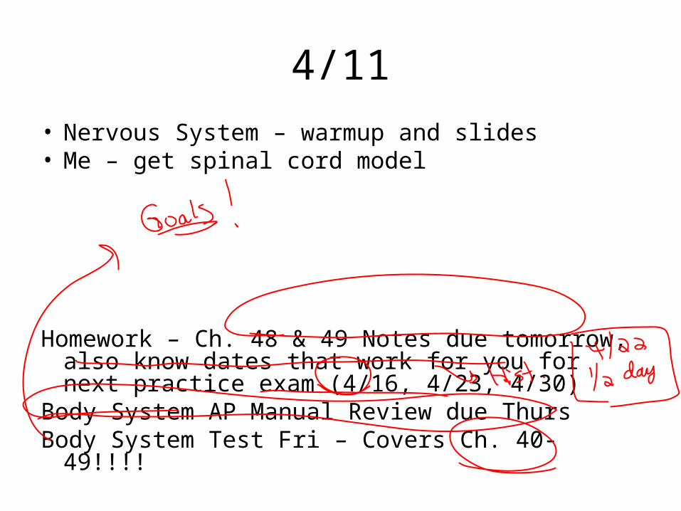
Nervous System• Functions? (think about
three main roles and types of neurons)
• What is a nerve? Ganglion?
Nucleus?

Copyright © 2002 Pearson Education, Inc., publishing as Benjamin Cummings
Fig. 48.1
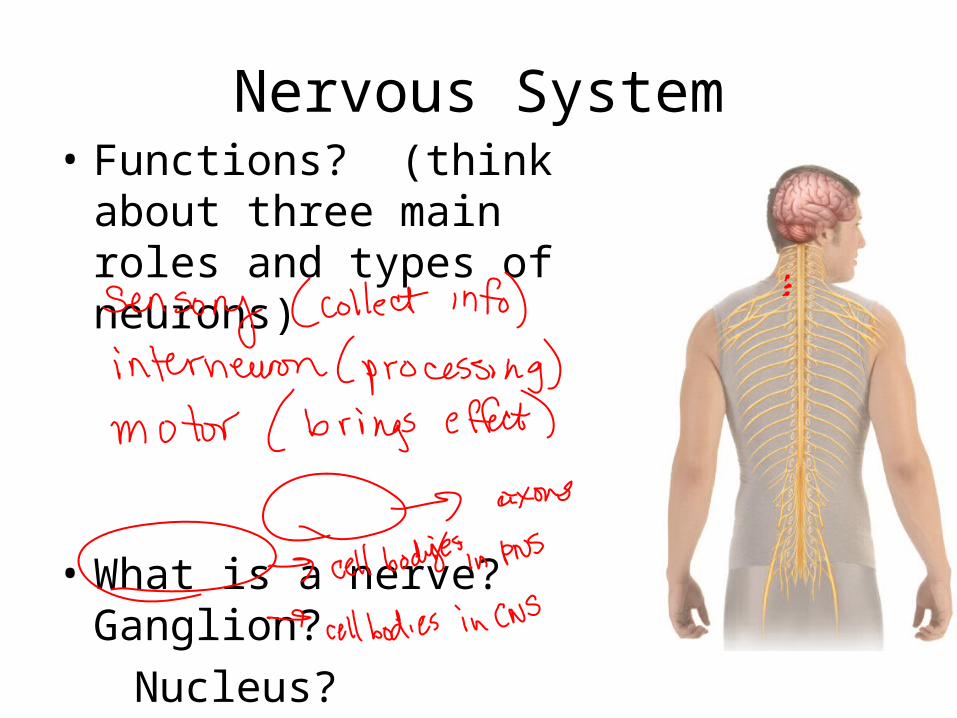
Neuron – let’s label

Copyright © 2002 Pearson Education, Inc., publishing as Benjamin Cummings
Fig. 48.2

• A Simple Nerve Circuit – the Reflex Arc.– A reflex is an autonomic response.
Copyright © 2002 Pearson Education, Inc., publishing as Benjamin Cummings
Fig. 48.3
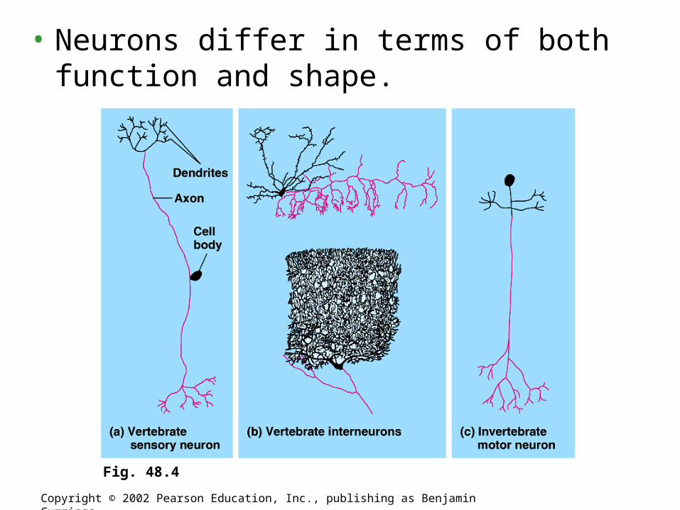
• Neurons differ in terms of both function and shape.
Copyright © 2002 Pearson Education, Inc., publishing as Benjamin Cummings
Fig. 48.4

• Types of Nerve Circuits.– Single presynaptic neuron several
postsynaptic neurons.– Several presynaptic neurons single
postsynaptic neuron.– Circular paths.
Copyright © 2002 Pearson Education, Inc., publishing as Benjamin Cummings
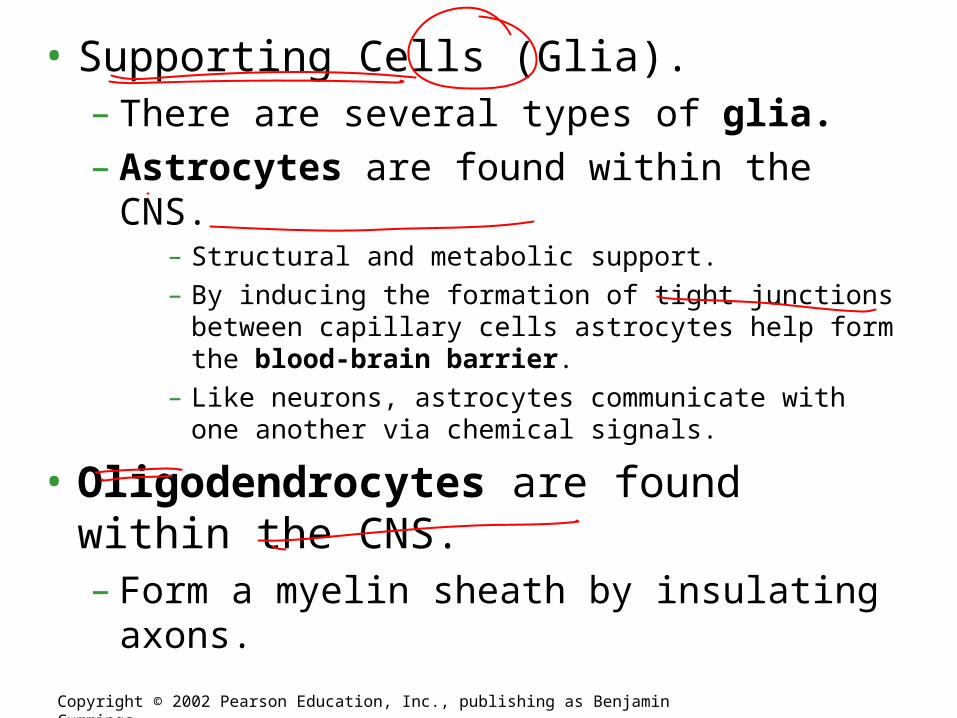
• Supporting Cells (Glia).– There are several types of glia.– Astrocytes are found within the CNS.
– Structural and metabolic support.– By inducing the formation of tight junctions between
capillary cells astrocytes help form the blood-brain barrier.– Like neurons, astrocytes communicate with one another via
chemical signals.
• Oligodendrocytes are found within the CNS.– Form a myelin sheath by insulating axons.
Copyright © 2002 Pearson Education, Inc., publishing as Benjamin Cummings

• Schwann cells are found within the PNS.– Form a myelin sheath by insulating axons.
Copyright © 2002 Pearson Education, Inc., publishing as Benjamin Cummings
Fig. 48.5

Neural Impulse
• Electrochemical =
Electric through neuron
Chemical between neurons

• At rest the sodium ions (Na+) build up outside the cell and K+ build up inside the cell.

http://www.biology4all.com/resources_library/source/63.swf
Review of the nerve impulse – THIS LINK WORKS!!!
• Types of gated ions.– Chemically-gated ion channels open or close
in response to a chemical stimulus.– Voltage-gated ion channels open or close in
response to a change in membrane potential.
Copyright © 2002 Pearson Education, Inc., publishing as Benjamin Cummings

• Graded Potentials: Hyperpolarization and Depolarization– Graded potentials are changes in membrane
potential
Copyright © 2002 Pearson Education, Inc., publishing as Benjamin Cummings
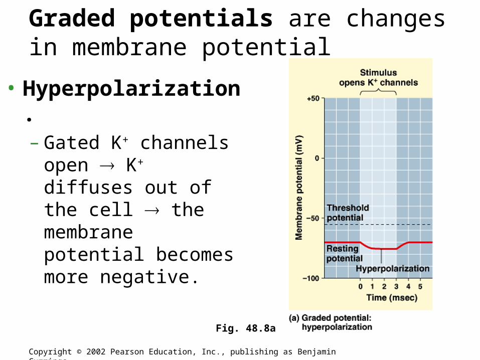
• Hyperpolarization.– Gated K+ channels
open K+ diffuses out of the cell the membrane potential becomes more negative.
Copyright © 2002 Pearson Education, Inc., publishing as Benjamin Cummings
Fig. 48.8a
Graded potentials are changes in membrane potential
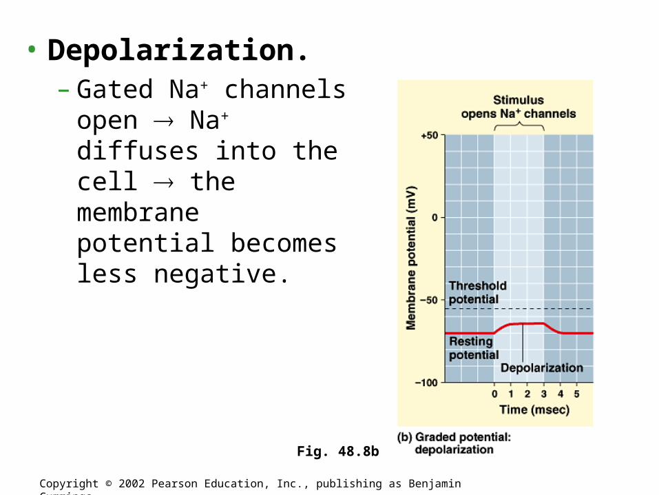
• Depolarization.– Gated Na+ channels
open Na+ diffuses into the cell the membrane potential becomes less negative.
Copyright © 2002 Pearson Education, Inc., publishing as Benjamin Cummings
Fig. 48.8b
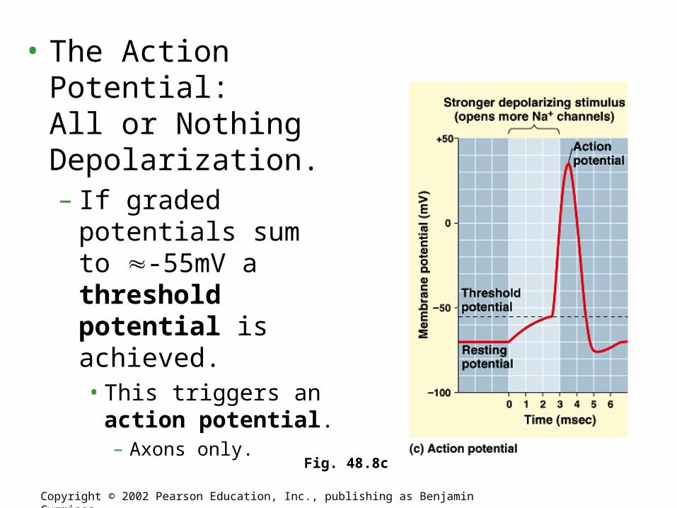
• The Action Potential: All or Nothing Depolarization.– If graded potentials
sum to -55mV a threshold potential is achieved.• This triggers an action
potential.– Axons only.
Copyright © 2002 Pearson Education, Inc., publishing as Benjamin Cummings
Fig. 48.8c

• 5 Steps of Action Potential
Copyright © 2002 Pearson Education, Inc., publishing as Benjamin Cummings
Fig. 48.9
• During the undershoot both the Na+
channel’s activation and inactivation gates are closed.– At this time the neuron cannot depolarize in
response to another stimulus: refractory period.
Copyright © 2002 Pearson Education, Inc., publishing as Benjamin Cummings

• Saltatory conduction.– In myelinated neurons only unmyelinated
regions of the axon depolarize.• Thus, the impulse moves faster than in unmyelinated
neurons.
Copyright © 2002 Pearson Education, Inc., publishing as Benjamin Cummings
Fig. 48.11

The small gap between the axon of one neuron and the dendrite of another neuron is called a synapse.
Nervous System
An action potential is carried across these gaps by neurotransmitters.
The Synapse
33.1 Structure of the Nervous System
Chapter 33
• The neuron before the synapse is called the presynaptic neuron – what do you think the neuron after the synapse is called?
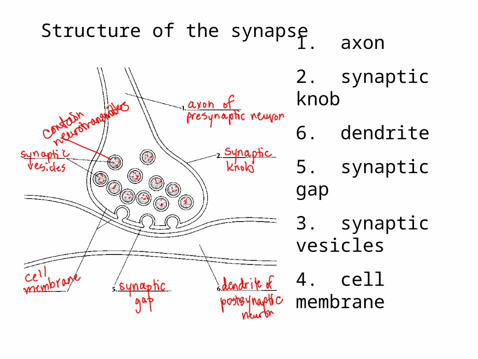
1. axon
2. synaptic knob
6. dendrite
5. synaptic gap
3. synaptic vesicles
4. cell membrane
Structure of the synapse

• The neurotransmitter diffuses across the synapse.
• Binds to receptors on the dendrite of a neuron.
• More nerve impulses are generated (or muscle or gland stimulated).
• The neurotransmitter is broken down by enzymes.
Fast forward to end in this animation.

Nervous SystemChapter 33
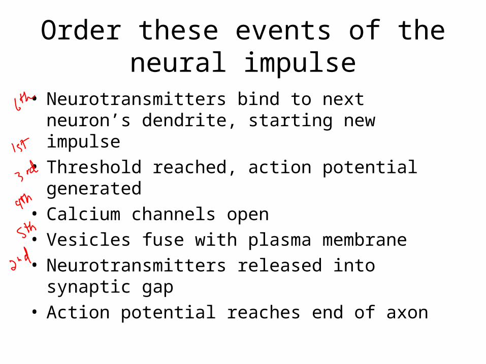
Order these events of the neural impulse
• Neurotransmitters bind to next neuron’s dendrite, starting new impulse
• Threshold reached, action potential generated• Calcium channels open • Vesicles fuse with plasma membrane • Neurotransmitters released into synaptic gap• Action potential reaches end of axon
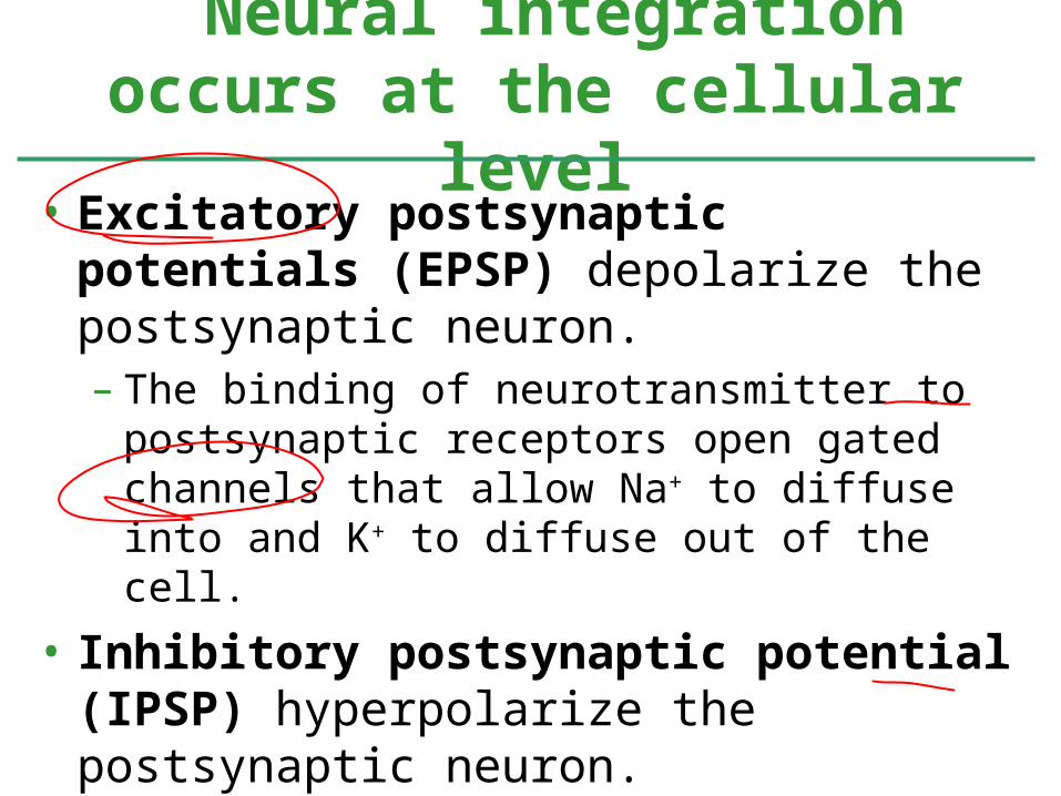
• Excitatory postsynaptic potentials (EPSP) depolarize the postsynaptic neuron.– The binding of neurotransmitter to postsynaptic
receptors open gated channels that allow Na+ to diffuse into and K+ to diffuse out of the cell.
• Inhibitory postsynaptic potential (IPSP) hyperpolarize the postsynaptic neuron.– The binding of neurotransmitter to postsynaptic
receptors open gated channels that allow K+ to diffuse out of the cell and/or Cl- to diffuse into the cell.
Neural integration occurs at the cellular level
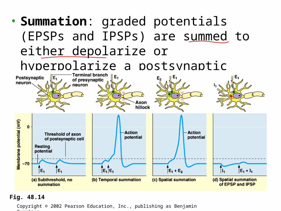
• Summation: graded potentials (EPSPs and IPSPs) are summed to either depolarize or hyperpolarize a postsynaptic neuron.
Copyright © 2002 Pearson Education, Inc., publishing as Benjamin Cummings
Fig. 48.14
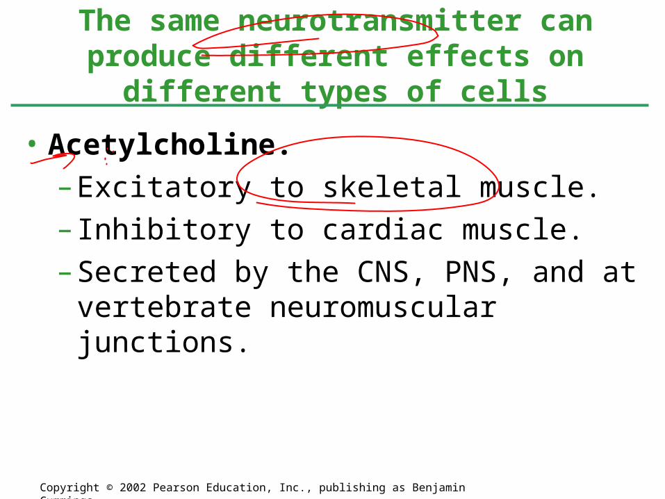
• Acetylcholine.
– Excitatory to skeletal muscle.
– Inhibitory to cardiac muscle.
– Secreted by the CNS, PNS, and at vertebrate neuromuscular junctions.
The same neurotransmitter can produce different effects on different types of cells
Copyright © 2002 Pearson Education, Inc., publishing as Benjamin Cummings

• Biogenic Amines.
– Epinephrine and norepinephrine.• Can have excitatory or inhibitory effects.• Secreted by the CNS and PNS.• Secreted by the adrenal glands.
Copyright © 2002 Pearson Education, Inc., publishing as Benjamin Cummings

• Dopamine
– Generally excitatory; may be inhibitory at some sites.• Widespread in the brain.• Affects sleep, mood, attention, and learning.
– Secreted by the CNS and PNS.
– A lack of dopamine in the brain is associated with Parkinson’s disease.
– Excessive dopamine is linked to schizophrenia.
Copyright © 2002 Pearson Education, Inc., publishing as Benjamin Cummings

• Serotonin.
– Generally inhibitory.• Widespread in the brain.• Affects sleep, mood, attention, and learning
– Secreted by the CNS.
Copyright © 2002 Pearson Education, Inc., publishing as Benjamin Cummings
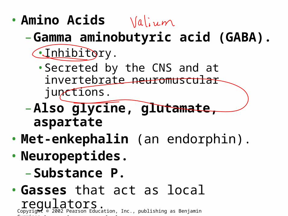
• Amino Acids– Gamma aminobutyric acid (GABA).
• Inhibitory.• Secreted by the CNS and at invertebrate
neuromuscular junctions.
– Also glycine, glutamate, aspartate• Met-enkephalin (an endorphin).• Neuropeptides.
– Substance P.• Gasses that act as local regulators.
– Nitric oxide.– Carbon monoxide.
Copyright © 2002 Pearson Education, Inc., publishing as Benjamin Cummings
4/12
• Finish Nervous system and intro Muscle Contraction / Neuromuscular Junction
• Muscle tutorial – in Math Lab (this year not able to intro – just went to lab to do tutorial and kids had to finish for homework)
• I will be checking 48 & 49 (also late Animal Dvpt questions), and dates for next practice exam
Homework – Body System AP Manual Review due Thurs
Body System Test Fri

Match the animal with nervous system
• Hollow dorsal nerve cord
• Small brain and longitudinal nerve cord
• Ventral nerve cord• Nerve Net
• Annelids and Arthropods
• Vertebrates• Cnidarians• Flatworms
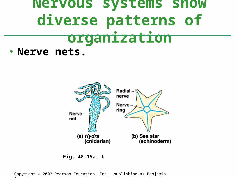
• Nerve nets.
Nervous systems show diverse patterns of organization
Copyright © 2002 Pearson Education, Inc., publishing as Benjamin Cummings
Fig. 48.15a, b
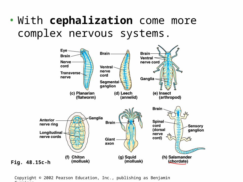
• With cephalization come more complex nervous systems.
Copyright © 2002 Pearson Education, Inc., publishing as Benjamin Cummings
Fig. 48.15c-h
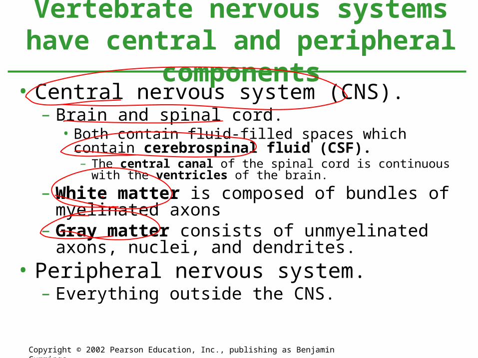
Vertebrate nervous systems have central and peripheral components
Copyright © 2002 Pearson Education, Inc., publishing as Benjamin Cummings
• Central nervous system (CNS).– Brain and spinal cord.
• Both contain fluid-filled spaces which contain cerebrospinal fluid (CSF).– The central canal of the spinal cord is continuous with the ventricles
of the brain.
– White matter is composed of bundles of myelinated axons
– Gray matter consists of unmyelinated axons, nuclei, and dendrites.
• Peripheral nervous system.– Everything outside the CNS.
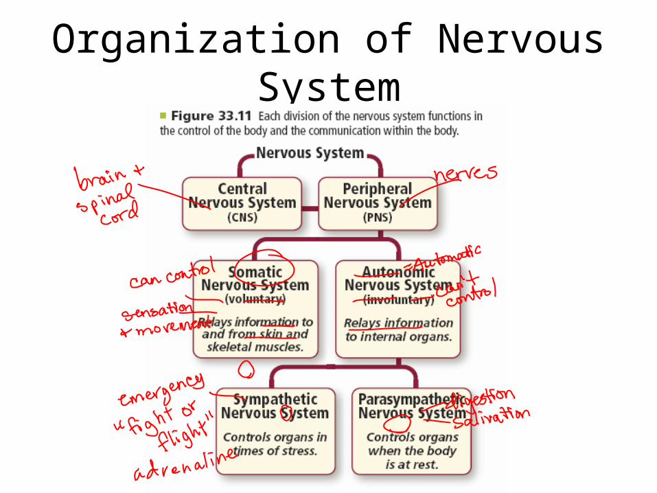
Organization of Nervous System
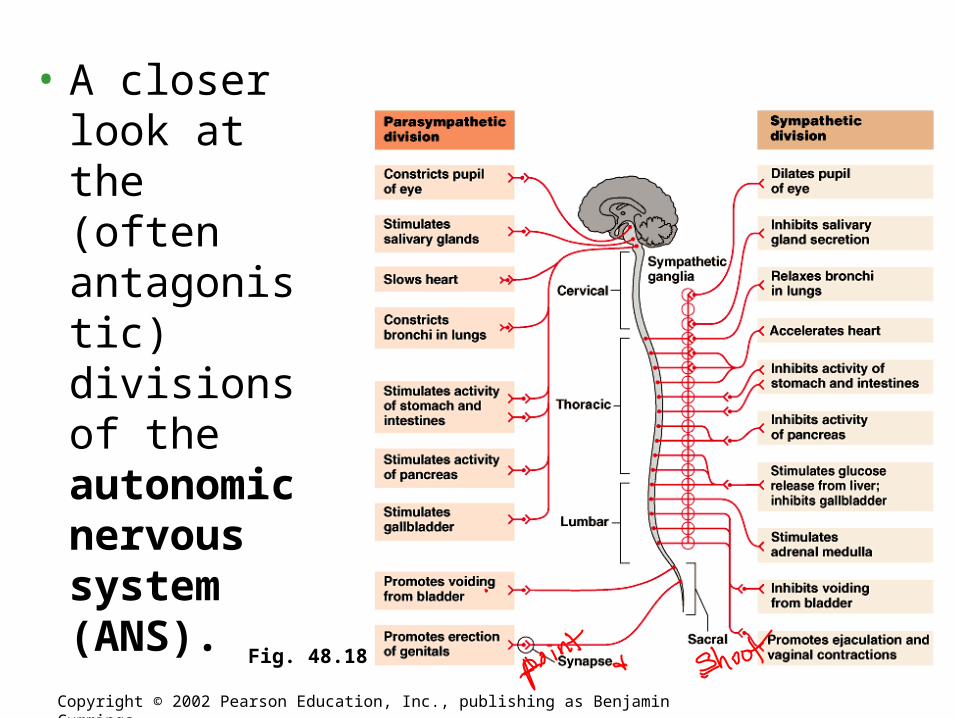
• A closer look at the (often antagonistic) divisions of the autonomic nervous system (ANS).
Copyright © 2002 Pearson Education, Inc., publishing as Benjamin Cummings
Fig. 48.18

Nervous SystemChapter 33

Embryonic development of the vertebrate brain reflects its evolution from three
anterior bulges of the neural tube
Copyright © 2002 Pearson Education, Inc., publishing as Benjamin Cummings
Fig. 48.19

Copyright © 2002 Pearson Education, Inc., publishing as Benjamin Cummings
Fig. 48.20
Let’s label functions – use p. 270

Self - Check
• Which part of the brain regulates thirst?
• Which part of the brain would be highly developed in an animal that is extremely coordinated like a monkey or cat?
• Which part of the brain do all your conscious thoughts come from?
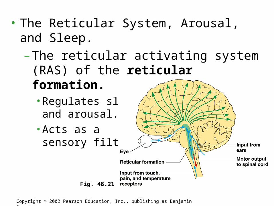
• The Reticular System, Arousal, and Sleep.
– The reticular activating system (RAS) of the reticular formation.• Regulates sleep
and arousal.• Acts as a
sensory filter.
Copyright © 2002 Pearson Education, Inc., publishing as Benjamin Cummings
Fig. 48.21
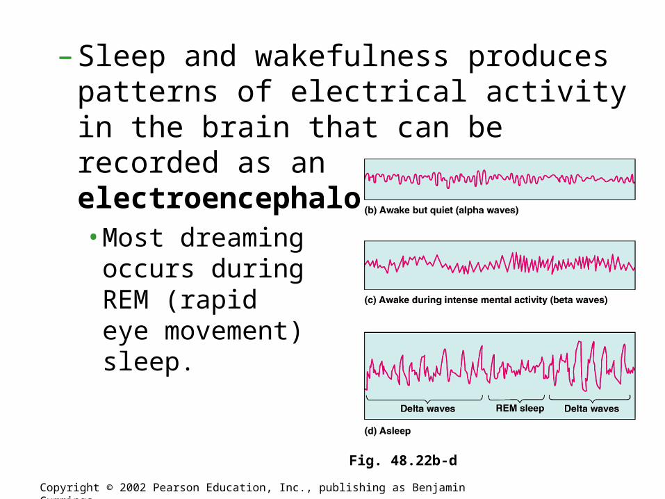
– Sleep and wakefulness produces patterns of electrical activity in the brain that can be recorded as an electroencephalogram (EEG).• Most dreaming
occurs during REM (rapid eye movement) sleep.
Copyright © 2002 Pearson Education, Inc., publishing as Benjamin Cummings
Fig. 48.22b-d
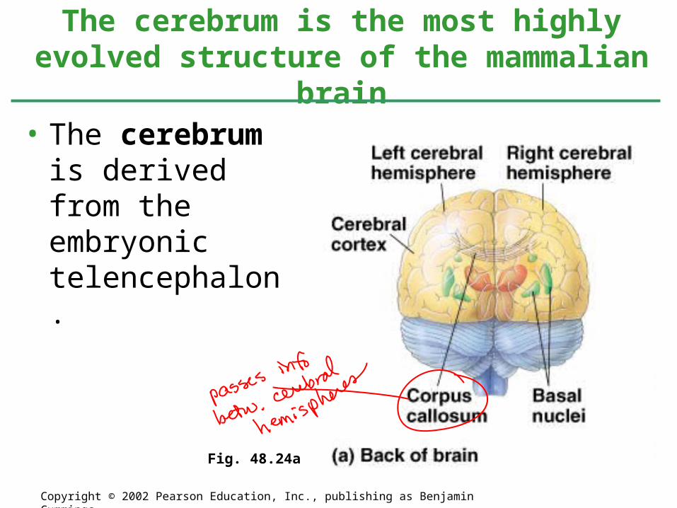
• The cerebrum is derived from the embryonic telencephalon.
The cerebrum is the most highly evolved structure of the mammalian brain
Copyright © 2002 Pearson Education, Inc., publishing as Benjamin Cummings
Fig. 48.24a
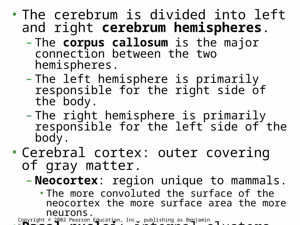
• The cerebrum is divided into left and right cerebrum hemispheres.– The corpus callosum is the major connection
between the two hemispheres.– The left hemisphere is primarily responsible for
the right side of the body.– The right hemisphere is primarily responsible for
the left side of the body.• Cerebral cortex: outer covering of gray
matter. – Neocortex: region unique to mammals.
• The more convoluted the surface of the neocortex the more surface area the more neurons.
• Basal nuclei: internal clusters of nuclei.
Copyright © 2002 Pearson Education, Inc., publishing as Benjamin Cummings
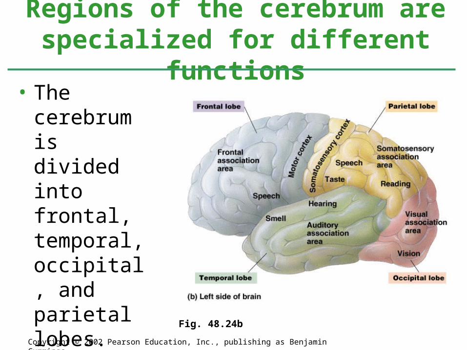
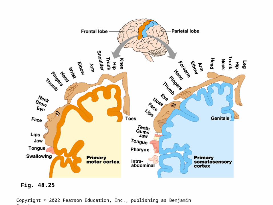
• The cerebrum is divided into frontal, temporal, occipital, and parietal lobes.
Regions of the cerebrum are specialized for different functions
Copyright © 2002 Pearson Education, Inc., publishing as Benjamin Cummings
Fig. 48.24b
Copyright © 2002 Pearson Education, Inc., publishing as Benjamin Cummings
Fig. 48.25

• Lateralization of Brain Function.– The left hemisphere.
• Specializes in language, math, logic operations, and the processing of serial sequences of information, and visual and auditory details.
• Specializes in detailed activities required for motor control.
– The right hemisphere.• Specializes in pattern recognition, spatial relationships,
nonverbal ideation, emotional processing, and the parallel processing of information.
Copyright © 2002 Pearson Education, Inc., publishing as Benjamin Cummings
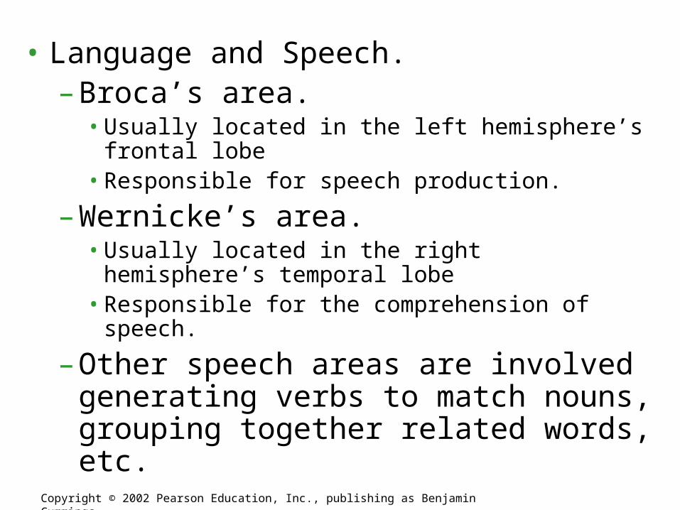
• Language and Speech.– Broca’s area.
• Usually located in the left hemisphere’s frontal lobe• Responsible for speech production.
– Wernicke’s area.• Usually located in the right hemisphere’s temporal lobe• Responsible for the comprehension of speech.
– Other speech areas are involved generating verbs to match nouns, grouping together related words, etc.
Copyright © 2002 Pearson Education, Inc., publishing as Benjamin Cummings

• Emotions.– In mammals, the limbic system is composed
of the hippocampus, olfactory cortex, inner portions of the cortex’s lobes, and parts of the thalamus and hypothalamus.• Mediates basic emotions (fear, anger), involved in
emotional bonding, establishes emotional memory– For example,
the amygdala is involved in recognizing the emotional content of facial expression.
Copyright © 2002 Pearson Education, Inc., publishing as Benjamin CummingsFig. 48.27

• Memory and Learning.– Short-term memory stored in the frontal
lobes.– The establishment of long-term memory
involves the hippocampus.• The transfer of information from short-term to
long-term memory.– Is enhanced by repetition (remember that when you are
preparing for an exam).– Influenced by emotional states mediated by the
amygdala.– Influenced by association with previously stored
information.
Copyright © 2002 Pearson Education, Inc., publishing as Benjamin Cummings
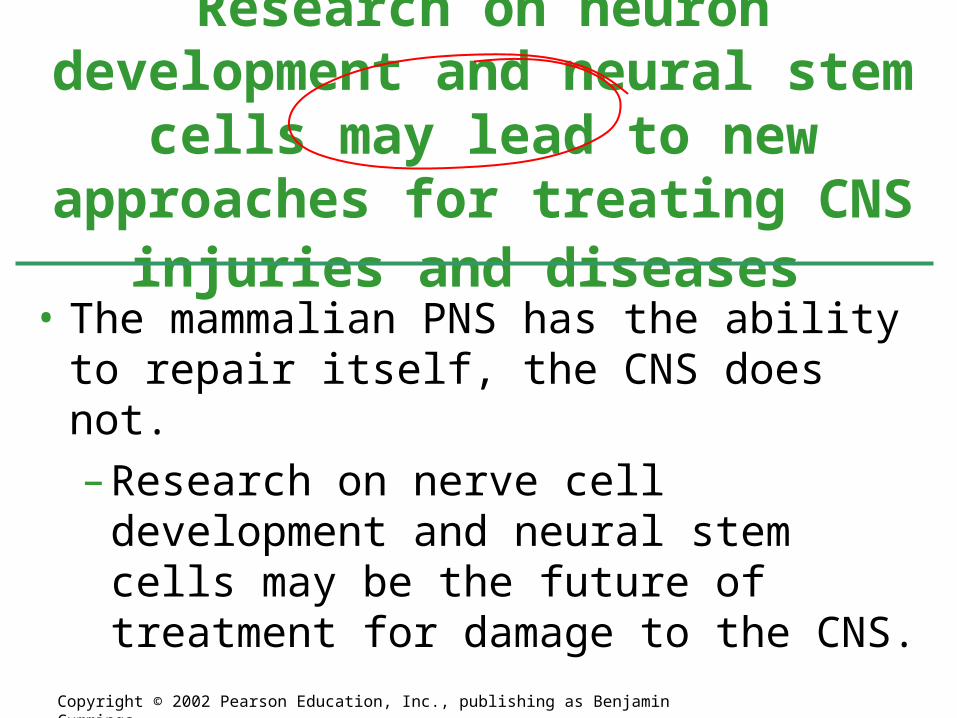
• The mammalian PNS has the ability to repair itself, the CNS does not.
– Research on nerve cell development and neural stem cells may be the future of treatment for damage to the CNS.
Research on neuron development and neural stem cells may lead to new approaches for treating CNS
injuries and diseases
Copyright © 2002 Pearson Education, Inc., publishing as Benjamin Cummings

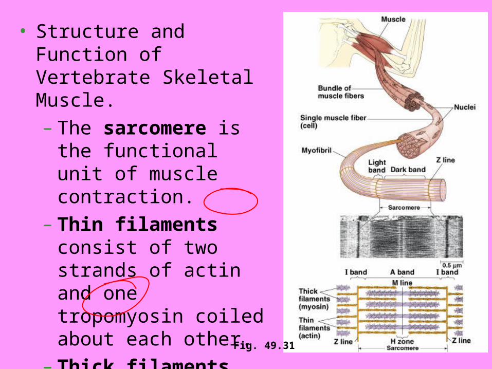
• Structure and Function of Vertebrate Skeletal Muscle.– The sarcomere is the
functional unit of muscle contraction.
– Thin filaments consist of two strands of actin and one tropomyosin coiled about each other.
– Thick filaments consist of myosin molecules.
Fig. 49.31
To computer lab
• You will do tutorial that reviews anatomy and physiology of muscle fibers

4/13
• Muscular System• Special Senses• Skeletal• Eye dissection/ Brain if time
Homework – Free Response Questions due tomorrowBody System AP Manual Review due tomorrowBody System Test Fri

Ch. 49 Goals
• Know the overall function of sensory organs and the muscular system
• Include a picture of the eye, ear, skeletal system, muscular system, sarcomere – know the anatomy of these
• Know the location and function of several types of sensory receptors
• Know the function of the cochlea and semicircular canals• Know the pathway of light through the eye and how
vision is sensed (rods & cones etc.)• Know the different types of skeletons• Know skeletal muscle organization and how sarcomeres
contracting lead to whole muscle contraction • Know the cellular events that lead to muscle contraction
at the neuromuscular junction – “sliding-filament model”

• Muscles come in antagonistic pairs.
Muscles move skeletal parts by contracting
Copyright © 2002 Pearson Education, Inc., publishing as Benjamin Cummings
Fig. 49.30

Skeletal Muscle Contraction
Most skeletal muscles are arranged in opposing, or antagonistic pairs.
Integumentary, Skeletal, and Muscular Systems
32.3 The Muscular System
Chapter 32
Tendons connect muscles to bones.

Integumentary, Skeletal, and Muscular SystemsChapter 32
• Structure and Function of Vertebrate Skeletal Muscle.– The sarcomere is the
functional unit of muscle contraction.
– Thin filaments consist of two strands of actin and one tropomyosin coiled about each other.
– Thick filaments consist of myosin molecules.
Fig. 49.31

Integumentary, Skeletal, and Muscular Systems
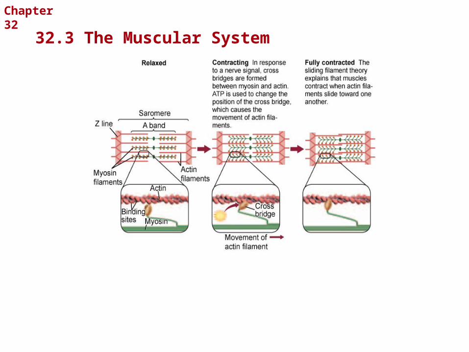
Sliding Filament Theory
Once a nerve signal reaches a muscle, the actin filaments slide toward one another, causing the muscle to contract.
Let’s visualize it
32.3 The Muscular System
Chapter 32
Integumentary, Skeletal, and Muscular Systems
32.3 The Muscular System
Chapter 32

More from Online tutorial
• http://www.getbodysmart.com/ap/muscletissue/menu/menu.html
• Click on Nerve Supply to Muscle Fiber• Show Neurotransmitter Release from
Motor Neuron• Then Physiology of Contraction• Click on Contraction Cycle and How
Multiple Myosin Heads Move Sarcomeres – hold down to play movie for each

• At rest tropomyosin blocks the myosin binding sites on actin.
• When calcium binds to the troponin complex a conformational change results in the movement of the tropomyosin-tropinin complex and exposure of actin’s myosin binding sites.
Calcium ions and regulatory proteins control muscle contraction
Copyright © 2002 Pearson Education, Inc., publishing as Benjamin CummingsFig. 49.34

• But, wherefore the calcium ions?– Follow the
action potential.
– When an action potential meets the muscle cell’s sarcoplasmic reticulum (SR) stored Ca2+ is released.
Copyright © 2002 Pearson Education, Inc., publishing as Benjamin Cummings
Fig. 49.35
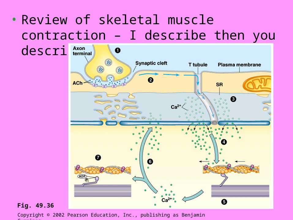
• Review of skeletal muscle contraction – I describe then you describe to neighbor.
Copyright © 2002 Pearson Education, Inc., publishing as Benjamin Cummings
Fig. 49.36

• Sensations are action potentials that reach the brain via sensory neurons.
• Reception occurs when a receptor detects a stimulus.
• Perception is the awareness and interpretation of the sensation.
Copyright © 2002 Pearson Education, Inc., publishing as Benjamin Cummings

Sensory receptors transduce stimulus energy and transmit signals to the nervous system
Copyright © 2002 Pearson Education, Inc., publishing as Benjamin Cummings
Fig. 49.2

Sensory receptors are categorized by the type of energy they transduce
Copyright © 2002 Pearson Education, Inc., publishing as Benjamin Cummings
Fig. 49.3

• Mechanoreceptors respond to mechanical energy/physical stimulus in the form of pressure, touch, stretch, motion, or sound.– For example, a muscle spindle is an
interoreceptor that responds to the stretching of skeletal muscle.
– For example, hair cells detect motion.
Copyright © 2002 Pearson Education, Inc., publishing as Benjamin Cummings

• Pain receptors = nocioceptors.– Different types of pain receptors respond to
different types of pain such as excess heat, pressure, or prostaglandins released from damaged or inflamed tissues (this is how antiinflammatories work – by blocking prostaglandin release)
Copyright © 2002 Pearson Education, Inc., publishing as Benjamin Cummings
• Thermoreceptors respond to heat or cold.– Respond to both surface and body core
temperature.
• Chemoreceptors respond to chemical stimuli.– Internal chemoreceptors respond to glucose,
O2, CO2, amino acids, etc.
– External chemoreceptors are gustatory receptors and olfactory receptors.
Copyright © 2002 Pearson Education, Inc., publishing as Benjamin Cummings

• Electromagnetic receptors respond to electromagnetic energy.– Photoreceptors respond to the radiation we
know as visible light.– Electroreceptors: some fish use electric
currents to locate objects.
Copyright © 2002 Pearson Education, Inc., publishing as Benjamin Cummings

Now check yourself – what is name of type of receptor for each
Copyright © 2002 Pearson Education, Inc., publishing as Benjamin Cummings
Fig. 49.3

A diversity of photoreceptors has evolved among invertebrates
Copyright © 2002 Pearson Education, Inc., publishing as Benjamin Cummings
Fig. 49.7
• Eye cups are among the simplest photoreceptors– Detect light intensity and direction — no image
formation.– The movement
of a planarian is integrated with photoreception.
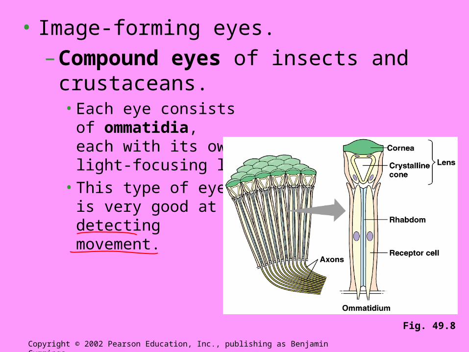
• Image-forming eyes.
– Compound eyes of insects and crustaceans.• Each eye consists
of ommatidia, each with its own light-focusing lens.
• This type of eye is very good at detecting movement.
Copyright © 2002 Pearson Education, Inc., publishing as Benjamin Cummings
Fig. 49.8

• Single-lens eyes of invertebrates such as jellies, polychaetes, spiders, and mollusks.
– The eye of an octopus works much like a camera and is similar to the vertebrate eye.
Copyright © 2002 Pearson Education, Inc., publishing as Benjamin Cummings

Vertebrates have single-lens eyes
Copyright © 2002 Pearson Education, Inc., publishing as Benjamin Cummings
• Is structurally analogous to the invertebrate single-lens eye. Let’s trace pathway of light
Fig. 49.9
• Sclera: a tough white layer of connective tissue that covers all of the eyeball except the cornea.
– Conjunctiva: external cover of the sclera — keeps the eye moist.
Copyright © 2002 Pearson Education, Inc., publishing as Benjamin Cummings
I skip these next several slides in lecture…

• Cornea: transparent covering of the front of the eye.
– Allows for the passage of light into the eye and functions as a fixed lens.
Copyright © 2002 Pearson Education, Inc., publishing as Benjamin Cummings

• Choroid: thin, pigmented layer lining the interior surface of the sclera.
– Prevents light rays from scattering and distorting the image.
– Anteriorly it forms the iris.• The iris regulates the size of the pupil.
Copyright © 2002 Pearson Education, Inc., publishing as Benjamin Cummings

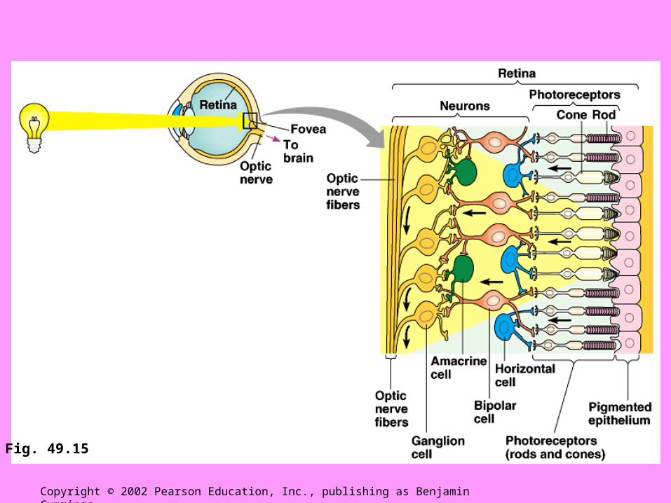
• Retina: lines the interior surface of the choroid.
– Contains photoreceptors.• Except at the optic disk (where the optic
nerve attaches).
Copyright © 2002 Pearson Education, Inc., publishing as Benjamin Cummings

• The lens and ciliary body divide the eye into two cavities.
– The anterior cavity is filled with aqueous humor produced by the ciliary body.• Glaucoma results when the duct that drain
aqueous humor are blocked.
– The posterior cavity is filled with vitreous humor.
– The lens, the aqueous humor, and the vitreous humor all play a role in focusing light onto the retina.
Copyright © 2002 Pearson Education, Inc., publishing as Benjamin Cummings

• Accommodation is the focusing of light in the retina.
– In squid, octopuses, and many fish this is accomplished by moving the lens forward and backward.
Copyright © 2002 Pearson Education, Inc., publishing as Benjamin Cummings
Discuss this…

– In mammals accommodation is accomplished by changing the shape of the lens.• The lens is
flattened for focusing on distant objects.
• The lens is rounded for focusing on near objects.
Copyright © 2002 Pearson Education, Inc., publishing as Benjamin Cummings
Fig. 49.10

• Photoreceptors of the retina.
– About 125 million rod cells.• Rod cells are light sensitive but do not distinguish
colors.
– About 6 million cone cells.• Not as light sensitive as rods but provide color
vision.• Most highly concentrated on the fovea – an area of
the retina that lacks rods.
Copyright © 2002 Pearson Education, Inc., publishing as Benjamin Cummings

The light-absorbing pigment rhodopsin triggers a signal-transduction pathway
Copyright © 2002 Pearson Education, Inc., publishing as Benjamin Cummings
• Rhodopsin (retinal + opsin) is the visual pigment of rods.
• The absorption of light by rhodopsin initiates a signal-transduction pathway.
Fig. 49.13
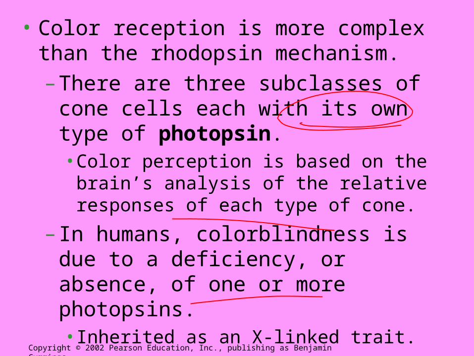
• Color reception is more complex than the rhodopsin mechanism.
– There are three subclasses of cone cells each with its own type of photopsin.• Color perception is based on the brain’s
analysis of the relative responses of each type of cone.
– In humans, colorblindness is due to a deficiency, or absence, of one or more photopsins.• Inherited as an X-linked trait.
Copyright © 2002 Pearson Education, Inc., publishing as Benjamin Cummings
Copyright © 2002 Pearson Education, Inc., publishing as Benjamin Cummings
Fig. 49.15
Copyright © 2002 Pearson Education, Inc., publishing as Benjamin Cummings
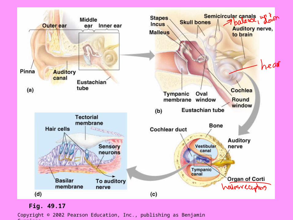
Fig. 49.17
• From the tympanic membrane sound waves are transmitted through the middle ear.
– Malleus incus stapes.
– From the stapes the sound wave is transmitted to the oval window and on to the inner ear.
– Eustachian tube connects the middle ear with the pharynx.
Copyright © 2002 Pearson Education, Inc., publishing as Benjamin Cummings
• The inner ear consists of a labyrinth of channels housed within the temporal bone.
– The cochlea is the part of the inner ear concerned with hearing.• Structurally it consists of the upper vestibular
canal and the lower tympanic canal, which are separated by the cochlear duct.
• The vestibular and tympanic canals are filled with perilymph.
Copyright © 2002 Pearson Education, Inc., publishing as Benjamin Cummings
– The cochlear duct is filled with endolymph.
– The organ of Corti rests on the basilar membrane.• The tectorial membrane rests atop the hair
cells of the organ of Corti.
Copyright © 2002 Pearson Education, Inc., publishing as Benjamin Cummings

• From inner ear structure to a sensory impulse: follow the vibrations.
– The round window functions to dissipate the vibrations.
• Vibrations in the cochlear fluid basilar membrane vibrates hair cells brush against the tectorial membrane generation of an action potential in a sensory neuron.
Copyright © 2002 Pearson Education, Inc., publishing as Benjamin Cummings

Copyright © 2002 Pearson Education, Inc., publishing as Benjamin Cummings
Fig. 49.18

• Behind the oval window is a vestibule that contains the utricle and saccule.
– The utricle opens into three semicircular canals.
2. The inner ear also contains the organs of equilibrium
Copyright © 2002 Pearson Education, Inc., publishing as Benjamin Cummings

Copyright © 2002 Pearson Education, Inc., publishing as Benjamin Cummings
Fig. 49.19
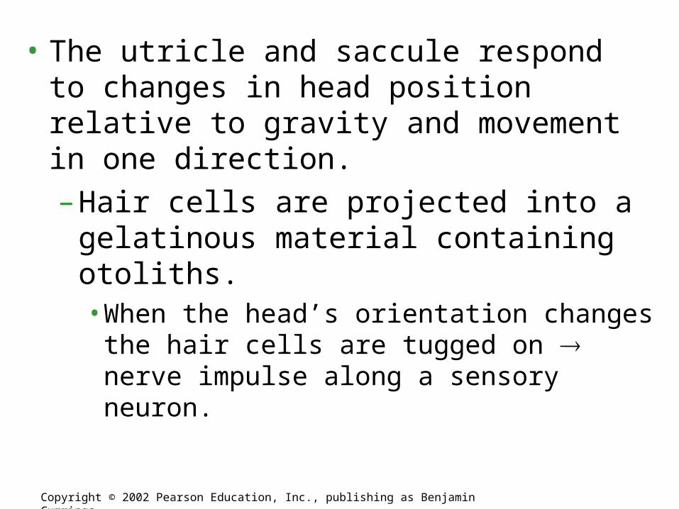
• The utricle and saccule respond to changes in head position relative to gravity and movement in one direction.
– Hair cells are projected into a gelatinous material containing otoliths.• When the head’s orientation changes the hair
cells are tugged on nerve impulse along a sensory neuron.
Copyright © 2002 Pearson Education, Inc., publishing as Benjamin Cummings

• The semicircular canals respond to rotational movements of the head.
– The mechanism is similar to that associated with the utricle and saccule.
Copyright © 2002 Pearson Education, Inc., publishing as Benjamin Cummings
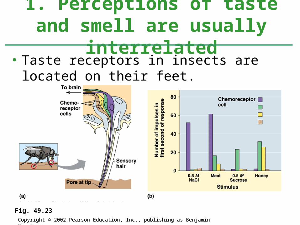
• Taste receptors in insects are located on their feet.
1. Perceptions of taste and smell are usually interrelated
Copyright © 2002 Pearson Education, Inc., publishing as Benjamin Cummings
Fig. 49.23
• In mammals, taste receptors are located in taste buds most of which are on the surface of the tongue.
• Each taste receptor responds to a wide array of chemicals.– It is the pattern of taste receptor response that
determines something’s perceived flavor.
Copyright © 2002 Pearson Education, Inc., publishing as Benjamin Cummings

• In mammals, olfactory receptors line the upper portion of the nasal cavity.– The binding of odor molecules to olfactory
receptors initiate signal transduction pathways involving a G-protein-signaling pathway and, often, adenylyl cyclase and cyclic AMP.
Copyright © 2002 Pearson Education, Inc., publishing as Benjamin Cummings
Fig. 49.24
3 main types of skeletons?
• Hard outer covering = ?
• Use fluid internally for structure = ?
• Inner hard supporting elements = ?

• Hydrostatic skeleton: consists of fluid held under pressure in a closed body compartment.– Form and movement is controlled by changing
the shape of this compartment.– The hydrostatic skeleton of earthworms allow
them to move by peristalsis.– Advantageous in aquatic environments and can
support crawling and burrowing.– Do not allow for running or walking.
Skeletons support and protect the animal body and are essential to
movement

• Exoskeletons: hard encasements deposited on the surface of an animal.– Mollusks are enclosed in a calcareous
exoskeleton.
– The jointed exoskeleton of arthropods is composed of a cuticle.• Regions of the cuticle can vary in hardness and
degree of flexibility.• About 30 – 50% of the cuticle consists of chitin.• Muscles are attached to the interior surface of the
cuticle.• This type of exoskeleton must be molted to allow for
growth.
Copyright © 2002 Pearson Education, Inc., publishing as Benjamin Cummings

• Endoskeletons: consist of hard supporting elements within soft tissues.
– Sponges have spicules.
– Echinoderms have plates composed of magnesium carbonate and calcium carbonate.
– Chordate endoskeletons are composed of cartilage and bone.• The bones of the mammalian skeleton are
connected at joints by ligaments.
Copyright © 2002 Pearson Education, Inc., publishing as Benjamin Cummings
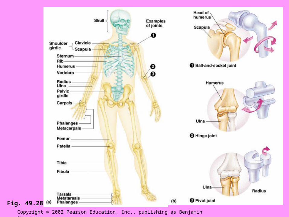
Copyright © 2002 Pearson Education, Inc., publishing as Benjamin Cummings
Fig. 49.28

4/14
• Correct Free Response• Virtual Pig Dissection in Math Lab- I check Free
Response and Review Manual• Veera
Homework –
Study for Body System Test tomorrow!!! Covers Ch. 40-49 USE YOUR GOALS!!!!

4/15
• Body System Test
Homework – Have fun at Junior Prom! Bring your review
manual on Monday and every day after until exam!!!


![The Nervous System. Divisions of the Nervous System Central Nervous System [CNS] = Spinal Cord Brain Peripheral Nervous System [PNS]= Spinal Nerves.](https://static.fdocuments.in/doc/165x107/56649d6c5503460f94a4c71d/the-nervous-system-divisions-of-the-nervous-system-central-nervous-system.jpg)