4 left ventricular enlargement
13
4 Left Ventricular Enlargement
-
Upload
muhammad-bin-zulfiqar -
Category
Education
-
view
202 -
download
7
Transcript of 4 left ventricular enlargement

4 Left Ventricular Enlargement

CLINICAL IMAGAGINGAN ATLAS OF DIFFERENTIAL DAIGNOSIS
EISENBERG
DR. Muhammad Bin Zulfiqar PGR-FCPS III SIMS/SHL

• Fig CA 4-1 Arteriosclerotic heart disease. (A) Frontal and (B) lateral views of the chest show marked enlargement of the left ventricle. There is also tortuosity of the aorta and bilateral streaks of fibrosis.

• Fig CA 4-2 Acute myocardial infarction. Lateral view of the chest shows marked prominence of the left ventricle (arrows).

• Fig CA 4-3 Aortic insufficiency. Frontal chest radiograph shows left ventricular enlargement with downward and lateral displacement of the cardiac apex. Note that the cardiac shadow extends below the dome of the left hemidiaphragm. The ascending aorta is strikingly dilated (arrows), suggesting some underlying aortic stenosis.
• Fig CA 4-4 Aortic stenosis. (A) Frontal view shows downward displacement of the cardiac apex. (B) On a lateral view, the bulging of the lower half of the posterior cardiac silhouette causes a broad indentation on the barium-filled esophagus (arrows).
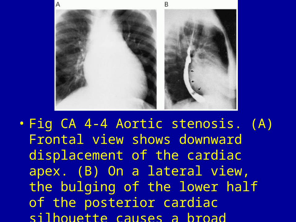
• Fig CA 4-5 Mitral insufficiency. (A) Frontal and (B) lateral views of the chest demonstrate cardiomegaly with enlargement of the left ventricle and left atrium. Note the striking double-contour configuration (closed arrows) and elevation of the left main-stem bronchus (open arrow), characteristic signs of left atrial enlargement.

• Fig CA 4-6 Glycogen storage disease. Generalized globular cardiac enlargement with a left ventricular prominence.

• Fig CA 4-7 Alcoholic cardiomyopathy. Generalized cardiac enlargement that involves all chambers but has a left ventricular predominance. There is pulmonary vascular congestion and a right pleural effusion.
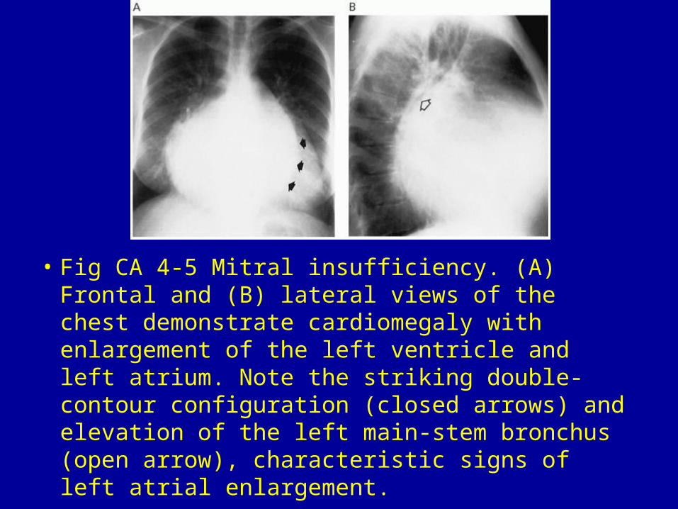
• Fig CA 4-8 Endocardial fibroelastosis. Generalized cardiomegaly with prominence of the left ventricle.