


4 Janneth Peralta-Ramírez1, J. Manuel Hernandez1, Rebeca...
48
Peralta et al. 1 EspF interacts with nucleation-promoting factors to recruit junctional proteins into 1 pedestals for their maturation and disruption of the paracellular permeability 2 3 Janneth Peralta-Ramírez 1 , J. Manuel Hernandez 1 , Rebeca Manning-Cela 2 , José Luna-Muñoz 3 , 4 Carlos Garcia-Tovar 4 , Jean-Philippe Nougayréde 5 , Eric Oswald 5 and Fernando Navarro-Garcia 1* 5 6 1 Departments of Cell Biology, 2 Molecular Biomedicine and 3 Physiology, Biophysics and 7 Neurosciences, Centro de Investigación y de Estudios Avanzados (CINVESTAV-IPN), Ap. Postal 8 14-740, 07000, Mexico DF, Mexico; 4 Morphology Unit, Universidad Nacional Autónoma de 9 México (UNAM-FES Cuautitlan), Km. 2.5 Carr. México-Cuautitlan, Ap. Postal 54714; 5 Unité 10 des Microbiologie Moléculaire (INRA-ENVT), Ecole Nationale Vétérinaire 31000 Toulouse 11 Cedex 3, France. 12 13 Running title: Role of EspF in pedestal maturation and TJ disruption 14 15 Key words: Enteropathogenic E. coli, EspF protein, Tight junction, Actin cytoskeleton, ZO-1, 16 ZO-2, Occludin, Claudin, Profilin, Arp2/3, N-WASP. 17 18 *Corresponding author 19 Department of Cell Biology, Cinvestav-Zacatenco, Ap. Postal 14-740, 07000 Mexico, DF, 20 Mexico. Phone: (525) 5061-3990. Fax: (525) 5061-3393. E-mail: [email protected] 21 ACCEPTED Copyright © 2008, American Society for Microbiology and/or the Listed Authors/Institutions. All Rights Reserved. Infect. Immun. doi:10.1128/IAI.00072-08 IAI Accepts, published online ahead of print on 16 June 2008 on May 4, 2018 by guest http://iai.asm.org/ Downloaded from
Transcript of 4 Janneth Peralta-Ramírez1, J. Manuel Hernandez1, Rebeca...

Peralta et al. 1
EspF interacts with nucleation-promoting factors to recruit junctional proteins into 1
pedestals for their maturation and disruption of the paracellular permeability 2
3
Janneth Peralta-Ramírez1, J. Manuel Hernandez
1, Rebeca Manning-Cela
2, José Luna-Muñoz
3, 4
Carlos Garcia-Tovar4, Jean-Philippe Nougayréde
5, Eric Oswald
5 and Fernando Navarro-Garcia
1* 5
6
1Departments of Cell Biology,
2Molecular Biomedicine and
3Physiology, Biophysics and 7
Neurosciences, Centro de Investigación y de Estudios Avanzados (CINVESTAV-IPN), Ap. Postal 8
14-740, 07000, Mexico DF, Mexico; 4Morphology Unit, Universidad Nacional Autónoma de 9
México (UNAM-FES Cuautitlan), Km. 2.5 Carr. México-Cuautitlan, Ap. Postal 54714; 5Unité 10
des Microbiologie Moléculaire (INRA-ENVT), Ecole Nationale Vétérinaire 31000 Toulouse 11
Cedex 3, France. 12
13
Running title: Role of EspF in pedestal maturation and TJ disruption 14
15
Key words: Enteropathogenic E. coli, EspF protein, Tight junction, Actin cytoskeleton, ZO-1, 16
ZO-2, Occludin, Claudin, Profilin, Arp2/3, N-WASP. 17
18
*Corresponding author 19
Department of Cell Biology, Cinvestav-Zacatenco, Ap. Postal 14-740, 07000 Mexico, DF, 20
Mexico. Phone: (525) 5061-3990. Fax: (525) 5061-3393. E-mail: [email protected]
ACCEPTED
Copyright © 2008, American Society for Microbiology and/or the Listed Authors/Institutions. All Rights Reserved.Infect. Immun. doi:10.1128/IAI.00072-08 IAI Accepts, published online ahead of print on 16 June 2008
on May 4, 2018 by guest
http://iai.asm.org/
Dow
nloaded from

Peralta et al. 2
1
Many pathogenic bacteria subvert normal host-cell processes by delivering effector 2
proteins, which mimic eukaryotic functions, directly into target cells. EspF is a 3
multifunctional protein injected into host cells by attaching and effacing (A/E) pathogens, 4
but its mechanism of action is not understood completely. In silico analyses of EspF 5
revealed two key motifs: proline-rich domains and PDZ domain-binding motifs. Such 6
functional domains may allow EspF to act as an actin nucleation-promoting factor by 7
mimicking host proteins. In agreement with these predictions, we found that EspF from 8
REPEC (E22) participates in the regulation of actin polymerization by binding to a 9
complex of proteins at the tight junctions (TJ). EspF bound to actin and profilin throughout 10
the course of infection. However, after 2 h of infection, EspF also bound to the N-WASP, 11
Arp2/3, ZO-1 and ZO-2 proteins. Moreover, EspF caused occludin, claudin, ZO-1 and ZO-12
2 redistribution and loss of transepithelial electrical resistance suggesting actin 13
sequestration by EspF may cause local actin depolymerization leading to EspF-induced TJ 14
disruption. Furthermore, EspF caused recruitment of these TJ proteins into the pedestals. 15
An E22 strain lacking EspF did not cause TJ disruption and pedestals were smaller than 16
those induced by the wild type strain. Additionally, the pedestals were mainly located in the 17
TJ. Overexpression of EspF caused bigger pedestals located along the length of the cells. 18
Thus, actin sequestration by EspF allows the recruitment of junctional proteins into the 19
pedestals, leading to maturation of actin-pedestals and disruption of paracellular 20
permeability. 21
ACCEPTED
on May 4, 2018 by guest
http://iai.asm.org/
Dow
nloaded from
Peralta et al. 3
Many pathogenic bacteria subvert normal host-cell processes through a complex crosstalk with 1
their mammalian hosts by delivering a collection of virulence factors, named effector proteins, 2
directly into target cells (8). A common and recurring target of such effector molecules is the host 3
cytoskeleton (13). Although structurally divergent due to their different tasks, these sophisticated 4
effectors often mimic the functions of eukaryotic proteins (43). Both intracellular and 5
extracellular bacteria that produce such targeted effector proteins often possess the ability to 6
produce unique actin-rich structures within distinct regions of the host cells. In contrast to 7
intracellular bacteria, which subvert cellular actin dynamics to facilitate their movement within 8
the host cytosol and infection of neighboring cells, the attaching and effacing (A/E) pathogens do 9
not enter the host cell but attach intimately to the cell surface, inducing motile actin-rich 10
pedestals (13, 39). 11
A/E pathogens comprise enteropathogenic Escherichia coli (EPEC), enterohaemorrhagic 12
E.coli, Citrobacter rodentium, Hafnia alvei, as well as animal EPEC strains such as rabbit EPEC 13
(REPEC). EPEC, a diarrhogenic pathogen of importance in developing countries, is a Gram-14
negative bacterium that stimulates the formation of A/E lesions in order to promote colonization 15
of the intestine, resulting in damage to epithelial surfaces and diarrhea (17). A/E lesions are 16
characterized by a localized loss of microvilli and intimate adherence of bacteria to the 17
mammalian cell plasma membrane, followed by recruitment of F-actin to sites of bacterial 18
attachment and ultimately resulting in the formation of actin-rich structures called pedestals (29). 19
The genes necessary for A/E lesion formation in EPEC map to a 35-kb chromosomal 20
pathogenicity island designated the locus of enterocyte effacement (LEE) (26). The LEE encodes 21
components of the type III secretion system (T3SS), transcriptional regulators, chaperones and 22
T3SS effector proteins; the latter are translocated directly into host cells. One effector that is 23
essential for actin assembly by A/E pathogens is the translocated intimin receptor, Tir (19). Upon 24
ACCEPTED
on May 4, 2018 by guest
http://iai.asm.org/
Dow
nloaded from

Peralta et al. 4
entry into the cells, Tir is inserted into the plasma membrane in a hairpin-loop conformation 1
exposing a central extracellular domain that binds to intimin, a bacterial adhesin of these A/E 2
pathogens. Intimin clusters Tir in the plasma membrane and initiates pedestal formation (7). 3
Tyrosine-474, which is present in the cytoplasmically located C-terminal domains of 4
EPEC Tir, is phosphorylated by mammalian kinases (37), a modification required for efficient 5
initiation of actin polymerization. A phosphorylated 12-residue peptide encompassing Y474 6
directly recruits the mammalian adaptor proteins Nck1 and Nck2 (5) which are known activators 7
of the N-WASP-Arp2/3 pathway of actin assembly in host cells (38). This actin nucleation 8
activity can be triggered by binding of N-WASP, a member of the Wiskott-Aldrich syndrome 9
protein (WASP)/Scar family of cellular actin nucleation-promoting factors (31). N-WASP has a 10
highly modular domain structure: the C-terminal WA domain binds directly to and activates the 11
Arp2/3 complex, while the N-proximal WASP homology 1 (WH1) domain binds the proline-rich, 12
F-actin- and Nck-binding protein WIP (WASP interacting protein) (25, 30). N-WASP and Arp2/3 13
localize to both EPEC and EHEC pedestals (12, 16), and N-WASP is essential for the local 14
recruitment of Arp2/3 and for pedestal formation by EPEC (16). The ability of N-WASP to 15
stimulate the Arp2/3 complex can be activated by Rho-family GTPases, but inhibitors of these 16
GTPases do not inhibit pedestal formation by EPEC (4). However, the adapter protein Nck, 17
which binds to phosphotyrosine residues via a Src homology 2 (SH2) domain, can functionally 18
substitute for Cdc42 in stimulating N-WASP to trigger actin polymerization in vitro (38). 19
Furthermore, tyrosine-454, which is also C-terminally located within EPEC Tir, is also 20
phosphorylated, although inefficiently. This residue contributes to an Nck-independent signalling 21
cascade leading to actin assembly at relatively low levels (6). 22
In addition to Tir, EPEC delivers other effector proteins such as EspF into the host cell via 23
the T3SS (26). In vitro studies have demonstrated that the EPEC effector protein EspF plays a 24
ACCEPTED
on May 4, 2018 by guest
http://iai.asm.org/
Dow
nloaded from

Peralta et al. 5
central role in decreasing transepithelial resistance (TER) and altering the intestinal epithelial TJ 1
structure (28, 42, 48). Specifically, EPEC disrupts the TJ architecture as evidenced by a loss of 2
TJ protein-protein interactions, redistribution of TJ proteins, and the appearance of aberrant TJ 3
strands in the lateral membrane (32). Although the mechanism by which EspF perturbs the 4
intestinal barrier function in vitro has not been defined, it is known that strains deficient in espF 5
are not attenuated in their ability to form characteristic A/E lesions (27, 28). In addition to these 6
morphological and physiological changes, EPEC also causes the dephosphorylation of occludin 7
(42). ZO-1 distribution has also been shown to be altered following EPEC infection (36). 8
However, the effects on TJ strands and individual TJ protein-protein interactions have not been 9
examined. 10
Thus, EspF has clearly been associated with TJ disruption but not with pedestal formation 11
(27). However, here we showed that EspF contains proline-rich sequences and PDZ domain 12
binding motifs, domains which are relevant for protein interactions with actin regulator proteins 13
as well as TJ proteins, suggesting a key role for EspF in pedestal maturation and disruption of 14
paracellular permeability. 15
16
MATERIALS AND METHODS 17
Bacterial strains, plasmids and culture conditions. Strains used in this study are listed in Table 18
1. Bacterial cultures were grown overnight in Luria-Bertani (LB) broth in a shaking incubator 19
(150 rpm). Depending on the plasmids (see Table 1), ampicillin and/or kanamycin were added to 20
the LB media (100 µg/ml or 35 µg/ml, respectively). The E. coli E22 wild type strain was 21
transformed with a plasmid pDsRed1-1 to obtain the red fluorescent bacteria or pespF-His to 22
obtain an EspF-overexpressing strain. Other strains were transformed with a plasmid that encodes 23
ACCEPTED
on May 4, 2018 by guest
http://iai.asm.org/
Dow
nloaded from

Peralta et al. 6
EspF recombinant protein (pespF-His). Unless otherwise mentioned, all the reagents used were 1
obtained from Sigma (Sigma-Aldrich Inc.). 2
pespF-His construction. The espF gene was amplified from genomic DNA of E. coli 3
E22 using Herculase polymerase (New England Biolabs Inc) and the primers, forward 5’: GGC 4
CCA AGC TTG ATG CTT AAT GGA ATT AGT CAA G and reverse 5’: CGT CGA CGC GTC 5
TGC AGT TTC TGC CTT TTT CGA CAG TTC ATA G. The PCR product (645 bp) was 6
purified by QIAquick PCR kit (Qiagen Inc) and cloned into pGEM®-T Easy vector (Promega 7
Corp.) according to the manufacturer’s instructions to create pGEM®-T-EspF. Subsequently, the 8
espF gene was subcloned into pET32b vector by digestion with Hind III/SalI (New England 9
Biolabs, Inc.), to obtain the poly-histidine tagged EspF fusion protein EspF-His. DH10B bacteria 10
were transformed with pespF-His to obtain the recombinant histidine-tagged EspF. 11
espF mutant construction. To generate an espF deletion mutant of strain E22, the espF 12
gene was replaced by a gene encoding kanamycin resistance using the lambda red recombinase 13
system (9). The kanamycin resistance gene was amplified from pKD4 by PCR with primers 14
espF-FRT-sense (5’-AAT TAG TCA AGC TGT TTC TAC ACT AGG ACG GCA TAT TAC 15
TAG TGC GGC AAT GTA GGC TGG AGC TGC TTC G) and espF-FRT-antisense (5’-CCG 16
GGC GGC TTG GCT TAA GAC CTG AAG TAT CAA GAC TTT TCG ATT TTT CAC ATA 17
TGA ATA TCC TCC TTA G). The product was treated with DpnI and introduced into E22 18
carrying pKD46. Colonies containing the espF::kan knock-out were then obtained as described 19
previously (9). 20
Cell Culture. RK13 rabbit kidney cells (ATCC: CCL-37) were propagated in humidified 21
5% CO2-95% air at 37ºC in Dulbecco's modified Eagle's medium (DMEM; Invitrogen Ltd.) 22
supplemented with 10% fetal bovine serum (Hyclone Ltd.), 0.1 mM non-essential amino acids, 2 23
mM L-glutamine, penicillin (1000 units/ml), and streptomycin (100 µg/ml). The cells were 24
ACCEPTED
on May 4, 2018 by guest
http://iai.asm.org/
Dow
nloaded from

Peralta et al. 7
serially propagated after harvesting with 10 mM EDTA and 0.25% trypsin (Invitrogen Ltd.) in 1
phosphate buffered saline solution (PBS, pH 7.4). For experimental use, subconfluent RK13 cells 2
were resuspended with EDTA-trypsin, plated into eight-well LabTek slides (VWR Ltd.), and 3
allowed to grow to 95% confluence. 4
Transfection of RK13 cells. RK13 cells were transfected with the plasmids pActin-5
EGFP, or pEGFP as a control (BD Biosciences Clontech Ltd.), using the calcium phosphate 6
method. Transfection was performed with 10 µg of cesium chloride purified plasmids. The 7
transfection efficiencies were 20 and 15% respectively. Stable transformed RK13 cell lines 8
(RK13-Actin-GFP or RK13GFP) were selected adding (48 h post-transfection) 350 µg/ml of 9
geneticin G-418 (Invitrogen Ltd.) and then maintained in DMEM. RK13-actin-GFP cells were 10
cloned by limiting dilution on 96 well tissue plates and the clones selected by epifluorescence 11
were analyzed and sorted by FACS in order to obtain clones expressing fluorescent actin. Cells 12
containing exogenous fluorescent actin maintained the sensitivity to actin-altering drugs and did 13
not show any alteration in the actin cytoskeleton. 14
Infection assay. Overnight bacterial cultures, grown in LB, were diluted (1:15) with 15
serum- and antibiotic-free DMEM, incubated at 37°C until midlog phase of the growth was 16
achieved. RK13 cells (6×10⁵) expressing actin-GFP or GFP were seeded on 8-well chamber 17
slides (NUNC, Lab-Tek). When the cells reached a confluence of 95%, the monolayer was 18
washed with PBS, then infected with bacteria to a 100 MOI (multiplicity of infection) in DMEM 19
and maintained for 1 h in a humid incubator at 37°C with an atmosphere of 5% CO2. Afterwards 20
the medium was aspirated, and the non adherent bacteria were removed by gentle washing with 21
PBS. The monolayer was covered with fresh medium and the remaining adherent bacteria were 22
allowed to infect the cell for 6 h (or the indicated times). 23
ACCEPTED
on May 4, 2018 by guest
http://iai.asm.org/
Dow
nloaded from

Peralta et al. 8
Measurement of Transepithelial Electrical Resistance (TER). TER of cultured 1
monolayers was determined directly using an EVOM™ Epithelial Voltohmmeter. Briefly, 1×10⁵ 2
RK13 cells or 6×10⁴ MDCK cells were seeded in 6.5 mm permeable transwell supports (3.0 µm 3
pore size, Corning, Inc.) The electrodes were cleaned with 70% ethanol and rinsed with sterile 4
PBS before measurements were taken. One electrode was immersed in the medium over the 5
monolayers while the other one was immersed in the medium outside the transwell. The 6
resistance was registered in Ω⋅⋅⋅⋅cm2. When the resistance was stable, the monolayers were washed 7
with PBS and infected as described above with strains listed in Table 1, and TER was quantified 8
every hour, lasting 6 h for RK13 and 12 h for MDCK. The resistance data obtained from three 9
independently analyzed monolayers were adjusted to the control and reported as relative 10
percentages. 11
Immunoprecipitation assays. Confluent monolayers of RK13 cells were infected with E. 12
coli E22 (MOI of 100) for 1, 1.5, 2.0, 2.5 and 3.0 h. Uninfected and infected monolayers were 13
washed with cold PBS, then the cells were removed with a scraper and lysed by incubation with 14
RIPA buffer (50 mM Tris-HCl, 150 mM NaCl, 1% NP40, 0.5% DOC, 1 mM Na-vanadate 1 mM 15
phenylmethylsulphonyl fluoride and protease-inhibitor mix) for 30 min at 4ºC. The cell extracts 16
were centrifuged at 13 000 × g for 15 min at 4ºC and the supernatant fraction, containing soluble 17
‘cytoplasmic’ components, was removed and kept on ice. Subsequently, the cell pellet was rinsed 18
in PBS, resuspended in a buffer (5 mM HEPES, 5 mM CHAPS, 5 mM DTT and 150 mM NaCl, 19
pH 7.5), centrifuged again (13 000 × g, 15 min at 4ºC) and the CHAPS soluble ‘membrane’ 20
fraction was removed and kept on ice. The protein concentration of all samples was determined 21
by the Bradford method and equal amounts of protein were used for the immunoprecipitation 22
assays. For EspF immunoprecipitation experiments, 5 µg of anti-EspF monoclonal antibody (a 23
ACCEPTED
on May 4, 2018 by guest
http://iai.asm.org/
Dow
nloaded from
Peralta et al. 9
gift from Michael Donnenberg, Department of Microbiology and Immunology, University of 1
Maryland, Baltimore MA, USA) and Protein A beads (Sigma) were incubated with cell lysates 2
for 3 h at 4°C, to precipitate the protein-antibody complex. The beads were washed three times 3
with RIPA buffer. The protein was eluted with Laemmli sample buffer (21) and boiled for 10 min 4
before Western analysis. 5
Western Blot. Protein samples from immunocomplexes were loaded and resolved by 6
sodium dodecyl sulphate-polyacrylamide gel electrophoresis (SDS-PAGE). Electrophoresed 7
proteins were transferred to nitrocellulose membranes (45) and then blocked with 5% skim milk. 8
Membranes were then probed with antibodies directed against occludin (mouse monoclonal 9
antibody 1:200), claudin (rabbit polyclonal antibody 1:200), ZO-1 (rabbit polyclonal antibody 10
1:200), ZO-2 (rabbit polyclonal antibody1:200) (Zymed Laboratories Inc.), profilin (goat 11
polyclonal antibody 1:100); Arp-2 (goat polyclonal antibody 1:100), WASP (rabbit polyclonal 12
antibody 1:100; Santa Cruz Biotechnology Inc.), EspF (mouse monoclonal antibody 1: 200; 13
described above) and actin (mouse monoclonal 1:200; prepared in our laboratory). Primary 14
antibodies were probed with peroxidase-conjugated secondary antibodies (Zymed Laboratories) 15
and detected using the ECL chemiluminescence substrates according to the manufacturer’s 16
instructions (GE Healthcare). 17
Measurement of Pedestals number and size. Pedestals were visualized as elongated 18
structures characterized by actin-GFP-enriched structures, as previously reported for EPEC 19
infections using fluorescence actin staining (FAS) test (20). For pedestal quantification, a 20
temporal course of REPEC infection was performed, every 30 min up 4 h post-infection. Fixed 21
cells were analyzed under confocal microscopy by taking 5 photos of randomly selected fields for 22
each time point. Each experiment represented 50 cells and was repeated at least three times. Each 23
photograph was analyzed by the Image-Pro Plus software (version 6.0) using the manual count 24
ACCEPTED
on May 4, 2018 by guest
http://iai.asm.org/
Dow
nloaded from

Peralta et al. 10
mode and spatial calibration (µm units calibrated by using the confocal microscopy scale) to 1
establish the number and size of the pedestals, respectively. Pedestal number data were plotted as 2
the average number of pedestals by cell. Pedestal size data were plotted as the average size of 3
pedestals during the temporal course of infections. 4
Immunofluorescence and confocal microscopy. At different times post-infection, 5
uninfected and REPEC-infected RK13 cells expressing actin-GFP were processed at room 6
temperature for immunofluorescence to analyze infection progress, pedestal formation and the 7
presence of TJ junction proteins. Cell monolayers were fixed with 3.7 % formaldehyde, 8
permeabilized with 0.5% Triton-X-100 for 10 min and blocked with 0.1% Bovine serum albumin 9
for 20 min (all solutions were diluted with PBS). The fixed monolayers were stained using 10
commercially available antibodies (mouse anti-occludin (1:20), rabbit anti-claudin (1:20), rabbit 11
anti-ZO-1 (1:20) and rabbit anti ZO-2 (1:20) (Zymed Laboratories), followed by incubation with 12
CY5 or rodamine-conjugated goat anti-rabbit or goat anti-mouse secondary antibodies (1:50) for 13
1 h. The samples were mounted on slides in gelvatol 20/30 (Monsanto Inc.), and analyzed on a 14
Leica TCS-P2 confocal laser scanning inverted microscope using the 100 × oil-immersion plan 15
apochromatic objective (NA 1.4). Six to ten consecutive single sections were obtained per sample 16
in the z-axis plane. To avoid the fluorescence filter each channel was captured independently. 17
GFP was excited at 488 nm, DsRed1-1 protein or rhodamine at 560 nm, CY5 at 650 nm. The 18
images obtained were grouped, projected and analyzed with Leica lite software and/or Image-Pro 19
Plus version 6.0 software. 20
21
22
23
ACCEPTED
on May 4, 2018 by guest
http://iai.asm.org/
Dow
nloaded from

Peralta et al. 11
RESULTS 1
EspF has affinity by profilin, Arp2/3 and actin 2
Since EspF is a multifunctional protein and many T3SS effectors mimic actin-associated 3
proteins, we investigated by using Position-Specific Iterated (PSI)-BLAST, which is useful for 4
finding very distantly related proteins, whether the E. coli E22 allele of EspF had homology with 5
any eukaryotic actin-associated proteins EspF had 28% of identity to human WASP, 28% to 6
human and mouse WAVE-Scar, 30% to human and mouse WIP, 31% to RickA (a WASP-like 7
protein from Rickettsia sp.) and 32% to human and mouse N-WASP and mouse Enah (protein 8
defined structurally by a proline-rich core domain, which is important for interaction with the 9
small actin-binding protein profilin). In fact, the homology between EspF and these WASP 10
related proteins was based on their proline-rich repeats. EspF analysis by Eukaryotic Linear 11
Motif software (ELM) showed that EspF from REPEC E22 has three identical proline-rich 12
sequences (RPAPPPP) separated each one for 41 amino acids, which can be recognized by class I 13
SH3 domain, such as those proteins interacting with the WASP related proteins. Additionally, 14
REPEC EspF has three identical class III PDZ domain binding motifs (KDHL) between each 15
proline-rich repeat and separated each one by 43 amino acids (Fig. 1). Furthermore, EspF from 16
EPEC strain E2348/69 has exactly the same motifs in the same positions plus two other class III 17
PDZ domain binding motifs (SETV), each one exactly located between a KDHL motif and a 18
proline-rich repeat (Fig. 1). Interestingly, EspF from EHEC has exactly the same motifs in the 19
same positions as those present in EPEC EspF (with slight sequence variations), but containing 20
another set of PDZ domain binding motifs (PEAL and KDHL) and between them a proline-rich 21
repeat. Whereas, EspF from C. rodentium is much likely as REPEC EspF but containing two 22
other sets of proline-rich repeats and KDHL motifs (Fig. 1). 23
ACCEPTED
on May 4, 2018 by guest
http://iai.asm.org/
Dow
nloaded from

Peralta et al. 12
Using the aligned amino acid sequences of the WASP related proteins, RickA proteins 1
and E. coli EspF , we generated a phylogenetic tree (CLC Free Workbench 4.0). In contrast to the 2
bacterial RickA protein, which clustered with the WASP and N-WASP proteins (Fig. 2A), E.coli 3
EspF clustered with the WIP proteins (Fig. 2A), suggesting that EspF may be functionally related 4
to the WIP proteins. Furthermore, alignment of E. coli EspF, N-WASP and WIP showed that 5
these proteins share conserved motifs important for binding to profilin, Arp2/3 and actin (Fig. 6
2B). Together, these data suggest that proline-rich repeats may be essential to the function of 7
EspF and that it may play a role in pedestal formation through recruitment of host cytoskeletal 8
proteins. 9
Since EspF may possess WIP-like features, we tested by immunoprecipitation whether 10
EspF had affinity for WIP-interacting proteins such as profilin, Arp2/3, WASP and actin. RK13 11
cells were infected with REPEC E22 at various times. At different time points after infection, 12
cells were lysed and subjected to immunoprecipitation with anti-EspF antibodies. The 13
immunoprecipitates were analyzed by Western blot using anti-profilin, anti-Arp2, anti-WASP 14
and anti-actin antibodies. Arp2/3 and WASP clearly co-immunoprecipitated with EspF at 1 and 15
1.5 h postinfection, slightly at 2 and 2.5 h but no interaction was observed at 3 h postinfection 16
(Fig. 3A). Interestingly, profilin co-immunoprecipitated with EspF at all timepoints tested (1 to 3 17
h postinfection) (Fig. 3A). Similarly, actin also co-immunoprecipitated with EspF at all 18
timepoints (see Fig. 12A). Surprisingly, the anti-EspF co-immunoprecipitated a significant 19
amount of actin, suggesting that EspF may interact directly with actin, but also indirectly through 20
profilin (since both proteins co-immunoprecipitated with EspF through the course of infection). 21
To test this hypothesis, purified actin and EspF were mixed together and subsequently subjected 22
to immunoprecipitation with either anti-EspF or anti-actin before analysis by Western blotting 23
using either anti-EspF or anti-actin. As negative control, purified EspF was mixed with 24
ACCEPTED
on May 4, 2018 by guest
http://iai.asm.org/
Dow
nloaded from

Peralta et al. 13
commercial Hsc70, a protein unable to interact with EspF (34). Additionally, purified actin was 1
added to soluble fractions from lysed REPEC-infected cells and was also subjected to the same 2
analysis. Both anti-actin and anti-EspF were able to co-immunoprecipitate EspF and actin 3
respectively (Fig. 3B), indicating that EspF is able to bind directly actin. Interestingly, the anti-4
EspF antibody co-immunoprecipitated actin from the cytosolic fraction of REPEC-infected cells 5
supplemented with an excess of purified actin; indicating that endogenous EspF (present in the 6
cytosolic fraction) was also able to bind exogenous purified actin, since the band of 7
immunoprecipitated actin was similar to that observed in the in vitro experiment, where a mixture 8
of 5 µg of EspF-actin purified proteins was used. In contrast, immunoprecipitation of EspF by the 9
anti-actin antibody was significantly reduced by the presence of an excess amount of actin (Fig. 10
3B). All these data suggest that EspF binds directly to actin and also indirectly to actin by 11
binding profilin. 12
13
Rabbit enteropathogenic E. coli induces A/E lesions in actin-GFP transfected RK13 cells 14
An experimental cellular model was used to investigate the role of EspF as a mimic of actin-15
associated proteins during an EPEC infection. RK13 cells from rabbit kidneys were transfected 16
with the plasmid p-actin-GFP and these transfected cells were subsequently used in infection 17
assays with E. coli E22 that had been transformed with the plasmid pDsRed1-1 to obtain red 18
fluorescent bacteria (E22-RFP). Actin-GFP transfected RK13 cells (Fig. 4A) were stained with 19
phalloidin-rhodamine (Fig. 4B) to determine the formation of exogenous actin filaments, which 20
were detected in yellow in merged images from transfected cells (Fig. 4A), while untransfected 21
cells showed red actin filaments. Transfected RK13 cell clones were selected with 350 µg/ml of 22
geneticin G418 to obtain stable clones expressing actin-GFP. These actin-GFP transfected cells 23
(green) were infected with E22-RFP (red) for 6 h and then fixed and directly observed by 24
ACCEPTED
on May 4, 2018 by guest
http://iai.asm.org/
Dow
nloaded from

Peralta et al. 14
confocal microscopy. Under these conditions, the pedestals (green) were easily observed (Fig. 1
4D) beneath attached bacteria (red) throughout the cytoplasm (Fig. 4F). Therefore, this model can 2
be used to study the dynamics of pedestal formation and the bacterial effector proteins that mimic 3
the functions of host cell proteins during actin polymerization. 4
5
REPEC EspF redistributes TJ proteins and increases the pedestal length 6
An interesting hypothesis, arising from the previous results, is that actin and profilin 7
sequestration by EspF may influence the actin polymerization-depolymerization activity at the 8
TJs thereby leading to redistribution of TJ proteins (46). To test this hypothesis, RK13 cells 9
expressing actin-GFP were infected for 4 h with wild type E22, an isogenic mutant in EspF 10
(E22∆espF), the isogenic mutant complemented with pespF (E22∆espF + pespF), or the wild 11
type E22 transformed with pespF (E22 + pespF). Redistribution of TJ proteins was 12
immunodetected by using anti-ZO-1 and anti-ZO-2 antibodies. We used a mutant in the T3SS 13
(E22∆escN) as a negative control as it was previously demonstrated that it could not cause 14
pedestal formation and redistribution of junctional proteins, such as ZO-1 or ZO-2 (Fig. 5A and 15
B). Also, as previously shown (24), wild-type E22 was able to cause pedestal formation and it 16
was also able to cause redistribution of the ZO-1 and ZO-2 junctional proteins (Fig. 5C and D; 17
arrows on the red mark). Thus, these data suggest that these events may be related as 18
redistribution of TJ proteins has been observed in all the wild type A/E pathogens. EspF has been 19
associated with TJ protein redistribution but not associated with pedestal formation (28). 20
However, by using E22∆espF, we were able to find smaller pedestals than those seen in the wild 21
type strain and no ZO-1 and ZO-2 redistribution as evidenced by the absence of red marks in the 22
cytoplasm and their presence in the intercellular junctions (Fig. 5E and F; arrowheads). 23
ACCEPTED
on May 4, 2018 by guest
http://iai.asm.org/
Dow
nloaded from
Peralta et al. 15
Furthermore, these smaller pedestals were mainly located at the intercellular junctions. 1
Complementation of E22∆espF with the plasmid pespF reestablished the effects observed for the 2
wild type strain i.e. normal sized pedestals, cytoplasmic redistribution of ZO-1 and ZO-2 and 3
pedestal formation throughout the cells (Fig. 5G and H). Additionally, perhaps due to increased 4
production of EspF in the complemented strain, a clearer colocalization of pedestals and ZO-1 5
and ZO-2 was seen (Fig. 5G and H; see yellow pedestals). This was recently reported by 6
Hanajima-Ozawa et al. (15) who showed recruitment of ZO-1 into pedestals caused by a human 7
EPEC strain. Moreover, cells infected with the wild type transformed with pespF showed bigger 8
pedestals than those seen in cells infected with the untransformed wild type (Fig. 5I and J; blue 9
arrows). 10
To understand this relationship, we determined the pedestal length and number, and 11
recorded the electrical resistance of RK13 monolayer infected with these REPEC variants. The 12
length and number of pedestals were quantified by the relative size (µm) of fluorescent actin 13
bundles and the number of pedestals per cell, respectively. Interestingly, the increase in pedestal 14
size depended on the presence of EspF. Thus, in a time course experiment the pedestals were 15
detected at 1 h postinfection with E22∆espF, wild type E22 and E22∆espF + pespF, with 16
variable sizes (0.7, 1.4 and 1.7 µm, respectively), whereas the pedestals induced by the strain 17
overproducing EspF (E22∆ + pespF) were detected at 30 min postinfection with sizes of about 18
1.2 µm (Fig. 6A). All pedestals grew to reach a stationary phase at 2 h postinfection with variable 19
sizes (E22∆espF = 1.2 µm, E22 = 2 µm, E22∆espF + pespF = 2.7 µm and E22 + pespF 3.2 µm). 20
However, the number of pedestals induced in the infected cells with any strain was almost 21
similar, except for the espF isogenic mutant that had less pedestals per cell, but only at 3 and 3.5 22
h postinfection (Fig. 6B). Interestingly, an increase in pedestal size for each REPEC variant 23
ACCEPTED
on May 4, 2018 by guest
http://iai.asm.org/
Dow
nloaded from

Peralta et al. 16
correlated with a decrease in TER induced by these REPEC variants on RK13 cell monolayers 1
(Fig. 6C). As RK13 cell monolayers have not been used previously to record TER values (which 2
had TER values of about 300 Ω⋅⋅⋅⋅cm2) we also corroborated the effects of the REPEC variants on 3
MDCK cells, a classical model that forms strong TJs (with TER values of about 1000 Ω⋅⋅⋅⋅cm2). 4
We found similar results for both cell types (Fig. 6D). It is well know that EHEC forms smaller 5
pedestals than EPEC, therefore we transformed EHEC EDL933 with pespF and used these 6
constructs to infect HEp-2 cells to compare pedestal sizes against those induced by the EHEC 7
wild type. EHEC+pespF induced pedestals of 1.3 ± 0.4 µm, while those induced by EHEC wild 8
type were of 0.6 ± 0.3 µm. The same results were found for similar experiments with RK13 cells 9
(data not shown). 10
11
EPEC redistributes TJ proteins to recruit them into A/E lesions 12
Previous data indicated that EspF sequestered actin and profilin leading to actin depolymerization 13
and TJ redistribution and suggest that TJ proteins may act as link proteins between the plasma 14
membrane and actin-rich pedestals. To test this, we searched for redistribution of transmembrane 15
junctional proteins (which may interact with ZO-1 and ZO-2) after REPEC infection. RK13 cells 16
expressing actin-GFP were infected with red fluorescent E. coli E22, fixed and immunostained 17
with either anti-occludin or anti-claudin antibodies. Untreated cells showed both occludin and 18
claudin proteins in the cell periphery (Fig. 7A-C and 8A-C, respectively). In contrast, infected 19
cells showed redistribution of both transmembrane junctional proteins from TJs to the cytoplasm 20
(Fig. 7D-F and 8D-F). Interestingly, occludin colocalized with the actin-rich pedestals and the 21
colocalization was detected along the pedestals and beneath the infecting bacteria (Fig. 7G-J), 22
whereas claudin also colocalized with the pedestals but the colocalization was mainly at the tip of 23
ACCEPTED
on May 4, 2018 by guest
http://iai.asm.org/
Dow
nloaded from

Peralta et al. 17
the pedestal and just under the infecting bacteria (Fig. 8G-J). These data indicate that EspF is able 1
to cause recruitment of transmembrane junctional proteins into the actin pedestals and suggest 2
that these junctional proteins are important for maturation of the actin-rich pedestals, maybe 3
through connecting the membrane and the actin bundles. 4
5
Interaction of EspF with TJ proteins 6
To understand the relationship of EspF with actin pedestals or the TJ proteins in RK13 cells 7
infected with the REPEC strain E22, we searched for EspF localization and its colocalization 8
with ZO-1, ZO-2, claudin and actin pedestals by immunofluorescence and confocal microscopy. 9
RK13 cells expressing actin-GFP were infected with wild type E22, E22∆espF or E22 + pespF 10
for 3 h. Triple staining showed that EspF did not colocalize with ZO-1 (Fig. 9B-D), ZO-2 (Fig. 11
9F-H), claudin (Fig. 9J-L), or with actin pedestals (Fig. 9A, E and I), even though the three 12
junctional proteins colocalized with the actin pedestals (Fig. 9D, H and L). At 3 h postinfection, 13
EspF was localized throughout the cytoplasm (Fig. 9B, F and J). EspF was not detected in cells 14
infected with E22∆espF (Fig. 10B, F and J) but small pedestals were detected (Fig. 10A, E and 15
I). These small pedestals were localized mainly in the intercellular junctions and they colocalized 16
with the junctional proteins (Fig. 10D, H and L), suggesting a key role for the junctional proteins 17
in pedestal formation. In contrast, overexpression of EspF did not induce colocalization of EspF 18
with the junctional proteins or the actin pedestals, but the three junctional proteins colocalized 19
with the actin pedestals (Fig. 11D, H and L) and many pedestals increased in length (Fig. 11A, E 20
and I). At 3 h postinfection, EspF was localized throughout the cytoplasm apparently associated 21
to vesicles or organelles (Fig, 11B. F and J). 22
ACCEPTED
on May 4, 2018 by guest
http://iai.asm.org/
Dow
nloaded from

Peralta et al. 18
To further study the interaction of EspF with the junctional proteins and its relationship 1
with actin, RK13 cells were infected with wild type E22 at various times (every 30 min from 1 to 2
3 h) and the infected cells were fractionated into soluble (cytosol) and insoluble (membrane and 3
cytoskeleton) fractions. Both fractions were treated with anti-EspF antibody and the 4
immunoprecipitates were subjected to Western blot using anti-ZO-1, anti-ZO-2, anti-occludin, 5
anti-claudin, anti-actin and anti-EspF antibodies. In the soluble fraction, EspF did not interact 6
with ZO-1 at 1, 1.5 and 2 h but EspF-ZO-1 interactions were detected at 2.5 and 3 h postinfection 7
(Fig. 12A). In contrast, in the insoluble fraction, EspF strongly interacted with ZO-1 at 1 h 8
postincubation and this interaction decreased with time until almost disappearing at 3 h 9
postinfection (Fig. 12B). In the case of ZO-2, no interaction was detected with EspF in the 10
soluble fraction at all times tested (Fig. 12A), but a weak interaction was observed in the 11
insoluble fraction at 1 and 1 .5 h postinfection which almost disappeared at subsequent time 12
points (Fig. 12B). Furthermore, occludin and claudin were not co-immunoprecipitated by the 13
anti-EspF antibody, suggesting that these two proteins do not interact directly (data not shown). 14
Interestingly, in the soluble fraction, EspF had a strong interaction with actin at all time points 15
tested (Fig. 12A), whereas only a slight interaction was detected in the insoluble fraction mainly 16
at 1 h postinfection, and it decreased until it disappeared 2 h after infection (Fig. 12B). Finally, 17
anti-EspF immunoprecipitated EspF from the both soluble and insoluble fractions of E22-18
infected RK13 cells (Fig. 12A and B). However, the 13-kDa protein species recognized by the 19
anti-EspF is not present in the immunoprecipitates from the insoluble fraction. As previously 20
reported, two EspF species were detected in culture supernatants from E22 infected cells: the 23 21
and 13 kDa proteins. It is not clear yet why EspF appears on PAGE as multiple bands, but it 22
seems that the low molecular weight EspF correlates to intracellular EspF following 23
mitochondrial targeting signal cleavage upon mitochondrial import (33). These two proteins were 24
ACCEPTED
on May 4, 2018 by guest
http://iai.asm.org/
Dow
nloaded from
Peralta et al. 19
not immunoprecipitated in uninfected cells but both were detected in lysates from infected cells 1
(data not shown). These latter results indicate that the 13-kDa protein is only present in the 2
soluble fraction. 3
All these data suggest that EspF does not interact directly with the transmembrane 4
proteins occludin and claudin but does interact with the scaffold proteins ZO-1 and ZO-2 in the 5
first hours of infection. This latter interaction maybe related with the strong actin interaction 6
observed at all the infection times tested and this may lead to actin depolymerization in the TJ 7
zones. 8
9
DISCUSSION 10
EspF is considered a multifunctional protein injected into host cells by A/E pathogens, but the 11
mechanism of action of this effector is not understood completely. Here we show that EspF may 12
act as a nucleation-promoting factor that participates in the regulation of actin polymerization by 13
binding to actin directly or by binding indirectly through its interaction with profilin. EspF is able 14
to bind ZO-1 and ZO-2 scaffold proteins causing local actin depolymerization by sequestering 15
actin and profilin and leading to disequilibrium of the polymerization-depolymerization cycles. 16
EspF-induced actin depolymerization in the TJ could enable endocytosis of transmembrane 17
junctional proteins (rearrangement of occludin and claudin). Thus, EspF favors recruitment of 18
junctional proteins into the pedestals by sequestering actin, leading to maturation of actin-19
pedestals and paracellular permeability disruption. 20
All the A/E pathogens containing the LEE pathogenicity island induce actin-rich pedestals 21
and also TJ disruption (17), events which appear to be related. EspF is clearly associated with TJ 22
ACCEPTED
on May 4, 2018 by guest
http://iai.asm.org/
Dow
nloaded from

Peralta et al. 20
disruption, but it has not been previously associated with pedestal formation (28). However, 1
EspF contains motifs that may be related to these two aforementioned activities. 2
The PDZ domain binding motifs present in EspF (Fig. 1) may be important in disruption 3
of the TJ since these motifs might be interacting with PDZ domains present in the membrane-4
associated guanylate kinase (MAGUK) family, which is characterized by the presence of several 5
protein-protein interaction domains and function as scaffolding factors that recruit signaling 6
molecules to cell junctions and synaptic termini, including the ZO-1, ZO-2 and ZO-3 junctional 7
proteins. Structural and molecular studies have shown that PDZ domains are pivotal features of 8
scaffolding proteins and localize MAGUKs and their interaction partners to specialized 9
membrane domains of neuronal and epithelial cells (10, 11). The domain architecture of the 10
MAGUKs enables interaction with receptors, the actin-cytoskeleton and ion channels, but also 11
allows for tethering of different MAGUK subfamily proteins together (50), which may contain up 12
to six PDZ domains (44). 13
EspF sequences from REPEC, EPEC, EHEC and C. rodentium contain conserved 14
repeated motifs that are located in the same position in their sequences although the number of 15
repeats can be variable. These repeated sequences, proline-rich motifs and PDZ domain binding 16
motifs, may be related to actin rearrangement and TJ disruption. These motifs in EspF from 17
different A/E pathogens may play a relevant role, since in all these A/E pathogens EspF has a 18
similar function and are interchangeable (48). Here we showed that all EspFs share the proline-19
rich motif with WASP related proteins, such as WASP, N-WASP, WAVE, WIP, as well as Enah. 20
The WASP family, WIP family and Ena/VASP family share a related function as actin regulatory 21
proteins in which their proline-rich motifs play a key role. WASP and neural (N-)WASP contain 22
proline-rich sequences that can bind to Src homology-3 (SH3) domains and profilin. Profilin is a 23
small protein that binds to an actin monomer and then supplies it to the barbed end (fast-growing 24
ACCEPTED
on May 4, 2018 by guest
http://iai.asm.org/
Dow
nloaded from

Peralta et al. 21
end) of an actin filament (35). WIP, CR16, and WICH/WIRE contain between three and six 1
potential profilin-binding sites named ABM-2 (actin based motility homology-2 including the 2
sequence XPPPPP, where X is A, S, L, or G). In addition, profilin binds to both G- and F-actin 3
(3). Our alignment and phylogenetic analysis of these proteins and EspF, revealed that EspF is 4
more closely related to WIP than the WASP family and that EspF and these actin regulatory 5
proteins share binding motifs, including APPPPP sequences, to profilin, actin and Arp2/3 that are 6
important proteins in the dynamics of actin polymerization. Interestingly, EspF was able to bind 7
profilin, actin, WASP and Arp2/3. 8
During REPEC infection EspF interacts with WASP and Arp2/3 before 2 h postinfection, 9
suggesting that these three proteins interact at the beginning of the infection and before the 10
pedestal is formed; we believe that EspF is interacting directly with both proteins or mainly with 11
WASP (and thereby indirectly to Arp2/3), but no experiments were performed to confirm this. 12
Interestingly, EspF also interacts with ZO-1 and ZO-2 junctional proteins at the same times 13
(before 2 h posinfection) in the insoluble fraction of infected cells, suggesting that all of these 14
proteins are interacting in the polymerization-depolymerization cycles at the TJ (46). This idea is 15
supported by the fact that EspF also contains PDZ-domain binding motifs at exactly the same 16
positions in the EspF orthologues (Fig. 1). PDZ domains are structurally conserved modules of 17
80 to 90 amino acids present in intracellular proteins. Their name originates from the three 18
proteins where they were first identified (named PSD-95/Discs-large/ZO-1). The ability of PDZ 19
domains to interact with proteins is well documented. They recognize motifs of 3-7 amino acids, 20
termed PDZ binding motifs or PDZBM, that are generally present at the C-terminal end of 21
membrane proteins (51). With respect to the growing number of components constituting the 22
multimolecular TJ complex, there is a group of proteins, termed the TJ plaque proteins, many of 23
which express PDZ domains that serve as links between the integral TJ proteins and the actin 24
ACCEPTED
on May 4, 2018 by guest
http://iai.asm.org/
Dow
nloaded from

Peralta et al. 22
cytoskeleton and as adapters for the recruitment of cytosolic molecules implicated in cell 1
signaling (40). Unlike the interaction of EspF with N-WASP, Arp2/3 and ZO proteins, EspF 2
interacts with profilin and actin throughout the course of REPEC infection (from 1 to 3 h). It this 3
case, we were able to show that EspF interacts with both proteins as the immunoprecipitation 4
experiments showed that the anti-EspF antibody co-immunoprecipitated a large amount of actin 5
in the soluble fraction (G-actin) and it also co-immunoprecipitated the actin that is binding to 6
profilin. EspF also has homology with RickA, a Rickettsia WASP-like protein that activates the 7
Arp2/3 complex and mediates actin-based motility. 8
It has been reported an unusual WIP behavior in actin pedestals formed by EPEC and 9
EHEC (22). Furthermore, WIP also inhibits actin depolymerization rates in a concentration-10
dependent manner; thus the WIP family may increase cellular F-actin content by virtue of their 11
ability to stabilize F-actin (18, 25). Additionally, WIP increases the efficiency of cortactin-12
mediated activation of the Arp2/3 complex in vitro, and stimulates membrane protrusion in a 13
manner dependent on an intact cortactin SH3 domain, which is responsible for binding the 14
proline-rich region of WIP. Since TJs are intimately related to the perijunctional cytoskeleton, 15
actin and/or profilin sequestration must cause actin depolymerization-induced TJ disruption (46). 16
It has been shown that actin depolymerization disrupts TJs via caveolae-mediated endocytosis of 17
TJ proteins, such as occludin (41). Interestingly, here we are showing that actin depolymerization 18
causes structural and functional TJ disruption by a pathophysiological process induced by EspF-19
producing bacteria, unlike the studies using pharmacological actin disruptors. This hypothesis is 20
supported by the fact that EspF-producing REPEC, but not the espF isogenic mutant, caused 21
redistribution of TJ proteins from the intercellular junction and a drop in TER values. These 22
findings also indicate the key role of TJ proteins in the formation of pedestals due to recruitment 23
ACCEPTED
on May 4, 2018 by guest
http://iai.asm.org/
Dow
nloaded from

Peralta et al. 23
of these proteins into the pedestals into the cells by the wild type strain or in the intracellular 1
junctions by the isogenic mutant, these results also support the recently finding of ZO-1 2
recruitment into pedestals by a human EPEC strain (15). Moreover, the EspF mutant is unable to 3
form mature pedestals and those that did form were very short in comparison with those induced 4
by the wild type strain and were even bigger when EspF was overexpressed by using E22 5
transformed with pespF, even though the number of pedestals appeared to be similar. 6
TJ protein endocytosis due to actin depolymerization, which is caused by actin and 7
profilin sequestration by EspF, is also supported by recent findings on TJ disruption and pedestal 8
formation. For instance, internalized occludin colocalizes with caveolin-1 and dynamin II, which 9
is blocked by dominant negative dynamin II (K44A) and inhibition of caveolae-mediated 10
endocytosis by cholesterol extraction prevented both Latrunculin A-induced TER loss and 11
occludin internalization (41). Conversely in EPEC infections, but with a different interpretation, 12
other authors have found that dynamin is required for F-actin assembly and pedestal formation by 13
EPEC E2348/69 (47), and that EPEC E2348/69 Tir translocation and pedestal formation require 14
membrane cholesterol (2). Recently, Alto et al. (1) have also reported that that the type III 15
effector EspF coordinates membrane remodeling and F-actin polymerization during EPEC 16
pathogenesis, since EspF activated both SNX9 and N-WASP in a coordinated spatiotemporal 17
pattern at clathrin-coated pits, again indicating a relationship between endocytosis and actin 18
polymerization. However, even though EspF may actually represent a pathogenic strategy to 19
mimic a natural host SNX9/N-WASP signaling complex (1), our data support the hypothesis that 20
membrane trafficking is required for epithelial tight junction maintenance and the formation of 21
the apical/basolateral poles (49), since EspF promotes the internalization of tight junction 22
proteins in vivo (14), potentially through a membrane-trafficking phenotype. Moreover, SNX9, 23
with an accessory role in the endocytic processes as it binds clathrin, was initially identified as a 24
ACCEPTED
on May 4, 2018 by guest
http://iai.asm.org/
Dow
nloaded from

Peralta et al. 24
host cell EspF binding partner protein; EspF specifically interacts with membrane-bound SNX9 1
by using the SH3 domain and no co-IP was detected with the cytosolic fraction of SNX9 (23). 2
3
ACKNOWLEDGMENTS 4
We thank Ian Henderson for invaluable critical review of the manuscript and Lourdes Ruiz, 5
Hector Salazar-Gonzalez, Lucia Chavez and Claudia Marquez for their technical help. This work 6
was supported by grants from Consejo Nacional de Ciencia y Tecnología de México 7
(CONACYT, 60714 and C02-44660) to FNG. 8
9
REFERENCES 10
1. Alto, N. M., A. W. Weflen, M. J. Rardin, D. Yarar, C. S. Lazar, R. Tonikian, A. 11
Koller, S. S. Taylor, C. Boone, S. S. Sidhu, S. L. Schmid, G. A. Hecht, and J. E. Dixon. 12
2007. The type III effector EspF coordinates membrane trafficking by the spatiotemporal 13
activation of two eukaryotic signaling pathways. J Cell Biol 178:1265-1278. 14
2. Allen-Vercoe, E., B. Waddell, S. Livingstone, J. Deans, and R. DeVinney. 2006. 15
Enteropathogenic Escherichia coli Tir translocation and pedestal formation requires 16
membrane cholesterol in the absence of bundle-forming pili. Cell Microbiol 8:613-624. 17
3. Aspenstrom, P. 2004. The mammalian verprolin homologue WIRE participates in 18
receptor-mediated endocytosis and regulation of the actin filament system by distinct 19
mechanisms. Exp Cell Res 298:485-498. 20
4. Ben-Ami, G., V. Ozeri, E. Hanski, F. Hofmann, K. Aktories, K. M. Hahn, G. M. 21
Bokoch, and I. Rosenshine. 1998. Agents that inhibit Rho, Rac, and Cdc42 do not block 22
ACCEPTED
on May 4, 2018 by guest
http://iai.asm.org/
Dow
nloaded from

Peralta et al. 25
formation of actin pedestals in HeLa cells infected with enteropathogenic Escherichia coli. 1
Infect Immun 66:1755-1758. 2
5. Campellone, K. G., A. Giese, D. J. Tipper, and J. M. Leong. 2002. A tyrosine-3
phosphorylated 12-amino-acid sequence of enteropathogenic Escherichia coli Tir binds the 4
host adaptor protein Nck and is required for Nck localization to actin pedestals. Mol 5
Microbiol 43:1227-1241. 6
6. Campellone, K. G., and J. M. Leong. 2005. Nck-independent actin assembly is mediated 7
by two phosphorylated tyrosines within enteropathogenic Escherichia coli Tir. Mol 8
Microbiol 56:416-432. 9
7. Campellone, K. G., S. Rankin, T. Pawson, M. W. Kirschner, D. J. Tipper, and J. M. 10
Leong. 2004. Clustering of Nck by a 12-residue Tir phosphopeptide is sufficient to trigger 11
localized actin assembly. J Cell Biol 164:407-416. 12
8. Cossart, P., and P. J. Sansonetti. 2004. Bacterial invasion: the paradigms of 13
enteroinvasive pathogens. Science 304:242-248. 14
9. Datsenko, K. A., and B. L. Wanner. 2000. One-step inactivation of chromosomal genes in 15
Escherichia coli K-12 using PCR products. Proc Natl Acad Sci U S A 97:6640-6645. 16
10. Funke, L., S. Dakoji, and D. S. Bredt. 2005. Membrane-associated guanylate kinases 17
regulate adhesion and plasticity at cell junctions. Annu Rev Biochem 74:219-245. 18
11. Gonzalez-Mariscal, L., A. Betanzos, and A. Avila-Flores. 2000. MAGUK proteins: 19
structure and role in the tight junction. Semin Cell Dev Biol 11:315-324. 20
12. Goosney, D. L., R. DeVinney, and B. B. Finlay. 2001. Recruitment of cytoskeletal and 21
signaling proteins to enteropathogenic and enterohemorrhagic Escherichia coli pedestals. 22
Infect Immun 69:3315-3322. 23
ACCEPTED
on May 4, 2018 by guest
http://iai.asm.org/
Dow
nloaded from

Peralta et al. 26
13. Gruenheid, S., and B. B. Finlay. 2003. Microbial pathogenesis and cytoskeletal function. 1
Nature 422:775-781. 2
14. Guttman, J. A., Y. Li, M. E. Wickham, W. Deng, A. W. Vogl, and B. B. Finlay. 2006. 3
Attaching and effacing pathogen-induced tight junction disruption in vivo. Cell Microbiol 4
8:634-645. 5
15. Hanajima-Ozawa, M., T. Matsuzawa, A. Fukui, S. Kamitani, H. Ohnishi, A. Abe, Y. 6
Horiguchi, and M. Miyake. 2007. Enteropathogenic Escherichia coli, Shigella flexneri, 7
and Listeria monocytogenes recruit a junctional protein, zonula occludens-1, to actin tails 8
and pedestals. Infect Immun 75:565-573. 9
16. Kalman, D., O. D. Weiner, D. L. Goosney, J. W. Sedat, B. B. Finlay, A. Abo, and J. M. 10
Bishop. 1999. Enteropathogenic E. coli acts through WASP and Arp2/3 complex to form 11
actin pedestals. Nat Cell Biol 1:389-391. 12
17. Kaper, J. B., J. P. Nataro, and H. L. Mobley. 2004. Pathogenic Escherichia coli. Nat Rev 13
Microbiol 2:123-140. 14
18. Kato, M., H. Miki, S. Kurita, T. Endo, H. Nakagawa, S. Miyamoto, and T. Takenawa. 15
2002. WICH, a novel verprolin homology domain-containing protein that functions 16
cooperatively with N-WASP in actin-microspike formation. Biochem Biophys Res 17
Commun 291:41-47. 18
19. Kenny, B., R. DeVinney, M. Stein, D. J. Reinscheid, E. A. Frey, and B. B. Finlay. 1997. 19
Enteropathogenic E. coli (EPEC) transfers its receptor for intimate adherence into 20
mammalian cells. Cell 91:511-520. 21
20. Knutton, S., T. Baldwin, P. H. Williams, and A. S. McNeish. 1989. Actin accumulation 22
at sites of bacterial adhesion to tissue culture cells: basis of a new diagnostic test for 23
enteropathogenic and enterohemorrhagic Escherichia coli. Infect Immun 57:1290-1298. 24
ACCEPTED
on May 4, 2018 by guest
http://iai.asm.org/
Dow
nloaded from

Peralta et al. 27
21. Laemmli, U. K. 1970. Cleavage of structural proteins during the assembly of the head of 1
bacteriophage T4. Nature 227:680-685. 2
22. Lommel, S., S. Benesch, M. Rohde, J. Wehland, and K. Rottner. 2004. 3
Enterohaemorrhagic and enteropathogenic Escherichia coli use different mechanisms for 4
actin pedestal formation that converge on N-WASP. Cell Microbiol 6:243-254. 5
23. Marches, O., M. Batchelor, R. K. Shaw, A. Patel, N. Cummings, T. Nagai, C. 6
Sasakawa, S. R. Carlsson, R. Lundmark, C. Cougoule, E. Caron, S. Knutton, I. 7
Connerton, and G. Frankel. 2006. EspF of enteropathogenic Escherichia coli binds 8
sorting nexin 9. J Bacteriol 188:3110-3115. 9
24. Marches, O., J. P. Nougayrede, S. Boullier, J. Mainil, G. Charlier, I. Raymond, P. 10
Pohl, M. Boury, J. De Rycke, A. Milon, and E. Oswald. 2000. Role of Tir and intimin in 11
the virulence of rabbit enteropathogenic Escherichia coli serotype O103:H2. Infect Immun 12
68:2171-2182. 13
25. Martinez-Quiles, N., R. Rohatgi, I. M. Anton, M. Medina, S. P. Saville, H. Miki, H. 14
Yamaguchi, T. Takenawa, J. H. Hartwig, R. S. Geha, and N. Ramesh. 2001. WIP 15
regulates N-WASP-mediated actin polymerization and filopodium formation. Nat Cell Biol 16
3:484-491. 17
26. McDaniel, T. K., K. G. Jarvis, M. S. Donnenberg, and J. B. Kaper. 1995. A genetic 18
locus of enterocyte effacement conserved among diverse enterobacterial pathogens. Proc 19
Natl Acad Sci U S A 92:1664-1668. 20
27. McNamara, B. P., and M. S. Donnenberg. 1998. A novel proline-rich protein, EspF, is 21
secreted from enteropathogenic Escherichia coli via the type III export pathway. FEMS 22
Microbiol Lett 166:71-78. 23
ACCEPTED
on May 4, 2018 by guest
http://iai.asm.org/
Dow
nloaded from

Peralta et al. 28
28. McNamara, B. P., A. Koutsouris, C. B. O'Connell, J. P. Nougayrede, M. S. 1
Donnenberg, and G. Hecht. 2001. Translocated EspF protein from enteropathogenic 2
Escherichia coli disrupts host intestinal barrier function. J Clin Invest 107:621-629. 3
29. Moon, H. W., S. C. Whipp, R. A. Argenzio, M. M. Levine, and R. A. Giannella. 1983. 4
Attaching and effacing activities of rabbit and human enteropathogenic Escherichia coli in 5
pig and rabbit intestines. Infect Immun 41:1340-1351. 6
30. Moreau, V., F. Frischknecht, I. Reckmann, R. Vincentelli, G. Rabut, D. Stewart, and 7
M. Way. 2000. A complex of N-WASP and WIP integrates signalling cascades that lead to 8
actin polymerization. Nat Cell Biol 2:441-448. 9
31. Mullins, R. D. 2000. How WASP-family proteins and the Arp2/3 complex convert 10
intracellular signals into cytoskeletal structures. Curr Opin Cell Biol 12:91-96. 11
32. Muza-Moons, M. M., E. E. Schneeberger, and G. A. Hecht. 2004. Enteropathogenic 12
Escherichia coli infection leads to appearance of aberrant tight junctions strands in the 13
lateral membrane of intestinal epithelial cells. Cell Microbiol 6:783-793. 14
33. Nougayrede, J. P., and M. S. Donnenberg. 2004. Enteropathogenic Escherichia coli EspF 15
is targeted to mitochondria and is required to initiate the mitochondrial death pathway. Cell 16
Microbiol 6:1097-1111. 17
34. Nougayrede, J. P., G. H. Foster, and M. S. Donnenberg. 2007. Enteropathogenic 18
Escherichia coli effector EspF interacts with host protein Abcf2. Cell Microbiol 9:680-693. 19
35. Pantaloni, D., and M. F. Carlier. 1993. How profilin promotes actin filament assembly in 20
the presence of thymosin beta 4. Cell 75:1007-1014. 21
36. Philpott, D. J., D. M. McKay, P. M. Sherman, and M. H. Perdue. 1996. Infection of T84 22
cells with enteropathogenic Escherichia coli alters barrier and transport functions. Am J 23
Physiol 270:G634-645. 24
ACCEPTED
on May 4, 2018 by guest
http://iai.asm.org/
Dow
nloaded from

Peralta et al. 29
37. Phillips, N., R. D. Hayward, and V. Koronakis. 2004. Phosphorylation of the 1
enteropathogenic E. coli receptor by the Src-family kinase c-Fyn triggers actin pedestal 2
formation. Nat Cell Biol 6:618-625. 3
38. Rohatgi, R., P. Nollau, H. Y. Ho, M. W. Kirschner, and B. J. Mayer. 2001. Nck and 4
phosphatidylinositol 4,5-bisphosphate synergistically activate actin polymerization through 5
the N-WASP-Arp2/3 pathway. J Biol Chem 276:26448-26452. 6
39. Sanger, J. M., R. Chang, F. Ashton, J. B. Kaper, and J. W. Sanger. 1996. Novel form of 7
actin-based motility transports bacteria on the surfaces of infected cells. Cell Motil 8
Cytoskeleton 34:279-287. 9
40. Schneeberger, E. E., and R. D. Lynch. 2004. The tight junction: a multifunctional 10
complex. Am J Physiol Cell Physiol 286:C1213-1228. 11
41. Shen, L., and J. R. Turner. 2005. Actin depolymerization disrupts tight junctions via 12
caveolae-mediated endocytosis. Mol Biol Cell 16:3919-3936. 13
42. Simonovic, I., J. Rosenberg, A. Koutsouris, and G. Hecht. 2000. Enteropathogenic 14
Escherichia coli dephosphorylates and dissociates occludin from intestinal epithelial tight 15
junctions. Cell Microbiol 2:305-315. 16
43. Stebbins, C. E., and J. E. Galan. 2001. Structural mimicry in bacterial virulence. Nature 17
412:701-705. 18
44. Te Velthuis, A. J., J. F. Admiraal, and C. P. Bagowski. 2007. Molecular Evolution of the 19
MAGUK Family in Metazoan Genomes. BMC Evol Biol 7:129. 20
45. Towbin, H., T. Staehelin, and J. Gordon. 1979. Electrophoretic transfer of proteins from 21
polyacrylamide gels to nitrocellulose sheets: procedure and some applications. Proc Natl 22
Acad Sci U S A 76:4350-4354. 23
ACCEPTED
on May 4, 2018 by guest
http://iai.asm.org/
Dow
nloaded from

Peralta et al. 30
46. Turner, J. R. 2006. Molecular basis of epithelial barrier regulation: from basic 1
mechanisms to clinical application. Am J Pathol 169:1901-1909. 2
47. Unsworth, K. E., P. Mazurkiewicz, F. Senf, M. Zettl, M. McNiven, M. Way, and D. W. 3
Holden. 2007. Dynamin is required for F-actin assembly and pedestal formation by 4
enteropathogenic Escherichia coli (EPEC). Cell Microbiol 9:438-449. 5
48. Viswanathan, V. K., A. Koutsouris, S. Lukic, M. Pilkinton, I. Simonovic, M. 6
Simonovic, and G. Hecht. 2004. Comparative analysis of EspF from enteropathogenic and 7
enterohemorrhagic Escherichia coli in alteration of epithelial barrier function. Infect Immun 8
72:3218-3227. 9
49. Wells, C. D., J. P. Fawcett, A. Traweger, Y. Yamanaka, M. Goudreault, K. Elder, S. 10
Kulkarni, G. Gish, C. Virag, C. Lim, K. Colwill, A. Starostine, P. Metalnikov, and T. 11
Pawson. 2006. A Rich1/Amot complex regulates the Cdc42 GTPase and apical-polarity 12
proteins in epithelial cells. Cell 125:535-548. 13
50. Zhang, Y., S. Yeh, B. A. Appleton, H. A. Held, P. J. Kausalya, D. C. Phua, W. L. 14
Wong, L. A. Lasky, C. Wiesmann, W. Hunziker, and S. S. Sidhu. 2006. Convergent and 15
divergent ligand specificity among PDZ domains of the LAP and zonula occludens (ZO) 16
families. J Biol Chem 281:22299-22311. 17
51. Zimmermann, P. 2006. The prevalence and significance of PDZ domain-phosphoinositide 18
interactions. Biochim Biophys Acta 1761:947-956. 19
20
ACCEPTED
on May 4, 2018 by guest
http://iai.asm.org/
Dow
nloaded from

Peralta et al. 31
FIGURE LEGENDS 1
Fig. 1. Schematic representation of repeat motifs into EspF orthologs. EspF sequences from 2
REPEC (E22) AAF03351, EPEC (2348/69) AAC38400, EHEC (O157:H7, sakai) BAB37973 3
and Citrobacter rodentium AAL06387 were analyzed by Eukaryotic Linear Motif software 4
(ELM) and the common motifs were drawn as boxes in the indicated position into the amino acid 5
sequences. 6
7
Fig. 2. Alignment of the EspF orthologs with human N-WASP and WIP. (A) Phylogram of 8
EspF sequences, WASP, N-WASP and WIP and RickA genes currently available in public 9
databases. (B) Amino-acid sequence alignment of EspF orthologs, WIP and N-WASP. Two 10
relevant portions are shown and the position of the potential profilin-, G-actin- and Arp2/3-11
binding motifs are indicated over the amino acid sequences. 12
13
Fig. 3. EspF binds N-WASP, Arp2/3, profilin and actin. (A) co-immunoprecipitation of N-14
WASP, Arp2/3 and profilin by anti-EspF antibody. RK13 cells were infected with wild type E22 15
at various times. After infection, cells were fractionated and the soluble fraction was 16
immunoprecipitated with an anti-EspF antibody. Immunocomplex were separated by SDS-17
PAGE, transferred to nitrocellulose membrane and probed with antibodies against N-WASP, 18
Arp2 and profilin by Western blot. Soluble fractions plus protein-A-agarose (cytosol+protA) or 19
anti-EspF antibody plus protein-A-agarose (α EspF+protA), as well as infection at time zero were 20
used as negative controls. (B) co-immunoprecipitation of actin and EspF. Five-µg of purified 21
actin and recombinant EspF-His were mixed. The mix was subjected to immunoprecipitation by 22
using anti-EspF or anti-actin antibodies. As negative control, purified EspF was mixed with 23
ACCEPTED
on May 4, 2018 by guest
http://iai.asm.org/
Dow
nloaded from

Peralta et al. 32
commercial Hsc70 and the immunoprecipitation was performed with anti-EspF antibodies. 1
Additionally, soluble fraction from infected cells were mixed with 5 µg of actin and also 2
subjected to immunoprecipitation and then analyzed by Western blot as indicated above. 3
4
Fig. 4. Red fluorescent REPEC (E22) induces actin filaments rearrangement and A/E 5
lesions in RK13 cells expressing actin-GFP. (A-C) Colocalization of stable actin-GFP 6
expressed in RK13 cells and F-actin decorated with phalloidin. Uninfected RK13 cells expressing 7
stable actin-GFP (A) were fixed and stained with rhodamine phalloidin (B) and the resultant 8
images were merged (C). Colocalization was integrated in stress fibers and lamellipodium 9
structures (arrows). (D-F) Cytoskeleton rearrangements and formation of actin-rich pedestals 10
beneath the attached bacteria in RK13 cells expressing actin-GFP. RK13 cells expressing stable 11
actin-GFP (D) were infected for 6 h with red fluorescent E22 (E) and the resultant images were 12
merged (F). Arrows show actin-rich pedestals beneath the attached bacteria. 13
14
Fig. 5. EspF expression by E22 causes junctional proteins recruitment to the pedestals and 15
increase of pedestal length. RK13 cells expressing actin-GFP were infected with wild type E22 16
(A and B), E22∆espF (C and D), E22∆espF complemented with pespF (E and F), or wild type 17
E22 transformed with pespF (G and H) for 4 h. Infected cells were stained with anti-ZO-1 (A, C, 18
E and G) or anti-ZO-2 (B, D, F and H). The green and red channels were merged (white arrows 19
point out ZO proteins redistribution). Note that espF mutant (E22∆espF) was unable to cause 20
ZO-1 and ZO-2 rearrangements; thereby the pedestals are formed in the intercellular junction 21
(arrowheads). Also, the EspF-overexpressing strains (E22+pespF) increased the recruitment of 22
these junctional proteins to the pedestals as well as their length (blue arrows). 23
ACCEPTED
on May 4, 2018 by guest
http://iai.asm.org/
Dow
nloaded from

Peralta et al. 33
1
Fig. 6. Removing of junctional proteins by EspF-producing REPEC influences pedestals 2
maturation and disrupts tight junction barrier function. EspF increases the pedestals length. 3
Actin-GFP RK13 monolayer were infected with wild type E22, E22∆espF, E22∆espF+pespF or 4
E22+pespF for the times indicated in the graph. Temporal course of the infection was correlated 5
with the sizes of pedestals (A) as well as with the number of pedestals per cell (B). Data were 6
obtained using the Image-Pro Plus software version 5.1, from three independent experiments for 7
each infection. EspF decreases the transepithelial electrical resistance (TER). RK13 (C) and 8
MDCK (D) monolayers were infected with the different strains indicated above for the times 9
indicated in the graph, after which TER was recorded and expressed as resistance changes in 10
percentage. The data are averages of three measures, from three independent experiments. 11
12
Fig. 7. REPEC induces occludin rearrangement and recruitment to pedestals in RK13 cells. 13
(A-F) Rearrangement of occludin by REPEC (E22). Uninfected RK13 cells expressing actin-GFP 14
(A) were fixed and stained with anti-occludin (B), and both images were merged (C). RK13 cells 15
expressing actin-GFP (D) were infected with red fluorescent E22 (F), fixed and stained with anti-16
occludin (E), and the images were merged (F). (G-J) REPEC recruits occludin into the pedestals. 17
Magnification of panel F shows specific localization of actin-GFP pedestals (G) beneath the 18
attached red bacteria (H), which are enriched with occludin (I). The merged images show the 19
colocalization of actin-GFP pedestals and occludin. 20
21
Fig. 8. REPEC induces claudin rearrangement and recruitment to pedestals in RK13 cells. 22
(A-F) Rearrangement of claudin by REPEC (E22). Uninfected RK13 cells expressing actin-GFP 23
(A) were fixed and stained with anti-claudin (B), and both images were merged (C). RK13 cells 24
ACCEPTED
on May 4, 2018 by guest
http://iai.asm.org/
Dow
nloaded from

Peralta et al. 34
expressing actin-GFP (D) were infected with red fluorescent E22 (F), fixed and stained with anti-1
claudin (E), and the images were merged (F). (G-J) REPEC recruits claudin to the pedestals. 2
Magnification of panel F shows specific localization of actin-GFP pedestals (G) beneath the 3
attached red bacteria (H), which are enriched in claudin (I). The merged images show the 4
colocalization of actin-GFP pedestals and claudin. 5
6
Fig. 9. EspF does not colocalize with ZO-1, ZO-2 or claudin into the pedestals, but it does in 7
nearby sites. RK13 cells expressing actin-GFP were infected with wild type E22 for 3 h. 8
Infected cells expressing actin-GFP (A, E and I) were fixed and immunostaining by using anti-9
EspF (B, F and J) and with anti ZO-1 (C), anti-ZO-2 (G) or anti- Claudin 1 (K). Panel D is a 10
merged image from panels A, B and C; panel H is a merged image from panels E, F and G; panel 11
L is a merged image from panels I, J and K. 12
13
Fig. 10. espF isogenic mutant does not cause redistribution of ZO-1, ZO-2 or claudin and 14
short pedestals are formed in the intercellular junctions. RK13 cells expressing actin-GFP 15
were infected with EspF mutant (E22∆espF) for 3 h. Infected cells expressing actin-GFP (A, E 16
and I) were fixed and immunostaining by using anti-EspF (B, F and J) and with anti ZO-1 (C), 17
anti-ZO-2 (G) or anti- Claudin1 (K). Panel D is a merged image from panels A, B and C; panel H 18
is a merged image from panels E, F and G; panel L is a merged image from panels I, J and K. 19
20
Fig. 11. EspF-overexpressing E22 causes enrichment of ZO-1, ZO-2 and claudin into the 21
pedestals and increases of pedestals length. RK13 cells expressing actin-GFP were infected 22
with wild type E22 transformed with pespF (E22+pespF) for 3 h. Infected cells expressing actin-23
ACCEPTED
on May 4, 2018 by guest
http://iai.asm.org/
Dow
nloaded from

Peralta et al. 35
GFP (A, E and I) were fixed and immunostaining by using anti-EspF (B, F and J) and with anti 1
ZO-1 (C), anti-ZO-2 (G) or anti- Claudin1 (K). Panel D is a merged image from panels A, B and 2
C; panel H is a merged image from panels E, F and G; panel L is a merged image from panels I, J 3
and K. 4
5
Fig. 12. EspF interacts with ZO-1, ZO-2 and actin in RK13 cells infected with E22. RK13 6
cells were infected with wild type E22 for the indicated times. Infected cells were lysed by a 3 7
cycles of freezing-thawing and passing through a needle. Lysed cells were centrifuged to obtain 8
the soluble fraction. The pellet was washed in RIPA-0.1% SDS for 30 min and then this fraction 9
was recentrifuged to obtain the insoluble fraction (membrane proteins). Soluble (A) and insoluble 10
(B) fractions were subjected to immunoprecipitation by using anti-EspF antibodies. 11
Immunoprecipitates were separated by SDS-PAGE and transfer to nitrocellulose membrane to be 12
probed with anti- ZO-1, anti-ZO-2 or anti-actin antibodies. Soluble or insoluble fractions plus 13
protein-A-agarose (Extract+protA) or anti-EspF antibody plus protein-A-agarose (α 14
EspF+protA), as well as infection at time zero were used as negative controls. 15
16 ACCEPTED
on May 4, 2018 by guest
http://iai.asm.org/
Dow
nloaded from
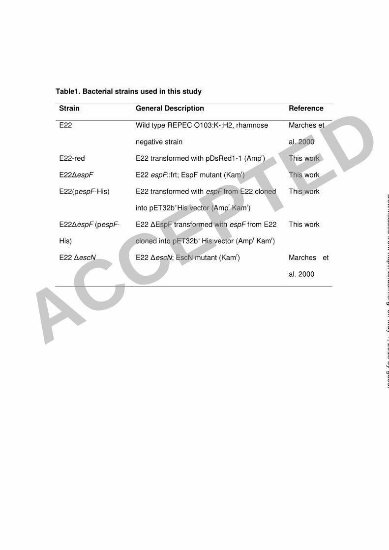
Table1. Bacterial strains used in this study Strain General Description Reference
E22 Wild type REPEC O103:K-:H2, rhamnose
negative strain
Marches et
al. 2000
E22-red E22 transformed with pDsRed1-1 (Ampr) This work
E22∆espF E22 espF::frt; EspF mutant (Kamr) This work
E22(pespF-His) E22 transformed with espF from E22 cloned
into pET32b+His vector (Ampr Kamr)
This work
E22∆espF (pespF-
His)
E22 ∆EspF transformed with espF from E22
cloned into pET32b+ His vector (Ampr Kamr)
This work
E22 ∆escN E22 ∆escN; EscN mutant (Kamr) Marches et
al. 2000
ACCEPTED
on May 4, 2018 by guest
http://iai.asm.org/
Dow
nloaded from


