4 Cabeza Biceps Braquial y 3 Coracobraquial Mehmet Mutlu 2012
4
Case Report Corresponding author: Levent Sarikcioglu Department of Anatomy, Faculty of Medicine, Akdeniz University, 07070 Antalya, Turkey Tel: +90-242-2496952, Fax: +90-242-2274495, E-mail: levent@akdeniz. edu.tr, [email protected] is is an Open Access article distributed under the terms of the Creative Commons Attribution Non-Commercial License (http://creativecommons.org/licenses/by-nc/3.0/) which permits unrestricted non-commercial use, distribution, and reproduction in any medium, provided the original work is properly cited. Copyright © 2012. Anatomy & Cell Biology http://dx.doi.org/10.5115/acb.2012.45.2.136 pISSN 2093-3665 eISSN 2093-3673 Four-headed biceps brachii, three-headed coracobrachialis muscles associated with arterial and nervous anomalies in the upper limb Mehmet Mutlu Catli, Umut Ozsoy, Yasemin Kaya, Arzu Hizay, Fatos Belgin Yildirim, Levent Sarikcioglu Department of Anatomy, Faculty of Medicine, Akdeniz University, Antalya, Turkey Abstract: A four-headed biceps brachii muscle and three-headed coracobrachialis muscle, high-originated radial artery and communication between the median and musculocutaneous nerves have been well documented in the available literature. However co-existence of these variations is rare. In this study we aimed to describe multiple variations in the upper limb and discuss their co-existence from clinical and embryological points of view. Key words: Upper extremity, Biceps brachii muscle, Coracobrachialis muscle, Musculocutaneous nerve, Median nerve Received May 7, 2012; Revised May 28, 2012; Accepted June 1, 2012 Case Report Methods During a routine cadaveric dissection at the Department of Anatomy, Akdeniz University Faculty of Medicine, Antalya, Turkey, we found multiple variations on the left side of a 51-year-old Turkish male cadaver. The arm was dissected carefully to display all structures. All other related structures were also exposed. Results Arterial variations: e brachial artery was split into two main branches at the level of middle third of the humerus. e radial artery coursed downwards and crossed the median nerve from medial to lateral side (Fig. 1A). Branches and terminations of the radial artery were normal in every aspect. e remaining branch coursed downwards as the ulnar artery. It passed under the bicipital aponeurosis and then gave its main branches. Nervous variations: A communicating branch originated from the median nerve at the level of upper third of the humerus, coursed downwards through two supernumerary Introduction Anatomical variations in the upper limb are frequent, but the coexistence of multiple combined neuromuscular and vascular variations are rare. ese variations are worthy of note for clinicians since they should be kept in mind in surgical and diagnostic procedures. In the present study we aimed to describe the co-existence of a high originated radial artery, supernumerary heads of the coracobrachialis and biceps brachii muscles and communication between the median and musculocutaneous nerves. e presence of this co-existence was discussed from embryological and clinical points of view.
-
Upload
arcadiafisioterapia -
Category
Documents
-
view
216 -
download
0
description
Articulo sobre anatomía
Transcript of 4 Cabeza Biceps Braquial y 3 Coracobraquial Mehmet Mutlu 2012

Case Report
Corresponding author: Levent Sarikcioglu Department of Anatomy, Faculty of Medicine, Akdeniz University, 07070 Antalya, TurkeyTel: +90-242-2496952, Fax: +90-242-2274495, E-mail: [email protected], [email protected]
Th is is an Open Access article distributed under the terms of the Creative Commons Attribution Non-Commercial License (http://creativecommons.org/licenses/by-nc/3.0/) which permits unrestricted non-commercial use, distribution, and reproduction in any medium, provided the original work is properly cited.
Copyright © 2012. Anatomy & Cell Biology
http://dx.doi.org/10.5115/acb.2012.45.2.136pISSN 2093-3665 eISSN 2093-3673
Four-headed biceps brachii, three-headed coracobrachialis muscles associated with arterial and nervous anomalies in the upper limbMehmet Mutlu Catli, Umut Ozsoy, Yasemin Kaya, Arzu Hizay, Fatos Belgin Yildirim, Levent SarikciogluDepartment of Anatomy, Faculty of Medicine, Akdeniz University, Antalya, Turkey
Abstract: A four-headed biceps brachii muscle and three-headed coracobrachialis muscle, high-originated radial artery and communication between the median and musculocutaneous nerves have been well documented in the available literature. However co-existence of these variations is rare. In this study we aimed to describe multiple variations in the upper limb and discuss their co-existence from clinical and embryological points of view.
Key words: Upper extremity, Biceps brachii muscle, Coracobrachialis muscle, Musculocutaneous nerve, Median nerve
Received May 7, 2012; Revised May 28, 2012; Accepted June 1, 2012
Case Report
MethodsDuring a routine cadaveric dissection at the Department of
Anatomy, Akdeniz University Faculty of Medicine, Antalya, Turkey, we found multiple variations on the left side of a 51-year-old Turkish male cadaver. The arm was dissected carefully to display all structures. All other related structures were also exposed.
ResultsArterial variations: Th e brachial artery was split into two
main branches at the level of middle third of the humerus. Th e radial artery coursed downwards and crossed the median nerve from medial to lateral side (Fig. 1A). Branches and terminations of the radial artery were normal in every aspect. Th e remaining branch coursed downwards as the ulnar artery. It passed under the bicipital aponeurosis and then gave its main branches.
Nervous variations: A communicating branch originated from the median nerve at the level of upper third of the humerus, coursed downwards through two supernumerary
Introduction
Anatomical variations in the upper limb are frequent, but the coexistence of multiple combined neuromuscular and vascular variations are rare. Th ese variations are worthy of note for clinicians since they should be kept in mind in surgical and diagnostic procedures. In the present study we aimed to describe the co-existence of a high originated radial artery, supernumerary heads of the coracobrachialis and biceps brachii muscles and communication between the median and musculocutaneous nerves. Th e presence of this co-existence was discussed from embryological and clinical points of view.
Multiple variations in the upper limb
http://dx.doi.org/10.5115/acb.2012.45.2.136
Anat Cell Biol 2012;45:136-139 137
www.acbjournal.org
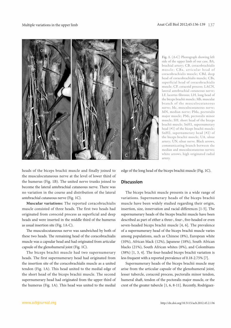
heads of the biceps brachii muscle and finally joined to the musculocutaneous nerve at the level of lower third of the humerus (Fig. 1B). The united nerve trunks joined to become the lateral antebrachial cutaneous nerve. There was no variation in the course and distribution of the lateral antebrachial cutaneous nerve (Fig. 1C).
Muscular variations: The reported coracobrachialis muscle consisted of three heads. The first two heads had originated from corocoid process as superficial and deep heads and were inserted in the middle third of the humerus as usual insertion site (Fig. 1A-C).
Th e musculocutaneous nerve was sandwiched by both of these two heads. Th e remaining head of the coracobrachialis muscle was a capsular head and had originated from articular capsule of the glenohumeral joint (Fig. 1C).
The biceps brachii muscle had two supernumerary heads. The first supernumerary head had originated from the insertion site of the coracobrachialis muscle as a united tendon (Fig. 1A). This head united to the medial edge of the short head of the biceps brachii muscle. The second supernumerary head had originated from the upper third of the humerus (Fig. 1A). This head was united to the medial
edge of the long head of the biceps brachii muscle (Fig. 1C).
Discussion
The biceps brachii muscle presents in a wide range of variations. Supernumerary heads of the biceps brachii muscle have been widely studied regarding their origin, insertion, size, innervation and racial differences [1-5]. The supernumerary heads of the biceps brachii muscle have been described as part of either a three-, four-, fi ve-headed or even seven-headed biceps brachii muscle [4, 6]. The prevalence of a supernumerary head of the biceps brachii muscle varies among populations, such as Chinese (8%), European white (10%), African black (12%), Japanese (18%), South African blacks (21%), South African whites (8%), and Colombians (38%) [1, 3, 4]. The four-headed biceps brachii variation is less frequent with a reported prevalence of 0.18-2.75% [7].
Supernumerary heads of the biceps brachii muscle may arise from the articular capsule of the glenohumeral joint, lesser tubercle, coracoid process, pectoralis minor tendon, humeral shaft , tendon of the pectoralis major muscle, or the crest of the greater tubercle [1, 6, 8-11]. Recently, Rodríguez-
Fig. 1. (A-C) Photograph showing left side of the upper limb of our case. BA, brachial artery; CB, coracobrachialis m u s c l e ; C B a , a r t i c u l a r h e a d o f coracobrachialis muscle; CBd, deep head of coracobrachialis muscle; CBs, superficial head of coracobrachialis muscle; CP, coracoid process; LACN, lateral antebrachial cutaneous nerve; LF, lacertus fibrosus; LH, long head of the biceps brachii muscle; Mb, muscular branch of the musculo cutane ous nerve; Mc, musculocutaneous nerve; MN, median nerve; PMa, pectoralis major muscle; PMi, pectoralis minor muscle; SH, short head of the biceps brachii muscle; SnH1, supernumerary head (#1) of the biceps brachii muscle; SnH2, supernumerary head (#2) of the biceps brachii muscle; UA, ulnar artery; UN, ulnar nerve. Black arrows, communicating branch between the median and musculocutaneous nerves; white arrows, high originated radial artery.
Anat Cell Biol 2012;45:136-139 Mehmet Mutlu Catli, et al138
www.acbjournal.orghttp://dx.doi.org/10.5115/acb.2012.45.2.136
Niedenführ et al. [3] studied on a series of 350 arms and classified the supernumerary heads of the biceps brachii into three diff erent types: superior, inferomedial, and infero-lateral humeral head. They found five cases (1.5%) with a supernumerary head that originated from the surface of the humerus between the lesser tubercle and the attachments of the coracobrachialis and brachialis muscles and fused with the muscular fibers of the short head of the biceps brachii muscle at its union with the long head.
The presence of the supernumerary heads of the biceps brachii muscle has been associated with variations of the surrounding muscles. El-Naggar and Zahir [12] described that two bellies of the coracobrachialis muscle associated with a third head of the biceps brachii muscle, although the coracobrachialis muscle was found to have a normal origin, and the short head of the biceps brachii muscle had separate bellies. From this point of view, the present case also had multiple variations (arterial, nervous and muscular) of which they were encountered unilaterally.
Anomalies of the coracobrachialis muscle are common [1, 13]. Numerous studies have documented variations of the form and origins of the coracobrachialis muscle. The reported morphological variations of the coracobrachialis muscle that included accessory slips inserting to the medial epicondyle of the humerus, medial supracondylar ridge, medial intermuscular septum, the lesser tubercle and a supernumerary head passing over the shoulder joint [14]. Th e morphological variations of the coracobrachialis muscle may be due to failure of muscle primordia disappearing during embryological development [15]. Th e origin of morphologic variations of the coracobrachialis muscle may be explained on the basis of the embryogenesis of the muscles of the arm. Th e intrinsic muscles of the upper limb diff erentiate in situ from the limb bud mesenchyme of the lateral plate mesoderm. At a certain stage of development, the muscle primordia within the diff erent layers of the arm fuse to form a single muscle mass; thereafter, some muscle primordia disappear through cell death [15]. Failure of muscle primordia to disappear during embryologic development may account for the presence of the supernumerary heads of coracobrachialis muscle as reported in this case. Th e coexistence of these variations may be the result of an abnormal embryological formation of the limb muscles, peripheral nerves and arteries.
In most species, the coracobrachialis muscle has three portions: the longus, medius and brevis. In humans, the medius and longus fuse to form the coracobrachialis
muscle [1]. The original third head of the muscle, termed coracobrachialis brevis, occurs rather rarely [1, 13]. Kyou-Jouff roy et al. [16] described three portions in coracobrachialis muscle that originated from the coracoid process and inserted into the medial epicondyle of the humerus (coracobrachialis longus or superfi cialis), humeral diaphysis (coracobrachialis medius) and humeral neck (coracobrachialis profundus or brevis). Mori [17] found that coracobrachialis was completely separated into its superficial and deep layers in eight arms (16%) and incompletely in four arms (8%).
Variations in the connections of musculocutaneous and median nerves have been classified in many ways. In two separate reports Testut [18] and Monden [19] reported that the incidence of the communications between musculocutaneus and median nerves was 36.2% (38 of 105 cases), and 27.5% (33 of 120 cases), respectively. Numerous communications between the median and musculocutaneous nerve have been classified regarding their presence, formations and racial differences [20-22]. In the present case we found that communication originated from the median nerve at the level of upper third of the humerus, coursed downwards through two supernumerary heads of the biceps brachii muscle and finally joined to the musculocutaneous nerve at the level of lower third of the humerus. A clinician’s knowledge of musculocutaneous-median nerve communication is important while evaluation of clinical neurophysiology, planning a surgery aft er trauma and understanding of median and musculocutaneous nerve dysfunction [23-25].
Presence of the multiple variations has also been reported in a previous report [26]. In our previous study we found a high originated radial artery accompanied by muscular and neural abnormalities (three-headed biceps brachii, absence of the palmaris longus muscle, and communication between median and musculocutaneous nerves). In the present case, there was a four-headed biceps brachii muscle and three-headed coracobrachialis muscle accompanied by high originated radial artery and communication between median and musculocutaneous nerves. Rodríguez-Niedenführ et al. [3] reported a case with four-headed biceps brachii muscle of the right arm, but they did not observe any connection between the median and musculocutaneous nerves. Although it is not usually reported, the presence of the multiple variations is a common case that encountered during educational and diagnostic procedures. We think that the presence of the multiple variations is worth of note
Multiple variations in the upper limb
http://dx.doi.org/10.5115/acb.2012.45.2.136
Anat Cell Biol 2012;45:136-139 139
www.acbjournal.org
not only for anatomist but also for clinicians. Because the upper extremity is a frequent site of injury, various surgical and invasive procedures are performed in this region; consequently, it is of utmost importance to be aware of such variations. Th e anatomical variations and abnormalities of the muscles of the upper limb have become signifi cant because of physicians may encounter such abnormalities during imaging with computed tomography and magnetic resonance. Also, these variations are important in order to define the anatomical features of each in relation to the clinical diagnosis and for surgical procedures [27]. Therefore, it should be kept in mind during routine dissection studies, and surgical/diagnostic procedures.
Acknowledgements
We thank Mr. N. Sagiroglu and Mr. H. Gezer and Mr. H. Savcili for their technical assistance. Th is study was supported by Akdeniz University Research Fund (project number: 2005.05.0103.127).
References
1. Bergman RA, Thompson SA, Afifi AK. Catalog of human variation. Baltimore: Urban and Schwarzenberg; 1985. p.108-14.
2. Kopuz C, Sancak B, Ozbenli S. On the incidence of third head of biceps brachii in Turkish neonates and adults. Kaibogaku Zasshi 1999;74:301-5.
3. Rodríguez-Niedenführ M, Vázquez T, Choi D, Parkin I, Sañudo JR. Supernumerary humeral heads of the biceps brachii muscle revisited. Clin Anat 2003;16:197-203.
4. Abu-Hijleh MF. Th ree-headed biceps brachii muscle associated with duplicated musculocutaneous nerve. Clin Anat 2005;18: 376-9.
5. Lee SE, Jung C, Ahn KY, Nam KI. Bilateral asymmetric super-numerary heads of biceps brachii. Anat Cell Biol 2011;44:238-40.
6. Nakatani T, Tanaka S, Mizukami S. Bilateral four-headed biceps brachii muscles: the median nerve and brachial artery passing through a tunnel formed by a muscle slip from the accessory head. Clin Anat 1998;11:209-12.
7. Khaledpour C. Anomalies of the biceps muscle of the arm. Anat Anz 1985;158:79-85.
8. de Burlet HM, Correljé J. Über variationen des menschlichen musculus biceps brachii. Gegenbaurs Morphol Jahrb 1919;50: 403-16.
9. Vollala VR, Nagabhooshana S, Bhat SM, Potu BK, Rodrigues V, Pamidi N. Multiple arterial, neural and muscular variations in upper limb of a single cadaver. Rom J Morphol Embryol
2009;50:129-35.10. Sargon MF, Tuncali D, Celik HH. An unusual origin for the
accessory head of biceps brachii muscle. Clin Anat 1996;9:160-2.11. Kosugi K, Shibata S, Yamashita H. Supernumerary head of
biceps brachii and branching pattern of the musculocutaneus nerve in Japanese. Surg Radiol Anat 1992;14:175-85.
12. El-Naggar MM, Zahir FI. Two bellies of the coracobrachialis muscle associated with a third head of the biceps brachii muscle. Clin Anat 2001;14:379-82.
13. Beattie PH. Description of bilateral coraco-brachialis brevis muscle, with a note on its signifi cance. Anat Rec 1947;97:123-6.
14. Vollala VR, Nagabhooshana S, Bhat SM, Potu BK, Rakesh V. Multiple accessory structures in the upper limb of a single cadaver. Singapore Med J 2008;49:e254-8.
15. Grim M. Ultrastructure of the ulnar portion of the contrahent muscle layer in the embryonic human hand. Folia Morphol (Praha) 1972;20:113-5.
16. Kyou-Jouffroy MK, Lessertisseur J, Saban R, Souteyrand-Boulenger JD. Musculature des membres, membre pectoral, groupe branchial ventral. In: Grassé PP, editor. Traité de Zoologie. Mammifères: Anatomie et Reproduction. Paris: Masson; 1971. p.96-8.
17. Mori M. Statistics on the musculature of the Japanese. Okajimas Folia Anat Jpn 1964;40:195-300.
18. Testut L. Mémoire sur la portion brachiale du nerf musculo-cutané. Int Monatsschr Anat Physiol 1884;1:305-41.
19. Monden M. Anastomosis between the median and musculo-cutaneous nerves in the upper arm. Juzen Ishi 1942;47:2045-55.
20. Choi D, Rodríguez-Niedenführ M, Vázquez T, Parkin I, Sañudo JR. Patterns of connections between the musculocutaneous and median nerves in the axilla and arm. Clin Anat 2002;15:11-7.
21. Kosugi K, Morita T, Koda M, Yamashita H. Branching pattern of musculocutaneous nerve. 2. Cases possessing supernumerary head of bicipital brachial muscle. Jikeikai Med J 1986;33:195-208.
22. Venieratos D, Anagnostopoulou S. Classifi cation of communi-cations between the musculocutaneous and median nerves. Clin Anat 1998;11:327-31.
23. Flatow EL, Bigliani LU, April EW. An anatomic study of the musculocutaneous nerve and its relationship to the coracoid process. Clin Orthop Relat Res 1989;(244):166-71.
24. Sonck WA, Francx MM, Engels HL. Innervation anomalies in upper and lower extremities: potential clinical implications. How to identify with electrophysiologic techniques. Electromyogr Clin Neurophysiol 1991;31:67-80.
25. Sunderland S. Nerves and nerve injuries. Edinburgh, London, NewYork: Churchill Livingstone; 1978.
26. Sarikcioglu L, Yildirim FB. High origin of the radial artery accompanied by muscular and neural anomalies. Ann Anat 2003;185:179-82.
27. Kopuz C, Fidan B, Islam A. An unusually distal and complete additional flexor profundus muscle to the index finger. J Anat 1997;191(Pt 3):465-7.