3Wallace H. Coulter Department of Biomedical Engineering ... · PDF file3Wallace H. Coulter...
33
RECENT ADVANCES IN POLYMERIC HEART VALVES RESEARCH Recent Advances in Polymeric Heart Valves Research Yee Han, Kuan 1 , Prasad Dasi, Lakshmi 2 , Ajit P., Yoganathan 3 , Hwa Liang, Leo 1* 1 Division of Bioengineering, National University of Singapore 2 Department of Mechanical Engineering, Colorado State University 3 Wallace H. Coulter Department of Biomedical Engineering, Georgia Institute of Technology *Correspondence author: Leo Hwa Liang, Division of Bioengineering, National University of Singapore, Singapore; Tel: +65-6516-5608, Fax: +65-6872-3069, email address: [email protected]
Transcript of 3Wallace H. Coulter Department of Biomedical Engineering ... · PDF file3Wallace H. Coulter...

RECENT ADVANCES IN POLYMERIC HEART VALVES RESEARCH
Recent Advances in Polymeric Heart Valves Research
Yee Han, Kuan1, Prasad Dasi, Lakshmi2, Ajit P., Yoganathan 3, Hwa Liang, Leo1*
1Division of Bioengineering, National University of Singapore
2Department of Mechanical Engineering, Colorado State University
3Wallace H. Coulter Department of Biomedical Engineering, Georgia Institute of Technology
*Correspondence author:
Leo Hwa Liang, Division of Bioengineering, National University of Singapore, Singapore; Tel: +65-6516-5608, Fax: +65-6872-3069, email address: [email protected]

RECENT ADVANCES IN POLYMERIC HEART VALVES RESEARCH
Abstract
Heart valve replacement is fast becoming a routine surgery worldwide and heart valve
prostheses are today considered among the most widely used cardiovascular devices.
Mechanical and bioprostheses have been the traditional choices to the replacement surgeries.
However, such valves continue to exposed patients to risks, including thrombosis, infection
and limited valve durability. In recent years, advances in polymer science give rise to an
important and new class of artificial heart valve made predominantly of polyurethane-based
materials, which show improved biocompatibility and biostability. These polymeric heart
valves have demonstrated excellent hemodynamic performance and good durability with
excellent fatigue stress resistance. Advancements in the designs and manufacturing methods
also suggested improved in the durability of polymeric heart valves. Animal studies with
these valves have also shown good biocompatibility with minimal calcification of the valve
leaflets. With these promising progresses, polymeric heart valves could be a viable alternative
in the heart valve replacement surgeries in the near future.
Keywords: Polyurethane, Hemodynamic, Reynolds shear stress, Bioprostheses, Calcification.

RECENT ADVANCES IN POLYMERIC HEART VALVES RESEARCH
Recent Advances in Polymeric Heart Valves Research
Existing commercial prosthetic heart valve designs can be classified into two groups:
bioprosthetic, or tissue valves, and mechanical heart valves. The former is made from a
combination of synthetic materials and chemically treated animal tissue mainly porcine in
origin, whereas the latter is manufactured entirely from synthetic materials. In the 1960s, Dr
Charles Hufnagel performed one of the earliest successful mechanical heart valve surgeries
where six of the eight patients who received a caged ball heart valve survived the operation
(Hufnagel, Gillespie, Conrad, Mercier, & Evangelist, 1966). However, current artificial heart
valves are still exposed to risks of thrombosis and thromboembolism, tissue overgrowth,
infection, anti-coagulant related hemorrhage, and valve failure due to material fatigue or
chemical change (Black, & Drury, 1994; Yoganathan, 2000; Starr, Fessler, Grunkemeier, &
He, 2002; Korossis, Fisher, & Ingham, 2000).
One major drawbacks associated with the implantation of mechanical heart valves is
the need for daily chronic anticoagulation therapy to reduce the risk of thrombosis and
thromboembolic complications. These patients are expose to an increased risk of bleeding,
infection, and/or autoimmune response (Walker, & Yoganathan, 1992). Blood flow through
mechanical prostheses can lead to high turbulent stresses that may damage and/or activate
blood elements and initiate platelet aggregation. Platelet aggregation can lead to thrombus
formation with disastrous consequences for the patient. Thrombi may even detach from the
valve and become lodged in a downstream blood vessel, thus reducing or even cutting off the
blood supply to vital tissues.
An alternative to mechanical valves is using tissue valves which utilize the concept of
a trileaflet configuration with one central orifice that mimics the design of the native valve.
These valves have a lower potential for blood element damage than their mechanical
counterparts. Nevertheless, the mechanical properties of tissue valves appear to degrade more

RECENT ADVANCES IN POLYMERIC HEART VALVES RESEARCH
rapidly, and they are prone to calcification (Black, & Drury, 1994). Often, implanted tissue
valves do not last for more than ten years, and reoperation is necessary.
The polyurethane (PU) trileaflet polymeric heart valve is the latest development in
prosthetic heart valve research (Hyde, Chinn, & Phillips, 1999). The design is based on the
natural aortic valve and is inherently appealing from a hemodynamic viewpoint. Although
this particular valve design is still in a developmental stage, preliminary studies have shown
excellent forward flow hemodynamic properties equivalent to that of a tissue heart valve and
promise a durability comparable to that of a mechanical heart valve (Bernacca, Mackay,
Wilkinson, & Wheatley, 1995; Jansen et al., 1991). However, recent animal trials involving
polymeric valves have reported problems mainly related to tearing of the leaflets and
thrombus formation occuring along the stent region of the valve (Wheatley et al., 2001). In
addition, results of long-term in vivo evaluation have suggested that calcification could be a
limiting factor to long-term function of polymeric valves (Bodnar, Frater, & eds, 1991). The
leaflets and basal attachments, such as the commissural region of the polymer valves, have
experienced extrinsic calcification associated with surface microthrombi that appear to be
independent of structural defects suggesting that the flow characteristics inside the polymeric
valve may be a contributor to the observed blood clots.
This review will discuss the recent development of polymeric heart valves,
particularly on its choice of materials. Much attention will also focus on the in vitro
hemodynamics characterization these polymeric heart valves. The review will also discuss
the in vivo experiments involving these polymeric heart valves and their susceptibility to
calcifications.
Introduction of Polymeric Heart Valves
Pioneering works in polymeric heart valve replacement in human was first reported in

RECENT ADVANCES IN POLYMERIC HEART VALVES RESEARCH
1958 (Roe, Owsley, & Boudoures, 1958). This is followed by a single trileaflet silicone
rubber (SR) aortic valve implanted in several patients between 1960 and 1962 (Roe, Kelly,
Myers, & Moore, 1966). The clinical trials were discontinued due to the high occurrence of
thromboembolic events. The choice of material is an important factor in the development of
polymeric heart valve. The chosen polymer should have acceptable characteristics with
regard to biocompatibility, hemocompatibility, anti-thrombogenicity, resistance to
degradation and calcification (Daebritz et al., 2004; Gallocher, Aguirre, Kasyanov, Pinchuk,
& Schoephoerster, 2006). Since then, several synthetic polymers have been tested as leaflet
materials, including silicone, polyofelin rubber, Silastic, SR-impregnated Dacron mesh,
polytetrafluoroethylene (PTPE) and PU.
In 1960s, silicone was considered a choice due to its great flexibility and
biocompatibility. However, in clinical investigations, formation of thrombosis and thickening
of valve leaflets were found. Silicone suffered from structural failure and short durability, and
was abandoned as a possible material replacement (Roe, Kelly, Myers, & Moore, 1966).
Another study also showed the inadequate durability of silicone and polyofeline rubbers
while in vivo (Cheeta, & Llyod, 1980). PTFE or Teflon valves were considered as a
polymeric heart valve material attributing to their good hemodynamic properties. However,
its low resistance to thromboembolism and stiffening of the leaflets prevented PTFE from
further development (Nistal, 1990). Calcification was also found on PTFE valves (Imamura,
& Kaye, 1977).
In 1982, a breakthrough came when Wisman and colleagues invented a trileaflet valve
fabricated from segmented polyurethane (SPU) (Wisman et al., 1982). The large central flow
orifice of the leaflet valve is similar to tissue valves, which reduces turbulence and blood
trauma as a result of lower transvalvular pressure drop and better hydraulic efficiency. The
valve demonstrated high flexure endurance, great strength and inherent nonthrombogenic

RECENT ADVANCES IN POLYMERIC HEART VALVES RESEARCH
characteristics.
Choice of Materials
PU has been widely used in biomedical application and it exhibits several favourable
properties due to its two-phased microstructure consisting of hard crystalline segments and
soft elastomeric segments. The hard segment confers structural strength while the soft
segment determines the flexibility and elasticity of the material.
Polyurethanes elastomers are copolymers containing blocks of low molecular weight
polyesters or polyethers linked together by a urethane group. These elastomers are based on
three monomers: (1) an isocyanate source, (2) a macroglycol, and (3) a chain extender, or
curative. The isocyanate can either be aromatic or aliphatic. One example of aromatic
isocyanate is the methylene diphenyl diisocyanate (MDI), which is a very reactive isocyanate.
Usually, it does not require a catalyst for synthesis if a diamine chain extender is used. An
example of an aliphatic isocyanate is the cyclo-aliphatic diisocyanate H12MDI. The aromatic
isocyanate containing polymers have a tendency to yellow upon exposure to ultraviolet
radiation, and some aromatic polyurethanes, such as those made from methylene dianiline, or
MDA, are known carcinogens and display immediate cell toxicity. However, elastomers
produced from less toxic aliphatic isocyanates have inferior physical properties and are
significantly more expensive (Szycher, 1988).
The reaction of a diisocynate with a short chain diol or diamine (‘chain extenders’)
forms the hard segments. Commonly used extenders for medical use are the 1,4 butane diol
or ethylene diamine. On the other hand, the reaction of the diisocynate with higher molecular
weight polyols, such as polyether, polyesther or polycarbonate, forms the soft segments.
Medical grade segmented polyurethane (SPU) have been applied successfully in various
cardiovascular devices, including artificial hearts, ventricle assist devices and blood pumps

RECENT ADVANCES IN POLYMERIC HEART VALVES RESEARCH
(Bernacca, Mackay, Gulbransen, Donn, & Wheatley, 1997; Bernacca, Straub, & Wheatley,
2002).
In recent years, remarkable advancement in polymer synthesis has resulted in a more
bio-stable PU. Recent studies have yielded a wider range of SPUs for polymeric heart valve
leaflets (Alferiev et al., 2001; Bernacca, Mackay, Gulbransen, Donn, & Wheatley, 1997;
Seifalian et al., 2003; Wheatley et al., 2001). The main types of PU which have been
developed and tested are polyester urethane, polyether urethane (PEU) and polycarbonate
urethane (PCU).
PEU were found to be unsuitable for long-term implants due to the hydrolytic
degradation. Such polymers are vulnerable to oxidative degradation and suffers
environmental stress cracking and metal ion oxidation under in vivo conditions (Stokes,
McVenes, & Anderson, 1995). PCU were tested and proven to have a higher oxidative
stability (Salacinski, Odlyha, Hamilton, & Seifalian, 2002). When compared to PEU, the
biodegradation of PCU was significantly lower and limited to a thinner surface layer
(Christenson, Anderson, & Hiltner, 2004).
In order to improve the biostability of PU, the chemical structures have been
modified. Bernacca and colleagues developed a polyurethane valve which was chain-
extended with ethylene diamine (PEUE). Good phase separation of polyurethane soft and
hard segments with good rubbery characteristics helped to explain the excellent PEUE valve
durability (Bernacca, Mackay, & Wheatley, 1996).
Another nanocomposite PU polymer, PCU and polyhedral oligomeric silsesquioxanes
(POSS) was developed and studies have shown that the polymer possesses greater
thromboresistance than PTFE (Kannan et al., 2006). In vivo study conducted using the POSS-
PCU composite found minimal inflammation and capsular formation in a sheep model
compared to control. No degradation was found even after 36 months of post-implantation in

RECENT ADVANCES IN POLYMERIC HEART VALVES RESEARCH
the sheep model (Kannan et al., 2007). The increased fibrinogen adsorption on POSS-PCU,
and large contact-angle hysteresis suggested that POSS-PCU inhibits inflammation by
adsorbing and inactivating fibrinogen on its surface. This composite has enhanced interfacial
biocompatibility and better biological stability as compared with conventional silicone
biomaterials.
In a study by Kidane and his team, it was found that POSS-PCU nanocomposite
possess excellent mechanical strength, good surface properties and resistance to platelet
adhesion. It has also shown comparable hardness and tear strength to commercially available
products such as Estane, Chronoflex C and Elasteon (Kidane et al., 2009). The inclusion of
nanoscale POSS particles into the polymer matrix was suggested as the contributing factor to
the increment in tensile strength and hardness of POSS-PCU. The hydrophobicity factor of
the polymer helps resist the adsorption of blood proteins, which is necessary in preventing
thrombosis and calcification on the heart valve surfaces.
In 2004, Jiang and colleagues developed and demonstrated a novel one-piece,
tricuspid valve made entirely from polyvinyl alcohol cryogel (PVA-C). The mechanical
properties of the valve were successfully demonstrated with reduction in flexural stresses of
the leaflets. The stress concentrations at the commissural areas where the leaflets are attached
to the stent were also avoided (Jiang, Campbell, Boughner, Wan, & Quantz, 2004). However
further studies are required to evaluate the potential of PVA-C valve as a superior alternative
to the current valves.
More recently, a novel polyolefin poly(styrene-b-isobutylene-b-styrene) (SIBS) was
being proposed as a viable polymer for use as valve due to its oxidation resistance. SIBS also
showed suitable hemocompatibility and mechanical durability, with over 350 million cycles
evaluated in vitro in the early stages. The material was comparable to PU in platelet
deposition tests, as well as tensile and fatigue properties after reinforcement of polypropylene

RECENT ADVANCES IN POLYMERIC HEART VALVES RESEARCH
fibres. Platelet activation due to the polymer valve was not found to be significantly different
from either a St. Jude bileaflet valve or a St. Jude Tissue valve. (Gallocher, Aguirre,
Kasyanov, Pinchuk, & Schoephoerster, 2006). Such encouraging traits open the possibility of
SIBS as a potential material choice for further development in polymeric heart valve.
Polymeric Heart Valve Design
There are several essential requirements of a heart valve design. The valve should fit
nicely into the host anatomy with minimal thrombogenicity and damage to blood cells. The
valve leaflets should also ensure minimum resistance to the forward flow and open at a
minimum systolic trans-valvular pressure difference. This promote smooth washout and
hence are less susceptible to trauma-promotion (causing thrombus, atherosclerosis and
hemolysis) (Bellhouse, & Reid, 1969). There should also be appropriate sealing in the close
position to minimize backward flow. The stress peaks in valve components should be low to
ensure durability and minimal change to the geometric features such as creep (Mercer,
Benedicty, & Bahnson, 1973). Figure 1 shows an example of a valve design.
Attempts to manufacture leaflet valves from polymers started in the 1950s and as
early as the inception of the first mechanical heart valve prostheses. However, only recently
the first realistic solution was developed. The major hurdles for the success of the polymeric
valves pertained to valve design and its chemical composition to eliminate thrombogenicity,
calcification, mechanical stress, and durability. The recent design of polyurethane valve is the
stent-supported bioprosthetic heart valve. Here the polymeric leaflets are mounted onto
flexible stents that lead to a circular orifice during forward flow. However, the leaflets, stent,
and the sewing ring in polymeric valves are made of a multilayered cohesively bonded single
material and are available at various degrees of hardness instead of each leaflet being sewn
separately onto the stents as in the bioprosthetic valve (Daebritz et al., 2004). The leaflets at
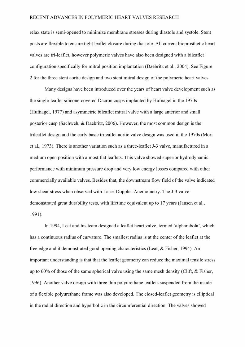
RECENT ADVANCES IN POLYMERIC HEART VALVES RESEARCH
relax state is semi-opened to minimize membrane stresses during diastole and systole. Stent
posts are flexible to ensure tight leaflet closure during diastole. All current bioprosthetic heart
valves are tri-leaflet, however polymeric valves have also been designed with a bileaflet
configuration specifically for mitral position implantation (Daebritz et al., 2004). See Figure
2 for the three stent aortic design and two stent mitral design of the polymeric heart valves
Many designs have been introduced over the years of heart valve development such as
the single-leaflet silicone-covered Dacron cusps implanted by Hufnagel in the 1970s
(Hufnagel, 1977) and asymmetric bileaflet mitral valve with a large anterior and small
posterior cusp (Sachweh, & Daebritz, 2006). However, the most common design is the
trileaflet design and the early basic trileaflet aortic valve design was used in the 1970s (Mori
et al., 1973). There is another variation such as a three-leaflet J-3 valve, manufactured in a
medium open position with almost flat leaflets. This valve showed superior hydrodynamic
performance with minimum pressure drop and very low energy losses compared with other
commercially available valves. Besides that, the downstream flow field of the valve indicated
low shear stress when observed with Laser-Doppler-Anemometry. The J-3 valve
demonstrated great durability tests, with lifetime equivalent up to 17 years (Jansen et al.,
1991).
In 1994, Leat and his team designed a leaflet heart valve, termed ‘alpharabola’, which
has a continuous radius of curvature. The smallest radius is at the center of the leaflet at the
free edge and it demonstrated good opening characteristics (Leat, & Fisher, 1994). An
important understanding is that that the leaflet geometry can reduce the maximal tensile stress
up to 60% of those of the same spherical valve using the same mesh density (Clift, & Fisher,
1996). Another valve design with three thin polyurethane leaflets suspended from the inside
of a flexible polyurethane frame was also developed. The closed-leaflet geometry is elliptical
in the radial direction and hyperbolic in the circumferential direction. The valves showed

RECENT ADVANCES IN POLYMERIC HEART VALVES RESEARCH
promising durability tests, with all six valves exceeded the lifetime equivalent of 10 years.
Nevertheless, one valve failed due to a leaflet tear after reaching 12 years cycling (Mackay,
Wheatley, Bernacca, Fisher, & Hindle, 1996).
Another interesting design is a stentless and fiber-reinforced synthetic valve as shown
in Figure 3. This design mimics stress-reduction and may decrease the tears and perforation
in the leaflets. The prototype also demonstrated low resistance to forward flow. However, the
manufacturing procedure to produce a stentless valve is complicated due to the irregular
shape of the mold to keep the fiber manually in place during winding procedure (Cacciola,
Peters, & Baaijens, 2000).
Leaflet Thickness
Thin SPU leaflets between 100-150 µm perform well in terms of trans-valvular
pressure gradients and energy losses. This leaflet thickness may achieve good hydrodynamic
flows but may compromise the durability. Although the leaflets can be manufactured as thin
as 100 µm to be flexible and durable, there are concerns at thickness less than 150 µm
(Mackay, Wheatley, Bernacca, Fisher, & Hindle, 1996). In order to reduce the imparted
bending stress, a valve that undergoes cyclical flexion must be manufactured from low elastic
modulus material.
SPU is widely used because it can flex easily. This helps to minimize blood flow
disturbance and resistance. The ease of flexion criterion must be counterbalanced by the
ability to withstand the high reverse pressures and mechanical stresses present when the valve
opens and closes in a normal physiological cycle.
Bernacca and his team examined the hydrodynamic behavior of biostable
polyurethane valves varying Young's modulus from 5 to 63.6 MPa and a mean leaflet
thickness from 48 to 238 µm. They demonstrated that valve hydrodynamic function is not

RECENT ADVANCES IN POLYMERIC HEART VALVES RESEARCH
significantly affected by the material modulus with little difference in the pressure gradient.
However, the increasing leaflet thickness is produces a significant rise in pressure gradient.
Energy loss during forward flow is proportional to the behaviour of the pressure gradient.
Increased in leaflet thickness is associated with the decrease in valve closing efficiency
(Bernacca, O'Connor, Williams, & Wheatley, 2002).
In another study by Bernacca and his colleagues, leaflet thickness was varied from 60
to 200 µm and it was also found that the thickness of a polyurethane valve is critical to its
function. Durability of the valve was directly related to leaflet thickness, with good durability
achieved with median leaflet thickness of approximately 150 µm. The polymeric heart valves
exceeded 800 million cycles (Bernacca, Mackay, Gulbransen, Donn, & Wheatley, 1997). In a
more recent in vitro fatigue testing, the durability of PCU valves with leaflet thickness
varying between 100 and 300 µm was between 600 million (15.8 years) to 1 billion cycles
(26 years) of normal human function (Daebritz et al., 2003).
Fabrication processes
Today, all polymeric heart valves are made mainly out of polyurethane, with a
combination of solution casting and injection molding. The stents or frames are injection
molded and typically have a thickness of approximately 3 mm. The polyurethane frames are
then molded onto steel formers of ellipto-hyperbolic leaflet shape (Mackay, Wheatley,
Bernacca, Fisher, & Hindle, 1996) and dipped into a concentrated polyurethane solution so
that they coat the whole valve to form the leaflets. The polymer valve is then allowed to dry
while hanging free edge downward. The leaflet edge is later cut and trimmed by precision a
laser cutting tool. The thickness of the leaflet ranges from 80 to 300 µm (Bernacca, Mackay,
Gulbransen, Donn, & Wheatley, 1997; Daebritz et al., 2003; Daebritz et al., 2004; Mackay,
Wheatley, Bernacca, Fisher, & Hindle, 1996). Some polyurethane valves contain a stiffening

RECENT ADVANCES IN POLYMERIC HEART VALVES RESEARCH
ring of radio-opaque MRI compatible titanium alloy to facilitate radiographic imaging.
There are several types of fabrication techniques for polymeric heart valves; (1) Dip
casting, (2) Film fabrication, and (3) Cavity moulding. The fabrication of the polymeric valve
usually involves coating a semi-rigid stent made of poly-ether-ether-ketone (PEEK) in
polyurethane (Wisman et al., 1982). Traditionally, SPU valves have been manufactured by
dip-coating, which involves the use of a specifically designed mandrel. It involved multiple
dips in a weak polymer solution (Jansen et al., 1991). The major disadvantage for this method
is that the leaflet thickness distribution cannot be control accurately.
In film fabrication, pre-cast SPU film is solvent-bonded to the valve frame and
thermally formed to the leaflet shape. This method allows greater control over the desired
geometry of the valve (Leat, & Fisher, 1995). However, this method would also yield a valve
that was inferior in term of durability due to an inconsistent leaflet-frame interface.
Jiang et al showed a cavity mould consists of a static assembly and a moving
assembly can be used to fabricate the whole valve structure. Hot polymer is poured into the
mould and placed in a water bath. The cyclic freezing and thawing process at – 20°C and +
20°C forms a thin film of polymer (Jiang, Campbell, Boughner, Wan, & Quantz, 2004). The
valve manufacturing method has shown to significantly influence the performance of the
valve leaflets. Valves manufactured using a dip-casting method has shown up to 31%
reduction in open leaflet bending strains compared to valves manufactured from solvent-cast
sheets of polyurethane (Corden, David, & Fisher, 1995).
The longer durability of dip cast valves compared to thermally formed film fabricated
leaflets was also verified by Leat and Fisher, although no significant difference was found in
the hydrodynamic function of the valves between the two prescribed methods. In their
comparison study, one method is by thermally forming the leaflets from film and bonded to
the frame while the other method is by dip casting the leaflets on to the frame using s-shaped

RECENT ADVANCES IN POLYMERIC HEART VALVES RESEARCH
mandrel. A simple and reproducible fabrication method using a combination of solution
casting and injection moulding was used by Mackay et al in their polyurethane heart valve
development (Mackay, Wheatley, Bernacca, Fisher, & Hindle, 1996). These valves were
tested and found to exhibit pressure gradients similar to those for a bioprosthesic valve with
considerably lower levels of regurgitation and leakage than bileaflet MHVs or bioprosthesic
valves.
In vitro studies of polymeric heart valves
In vitro studies have been conducted to determine the performance of polymeric heart
valves. Many of these valves have been tested in vitro under pulsatile flow conditions for the
hydrodynamic functions. The levels of regurgitation and leakage are considerably less than
the mechanical and porcine bioprosthetic valves (Mackay, Wheatley, Bernacca, Fisher, &
Hindle, 1996).
It has been shown that polymeric heart valves generate a centralized flow with a
relatively flat profile similar to that of bioprostheses and native heart valves in vitro studies.
The flow patterns of four different trileaflet polyurethane heart valves were compared to the
Hancock porcine heart valve and Ionescu-Shiley 3 pericardial valve by measuring the down-
stream velocity distributions using Laser-Doppler-Anemometry (Herold et al., 1987). It was
observed that the velocity distributions at peak systole are very similar between the
polyurethane valve and Hancock prosthesis. Both flow fields can be characterized as a
triangular jet-flow, with the recirculation areas located near the right aortic wall downstream
of the stent post and in the opposite sinus cavity. However, flat plug type flow was observed
for the Ionescu-Shiley 3 valve. The flow is positive with recirculation in the aortic sinus
cavity and one small transient recirculating vortex near the right aortic wall. Overall, the flat
flow profile of polyurethane valves is similar to those of the native and bioprostheses valves

RECENT ADVANCES IN POLYMERIC HEART VALVES RESEARCH
with vortex formation observed behind the leaflets in the sinus cavity.
De’Souza and colleagues characterized the velocity and turbulence waveforms distal
to several trileaflet polymeric heart valves with varying degrees of stenosis (D'Souza,
Butterfield, & Fisher, 2003). They observed that the shape and orientation of the spatial
velocity profile in a polyurethane heart valve depicts the effect of the constriction caused by
the valve leaflet in a similar manner to that observed in bioprosthetic valves.
In another study, the flow dynamics of two polyurethane valves are investigated; one
with leaflets bound to the edge of the supporting stents (PUI) and the other with leaflets
mounted outside the stents (PUII) (Chandran et al., 1989). The PUII valves have larger inlet
orifice diameter with a smaller tissue annulus diameter, which provide more unobstructed
area for blood flow. Such valve design was also found to reduce the maximum turbulent
shear stresses distal to the valves, which appears more advantageous. Furthermore, the
turbulent shear stress in the PUII valve was half that in the PUI valve showing that the
increase in orifice diameter can help reduce the turbulent shear stresses and corresponding
damage to blood elements. When compared to tissue valves, polymeric valves have larger
turbulent shear stresses. The orifice diameters in polymer valves are usually smaller than
those in bioprostheses, and as a result, polyurethane valves have larger observed velocity
magnitudes and larger velocity gradients than the bioprostheses.
Recent studies have also shown that the trileaflet design is more efficient than double
or quadruple arrangements (Cheeta, & Llyod, 1980; Jansen et al., 1991; Leat, & Fisher, 1995;
Mackay, Wheatley, Bernacca, Fisher, & Hindle, 1996), and that the maximal stresses in these
trileaflet configurations were found near the commissural attachment, which is consistent
with other synthetic and bioprosthetic valves (Leat, & Fisher, 1995). Corden and team
calculated the bending stresses and strains at the free leaflet edge by measuring the curvature
and the study showed that the highest curvatures occurred at the commissural regions when

RECENT ADVANCES IN POLYMERIC HEART VALVES RESEARCH
the valves are fully open (Corden, David, & Fisher, 1995).
Leo and colleagues performed Laser Doppler Velocimetry (LDV) and Particle
Imaging Velocimetry (PIV) studies of three Aortech polymeric heart valves investigating the
effect of commissural design and leaflet thickness on the hemodynamic performance of the
heart valves. These studies indicated that commisural design and leaflet thickness can
influence the thrombogenic potential of tri-leaflet polymeric valves. The studies, as seen in
Figure 4 and 5, identified the following regions of high shear stress and high velocity flow in
the three polymeric heart valves: 1) the leakage jet inside the valve during diastole; 2) the
trailing edge of the valve; 3) the vortex ring surrounding the forward jet during the
acceleration phase; 4) the edge of the central orifice jet, which extended from the inside of
the valve into the distal part of the flow chamber; and 5) the distal region of the flow chamber
where turbulence mixing occurred during systole. Low flow velocities were observed in the
following regions of the three polymeric heart valves: 1) the split flow inside the valve during
diastole; 2) a vortex inside the sinus region; and 3) flow inside the recirculation zone along
the chamber wall during diastole. Additional low flow regions were observed in prototype A:
1) the inflow stent region during late systole; and 2) the closed commissural region during
systole (Leo, 2005).
Using flow visualization and LDV, several studies have demonstrated that trileaflet
bioprostheses produced jet-type flow fields like those observed in the polymeric heart valves
(Woo, Williams, & Yoganathan, 1983; Woo, Williams, & Yoganathan, 1983; Yoganathan et
al., 1983; Yoganathan et al., 1986; Yoganathan, Woo, & Sung, 1986). In general, the
pericardial and Aortech polymeric heart valve designs created flatter velocity profiles than
the porcine and Abiomed valve designs. All three Aortech polymeric heart valves produced
jets with flat velocity profiles at peak systole, identical to those observed in the Ionescus-
Shirley pericardial, Carpentier-Edwards 2650 porcine, Hancock pericardial, and Carpentier-

RECENT ADVANCES IN POLYMERIC HEART VALVES RESEARCH
Edwards pericardial valves. In contrast, the velocity profiles of the central jets in the
Carpentier-Edwards 2625 porcine, Hancock modified-orifice porcine, Hancock II porcine,
and the Abiomed valves were more parabolic in shape.
The diameter of the central jets in the tissue bioprostheses at peak systole is typically
larger than that in the polymeric heart valves. The bioprosthetic valves studied have larger
tissue annular diameters compared with the polymeric prostheses (see Table 1). For a given
cardiac output, valves with larger tissue annular diameters typically have larger jet diameter
compared to those with smaller tissue annular diameters. Central orifice jet diameters
between 15 - 25 mm were observed in the bioprostheses at peak systole with the smallest
occurring in Carpentier-Edwards 2625 and Hancock standard-orifice porcine valves, and the
largest in the Hancock pericardial and Carpentier-Edwards pericardial valves(Yoganathan et
al., 1986). The jet diameters for the Aortech polymeric heart valves were comparable to that
of the Abiomed trileaflet polymeric valve prosthesis, which had a jet diameter of 14 mm
(Woo, Williams, & Yoganathan, 1983; Woo, Williams, & Yoganathan, 1983). The Aortech
prototype A had a jet diameter of 13 mm, while both Aortech prototypes B and C a jet
diameter of 16 mm.
Flow separation was observed at the edges of the valve leaflets and around the jet.
Flow reattachment occurred when the central orifice jet diverged and impinged on the
chamber wall downstream of the valve. The location of reattachment points varies from valve
to valve. Under the same cardiac output, valves with larger orifice sizes tend to have
reattachment points closer to the trailing edge of the valve leaflet. For instance, the central
orifice jets of the Abiomed 25 mm and 21 mm valves were observed to reattach 40 mm and
60 mm downstream from the valves, respectively (Woo, Williams, & Yoganathan, 1983). A
common observation pertaining to the reattachment points of the central orifice downstream
of the valve was made in the flow visualization studies as well as in current studies.
RECENT ADVANCES IN POLYMERIC HEART VALVES RESEARCH
Reattachment of the orifice jet occurred at different downstream locations in the upper and
lower parts of the flow chamber. This observation is attributed to the asymmetric nature of
the central orifice jet. In the Abiomed 25 mm and 21 mm valves, the central orifice jet
reattached approximately 75 mm and 40 mm downstream of the sewing ring in the upper and
lower parts of the flow chamber, respectively (Woo, Williams, & Yoganathan, 1983). For the
tissue bioprostheses, the jet typically reattached in the upper part of the flow chamber at a
location between 55 – 90 mm downstream from the valve sewing ring, and at a location
between 35 – 50 mm in the lower part of the flow chamber. These findings are consistent
with those made for the three Aortech polymeric heart valves; the central jet in the three
prototypes valves reattached at a location between 45 - 55 mm downstream from the valve
sewing ring in the lower part of the flow chamber. For both prototypes A and C, the jet
reattachment point in the upper part of the flow chamber seem to occur beyond the
measurement plane, which only extended 77.5 mm downstream from the valve sewing ring.
The exception to the jet reattachment location in the upper part of the flow chamber was
observed in both the Hancock pericardial and the Aortech prototype B valves. The central
orifice jets in both valves were observed to reattach closer to the valve at approximately 55
mm downstream from the valve sewing ring in the upper part of the flow chamber.
The leaflet profile and flow patterns of the three Aortech polymeric heart valves at
peak systole was similar to that seen in the pericardial heart valve where a small recirculation
flow was observed at the edge of the leaflet during systole as illustrated in Figure 6 (Black,
Cochrane, Lawford, Reul, & Yoganathan, 1991). The shape of the open leaflet may explain
the formation of a vena contracta in the central orifice jet. This narrowing of the central
orifice jet immediately downstream of the valve was due to the opening configuration of the
valve during systole where the shape of the leaflet was such that the flow area at the leaflet’s
trailing edge was more constricted than that inside the valve.

RECENT ADVANCES IN POLYMERIC HEART VALVES RESEARCH
Table 1 compares the peak velocities and Reynolds shear stress values of previous
valve studies with those obtained in the current polymeric valve experiments. It can be seen
that the peak forward velocities obtained from previous studies were comparable to those
recorded in the current study. The peak Reynolds shear stress levels calculated downstream
of all valves during systole was typically between 1,000 – 2,900 dynes/cm2. The highest
Reynolds shear stress of 8,347 dynes/cm2 was recorded in Aortech prototype A, while both
the Abiomed 21 mm and the Carpentier-Edwards 27 mm 2625 porcine valves recorded
Reynolds shear stresses of 4,500 dynes/cm2. Reynolds shear stress studies performed with the
trileaflet bioprostheses revealed a Reynolds shear stress distribution identical to those from
the current polymeric valves. Elevated Reynolds shear stress levels were typically observed
along the edge of the central orifice jet and were spread out over a wider area farther
downstream from the valve as the energy of the central jet becomes dissipated. All of the
aortic valves studied produced Reynolds shear stress in excess of 200 dynes/cm2 during the
majority portion of systole. It is therefore clear that the elevated Reynolds shear stress levels
could lead to sub-lethal and/or lethal damage to the blood elements (Leo, 2005).
Calcification in polymeric heart valves
Calcification remains a major cause of clinical failure in polymeric heart valves
(Akutsu, Dreyer, & Kolff, 1959). Calcification occurs when nodular deposits of calcium
phosphates or other calcium-containing compounds form on cardiovascular devices. There
are several causes of heart valve calcification; (1) Host factors, (2) Fixation conditions, and
(3) Mechanical effects: deposition of calcium phosphates occurs more readily at regions of
stress concentrations in cusps (Vyavahare et al., 1997).
The hemodynamic function of the polymeric heart valves will be affected when
undesired calcium phosphates deposit on the valve surface. This often leads to valve

RECENT ADVANCES IN POLYMERIC HEART VALVES RESEARCH
obstruction and subsequently stiffening of leaflet cusps (Nistal et al., 1990). The most
common mineral deposit found on biomaterials is a poorly crystalline calcium phosphate
known as apatite, which is related to calcium hydroxyapatite, the mineral that is responsible
for the structural integrity in the bones. Calcification was observed. It was also suggested
that the tendency of calcification occurrence may be more related to the presence of small
molecular weight contaminants and oligomers than the bulk polymer itself (Fisher et al.,
1992).
Calcification may occur on regions exposed to high mechanical deformation such as
flexure points in the polymeric valve leaflets. Also calcification may occur on the implant
surface associated with attached tissue or cells or within structural components. Studies have
shown that the commissural region of the valve leaflets have the higher tendency of
calcification, mainly due to the high bending deformations and bending strains that occur in
this region (Corden, David, & Fisher, 1995). In 1987, Glasmacher et al investigated in vitro
calcification behaviour of polyurethane biomaterials but encounter problems in finding
suitable calcifying solution. They also noticed that high mechanical stresses due to cyclical
flexing and other elastomer deformations caused calcification deposits (Glasmacher, Reul,
Rau, & Wieland, 1987). The calcification modelling issue was addressed by Fisher et al by
examining a range of methods of studying the calcification process. Test protocols were
described are proven useful in the early calcification investigation (Fisher et al., 1992).
Calcification in polymeric heart valves can either be intrinsic or extrinsic. If damaged
tissue or cellular components become trapped in these surface defects, they may eventually
calcify leading to extrinsic calcification. However, intrinsic calcification of calcium deposits
formed within the leaflet surfaces have also been observed without any surface defects
(Bernacca, Mackay, Wilkinson, & Wheatley, 1995; Herold et al., 1987). Studies have shown
that surface defects that occur during the fabrication process can lead to extrinsic calcification

RECENT ADVANCES IN POLYMERIC HEART VALVES RESEARCH
whereas material composition has more influence on the occurrence of intrinsic calcification
(Bernacca, Mackay, Wilkinson, & Wheatley, 1997; Bernacca, Straub, & Wheatley, 2002).
In another study, attempts were made to modify the surface of polyurethane valves in
order to resist calcification. Alferiev and colleagues showed that biophosphonate derivatized
polyurethanes improve calcification resistance in polymeric heart valves. Biophosphonates
resist calcification by disrupting the growth of the calcium phosphate crystal (Alferiev et al.,
2001).
In vivo valve performance on animal models
Pre-clinical assessment for polymeric heart valves includes in vivo testing in animal
models. Animal models remain the best means for evaluating anticalcification strategies in
polymeric heart valves. Typical animal studies involve both subdermal (rat) and circulatory
(sheep or calf) implants. Sheep is considered less thrombogenic than human and is widely use
to investigate thromboembolic complications. Meanwhile, a growing calf model represents an
extreme calcification model, and more thrombogenic than sheep, due to their fast growth and
extreme hemodynamic workload (Schoen, Levy, Hilbert, & Bianco, 1992).
In animal studies performed in the 1950s, polyurethane trileaflet valves were
implanted in dogs. However, most dogs only survived for a few hours post operation
(Akutsu, Dreyer, & Kolff, 1959). Following that, studies on calves by comparing various
polyurethane valves with two Ionescus-Shirley bovine pericardial valves demonstrated that
polyurethane valves have similar advantages to bioprostheses in term of thrombus formation
(Herold et al., 1987). However, all explanted valves were calcified and thrombosed, likely
due to the severe hemodynamic and surface alterations by the calcium deposits on the
upstream and downstream sides of the leaflets.
Polyurethane valves were assessed in vivo in a growing sheep model and showed

RECENT ADVANCES IN POLYMERIC HEART VALVES RESEARCH
good comparison with bioprosthesis and other mechanical valves (Wheatley et al., 2000). The
hemodynamic performance is better than the bioprosthesis and lower thrombogenicity when
compared to mechanical valves. The team found that high intensity transient signals (HITS)
were more frequent in the mechanical valve group than in either the porcine or the
polyurethane valve groups. There is also a correlation between the HITS count and platelet
aggregation in the valves, suggesting that these phenomenons are related and that changes in
valve factors, such as reduction in flow orifice area, that tend to increase valve
thrombogenicity could also increase the number of HITS. However, three of the eight valves
showed serious thrombosis, which highlighted a concern as sheep are less thrombogenic than
humans.
In a research conducted by Daebritz and his team, PCU valve prostheses for mitral
position showed great durability and hemodynamic performance in vivo for 22 weeks using a
growing calf model. When implanted correctly, the valve showed minimal degradation of
material and pathological findings showed that animals with PCU valves did clinically better
than those with Perimount and Mosaic commercial valves (Daebritz et al., 2003). Minor
thrombus deposits were observed at the commissural regions of the SPU valves, but the
bioprostheses showed moderate to severe thickening and deformation of the leaflets. One
bioprosthesis in the mitral position was completely covered by a huge thrombus.
Future prospects of polymeric heart valves
Polymeric heart valve designs have improved significantly to address the flow
characteristics and stress reduction. Structural and surface modification of materials also
helped to developed better degradation-resistant materials. Manufacturing methods have also
been improved to allow reproducible polymeric heart valves. Surface modifications of
materials have helped to address the thrombogenicity and calcification issue but it still

RECENT ADVANCES IN POLYMERIC HEART VALVES RESEARCH
remains a challenge in the development of polymeric heart valves to improve the durability.
The hemodynamic performance of the valve can be continued to improve by designing valves
with minimal stresses on the leaflets. Perhaps the biocompatibility and the overall
hemodynamic function and valve durability can be improved by increasing the capability of
the synthetic surface to attract endothelial cells (Ghanbari et al., 2009). Looking forward, the
full potential of polymeric heart valves may be realized through the integration of living cells
into the valve structures.

RECENT ADVANCES IN POLYMERIC HEART VALVES RESEARCH
References Akutsu, T., Dreyer, B., & Kolff, W. (1959). Polyurethane artificial heart valves in animals. J Appl Physiol, 14, 1045-1048. Alferiev, I., Vyavahare, N., Song, C., Connolly, J., Hinson, J. T., & Lu, Z., et al. (2001). Bisphosphonate derivatized polyurethanes resist calcification. Biomaterials, 22, 2683- 2693. Bellhouse, B. J., & Reid, K. G. (1969). Fluid mechanics of the aortic valve. Br Heart J, 31, 391. Bernacca, G. M., Mackay, T. G., Gulbransen, M. J., Donn, A. W., & Wheatley, D. J. (1997). Polyurethane heart valve durability: effects of leaflet thickness and material. Int J Artif Organs, 20, 327-331. Bernacca, G. M., Mackay, T. G., & Wheatley, D. J. (1996). In vitro function and durability of a polyurethane heart valve: material considerations. J Heart Valve Dis, 5, 538-542. Bernacca, G. M., Mackay, T. G., Wilkinson, R., & Wheatley, D. J. (1995). Calcification and fatigue failure in a polyurethane heart value. Biomaterials, 16, 279-285. Bernacca, G. M., Mackay, T. G., Wilkinson, R., & Wheatley, D. J. (1997). Polyurethane heart valves: fatigue failure, calcification, and polyurethane structure. J Biomed Mater Res, 34, 371-379. Bernacca, G. M., O'Connor, B., Williams, D. F., & Wheatley, D. J. (2002). Hydrodynamic function of polyurethane prosthetic heart valves: influences of Young's modulus and leaflet thickness. Biomaterials, 23, 45-50. Bernacca, G. M., Straub, I., & Wheatley, D. J. (2002). Mechanical and morphological study of biostable polyurethane heart valve leaflets explanted from sheep. J Biomed Mater Res, 61, 138-145. Black, M. M., & Drury, P. J. (1994). Mechanical and other problems of artificial valves. Curr Top Pathol, 86, 127-159. Black, M., Cochrane, T., Lawford, P., Reul, H., & Yoganathan, A. (1991). Design and flow characteristic. Replacement cardiac valves pp. 482). Pergamon Press: New York. Bodnar, E., Frater, R., & eds (1991). Replacement cardiac valves. pp. 482). Pergamon press: New York. Cacciola, G., Peters, G. W., & Baaijens, F. P. (2000). A synthetic fiber-reinforced stentless

RECENT ADVANCES IN POLYMERIC HEART VALVES RESEARCH
heart valve. J Biomech, 33, 653-658. Chandran, K. B., Fatemi, R., Schoephoerster, R., Wurzel, D., Hansen, G., & Pantalos, G., et al. (1989). In vitro comparison of velocity profiles and turbulent shear distal to polyurethane trileaflet and pericardial prosthetic valves. Artif Organs, 13, 148-154. Cheeta, G., & Llyod, J. (1980). The design, fabrication and evaluation of a trileaflet prosthetic heart valve. J Biomech Eng, 102, 34-41. Christenson, E. M., Anderson, J. M., & Hiltner, A. (2004). Oxidative mechanisms of poly(carbonate urethane) and poly(ether urethane) biodegradation: in vivo and in vitro correlations. J Biomed Mater Res A, 70, 245-255. Clift, S. E., & Fisher, J. (1996). Finite element stress analysis of a new design of synthetic leaflet heart valve. Proc Inst Mech Eng [H], 210, 267-272. Corden, J., David, T., & Fisher, J. (1995). Determination of the curvatures and bending strains in open trileaflet heart valves. Proc Inst Mech Eng [H], 209, 121-128. Corden, J., David, T., & Fisher, J. (1995). In vitro determination of the curvatures and bending strains acting on the leaflets of polyurethane trileaflet heart valves during leaflet motion. Proc Inst Mech Eng [H], 209, 243-253. D'Souza, S. S., Butterfield, M., & Fisher, J. (2003). Kinematics of synthetic flexible leaflet heart valves during accelerated testing. J Heart Valve Dis, 12, 110-9; discussion 119. Daebritz, S. H., Fausten, B., Hermanns, B., Franke, A., Schroeder, J., & Groetzner, J., et al. (2004). New flexible polymeric heart valve prostheses for the mitral and aortic positions. Heart Surg Forum, 7, E525-E532. Daebritz, S. H., Sachweh, J. S., Hermanns, B., Fausten, B., Franke, A., & Groetzner, J., et al. (2003). Introduction of a flexible polymeric heart valve prosthesis with special design for mitral position. Circulation, 108 Suppl 1, II134-II139. Fisher, A. C., Bernacca, G. M., Mackay, T. G., Dimitri, W. R., Wilkinson, R., & Wheatley, D. J., et al. (1992). Calcification modelling in artificial heart valves. Int J Artif Organs, 15, 284-288. Gallocher, S. L., Aguirre, A. F., Kasyanov, V., Pinchuk, L., & Schoephoerster, R. T. (2006). A novel polymer for potential use in a trileaflet heart valve. J Biomed Mater Res B Appl Biomater, 79, 325-334. Ghanbari, H., Viatge, H., Kidane, A. G., Burriesci, G., Tavakoli, M., & Seifalian, A. M., et al. (2009). Polymeric heart valves: new materials, emerging hopes. Trends Biotechnol,

RECENT ADVANCES IN POLYMERIC HEART VALVES RESEARCH
27, 359-367. Glasmacher, B., Reul, H., Rau, G., & Wieland, J. (1987). In vitro investigation of the calcification behaviour of polyurethane biomaterials. Polyurethanes in Biomedical Engineering II, Herold, M., Lo, H., Reul, H., Muckter, H., Taguchi, K., & Giersiepen, M., et al. (1987). The Helmholtz-Institute-tri-leaflet-polyurethane-heart valve prosthesis: design, manufacturing and first in-vitro and in-vivo results. Polyurethanes in Biomedical Engineering II, 231-268. Hufnagel, C. A. (1977). Reflections on the development of valvular prostheses. Med Instrum, 11, 74-76. Hufnagel, C. A., Gillespie, J. F., Conrad, P. W., Mercier, C., & Evangelist, F. A. (1966). Cardiac valve replacement-its current status. Med Ann Dist Columbia, 35, 457-462. Hyde, J. A., Chinn, J. A., & Phillips, R. E. (1999). Polymer heart valves. J Heart Valve Dis, 8, 331-339. Imamura, E., & Kaye, M. P. (1977). Function of expanded-polytetrafluoroethylene laminated trileaflet valves in animals. Mayo Clin Proc, 52, 770-775. Jansen, J., Willeke, S., Reiners, B., Harbott, P., Reul, H., & Lo, H. B., et al. (1991). Advances in design principle and fluid dynamics of a flexible polymeric heart valve. ASAIO Trans, 37, M451-M453. Jiang, H., Campbell, G., Boughner, D., Wan, W. K., & Quantz, M. (2004). Design and manufacture of a polyvinyl alcohol (PVA) cryogel tri-leaflet heart valve prosthesis. Med Eng Phys, 26, 269-277. Kannan, R. Y., Salacinski, H. J., De Groot, J., Clatworthy, I., Bozec, L., & Horton, M., et al. (2006). The antithrombogenic potential of a polyhedral oligomeric silsesquioxane (POSS) nanocomposite. Biomacromolecules, 7, 215-223. Kannan, R. Y., Salacinski, H. J., Ghanavi, J. E., Narula, A., Odlyha, M., & Peirovi, H., et al. (2007). Silsesquioxane nanocomposites as tissue implants. Plast Reconstr Surg, 119, 1653-1662. Kidane, A. G., Burriesci, G., Edirisinghe, M., Ghanbari, H., Bonhoeffer, P., & Seifalian, A. M., et al. (2009). A novel nanocomposite polymer for development of synthetic heart valve leaflets. Acta Biomater, 5, 2409-2417. Korossis, S. A., Fisher, J., & Ingham, E. (2000). Cardiac valve replacement: a bioengineering
RECENT ADVANCES IN POLYMERIC HEART VALVES RESEARCH
approach. Biomed Mater Eng, 10, 83-124. Leat, M. E., & Fisher, J. (1994). A synthetic leaflet heart valve with improved opening characteristics. Med Eng Phys, 16, 470-476. Leat, M. E., & Fisher, J. (1995). The influence of manufacturing methods on the function and performance of a synthetic leaflet heart valve. Proc Inst Mech Eng [H], 209, 65-69. Leo, H. (2005). Ph.D. Thesis: An In Vitro Investigation of the Flow Fields Through Bileaflet and Polymeric Prosthetic Heart Valves. The Journal of thoracic surgery, Mackay, T. G., Wheatley, D. J., Bernacca, G. M., Fisher, A. C., & Hindle, C. S. (1996). New polyurethane heart valve prosthesis: design, manufacture and evaluation. Biomaterials, 17, 1857-1863. Mercer, J. L., Benedicty, M., & Bahnson, H. T. (1973). The geometry and construction of the aortic leaflet. J Thorac Cardiovasc Surg, 65, 511-518. Mori, H., Hessel, E. A., Nelson, R. J., Anderson, H. N., Dillard, D. H., & Merendino, K. A., et al. (1973). Design and durability test of Silastic trileaflet aortic valve prostheses. J Thorac Cardiovasc Surg, 65, 576-582. Nistal, F., García-Martínez, V., Arbe, E., Fernández, D., Artiñano, E., & Mazorra, F., et al. (1990). In vivo experimental assessment of polytetrafluoroethylene trileaflet heart valve prosthesis. J Thorac Cardiovasc Surg, 99, 1074-1081. Roe, B. B., Kelly, P. B., Myers, J. L., & Moore, D. W. (1966). Tricuspid leaflet aortic valve prosthesis. Circulation, 33, I124-I130. Roe, B. B., Owsley, J. W., & Boudoures, P. C. (1958). Experimental results with a prosthetic aortic valve. J Thorac Surg, 36, 563-570. Sachweh, J. S., & Daebritz, S. H. (2006). Novel "biomechanical" polymeric valve prostheses with special design for aortic and mitral position: a future option for pediatric patients? ASAIO J, 52, 575-580. Salacinski, H. J., Odlyha, M., Hamilton, G., & Seifalian, A. M. (2002). Thermo-mechanical analysis of a compliant poly(carbonate-urea)urethane after exposure to hydrolytic, oxidative, peroxidative and biological solutions. Biomaterials, 23, 2231-2240. Schoen, F. J., Levy, R. J., Hilbert, S. L., & Bianco, R. W. (1992). Antimineralization treatments for bioprosthetic heart valves. Assessment of efficacy and safety. J Thorac Cardiovasc Surg, 104, 1285-1288.

RECENT ADVANCES IN POLYMERIC HEART VALVES RESEARCH
Seifalian, A. M., Salacinski, H. J., Tiwari, A., Edwards, A., Bowald, S., & Hamilton, G., et al. (2003). In vivo biostability of a poly(carbonate-urea)urethane graft. Biomaterials, 24, 2549-2557. Starr, A., Fessler, C. L., Grunkemeier, G., & He, G. W. (2002). Heart valve replacement surgery: past, present and future. Clin Exp Pharmacol Physiol, 29, 735-738. Stokes, K., McVenes, R., & Anderson, J. M. (1995). Polyurethane elastomer biostability. J Biomater Appl, 9, 321-354. Szycher, M. (1988). Biostability of polyurethane elastomers: a critical review. J Biomater Appl, 3, 297-402. Vyavahare, N., Chen, W., Joshi, R., Lee, C., Hirsch, D., & Levy, J., et al. (1997). Current Progress in Anticalcification for Bioprosthetic. Cardiovasc Pathol, 6, 219-229. Walker, P. G., & Yoganathan, A. P. (1992). In vitro pulsatile flow hemodynamics of five mechanical aortic heart valve prostheses. Eur J Cardiothorac Surg, 6 Suppl 1, S113- S123. Wheatley, D. J., Bernacca, G. M., Tolland, M. M., O'Connor, B., Fisher, J., & Williams, D. F., et al. (2001). Hydrodynamic function of a biostable polyurethane flexible heart valve after six months in sheep. Int J Artif Organs, 24, 95-101. Wheatley, D. J., Raco, L., Bernacca, G. M., Sim, I., Belcher, P. R., & Boyd, J. S., et al. (2000). Polyurethane: material for the next generation of heart valve prostheses? Eur J Cardiothorac Surg, 17, 440-448. Wisman, C. B., Pierce, W. S., Donachy, J. H., Pae, W. E., Myers, J. L., & Prophet, G. A., et al. (1982). A polyurethane trileaflet cardiac valve prosthesis: in vitro and in vivo studies. Trans Am Soc Artif Intern Organs, 28, 164-168. Woo, Y. R., Williams, F. P., & Yoganathan, A. P. (1983). In-vitro fluid dynamic characteristics of the abiomed trileaflet heart valve prosthesis. J Biomech Eng, 105, 338-345. Woo, Y. R., Williams, F. P., & Yoganathan, A. P. (1983). Steady and pulsatile flow studies on a trileaflet heart valve prosthesis. Scand J Thorac Cardiovasc Surg, 17, 227-236. Yoganathan, A. P., Woo, Y. R., & Sung, H. W. (1986). Turbulent shear stress measurements in the vicinity of aortic heart valve prostheses. J Biomech, 19, 433-442. Yoganathan, A. P., Woo, Y. R., Sung, H. W., Williams, F. P., Franch, R. H., & Jones, M., et al. (1986). In vitro hemodynamic characteristics of tissue bioprostheses in the aortic position. J Thorac Cardiovasc Surg, 92, 198-209.

RECENT ADVANCES IN POLYMERIC HEART VALVES RESEARCH
Yoganathan, A. P., Woo, Y. R., Williams, F. P., Stevenson, D. M., Franch, R. H., & Harrison, E. C., et al. (1983). In vitro fluid dynamic characteristics of Ionescu-Shiley and Carpentier-Edwards tissue bioprostheses. Artif Organs, 7, 459-469. Yoganathan, A. (2000). Cardiac Valve Prostheses. The Biomedical Engineering Handbook pp. 127-121). CRC Press LLc.

RECENT ADVANCES IN POLYMERIC HEART VALVES RESEARCH
Figure 1. An Aortech 23 mm tri-leaflet polymeric heart valve (Leo, 2005).
Figure 2. Computer aided design of the polymeric heart valves; (a) three stent aortic design,
(b) two stent mitral design (Daebritz et al., 2003).
�
Figure 3. Stentless valve prototype with the leaflets in an open configuration (Cacciola,
Peters, & Baaijens, 2000).
Commissure region �
Coaptation region
High central region
Stent
(a) (b)

RECENT ADVANCES IN POLYMERIC HEART VALVES RESEARCH
Figure 4. Velocity fields at the center plane of an Aortech polymeric heart valve during peak
systole. Velocity central orifice jet of 2.3 m/s was observed issuing from the valve orifice
with flow separation occurring at the trailing edge of the leaflet. Plug flow profile was
evident at the immediate downstream of leaflet trailing edge and became more parabolic at
the distal end of the flow chamber (Leo, 2005).
Figure 5. Reynolds shear stress contour plot of the center plane of an Aortech polymeric
heart valve during peak systole. High Reynolds shear stress values coincided with regions of
high velocity gradient typically observed at the edge of the central orifice jet and at the distal
part of the flow chamber (Leo, 2005).
Recirculation zone
Vena contracta
Distal region of flow chamber
����� ����������
�� �������

RECENT ADVANCES IN POLYMERIC HEART VALVES RESEARCH
Figure 6. Comparison of flow patterns and leaflet configuration at peak systole among the
three heart valve prostheses: (a) porcine bioprosthesis, (b) pericardial bioprosthesis, and (c)
Aortech polymeric heart valve (Black, Cochrane, Lawford, Reul, & Yoganathan, 1991; Woo,
Williams, & Yoganathan, 1983)
(a)
(b)
(c)
Front view
Side view

RECENT ADVANCES IN POLYMERIC HEART VALVES RESEARCH
Table 1 Comparison of the velocity magnitudes and Reynolds shear stresses from various trileaflet valve designs (Leo, 2005).
Effective annular orifice diameter (mm)
Maximum phase averaged velocity during systole (m/s)
Maximum phase averaged TSS during systole (dynes/cm2)
Carpentier-Edwards 2625 porcine valve (27 mm)
23.0 3.3 4,500
Hancock modified orifice porcine valve (25 mm)
21.8 3.0 2,900
Ionesuc-Shiley pericardial valve (27 mm)
23.4 2.3 2,500
Carpentier-Edwards 2650 porcine valve (27 mm)
25.0 2.0 2,000
Hancock II porcine valve (27 mm)
24.0 2.6 2,500
Hancock pericardial valve (27 mm)
23.3 1.8 2,100
Ionescuc-Shiley low profile pericardial valve (27 mm)
23.0 2.2 2,400
Carpentier-Edwards pericardial valve (27 mm)
25.7 1.8 1,000
Abiomed (21 mm) 18.6 3.7 4,500
Abiomed (25 mm) 22.8 2.2 2,200
Aortech prototype A (23 mm)
23.0 2.8
8,347
Aortech prototype B (23 mm)
23.0 2.5
1,725
Aortech prototype C (23 mm)
23.0 2.3 1,327