3D Visualization of FreeSurfer Data
64
-1- Pujol S et al. National Alliance for Medical Image Computing 3D Visualization of FreeSurfer Data Sonia Pujol, Ph.D. Silas Mann, B.Sc. Randy Gollub, MD., Ph.D. Surgical Planning Laboratory Athinoula A. Martinos Center Harvard University
Transcript of 3D Visualization of FreeSurfer Data

-1-
Pujol S et al.
National Alliance for Medical Image Computing
3D Visualization of FreeSurfer
DataSonia Pujol, Ph.D.Silas Mann, B.Sc.
Randy Gollub, MD., Ph.D.
Surgical Planning Laboratory
Athinoula A. Martinos Center
Harvard University

-2-
Pujol S et al.
National Alliance for Medical Image Computing
National Alliance for Medical Image Computing
NIH U54EB005149
Neuroimage Analysis Center
NIH P41RR013218
Morphometry Biomedical Informatics Research Network
NIH U24RRO21382
Surgical Planning Laboratory (BWH)
Thanks to Nicole Aucoin
Center for Functional Neuroimaging Technology
NIH P41RR14075
Acknowledgements
-3-
Pujol S et al.
National Alliance for Medical Image Computing
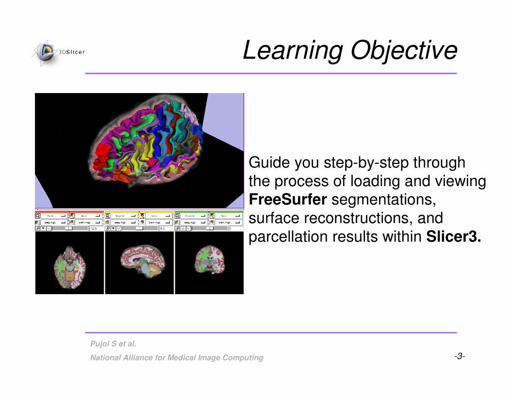
Guide you step-by-step through
the process of loading and viewingFreeSurfer segmentations,
surface reconstructions, and
parcellation results within Slicer3.
Learning Objective

-4-
Pujol S et al.
National Alliance for Medical Image Computing
Prerequisites
This tutorial assumes that you have completed the course
Slicer3Visualization Tutorial.
Tutorials for Slicer3.4 are available on the Slicer101 page:
http://www.slicer.org/slicerWiki/index.php/Slicer3.4:Training#Software_tutorials

-5-
Pujol S et al.
National Alliance for Medical Image Computing
Prerequisites
This tutorial assumes a working
knowledge of how to use FreeSurferto generate segmentation and
surface files.
Tutorials for FreeSurfer are available
at the following location:
http://surfer.nmr.mgh.harvard.edu/fswiki/Tutorials/

-6-
Pujol S et al.
National Alliance for Medical Image Computing
Materials
•This tutorial requires the installation of the Slicer3.4
software and the tutorial dataset.
•Download and install the Slicer3.4 software from the
Slicer web site
http://www.slicer.org/pages/Special:SlicerDownloads
Disclaimer: It is the responsibility of the user of Slicer to comply with both the terms of the license and with the applicable laws, regulations, and rules.

-7-
Pujol S et al.
National Alliance for Medical Image Computing
Materials
This tutorial makes use of the same T1 weighted image dataset (bert) that is
used for the FreeSurfer tutorial available at the following location:
http://surfer.nmr.mgh.harvard.edu/fswiki/FsTutorial
If you already have the FreeSurfer subject ‘bert’ on your computer, then just
download the file ‘slicerGenericScene.mrml’
http://www.na-mic.org/Wiki/index.php/Image:SlicerGenericScene.mrml
If you don’t have the FreeSurfer tutorial dataset known as ‘bert’ on your
computer, then download the archive below:
http://www.na-mic.org/Wiki/index.php/Image:FreeSurferData.tar.gz

-8-
Pujol S et al.
National Alliance for Medical Image Computing
• Brain volumes . . . . . . . . . . . . . . . . . . . . . . . . .
• ASEG volumes . . . . . . . . . . . . . . . . . . . . . . . . . . .
• Surfaces . . . . . . . . . . . . . . . . . . . . . . . . . . . . . . . . . .
• Parcellation Maps . . . . . . . . . . . . . . . . . . . . . . . . . . . .
• All of the above, via a scene file. . . . . . . . . . . . . . .
From FreeSurfer, Slicer3 can load:
Overview
-9-
Pujol S et al.
National Alliance for Medical Image Computing
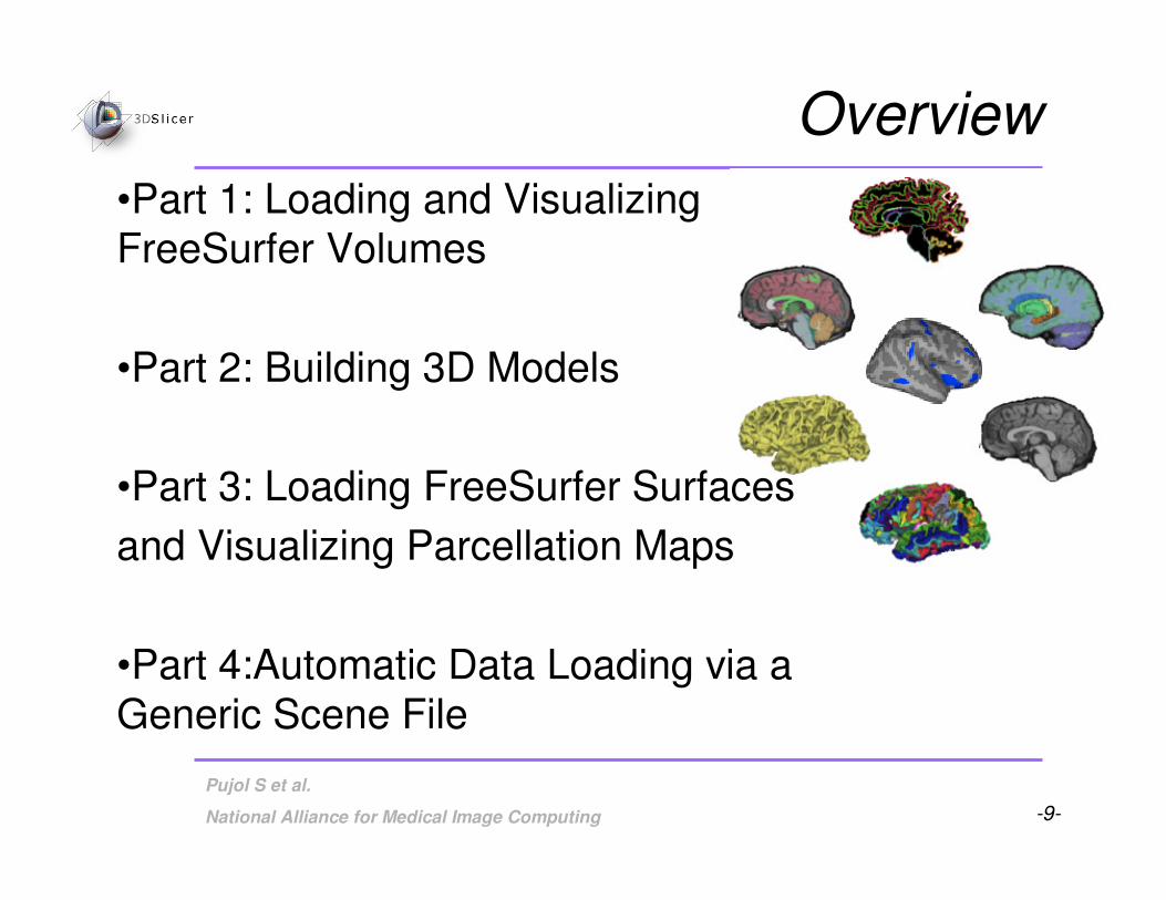
Overview
•Part 1: Loading and VisualizingFreeSurfer Volumes
•Part 2: Building 3D Models
•Part 3: Loading FreeSurfer Surfaces
and Visualizing Parcellation Maps
•Part 4:Automatic Data Loading via a Generic Scene File

-10-
Pujol S et al.
National Alliance for Medical Image Computing
Part 1: Loading and Visualizing FreeSurfer
Volumes

-11-
Pujol S et al.
National Alliance for Medical Image Computing
Loading a Brain File
Intensity corrected
T1 volume
Skull Stripping and Noise Filtering
Watershed Algorithm
brain.mgz
FreeSurfer pipeline

-12-
Pujol S et al.
National Alliance for Medical Image Computing
Loading a Brain File
Select Volumes from
the Module menu

-13-
Pujol S et al.
National Alliance for Medical Image Computing
Loading a Brain File
Click on Select Volume File

-14-
Pujol S et al.
National Alliance for Medical Image Computing
Loading a Brain File
Browse to find the
dataset brain.mgzlocated in the
directory
/subjects/bert/mri/
and click on Open

-15-
Pujol S et al.
National Alliance for Medical Image Computing
Loading a Brain File
Choose Image Origin: Centeredand click Apply

-16-
Pujol S et al.
National Alliance for Medical Image Computing
Loading a Brain File
The volume brain.mgz
appears in the Slice Viewer

-17-
Pujol S et al.
National Alliance for Medical Image Computing
Loading a Brain File
Click on the linksicon to link the three anatomical
slices.
Click on the Slice
Visibility icon to display the slices in
the 3D Viewer

-18-
Pujol S et al.
National Alliance for Medical Image Computing
Loading a Brain File
The three anatomical
slices appear in the 3D Viewer

-19-
Pujol S et al.
National Alliance for Medical Image Computing
Loading an ASEG File
Intensity corrected
T1 volume
Subcortical processing
aseg.mgz
FreeSurfer pipeline
Segmentation

-20-
Pujol S et al.
National Alliance for Medical Image Computing
Loading an ASEG File
Click on Select Volume File, and browse to find
the dataset aseg.mgz located in the directory
/subjects/bert/mri/ and click on Open
-21-
Pujol S et al.
National Alliance for Medical Image Computing
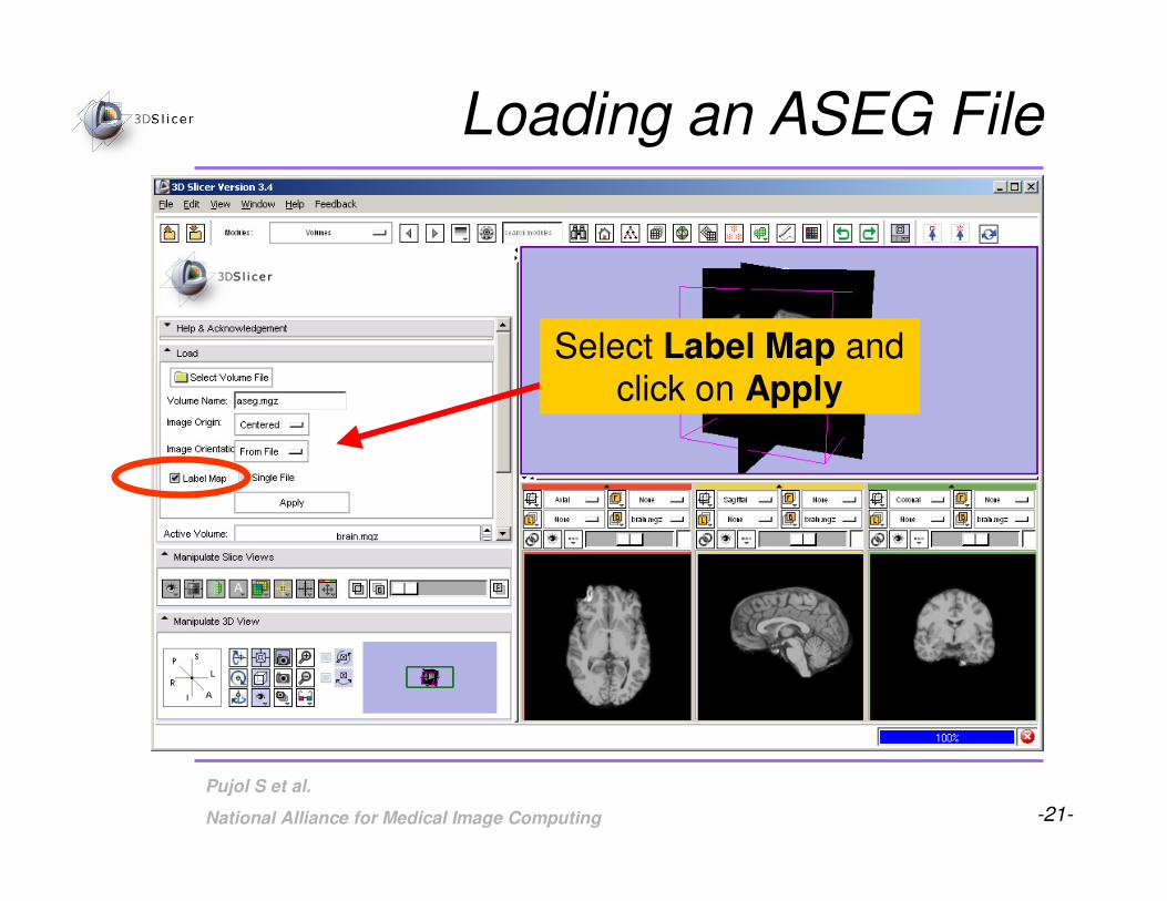
Loading an ASEG File
Select Label Map and
click on Apply

-22-
Pujol S et al.
National Alliance for Medical Image Computing
Loading an ASEG File
The volume aseg.mgz
appears in the Viewer
The labels are
superimposed on the
gray brain images
-23-
Pujol S et al.
National Alliance for Medical Image Computing
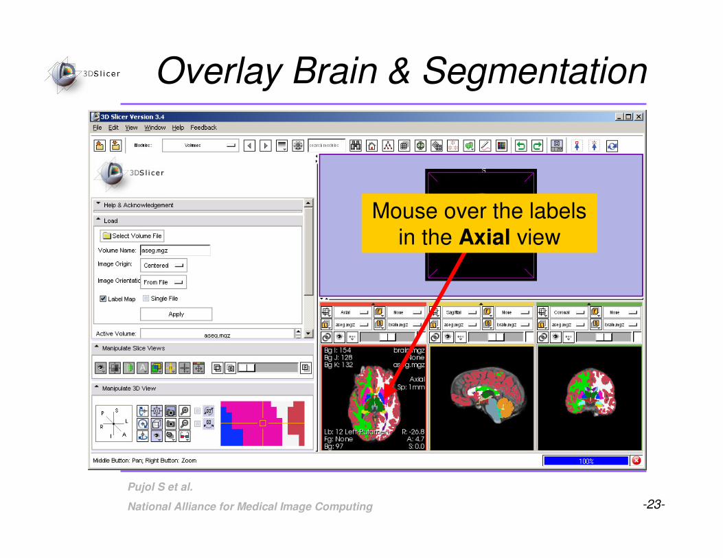
Overlay Brain & Segmentation
Mouse over the labels
in the Axial view

-24-
Pujol S et al.
National Alliance for Medical Image Computing
Overlay Brain & SegmentationOverlay Brain & Segmentation
The names of the
labels appear in the
window

-25-
Pujol S et al.
National Alliance for Medical Image Computing
Overlay Brain & Segmentation
Find the labels corresponding to the
Left Thalamus Proper, the Left
Caudate, and the Left Putamen in
the three anatomical views

-26-
Pujol S et al.
National Alliance for Medical Image Computing
Overlay Brain & Segmentation
Left Thalamus Proper = #10
Left Putamen = #12
Left Caudate = #11

-27-
Pujol S et al.
National Alliance for Medical Image Computing
Part 2: Building
3D Models

-28-
Pujol S et al.
National Alliance for Medical Image Computing
• Building a Single Model
• Building Multiple Models
Building 3D Models

-29-
Pujol S et al.
National Alliance for Medical Image Computing
Building a Single Model
Select the module
Model Maker from the
category Surface
Models

-30-
Pujol S et al.
National Alliance for Medical Image Computing
Building a Single Model
Choose Input Volume:aseg.mgz
Select Models: Create New
ModelHierarchy

-31-
Pujol S et al.
National Alliance for Medical Image Computing
Building a Single Model
Enter right-hippocampus as Model Name and uncheck the
box for Generate All Models

-32-
Pujol S et al.
National Alliance for Medical Image Computing
In the Model Maker Parameters tab, type
in label #53, which
corresponds to the
label for the Right Hippocampus
Building a Single Model

-33-
Pujol S et al.
National Alliance for Medical Image Computing
Turn off Slice visibility
and Click Apply
Building a Single Model

-34-
Pujol S et al.
National Alliance for Medical Image Computing
Building a Single Model
The 3-dimensional model of the Right
Hippocampus appears
in the 3D Viewer

-35-
Pujol S et al.
National Alliance for Medical Image Computing
• Building a Single Model
• Building Multiple Models
Building 3D Models

-36-
Pujol S et al.
National Alliance for Medical Image Computing
Delete the Model Name.
Delete label #53, and set the Start Label to label
#10, which corresponds
to the Left Thalamus
Proper
Set the End Label to label #13, which
corresponds to the LeftPallidum
Check Joint Smoothing
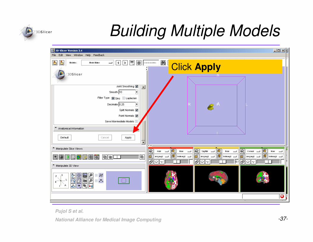
Building Multiple Models
-37-
Pujol S et al.
National Alliance for Medical Image Computing
Click Apply
Building Multiple Models

-38-
Pujol S et al.
National Alliance for Medical Image Computing
Building Multiple Models
The 3-dimensional models of
the Left Thalamus Proper(label #10), Left Caudate (label
#11), Left Putamen (label #12),
and Left Pallidum (label #13)
appear in the 3D Viewer

-39-
Pujol S et al.
National Alliance for Medical Image Computing
Part 3: LoadingFreeSurfer Surfaces and VisualizingParcellation Maps

-40-
Pujol S et al.
National Alliance for Medical Image Computing
Building Multiple Models
Select the module
Models from the
Module menu

-41-
Pujol S et al.
National Alliance for Medical Image Computing
Loading Surfaces
Click on Add 3D model or a
model directory, and click Select Model.

-42-
Pujol S et al.
National Alliance for Medical Image Computing
Loading Surfaces
Browse to find the surface
lh.white located in the directory
/subjects/bert/surf/
Click on Open

-43-
Pujol S et al.
National Alliance for Medical Image Computing
Loading Surfaces
The surface of the White Matter of the
Left Hemisphere
appears in the 3D Viewer

-44-
Pujol S et al.
National Alliance for Medical Image Computing
Click on Add scalar overlay
Visualizing Parcellation Maps

-45-
Pujol S et al.
National Alliance for Medical Image Computing
Click on Select model for overlay and selectlh.white. Click Select
a scalar overlay.
Visualizing Parcellation Maps

-46-
Pujol S et al.
National Alliance for Medical Image Computing
Visualizing Parcellation Maps
Browse to find the Parcellation Map
lh.aparc.annot located in the directory
/subjects/bert/label/
and click on Open

-47-
Pujol S et al.
National Alliance for Medical Image Computing
Visualizing Parcellation Maps
The Parcellation Map
is overlaid on the
White Matter surface
in the 3D Viewer

-48-
Pujol S et al.
National Alliance for Medical Image Computing
Part 4: Automatic Data Loading via a Generic Scene File

-49-
Pujol S et al.
National Alliance for Medical Image Computing
Loading a Generic Scene File
Click on Close Scene in
the File menu to close
the current sceneClick OK to confirm

-50-
Pujol S et al.
National Alliance for Medical Image Computing
Loading a Generic Scene File
• The generic scene file works by looking in the subject
directory created by FreeSurfer, and loading all available volumes and models based on known
subdirectory names and filenames.
• The file slicerGenericScene.mrml will work properly if
the subdirectory names and filenames have not been changed by the user.

-51-
Pujol S et al.
National Alliance for Medical Image Computing
Loading a Generic Scene File
Copy the file slicerGenericScene.mrml into the
directory /subjects/ of our tutorial dataset.
/subjects/

-52-
Pujol S et al.
National Alliance for Medical Image Computing
Loading a Generic Scene File
Copy the file slicerGenericScene.mrml located in the
directory /subjects/, into the directory /subjects/bert/ of our sample subject.
/subjects/bert/

-53-
Pujol S et al.
National Alliance for Medical Image Computing
Loading a Generic Scene File
Rename the file ‘slicerGenericScene.mrml’ located in
the directory /subjects/bert/ ‘slicerBertScene.mrml’
/subjects/bert/

-54-
Pujol S et al.
National Alliance for Medical Image Computing
Loading a Generic Scene File
Click on Load Scene in the Filemenu, and select the scene
slicerBertScene.mrml located in
the directory /subjects/bert/

-55-
Pujol S et al.
National Alliance for Medical Image Computing
Loading a Generic Scene File
The scene appears with a
list of files which have been automatically loaded
from the subject directory
bert.

-56-
Pujol S et al.
National Alliance for Medical Image Computing
Loading a Generic Scene File
Select the mode 3D only layout
from the Viewer menu

-57-
Pujol S et al.
National Alliance for Medical Image Computing
Loading a Generic Scene File
Select the module
Models, and expand the tab Hierarchy &
Display to display the
list of models that
were loaded

-58-
Pujol S et al.
National Alliance for Medical Image Computing
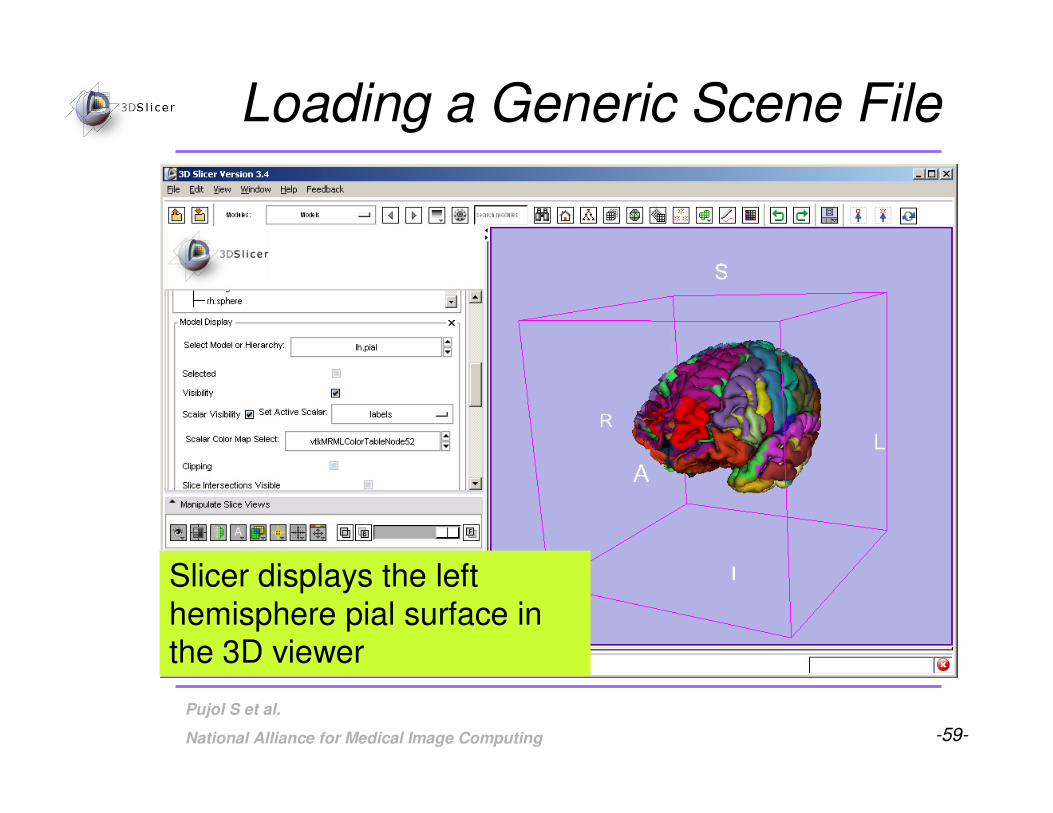
Select the surface lh_pial,
and turn on the visibility of the model
Loading a Generic Scene File
-59-
Pujol S et al.
National Alliance for Medical Image Computing
Loading a Generic Scene File
Slicer displays the left
hemisphere pial surface in the 3D viewer

-60-
Pujol S et al.
National Alliance for Medical Image Computing
The generic scene includes
three snapshots that provide a variety of scene setups:
-Left and Right Annotations
-Left and Right Pial curve
-Left and right white sulc
Loading a Generic Scene File

-61-
Pujol S et al.
National Alliance for Medical Image Computing
Click on the restore snapshots
icon, select the snapshot Left and right white sulc, and
click on restore
Loading a Generic Scene File

-62-
Pujol S et al.
National Alliance for Medical Image Computing
Loading a Generic Scene File
The snapshot displays the
left and right sulci using theGreenRed colorscheme.

-63-
Pujol S et al.
National Alliance for Medical Image Computing
• Brain volumes . . . . . . . . . . . . . . . . . . . . . . . . .
• ASEG volumes . . . . . . . . . . . . . . . . . . . . . . . . . . .
• Surfaces . . . . . . . . . . . . . . . . . . . . . . . . . . . . . . . . . .
• Parcellation Maps . . . . . . . . . . . . . . . . . . . . . . . . . . . .
• All of the above, via a scene file. . . . . . . . . . . . . . .
From FreeSurfer, Slicer3 can load:
Summary

-64-
Pujol S et al.
National Alliance for Medical Image Computing
• 3D visualization of brain segmented surfaces and parcellation maps
• Intuitive graphical user interface to interact withFreeSurfer data
• Multi platforms open-source environment
Conclusion