380816 pdfconv 393606 0A8B2BC0-4449-11E5-98B5 …
38
UC Irvine UC Irvine Electronic Theses and Dissertations Title Comparing Conventional and Valveless Trocar Insufflation During Laparoscopic Renal Surgery Permalink https://escholarship.org/uc/item/6dx8t1cj Author Bucur, Philip Laurence Publication Date 2015 Peer reviewed|Thesis/dissertation eScholarship.org Powered by the California Digital Library University of California
Transcript of 380816 pdfconv 393606 0A8B2BC0-4449-11E5-98B5 …

UC IrvineUC Irvine Electronic Theses and Dissertations
TitleComparing Conventional and Valveless Trocar Insufflation During Laparoscopic Renal Surgery
Permalinkhttps://escholarship.org/uc/item/6dx8t1cj
AuthorBucur, Philip Laurence
Publication Date2015 Peer reviewed|Thesis/dissertation
eScholarship.org Powered by the California Digital LibraryUniversity of California

UNIVERSITY OF CALIFORNIA,
IRVINE
Comparing Conventional and Valveless Trocar Insufflation During Laparoscopic Renal
Surgery
THESIS
submitted in partial satisfaction of the requirements
for the degree of
MASTER OF SCIENCE
In Biomedical and Translational Science
by
Philip Bucur
Thesis Committee:
Clinical Professor Jaime Landman, Chair
Associate Professor Sheldon Greenfield
Assistant Professor John Billimek
2015

Copyright 2015 Philip Bucur

ii
DEDICATION
To
my parents, friends, co-workers, and mentors
in recognition of the support and opportunity I have been given daily
Thank you

iii
TABLE OF CONTENTS
Page
LIST OF FIGURES iv
LIST OF TABLES v
ACKNOWLEDGMENTS vi
ABSTRACT OF THE THESIS vii
INTRODUCTION 1
CHAPTER 1: Methods 4
CHAPTER 2: Results 9
CHAPTER 3: Discussion 23
REFERENCES (OR BIBLIOGRAPHY) 28

iv
LIST OF FIGURES
Page
Figure 2.1 Average Variation in Pneumoperitoneum 12

v
LIST OF TABLES
Page
Table 2.1 Patient Characteristics and Procedures 9
Table 2.2 Comparison of Intra-abdominal Pressure Variation 11
Table 2.3 Intraoperative Outcomes 14
Table 2.4 Postoperative Outcomes 15
Table 2.5 Comparison of Ventilatory Parameters 17
Table 2.6 Comparison of Hemodynamic Parameters 20

vi
ACKNOWLEDGMENTS
I would like to express the deepest appreciation to my committee chair, Professor Jaime
Landman, who continues to inspire me daily with his creativity and passion for research
and Urology.
I would like to thank my committee members, Professor Sheldon Greenfield and Professor
John Billimek, for their teaching, guidance, and mentorship during the MS-BATS program.
I have no financial disclosures as all research was performed at the University of California,
Irvine using anesthesia equipment supplied in the operating room.

vii
ABSTRACT OF THE THESIS
Comparing Conventional and Valveless Trocar Insufflation During Laparoscopic Renal
Surgery
By
Philip Bucur
Master of Science – Biomedical and Translational Science
University of California, Irvine, 2015
Professor Jaime Landman, Chair
We compared the variation in pneumoperitoneum, physiologic effects, and
postoperative outcomes of patients undergoing laparoscopic renal surgery using a
conventional insufflation system (CI) versus the valveless trocar insufflation (VI) system.
This is a prospective, randomized comparative study with fifty-six patients
undergoing laparoscopic renal surgery with valveless trocar insufflation or conventional
insufflation. Patients in the valveless insufflation arm (n=28) underwent surgery using the
AirSeal valveless trocar insufflation system whereas patients in the conventional treatment
arm (n=28) underwent surgery using standard laparoscopic trocars connected to a Storz
insufflator with the insufflation pressure set to 15 mm Hg. We compared the groups with
respect to stability of pneumoperitoneum, intraoperative and postoperative outcomes, and
physiologic parameters.
The coefficient of variation in pressures was significantly lower in the valveless
trocar group compared to the conventional treatment group (7.9% vs. 15.6%, p<0.001)
with significantly less time spent above insufflation pressures of 20 mm Hg. Estimated

viii
blood loss was significantly higher in the valveless trocar group than conventional group
(155 vs. 75 cc, p=0.03). End-tidal CO2 (ET CO2) was significantly lower at 10 minutes (34.3
vs. 36.6 mmHg, p=0.029) and 25 minutes (35.8 vs. 37.6 mmHg, p=0.047) in the valveless
trocar group compared to the conventional treatment group. There were no other
significant differences across physiologic parameters or outcomes.
In conclusion, compared with a conventional insufflation system, the valveless
trocar insufflation system provides a significantly more stable pneumoperitoneum during
laparoscopic renal surgery and lower end-tidal CO2 at 10 minutes, but with an increased
risk of blood loss.
1
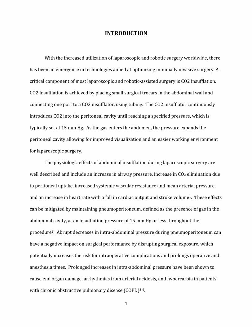
INTRODUCTION
With the increased utilization of laparoscopic and robotic surgery worldwide, there
has been an emergence in technologies aimed at optimizing minimally invasive surgery. A
critical component of most laparoscopic and robotic-assisted surgery is CO2 insufflation.
CO2 insufflation is achieved by placing small surgical trocars in the abdominal wall and
connecting one port to a CO2 insufflator, using tubing. The CO2 insufflator continuously
introduces CO2 into the peritoneal cavity until reaching a specified pressure, which is
typically set at 15 mm Hg. As the gas enters the abdomen, the pressure expands the
peritoneal cavity allowing for improved visualization and an easier working environment
for laparoscopic surgery.
The physiologic effects of abdominal insufflation during laparoscopic surgery are
well described and include an increase in airway pressure, increase in CO2 elimination due
to peritoneal uptake, increased systemic vascular resistance and mean arterial pressure,
and an increase in heart rate with a fall in cardiac output and stroke volume1. These effects
can be mitigated by maintaining pneumoperitoneum, defined as the presence of gas in the
abdominal cavity, at an insufflation pressure of 15 mm Hg or less throughout the
procedure2. Abrupt decreases in intra-abdominal pressure during pneumoperitoneum can
have a negative impact on surgical performance by disrupting surgical exposure, which
potentially increases the risk for intraoperative complications and prolongs operative and
anesthesia times. Prolonged increases in intra-abdominal pressure have been shown to
cause end organ damage, arrhythmias from arterial acidosis, and hypercarbia in patients
with chronic obstructive pulmonary disease (COPD)3-6.

2
Since 2007, there have been two types of insufflation devices, which have been used
routinely in laparoscopic surgery, conventional automated insufflation (CI) and valveless
trocar insufflation (VI). The mechanical insufflator was introduced in 1960 by Semm and
colleagues, which eventually gave rise to conventional automated insufflation after
laparoscopy became widely accepted in the 1980’s7. CI uses a one-way valve trocar, which
allows instruments to be passed in and out of the peritoneum. An advantage of CI has been
the familiarity of the device amongst more experienced surgeons and lower cost in
comparison to the valveless trocar insufflator. An adverse effect of CI has been the loss of
gas in the abdomen when CO2 escapes as instruments are passed through the trocar or
when suction is used. Additionally, CI with one-way trocars is associated with moisture
accumulation at the camera lens and the need for surgical smoke plume evacuation with a
suction device, or manual venting into the operating room through the stopcock of a
conventional trocar.
A valveless trocar insufflation system (AirSeal, SurgiQuest, Milford, CT) was
developed in 2007 designed to improve upon the difficulties associated with CI. The goal of
the newer system was to maintain a stable pneumoperitoneum as instruments are passed
through the trocars by using a pressure barrier, which expels CO2 into the environment if
intra-abdominal CO2 levels are too high. In addition, the VI continuously evacuates smoke
without the need of an additional suctioning device or manual venting into the operating
room8. Other advantages, in initial evidence in both retrospective and prospective, non-
randomized studies, has suggested VI may lower the rate of CO2 uptake, decrease the
volume of CO2 consumed, and decrease operative time9-11. Disadvantages of VI have been
less familiarity among experienced surgeons, increased cost, and initial retrospective

3
evidence suggesting blunting of end tidal CO2 levels may mask detection of intraoperative
pneumothorax11.
There is increasing pressure for medical device companies to compete with existing
technology. Therefore, new devices enter the market with FDA approval but with limited
data and few head to head comparisons against the current standard of care. Frequently in
surgery, the differences in devices are not truly understood by the operator and selection
of the device is driven by comfort or the hospital’s accessibility to the device. Laparoscopic
insufflators have been traditionally used for the same reasons, driven by surgeon
familiarity and availability. The performance of the insufflators and the impact of this
performance on patient’s outcomes have yet to be compared in vivo in a randomized,
comparative study. The primary aim of this study is to investigate how well
pneumoperitoneum is maintained during laparoscopic surgery by comparing the variation
in intra-abdominal pressure when using CI compared to VI. The secondary aims of this
study are to investigate the physiologic impact, intraoperative outcomes, and post-
operative outcomes of CI compared to VI.
4
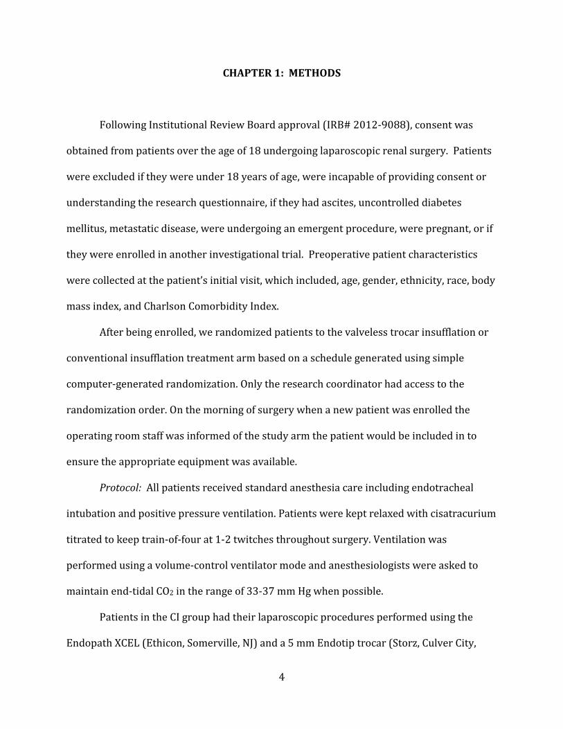
CHAPTER 1: METHODS
Following Institutional Review Board approval (IRB# 2012-9088), consent was
obtained from patients over the age of 18 undergoing laparoscopic renal surgery. Patients
were excluded if they were under 18 years of age, were incapable of providing consent or
understanding the research questionnaire, if they had ascites, uncontrolled diabetes
mellitus, metastatic disease, were undergoing an emergent procedure, were pregnant, or if
they were enrolled in another investigational trial. Preoperative patient characteristics
were collected at the patient’s initial visit, which included, age, gender, ethnicity, race, body
mass index, and Charlson Comorbidity Index.
After being enrolled, we randomized patients to the valveless trocar insufflation or
conventional insufflation treatment arm based on a schedule generated using simple
computer-generated randomization. Only the research coordinator had access to the
randomization order. On the morning of surgery when a new patient was enrolled the
operating room staff was informed of the study arm the patient would be included in to
ensure the appropriate equipment was available.
Protocol: All patients received standard anesthesia care including endotracheal
intubation and positive pressure ventilation. Patients were kept relaxed with cisatracurium
titrated to keep train-of-four at 1-2 twitches throughout surgery. Ventilation was
performed using a volume-control ventilator mode and anesthesiologists were asked to
maintain end-tidal CO2 in the range of 33-37 mm Hg when possible.
Patients in the CI group had their laparoscopic procedures performed using the
Endopath XCEL (Ethicon, Somerville, NJ) and a 5 mm Endotip trocar (Storz, Culver City,

5
CA), with insufflation provided via a 12 mm assistant port connected to a standard
insufflator (Storz, Culver City, CA). In the AirSeal valveless trocar treatment arm,
insufflation was provided via a 12 mm AirSeal Access Port (SurgiQuest, Milford, CT)
connected to an AirSeal IFS insufflator (SurgiQuest, Milford, CT), with Endopath XCEL and
5mm Endotip trocar used for assistant and other instrument ports. A single fellowship-
trained laparoscopic surgeon at one institution performed all surgical procedures.
Primary Outcome: The primary outcome for the study was variabilty in
pneumoperitoneum around the standard 15 mm Hg set point during all laparoscopic cases.
The device with less variability in pneumoperitoneum was considered to have higher
intraoperative performance. True insufflation pressure was measured by an independent
pressure transducer connected to a side port of a non-insufflating trocar. Data from this
transducer were continuously recorded throughout the case using custom data collection
software. Variability was assessed in two different ways. First, we computed the mean
coefficient of variation by taking the coefficient of variation of within each individual case
for each group and than calculating the mean coefficient over variation across all cases for
each group. Second, we computed the percentage of time spent within three insufflation
pressure ranges defined a priori as “acceptable” (12-18 mm Hg), “borderline” (10-12 and
18-20 mm Hg), and “unacceptable” (less than 10 mm Hg and greater than 20 mm Hg).
Secondary Outcomes: Secondary outcomes collected during the study included
intraoperative outcomes, postoperative outcomes, and physiologic parameters.
Intraoperative outcomes collected include duration of surgery, estimated blood loss, urine
output, lack of pneumoperitoneum, passage of instruments, cleaning of the camera lens,
and smoke evacuations. Intraoperative complications were reviewed from the surgery
6
dictation postoperatively. Surgeon assessment of the image quality was documented
during each procedure.
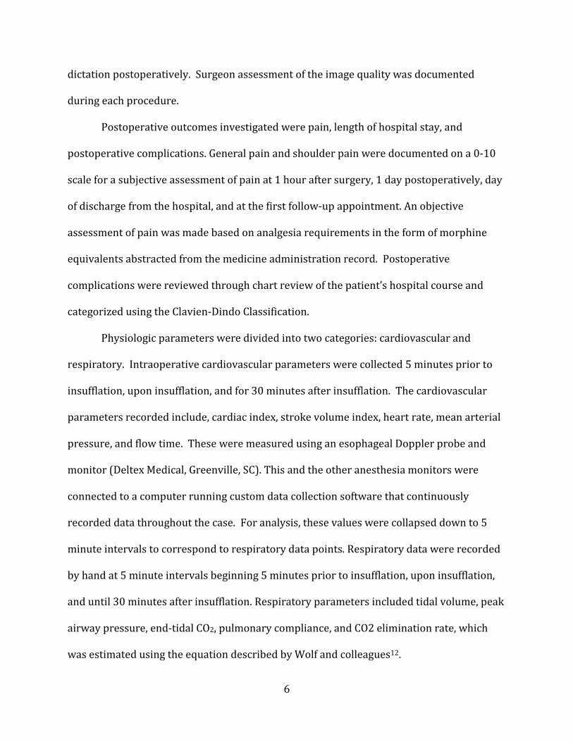
Postoperative outcomes investigated were pain, length of hospital stay, and
postoperative complications. General pain and shoulder pain were documented on a 0-10
scale for a subjective assessment of pain at 1 hour after surgery, 1 day postoperatively, day
of discharge from the hospital, and at the first follow-up appointment. An objective
assessment of pain was made based on analgesia requirements in the form of morphine
equivalents abstracted from the medicine administration record. Postoperative
complications were reviewed through chart review of the patient’s hospital course and
categorized using the Clavien-Dindo Classification.
Physiologic parameters were divided into two categories: cardiovascular and
respiratory. Intraoperative cardiovascular parameters were collected 5 minutes prior to
insufflation, upon insufflation, and for 30 minutes after insufflation. The cardiovascular
parameters recorded include, cardiac index, stroke volume index, heart rate, mean arterial
pressure, and flow time. These were measured using an esophageal Doppler probe and
monitor (Deltex Medical, Greenville, SC). This and the other anesthesia monitors were
connected to a computer running custom data collection software that continuously
recorded data throughout the case. For analysis, these values were collapsed down to 5
minute intervals to correspond to respiratory data points. Respiratory data were recorded
by hand at 5 minute intervals beginning 5 minutes prior to insufflation, upon insufflation,
and until 30 minutes after insufflation. Respiratory parameters included tidal volume, peak
airway pressure, end-tidal CO2, pulmonary compliance, and CO2 elimination rate, which
was estimated using the equation described by Wolf and colleagues12.
7
Statistical Analysis: This study was powered to our primary outcome. The target
sample size for this study was 60 patients, 30 patients in both arms, which was calculated
using a 92% power to achieve a 0.05 significance level to a ratio of 3.6 between the
variances of the pressure measurements. All Statistical analysis was performed using SPSS
software (IBM, Armonk, NY) and variables were considered significant with a p-value <
0.05. All preoperative patient characteristics and procedures performed were analyzed
using an unpaired T test and Fisher exact test. To determine the primary outcome, the
mean coefficient of variation each group was determined and compared using an F-test.
Percentage of time spent within each pressure range and median number of pressure
spikes per case of each group were calculated and compared between groups using a
Wilcox-Mann-Whitney test. All physiologic parameters were compared at each 5 minute
interval between groups using an unpaired T test. Intraoperative and postoperative
outcomes were analyzed using an unpaired T test was used for continuous variables and a
Fisher exact test was used for categorical variables.

8
CHAPTER 2: RESULTS
A total of 60 patients met the inclusion criteria and were included in this study;
there were 30 patients in each arm. 3 patients were excluded following surgery due to
metastatic disease found intraoperatively. 1 patient had no intraoperative data captured
from both respiratory and cardiovascular devices and was excluded. The 56 patients who
were used for analysis included 28 patients in each arm. Patient demographic and surgery
data is presented in Table 2.1. There were no statistically significant differences between
groups when analyzing patient characteristics and procedures performed. Of these, 20
patients underwent laparoscopic partial nephrectomy, followed by 17 radical
nephrectomies, 9 cryoablation procedures, 4 nephroureterectomies, 3 pyeloplasties, 1
simple nephrectomy, 1 retroperitoneal mass excision, and 1 ureteral re-implant. There
were 41 males and 15 females with a mean age of 63.7 years. The mean body mass index
(BMI) was 28.3 and mean Charlson Comorbidity Index was 3.4.

9
Table 2.1: Patient characteristics and procedures performed
Variable Overall
N=56
VI
n=28
CI
N=28
P-Value
Age, mean (sd) 63.7 (14.1) 62.5 (15.3) 64.8 (13.1) .544
BMI, mean (sd) 28.3 (5.2) 27.9 (6.2) 28.7 (4.1) .580
CCI, mean (sd) 3.4 (2.1) 3.6 (2.4) 3.2 (1.7) .408
Gender, n (%)
Male
Female
41 (73.2)
15 (26.8)
23 (82.1)
5 (17.9)
18 (64.3)
10 (35.7)
.227
Ethnicity, n (%)
Hispanic
Non-Hispanic
3 (5.4)
53 (94.6)
1 (3.6)
27 (96.4)
2 (7.1)
26 (92.9)
.500
Race, n (%)
Caucasian
Black
Asian
Hawaiian or PI
Other
46 (82.1)
2 (3.6)
3 (5.4)
1 (1.8)
4 (7.1)
22 (78.6)
1 (3.6)
2 (7.1)
1 (3.6)
2 (7.1)
24 (85.7)
1 (3.6)
1 (3.6)
0 (0.0)
2 (7.1)
.841
Treatment, n (%)
Radical nephrectomy
Simple nephrectomy
Partial nephrectomy
Nephroureterectomy
Cryoablation
Pyeloplasty
Ureteral implantation
Retroperitoneal mass
excision
17 (30.4)
1 (1.8)
20 (35.7)
4 (7.1)
9 (16.1)
3 (5.4)
1 (1.8)
1 (1.8)
9 (32.1)
0 (0.0)
9 (32.1)
3 (10.7)
4 (14.3)
2 (7.1)
1 (3.6)
0 (0.0)
8 (28.6)
1 (3.6)
11 (39.3)
1 (3.6)
5 (17.9)
1 (3.6)
0 (0.0)
1 (3.6)
.696
Primary Outcome: All 56 cases were included in analysis of the primary outcome.
There was significantly less variability in pressure readings with a lower mean coefficient
of variation during VI compared to CI (7.9% vs. 15.6%, p<0.001) (Table 2.2). The average
variability between groups can be further illustrated across all 56 cases in Figure 2.1. There
was significantly less time spent within the ‘borderline’ range with pressure readings ≥ 18
mm Hg (0.2% vs. 9.2%, p<0.0005) and ≤ 12 mm Hg (12.5% vs. 12.9%, p=0.013) during VI

10
compared to CI. Additionally, there was significantly less time spent with pressure readings
in the ‘unacceptable’ range of ≥ 20 mm Hg (0.1% vs. 2.1%, p<0.0005) and ≤ 10 mm Hg
(1.8% vs. 7.2%, p<0.0005) during the cases with VI compared to CI. There were
significantly fewer median pressure spikes above 20 mm Hg when using VI compared to CI
(0 vs. 16, p<0.0005) (Table 2.2).

11
Table 2.2: Comparing intra-abdominal pressure variation and percentage of time in each
pressure range between conventional and valveless trocar insufflation.
Valveless
Insufflation
n=28
Conventional
Insufflation
n=28
P-value
Intra-abdominal pressure variation
Mean pressure, mean (sd) 14.0 (1.3) 14.7 (1.7)
Mean SD (per patient), mean (sd) 1.1 (0.4) 2.3 (0.7)
Mean coefficient of variation,
mean (sd)
7.9 (3.1) 15.6 (5.3) <0.001
Percentage of operative time in each pressure range
Pressures ≥18 mm Hg, mean %
(sd)
0.2 (0.8) 9.2 (14.2) <0.0005
Pressures ≤12 mm Hg, mean %
(sd)
12.5 (25.5) 12.9 (20.3) 0.013
Pressures ≥20 mm Hg, mean %
(sd)
0.1 (0.2) 2.1 (7.0) <0.0005
Pressures ≤10 mm Hg, mean %
(sd)
1.8 (4.8) 7.2 (18.0) <0.0005
Spikes >20 mm Hg, median
number per case
0 16 <0.0005

12
Figure: 2.1: The average variation in pneumoperitoneum between conventional
insufflation (A) and valveless trocar insufflation (B) for the first 30 minutes of all 56
laparoscopic cases.

13
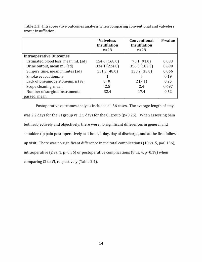
Secondary Outcomes: Intraoperative outcomes analysis included all 56 patients.
Estimated blood loss was significantly higher in the VI group compared to the CI group
(155 vs. 75 mL, p=0.033). There were no significant differences in urine output (356 vs.
334 mL, p=0.69) or surgery length (151 vs. 130 mins, p=0.066) between the VI group and
CI group, respectively. There were no significant differences in the mean number of
laparoscope cleanings (2.5 vs. 2.4, p=0.697) or mean number of surgical instruments
passed through the trocars (32.4 vs. 17.4, p=0.51) between the VI and CI groups,
respectively. The CI group required smoke evacuation in 5 cases compared to 1 case in the
VI group (p=0.19). Pneumoperitoneum was lost in 2 cases in the CI group compared to
none in the VI group, (p=0.25; see Table 2.3). Surgeon assessment noted less smoke in the
surgical field of view, superior image quality, and less suctioning needed by the first
assistant when using VI insufflation compared to CI insufflation.
14
Table 2.3: Intraoperative outcomes analysis when comparing conventional and valveless
trocar insufflation.
Valveless
Insufflation
n=28
Conventional
Insufflation
n=28
P-value
Intraoperative Outcomes
Estimated blood loss, mean mL (sd) 154.6 (168.0) 75.1 (91.0) 0.033
Urine output, mean mL (sd) 334.1 (224.0) 356.0 (182.3) 0.690
Surgery time, mean minutes (sd) 151.3 (48.0) 130.2 (35.0) 0.066
Smoke evacuations, n 1 5 0.19
Lack of pneumoperitoneum, n (%) 0 (0) 2 (7.1) 0.25
Scope cleaning, mean 2.5 2.4 0.697
Number of surgical instruments
passed, mean
32.4 17.4 0.52
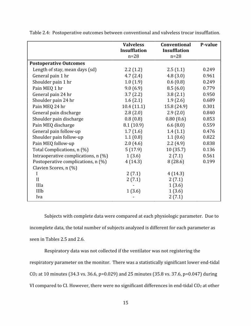
Postoperative outcomes analysis included all 56 cases. The average length of stay
was 2.2 days for the VI group vs. 2.5 days for the CI group (p=0.25). When assessing pain
both subjectively and objectively, there were no significant differences in general and
shoulder-tip pain post-operatively at 1 hour, 1 day, day of discharge, and at the first follow-
up visit. There was no significant difference in the total complications (10 vs. 5, p=0.136),
intraoperative (2 vs. 1, p=0.56) or postoperative complications (8 vs. 4, p=0.19) when
comparing CI to VI, respectively (Table 2.4).
15
Table 2.4: Postoperative outcomes between conventional and valveless trocar insufflation.
Valveless
Insufflation
n=28
Conventional
Insufflation
n=28
P-value
Postoperative Outcomes
Length of stay, mean days (sd) 2.2 (1.2) 2.5 (1.1) 0.249
General pain 1 hr 4.7 (2.4) 4.8 (3.0) 0.961
Shoulder pain 1 hr 1.0 (1.9) 0.6 (0.8) 0.249
Pain MEQ 1 hr 9.0 (6.9) 8.5 (6.0) 0.779
General pain 24 hr 3.7 (2.2) 3.8 (2.1) 0.950
Shoulder pain 24 hr 1.6 (2.1) 1.9 (2.6) 0.689
Pain MEQ 24 hr 10.4 (11.1) 15.8 (24.9) 0.301
General pain discharge 2.8 (2.0) 2.9 (2.0) 0.840
Shoulder pain discharge 0.8 (0.8) 0.80 (0.6) 0.853
Pain MEQ discharge 8.1 (10.9) 6.6 (8.0) 0.559
General pain follow-up 1.7 (1.6) 1.4 (1.1) 0.476
Shoulder pain follow-up 1.1 (0.8) 1.1 (0.6) 0.822
Pain MEQ follow-up 2.0 (4.6) 2.2 (4.9) 0.838
Total Complications, n (%) 5 (17.9) 10 (35.7) 0.136
Intraoperative complications, n (%) 1 (3.6) 2 (7.1) 0.561
Postoperative complications, n (%) 4 (14.3) 8 (28.6) 0.199
Clavien Scores, n (%)
I
II
IIIa
IIIb
Iva
2 (7.1)
2 (7.1)
-
1 (3.6)
-
4 (14.3)
2 (7.1)
1 (3.6)
1 (3.6)
2 (7.1)
Subjects with complete data were compared at each physiologic parameter. Due to
incomplete data, the total number of subjects analyzed is different for each parameter as
seen in Tables 2.5 and 2.6.
Respiratory data was not collected if the ventilator was not registering the
respiratory parameter on the monitor. There was a statistically significant lower end-tidal
CO2 at 10 minutes (34.3 vs. 36.6, p=0.029) and 25 minutes (35.8 vs. 37.6, p=0.047) during
VI compared to CI. However, there were no significant differences in end-tidal CO2 at other

16
time points between the VI and CI groups. There were also no significant differences in
peak airway pressure, tidal volume, pulmonary compliance, and CO2 elimination rate
between the two groups (Table 2.5).
Cardiovascular data was lost as a result of poor esophageal probe placement and
loss of signal during procedures due to factors including interference from electrocautery
devices, patient repositioning, or movement of the probe. In the patients with complete
data, there were no significant differences in cardiac index, stroke volume index, heart rate,
mean arterial pressure, and flow time between the two groups (Table 2.6).

17
Table 2.5: Ventilation parameters compared between conventional and valveless trocar
insufflation prior to insufflation (-5), at insufflation (0), and post-insufflation (5-30) for 30
minutes. TV - tidal volume, Peak – peak airway pressure, ET CO2 – end-tidal CO2, MV –
minute ventilation, Comp – pulmonary compliance, CO2 Elim – CO2 elimination rate
Ventilation
Parameter at Time,
units
Group N Mean Std. Deviation P-Value
TV -5, mL Valveless 26 532.8 70.5 .303
Conventional 25 556.0 87.8
TV 0, mL Valveless 27 526.9 70.3 .051
Conventional 28 572.5 95.9
TV 5, mL Valveless 27 520.9 73.8 .159
Conventional 27 555.0 100.0
TV 10, mL Valveless 27 541.0 85.4 .640
Conventional 27 552.9 100.0
TV 15, mL Valveless 28 538.6 80.0 .321
Conventional 28 562.6 98.3
TV 20, mL Valveless 28 521.4 118.1 .236
Conventional 28 557.5 107.3
TV 25, mL Valveless 28 547.7 76.0 .809
Conventional 28 553.4 99.3
TV 30, mL Valveless 28 558.1 75.5 .656
Conventional 28 569.0 103.5
Peak -5, cmH20 Valveless 19 19.4 4.9 .309
Conventional 19 21.1 5.4
Peak 0, cmH20 Valveless 20 20.2 5.0 .140
Conventional 23 22.5 4.9
Peak 5, cmH20 Valveless 26 23.8 4.1 .114
Conventional 26 25.9 5.3
Peak 10, cmH20 Valveless 26 24.0 4.1 .128
Conventional 26 27.0 5.3
Peak 15, cmH20 Valveless 27 23.9 4.1 .120
Conventional 27 26.6 4.0
Peak 20, cmH20 Valveless 27 23.6 5.5 .138
Conventional 27 26.3 4.0
Peak 25, cmH20 Valveless 27 24.8 4.3 .112
Conventional 28 26.6 4.1
Peak 30, cmH20 Valveless 27 24.7 3.9 .063
Conventional 28 26.6 3.6

18
ET C02 -5, mm Hg Valveless 26 32.3 2.6 .156
Conventional 26 33.8 4.9
ET C02 0, mmHg Valveless 26 32.7 2.5 .375
Conventional 28 33.5 3.9
ET CO2 5, mmHg Valveless 27 33.0 4.3 .156
Conventional 27 34.6 4.0
ET CO2 10, mm Hg Valveless 27 34.3 4.2 .029
Conventional 27 36.6 3.4
ET CO2 15, mm Hg Valveless 28 35.4 3.6 .158
Conventional 28 36.7 3.3
ET CO2 20, mm Hg Valveless 28 35.6 3.4 .107
Conventional 28 37.0 2.9
ET CO2 25 mm Hg Valveless 28 35.8 3.4 .047
Conventional 28 37.6 3.3
ET CO2 30, mm Hg Valveless 28 35.9 3.2 .156
Conventional 28 37.3 3.9
MV -5, mL/min Valveless 26 5781.2 1176.0 .449
Conventional 23 6072.4 1493.0
MV 0, mL/min Valveless 27 5677.0 1169.7 .412
Conventional 26 6009.9 1717.5
MV 5, mL/min Valveless 27 5777.2 1053.1 .980
Conventional 26 5768.2 1536.2
MV 10, mL/min Valveless 27 6072.3 1201.3 .558
Conventional 26 5826.7 1784.8
MV 15, mL/min Valveless 28 6041.0 1143.1 .714
Conventional 27 5896.0 1725.6
MV 20, mL, min Valveless 28 5814.5 1482.3 .988
Conventional 27 5821.4 1807.0
MV 25, mL/min Valveless 28 6144.0 1050.3 .416
Conventional 28 5826.5 1762.0
MV 30, mL/min Valveless 28 6285.2 1104.6 .617
Conventional 28 6093.0 1691.1
Comp -5, L/cmH20 Valveless 19 29.1 6.5 .519
Conventional 19 27.7 7.4
Comp 0, L/cmH20 Valveless 21 27.2 7.9 .952
Conventional 23 27.0 6.6
Comp 5 L/cmH20 Valveless 26 22.6 4.2 .696
Conventional 26 22.1 5.0
Comp 10 L/cmH20 Valveless 26 22.9 5.2 .257

19
Conventional 26 21.2 5.3
Comp 15 L/cmH20 Valveless 27 22.9 4.8 .290
Conventional 27 21.5 4.4
Comp 20, L/cmH20 Valveless 27 22.3 5.1 .530
Conventional 27 21.4 4.8
Comp 25, L/cmH20 Valveless 27 22.5 4.5 .334
Conventional 28 21.2 5.0
Comp 30 L/cmH20 Valveless 27 22.9 4.4 .400
Conventional 28 21.8 5.3
CO2 Elim -5,
mL/min
Valveless 26 2.98 0.6 .308
Conventional 27 3.18 0.7
CO2 Elim 0, mL/min Valveless 27 2.97 0.6 .541
Conventional 27 3.08 0.7
CO2 Elim 5, mL/min Valveless 28 3.06 0.8 .924
Conventional 27 3.04 0.7
CO2 Elim 10,
mL/min
Valveless 26 3.33 0.8 .528
Conventional 28 3.21 0.7
CO2 Elim 15,
mL/min
Valveless 27 3.46 1.0 .494
Conventional 27 3.30 0.7
CO2 Elim 20,
mL/min
Valveless 28 3.33 1.1 .811
Conventional 27 3.27 0.7
CO2 Elim 25,
mL/min
Valveless 28 3.55 0.9 .358
Conventional 27 3.35 0.7
CO2 Elim 30,
mL/min
Valveless 27 3.64 0.9 .449
Conventional 26 3.47 0.7

20
Table 2.6: Hemodynamic parameters compared between conventional and valveless trocar
insufflation prior to insufflation (-5), at insufflation (0), and post-insufflation (5-30) for 30
minutes. CI – cardiac index, SVI – stroke volume index, HR – heart rate, MAP – mean
arterial pressure, FT – flow time.
Hemodynamic
Parameter at
Time, units
Group N Mean Std.
Deviation
P-Value
CI -5, L/min/m2 Valveless 17 5.1 1.7 .967
Conventional 17 5.1 1.4
CI 0, L/min/m2 Valveless 21 4.5 1.3 .492
Conventional 18 4.8 1.1
CI 5, L/min/m2 Valveless 17 4.1 1.1 .927
Conventional 14 4.1 .9
CI 10, L/min/m2 Valveless 21 4.4 1.4 .519
Conventional 16 4.7 1.0
CI 15, L/min/m2 Valveless 22 4.4 1.7 .596
Conventional 23 4.6 1.9
CI 20, L/min/m2 Valveless 22 4.9 1.3 .968
Conventional 23 4.9 1.3
CI 25, L/min/m2 Valveless 20 4.7 1.2 .343
Conventional 23 5.1 1.4
CI 30, L/min/m2 Valveless 20 4.7 1.2 .154
Conventional 24 5.3 1.5
SVI -5, ml/m2/beat Valveless 18 37.9 7.8 .524
Conventional 17 40.1 12.0
SVI 0, ml/m2/beat Valveless 22 35.3 10.6 .367
Conventional 19 38.2 9.3
SVI 5, ml/m2/beat Valveless 21 29.5 6.4 .937
Conventional 17 29.6 7.1
SVI 10,
ml/m2/beat
Valveless 23 34.1 7.8 .390
Conventional 19 31.5 11.3
SVI 15,
ml/m2/beat
Valveless 26 30.8 13.6 .073
Conventional 23 37.0 9.6
SVI 20,
ml/m2/beat
Valveless 24 36.6 8.2 .823
Conventional 24 37.2 9.2
SVI 25,
ml/m2/beat
Valveless 21 37.2 7.9 .679
Conventional 24 38.5 12.4
SVI 30,
ml/m2/beat
Valveless 21 36.9 10.9 .482
Conventional 26 39.2 11.7

21
HR -5, beats/min Valveless 17 61.4 9.6 .323
Conventional 19 65.1 12.3
HR 0, beats/min Valveless 21 62.7 10.6 .509
Conventional 21 65.0 12.1
HR 5, beats/min Valveless 24 66.2 12.1 .882
Conventional 21 65.7 11.8
HR 10, beats/min Valveless 26 65.5 11.3 .437
Conventional 20 68.4 13.6
HR 15, beats/min Valveless 27 62.8 17.3 .453
Conventional 21 66.2 11.8
HR 20, beats/min Valveless 26 65.7 12.7 .976
Conventional 22 65.6 11.7
HR 25, beats/min Valveless 25 65.7 12.4 .827
Conventional 23 65.0 11.0
HR 30, beats/min Valveless 26 59.3 21.3 .243
Conventional 23 65.2 11.5
MAP -5, mm Hg Valveless 19 78.4 10.0 .115
Conventional 20 72.9 11.3
MAP 0, mm Hg Valveless 24 79.2 13.7 .464
Conventional 22 76.4 12.2
MAP 5, mm Hg Valveless 26 86.3 23.2 .996
Conventional 22 86.3 26.8
MAP 10, mm Hg Valveless 27 93.0 15.1 .344
Conventional 22 97.3 16.4
MAP 15, mm Hg Valveless 26 83.8 33.1 .279
Conventional 24 91.8 13.9
MAP 20, mm Hg Valveless 26 91.9 12.8 .744
Conventional 22 90.6 15.2
MAP 25, mm Hg Valveless 24 89.6 13.5 .273
Conventional 24 85.4 12.3
MAP 30, mm Hg Valveless 23 89.1 12.7 .179
Conventional 24 84.1 12.4
FT -5, mL/min Valveless 18 350.8 29.6 .127
Conventional 17 329.8 48.0
FT 0, mL/min Valveless 23 326.4 40.2 .797
Conventional 20 322.1 66.4
FT 5, mL/min Valveless 25 304.0 55.0 .348
Conventional 17 318.1 32.3

22
FT 10 mL/min Valveless 27 319.6 50.6 .456
Conventional 20 302.9 99.0
FT 15 mL/min Valveless 27 299.2 103.0 .091
Conventional 23 341.2 59.4
FT 20, mL/min Valveless 25 322.5 84.7 .478
Conventional 24 337.1 54.4
FT 25, mL/min Valveless 25 304.5 98.7 .710
Conventional 25 314.7 92.8
FT 30 mL/min Valveless 25 294.1 99.3 .062
Conventional 26 336.8 55.0

23
CHAPTER 3: DISCUSSSION
The incidence of minimally invasive surgery continues to rise as technology
continues to improve with roughly 3 million procedures performed during 2009 in the
United States alone13. Understanding the performance of the surgical devices being used
on a daily basis is imperative to enhancing procedure quality and improved surgical
outcomes for patients. The AirSeal valveless trocar insufflation system entered the market
in 2009 and has been used routinely in laparoscopic urologic, bariatric, and robot-assisted
laparoscopic surgeries. However, there are no randomized, comparative studies showing
the intraoperative performance of conventional insufflation to valveless trocar insufflation
for maintaining pneumoperitoneum within an acceptable (12–18 mm Hg) range.
The results of this study suggest valveless trocar insufflation maintains more
precise control of pneumoperitoneum with pressures remaining in the acceptable range
87.3% of the time compared to 77.9% using conventional insufflation. This was previously
only supported by in vitro studies which suggested VI maintains pneumoperitoneum within
a more precise pressure range than CI during periods of suctioning and passage of
instruments into the abdomen8. Procedures performed with CI also spent 7.2% of the time
compared with 1.8% using VI in the ‘unacceptable’ range of less than 10 mm Hg. Spending
more time in this range may have been the reason there was an unacceptable loss of
pneumoperitoneum in 2 CI cases when compared with no episodes in the VI group.
Though this was not statistically significant, loss of pneumoperitoneum places the patient
at inadvertent risk of injury to tissue. Fortunately, we did not observe any complications
during both cases of unacceptable loss of pneumoperitoneum. Cases using CI also spent

24
2.1% of the time compared with 0.1% using VI with insufflation pressures greater than 20
mm Hg, and had significantly more pressure spikes throughout the cases compared to VI.
Previous documentation suggested higher insufflation pressures may lead to decreased
urine output, decreased cardiac output, increased peak airway pressures, and increased
end-tidal CO2; however in this study only end-tidal CO2 was found to be higher in the CI
group at 10 and 25 minutes4,14,15. Upon subjective assessment by the surgeon, VI provided
a clearer surgical view with less smoke obscuring vision consistently throughout the cases.
This did not translate to any significant difference in objective outcomes, which included
the number of smoke evacuations, scope cleanings, or mean operative time, as previously
seen in retrospective and in vitro studies8,9.
Though VI was able to maintain pneumoperitoneum with less variation compared to
CI, this effect did cause any significant improvement in intraoperative or postoperative
outcomes for patients randomized to VI. In fact, the average estimated blood loss was
significantly higher in the VI group despite a subjectively clearer working view and no loss
of pneumoperitoneum. This may suggest consistently higher pneumoperiteum pressures
in the CI group may have had a tamponade effect on venous oozing throughout the cases.
Differences in all other intraoperative and postoperative outcomes were not statistically
significant. Of those outcomes, general pain and hospital length of stay were of most
interest as these may be used as quality of care metrics for hospitals in the near future.
Unfortunately, a small sample size most likely contributed to our study not reporting any
statistically significant difference in either group between these outcomes. Complications
are another important outcome measured as these can contribute to higher readmission
rates and longer hospital stays. There were fewer total complications in the VI group,

25
intra-operatively and post-operatively, which was not statistically significant. The majority
of these complications were post-operative urinary retention, possibly a result of residual
anesthetic effects on the bladder at the time of Foley catheter removal. Prior studies have
suggested VI may be associated with higher incidence of subcutaneous emphysema and
may mask the ability to detect intraoperative pneumothorax due to blunted end-tidal CO2
levels 9-11,16. This study did not have any cases of subcutaneous emphysema or
pneumothorax as complications in either group.
This study also attempted to investigate any physiologic benefit to using VI
compared to CI. No significant benefit was seen amongst the cardiovascular parameters,
which was limited by using the esophageal doppler as a measuring device. The doppler
was frequently displaced and lost signal causing incomplete data collection. With a more
reliable measuring device and a larger sample size, cardiovascular differences may be
identified between the two devices leading to preferred usage in patients with high
cardiovascular risk factors.
Among the respiratory parameters evaluated, end-tidal CO2 at 10 minutes and 25
minutes after initial insufflation was different between groups, however end-tidal CO2 was
not different for all other time points. These results are partially consistent with previous
retrospective and prospective nonrandomized studies, which showed decreased end-tidal
CO2 in the VI group potentially leading to lower volumes of CO2 elimination rates and lower
CO2 absorption9,10. Due to the lack of difference in end-tidal CO2 throughout most of the
case, there were no significant differences in CO2 elimination rates between the 2 groups at
any time points. The inconsistency of our results with previous studies may be a
consequence of our anesthesia team noticing the steeper rise in end-tidal CO2 after
26
insufflation and making intra-operative adjustments with the ventilator to blow off CO2 in
the CI group. With a larger sample size, further respiratory differences may have been
seen, which would make one device more preferred for patients with obstructive lung
disease such as COPD.
This study is also limited by potential bias as all procedures and subjective
assessments were performed by a single surgeon at a single site. Given the nature of the
intervention, blinding the surgeon to the group assignment at the time of surgery was not
feasible because of differences in the equipment used. Additionally, the study was
statistically powered to achieve significance with our primary outcome of intra-abdominal
pressure variation, and may not have been sufficiently powered to demonstrate
physiologic, intraoperative, and post-operative outcomes that may have reached
significance with a larger sample size.
Further study incorporating larger sample sizes is warranted to understand the true
physiologic benefits of each insufflation device. Since valveles trocar insufflation has been
shown to consistently maintain pneumoperitoneum within a specified pressure range,
further research should also explore the cardiovascular and respiratory effects of
performing procedures at a lower pressure ranges and comparing them to the standard
range to improve postoperative outcomes. Understanding the physiologic effects and
outcomes of each device in different patient sub-populations would also benefit
laparoscopic surgeons moving forward as bariatric laparoscopy and minimally invasive
cardiothoracic surgery fields target patients with higher co-morbidities and higher
intraoperative risk factors.

27
In conclusion, this randomized, comparative study shows valveless trocar
insufflation is able to maintain pneumoperitoneum within an acceptable range more
consistently than conventional insufflation during laparoscopic renal surgery. However,
the clinical benefits of maintaining more stable pneumoperitoneum are still not well
understood with the only beneficial respiratory effect being lower end-tidal CO2 10 and 25
minutes after insufflation, but with the added risk of increased perioperative bleeding.

28
REFERENCES:
1. Sharma KC, Brandstetter RD, Brensilver JM, Jung LD. Cardiopulmonary physiology
and pathophysiology as a consequence of laparoscopic surgery. Chest 1996;110:810-5.
2. Smith JA, Howards SS, Preminger GM. Hinman's Atlas of Urologic Surgery. Third ed.
Philadelphia, PA: Elsevier Saunders; 2012.
3. Chang DT, Kirsch AJ, Sawczuk IS. Oliguria during laparoscopic surgery. Journal of
endourology / Endourological Society 1994;8:349-52.
4. McDougall EM, Monk TG, Wolf JS, et al. The effect of prolonged pneumoperitoneum
on renal function in an animal model. J Am Coll Surg 1996;182:317-28.
5. Nguyen NT, Wolfe BM. The physiologic effects of pneumoperitoneum in the
morbidly obese. Annals of surgery 2005;241:219-26.
6. Venkatesh R, Landman J, et al. Prevention, Recognition, and management of
complications in urologic surgery. AUA Updated Series 2003;XXII.
7. Litynski G. Kurt Semm and an Automated Insufflator. JSLS1998:197-200.
8. Nepple KG, Kallogjeri D, Bhayani SB. Benchtop evaluation of pressure barrier
insufflator and standard insufflator systems. Surg Endosc 2013;27:333-8.
9. Herati AS, Atalla MA, Rais-Bahrami S, Andonian S, Vira MA, Kavoussi LR. A new
valve-less trocar for urologic laparoscopy: initial evaluation. J Endourol 2009;23:1535-9.
10. Herati AS, Andonian S, Rais-Bahrami S, et al. Use of the valveless trocar system
reduces carbon dioxide absorption during laparoscopy when compared with standard
trocars. Urology 2011;77:1126-32.
11. Hillelsohn JH, Friedlander JI, Bagadiya N, et al. Masked pneumothorax: risk of
valveless trocar systems. J Urol 2013;189:955-9.
12. Wolf JS, Monk TG, McDougall EM, McClennan BL, Clayman RV. The extraperitoneal
approach and subcutaneous emphysema are associated with greater absorption of carbon
dioxide during laparoscopic renal surgery. J Urol 1995;154:959-63.
13. Thomson Reuters In-Patient and Out-Patient Database. 2009.
14. Perrin M, Fletcher A. Laparoscopic abdominal surgery. Continuing Education in
Anaestheis, Critical Care & Pain2004:107-10.
15. Collins S, Lehman D, McDougall E, Clayman R, Landman J. AUA BLUS Handbook of
Laparoscopic and Robotic Fundamentals. In: American Urologic Association of Education
and Research I, ed.:12.
16. Horstmann M, Horton, K., Kurz, M., Padevit, C. & John, H. Prospective comparison
between the AirSeal® System valve-less Trocar and a standard VersaportTM Plus V2
Trocar in robotic-assisted radical prostatectomy. J. Endourolo; 2013:579-82.