3680 014 kapitel 10 UK RZ - GBO - Greiner Bio-One 2010... · Additionally modern robotic spotters...
14
www.gbo.com/bioscience 13 Reaction Tubes/ Analyser Cups 1 Cell/ Tissue Culture 2 HTS- Microplates 3 Immunology/ HLA 4 Microbiology/ Bacteriology 5 Tubes/Multi- Purpose Beakers 6 Liquid Handling 7 Molecular Biology 8 Protein Crystallisation 9 Separation 10 Biochips/ Microfluidics 11 Cryo- Technics 12 Lids/Sealers/ CapMats 14 Accessories
Transcript of 3680 014 kapitel 10 UK RZ - GBO - Greiner Bio-One 2010... · Additionally modern robotic spotters...

www.gbo.com/bioscience
13 R
eact
ion
Tube
s/An
alys
er C
ups
1 C
ell/
Tiss
ue C
ultu
re2
HTS
-M
icro
plat
es3
Imm
unol
ogy/
HLA
4 M
icro
biol
ogy/
Bac
terio
logy
5 Tu
bes/
Mul
ti-
Pur
pose
Bea
kers
6 Li
quid
H
andl
ing
7 M
olec
ular
Bio
logy
8 P
rote
in
Cry
stal
lisat
ion
9 S
epar
atio
n10
Bio
chip
s/M
icro
fluid
ics
11 C
ryo-
Tech
nics
12 L
ids/
Sea
lers
/C
apM
ats
14 A
cces
sorie
s

www.gbo.com/bioscience
Biochips
Technical Information 10 I 2
HTATMPlatforms 10 I 4HTATMSlides 10 I 4HTATMPlate 10 I 6HTATM Buffer Systems 10 I 7CheckScannerTM 10 I 8CheckReportTMSoftware 10 I 8
Diagnostic Kits 10 I 9PapilloCheck® 10 I 9PelvoCheck® 10 l 9ParoCheck® 10 I 10CarnoCheck® 10 I 10
Mycoplasma Detection 10 I 11CytoCheck® 10 I 11Mycoplasma Service 10 l 11
Microfluidics
Customised Microfluidic Platforms 10 I 12
10 Biochips / Microfluidics
1 C
ell/
Tiss
ue C
ultu
re2
HTS
-M
icro
plat
es3
Imm
unol
ogy/
HLA
4 M
icro
biol
ogy/
Bac
terio
logy
5 Tu
bes/
Mul
ti-
Pur
pose
Bea
kers
6 Li
quid
H
andl
ing
7 M
olec
ular
Bio
logy
8 P
rote
in
Cry
stal
lisat
ion
9 S
epar
atio
n10
Bio
chip
s/M
icro
fluid
ics
11 C
ryo-
Tech
nics
12 L
ids/
Sea
lers
/C
apM
ats
13 R
eact
ion
Tube
s/An
alys
er C
ups
14 A
cces
sorie
s

10 2 www.gbo.com/bioscience
HTATM High-Throughput microArraying
Introduction
The complete deciphering of the human genome and the rapidly growing number of completely sequenced animal, plant and microbial genomes open up new investigative possibilities and techniques. Complex functional analyses, mutation detection or genotyping can be performed within a few hours with the aid of microarrays or „biochips”.Greiner Bio-One develops and produces high-quality biochips for human diagnostics, consumer protection and quality control in the pharmaceutical industry. Our expertise encompasses the development and production of our own innovative biochip platforms for the parallel analysis of samples, the selection of probes, the validation and production of biochips as well as their certification as In-Vitro Diagnostic Devices according to the European Guideline 98/79/EC.
HTATMPlatforms
In academic research with “high-density microarrays”, some samples are investigated for thousands of parameters whilst compared to diagnostics thousands of samples are analysed for a few expressive markers. Therefore parallel comparison and automation play a decisive role. In order to take this into account, Greiner Bio-One has developed new HTS platforms made from plastic especially for use in biochip technology – the HTATMPlatforms (High-Throughput microArraying).
•HTATMSlide1: slide with even surface
The following low-autofluorescence HTATMPlatforms are available:
•HTATMPlate: plate with 96 wells
13 R
eact
ion
Tube
s/An
alys
er C
ups
1 C
ell/
Tiss
ue C
ultu
re2
HTS
-M
icro
plat
es3
Imm
unol
ogy/
HLA
4 M
icro
biol
ogy/
Bac
terio
logy
5 Tu
bes/
Mul
ti-
Pur
pose
Bea
kers
6 Li
quid
H
andl
ing
7 M
olec
ular
Bio
logy
8 P
rote
in
Cry
stal
lisat
ion
9 S
epar
atio
n10
Bio
chip
s/M
icro
fluid
ics
11 C
ryo-
Tech
nics
12 L
ids/
Sea
lers
/C
apM
ats
14 A
cces
sorie
s
• HTATMSlide12: slide with 12 wells
All HTATMPlatforms are barcode-labelled on request.!
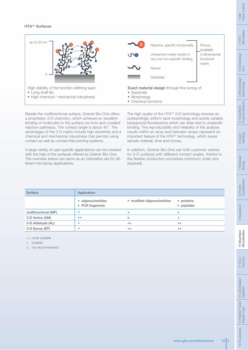
Surface
multifunctional (MF)
3-D Amino (AM)
3-D Aldehyde (AL)
3-D Epoxy (EP)
++ most suitable+ suitableo not recommended
Application
• oligonucleotides• PCR fragments
+
++
+
+
• proteins• peptides
+
+
++
++
• modified oligonucleotides
+
o
++
++
HTATM Surfaces
up to 50 nm
0
Reactive, specific functionality
Unreactive moiety results in very low non-specific binding
Spacer
Substrate
Porous, swellable3-dimensionalfunctional matrix
High stability of the function-defining layer:• Long shelf life• High chemical / mechanical robustness
Exact material design through fine tuning of:• Substrate• Morphology• Chemical functions
Beside the multifunctional surface, Greiner Bio-One offers a proprietary 3-D chemistry, which achieves an excellent binding of molecules to the surface via ionic and covalent reaction pathways. The contact angle is about 45°. The advantages of the 3-D matrix include high sensitivity and a chemical and mechanical robustness that permits using contact as well as contact-free printing systems.
A large variety of user-specific applications can be covered with the help of the surfaces offered by Greiner Bio-One. The overview below can serve as an orientation aid for dif-ferent microarray applications.
The high quality of the HTATM 3-D technology ensures an outstandingly uniform spot morphology and avoids variable background fluorescence which can arise due to unspecific binding. The reproducibility and reliability of the analysis results within an array and between arrays represent an important feature of the HTATM technology, which saves sample material, time and money.
In addition, Greiner Bio-One can fulfil customer wishes for 3-D surfaces with different contact angles, thanks to the flexible production procedure (minimum order size required).
1 C
ell/
Tiss
ue C
ultu
re2
HTS
-M
icro
plat
es3
Imm
unol
ogy/
HLA
4 M
icro
biol
ogy/
Bac
terio
logy
5 Tu
bes/
Mul
ti-
Pur
pose
Bea
kers
6 Li
quid
H
andl
ing
7 M
olec
ular
Bio
logy
8 P
rote
in
Cry
stal
lisat
ion
9 S
epar
atio
n10
Bio
chip
s/M
icro
fluid
ics
11 C
ryo-
Tech
nics
12 L
ids/
Sea
lers
/C
apM
ats
13 R
eact
ion
Tube
s/An
alys
er C
ups
14 A
cces
sorie
s
10 3www.gbo.com/bioscience

HTATMPlatforms
10 4 www.gbo.com/bioscience
Cat.-No.
Description
Surface functionalisation
Tests per slide
Slide material
Slides per case
445 825
HTATMSlide1
multifunctional
1
plastic
25
446 820
HTATMSlide12
multifunctional
12
plastic
5
445 830
HTATMSlide1
3-D Amino
1
plastic
5
445 835
HTATMSlide1
3-D Amino
1
plastic
25
446 830
HTATMSlide12
3-D Amino
12
plastic
5
HTATMSlides Multifunctional3-D Amino3-D Aldehyde3-D Epoxy
• HTATMSlide12: 36 mm2 printable area/well
• HTATMSlide12: Low well rim of only 0.5 mm
• Low autofluorescence
• Barcode-labelling on request
446 8XX
445 8XX
HTATM Platforms HTATMSlides
HTATMSlide1The plastic slide HTATMSlide1 has the dimensions of a standard 25 x 75 mm glass slide and can be used for the processing of one sample (Fig. 1).
HTATMSlide12The HTATMSlide12 is also a plastic slide and has the same dimensions as the HTATMSlide1 but is partitioned into 12 flat compartments, each with a printable surface of 6 x 6 mm and a low well rim of only 0.5 mm (Fig. 2). 12 samples can be processed simultaneously. Therefore the HTATMSlide12 is able to combine the advantages of the universal microplate platform with microarrays for diagnostic applications due to an identical well geometry compared with a standard 96 well microplate.
Figure 1: Slide geometry of HTATMSlide1
Figure 2: Slide geometry of HTATMSlide12
A = 75 +/- 0.2 mmB = 25 +/- 0.1 mmC = 1.0 +/- 0.05 mmD = 18.5 mmH = Flatness –< 0.05 mm
A = 75 +/- 0.2 mmB = 25 +/- 0.1 mmC = 1.0 +/- 0.05 mmD = 0.5 mmE/F = 9 +/- 0.02 mmG = 3.0 mmH = Flatness –< 0.05 mm
13 R
eact
ion
Tube
s/An
alys
er C
ups
1 C
ell/
Tiss
ue C
ultu
re2
HTS
-M
icro
plat
es3
Imm
unol
ogy/
HLA
4 M
icro
biol
ogy/
Bac
terio
logy
5 Tu
bes/
Mul
ti-
Pur
pose
Bea
kers
6 Li
quid
H
andl
ing
7 M
olec
ular
Bio
logy
8 P
rote
in
Cry
stal
lisat
ion
9 S
epar
atio
n10
Bio
chip
s/M
icro
fluid
ics
11 C
ryo-
Tech
nics
12 L
ids/
Sea
lers
/C
apM
ats
14 A
cces
sorie
s
Free of detectable DNase, RNase
HTATMPlatforms
10 5www.gbo.com/bioscience
Cat.-No.
Description
Surface functionalisation
Tests per slide
Slide material
Slides per case
445 850
HTATMSlide1
3-D Epoxy
1
plastic
5
445 855
HTATMSlide1
3-D Epoxy
1
plastic
25
446 850
HTATMSlide12
3-D Epoxy
12
plastic
5
Cat.-No.
Description
Surface functionalisation
Tests per slide
Slide material
Slides per case
445 840
HTATMSlide1
3-D Aldehyde
1
plastic
5
445 845
HTATMSlide1
3-D Aldehyde
1
plastic
25
446 840
HTATMSlide12
3-D Aldehyde
12
plastic
5
1 C
ell/
Tiss
ue C
ultu
re2
HTS
-M
icro
plat
es3
Imm
unol
ogy/
HLA
4 M
icro
biol
ogy/
Bac
terio
logy
5 Tu
bes/
Mul
ti-
Pur
pose
Bea
kers
6 Li
quid
H
andl
ing
7 M
olec
ular
Bio
logy
8 P
rote
in
Cry
stal
lisat
ion
9 S
epar
atio
n10
Bio
chip
s/M
icro
fluid
ics
11 C
ryo-
Tech
nics
12 L
ids/
Sea
lers
/C
apM
ats
13 R
eact
ion
Tube
s/An
alys
er C
ups
14 A
cces
sorie
s

Cat.-No.
Description
Surface functionalisation
Tests per plate
Material
Quantity per bag/case
791 840
HTATMPlate
3-D Aldehyde
96
plastic
5
791 830
HTATMPlate
3-D Amino
96
plastic
5
791 820
HTATMPlate
multifunctional
96
plastic
5
791 850
HTATMPlate
3-D Epoxy
96
plastic
5
With 96 shallow wells and a low well rim of only 0.3 mm, the HTATMPlate has been optimised for the simultaneous analysis of multiple samples. The low height of the rims facilitates both quick and cost-effective printing of a wide range of analytes into the 96 „wells“ of the plate. Additionally modern robotic spotters and arrayers are already equipped to handle this 96 well format. The partition of the plate into 4 individual strips containing 24 wells each gives the user a high degree of flexibility permitting processing of a variable number of samples each day. For processing, a removable wash collar allows a temporary increase in volume for hybridisation and washing procedures, and is removed prior to detection. The dimensions of the HTATMPlate are compatible with automated systems.
Figure 3: Schematic structure of the HTATMPlate: (1) frame, (2) 24 well strip, (3) 24 well wash collar
Figure 4: Geometry of a HTATMPlate strip with 24 wells
HTATMPlatforms
HTATMPlate Multifunctional 3-D Amino3-D Aldehyde3-D Epoxy
• 96 elevated wells/ stages with a printable area of 36 mm2 each
• 4 individual strips with 24 wells each
• Low autofluorescence
• Low well rim of only 0.3 mm
• Barcode-labelling on request
10 6 www.gbo.com/bioscience
791 8XX
HTATMPlate
A = 81.5 mmB = 26.95 mmC = 6.4 mmD = 9.0 mm
1
2
3
13 R
eact
ion
Tube
s/An
alys
er C
ups
1 C
ell/
Tiss
ue C
ultu
re2
HTS
-M
icro
plat
es3
Imm
unol
ogy/
HLA
4 M
icro
biol
ogy/
Bac
terio
logy
5 Tu
bes/
Mul
ti-
Pur
pose
Bea
kers
6 Li
quid
H
andl
ing
7 M
olec
ular
Bio
logy
8 P
rote
in
Cry
stal
lisat
ion
9 S
epar
atio
n10
Bio
chip
s/M
icro
fluid
ics
11 C
ryo-
Tech
nics
12 L
ids/
Sea
lers
/C
apM
ats
14 A
cces
sorie
s
Free of detectable DNase, RNase

Cat.-No.
Description
Concentration
Volume [ml]
Quantity per case
445 055
sciSPOT-Protein
2x
25
1
445 052
sciBLOCK
5x
500
1
445 054
sciBIND
2x
1.6
1
445 051
sciWASH-Protein
8x
500
1
Cat.-No.
Description
Concentration
Volume [ml]
Quantity per case
445 016
sciHYB
1x
1.5
1
445 011
sciWASH I
5x
500
1
445 012
sciWASH II
5x
500
1
445 013
sciWASH III
5x
500
1
Cat.-No.
Description
Concentration
Volume [ml]
Quantity per case
445 015
sciSPOT-MF
2x
50
1
445 018
sciSPOT-AM
2x
50
1
445 017
sciSPOT-AL
2x
50
1
445 014
sciPROCESS-MF
2x
500
1
445 020
sciPROCESS-AM
2x
500
1
445 021
sciPROCESS-AL
2x
500
1
Greiner Bio-One and SCIENION have developed integrated and complete systems for the production and use of DNA- and protein microarrays on the HTA™Platform.
Buffer systems for DNA-microarrays Optimised buffers for each of the three surface modifications are available guaranteeing optimal conditions for spotting and processing:• sciSPOT-MF and sciPROCESS-MF for spotting on multifunctional surfaces• sciSPOT-AM and sciPROCESS-AM for spotting on 3-D Amino surfaces
• sciSPOT-AL and sciPROCESS-AL for spotting on 3-D Aldehyde surfacesThe hybridisation buffer sciHYB creates the most favourable environment for hybridisation, while minimising cross-hybridisation. The washing buffers sciWASH I – III reduce the background and deliver a high level of reproducibility.
Buffer systems for protein microarrayssciSPOT-Protein is a unique spotting solution which facilitates a uniform binding of proteins, antibodies and peptides to HTA™Slides. sciBLOCK buffer removes unbound proteins after spotting and blocks the HTA™ surfaces during incubation. sciBIND buffer creates the most favourable environment for binding reactions. Finally, sciWASH-Protein buffer is a microarray wash buffer which is formulated to promote specific binding and to reduce background signals.
HTATMPlatforms
HTATM Buffer Systems
10 7www.gbo.com/bioscience
445 052445 051
445 055
445 054
HTATM Buffer Systems
• Complete and integrated systems for DNA and protein applications
• Optimised buffer systems for each HTA™ surface modification
• Ultra-pure reagents
• Purified by sterile filtration
• Premixed
• Minimised array-to-array variation
• Expedited data analysis
Detailed protocols for each application are available on www.gbo.com/bioscience/manuals.!
1 C
ell/
Tiss
ue C
ultu
re2
HTS
-M
icro
plat
es3
Imm
unol
ogy/
HLA
4 M
icro
biol
ogy/
Bac
terio
logy
5 Tu
bes/
Mul
ti-
Pur
pose
Bea
kers
6 Li
quid
H
andl
ing
7 M
olec
ular
Bio
logy
8 P
rote
in
Cry
stal
lisat
ion
9 S
epar
atio
n10
Bio
chip
s/M
icro
fluid
ics
11 C
ryo-
Tech
nics
12 L
ids/
Sea
lers
/C
apM
ats
13 R
eact
ion
Tube
s/An
alys
er C
ups
14 A
cces
sorie
s
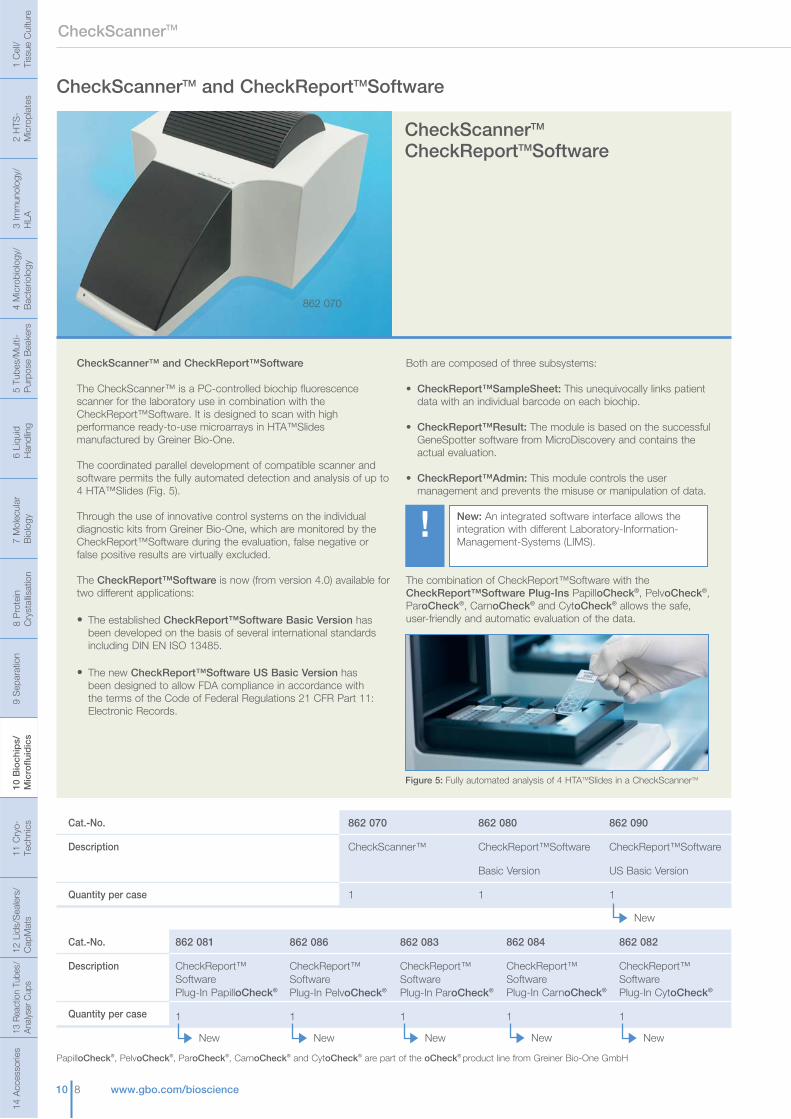
CheckScannerTM
10 8 www.gbo.com/bioscience
CheckScanner™ and CheckReport™Software
The CheckScanner™ is a PC-controlled biochip fluorescence scanner for the laboratory use in combination with the CheckReport™Software. It is designed to scan with high performance ready-to-use microarrays in HTA™Slides manufactured by Greiner Bio-One.
The coordinated parallel development of compatible scanner and software permits the fully automated detection and analysis of up to 4 HTA™Slides (Fig. 5).
Through the use of innovative control systems on the individual diagnostic kits from Greiner Bio-One, which are monitored by the CheckReport™Software during the evaluation, false negative or false positive results are virtually excluded.
The CheckReport™Software is now (from version 4.0) available for two different applications:
• The established CheckReport™Software Basic Version has been developed on the basis of several international standards including DIN EN ISO 13485.
• The new CheckReport™Software US Basic Version has been designed to allow FDA compliance in accordance with the terms of the Code of Federal Regulations 21 CFR Part 11: Electronic Records.
Both are composed of three subsystems: • CheckReport™SampleSheet: This unequivocally links patient data with an individual barcode on each biochip.
• CheckReport™Result: The module is based on the successful GeneSpotter software from MicroDiscovery and contains the actual evaluation.
• CheckReport™Admin: This module controls the user management and prevents the misuse or manipulation of data.
The combination of CheckReport™Software with the CheckReport™Software Plug-Ins PapilloCheck®, PelvoCheck®, ParoCheck®, CarnoCheck® and CytoCheck® allows the safe, user-friendly and automatic evaluation of the data.
CheckScannerTM CheckReportTMSoftware
862 070
CheckScannerTM and CheckReportTMSoftware
New: An integrated software interface allows the integration with different Laboratory-Information-Management-Systems (LIMS).
!
Cat.-No.
Description
Quantity per case
862 080
CheckReport™Software
Basic Version
1
862 070
CheckScanner™
1
862 090
CheckReport™Software
US Basic Version
1
New
Cat.-No.
Description
Quantity per case
862 084
CheckReport™SoftwarePlug-In CarnoCheck®
1
New
862 082
CheckReport™SoftwarePlug-In CytoCheck®
1
New
862 083
CheckReport™SoftwarePlug-In ParoCheck®
1
New
862 086
CheckReport™SoftwarePlug-In PelvoCheck®
1
New
862 081
CheckReport™SoftwarePlug-In PapilloCheck®
1
New
Figure 5: Fully automated analysis of 4 HTATMSlides in a CheckScannerTM
13 R
eact
ion
Tube
s/An
alys
er C
ups
1 C
ell/
Tiss
ue C
ultu
re2
HTS
-M
icro
plat
es3
Imm
unol
ogy/
HLA
4 M
icro
biol
ogy/
Bac
terio
logy
5 Tu
bes/
Mul
ti-
Pur
pose
Bea
kers
6 Li
quid
H
andl
ing
7 M
olec
ular
Bio
logy
8 P
rote
in
Cry
stal
lisat
ion
9 S
epar
atio
n10
Bio
chip
s/M
icro
fluid
ics
11 C
ryo-
Tech
nics
12 L
ids/
Sea
lers
/C
apM
ats
14 A
cces
sorie
s
PapilloCheck®, PelvoCheck®, ParoCheck®, CarnoCheck® and CytoCheck® are part of the oCheck® product line from Greiner Bio-One GmbH

10 9www.gbo.com/bioscience
Diagnostic Kits
PapilloCheck® PelvoCheck®
Diagnostic Kits
All of the Greiner Bio-One ready-to-use kits consist of complete test systems with all the necessary buffers and solutions. Extensive on-chip controls ensure error-free results.
PapilloCheck®
High-risk types of human papilloma viruses (HPV) are causative agents in the occurrence of cervical cancer.
The PapilloCheck® CE-IVD diagnostic kit is intended to be used for the qualitative detection and genotyping of 24 types (18 high-risk and 6 low-risk types) of the human papilloma virus in DNA preparations from human cervical smears. The assay is based on the detection of a fragment of the E1 gene of the human papilloma virus and allows the simultaneous processing of 12 cervical specimens.
Prior to the application of the PapilloCheck® diagnostic kit, DNA has to be extracted from a cervical smear specimen previously collected from the patient using the PapilloCheck® Collection Kit. After the following amplification of a 350 bp fragment of the E1 gene via polymerase chain reaction (PCR) the fluorescence-labelled amplification products are hybridised to specific DNA probes fixed on the PapilloCheck® chip. Due to the fluorescence signal the presence of HPV DNA is visualised by the CheckScannerTM and analysed by the corresponding CheckReportTMSoftware.
Nearly error-free results are guaranteed due to the integration of controls for the quality of the DNA extraction, PCR, spot homogeneity and the hybridisation efficiency. In addition, the integration of dUTP in the PapilloCheck® MasterMix ensures the elimination of carry-over contaminations from previous PCR reactions.
PelvoCheck®
Pelvic inflammmatory diseases (PID) are mostly associated with sexually transmitted bacteria causing millions of new infections worldwide per year. Unidentified infections may lead to dramatic symptoms including complications during pregnancy, blindness of the newborn and infertility.
The newly developed diagnostic kit enables the identification of the six most frequently detected bacteria associated with sexually transmitted diseases (Chlamydia trachomatis, Neisseria gonorrhoeae, Treponema pallidum, Mycoplasma hominis, Mycoplasma genitalium, Ureaplasma urealyticum). Both cervical and pooled urine samples can be analysed.
The test is based on the detection of a specific DNA fragment of the 16S rRNA Gene using polymerase chain reaction (PCR). The integration of dUTP in the PelvoCheck® MasterMix ensures the elimination of carry-over contaminations from previous PCR reactions.
The scanning procedure, data evaluation and report generation are performed using CheckScannerTM and CheckReportTMSoftware.
s
1 C
ell/
Tiss
ue C
ultu
re2
HTS
-M
icro
plat
es3
Imm
unol
ogy/
HLA
4 M
icro
biol
ogy/
Bac
terio
logy
5 Tu
bes/
Mul
ti-
Pur
pose
Bea
kers
6 Li
quid
H
andl
ing
7 M
olec
ular
Bio
logy
8 P
rote
in
Cry
stal
lisat
ion
9 S
epar
atio
n10
Bio
chip
s/M
icro
fluid
ics
11 C
ryo-
Tech
nics
12 L
ids/
Sea
lers
/C
apM
ats
13 R
eact
ion
Tube
s/An
alys
er C
ups
14 A
cces
sorie
s
Cat.-No.
Description
Tests per case
465 060
PapilloCheck® (CE-IVD)
identification of 24
human papilloma
viruses
60
465 070
PapilloCheck®
Collection Kit
10
504 060*)
PelvoCheck® (CE-IVD)
identification of 6 bacteria
associated with sexually
transmitted diseases
60
NewNew
*) available 2010
465 060
PapilloCheck® and PelvoCheck® are part of the oCheck® product line from Greiner Bio-One GmbH
Cat.-No.
Description
Tests per case
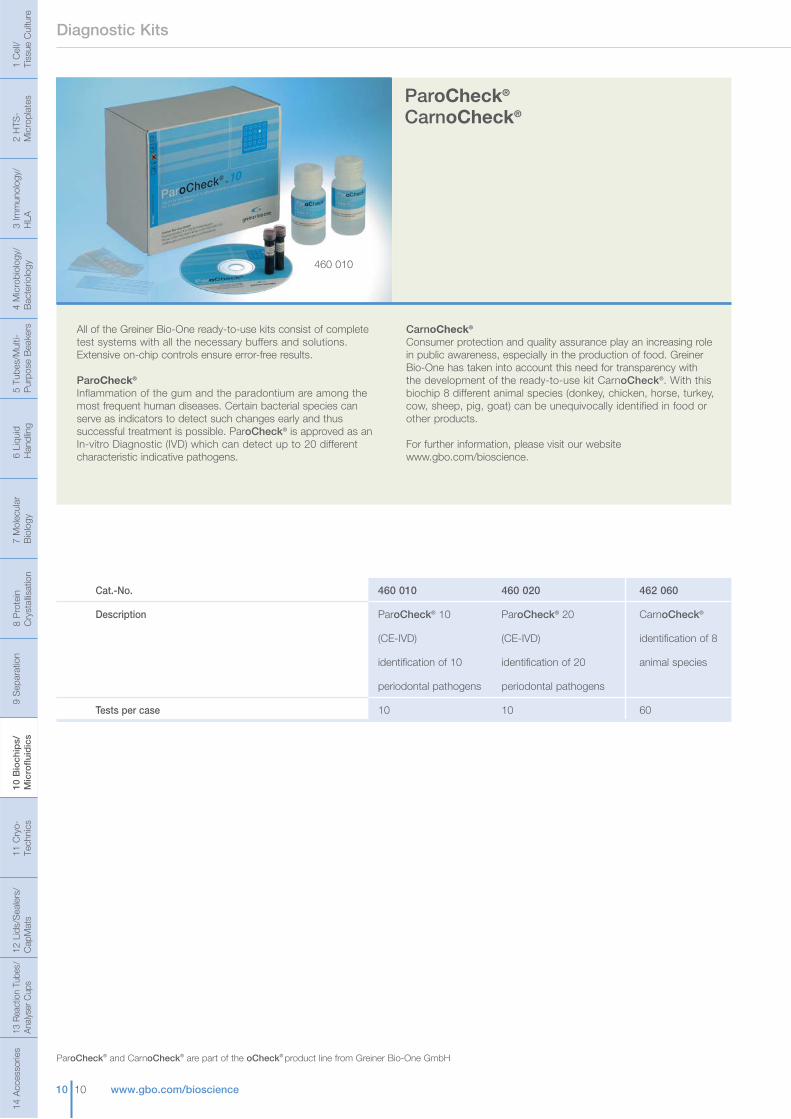
462 060
CarnoCheck®
identification of 8
animal species
60
460 010
ParoCheck® 10
(CE-IVD)
identification of 10
periodontal pathogens
10
460 020
ParoCheck® 20
(CE-IVD)
identification of 20
periodontal pathogens
10
10 10 www.gbo.com/bioscience
Diagnostic Kits
ParoCheck® CarnoCheck®
All of the Greiner Bio-One ready-to-use kits consist of complete test systems with all the necessary buffers and solutions. Extensive on-chip controls ensure error-free results.
ParoCheck®
Inflammation of the gum and the paradontium are among the most frequent human diseases. Certain bacterial species can serve as indicators to detect such changes early and thus successful treatment is possible. ParoCheck® is approved as an In-vitro Diagnostic (IVD) which can detect up to 20 different characteristic indicative pathogens.
CarnoCheck®
Consumer protection and quality assurance play an increasing role in public awareness, especially in the production of food. Greiner Bio-One has taken into account this need for transparency with the development of the ready-to-use kit CarnoCheck®. With this biochip 8 different animal species (donkey, chicken, horse, turkey, cow, sheep, pig, goat) can be unequivocally identified in food or other products.
For further information, please visit our website www.gbo.com/bioscience.
460 010
13 R
eact
ion
Tube
s/An
alys
er C
ups
1 C
ell/
Tiss
ue C
ultu
re2
HTS
-M
icro
plat
es3
Imm
unol
ogy/
HLA
4 M
icro
biol
ogy/
Bac
terio
logy
5 Tu
bes/
Mul
ti-
Pur
pose
Bea
kers
6 Li
quid
H
andl
ing
7 M
olec
ular
Bio
logy
8 P
rote
in
Cry
stal
lisat
ion
9 S
epar
atio
n10
Bio
chip
s/M
icro
fluid
ics
11 C
ryo-
Tech
nics
12 L
ids/
Sea
lers
/C
apM
ats
14 A
cces
sorie
s
ParoCheck® and CarnoCheck® are part of the oCheck® product line from Greiner Bio-One GmbH

10 11www.gbo.com/bioscience
Mycoplasma Detection Kit
Cat.-No.
Description
Tests
464 060
CytoCheck®
identification of
mycoplasma species
10
CytoCheck® Mycoplasma Service
Mycoplasma Detection
464 060
CytoCheck®
In the biopharmaceutical industry, cell cultures are being used increasingly for the manufacture of therapeutic products. Mycoplasma contamination of these cell cultures is a serious problem, as it potentially compromises the safety of the drugs being produced and reduces the final yield. CytoCheck® is a premium test kit for the identification of mycoplasma species in cell cultures and other biological materials. It has been validated in accordance with the guidelines of the European Pharmacopoeia (Ph. Eur. 2.6.7, 2.6.21).
CytoCheck® detects mycoplasma species with a universal mycoplasma probe (including Acholeplasma sp., Spiroplasma sp. and Ureaplasma sp.) and identifies 40 individual species using species-specific probes which can then be used to track the source of contamination. DNA is extracted from the sample and then PCR is performed on the extracted material. The PCR primers amplify conserved and species-specific portions of the “16S-23S rRNA intergenic transcribed spacer” of mycoplasma DNA. Implementation of dUTP prevents carry-over of PCR products, which could become a subsequent source of contamination. The fluorescently labelled amplified DNA fragments are then hybridised to the microarray chip. The chip contains probes for both species-specific targets and a universal probe that detects any species of mycoplasma present in the original sample. Finally, detection of any bound PCR products is achieved with a microarray scanner.
The CytoCheck® DNA-chip has a total of 225 individual measurement points which are used by the CheckScannerTM instrument and subsequently the analysis software, theCheckReportTMSoftware. The integrated platform allows automatic sample tracking, rapid report generation and digital data management (Fig. 6). The CheckReportTMSoftware was especially designed for 21 CFR part 11 conformity to allow FDA compliance of the test procedure. The test can be processed within only 5 hours and sensitivity and specificity are comparable to the cell culture and indicator cell culture methods.
CytoCheck® is offered as a complete ready-to-use kit and includes the CytoCheck® DNA-chips, the PCR MasterMix, buffers and a detailed instruction manual.
Figure 6: The CheckReportTMSoftware automatically generates a report showing the result of the 40 species-specific probes as well as the universal probe
1 C
ell/
Tiss
ue C
ultu
re2
HTS
-M
icro
plat
es3
Imm
unol
ogy/
HLA
4 M
icro
biol
ogy/
Bac
terio
logy
5 Tu
bes/
Mul
ti-
Pur
pose
Bea
kers
6 Li
quid
H
andl
ing
7 M
olec
ular
Bio
logy
8 P
rote
in
Cry
stal
lisat
ion
9 S
epar
atio
n10
Bio
chip
s/M
icro
fluid
ics
11 C
ryo-
Tech
nics
12 L
ids/
Sea
lers
/C
apM
ats
13 R
eact
ion
Tube
s/An
alys
er C
ups
14 A
cces
sorie
s
CytoCheck® is part of the oCheck® product line from Greiner Bio-One GmbH
Mycoplasma Service Greiner Bio-One offer a service for the detection of mycoplasma called Mycoplasma Service!
463 090
Mycoplasma Service
test for detection
of mycoplasma
1

10 12 www.gbo.com/bioscience
The finest structures for the finest ideasDiscover the potential of microfluidics
Through the use of modern manufacturing techniques it is now possible to produce microstructured components for the diagnostics, drug discovery, and research industry made from innovative plastic polymer materials. They offer substantial economical advantages, a large selection of quite diverse materials and surface treatments are available, and large format components can also be economically structured. The principal application areas are:
Analytics/Diagnostics (electrophoresis, lab-on-a-chip)
Chemistry (micromixers, reactors, heat exchangers)
Drug discovery and liquid handling
For the production of microfluidic systems as single-use products, novel and innovative manufacturing techniques are required. A number of manufacturing processes can be utilised and today injection moulding is widely used to produce microstructured parts from plastics. The choice of material depends on the final application of the components. Common selection criteria are:
Transparency
Stability
Chemical resistance
Biocompatibility
Surface properties (e.g. hydrophobicity)
A large number of different polymers are available (e.g. polystyrene, polymethylmethacrylate, polycarbonate, polypropylene). Additional surface treatments after moulding can further improve the properties of the chosen material, and compared to glass, a larger variety of options for surface modifications are available.
13 R
eact
ion
Tube
s/An
alys
er C
ups
1 C
ell/
Tiss
ue C
ultu
re2
HTS
-M
icro
plat
es3
Imm
unol
ogy/
HLA
4 M
icro
biol
ogy/
Bac
terio
logy
5 Tu
bes/
Mul
ti-
Pur
pose
Bea
kers
6 Li
quid
H
andl
ing
7 M
olec
ular
Bio
logy
8 P
rote
in
Cry
stal
lisat
ion
9 S
epar
atio
n10
Bio
chip
s/M
icro
fluid
ics
11 C
ryo-
Tech
nics
12 L
ids/
Sea
lers
/C
apM
ats
14 A
cces
sorie
s
Figure 1: Production of microstructured parts by injection moulding.
Precise manufacturing of the mould insert is critical for the quality of the plastic component. Different methods are used depending on the structural size, precision and aspect ratio of the structure being moulded. Routinely, mechanical micromachining is used to create the required metal moulding tools. Using high-speed tools, microstructures can be produced even on large areas. For extremely precise mould inserts, galvanic/lithographic techniques are available.
Figure 2a: Mould insert made of brass, manufactured by micro-milling.Figure 2b: Detail of a mould insert made of nickel, manufactured by UV-LIGA.
Standard formats allow for fast and cost-efficient production of customer-specific designs from prototyping to production level. A common format in the life science is the microscope slide with a footprint of 25 mm x 75 mm.
Figure 3: Microfluidic Slide. Microchannels and macro-features (reservoirs, through-holes) were produced in one step.
Greiner Bio-One can produce microstructured components in a variety of shapes and dimensions and also larger formats, such as the microplate format, can be structured. In many cases, the microstructured surface must be sealed with a lid in order to produce a closed channel system. Different joining methods, such as laser welding, ultrasonic welding, adhesive techniques and diffusion bonding are used. The incorporation of additional components such as electrodes or membranes into the microstructured part is also possible. To combine different materials, we have special welding and adhesive bonding techniques that have been adapted for microstructures.
Figure 4a: Partly sealed structures for capillary electrophoresis. Figure 4b: SEM image of the cross section through a closed microchannel (100 µm x 50 µm).
10 13www.gbo.com/bioscience
Our serviceWith our team of biologists, chemists, physicists, and engineers, we work closely with our customers. Built on our experience in plastics manufacturing, we can supply an innovative solution, custom-tailored to your requirements.
A large variety of materials is available to realise your design, in addition to which we offer several modification technologies to tailor the surface properties of your microstructured component to optimise it for your application.
We offer our services from the first draft to the product:
Prototyping
Small series production
Mass production
!2a) 2b)
4a) 4b)
Microchannel
Intersection
Reservoir
1000 µm
1 C
ell/
Tiss
ue C
ultu
re2
HTS
-M
icro
plat
es3
Imm
unol
ogy/
HLA
4 M
icro
biol
ogy/
Bac
terio
logy
5 Tu
bes/
Mul
ti-
Pur
pose
Bea
kers
6 Li
quid
H
andl
ing
7 M
olec
ular
Bio
logy
8 P
rote
in
Cry
stal
lisat
ion
9 S
epar
atio
n10
Bio
chip
s/M
icro
fluid
ics
11 C
ryo-
Tech
nics
12 L
ids/
Sea
lers
/C
apM
ats
13 R
eact
ion
Tube
s/An
alys
er C
ups
14 A
cces
sorie
s