34ème COLLOQUE SCIENTIFIQUE - RuminantiaHeli Lindeberg Henry Leese Iris Kaimio Isabelle Donnay...
181
34 th Annual Meeting A.E.T.E. – Nantes, France, 7 th –8 th September 2018 i 34 ème COLLOQUE SCIENTIFIQUE 34 th SCIENTIFIC MEETING * * * Dr. Patrice Humblot Special Celebration * * * Nantes, France, 7 th and 8 th September 2018
Transcript of 34ème COLLOQUE SCIENTIFIQUE - RuminantiaHeli Lindeberg Henry Leese Iris Kaimio Isabelle Donnay...

34th Annual Meeting A.E.T.E. – Nantes, France, 7th –8th September 2018 i
34ème COLLOQUE SCIENTIFIQUE
34th SCIENTIFIC MEETING
*
* *
Dr. Patrice Humblot
Special Celebration
* *
*
Nantes, France, 7th and 8th September 2018

34th Annual Meeting A.E.T.E. – Nantes, France, 7th –8th September 2018 ii

34th Annual Meeting A.E.T.E. – Nantes, France, 7th –8th September 2018 iii
Board of Governors
President Dimitrios Rizos, Spain
Vice President Leroy Jo, Belgium
Treasurer Jan Detterer, Germany
Secretary Teresa Mogas, Spain
Annual statistics Marja Mikkola, Finland
Newsletter Roger Sturmey, United Kingdom
AETE website Hilde Aardema, The Netherlands
Scientific Committee Jane Morrell, Sweden
French foundation Daniel Le Bourhis, France
and of ET Industry
ET Industry Hélène Quinton, France

34th Annual Meeting A.E.T.E. – Nantes, France, 7th –8th September 2018 iv

34th Annual Meeting A.E.T.E. – Nantes, France, 7th –8th September 2018 v
Local Organizing Committee

34th Annual Meeting A.E.T.E. – Nantes, France, 7th –8th September 2018 vi

34th Annual Meeting A.E.T.E. – Nantes, France, 7th –8th September 2018 vii
Main Sponsor
General Sponsors
Exhibitors
Supporters
Thanks to all our
Sponsors!

34th Annual Meeting A.E.T.E. – Nantes, France, 7th –8th September 2018 viii

34th Annual Meeting A.E.T.E. – Nantes, France, 7th –8th September 2018 ix
The Board Members of the European Embryo Technology Association would like to express their appreciations for the
scientific support to the following people
Section Chairs:
Hélène Quinton: TAI/FTET/AI OPU - IVF and ET Hilde Aardema: Folliculogenesis, ogenesis, and superovulation Jane Morrell: Physiology of reproduction in male and semen technology Marja Mikkola and Daniel Le Bourhis: Embryology, developmental biology, and physiology of reproduction Roger Sturmey: Cloning, transgenesis, and stem cells
Support biotechnologies: Cryopreservation and cryobiology, diagnosis through imaging, molecular biology, and “omics”
Jo Leroy: AETE Student Competition Teresa Mogas Workshops Dimitrios Rizos and Teresa Mogas: Invited Papers
Manuscript and Abstract Reviewers:
Adam Watkins
Anders Johannisson
Aurélie Bonnet
Benedicte Grimard
Benoit Guyonnet
Brigitte Leguienne
Carmen Alminana-Brines
Darryl Russell
Diego Bucci
Dimitrios Rizos
Eli Sellem
Eline Wydooghe
Elisabeth Blesbois
Fabienne constant
Felipe Martinez Pastor
Frederic Charreaux
Hélène Kiefer
Helene Quinton
Heli Lindeberg
Henry Leese
Iris Kaimio
Isabelle Donnay
isabelle Hue
Jaana Peippo
Jean-Francois Bruyas
Jennifer Schoen
Jessie De Bie
Jo Carvalho
Jo Leroy
Juhani Taponen
Julian Garde
Karolien Desmet
Katie Fowler
Lamia Briand
Leen Vandaele
Lies Jordaens
Margareta Wallgren
Maria Sabes Alsina
Mariana Sponchiado
Marie Saint-Dizier
Marja Mikkola
Miguel Velazquez
Nathalie Beaujean
Olivier Sandra
Pascal Mermillod
Pascal Salvetti
Pascale Chavatte-Palmer
Paul McKeegan
Peter Bols
Rebecca Krisher
Sebastien Elis
Sara Valckx
Serge Lacaze
Steven Van Cruchten
Sveltana Uzbekova
Sylvie Chastant-Maillard
Teresa Mogas
Thanapol Nongbua
Veronique Duranthon
Virginie Maillard
Waleed Marei
Yannis Sfontouris
Ylva Sjunnesson
Adam Watkins
Anders Johannisson
Aurélie Bonnet
Benedicte Grimard
Benoit Guyonnet
Brigitte Leguienne
Carmen Alminana-Brines
Darryl Russell
Diego Bucci

34th Annual Meeting A.E.T.E. – Nantes, France, 7th –8th September 2018 x
34th Annual Meeting A.E.T.E. – Nantes, France, 7th –8th September 2018 xi
C O N T E N T S
All the original articles and abstracts are published in Animal Reproduction.
For citation purposes, the Animal Reproduction should be the original source
(http://sbte.org.br/anais/pt)
AETE pioneer award 2018: Dr. Patrice Humblot
Dobson, H ................................................................................................. 3
From clinics to (cow)mics; a reproductive journey
P. Humblot ............................................................................................... 7
Commercial embryo transfer activity in EUROPE 2017
Mikkola M ............................................................................................... 21
INVITED LECTURES
Ovarian antral follicle populations and embryo production in cattle
A.F. Zangirolamo, F. Morotti, N.C. da Silva, T.K. Sanches, M.M. Seneda ................... 29
Preservation of female fertility in humans and animal species
H. Picton ................................................................................................ 35
Directions and applications of CRISPR 1 technology in livestock research
I. Lamas-Toranzo, P. Ramos-Ibeas, E. Pericuesta and P. Bermejo-Álvaro .................. 45
Oocyte related factors impacting on embryo quality: relevance for “in
vitro” embryo production
Fabienne Nuttinck ..................................................................................... 55
SHORT COMMUNICATIONS
TAI / FTET / AI
A set of conditions that could enhance fertility rates and litter size in Sardi Morrocan
sheep after exo-cervical insemination
Bouchra El Amiri, Anass Ben Moula, Abdelmoughit Badi, Naima Hamidallah, Larbi Allai,
Kaoutar EL Khalil, Elemer Gergatz, Erzsébet Gyökér, and Ottó Szenci ..................... 66

34th Annual Meeting A.E.T.E. – Nantes, France, 7th –8th September 2018 xii
Timed embryo transfer program from charolais heifers to holstein heifers
Recai Kulaksiz, Selvi Özge Köksal, Ismail Dogru, and Sükrü Metin Pancarci ................ 67
OPU - IVF and ET
Nobiletin supplementation in maturation media enhances in vitro oocyte maturation
and subsequent embryo development
Y.N. Cajas, K. Cañón-Beltrán, M. Ladrón de Guevara, M.G. Millán de la Blanca, M.E.
González, and D. Rizos ............................................................................... 70
Nuclear maturation rate of caprine oocytes after in vitro maturation in three
different media with base of TCM 199.
Antonio Santana Dos Santos Filho, Sebastião Inocêncio Guido, Claudio Coutinho
Bartolomeu, Wasim El Shebli, and Rita de kassia Oliveira de Andrade ...................... 71
Effects of an enriched n-3 polyunsaturated fatty acid diet on in vitro embryo
production in dairy cowsEffects of an enriched n-3 polyunsaturated fatty acid diet on
in vitro embryo production in dairy cows
S. Elis, D. Le Bourhis, M. Oseikria, P. Salvetti, O. Desnoes, E. Briant, M. Dupont, L. Le
Berre, A.Desmarchais, S. Uzbekova, V. Maillard, and S. Freret .............................. 72
Follicular wave synchronization and superstimulation prior to ovum pick-up for
improving in vitro embryo production in non-lactating Holstein cows
L.B. Ferré, C. Fresno, M.E. Kjelland, and P.J. Ross ............................................ 73
Risk of Coxiella burnetii transmission via embryo transfer using in vitro early caprine
embryos
F. Fieni, A. Alsaleh, J.M.G. Souza-Fabjan, P. Mermillod, E. Corbin, P. Nascimento, J.F.
Bruyas, and J.L. Pellerin ............................................................................ 74
A retrospective study on influence of weight of heifer’s donors at 12 months in
relation to age at first estrous, age at first embryo flushing and number of viable
embryos in a breeding program
Giselle Gamarra, Maxence Coquerelle, Michell Mouneyres, Brigitte Marquant Le Guienne,
and Serge Lacaze ...................................................................................... 75
Effects of melatonin on bovine embryonic developmental competence and kinetics in
vitro
J. C. Gutiérrez-Añez, P. Aldag, A. Lucas-Hahn, and H. Niemann ............................ 76
Detection of cell-free DNA in embryo culture medium: its potential application as a
noninvasive method for sex determination in cattle
Tsige Hailay, Samuel Gebremedhn, Franca Rings, Dessiie Salilew-Wondim, Dawit
Tesfaye, Schellander Karl, and Michael Hoelker ................................................ 77

34th Annual Meeting A.E.T.E. – Nantes, France, 7th –8th September 2018 xiii
The influence of flunixin meglumine (FM), hCG or a combination of hCG and FM on
the conception results in embryo recipient heifers including the passage time
through the cervix and the presence of a large follicle
M. Herudzinska, B.M. Jaskowski, J. Czeladko, J. Kulus, E. Wedrowska, M. Gehrke, J.M.
Jaskowski, and K.P. Brüssow ....................................................................... 78
The influence of dominant follicles and corpora lutea location on the conception rate
in embryo recipient heifers
B.M. Jaskowski, J. Czeladko, J. Kulus, J. Sobolewski, M. Gehrke, J.M. Jaskowski, M.
Rogoziewicz, and K.P. Brüssow ..................................................................... 79
Reproductive fluids added to embryo culture vs. standard culture in cow: first results
on pregnancy rates
J.S. Lopes, M. Hamdi, E. Alcázar-Triviño, B. Rodríguez-Alonso, A. Canha-Gouveia, E.
París-Oller, C. Soriano-Úbeda, S. Cánovas, D. Rizos, and P. Coy ............................. 80
Transcervical embryo recovery in Lacaune ewes superovulated with different doses
of FSH
J.M.G. Souza-Fabjan, L.M. Figueira, N.G. Alves, R.I.T.P. Batista, L.C. Souza, A.A. Arrais,
C.F. Silva, M.C.C. Morais, and J.F. Fonseca ...................................................... 81
Porcine follicular fluid as chemoattractant improves sperm attraction and in vitro
fertilization
Luis Alberto Vieira, Andrea Aguilera, Alessia Diana, and Carmen Matas .................... 82
Retrospective survey on equine embryo transfer activities in France in 2014 and
2015
E.Autard de Bragard; J.F. Bruyas ................................................................... 83
FOLLICULOGENESIS, OOGENESIS, and SUPEROVULATION
Retrospective analysis of superstimulation with Folltropin®-V in Wagyu versus other
beef breeds
C.B. Steinhauser, C.R. Looney, J.F. Hasler, P. Renaud ........................................ 86
PHYSIOLOGY of REPRODUCTION IN MALE, and SEMEN TECHNOLOGY
ADAM protein expression in avian sperm and female genital epithelial cells: relation
to sperm storage
L. Cordeiro, C. Riou, and N.Gérard ................................................................ 88
Analysis of zona pellucida binding properties of boar sperm subpopulations separated
by carbohydrate affinity
L. González-Brusi, R. López-Úbeda, J.G. Hamzé, P. Cots, J. Ballesta, M. Jiménez-
Movilla, M.J. Izquierdo-Rico, and M. Avilés ...................................................... 89

34th Annual Meeting A.E.T.E. – Nantes, France, 7th –8th September 2018 xiv
Sperm motility in thawed bull semen is increased by a short incubation before
analysis
I.B. Lima-Verde, T. Ntallaris, and J.M. Morrell ................................................. 90
Effect of cholescytokinin (CCK) protein on the motility of porcine sperm.
R. López-Úbeda, FA. García-Vázquez, M. Balastegui-Alarcón, P. Cots, M. Avilés, and MJ.
Izquierdo-Rico.......................................................................................... 91
Porcine periovulatory oviductal fluid decreases the occurrence of Protein Kinase A
(PKA) substrates trough sAC/cAMP/PKA during mouse sperm capacitation
C. Matás, M.G. Gervasi, C. Soriano-Úbeda, E. París-Oller, and P.E. Visconti .............. 92
Effect of swim-up on the activation of apoptosis in frozen bovine semen: A flow
cytometric study
Maria Moreira da Silva, Loide Soares, and F. Moreira da Silva ............................... 93
Progesterone induces sperm release from bovine oviductal epithelial cells by
modification of the sperm protein and lipid compositions
M. Ramal Sanchez, G. Tsikis, M.C. Blache, V. Labas, X. Druart, N. Bernabò, P. Mermillod,
and M. Saint-Dizier ................................................................................... 94
Motility and atomic force microscopy observation of avian spermatozoa incubated in
uterine fluid
C. Riou, L. Cordeiro, A. Chaiyasitdhi, O. Srikimkaew, S. Suebka, L. Nadal-Desbarats, W.
Kundhikanjana, and N. Gérard ...................................................................... 95
Sperm selection by density-gradient centrifugation of Merino ram semen cold-stored
up to 48 h improves viability and membrane integrity
D.A. Galarza, M. Ladrón de Guevara, P. Beltrán-Breña, D. Rizos, A. López-Sebastián, J.
Santiago-Moreno ....................................................................................... 96
EMBRYOLOGY, DEVELOPMENTAL BIOLOGY, and PHYSIOLOGY of REPRODUCTION
Oviduct extracellular vesicles: a new strategy to optimize porcine in vitro embryo
production
A. S. Alcântara-Neto, P. Mermillod, and C. Almiñana ......................................... 98
Physiological concentrations of steroid hormones during in vitro culture changed
lipid composition and improved cryosurvival of bovine embryos
Charles Banliat, Florine Dubuisson, Emilie Corbin, Daniel Tomas, Daniel Le Bourhis,
Pascal Salvetti, Valérie Labas, Pascal Mermillod, and Marie Saint-Dizier ................... 99
Lipidomic profiling of the bovine oviductal fluid across the estrous cycle
Charles Banliat, Daniel Tomas, Ana-Paula Teixeira-Gomes, Svetlana Uzbekova, Valérie
Labas, and Marie Saint-Dizier ...................................................................... 100

34th Annual Meeting A.E.T.E. – Nantes, France, 7th –8th September 2018 xv
Short- and long-term effects of progesterone and prolactin during the second phase
of IVM on metaphase-II chromosomes in bovine oocytes
I.Y. Lebedeva, G.N. Singina, E.N. Shedova, O.S. Mityashova, A.V. Lopukhov, and V.V.
Konnova ............................................................................................... 101
The effect of short-term cytoskeletal inhibitor treatment on embryo metabolism and
viability
B. Muller and R.G. Sturmey ........................................................................ 102
The effect of chilling on the viability of in vitro produced bovine embyos
M.E. Mutikainen, T.M. Hamama, M. Weldenegodguad, and J.M.H. Peippo ............... 103
Interferon tau exerts concentration dependent actions on bovine neutrophil gene
dynamics favoring implantation
Manjari Pasumarti, Ajay K Dang, and Iqbal Hyder ............................................. 104
Analyzing the effects of bovine Interferon tau and female sex steroids on neutrophil
pro and anti inflammatory triggers to understand implantation
Manjari Pasumarti and Iqbal Hyder ................................................................ 105
Role of RNA isoform expression in sex determination in mice
B. Planells, I. Gómez-Redondo, E. Pericuesta, P. Lonergan and A. Gutiérrrez-Adán .... 106
The effect of vitamin c on the developmental competences and quality of pig
blastocysts obtained after in vitro fertilization
I. Rajska, K. Poniedzialek-Kempny, K. Sobol, and B. Gajda ................................. 107
Sperm storage in hen’s reproductive tract: metabolic composition of the uterine
fluid after artificial insemination
C. Riou, L. Cordeiro, L. Nadal-Desbarats, and N. Gérard ..................................... 108
Local embryo effect on the transcriptomic response of the oviductal epithelial cells
results from in vivo and in vitro approaches
B. Rodríguez-Alonso, M. Hamdi, J.M. Sánchez, A. Gutierrez-Adan, P. Lonergan, and D.
Rizos .................................................................................................... 109
Investigating the impact of hyperglycaemia on bovine oviduct epithelial cell
physiology and secretions in vitro
K.K. Roussi and R.G. Sturmey ...................................................................... 110
Effects of cumulus cells and prolactin on histone acetylation during the prolonged
culture of matured bovine oocytes
E.N. Shedova, G.N. Singina, A.V. Lopukhov, and V.V. Konnova ............................ 111

34th Annual Meeting A.E.T.E. – Nantes, France, 7th –8th September 2018 xvi
Apoptosis resistance of bovine cumulus-oocyte complexes is modulated via
progesterone-dependent pathways at the terminal step of in vitro maturation
G.N. Singina, I.Y. Lebedeva, E.N. Shedova, E.K. Montvila, A.A. Smekalova, and E.V.
Tsyndrina .............................................................................................. 112
Rescue potential of supportive embryo culture conditions on bovine embryos derived
from metabolically-compromised oocytes
A. Smits, W.F.A. Marei, J. De Bie, P.E.J. Bols, B. Meulders, and J.L.M.R. Leroy ........ 113
Oviductal cells express Stearoyl-CoA desaturase that can protect the embryo against
saturated fatty acids
H. Aardema, P.K. Fontes, P.L.A.M. Vos, B.M. Gadella, B.A.J. Roelen, and
H.T.A. van Tol ........................................................................................ 114
Enhancement of the developmental capacity of metabolically compromised bovine
oocytes and embryos by water soluble vitamin E (TROLOX) depends on the timing of
the treatment
J. De Bie, W.F.A. Marei, P.E.J. Bols, and J.L.M.R. Leroy ..................................... 115
Elevated non-esterified fatty acid concentrations during in vitro maturation affect
the transcriptome profile of day 14 bovine embryos 7 days after transfer
K.L.J. Desmet, W.F.A. Marei, C. Richard, I. Hue, S. Andries, P.E.J. Bols, and J.L.M.R.
Leroy .................................................................................................... 116
The Effects of undernutrition and supplementation with cactus silage in Boujaâd
ewes on offspring growth performances
K. EL Khalil, N. Hamidallah, A. Benmoula, A. Badi, and B. El Amiri ........................ 117
Piglets obtained by transfer of embryos received after in vitro fertilization of
oocytes matured with thymosin: a preliminary study
K. Poniedzialek-Kempny, I. Rajska, L. Gajda, B. Gajda, and Z. Smorag .................. 118
PredOSEgenesis: A two-layer classifier for identifying oogenesis, spermatogenesis
and embryogenesis-related proteins
M. Rahimi, A. Mohammadi-Sangcheshmeh, and M.R Bakhtiarizadeh ....................... 119
Characterization of sperm-oviduct extracellular vesicles interactions at various
stages of the bovine estrous cycle and their effects on sperm physiology
J. Saulnier, X. Druart, K. Reynaud, M. Saint-Dizier, and C. Almiñana Brines ............. 120
Health status in the Latvian native breed of Latvian Brown dairy cows that are
intended for multiple ovulation and embryo transfer
I. Sematovica, V. Antane, and E. Eihvalde ...................................................... 121

34th Annual Meeting A.E.T.E. – Nantes, France, 7th –8th September 2018 xvii
CLONING, TRANSGENESIS, and STEM CELLS
Adipose mesenchymal stem cells: a new tool to restore interesting genotypes by
cloning in the rabbit
N. Daniel, G. Morin, C. Richard, and V. Duranthon ............................................ 124
Detection of adult stem cell marker leucine-rich repeat-containing G-protein-coupled
receptor 5 (LGR5) transcripts in bovine oviduct epithelial cells
P.K. Fontes, H.H.WHenning, H.T.A van Tol, P.L.A.M Vos, T.A.E. Stout, and B.M. Gadella
.......................................................................................................... 125
Early microinjection of bovine zygotes reduces mosaicism rates following CRISPR-
mediated genome edition
I. Lamas-Toranzo, F. Cornudella-Ardiaca, J. Cobos-Figueroa, O. Ousinde, and P.
Bermejo-Álvarez ...................................................................................... 126
The use of a novel microfluidic culture device and predictive metabolic profiling as a
means to improve murine embryo developmental competence in vitro
P.J. McKeegan, F. Colucci, H.M. Picton, and V. Pensabene .................................. 127
SUPPORT BIOTECHNOLOGIES: CRYOPRESERVATION and CRYOBIOLOGY, DIAGNOSIS
THROUGH IMAGING, MOLECULAR BIOLOGY, and "OMICS"
The use of different progestin devices in ovarian stimulation protocol affects gene
expression in sheep cumulus-oocyte complexes (COC)
G.M. Bragança, R.I.T.P. Batista, C.V. de Souza, J.D.R. dos Santos, E.K.N. Arashiro, J.F.
da Fonseca, F.Z. Brandão, and J.M.G. de Souza-Fabjan ...................................... 130
Analysis of correlation between zona pellucida birefringence using polarized light
microscopy and resistance to trypsin digestion in different mammals.
P. Cots, J. Sánchez-Férez, E. Gómez, L. Gónzalez-Brusi, M.J. Izquierdo-Rico, R.T. López-
Donaire, J. Ballesta, and M. Avilés ................................................................ 131
Comparison of survival rates of two thawing methods for vitrified biopsied bovine in
vitro-produced blastocysts
N. González, J. Scherzer, M. Reichenbach, and H. Zerbe .................................... 132
Design of pJuno-beads to study the molecular mechanisms of sperm-oocyte
interaction.
J.G. Hamze, R. Romar, and M. Jiménez-Movilla ................................................ 133
Effect of vitrification on the functional activity of mitochondria in porcine oocytes
during in vitro maturation
T. Kuzmina, T. Stanislavovich, and D. Tatarskaya ............................................. 134
34th Annual Meeting A.E.T.E. – Nantes, France, 7th –8th September 2018 xviii
Cryodamage of oocytes frozen in antral follicles of bovine ovarian tissue fragments
A.V. Makarevich, L. Olexikova, M. Foldesiova, E. Kubovicova, and J. Pivko ............. 135
MitoQ rescues early embryo development of metabolically-compromised bovine
oocytes in vitro
W.F.A. Marei, L. Van den Bosch, P.E.J. Bols, and J.L.M.R. Leroy .......................... 136
mtDNA content of bovine cumulus cells derived from oocytes with different
developmental competence following individual culture
A. Martínez-Moro, I. Lamas-Toranzo, and P. Bermejo-Álvarez ............................... 137
Good pregnancy rate of bovine biopsied and vitrified IVP embryos
E. Mullaart, F. Dotinga, H. Flapper, H. Knijn, and R. Schaap ............................... 138
Peri-natal basic blood biochemistry and health of calves born from frozen and
vitrified IVP cattle embryos
A.V. Murillo, M. Muñoz, D. Martin-Gonzalez, S. Carrocera, and E. Gomez ............... 139
Identification and mathematical prediction of different morphokinetic profiles of in
vitro developed bovine embryos.
A. P. Reis, M. Beghiti, S. Messoudi, B. Marquant-Le Guienne, L. Laffont, S. Ruffini, E.
Canon, P. Adenot, N. Le Brusq, V. Duranthon, and A. Trubuil ............................... 140
Enriched n-3 polyunsaturated fatty acid diet modified oocyte lipid composition and
may influence oocyte quality in Prim Holstein dairy cows.
S. Uzbekova, P.Bertevello, O. Ghazouani, A.P. Teixeira-Gomes, A. Seyer, V. Maillard, M.
Oseikria, C. Banliat, V. Labas, and S. Elis ....................................................... 141
Effect of seminal plasma proteins from stallions of proven fertility on frozen
epididymal sperm
L.A Vieira, C. Matas, and J. Gadea................................................................ 142
WORKSHOP I: Sanitary and regulations on embryo transfer
IETS and HASAC: the genesis of the World Organization for Animal Health (OIE)
recommendations for safe trade of embryos
P. Chavatte-Palmer, M. Thibier, J. Gard, and F. Fieni ........................................ 144
Viral emergences and consequences for the risk of disease transmission via in vivo
derived embryos
S. Zientara ............................................................................................ 145
The constrains for a registered Embryo Transfer Team concerning bovine embryo
importation and exportation
H. Quinton ............................................................................................ 146

34th Annual Meeting A.E.T.E. – Nantes, France, 7th –8th September 2018 xix
Consequences of sanitary issues (diseases outbreaks) on bovine semen exportations
from Europe to third countries: history and current situation
O. Gerard* ............................................................................................ 147
WORKSHOP II: Preservation of in vitro produced embryos
Cryopreservation of IVP bovine embryos
B.V. Sanches, M.M. Seneda, and A.F. Zangirolamo ............................................ 150
The challenge of vitrifying in vitro-produced porcine embryos
C. Cuello, C.A. Martinez, A. Nohalez, I. Parrilla, M.A. Gil, and E.A. Martinez ............ 151
Effects of (cryo)preservation on the quality of in vitro produced embryos
C. Wrenzycki ......................................................................................... 152
AUTHORS INDEX ...................................................................................... 157

34th Annual Meeting A.E.T.E. – Nantes, France, 7th –8th September 2018 xx

34thAnnual Meeting A.E.T.E. – Nantes, France, 7th – 8th September 2018 1
Dr. Patrice Humblot
A.E.T.E. Medalist 2018

34thAnnual Meeting A.E.T.E. – Nantes, France, 7th – 8th September 2018 2

34thAnnual Meeting A.E.T.E. – Nantes, France, 7th – 8th September 2018 3
Dr. Patrice Humblot
A.E.T.E. Medalist 2018
Commendation of Dr. Patrice Humblot for AETE Pioneer Award 2018
Firstly, I should explain how I first got to know Patrice. In 1980, before the fall of
the Iron Curtain, four Western Europeans were invited to Warsaw to participate in a
meeting about measurement of progesterone in cows’ milk as a means of pregnancy
diagnosis and clinical surveillance. We had to have special visas to enter Poland and I
knew only one other participant, Jean Saumande from France. Our hosts were very
grateful to us for sharing our scientific experiences so openly, and we were invited to
the home of Romeck Stupniski for dinner one evening – a brave thing for him to do in
those days (on leaving the country I was asked by the Polish border guards where I had
been during my stay in Warsaw….and they reminded me that I had been to his home). I
digress – to get to Romeck’s apartment, there wasn’t enough room in the cars, so
Patrice and I agreed to travel by public transport. Ask Patrice about waiting for Tram
number 8! This educative experience cemented a life-long friendship.
Patrice Humblot was very young when he was born 64 years ago.
Patrice is the younger of two brothers raised in suburban Paris by two of the most
wonderfully generous people I have had the honour to meet. Over the years, the whole
family very politely corrected my attempts at trying to converse in French, usually
quickly reverting to English.
Patrice graduated from the National Veterinary School in Maisons Alfort (Paris) in
1978, with a particular interest in reproductive physiology (…mainly of cows). He was
clearly quite astute and took further specialised training in statistics within 5 years of
graduating. This was a decision that has paid dividends for him – and all those with
whom he has worked and interacted since.

34thAnnual Meeting A.E.T.E. – Nantes, France, 7th – 8th September 2018 4
Immediately after graduation, Patrice joined UNCEIA (the French National Union of
Breeding Companies) becoming responsible for programmes to improve cow fertility.
This enabled him to apply his theoretical education to very practical issues with the
outcomes being of direct benefit to farmers. This typifies his scientific character –
pinpointing the cause of problems, working out solutions, then providing strategies
to avoid such difficulties in the future. But his work has also been pro-active – while
understanding contemporary advances in most complex aspects of science, he often
‘thinks out of the box’ to identify ways of applying this knowledge in very practical
ways. Thus, it was no surprise when he was appointed Scientific Director of UNCEIA
R&D Department in 2002, a role that involved supporting research and development of
reproductive technologies such as ET, embryo freezing, embryo sexing, IVF/IVP and
genomics. He was also very actively involved in the successful preparation of several
patents in the field of pregnancy diagnosis, bull sperm production and embryo
cryopreservation.
It may seem strange to some younger scientists that a person could work for the
same commercial employer for ~30 years but Patrice did this in dedicated fashion. He
didn’t stay hidden away in his office or lab. He made sure he could get out and about.
He talked to farmers to understand their businesses so he could ensure that UNCEIA
delivered what was required – this encompassed genetic progress through sensible
genetic selection and then provision of elite high-performance bulls; always seeking to
improve AI and ET techniques to be delivered for use in the field; developing ways to
monitor embryonic death by methods that could be applied easily on a large scale; as
well as considering the effect of other environmental factors, such as nutrition, on the
efficiency of cow fertility (beef and dairy). He also will be the first to admit that all his
work has benefitted from interaction with other scientists – whether he visits their labs,
they visit UNCEIA or Uppsala, or he meets them at conferences. He has had long-term
fruitful collaborations and friendly support of many at UNCEIA, INRA (Nouzilly, Jouy en
Josas and Rennes), different teams in Europe including more recently SLU, and
colleagues further away, especially in Brazil and Thailand.
In February 2010, Patrice was attracted to Sweden, becoming Professor in
Domestic Animal Reproduction at the Faculty of Veterinary Medicine and Animal
Sciences, Swedish University of Agricultural Sciences (SLU), Uppsala.

34thAnnual Meeting A.E.T.E. – Nantes, France, 7th – 8th September 2018 5
During both his working appointments, he has produced an impressive number of
cutting edge scientific papers (more than 500), popular science articles, and technical
notes. He has achieved this as well as supervising PhD students many from different
countries which helped to establish international collaborations. In addition, he is sought
after as a specialist lecturer on many international conferences and post-graduate
courses (including in the UK at the University of Liverpool). In parallel, Patrice has
developed internationally recognized expertise in the field of animal reproduction and
reproductive technologies through numerous aid-missions in developing countries. He has
developed a wide network of research collaborations having inspired many scientists in
his spheres of research throughout the world.
With this kind of experience, it is obvious that he has been invited to serve on
many Evaluation Panels, for example for INRA (France) and their research units, ANR
(Agencie Nationale pour la Recherche, France) and other European consortia (Ireland
and Estonia).
Patrice has handsomely contributed back to the scientific community by acting as a
reviewer for many international scientific journals, and especially as acting as a co-editor
of Livestock Production Science. He is also an active member of the European Society
for Reproduction in Domestic Animals (ESDAR), of the European Embryo Transfer
Association (AETE), of the International Embryo Transfer Society (IETS) and of World
Buiatrics Congress (WBC), having himself been on the Organising Committee of at least 6
international conferences. Indeed, early in his career he was involved in arranging the
first of many successful joint meetings between the Society for Study of Fertility (SSF, as
it was then) and the French Society for Study of Fertility. For the first meeting in
France, the English contingent arrived very late (after midnight due to bad weather in
the English/French Channel) but Patrice remained unflustered. He delayed the start of a
7-course banquet until we arrived, but I admit he was surprised by the amount of wine
consumed by the assembled scientists from both Societies on the second evening.
I have known Patrice for many years, as a scientific colleague and as a soul-mate.
His family (including his three daughters) have been closely associated with mine as we
have all grown up (?). If we disagree, we have another glass of wine (French, of course),
and all is resolved.

34thAnnual Meeting A.E.T.E. – Nantes, France, 7th – 8th September 2018 6
With many demands on his time, Patrice never forgets his main raison d’etre: to
make the world a better place, and where better to target than the oocyte, sperm and
embryo?
In short, Patrice Humblot is a most worthy recipient of the AETE Pioneer Award
2018.
Hilary Dobson,
University of Liverpool, UK
Sept 2018
Proceedings of the 34rd Meeting of the Association of Embryo Transfer in Europe (AETE); Nantes, France, September 7th and 8th, 2018.
_________________________________________ 1Corresponding author: [email protected]
Received: May 17, 2018 Accepted: June 26, 2018 7
From clinics to (cow)mics: a reproductive journey
Patrice Humblot1
Division of Reproduction, Department of Clinical Sciences, SLU, Uppsala, Sweden.
Abstract
This manuscript describes the different topics I
have been involved in the fields of reproductive
physiology and embryo biotechnologies with attempts to
address practical issues raised mainly by the breeding
industry. The journey started with phenotyping work in the field of reproductive physio-pathology. Other issues
were related to the optimization of reproductive
biotechnologies to favorize genetic selection. The
implementation of genomic selection raised opportunities
to develop the use embryo biotechnologies and showed
the interest of combining them in the case of embryo
genotyping. There is still a need to refine phenotyping for
reproductive traits especially for the identification of
markers of uterine dysfunction. It is believed that new
knowledge generated by combining different molecular
approaches will be the source of applications that may benefit AI practice and embryo technologies.
Keywords: cattle, genomics, reproductive phenotypes,
reproductive technologies.
Introduction
Warning!
Working most of my life for the breeding
industry had two major consequences. This led to
develop research based on application driven
approaches. In addition, although working mainly in the
field of animal reproduction, I have been almost
permanently at the border of different domains,
endocrinology at first, embryology and embryo
technologies, genetic selection and more recently reproductive genomics. I started with clinical medicine,
dealing with reproductive problems in high producing
dairy cows and approach today the mechanisms
underlying the development of inflammation and
resilience to stress, using cow, dog and cat endometrium
as models.
The following text is an attempt to describe the
context in the field of reproduction at the beginning of
my working life, the major developments in
reproductive physiology, veterinary medicine and
genetic selection, I have been witness too and their
promising applications followed or not by real development. The environment of present research
providing extremely powerful tools, especially for
genomics, stresses out the need for Bioinformatics to
integrate information when approaching reproductive
physiology or diseases with concepts referring to
precision medicine.
Hence, this text should be seen as just an overview produced by a “generalist” who approached too many topics. Despite the associated limitations, I hope that the description of existing gaps in knowledge and/or some of the perspectives drawn from it may be the source of research ideas for future adventurers discovering by mistake this text on a dusty shelve. AI and the birth of phenotyping for fertility
Clinical approaches for the control of fertility and
oestrus synchronization
I joined the world of Artificial Insemination
(AI) in 1977, soon after the end of its golden age.
Following a rapid growth after the creation of the first
French AI centre in 1946 the number of AI’s reached a
plateau in the 70’s and then started to decrease, due to
the decrease in cow numbers associated with the increase in cow productivity. This technique is still
widely used in France with a total of 7 millions of AI’s
in 2017 (Grimard et al., 2018) and represents the major
way of reproduction in dairy cows (in 2017, 80% of
calves issued from AI) whereas it’s development has
been limited in beef cows (only 13% of calves issued
from AI). Due to genetic selection oriented essentially
to improve milk traits, fertility after AI decreased
regularly between the 70’s and year 2000 (Barbat et al.,
2010). The above trend was not specific to France, and
was observed in all dairy producing countries (Royal et al., 2000; Lucy, 2001; Bousquet et al., 2004). The need
for a better characterization of reproduction and
treatment of reproductive disorders emerged from this
situation. In the 70’s major progresses in the
mechanisms regulating corpus luteum and pituitary
function and the commercialization of hormones such as
PGF2α and GnRH combined with the development of
accessible progesterone Radio-Immuno assays offered
new opportunities for the treatment of reproductive
disorders (Thibier et al., 1977; Humblot and Thibier
1980, 1981). Progesterone in plasma then milk allowed
the characterization of ovarian activity and the different types of postpartum reproductive disorders. Achieving
this was “the birth of precise reproductive phenotypes”

Humblot. From reproductive clinics to (cow) mics.
_________________________________________
Anim. Reprod., v.15, n.3, p.000-00, Jul./Sept. 2018 8
(As Mr Jourdain in “Le Bourgeois gentilhomme”
[Molière] who did speak “prose” without knowing it,
we were establishing the first reproductive
phenotypes…) and gave opportunities to develop
targeted treatment protocols depending on ovarian
cyclicity (Humblot and Thibier 1981, Thibier et al.,
1985). These studies were followed by the wave of systematic synchronisation treatments followed by fixed
time AI(s) known today as FTAIs (see for review
Sartori et al., 2016). All these first steps together with
the development of efficient protocols to synchronize
oestrus in dairy and beef cattle (Chupin et al., 1974;
Deletang, 1975; Grimard et al., 1995; Humblot et al.,
1996) were crucial for the subsequent emergence of
embryo based technologies.
Characterization of reproductive disorders Post AI /
embryo mortality:
In ruminants, the access to progesterone assays,
the discovery of pregnancy specific proteins from the
conceptus (Martal et al., 1979; Thatcher et al., 1989)
and later produced by placental cells (Butler et al.,1982;
Sasser et al.,1986; Beckers et al., 1999; Perenyi et al.,
2002) allowed deciphering the mechanisms by which
pregnancy was maintained or leading to embryo
mortality. In the cow, the consequences of embryonic
losses on luteal function were determined (Northey and
French, 1980; Humblot and Dalla Porta, 1984) showing
that contrary to later losses, embryonic mortality before day 14 post-AI do not induce any change in oestrus
cycle length. This information associated with the
characterization of the Pregnancy Associated
Glycoproteins (PSPB/PAGs) profiles were the basis to
determine the relative weight of early and late
embryonic losses (Humblot et al., 1988; Humblot
2001). The results, obtained from thousands of cows
(both dairy and beef breeds) showing the higher
frequencies of very early losses (either non fertilization
or early embryonic mortality) when compared to later
losses were further confirmed with other methods and in a different environment and breeds (Diskin et al., 2006).
In addition, PAG measurements when repeated
sequentially allowed the precise characterisation of the
time of embryo death or later abortions. While revealing
the strong gap between the time at which embryo death
occurred and clinical abortion (reaching often 2 months
or more) they represented a much better source of
information to identify the causes of pregnancy failures
and were the source of more precise phenotypes for
pregnancy failures (Dobson et al., 1993; Wallace et al.,
1997; Humblot, 2001). More recently, new systems
have been successfully developed for assaying PAGs in cow blood or milk (Ricci et al., 2015). At the same time
the PAGs family has been enriched with some new
members allowing their measurement at an earlier stage
of pregnancy (Touzard et al., 2013), but today there is
still a need for a specific and reliable marker of “non
pregnancy” which being predictive of return in oestrus,
would allow the planning of a new AI.
The bull as a major source of variation of fertility
When the decrease in fertility following AI was ascertain, investigating differences in fertility between
AI bulls became a concern. At the beginning of the 80’s,
we analysed data from AI centres reputed for their
proper management of information, gathering at this
occasion the results of several millions of AIs and the
available sources of variation which represented huge
data sets. The procedures for the transfer of data, their
validation and statistical analysis had to be customized
and computing time and resources appeared as
limitations. Although the bull factor was found
significant, the results revealed that differences originated from a few extreme individuals representing
less than 10% of the population (either with a very high
or a very low fertility). The sequential analysis of non
return (absence of oestrus following AI, being
predictive of pregnancy) rates recorded at different
times after AI did show that the bull used for AI
influenced almost exclusively non fertilization or early
embryonic mortality (before 14 days of pregnancy in the
cow), subsequent impacts on fertility (late embryonic
mortality or abortions) being very marginal (Humblot et
al., 1991). These results were further confirmed in one
field trial where fertility phenotypes were defined with more precision from few thousands of AIs (Grimard et
al., 2006). Beyond the results obtained, the above work
was an excellent opportunity to develop a fruitful
collaboration with bioinformaticians and
biostatisticians. This challenging experience, as a young
reproductive physiologist, strongly influenced my
education and way to approach research.
Changes in AI practices and sperm processing;
consequences for reproductive performances and the
environment
In cattle, the landscape of AI has changed
considerably since the 70’s. The changes are essentially
linked to increased herd size, improved automatization
and recordings associated to a relative reduction in man-
power. This combination was not necessarily favourable
to fertility as AI success is still related to the quality of
heat detection in the absence of FTAIs. Two major
changes in AI practice and technology occurred during
the past decades (Grimard et al., 2018). i) The number
of AI performed by farmers instead of specialized AI
technicians increases regularly especially in herds >100 cows (for instance +12% in France between 2015 and
2016). The impact of this practice on fertility is difficult
to evaluate as non-return rates are evaluated from records
deviating from the usual standards. ii) Semen sexing

Humblot. From reproductive clinics to (cow) mics.
_________________________________________
Anim. Reprod., v.15, n.3, p.000-00, Jul./Sept. 2018 9
became a commercial reality with patent advantages for
individual farmers and breeding companies. There is still
no alternative to flow cytometry and related logistics
(Galli, 2017). Today, there is still differences in fertility
(from 8 to 15% lower) in cows and even heifers
following AI’s performed with sexed and conventional
semen (Le Mezec, 2018 cf review from Grimard et al., 2018) showing that the unfavourable consequences of
sperm processing through flow cytometry are not fully
controlled.
The advantages of encapsulation of sperm
giving more flexibility in timing of AI have been put
forward for a while (Ghidoni et al., 2008), and
improvements of sperm quality have been reported in an
in vitro study (Alm-Kristiansen et al., 2018). However
so far field results are not so demonstrative
(Standerholen et al., 2015). Other issues relate to the
environmental impact of adding antibiotics during sperm processing. Due to the large amounts of
extenders to be prepared, inducing antibiotic resistance
may be of critical importance especially in the pig
(Morrell 2016; Morrell and Humblot, 2016). With this
perspective, all alternative solutions lowering the
potential impact of sperm handling on the environment
would be most useful.
For each of these fields related to semen
processing and AI practice, new technological
developments are awaited to improve fertility results
while limiting at the same time possible impact of AI
technology on the environment. In the near future, profit may be taken from the evaluation of seminal plasma
(SP). Effectively, protein patterns in SP, which are
representative of individuals, have been related to
resistance to freezing, survival of sperm in the genital
tract (Soleilhavoup et al., 2014; Rickard et al., 2015),
fertility (Morrell et al., 2018) and also to be involved in
immune-tolerance mechanisms which may be of
importance especially for the success of implantation
(Robertson, 2005).
Changes in genetic selection objectives and consequences for reproductive performances
In the seventies inheritance of fertility and its
relationship with dairy production was already a
concern (Foote, 1970; Maijala, 1976). However,
reproductive performances, still acceptable by this time
declined steadily and even more during and after the 80’s
(Royal et al., 2000; Barbat et al., 2010). Following
studies developed initially in Nordic countries, mostly
Sweden (Maijala, 1976), studies on the heritability of
fertility traits (Humblot and Denis, 1986) suggested that
genetic selection for milk yield could be partly responsible for the decline in reproductive performance.
These results were confirmed and much documented
from further studies demonstrating strong negative
genetic links between milk production traits and
reproductive traits in all French dairy breeds (Boichard
and Manfredi, 1994; Ducrocq et al., 2008). This led to
develop a genetic evaluation based on fertility and other
functional traits, which was routinely used in France
since year 2000 and helped to adjust breed selection
objectives.
In most European countries, genomic selection has now been implemented for about 10 years. It is well
established that genomic selection especially due to
increased precision is much more efficient than former
selection from quantitative genetics to orientate
favourably reproductive traits or other traits with low
heritability (Barbat et al., 2010). Considering selection
objectives which are more balanced than in the past, this
could lower considerably the decrease in reproductive
performance observed these last decades in most dairy
breeds or even lead to some recovery (Barbat et al.,
2010, Le Mezec, 2017). However, in countries where the use of FTIA protocols is very frequent, there is also
a risk to select cows for their responsiveness to oestrus
synchronization treatments instead of selecting for more
physiological fertility traits (Lucy, 2001). Despite a
more balanced selection, dairy farmers will have to deal
with individuals producing more and more (Britt et al.,
2018) and all problems related with high production are
far from being solved. Although improvements in diets
and management of feeding takes place there is still
strong individual variations in the way the cows are
dealing with the metabolic challenge they are faced too
(Bedere et al., 2017; Mellouk et al., 2017; Ntallaris et al., 2017). Responses to lactation and feeding are
associated to huge differences between individuals in
changes in Negative Energy Balance, body condition
and fat mobilization (Mellouk et al., 2017; Ntallaris et
al., 2017). Effects of energy restriction on reproductive
performance due to excessive fat mobilization can be
even more pronounced in suckled beef cows (Grimard
et al., 1995, 1997). Understanding these issues may help
to find solutions to lower the amplitude of these changes
during the postpartum period. This may lead in turn to a
better control of the reestablishment of ovarian activity and overall reproductive performance. In this
perspective, the impacts of fat mobilization on
inflammatory processes and sensitivity to post-partum
diseases such as endometritis (Wathes et al., 2009;
Valour et al., 2013) are major issues but practical
diagnostic tools are still missing. Such tools are needed
to diagnose cows with sub-clinical inflammation, decide
if AI is appropriate or not and define alternative
therapies. In addition, such markers will give the basis
for new phenotypes to be used in future selection
programs with the objective to produce more robust
animals, not only for reproductive traits but also for resistance to diseases.

Humblot. From reproductive clinics to (cow) mics.
_________________________________________
Anim. Reprod., v.15, n.3, p.000-00, Jul./Sept. 2018 10
Embryo technologies
The development of embryo based
biotechnologies started in the 70’s. The use of MOET’s
(Multiple Ovulation Embryo Transfer) programs
became very popular and the proportion of bulls
favourably tested issued from embryo transfer and used massively as AI sires, increased very quickly to reach
about 90% before the development of IVF-IVP (in vitro
fertilization- in vitro production) occurring in the 90’s.
The technical developments of in vivo embryo
transfer, which took place during these 20 years have
been reviewed by Ponsart et al., 2004. Superovulation
protocols included initially the use of eCG (equine
Chorionic Gonadotrophin), which has been replaced
successfully by FSH in the 80’s (Nibart and Humblot,
1997a). Crucial improvements occurred with the use of
non-surgical techniques for collection and subsequent
transfer of embryos together with the optimization of
freezing protocols ultimately allowing routine use of
direct embryo transfer which was thus performed as an
ordinary AI. The development of procedures insuring
the quality of field work and the safety of conditioning
fresh and frozen embryos made embryo transfer the
safest way to exchange genes as reviewed by Thibier
2001, 2011. I contributed marginally to these first
improvements, the corresponding work from our group
being performed mainly by M Nibart who was
coordinating the activities of embryo transfer
technicians in France and established first strong
connections with Brasil (Nibart et al., 1997) where the
success of the technique became exponential. In
commercial groups in France, as well as in other
countries in Europe mean pregnancy rate close to 60%
were easily achieved after on-farm non surgical transfer
of single fresh embryos (Nibart and Humblot, 1997b;
Ponsart et al., 2004). Embryo sexing became
considered, to better target the use of embryo transfer
either for the benefit of the farmer who wanted new
female calves of a high genetic merit or to provide male
calves as future sire candidates for breeding companies
(Thibier and Nibart, 1995). The success of embryo
development following biopsy and achieving
pregnancies from frozen and biopsied embryos became
critical (Lopes et al., 2001). Pregnancy rates over 60%
were rapidly obtained by different groups following the
transfer on farm of fresh biopsied in vivo produced
embryos (Lacaze et al., 2008; Ponsart et al., 2008) and
later on similar percentages were reported following use
of frozen biopsied embryos either on farm or in station
(Gonzalez et al., 2008).
IVF-IVP and subsequent embryo based
technologies such as cloning have been reviewed very
nicely and extensively by C. Galli (AETE pioneer
award 2017). As it would be inappropriate and vain to
develop the matter with a similar approach, we will
focus here on the practical issues we did try to address
with the group in the Research and Development
department of UNCEIA (Union Nationale des
Coopératives d’Insémination Animale). The advantages
and some of the questions raised by the use of these
technologies and emerging ones in selection schemes
especially in relation with genetic variability will be
discussed.
Improving the quality of oocytes and embryos
While postpartum dairy cows meet a more or
less pronounced status of negative energy balance
(NEB), investigations performed in donor cow and
heifers revealed that these do not usually suffer from
energy deficit (Humblot et al., 1998). On the contrary,
embryo donors were very frequently overfed and present high concentrations of glucose and insulin
associated to a high Body Condition Score (BCS). Due
to positive effects on follicular growth, these
characteristics may be favourable to the superovulatory
response but not necessarily to fertilization and early
embryo survival. In donor cows with high BCS, the
number of unfertilized oocytes was increased (Humblot
et al., 1998). Superovulated dairy heifers submitted to a
high growth rate presented high concentrations of
insulin (Freret et al., 2004) and blastocyst development
following repeated OPU (Ovum Pick up) and IVF was
decreased when compared to restricted ones (Freret et al., 2006). This led to the concept that a transient
increase of energy could be favourable to follicular
growth and superovulatory response whereas constant
exposure to high energy may affect negatively
fertilization and early embryonic development
(Humblot et al., 2008; Garnsworthy et al., 2009).
It was confirmed later on, that exposure of
restricted donor heifers to a transient increase in energy
brought by propylene glycol which increased insulin
levels, improved the superovulatory response and the
production of high quality embryos (Gamarra et al., 2015). Although the full mechanisms by which such
effects are induced is still to be deciphered, the
favourable changes observed could be related to
restoration of critical gene expression of the IGF system
in follicles associated to epigenetic effects in blastocysts
(Gamarra et al., 2018). There is still issues to be solved
while making the above diets attractive for donors and
their use practical. If successful, they may be applied
also more extensively in dairy or beef cows for which
energy supply is often a limitation (Grimard et al., 1995,
1997). The positive effects of improved diets could be
used to optimize the results of MOETs or OPU-IVP programs through more “personalized approaches”
when implementing superovulation protocols or even
before AI. Similarly, the measurement of anti-Müllerian

Humblot. From reproductive clinics to (cow) mics.
_________________________________________
Anim. Reprod., v.15, n.3, p.000-00, Jul./Sept. 2018 11
hormone (AMH) which helps to predict an animal's
response to superovulation (Rico et al., 2009; Mossa et
al., 2017), may be used to individualize treatment
protocols. This is probably more promising than
implementing selection on this phenotype that would
result in a drastic reduction of families in selection
schemes detrimental to genetic variability.
Embryo technologies and selection
At the same time we tried to improve
reproductive technologies and control better the factors influencing the success of superovulation, in vivo and
vitro production, pregnancy rates after embryo transfer
as fresh or frozen, work was done on the concept of
assembling different techniques for the sake of genetic
selection and later on in the emerging context of
genomic selection. These efforts were both, technically
and politically driven. Our work was supported by
breeding companies, thus, demonstrating the advantages
of the different embryo based biotechnologies for
selection purposes was a major concern mixed with the
necessity of making embryo based techniques
economically sustainable. Although being reproductive physiologists, our “Credo“ was; Genetic progress:
Δg = (selection pressure x precision x genetic variability)
Generation interval
We had to consider how reproductive
biotechnologies could serve each of the terms of this
basic equation. This was not so simple as for instance
selection pressure and the possible resulting genetic
variability of a given trait are antagonistic. Also, as
mentioned before, due to negative genetic correlations,
selecting exclusively for a given trait would be detrimental to other traits in a very “efficient” way (this
has been demonstrated from the example of milk
production and reproductive traits). The practical cost of
the techniques was an additional parameter conditioning
our activities and the respective development of each
embryo based technologies. Due to this combination of
constraints, the different techniques were more or less
affected by the evolution of genetic selection and the
environment of the milk market. In France and more
generally in Europe, AI and in vivo embryo transfer
were well implanted, considered as robust and not too expensive under well established routines. Implemented
by AI technicians/or specialized ones their application
by the breeding companies was not put into question.
On the contrary, although significant improvements in
embryo production related to oocyte maturation
(Humblot et al., 2005; Lequarre et al., 2005), culture
systems (Menck et al., 1997; Guyader Joly et al., 1998;
Holm et al., 1999, 2002) and embryo freezing (Vajta et
al., 1997, 1999; Guyader Joly et al., 1999; Diez et al.,
2001) have been achieved, IVF-IVP technologies were
chronically and sometimes acutely seen as expensive,
not always reliable or not efficient enough. In the
context of genetic schemes, using males of a high
genetic merit, not necessarily among the most fertile
ones was mandatory. Despite efforts were made to
customize the in vitro production system, especially
fertilization steps (Marquant Le Guienne et al., 1990; Marquant Le Guienne and Humblot, 1998), the direct
effect of the bull on fertilization and early development
rates as evoked above in the context of AI, represented
often (and still represents) an additional limitation for
the production of viable embryos. In addition, the need
for consistent investments, both in terms of facilities
(laboratory and station) and personnel, made them
regularly criticized. Despite strong advantages
especially in terms of generation interval and genetic
variability were seen (Humblot et al., 2010; Humblot,
2011) they did not balanced sufficiently the above limitations in the hands of European “genetic drivers”.
However, the economical situation and bases for
marketing genetics were totally different in other parts
of the world, especially South America and most
particularly Brazil, where the growth of IVF-IVP
techniques became exponential at the same time these
were confronted to limitations in their development in
Europe (the number of transfers with in vitro produced
embryos were reduced by -33% between years 2003 and
2004; Lonergan, 2004; Merton, 2005).
Things started to change and a new era opened
for embryo based biotechnologies with the emergence of the first generation of genomic selection. In the
bovine species, the discovery of DNA regions where
polymorphism was associated with phenotypic
performance for traits of interest (QTL; Quantitative
Trait Loci) was at the origin of the present revolution in
the selection process. From 2000 to 2005 a few QTL of
interest were available and the idea emerged to
genotype embryos for those markers before transferring
them with the main objective to increase selection
pressure. This was the birth of the programme
“TYPAGENAE” in which we planned to combine different embryo biotechnologies to perform genotyping
on embryonic material (Le Bourhis et al., 2008, 2010;
Humblot et al., 2010; Humblot, 2011). Due to the very
limited amount of biological material available from the
biopsy, it was planned to use biopsy culture techniques
and cloning of blastomeres to satisfy the DNA
requirements for typing. Although possible, such
procedures where quite heavy to set up for a routine use
and fortunately very quick improvements in DNA
typing techniques allow bypass these steps. All the work
previously done to improve the freezability of biopsed
in vivo or in vitro produced blastocysts was valorized in this application (Guyader Joly et al., 2008). By the time
the programme was initiated some advantages were
found in terms of genetic progress (Humblot et al.,
2010). Surprisingly we observed that the efficiency of

Humblot. From reproductive clinics to (cow) mics.
_________________________________________
Anim. Reprod., v.15, n.3, p.000-00, Jul./Sept. 2018 12
the technical steps were not among the major variables
influencing genetic gain. The need for a high selection
pressure and economical factors such as the price of
heifers had more weight than any of the reproductive
steps involved in the process. However this was found
in the context of selection for a single trait. As selection
for multiple trait is much more demanding in terms of genetic resources, it is likely that the efficiency of
reproductive techniques would be more important in
this context. By this time, it was clear that the
progresses made in genomic selection (genomic tools,
number of markers, genomic knowledge from
parents/former generations, precision of the genetic
estimation related to the size of reference populations)
increased the potential advantages of using embryo
genotyping. The technique is now used by the major
breeding companies in Europe and worldwide (Le
Bourhis et al., 2010; Shojaei Saadi et al., 2014). Two years ago, the workshop organized on this topic
(Association of Embryo Technology in Europe - AETE,
2016) revealed that the major limitations were related to
logistics i.e access to a typing center and delay of
response. This technique is also easier to handle for
breeding companies running field stations with OPU
donors and recipients. In this context, the percentage of
genotyped embryos is no more marginal, and reach
today 40% of the total number of transfer performed by
some selection units in Europe (S Lacaze, 2018; Auriva,
Denguin, France; personal communication, AETE
activity statistics 2017). Other benefits results from the systematic eradication of known genetic defects at an
early stage. In the future, embryo genotyping may
favours also the management of genetic variability
through the optimization of the use of available
recipients which is still a limiting factor in European
conditions.
There are many discussions today about
emerging technologies susceptible to change completely
the practices in genetic selection. Using methods
derived from rodents (Brinster and Avarbock, 1994),
advances in culturing cattle and pig spermatogonial stem cells (SSCs) have occurred over the past few years
(Oatley, 2018). These cells have the capacity to
regenerate spermatogenesis following transplantation
into testes of a recipient male that lacks endogenous
germline. There is still limitations in the proliferation of
SSCs to provide sufficient numbers of cells for transfer
into multiple recipient males. If successful, this ability
could be exploited in livestock production as a breeding
tool to shorten generation interval then enhancing
genetic gain. Another possibility raised from the recent
work of Bogliotti et al. (2018) would be to use
embryonic stem cells as donors for nuclear transfer to produce blastocysts. Both types of techniques could be
combined with gene editing to produce animals with
close specific characteristics. The potential interest of
Parental Allele Gene Editing (PAGE) for selection has
been put forward a few years ago (Jenko et al., 2015).
On the contrary, recent reports showed that the genetic
gain allowed by gene editing would be quite low
(especially if causal mutations corresponding to a given
trait are not perfectly identified) and extremely costly
(Simianer et al., 2018). It may be possible to overcome
all technical issues one day or another. However it is very difficult to see how the intensive production from a
limited set of donor animals induced by these
technologies would not be detrimental to genetic
variability. As its maintenance in the main dairy breeds
is of a crucial importance today to insure the
sustainability of dairy cattle productions (Colleau and
Sargolzaei, 2011; Colleau et al., 2017; V. Ducrocq, 2018;
INRA, Jouy en Josas, France; personal communication),
it is unlikely, due also to the limitations of PAGE in the
context of complex traits (Gao et al., 2017), that use of
these new technologies will develop quickly in dairy cattle selection schemes. Nevertheless, it will be
interesting to follow the development of these techniques
and the place they may find for other types of productions
and in other species. The social acceptability of these
techniques should also be discussed in the future when
considering the growing concern related to a more
natural approach of breeding practices.
Functional genomics in reproductive tissues
Relationships between reproductive phenotypes and
gene expression
By the end of the 90’s, methods based on use
of a limited set of informative genomic regions (initially
a few QTLs, quantitative trait loci) were implemented in
selection schemes (Meuwissen and Goddard, 1999). The
promising results obtained in terms of genetic progress
raised the need to enrich and refine the set of markers
available. An agreement between breeding companies
and the French national research funding agencies
created a favourable environment to run genomic
studies aiming at developing new methods and at identifying new markers for genomic selection. As other
functional traits, reproductive traits were among the
most difficult to select with conventional methods and
thus were susceptible to benefit largely from genomic
selection. This gave us the opportunity to initiate
projects to relate phenotypic and genomic information
in reproductive tissues. A QTL approach based on the
registration of precise phenotypic information obtained
in young bulls allowed the identification of 15 markers
for sperm quality (Druet et al., 2009). As early
embryonic mortality or lack of fertilization were major
sources of poor fertility (see above #1) and due to the strong relationships existing between oocyte growth,
maturation, the first cleavages and the success of
subsequent embryonic development and maintenance of
pregnancy (Lonergan et al., 1999; Sirard, 2001; Sirard

Humblot. From reproductive clinics to (cow) mics.
_________________________________________
Anim. Reprod., v.15, n.3, p.000-00, Jul./Sept. 2018 13
et al., 2006; Lequarré et al., 2005; Humblot et al.,
2005), several projects aimed at studying oocyte quality
and related gene expression (Pennetier et al., 2005).
Putative markers were identified from extreme
phenotypes (Guyader Joly et al., 2007) and differential
gene expression in relation with oocyte maturation
(Angulo et al., 2015). Benefits have been taken also from the experience obtained from the study of the
sources of variation of reproductive performance
(Grimard et al., 2006) to approach differences in
fertility between progeny groups from a large data base
and relate them to the existence of candidate mutations
in Holstein cows (Ledoux et al., 2015). This work
allowed the identification of one QTL for early
embryonic mortality in the Prim’Holstein cow (Lefebre
et al., 2011).
The results from these first functional studies
on reproductive genomics had so far a marginal impact on genomic selection progressing mainly today from the
use of a very large set of markers with whole genome
approaches (V. Ducrocq, 2018; INRA, Jouy en Josas,
France; personal communication). However, these
projects helped to clarify the specific impacts of genetic
variants on reproductive function (Coyral-Castel et al.,
2011; Ledoux et al., 2015). In other projects, attention
was paid on the relationships between reproduction and
metabolism. The ability of cows to be fertilized and
sustain pregnancy was investigated through the study of
the effects of diet on gene expression in the genital tract
(Valour et al., 2013). As a continuation, we use the cow endometrium and in vitro models to study the impacts
of metabolic and infectious stress on gene expression
and pro-inflammatory response in the endometrium
(Chanrot et al., 2017a, b; Guo et al., 2016; Piras et al.,
2017; Chankeaw et al., 2018). Together with others
(Oguejiofor et al., 2015a,b; Salilew-Wondim et al.,
2016 ), these studies reveal that infectious stress alters a
very large number of genes belonging to pro-
inflammatory, proliferative, metabolic and oxidative
stress (over-expressed) and to cell structure and cell
adhesion (under-expressed) pathways. Some of these changes have been documented in different cow models,
but the above studies show that alterations of
endometrial function concerns also a large number of
genes involved specifically in maternal recognition of
pregnancy (Cheng et al., 2017), immune-tolerance and
implantation (Guo et al., 2016; Piras et al., 2017). Such
studies bringing a more complete view of alterations
induced by pathogens, pave the way for in vivo work
and will probably be the source of alternative therapies
in the future.
In addition, some of the above studies confirm
the links between metabolic imbalance and increased sensitivity to infectious stress through increased gene
expression promoting pro-inflammatory reactions.
Together with other approaches based on metabolomics
(Munoz et al., 2014a, b) measurement of NEFA’s in
milk (Martin et al., 2015) or other biomarkers (Adnane
et al., 2017) they may allow developing predictive tools
to evaluate the ability of cows to re-establish ovarian
activity, be able to sustain pregnancy following AI or
embryo transfer and increase success rates.
The “renaissance” of Epigenetics and its potential for selection and precision medicine
Different theories have been put forward with
an evolutionary perspective. Among those the theory of
Lamark (1744-1829) proposed a “soft adaptation” of
species to their environment and possible transmission
of induced changes to next generations. This theory has
been debated for long but has taken over former
criticism with the accumulation of scientific evidence
obtained from examples showing the importance of
transgenerational epigenetics (Haig, 2007). The concept that gene expression is controlled by epigenetic
mechanisms and that DNA associated molecular
patterns can be transferred to next generations is now
well established (Segars and Aagaard-Tillery, 2009).
The knowledge accumulated in that field based on the
development of next generation sequencing
technologies raises numerous common challenges to be
addressed for public health and animal health. Humans
and animals share a large number of diseases, for
instance either metabolic diseases or those induced by
pathogens. The occurrence and severity of diseases are
often determined by the environment humans and animals are exposed to. There is now evidence that the
development/severity of many diseases is linked to
epigenetic mechanisms controlling for instance DNA
accessibility to pathogens and improper immune
response of host (Doherty et al., 2016). The fact that
some of the mechanisms initiating the development of
metabolic, cardio-vascular or neurological diseases are
taking place during the peri-conception period is now
largely documented (Van Soom and Fazeli, 2015; Fazeli
and Holt, 2017; Ord et al., 2017). This put the maternal
environment and more generally reproduction in a central place and associated knowledge particularly
critical for public and animal health. The involvement
of epigenetic mechanisms in the development of
diseases represents already a huge field of research in
the human species. Animals are intensively used as
experimental models or often as sentinels in the case of
wild life to evaluate the impact of the environment on
diseases (Guillette et al., 2016). However, there are
many fields where specific research made in animals
(including livestock species), can contribute to improve
animal productions and welfare. Progresses made in the
description of animal genomes and reduction of the costs, offers new opportunities to perform these studies
and it can be foreseen that epigenetic studies will be the
basis for new developments for reproductive physiology
and biotechnologies. Obtained from models or taken

Humblot. From reproductive clinics to (cow) mics.
_________________________________________
Anim. Reprod., v.15, n.3, p.000-00, Jul./Sept. 2018 14
from the environment, the identification of extreme
phenotypes combined with use of genetic and epigenetic
information allowed already the identification of critical
set of genes and epigenetic marks associated to cell
processes as altered responses to disease (Jhamat et al.,
2016). Although more complex such links can be
established between the embryo and its environment either metabolic (Laskowski et al., 2018) and or induced
by use of embryo technologies like cloning (Beaujean,
2014; Sepulveda-Rincon et al., 2016) or embryo culture
(Salvaing et al., 2016). Further steps include functional
studies to validate the differences observed at the
genetic and epigenetic levels and eventually the role of
critical genetic variants that would be associated with
increased risk to suffer from diseases. Due to the
complexity of the mechanisms involved, especially the
multiplicity of epigenetic marks and their respective
roles, it will take time to integrate epigenetics in selection schemes but its association with genomic
information can be anticipated (Britt et al., 2018; V.
Ducrocq, 2018; INRA. Jouy en Josas, France; personal
communication) especially in the field of resistance to
disease. There are also emerging fields such as inter-
cellular communication through extra-cellular vesicles
(Hwang, 2013). New knowledge about the molecular
and especially the epigenetic signals they vehicle, will
bring substantial improvements in the diagnostic and
therapy of diseases (Giebel et al., 2017) and probably be
the source of critical information for embryo survival
(Rizos et al., 2017).
Conclusions
This retrospective and present developments in
genetics biology, developmental biology and
reproductive techniques raise a lot of questions in many
different fields. It is not possible at the moment to give
the right answers for most of them. However there is no
doubt that these questions are very critical for future
generations especially in Livestock species due to the
weight of human decisions and the acceleration in the
selection process associated to genomic selection.
Reproduction and reproductive technologies will go on
to play a central role as their efficiency is a key element
to preserve genetic variability. New technologies such
as genome editing or reproduction techniques based on
germinal cells of a few individuals to shorten generation
interval are tempting for the breeding industry. On top
of technical limitations and high costs, there is a big risk
that they will increase dramatically consanguinity thus
altering many other traits such as reproductive
efficiency. It is not risky to predict that phenotyping will
go on to progress and this will be very positive to
facilitate the selection for new traits important for
animal health and or the environment. As part of the
picture, although still rather complex to obtain due to
limitations of the bovine genome, the integration of
epigenetic data in selection schemes is promising
especially for traits such as resistance to diseases.
Reproduction and especially embryo developmental
biology stands in the central place to approach the
epigenetic mechanisms translating the impact of the
environment on individuals especially at time of peri-
conception which induce the development of physio-
pathological processes leading to diseases. Strong
limitations still exist when approaching for instance
reproductive diseases or the impact of the environment
on reproduction from molecular studies generating huge
amounts of data to be integrated. The end of this
reproductive journey revealed the need to reinforce the
tiny links between the communities of reproductive
physiologists, molecular geneticists, bio-informaticians
and bio-statisticians to investigate arising problems with
common translational approaches.
Acknowledgments
By order of apparition in my working life, I
would like to acknowledge P Chalard, M Thibier, M
Parez, C Thibault, JM Duplan. I have been nourished by
their enthusiastic passion for Reproduction from their approaches in clinics, reproductive physiology and
genetics. As we should say today for the birth of a
publication, each of them stimulated me as a young
researcher, “contributing equally” to my education
while completing it with different values not only
technical or scientific.
I am also very grateful to the UNCEIA team I
did accompany for more than 30 years, to the colleagues
from selection units and INRA, also at SLU, and all the
friends I have been lucky to meet through that journey
which looks now like a day. I am very thankful to all those who did and
presently share my life, especially my daughters for
being resilient, tolerant and not emitting reproaches for
my repeated absences when I was travelling and even
sometimes when I was (am) with them.
References
Adnane M, Chapwanya A, Kaidi R, Meade KG,
O'Farrelly C. 2017. Profiling inflammatory biomarkers
in cervico-vaginal mucus (CVM) postpartum: Potential
early indicators of bovine clinical endometritis? Theriogenology, 103:117-122.
Alm-Kristiansen AH, Gaustad ER, Bai G,
Standerholen FB, Klinkenberg G, Kommisrud E,
Waterhouse KE. 2018. In vitro studies of Norwegian Red bovine semen immobilized and cryopreserved in
alginate solid gel network. Reprod Domest Anim,
53:365-370.

Humblot. From reproductive clinics to (cow) mics.
_________________________________________
Anim. Reprod., v.15, n.3, p.000-00, Jul./Sept. 2018 15
Angulo L, Guyader-Joly C, Auclair S, Hennequet-
Antier C, Papillier P, Boussaha M, Fritz S, Hugot K,
Moreews F, Ponsart C, Humblot P, Dalbies-Tran R.
2015. An integrated approach to bovine oocyte quality:
from phenotype to genes. Reprod Fertil Dev, 18. doi:
10.1071/RD14353.
Association of Embryo Technology in Europe. 2016. Workshop report: integration of reproductive
biotechnologies in the context of genomic selection.
Workshop at 32nd Scientific Meeting AETE, 2016,
Barcelona, Spain. Moderated by Patrice Humblot and
Pascal Salvetti. AETE Newsletter, (46):10-11.
Barbat A, Le Mézec P, Ducrocq V, Mattalia S, Fritz
S, Boichard D, Ponsart C, Humblot P. 2010. Female
fertility in French dairy breeds: current situation and
strategies for improvement. J Reprod Dev, 56:S15-S21.
Beaujean N. 2014. Epigenetics, embryo quality and
developmental potential. Reprod Fertil Dev, 27:53-62.
Beckers JF, Drion PV, Garbayo JM, Perényi Z,
Zarrouk A, Sulon J, Remy B, Szenci O. 1999.
Pregnancy associated glycoproteins in ruminants:
inactive members of the aspartic proteinase family. Acta
Vet Hung, 47:461-469.
Bedere N, Disenhaus C, Ducrocq V, Leurent-Colette
S, Delaby L. 2017. Ability of dairy cows to ensure
pregnancy according to breed and genetic merit for
production traits under contrasted pasture-based
systems. J Dairy Sci, 100:2812-2827.
Bogliotti YS, Wub J, Vilarinoa M, Okamurad D,
Sotoa DA, Zhonge C, Sakuraib M, Vilar Sampaioa R, Suzukie K, Izpisua Belmontee JC, Rossa PJ. 2018.
Efficient derivation of stable primed pluripotent
embryonic stem cells from bovine blastocysts. Proc
Natl Acad Sci USA, 115:2090-2095.
Boichard D, Manfredi E. 1994. Genetic analysis of
conception rate in French Holstein dairy cattle. Acta
Agric Scand Sect A, 44:138-145.
Bousquet D, Bouchard E, Du Tremblay B. 2004.
Decreasing fertility in dairy cows, myth or reality? In:
Proceedings of the 23th World Buiatrics Congress,
2004, Québec, Canada. Quebec:IVIS/WBC. 6 pp. Brinster RL, Avarbock MR. 1994. Germline
transmission of donor haplotype following
spermatogonial transplantation. Proc Natl Acad Sci,
USA, 91:11303-11307.
Britt JH, Cushman RA, Dechow CD, Dobson H,
Humblot P, Hutjens MF, Jones GA, Ruegg PS,
Sheldon IM, Stevenson JS. 2018. Invited review:
Learning from the future - A vision for dairy farms and
cows in 2067. J Dairy Sci, 101:3722-3741.
Butler JE, Hamilton WC, Sasser RG, Ruder C, Hass
GM, Williams RJ. 1982. Detection and partial
characterization of two bovine pregnancy specific proteins. Biol Reprod, 26:925-933.
Chankeaw W, Lignier S, Raliou M, Ntallaris T,
Richard C, Guo Y, Sandra O, Andersson G,
Humblot P, Charpigny G. 2018. Specific impacts of
mild feed restriction on gene expression of endometrial
luminal, glandular and stromal cells in postpartum dairy
dows. Reprod Fertil Dev, 30:196. (Abstract).
Chanrot M, Blomqvist G, Guo Y, Ullman K,
Juremalm M, Bage R, Donofrio G, Valarcher JF,
Humblot P. 2017a. Bovine herpes virus type 4 alters
TNF-a and IL-8 profiles and impairs the survival of bovine endometrial epithelial cells. Reprod Biol,
17:225-232.
Chanrot M, Guo Y, Dalin AM, Persson E, Båge R,
Svensson A, Gustafsson H, Humblot P. 2017b. Dose
related effects of LPS on endometrial epithelial cell
populations from dioestrus cows. Anim Reprod Sci,
177:12-24.
Cheng Z, Chauhan L, Barry AT, Abudureyimu A,
Oguejiofor CF, Chen X, Wathes DC. 2017. Acute
bovine viral diarrhea virus infection inhibits expression
of interferon tau-stimulated genes in bovine endometrium. Biol Reprod, 96:1142-1153.
Chupin D, Deletang F, Petit M, Pelot J, Le Provost
F, Ortavant R, Parez M, Mauléon P. 1974. Use of
progestagens in subcutaneous implants for the control of
sexual cycles in the cow. Ann Biol Anim Biochim
Biophys, 14:27-39.
Colleau JJ, Sargolzaei M. 2011. MIM: an indirect
method to assess inbreeding and coancestry in large
incomplete pedigrees of selected dairy cattle. J Anim
Breed Genet, 128:163-173.
Colleau JJ, Palhière I, Rodríguez-Ramilo ST,
Legarra A. 2017. A fast indirect method to compute functions of genomic relationships concerning
genotyped and ungenotyped individuals, for diversity
management. Genet Sel Evol, 49:87. doi:
10.1186/s12711-017-0363-9.
Coyral-Castel S, Ramé C, Monniaux D, Fréret S,
Fabre-Nys C, Fritz S, Monget P, Dupont J. 2011.
Ovarian parameters and fertility of dairy cows selected
for one QTL located on BTA3. Theriogenology,
75:1239-1250.
Deletang F. 1975. Synchronisation of oestrus in cattle
using a progestagen (SC 21,009) and a synthetic analogue of prostaglandin F2alpha (cloprostenol). Vet
Rec, 97:453-454.
Diez C, Heyman Y, Le Bourhis D, Guyader-Joly C,
Degrouard J, Renard JP. 2001. Delipidating in vitro-
produced bovine zygotes: effect on further development
and consequences for freezability. Theriogenology,
55:923-936.
Diskin MG, Murphy JJ, Sreenan JM. 2006. Embryo
survival in dairy cows managed under pastoral
conditions. Anim Reprod Sci, 96:297-311.
Dobson H, Rowan TG, Kippax IS, Humblot P. 1993.
Assessment of fetal number, and fetal and placental viability throughout pregnancy in cattle.
Theriogenology, 40:411-425.
Doherty R, Whiston R, Cormican P, Finlay EK,
Couldrey C, Brady C, O'Farrelly C, Meade KG.
Humblot. From reproductive clinics to (cow) mics.
_________________________________________
Anim. Reprod., v.15, n.3, p.000-00, Jul./Sept. 2018 16
2016. The CD4(+) T cell methylome contributes to a
distinct CD4(+) T cell transcriptional signature in
Mycobacterium bovis-infected cattle. Sci Rep, 6:31014.
doi: 10.1038/srep31014.
Druet T, Fritz S, Sellem E, Basso B, Gérard O,
Salas-Cortes L, Humblot P, Druart X, Eggen A.
2009. Estimation of genetic parameters and genome scan for 15 semen characteristics traits of Holstein bulls.
J Anim Breed Genet, 126:269-277.
Ducrocq V, Gion A, Druet T. 2008. Genetic
correlations between production, type and functional
traits in three French dairy cattle breeds. In:
Proceedings of the 59th EAAP Annual meeting,
Vilnius, Lituania. Vilnius: EAAP. Sect. 35, abstr. 269.
Fazeli A, Holt WV. 2017. Introduction: a brief guide to
the periconception environment. Adv Exp Med Biol,
1014:1-14. doi: 10.1007/978-3-319-62414-3_1.
Foote RH. 1970. Inheritance of fertility, facts opinion and speculations. J. Dairy Sci, 53:936-944.
Freret S, Ficheux C, Jeanguyot N, Joly C, Ponter A,
Ponsart C, Grimard B, Humblot P. 2004. Body
weight gain variation, hormonal and metabolic status
and in vitro embryo production in superovulated dairy
heifers. Reprod Fertil Dev, 16:254-254.
Freret S, Grimard B, Ponter AA, Joly C, Ponsart C,
Humblot P. 2006. Reduction of body-weight gain
enhances in vitro embryo production in overfed
superovulated dairy heifers. Reproduction, 131:783-
794.
Galli C. 2017. Achievements and unmet promises of assisted reproduction technologies in large animals: a
personal perspective. In Proceedings 33th AETE
Scientific Meeting; 2017, Bath, UK. Paris: AETE. pp 9-
32.
Gamarra G, Ponsart C, Lacaze S, Le Guienne B,
Humblot P, Deloche MC, Monniaux D, Ponter AA.
2015. Dietary propylene glycol and in vitro embryo
production after ovum pick-up in heifers with different
anti-Müllerian hormone profiles. Reprod Fertil Dev,
27:1249-1261.
Gamarra G, Ponsart C, Lacaze S, Nuttinck F,
Cordova A, Mermillod P, Marquant-Le Guienne B,
Monniaux D, Humblot P, Ponter AA. 2018. Oral
propylene glycol modifies follicular fluid and gene
expression profiles in cumulus-oocyte complexes and
embryos in feed-restricted heifers. Reprod Fertil Dev,
30:417-429.
Gao N, Martini JWR, Zhang Z, Yuan X, Zhang H,
Simianer H, Li J. 2017. Incorporating gene annotation
into genomic prediction of complex phenotypes.
Genetics, 207:489-501.
Garnsworthy PC, Fouladi-Nashta AA, Mann GE,
Sinclair KD, Webb R. 2009. Effect of dietary-induced changes in plasma insulin concentrations during the
early post partum period on pregnancy rate in dairy
cows. Reproduction, 137:759-768.
Ghidoni I, Chlapanidas T, Bucco M, Crovato F,
Marazzi M, Vigo D, Torre ML, Faustini M. 2008.
Alginate cell encapsulation: new advances in
reproduction and cartilage regenerative medicine.
Cytotechnology, 58:49-56.
Giebel B, Kordelas L, Börger V. 2017. Clinical
potential of mesenchymal stem/stromal cell-derived
extracellular vesicles. Stem Cell Investig, 4:84. doi: 10.21037/sci.2017.09.06.
Gonzalez C, Le Bourhis D, Guyader Joly C, Moulin
B, Heyman Y, Humblot P. 2008. Pregnancy rates after
single direct transfer of biopsed frozen-thawed bovine
embryos according to quality. Proceedings of the 24th
AETE, 2008, abstract 158.
Grimard B, Humblot P, Ponter AA, Mialot JP,
Sauvant D, Thibier M. 1995. Influence of postpartum
energy restriction on energy status, plasma LH and
oestradiol secretion and follicular development in
suckled beef cows. J Reprod Fertil, 104:173-179.
Grimard B, Humblot P, Mialot JP, Jeanguyot N,
Sauvant D, Thibier M. 1997. Absence of response to
oestrus induction and synchronization treatment is
related to lipid mobilization in suckled beef cows.
Reprod Nutr Dev, 37:129-140
Grimard B, Freret S, Chevallier A, Pinto A, Ponsart
C, Humblot P. 2006. Genetic and environmental
factors influencing first service conception rate and late
embryonic/foetal mortality in low fertility dairy herds.
Anim Reprod Sci, 91:31-44.
Grimard B, Le Mezec P, Bidan F, Journeaux L,
Salvetti P. 2018. Insemination en élevage bovin en France: nouveau paysage, nouvelles pratiques et marges
de progrès. Le Point Vétérinaire, in press.
Guillette LJ Jr, Parrott BB, Nilsson E, Haque MM,
Skinner MK. 2016. Epigenetic programming
alterations in alligators from environmentally
contaminated lakes. Gen Comp Endocrinol, 238:4-12.
Guo Y, Jahmat N, Van Shaik T, Chanrot M,
Valarcher JF, Charpigny G, Bongcam-Rudloff E,
Andersson G, Humblot P. 2016. Changes in gene
expression following exposure of bovine endometrial
epithelial cells (BEEC) to escherichia coli LPS; their possible effect on implantation. Reprod Fertil Dev,
29:170-171. (Abstract)
Guyader-Joly C, Guérin P, Renard JP, Guillaud J,
Ponchon S, Ménézo Y. 1998. Precursors of taurine in
female genital tract: effects on developmental capacity
of bovine embryo produced in vitro. Amino Acids,
15:27-42.
Guyader-Joly C, Ponchon S, Durand M, Heyman Y,
Renard JP, Ménézo Y. 1999. Effect of lecithin on in
vitro and in vivo survival of in vitro produced bovine
blastocysts after cryopreservation. Theriogenology,
52:1193-202.
Guyader-Joly C, Ponchon S, Gonzalez C, Marquant-
Le GuienneB, Clément L, Dalbiès-Tran R, Mermillod
P, Humblot P. 2007. Identification of contrasted
phenotypes in the bovine from repeated in vivo and in
https://www.ncbi.nlm.nih.gov/pubmed/?term=Deloche%20MC%5BAuthor%5D&cauthor=true&cauthor_uid=25226865
https://www.ncbi.nlm.nih.gov/pubmed/?term=Monniaux%20D%5BAuthor%5D&cauthor=true&cauthor_uid=25226865
https://www.ncbi.nlm.nih.gov/pubmed/?term=Nuttinck%20F%5BAuthor%5D&cauthor=true&cauthor_uid=28822459

Humblot. From reproductive clinics to (cow) mics.
_________________________________________
Anim. Reprod., v.15, n.3, p.000-00, Jul./Sept. 2018 17
vitro embryo production following superovulation.
Reprod Fertil Dev, 20:131-132. (Abstract).
Guyader-Joly C, Gonzalez C, Le Bourhis D, Moulin
B, Heyman Y, Humblot P. 2008. Pregnancy rates after
single direct transfer of biopsied frozen–thawed bovine
embryos according to quality. Reprod Fertil Dev,
21:167. (Abstract). Haig D. 2007. Weismann Rules! OK? Epigenetics and
the Lamarckian temptation. Biol Philos, 22:415-428
Holm P, Booth PJ, Schmidt MH, Greve T, Callesen
H. 1999. High bovine blastocyst development in a static
in vitro production system using SOFaa medium
supplemented with sodium citrate and myo-inositol with
or without serum-proteins. Theriogenology, 52:683-700.
Holm P, Booth PJ, Callesen H. 2002. Kinetics of early
in vitro development of bovine in vivoand in vitro
derived zygotes produced and/or cultured in chemically
defined or serum-containing media. Reproduction, 123:553-565.
Humblot P, Thibier M. 1980. Progesterone monitoring
of anestrous dairy cows and subsequent treatment with a
prostaglandin F2 alpha analog or gonadotropin-
releasing hormone. Am J Vet Res, 41:1762-1766.
Humblot P, Thibier M. 1981. Utilisation de schémas
thérapeutiques dans un programme de contrôle de
l'infécondité dans un troupeau de vaches laitières. Rec
Med Vet, 157:91-104.
Humblot P, Dalla Porta MA. 1984. Effect of
conceptus removal and intrauterine administrations of
conceptus tissue on luteal function in the cow. Reprod Nutr Dev, 24(5A):529-541.
Humblot P, Denis JB. 1986. Sire effects on cow
fertility and late embryonic mortality in the Montbeliard
breed. Livest Prod Sci, 14:139-148.
Humblot P, Camous S, Martal J, Charlery J,
Jeanguyot N, Thibier M, Sasser RG. 1988. Pregnancy
specific protein B, progesterone concentrations and
embryonic mortality during early pregnancy in dairy
cows. J Reprod Fertil, 83:215-223.
Humblot P, Decoux G, Dhorne T. 1991. Effects of the
sire and district of AI on cow fertility. Reprod Domest Anim, 26:225-234.
Humblot P, Grimard B, Ribon O, Khireddine B,
Dervishi V, Thibier M. 1996. Sources of variation of
post-partum cyclicity, ovulation and pregnancy rates in
primiparous charolais cows treated with Norgestomet
implants and PMSG. Theriogenology, 46:1085-1096.
Humblot P, Negrao S, Nibart M. 1998. Effects of high
energy supply and metabolic status on superovulatory
response and embryo production in dairy heifers.
Theriogenology, 49:378. (Abstract).
Humblot P. 2001. Use of pregnancy specific proteins
and progesterone assays to monitor pregnancy and determine the timing, frequencies and sources of
embryonic mortality in ruminants. Theriogenology,
56:1417-1433.
Humblot P, Holm P, Lonergan P, Wrenzycki C,
Lequarré AS, Joly CG, Herrmann D, Lopes A, Rizos
D, Niemann H, Callesen H. 2005. Effect of stage of
follicular growth during superovulation on
developmental competence of bovine oocytes.
Theriogenology, 63:1149-1166.
Humblot P, Grimard B, Freret S, Charpigny G,
Ponter AA, Seegers H, Ponsart C. 2008. Impact of energy balance on metabolic changes and reproductive
tissues; consequences for ovarian activity and fertility in
dairy and beef cattle. In: Garnsworthy PC, Wiseman J
(Ed.). Recent Advances in Animal Nutrition.
Nottingham, UK: Nottingham University Press.p. 1-14.
Humblot P, Le Bourhis D, Fritz S, Colleau JJ,
Gonzalez C, Guyader Joly C, Malafosse A, Heyman
Y, Amigues Y, Tissier M, Ponsart C. 2010.
Reproductive technologies and genomic selection in cattle.
Vet Med Int, 2010:192787. doi: 10.4061/2010/192787.
Humblot P. 2011. Reproductive technologies and epigenetics their implications for genomic selection in
cattle. Acta Sci Vet, 39(suppl. 1):S253-262.
Hwang I. 2013. Cell-cell communication via
extracellular membrane vesicles and its role in the
immune response. Mol Cells, 36:105-111.
Jhamat N, Guo Y, Niazi A, Ivanova E, Kelsey G,
Bongcam-Rudloff E, Andersson G, Humblot P. 2016.
Identification of differentially methylated regions in the
genome of bovine endometrial epithelial cells (BEEC)
challenged by E. coli LPS and its effect on transcription.
In: Proceedings of the COST Epiconcept Conference,
2016, Giardini-Naxos, Italy. Milan: University of Milan, CoST. Abstr. 32, pp. 41.
Jenko J, Gorjanc G, Cleveland M, Varshney R,
Whitelaw C, Woolliams J, Hickey J. 2015. Potential
of promotion of alleles by genome editing to improve
quantitative traits in livestock breeding programs. Genet
Sel Evol, 47:55. doi: 10.1186/s12711-015-0135-3.
Lacaze S, Ponsart C, Discala D, Richet L, Valadier
A, Pailhous C, Humblot P. 2008. Use of embryo
sexing and MOET in Aubrac cows to save the dairy
genetic type. In: Proceedings of the 24th Scientific
Meeting AETE, 2008; Pau, France. Pau: AETE. pp. 182. (Abstract).
Laskowski D, Humblot P, Sirard MA, Sjunnesson Y,
Jhamat N, Båge R, Andersson G. 2018. DNA
methylation pattern of bovine blastocysts associated to
hyperinsulinemia in vitro. Mol Reprod Dev. doi:
10.1002/mrd.22995.
Le Bourhis D, Amigues Y, Charreaux F, Lacaze S,
Tissier M, Guyader-Joly C, Mervant G, Moulin B,
Vignon X, Gonzalez C, Humblot P. 2008. Embryo
genotyping from in vivo biopsied bovine embryos after
whole genome amplification. Reprod Fertil Dev,
21:192-192. (Abstract).
Le Bourhis D, Mullaart E, Humblot P, Coppieters D,
Ponsart C. 2010. Bovine embryo genotyping using a
50k single nucleotide polymorphism chip. Reprod Fertil
Dev, 23:197. (Abstract).

Humblot. From reproductive clinics to (cow) mics.
_________________________________________
Anim. Reprod., v.15, n.3, p.000-00, Jul./Sept. 2018 18
Le Mezec P. 2017. Fertilité des principales races
laitières: Bilan 199-2015. Paris: Idele Ed. 45 pp.
Le Mezec P. 2018. Statistiques semence sexée.
Available on
http:/idele.fr/no_cache/recherché/publication/idelesolr/r
ecommends/statistique-semence-sexee.html.
Ledoux D, Ponsart C, Grimard B, Gatien J, Deloche MC, Fritz S, Lefebvre R, Humblot P. 2015. Sire
effect on early and late embryonic death in French
Holstein cattle. Animal, 9:766-774.
Lefebvre R, Fritz S, Ledoux D, Gatien J, Genestout
J, Rossignol MN, Grimard B, Boichard D, Humblot
P, Ponsart C. 2011. GENIFER: cartographie fine et
effets de QTL de fertilité en race bovine Holstein.
Rencontres Rech Rumin, 18:415. (Abstract).
Lequarre AS, Vigneron C, Ribaucour F, Holm P,
Donnay I, Dalbiès-Tran R, Callesen H, Mermillod P.
2005. Influence of antral follicle size on oocyte characteristics and embryo development in the bovine.
Theriogenology. 63:841-859.
Lonergan P, Khatir H, Piumi F, Rieger D, Humblot
P, Boland MP. 1999. Effect of time interval from
insemination to first cleavage on the developmental
characteristics, sex ratio and pregnancy rate after
transfer of bovine embryos. J Reprod Fertil, 117:159-
167.
Lonergan P. 2004. Overall bovine embryo transfer
activityin Europe in 2003. In: 20th Annual Meeting
AETE, 2004, Lyon, France. Lyon: AETE. pp.72-74.
Lopes RF, Forell F, Oliveira AT, Rodrigues JL. 2001. Splitting and biopsy for bovine embryo sexing
under field conditions. Theriogenology, 56:1383-1392.
Lucy MC. 2001. Reproductive losses in dairy cattle:
where it will end? J Dairy Sci, 84:1277-1293.
Maijala K. 1976. Possibilities for improving fertility in
cattle by selection. World Rev Anim Prod, 12:69-76.
Marquant-Le Guienne B, Humblot P, Thibier M,
Thibault C. 1990. Evaluation of bull semen fertility by
homologous in vitro fertilization tests. Reprod Nutr
Dev, 30:259-266.
Marquant-Le Guienne B, Humblot P. 1998. Practical measures to improve in vitro blastocyst production in
the bovine. Theriogenology, 49:3-11.
Martal J, Lacroix MC, Loudes C, Saunier M,
Wintenberger-Torrès S. 1979. Trophoblastin, an
antiluteolytic protein present in early pregnancy in
sheep. J Reprod Fertil, 56:63-73.
Martin AD, Afseth NK, Kohler A, Randby Å, Eknæs
M, Waldmann A, Dørum G 2nd, Måge I, Reksen O.
2015. The relationship between fatty acid profiles in
milk identified by Fourier transform infrared
spectroscopy and onset of luteal activity in Norwegian
dairy cattle. J Dairy Sci, 98:5374-5384.
Mellouk N, Rame C, Touze JL, Briant E, Ma L,
Guillaume D, Lomet D, Caraty A, Ntallaris T,
Humblot P, Dupont J. 2017. Involvment of plasma
adipokines in metabolic and reproductive parameters in
Holstein dairy cows fed with diets with differing energy
levels. J Dairy Sci, 100:8518-8533.
Menck MC, Guyader-Joly C, Peynot N, Le Bourhis
D, Lobo RB, Renard JP, Heyman Y. 1997. Beneficial
effects of Vero cells for developing IVF bovine eggs in
two different coculture systems. Reprod Nutr Dev,
37:141-150. Merton S. 2005. Overall bovine embryo transfer
activityin Europe in 2004. In: 21st Annual Meeting
AETE, 2005, Keszthely, Hungary. Keszthely: AETE.pp.
73-75.
Meuwissen THE, Goddard ME. 1999. Marker assisted
estimation of breeding values when marker information
is missing on many animals. Genet Sel Evol, 31:375-
394.
Morrell J. 2016. Practical applications of sperm
selection techniques for improving reproductive
efficiency. In: Proceedings 32th AETE Annual Meeting, 2016, Barcelona, Spain. Barcelona; AETE. pp 101-115.
Morrell J, Humblot P. 2016. Animal reproduction
technologies: future perspectives. J Agric Sci Technol,
6:301-313.
Morrell J, Willforss J, Resjö S, Levander F, Hallap
T, Andreasson E, De Koning DJ, Humblot P. 2018.
Identification of markers of bull fertility in seminal
plasma (SP) from protein patterns. In: Proceedings
BSAS Bull Fertility Conference 2018: Bull fertility -
Theory to practice, 2018, Westport, Ireland. Midlothian,
UK: BSAS. (Abstract).
Mossa F, Jimenez-Krassel F, Scheetz D, Weber-Nielsen M, Evans ACO, Ireland JJ. 2017. Anti-
müllerian hormone (AMH) and fertility management in
agricultural species. Reproduction, 154:R1-R11. doi:
10.1530/REP-17-0104.
Muñoz M, Uyar A, Correia E, Díez C, Fernandez-
Gonzalez A, Caamaño JN, Martínez-Bello D, Trigal
B, Humblot P, Ponsart C, Guyader-Joly C,
Carrocera S, Martin D, Marquant Le Guienne B,
Seli E, Gomez E. 2014a. Prediction of pregnancy
viability in bovine in vitro-produced embryos and
recipient plasma with Fourier transform infrared spectroscopy. J Dairy Sci, 97:5497-5507.
Muñoz M, Uyar A, Correia E, Ponsart C, Guyader-
Joly C, Martínez-Bello D, Marquant-Le Guienne B,
Fernandez-Gonzalez A, Díez C, Caamaño JN, Trigal
B, Humblot P, Carrocera S, Martin D, Seli E, Gomez
E. 2014b. Metabolomic prediction of pregnancy
viability in superovulated cattle embryos and recipients
with fourier transform infrared spectroscopy. Biomed
Res Int, 608579. doi: 10.1155/2014/608579.
Nibart M, Humblot P. 1997a. Utilisation des hormones
gonadotropes hypophysaires chez les bovins. In :
Combarnous Y, Volland-Nail P. (Ed.). Les gonadotropines. Paris: INRA. pp. 377-394.
Nibart M, Humblot P. 1997b. Pregnancy rates
following direct transfer of glycerol sucrose or ethylene
glycol cryopreserved bovine embryos. Theriogenology,
https://www.ncbi.nlm.nih.gov/pubmed/?term=Vigneron%20C%5BAuthor%5D&cauthor=true&cauthor_uid=15629802
https://www.ncbi.nlm.nih.gov/pubmed/?term=Callesen%20H%5BAuthor%5D&cauthor=true&cauthor_uid=15629802

Humblot. From reproductive clinics to (cow) mics.
_________________________________________
Anim. Reprod., v.15, n.3, p.000-00, Jul./Sept. 2018 19
47:371. (Abstract).
Nibart M, Marquant-Le Guienne B, Humblot P,
Guerin B. 1997. The application of new reproductive
technologies in France. Arq Fac Vet UFRGS, 25(suppl.
1):21-35.
Northey DL, French LR. 1980. Effect of embryo
removal and intrauterine infusion of embryonic homogenates on the lifespan of the bovine corpus
luteum. J Anim Sci, 50:298-302
Ntallaris T, Humblot P, Båge R, Sjunnesson Y,
Dupont J, Berglund B. 2017. Effect of energy balance
profiles on metabolic and reproductive response in
Holstein and Swedish Red cows. Theriogenology,
90:276-283.
Oatley JM. 2018. Recent advances for spermatogonial
stem cell transplantation in livestock. Reprod Fertil
Dev, 30:44-49.
Oguejiofor CF, Cheng Z, Abudureyimu A, Anstaett OL, Brownlie J, Fouladi-Nashta AA, Wathes DC.
2015a Global transcriptomic profiling of bovine
endometrial immune response in vitro. II. Effect of
bovine viral diarrhea virus on the endometrial response
to lipopolysaccharide. Biol Reprod, 93(4):101. doi:
10.1095/biolreprod.115.128876.
Oguejiofor CF, Cheng Z, Abudureyimu A, Fouladi-
Nashta AA, Wathes DC. 2015b. Global transcriptomic
profiling of bovine endometrial immune response in
vitro. I. Effect of lipopolysaccharide on innate
immunity. Biol Reprod. 93(4):100. doi:
10.1095/biolreprod.115.128868. Ord J, Fazeli A, Watt PJ. 2017. Long-term effects of
the periconception period on embryo epigenetic profile
and phenotype: the role of stress and how this effect is
mediated. Adv Exp Med Biol, 1014:117-135.
Pennetier S, Uzbekova S, Guyader-Joly C, Humblot
P, Mermillod P, Dalbies-Tran R. 2005. Genes
preferentially expressed in bovine oocytes revealed by
subtractive and suppressive hybridization. Biol Reprod,
73:713-720.
Perényi ZS, Szenci O, Drion PV, Banga-Mboko H,
Sousa NM, El Amiri B, Beckers JF. 2002. Aspartic proteinase members secreted by the ruminant placenta:
specificity of three radioimmunoassay systems for the
measurement of pregnancy-associated glycoproteins.
Reprod Domest Anim, 37:324-349.
Piras C, Guo Y, Soggiu A, Chanrot M, Greco V,
Urbani A, Charpigny G, Bonizzi L, Roncada P,
Humblot P. 2017. Changes in protein expression
profiles in bovine endometrial epithelial cells exposed
to E. coli LPS challenge. Mol Biosyst, 13:392-405.
Ponsart C, Marquant Leguienne B, Humblot P.
2004. Evolution and future trends for use of embryo
based biotechnologies in the bovine. In: Proceedings 11th Rencontre Recherche Ruminants (3R), 2004, Paris,
France. Paris: INRA. pp. 361-368.
Ponsart C, Salas Cortes L, Jeanguyot N, Humblot P.
2008. Le sexage des embryons a le vent en poupe.
Bulletin Technique de l’Insémination Artificielle, 129:
30-31.
Rico C, Fabre S, Médigue C, di Clemente N,
Clément F, Bontoux M, Touzé JL, Dupont M, Briant
E, Rémy B, Beckers JF, Monniaux D. 2009. Anti-
mullerian hormone is an endocrine marker of ovarian
gonadotropin-responsive follicles and can help to predict superovulatory responses in the cow. Biol
Reprod, 80:50-59.
Ricci A, Carvalho PD, Amundson MC, Fourdraine
RH, Vincenti L, Fricke PM. 2015. Factors associated
with pregnancy-associated glycoprotein (PAG) levels in
plasma and milk of Holstein cows during early
pregnancy and their effect on the accuracy of pregnancy
diagnosis. J Dairy Sci, 98:2502-2514.
Rickard JP, Leahy T, Soleilhavoup C, Tsikis G,
Labas V, Harichaux G, Lynch GW, Druart X, de
Graaf SP. 2015. The identification of proteomic markers of sperm freezing resilience in ram seminal
plasma. J Proteomics, 126:303-311.
Rizos D, Maillo V, Sánchez-Calabuig MJ, Lonergan
P. 2017. The consequences of maternal-embryonic cross
talk during the periconception period on subsequent
embryonic development. Adv Exp Med Biol, 1014:69-86.
Robertson SA. 2005. Seminal plasma and male factor
signalling in the female reproductive tract. Cell Tissue
Res, 322:43-52.
Royal M, Darwash AO, Flint APF, Webb R,
Wooliams JA, Lamming GE. 2000. Declining fertility
in dairy cattle: changes in traditional and endocrine parameters for fertility. Anim Sci, 70:487-501.
Salilew-Wondim D, Ibrahim S, Gebremedhn S,
Tesfaye D, Heppelmann M, Bollwein H, Pfarrer C,
Tholen E, Neuhoff C, Schellander K, Hoelker M.
2016. Clinical and subclinical endometritis induced
alterations in bovine endometrial transcriptome and
miRNome profile. BMC Genomics, 17:218. doi:
10.1186/s12864-016-2513-9.
Salvaing J, Peynot N, Bedhane MN, Veniel S, Pellier
E, Boulesteix C, Beaujean N, Daniel N, Duranthon
V. 2016. Assessment of 'one-step' versus 'sequential' embryo culture conditions through embryonic genome
methylation and hydroxymethylation changes. Hum
Reprod, 31:2471-2483.
Sartori R, Prata AB, Figuieiredo ACS, Sanches BV,
Pontes GCS, Viana JHM, Pontes JH, Vasconcelos
JLM, Pereira MHC, Dode MAN, Monteiro PLJ,
Baruselli PS. 2016. Update and overview on assited
reproductive technologies (ARTs) in Brazil. Anim
Reprod, 13:300-312.
Sasser RG, Ruder CA, Ivan, KA, Butler JE,
Hamilton WC. 1986. Detection of pregnancy by
radioimmunoassay of a novel pregnancy specific protein in serum of cows and a profile of serum concentrations
during gestation. Biol Reprod, 35:936-942.
Segars JH , Aagaard-Tillery KM. 2009. Epigenetics
in Reproduction. Semin Reprod Med, 27:349-350.

Humblot. From reproductive clinics to (cow) mics.
_________________________________________
Anim. Reprod., v.15, n.3, p.000-00, Jul./Sept. 2018 20
Sepulveda-Rincon LP, Solanas Edel L, Serrano-
Revuelta E, Ruddick L, Maalouf WE, Beaujean N.
2016. Early epigenetic reprogramming in fertilized,
cloned, and parthenogenetic embryos. Theriogenology.
86:91-98.
Shojaei Saadi HA, Vigneault C, Sargolzaei M, Gagné
D, Fournier É, de Montera B, Chesnais J, Blondin P, Robert C. 2014. Impact of whole-genome amplification
on the reliability of pre-transfer cattle embryo breeding
value estimates. BMC Genomics, 15:889. doi:
10.1186/1471-2164-15-889
Simianer H, Pook T, Schlather M. 2018. Turning the
PAGE - the potential of genome editing in breeding for
complex traits revisited. In: Proceedings of the 11st
World Congress on Genetics Applied to Livestock
Production, 2018, Auckland, New Zealand. Auckland:
WCGALP. pp. 190-194.
Sirard MA. 2001. Resumption of meiosis: mechanism involved in meiotic progression and its relation with
developmental competence. Theriogenology, 55:1241-
1254.
Sirard MA, Richard F, Blondin P, Robert C. 2006.
Contribution of the oocyte to embryo quality.
Theriogenology, 65:126-136. Review
Soleilhavoup C, Tsikis G, Labas V, Harichaux G,
Kohnke PL, Dacheux JL, Guérin Y, Gatti JL, de
Graaf SP, Druart X. 2014. Ram seminal plasma
proteome and its impact on liquid preservation of
spermatozoa. J Proteomics, 109:245-260. doi:
10.1016/j.jprot.2014.07.007.
Standerholen FB, Waterhouse KE, Larsgard AG,
Garmo RT, Myromslien FD, Sunde J, Ropstad E,
Klinkenberg G, Kommisrud E. 2015. Use of
immobilized cryopreserved bovine semen in a blind
artificial insemination trial. Theriogenology, 84:413-420.
Thatcher WW, Hansen PJ, Gross TS, Helmer SD,
Plante C, Bazer FW. 1989. Antiluteolytic effects of
bovine trophoblast protein-1. J Reprod Fertil Suppl,
37:91-99.
Thibier M, Petit M, Humblot P. 1977. Use of
progesterone concentrations in peripheral plasma or milk in cattle herd management. In: Sreenan JM (Ed.).
Control of reproduction in the cow. New York, NY:
Springer. pp. 576-595. (Current Topics in Veterinary
Medicine, 1).
Thibier M, Gouffe D, Jean O, Valognes J, Daunizeau
A, Humblot P. 1985. Enhancing the rate of recovery
and quality of the embryos in repeat breeding cows by
using a GnRH analogue injection at mid-luteal phase
prior to breeding. Theriogenology, 24:725-736.
Thibier M, Nibart M. 1995. The sexing of bovine
embryos in the field. Theriogenology, 43:71-80
Thibier M. 2001. Identified and unidentified challenges for reproductive biotechnologies regarding infectious
diseases in animal and public health. Theriogenology,
56:1465-1481.
Thibier M. 2011. Embryo transfer: a comparative
biosecurity advantage in international movements of
germplasm. Rev Sci Tech, 30:177-188.
Touzard E, Reinaud P, Dubois O, Guyader-Joly C,
Humblot P, Ponsart C, Charpigny G. 2013. Specific
expression patterns and cell distribution of ancient and
modern PAG in bovine placenta during pregnancy.
Reproduction, 146:347-362.
Valour D, Hue I, Degrelle SA, Déjean S, Marot G,
Dubois O, Germain G, Humblot P, Ponter AA,
Charpigny G, Grimard B. 2013. Preand post-partum
mild underfeeding influences gene expression in the
reproductive tract of cyclic dairy cows. Reprod Domest
Anim, 48:484-499.
Van Soom A, Fazeli A. 2015. Epigenetics and
periconception environment: an introduction. Reprod
Fertil Dev, 27(5):iii-v.
Vajta G, Holm P, Greve T, Callesen H. 1997.
Survival and development of bovine blastocysts produced in vitro after assisted hatching, vitrification
and in-straw direct rehydration. J Reprod Fertil, 111:65-
70.
Vajta G, Rindom N, Peura TT, Holm P, Greve T,
Callesen H. 1999. The effect of media, serum and
temperature on in vitro survival of bovine blastocysts
after Open Pulled Straw (OPS) vitrification.
Theriogenology, 52:939-948.
Wallace JM, Aitken RP, Cheyne MA, Humblot P.
1997. Pregnancy-specific protein B and progesterone
concentrations in relation to nutritional regimen,
placental mass and pregnacy outcome in growing adolescent ewes carrying singleton fetuses. J Reprod
Fertil, 109:53-58.
Wathes DC, Cheng Z, Chowdhury W, Fenwick MA,
Fitzpatrick R, Morris DG, Patton J, Murphy JJ.
2009. Negative energy balance alters global gene
expression and immune responses in the uterus of
postpartum dairy cows. Physiol Genomics, 39:1-13.
34thAnnual Meeting A.E.T.E. – Nantes, France, 7th – 8th September 2018 21
Commercial Embryo Transfer
Activity
in Europe 2017
Collated by Marja Mikkola

34thAnnual Meeting A.E.T.E. – Nantes, France, 7th – 8th September 2018 22
National data collectors
Country Collector
Austria Friedrich Führer
Belgium Peter Vercauteren, Isabelle Donnay
Bosnia Herzegovina Teodor Markovic
Croatia Mario Matkovic
Czech Republic Pavel Bucek
Denmark Henrik Callesen
Estonia Jevgeni Kurykin
Finland Seija Vahtiala
France Serge Lacaze
Germany Hubert Cramer
Greece Foteini Samartzi
Hungary Istvan Pentek
Ireland Patrick Lonergan
Israel Amir Shifman, Yoel Zeron
Italy Giovanna Lazzari
Latvia Vita Antane
Lithuania Rasa Nainiene
Luxembourg Marianne Vaessen
Macedonia Toni Dovenski
The Netherlands Helga Flapper, Hilde Aardema
Norway Eiliv Kummen
Poland Jędrzej Jaśkowski
Portugal João Nestor Chagas e Silva
Russian Federation Denis Knurow, Viktor Madison
Serbia Aleksandar Milovanovic
Slovakia Jozef Bires, Dalibor Polak
Slovenia Janko Mrkun
Spain Daniel Martinez Bello
Sweden Renée Båge
Switzerland Rainer Saner
Turkey Ebru Emsen
Ukraine Viktor Madison
United Kingdom Roger Sturmey, Brian Graham

34thAnnual Meeting A.E.T.E. – Nantes, France, 7th – 8th September 2018 23
Bovine In vivo embryo production
Country CollectionsViable
embryos
Embryos/
collection
Dairy breeds
(% collections)
Sexed semen
(% collections)
Austria 271 2014 7,4 93,0 % 3,7 %
Belgium 1160 5973 5,1 12,5 % 0,3 %
Bosnia and Herzegovina 0 0
Croatia 0 0
Denmark 759 4785 6,3 92,4 % 0,0 %
Estonia 0 0
Finland 310 1949 6,3 99,4 % 2,9 %
France 6729 35277 5,2 79,7 % 14,0 %
Germany 3412 22882 6,7 88,8 % 0,0 %
Greece 0 0
Hungary 60 371 6,2 30,0 % 28,3 %
Ireland 785 4328 5,5 100,0 % 0,0 %
Israel 60 212 3,5 100,0 % 3,3 %
Italy 2500 19883 8,0 94,0 % 40,0 %
Latvia 6 0 0,0 100,0 % 0,0 %
Lithuania 4 23 5,8 100,0 % 0,0 %
Luxembourg 212 1151 5,4 95,3 % 10,8 %
Macedonia 0 0
Netherlands 2493 21910 8,8 100,0 % 0,0 %
Norway 60 370 6,2 61,7 % 0,0 %
Poland 226 1177 5,2 93,8 % 42,0 %
Portugal 102 510 5,0 69,6 % 26,5 %
Russian Federation 1855 12832 6,9 30,8 % 26,4 %
Serbia 6 25 4,2 100,0 % 0,0 %
Slovakia 0 0
Slovenia 11 33 3,0 100,0 % 0,0 %
Spain 551 2930 5,3 67,0 % 45,0 %
Sweden 130 564 4,3 97,7 % 0,0 %
Switzerland 543 3911 7,2 94,5 % 49,2 %
Ukraine 10 21 2,1 100,0 % 0,0 %
United Kingdom 61 327 5,4 11,5 % 4,9 %
Total 22316 143458 6,4 79,1 % 14,1 % Bovine In vitro embryo production (OPU-IVP)
Country Sessions OocytesOocytes/
SessionEmbryos
Embryos/
Session
Stimulated
sessions (%)
Sexed semen
(%)
Dairy beeds
(%)
Finland 455 3748 8,24 1211 2,66 77,4 % 17,8 % 100,0 %
France 691 6171 8,93 1756 2,54 99,0 % 18,4 % 95,7 %
Germany 1020 13880 13,61 1794 1,76 55,2 % 0,0 % 98,3 %
Italy 482 4819 10,00 818 1,70 21,6 % 40,2 % 100,0 %
Netherlands 7345 83421 11,36 16695 2,27 80,0 % 0,0 % 100,0 %
Poland 14 89 6,36 34 2,43 0,0 % 78,6 % 100,0 %
Russian Federation 7758 87120 11,23 26762 3,45 0,0 % 73,6 % 42,2 %
Spain 839 11122 13,26 2746 3,27 12,0 % 65,8 % 86,1 %
Switzerland 41 312 7,61 81 1,98 0,0 % 56,1 % 100,0 %
United Kingdom 39 415 10,64 74 1,90 0,0 % 7,7 % 7,7 %
Total 18684 211097 11,30 51971 2,78 49,0 % 35,8 % 74,9 %
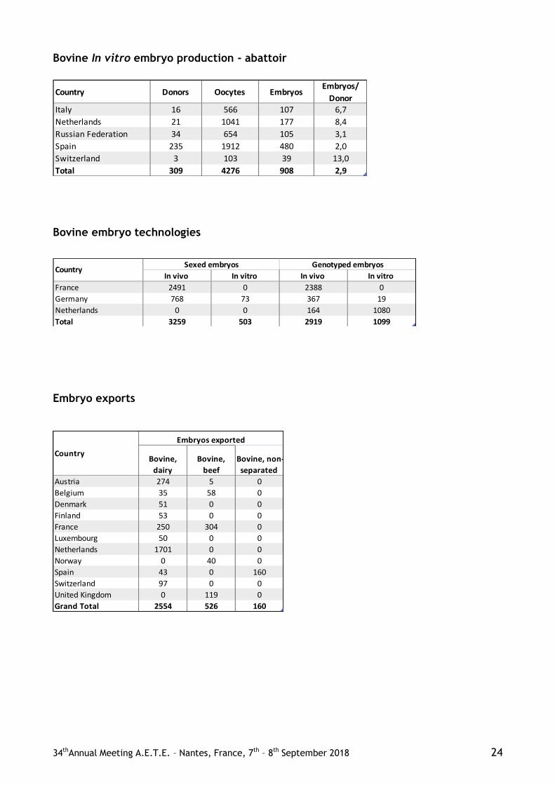
34thAnnual Meeting A.E.T.E. – Nantes, France, 7th – 8th September 2018 24
Bovine In vitro embryo production - abattoir
Country Donors Oocytes EmbryosEmbryos/
DonorItaly 16 566 107 6,7
Netherlands 21 1041 177 8,4
Russian Federation 34 654 105 3,1
Spain 235 1912 480 2,0
Switzerland 3 103 39 13,0
Total 309 4276 908 2,9
Bovine embryo technologies
In vivo In vitro In vivo In vitro
France 2491 0 2388 0
Germany 768 73 367 19
Netherlands 0 0 164 1080
Total 3259 503 2919 1099
Sexed embryos Genotyped embryosCountry
Embryo exports
Bovine,
dairy
Bovine,
beef
Bovine, non-
separated
Austria 274 5 0
Belgium 35 58 0
Denmark 51 0 0
Finland 53 0 0
France 250 304 0
Luxembourg 50 0 0
Netherlands 1701 0 0
Norway 0 40 0
Spain 43 0 160
Switzerland 97 0 0
United Kingdom 0 119 0
Grand Total 2554 526 160
Country
Embryos exported

34thAnnual Meeting A.E.T.E. – Nantes, France, 7th – 8th September 2018 25
Bovine In vivo embryo transfer
Fresh Frozen
domestic
Frozen
foreignFresh
Frozen
domestic
Frozen
foreignFresh
Frozen
domestic
Frozen
foreign
Austria 640 824 77 12 95 27 0 0 0 1675
Belgium 173 493 1616 995 3017 34 0 0 0 6328
Bosnia and Herzegovina 0 0 0 0 0 0 0 0 0 0
Croatia 0 0 0 0 0 0 0 0 0 0
Denmark 2494 1253 0 129 124 0 0 0 0 4000
Estonia 0 0 0 0 0 0 0 0 0 0
Finland 722 1484 212 9 1 33 0 0 0 2461
France 14216 12668 1615 2630 4548 289 56 0 0 36022
Germany 8047 11078 0 717 1351 0 0 0 0 21193
Greece 0 0 2 0 0 0 0 0 0 2
Hungary 60 58 0 29 129 0 0 0 0 276
Ireland 1698 2155 0 0 0 0 0 0 0 3853
Israel 192 8 0 0 0 0 0 0 0 200
Italy 7400 0 0 0 0 0 0 0 0 7400
Latvia 0 0 0 0 0 0 0 0 0 0
Lithuania 0 0 0 0 0 0 0 0 0 0
Luxembourg 280 800 35 20 70 0 0 0 0 1205
Macedonia 0 0 0 0 0 0 0 0 0 0
Netherlands 6303 17867 0 0 0 0 0 0 0 24170
Norway 35 142 0 25 28 113 0 0 0 343
Poland 760 288 123 20 3 0 0 0 0 1194
Portugal 92 339 0 31 32 13 0 0 0 507
Russian Federation 823 1484 24 45 11334 0 0 0 0 13710
Serbia 10 15 0 0 0 0 0 0 0 25
Slovakia 0 0 0 0 0 0 0 0 0 0
Slovenia 13 5 0 0 0 8 0 0 0 26
Spain 453 485 59 403 147 21 664 471 56 2759
Sweden 205 412 0 2 0 0 0 0 0 619
Switzerland 1012 2152 622 32 69 32 0 0 0 3919
Ukraine 21 0 0 0 0 0 0 0 0 21
United Kingdom 21 23 8 57 116 37 0 0 0 262
Total 45670 54033 4393 5156 21064 607 720 471 56 132170
Dairy breeds Beef breeds Non-separated breeds
CountryTotal
transfers
Bovine In vitro embryo transfer
Fresh Frozen domestic Frozen foreign Fresh Frozen domestic Frozen foreign
Finland 49 798 0 0 0 0 847
France 815 329 49 19 3 0 1215
Germany 1508 174 0 0 0 0 1682
Hungary 0 0 105 0 0 0 105
Italy 212 503 0 0 42 0 757
Netherlands 10196 4731 0 63 114 0 15104
Poland 16 7 0 0 0 0 23
Russian Federation 20302 7658 0 0 0 0 27960
Spain 1407 448 12 28 20 3 1918
Switzerland 2 7 117 0 0 0 126
United Kingdom 0 15 0 0 0 0 15
Total 34507 14670 283 110 179 3 49752
OPU AbattoirCountry Total transfers

34thAnnual Meeting A.E.T.E. – Nantes, France, 7th – 8th September 2018 26
Embryo production and transfer in other species – In vivo
Collections Viable embryos Fresh embryos Frozen domestic Frozen foreign
SHEEP
France 10 33 0 0 0
Hungary 5 39 0 0 45
Sweden 20 67 0 67 365
United Kingdom 557 2717 217 116 0
Total 592 2856 217 183 410
GOAT
France 9 76 0 0 0
Total 9 76 0 0 0
HORSE
Finland 4 1 1 0 0
France 1476 776 776 0 0
Italy 289 197 197 0 0
Netherlands 284 164 164 0 0
Poland 7 7 7 0 0
Russian Federation 3 2 2 4 0
Spain 15 12 12 0 0
Sweden 23 15 15 0 0
Switzerland 72 37 30 2 0
Total 2173 1211 1204 6 0
OTHER
Slovakia 10 20 0 0 0
Total 10 20 0 0 0
Embryo collection Embryo transfer
Country
Embryo production and transfer in other species – In vitro
OPU sessions Oocytes Embryos Fresh embryos Frozen embyos
BUFFALO
Italy 3 66 26 0 0
Total 3 66 26 0 0
HORSE
Italy 934 10866 885 82 203
Netherlands 213 2807 241 0 140
Switzerland 56 215 16 0 3
Total 1203 13888 1142 83 346
Country
Embryo transferEmbryo collection

34thAnnual Meeting A.E.T.E. – Nantes, France, 7th – 8th September 2018 27
INVITED LECTURES

34thAnnual Meeting A.E.T.E. – Nantes, France, 7th – 8th September 2018 28

Proceedings of the 34rd Meeting of the Association of Embryo Transfer in Europe (AETE); Nantes, France, September 7th and 8th, 2018.
_________________________________________ 3Corresponding author: [email protected]
Received: May 15, 2108 Accepted: July 16, 2018 29
Ovarian antral follicle populations and embryo production in cattle
Amanda Fonseca Zangirolamo1,2
, Fabio Morotti1, Nathalia Covre da Silva
1, Tamires Korchovei Sanches
1,
Marcelo M. Seneda
1,3
1Universidade Estadual de Londrina, Laboratório de Reprodução Animal (DCV-CCA-UEL), Londrina, PR, Brazil.
2National Institute of Science and Technology for Dairy Production Chain (INCT–LEITE), Universidade Estadual de Londrina, Londrina, PR, Brazil.
Abstract
Reproductive biotechniques such as embryo
production are important tools to increase the
reproductive performance in cattle in a short time. In this
context, the antral follicle count (AFC), which reflects the
population of antral follicles present in an ovary, has been
indicated as an important phenotypic characteristic
related to female fertility and closely correlated to the
performance of in vivo and in vitro embryo production
(IVEP). A positive correlation was evidenced between
AFC and oocyte retrieval by ovum pick up (OPU)
sessions and and with the number of embryos produced. Several studies have reported that females with a high
AFC had greater embryo yields compared to those with
medium and low AFC. However, controversial results
were obtained by studies conducted in different bovine
breeds. Many conflicting data may be due to the
differences in the experimental design, particularly
regarding the classification of animals in AFC groups,
subspecies particularities, herd aptitude or even issues
related to animal management. Therefore, aspects such as
the choice of donor, type of aspirated follicles and the
stage of follicular wave need to be clarified. Thus, this
text aims to discuss the use of AFC as a reproductive tool and its applications in the in vivo and in vitro production
of embryos, besides describing consistent results and new
challenges regarding AFC and embryo production.
Keywords: antral follicle count, Bos taurus, Bos indicus,
cow, fertility.
Introduction
In the world scenario for bovine embryos, in
the last decade, the in vitro embryo production (IVEP)
has expanded remarkably when compared to in vivo
embryo production (Watanabe et al., 2017). In the year
2016, a total of 666,215 in vitro embryos were
produced, exceeding for the first time the volume of
embryos produced in vivo (Perry, 2017). In this context,
Brazil has contributed to the consolidation of large-scale
application of this biotechnique and many challenges
have been faced to improve the IVEP (Perry, 2016).
Several studies have been conducted to maximize the reproductive efficiency of the herd. In this
way, the ovarian antral follicular count (AFC) as a tool
to evaluate the ovarian reserve, has been positively
correlated with parameters such as the number of viable
oocytes, blastocyst (Santos et al., 2016) and conception
rate after AI (Mossa et al., 2012). Different research groups investigated the embryo production performance
of females with different AFC. This parameter is
variable among females but highly constant in the same
female (Burns et al., 2005; Morotti et al., 2017). For
both indicus and taurus subspecies and indicus x taurus
crossbred, a positive correlation was found between
AFC and oocyte retrieval in the ovum pick up (OPU)
sessions and the number of embryos produced in vitro
(Ireland et al., 2008; Monteiro et al., 2017).
Considering embryonic production, females
with high AFC presented a larger number of in vitro embryos when compared to those with low AFC
(Ireland et al., 2008; Monteiro et al., 2017). Such
information is quite predictable, considering that more
follicles provide more oocytes to the in vitro embryo
production. On other hand, the influence of AFC on the
efficiency of embryonic production remains not well
understood. For example, Rosa et al. (2018), reported
no differences in both cleavage and blastocyst rates of
oocytes that came from ovaries with different AFC.
Also evaluating different AFC patterns, Rosa et al.
(2018) evaluated the genes involved in oogenesis and
folliculogenesis, which were differentially expressed in granulosa (progesterone receptor - PGR and Anti-
m llerian hormone receptor type II - AMHR2) and
cumulus cells (natriuretic peptide receptor 2 and 3 -
NPR2/ NPR3, fibroblast growth factor 10 - FGF10 and
signal transducer and activator of transcription 3 -
STAT3) from high versus low AFC cows. Mossa et al.
(2010) reported lower abundance of cytochrome P450
family 17 subfamily A member 1 - CYP17A1 mRNA in
thecal cells of low versus high AFC. Moreover, the study
by Ireland et al. (2009) reported that the abundance of
mRNAs for cytochrome P450 family 19 subfamily A member 1 - CYP19A1 in granulosal cells and estrogen
receptor 1 and 2 - ESR1/ ESR2, and cathepsin B - CTSB
in cumulus cells were greater, whereas mRNAs for AMH
in granulosal cells and TBC1 domain family member 1 -
TBC1D1 in thecal cells were lower for animals with low
compared to the high AFC group during follicle waves.
Considering a general view on AFC, many
points need to be addressed. For example, there is no
standard to the classification of AFC according to the
number of follicles. Comparing all the reports at the
literature, it is easy to recognize several patters of AFC groups. In this way, it is quite difficult to compare data
among the articles. Therefore, considering the

Zangirolamo et al. Antral follicle populations and embryo production.
_________________________________________
Anim. Reprod., v.15, n.3, p.000-00, Jul./Sept. 2018 30
importance of AFC on the embryo production this text
aims to discuss: i) The use of AFC as a reproductive
tool and the relationship between different fertility parameters (i.e. blastocyst rate and pregnancy, AMH
concentration, among others); ii) applications of AFC on
the in vivo and in vitro production of embryos and, iii)
controversial data and new challenges regarding to the
practical use of AFC as a reproductive tool for cattle.
Use of AFC as a reproductive tool
The association between reproductive
biotechniques and genetic improvement programs in
cattle has contributed to the development of the milk
and food production (Hansen, 2014). The selection of
females suitable for breeding is of great importance for
reproductive efficiency, mainly because some ovarian
physiological characteristics can directly influence the
number and quality of oocytes (Lonergan et al., 1994).
In this context, cows can be classified as low,
intermediate or high AFC, according to the number of
antral follicles (of 3 mm in diameter) detected via
ovarian ultrasonography.
The AFC has been related to fertility parameters such as ovarian size, corpus luteum diameter, number of healthy oocytes, endometrial thickness, progesterone concentration, pregnancy rate and calving interval (Jimenez-Krassel et al., 2009, Martinez et al., 2016). Thus, considering the relationship between AFC and several characteristics linked to fertility, the selection of donors could be performed using a single ultrasound examination at the beginning of reproductive life (Silva-Santos et al., 2014; Morotti et al., 2017). Also, the Anti-mullerian hormone (AMH), has been recognized as an indicator of ovarian
response to superovulation protocols (Souza et al., 2015), being AMH also highly correlated to the AFC and viability of the oocytes (Baruselli et al., 2016). It was also observed a positive association between AMH and fertility in dairy cows (Jimenez-Krassel et al., 2015).
The AFC is a characteristic of low to moderate heritability, and it was verified that there is no correlation between AFC and milk production during lactation in Holstein cows (Walsh et al., 2014). Similarly, for Braford (beef) cattle, some parameters related to meat production showed low correlation with AFC (Morotti et al., 2017). Then, based on these
studies, it can be suggested that AFC selection does not cause genetic demerit for the progeny.
In general, a greater number of antral follicles results in better OPU/IVEP quantitative efficiency. Many reports suggest that AFC is positively associated with the number of embryos produced by the donor in different breeds (Ireland et al., 2008; Silva-Santos et al., 2014, Santos et al., 2016). Although, more recently, Monteiro et al. (2017) did not find any advantage for Bos indicus females with high AFC relative to IVEP rates (Blastocyst rate 33.9% and 34.2% for low and high AFC animals, respectively).
In taurine females submitted to fixed time artificial insemination (FTAI) presenting high AFC, it was also found a higher pregnancy rate when compared
to those with low AFC (94% and 84%, respectively; Mossa et al., 2012). In contrast, studies with indicine cattle did not find a positive correlation between AFC and pregnancy rates with artificial insemination (Mendonça et al., 2013; Santos et al., 2016). Surprisingly, an evaluation of AFC and timed artificial insemination showed better reproductive performance in low AFC Nelore cows than high AFC females (conception rate 61.7% and 49.5%, respectively; Morotti et al., 2018). Recently, another unexpected result came from New Zealand with Bos taurus dairy cattle. The results showed that females with high AFC ( 25 follicles) had suboptimal fertility and shorter productive life when compared to those with low AFC (≤ 15 follicles) (Jimenez-Krassel et al., 2017). In contrast, also working with dairy cattle, Modina et al. (2014) had reported that those cows with intermediate number of antral follicles (n < 10) were identified with reduction in the fertility parameters, when compared to females with 10 follicles. Once again, the comparison between the articles is quite difficult considering that AFC was not the single condition that was different in the experimental design. As pointed by Martinez et al. (2016), the associations of AFC with other fertility parameters need for further evaluations to ensure the best way to use this strategy on reproductive programs.
In conclusion, the relationship between AFC and fertility needs to be clarified. Both in Bos taurus and Bos indicus cattle have shown conflicting data, mainly when different approaches, embryo transfer - ET or artificial insemination – AI, have been considered.
Applications of AFC for in vitro and in vivo
production of embryos
The embryo yield is highly variable in both in vitro and in vivo embryo production systems (Pontes et al., 2010). The IVP may be affected by oocyte recovery rate on OPU, as well as ET is affected by the response of the donor afterthe superovulation protocol (SOV) - (Ireland et al., 2007; Silva-Santos et al., 2014). Therefore, it is interesting to consider that the success of these techniques is dependent on the individual ovarian characteristics of the donor, which may affect the number and quality of the oocytes / embryos that are recovered (Stojsin-Carter et al., 2016).
In 2007, Ireland and collaborators performed a study with Bos taurus cattle and observed that the
number of IVF embryos per animal in the high AFC group ( 25 follicles) was four times higher (P < 0.05) than the low AFC group (≤ 15 follicles). In the multiple ovulation and embryo transfer (MOET), the number of embryos recovered after artificial insemination was 10.6 ± 2.7 vs. 4.7 ± 0.7 for the high and low AFC group, respectively (P < 0.05).
Similarly, indicus-taurus donors with high AFC ( 40 follicles), produced a higher (P < 0.05) number of blastocysts than animals of low AFC group (≤ 10 follicles) both in IVF and MOET approaches (Silva-Santos et al., 2014). However, the same team reported less consistent results for Bos indicus cattle. The high AFC showed better embryo production performance, but the cleavage rate was similar among

Zangirolamo et al. Antral follicle populations and embryo production.
_________________________________________
Anim. Reprod., v.15, n.3, p.000-00, Jul./Sept. 2018 31
low (≤ 7 follicles), intermediate (18 to 25 follicles) and high AFC ( 40 follicles) (Santos et al., 2016).
The relationship between of the ovarian pool of
follicles and the response to the superovulation has been
previously evaluated (Cushman et al., 1999). However,
possible applications of AFC and specific protocols to
SOV is topic that remains to be better understood.
Considering AFC and genes related to
folliculogenesis and oogenesis, it was reported differences in gene expression in cumulus and granulosa
cells collected from Nelore cows with low and high AFC.
Interestingly, in the study by Rosa et al. (2018), animals
com high AFC had genes upregulated in granulosa cells
while cows with a low AFC presented genes upregulated
only in cumulus cells. Taking together with data from
other studies, these findings suggest that AFC may
influence the molecular network that controls ovarian
function (Ireland et al., 2009; Mossa et al., 2010).
Considering the importance of communications between
oocyte and cumulus cells, as well as the influence of gonadotropins on the cAMP in the two cell types
(Luciano et al., 2004), it is quite predictable that AFC
may really interfere on the rates of embryonic production.
However, a precise experiment to consider only AFC and embryo production remains a challenge, since multiple
aspects need to be isolated in the experimental design.
As described in Table 1, the main problem for
comparing the AFC data is the classification of animals
in groups of high and low AFC. Each team considered a
specific strategy. Thus, it is quite difficult to compare
the results.
Controversial data among studies in AFC
classification also are present in both in vivo and in vitro
embryonic production, as shown in Table 2.
Considering data of pregnancy rate and AFC
after artificial insemination, it is also interesting to identify the clear differences among the groups, as shown
in the Table 3.
Table 1. Results (mean ± SD) from studies comparing in vitro embryo production in Bos taurus, Bos indicus and
Bos indicus-taurus (crossbred) between high and low AFC groups.
Author type AFC Animals
n
PU
n
COC’s
n
Blastocyst
n
Blastocyst
rate %
Ireland et al., 2007 Bos taurus
Low
(≤ 15 follicles)
68*** --- 7.5a 1.3
a 29.6
High
( 25 follicles)
37*** --- 29.5b 4.9
b 30.9
Silva-Santos et al., 2014 Bos indicus-taurus
Low (≤ 10 follicles)
20* 20 5.8 ± 3.4A
0.5 ± 0.8A 9.5
A
High
( 40 follicles)
20* 20 36.9 ± 13.7B 6.1 ± 4.5
B 16.5
B
Santos et al., 2016 Bos indicus Low (≤ 7 COC’s)
19* 19 3.8 ± 1.1α 0.6 ± 0.6
α 13.0
α
High
( 40 COC’s)
22* 22 40.4 ± 10.6β 18.4 ± 6.7
β 41.9
β
Monteiro et al., 2017 Bos indicus Low
(< 15 COC’s)
18** 216 10.8 ± 0.4 3.6 ± 0.2 33.9
High
( 15 COC’s)
18** 216 21.2 ± 1.0 7.1 ± 0.4 34.2
Rosa et al., 2018 Bos indicus Low
(≤ 31 follicles)
356*** --- 536 (total) 203 (total) 38.6
High
( 92 follicles)
356*** --- 617 (total) 251 (total) 40.6
*Animals submitted to OPU only once. **Animals submitted to OPU more than once. ***Animals submitted to postmortem ovarian aspiration. a,b/A,B/α,βFor the same author and variable were different (P ≤ 0.05) between the AFCs groups. Adapted from Ireland et al. (2007); Silva-Santos et al. (2014); Santos et al. (2016); Monteiro et al. (2017) and Rosa et al. (2018).

Zangirolamo et al. Antral follicle populations and embryo production.
_________________________________________
Anim. Reprod., v.15, n.3, p.000-00, Jul./Sept. 2018 32
Table 2. Results (mean ± SD) from studies comparing in vivo embryo production in Bos taurus and Bos indicus-
taurus (crossbred) between high and low AFC groups.
Author Animals Number of follicles Number of flushes Transferable embryos/ animals
Ireland et al., 2007
Bos taurus
Low (≤ 15 follicles)
21* 3.8 ± 0.8a
High
( 25 follicles)
19* 5.4 ± 1.3b
Silva-Santos et al., 2014 Bos indicus-taurus
Low
(≤ 10 follicles)
20** 1.9 ± 2.1A
High
( 40 follicles)
20** 6.9 ± 5.3B
*The same female may have been superovulated and collected up to twice. **One single collection per animal. a,b/A,BFor the same author were different (P ≤ 0.05) between the AFCs groups. Adapted from Ireland et al. (2007)
and Silva-Santos et al. (2014).
Table 3. Reproductive performance of females with high or low follicle count (AFC) after artificial insemination.
Author Group Pregnancy rate (%)
Mossa et al., 2012
Low AFC (≤ 15 follicles)
84
High AFC ( 25 follicles) 94
Mendonça et al., 2013 Low (≤ 12 follicles) 51.85
High AFC ( 30 follicles) 44.73
Santos et al., 2016 Low AFC (≤ 10 follicles) 58,6
High AFC ( 25 folículos) 51,9
Adapted from Mossa et al. (2012); Mendonça et al. (2013) and Santos et al. (2016).
AFC: consistent data versus new challenges
The strategy used to the classification of
females in AFC categories is very different in each
author. Each one exhibited a distinct experimental
design; some studies did not consider the AFC
intermediate group or, for example, the oocytes were
recovered in different ways - by OPU or ovaries from a slaughterhouse (Ireland et al., 2008; Monteiro et al.,
2017). In some cases, the parameters used to classify
females in AFC categories differed considerably among
authors (Morotti et al., 2015). Therefore, when
comparing studies concerning AFC, we should consider
how the separation of animals into the categories was
made, trying to establish a more balanced comparison.
Moreover, there are differences between the
studied subspecies inherent to the physiology of the
estrous cycle, which encompass divergences from
follicular wave number per cycle to the number of
follicles recruited per growth wave (Baruselli et al., 2007). For example, Holstein cows tend to present the
predominance of two or three waves of follicular growth
per estrus cycle (Ginther et al., 1989; Wolfenson et al.,
2004), whereas Zebu cows are more related to three to
four waves per estrus cycle (Rhodes et al., 1995; Nelore
- Figueiredo et al., 1997). Also, Bos indicus females
recruit more follicles per follicular growth wave than
Bos taurus females (33,4 ± 3,2 vs 25,4 ± 2,5; Carvalho et al., 2008). Taking those data together, it is easy to
realize the challenge to compare studies that present
results of distinct subspecies. So, the comparison of
AFC with fertility in different breeds should be studied
individually.
Furthermore, for a better precision of the
factors that interfere with AFC, it is necessary to
consider the development phase in which the follicles
are aspirated since there is a direct interference on the oocyte competence for IVEP. In this context, Cavalieri
et al. (2018), showed that cows submitted to follicular
wave synchronization had a better IVEP rate and
pregnancy rate after ET when compared to females
aspirated on a random day of estrous cycle. Because of
that, the strategy for oocyte recovery must be
considered when comparing different AFC cows.
In addition to the subspecies, it is also
important to consider the influence of the age of the
females in the AFC studies, since there are indications,
that the ovarian follicular reserve decreases after the
female reaches five years, suggesting a decrease in fertility (Cushman et al., 2009). The same authors also
reported the importance of birth weight as a parameter
that influences AFC, but the stage of estrous cycle
seems to not interfere in the AFC classification, which
facilitates the establishment of pattern (high,
intermediate or low AFC) to each cow (Cushman et al.,
2009).
In summary, according to the literature and
data cited above, AFC seems to be correlated with

Zangirolamo et al. Antral follicle populations and embryo production.
_________________________________________
Anim. Reprod., v.15, n.3, p.000-00, Jul./Sept. 2018 33
several fertility parameters, and it may be a tool that can
contribute to the success of embryo production both in
vivo and in vitro. However, there is a great need to study the real long term impact of AFC on fertility, to
establish specific parameters of AFC classification and
to understand the physiological causes of the variation
in the AFC among individual female cattle.
Final comments
Despite many studies on antral follicle
population, the relationship between AFC, fertility, and
efficiency of biotechniques are not fully understood.
However, in the context of embryo production, the AFC
can be used as an auxiliary tool for the selection of animals with the greater quantitative potential of
embryos. Finally, a better understanding of the factors
linked to AFC and of the reproductive characteristics
from Bos indicus and Bos taurus may provide
adjustments in cattle management and to improve the
efficiency of reproductive biotechniques.
References
Baruselli PS, Gimenes LU, Sales JNS. 2007.
Fisiologia reprodutiva de fêmeas taurinas e zebuínas. Rev Bras Reprod Anim, 31:205-211.
Baruselli PS, Batista EOS, Ferreira RM. 2016.
Plasma anti-müllerian hormone allows reproductive
selection of donors with greater potential of embryo
production. Spermova, 6:1-13.
Burns DS, Jimenez-Krassel F, Ireland JLH, Knight
PG, Ireland JJ. 2005. Numbers of antral follicles
during follicular waves in cattle: evidence for high
variation among animals, very high repeatability in
individuals, and an inverse association with serum
follicle-stimulating hormone concentrations. Biol Reprod, 73:53-62.
Carvalho JBP, Carvalho NAT, Reis EL, Nichi M,
Souza AH, Baruselli PS. 2008. Effect of early
luteolysis in progesterone-based timed AI protocols in
Bos indicus, Bos indicus x Bos taurus, and Bos taurus
heifers. Theriogenology, 69:167-175.
Cavalieri FLB, Morotti F, Seneda MM, Colombo
AHB, Andreazzi MA, Emanuelli IP, Rigolon LP.
2018. Improvement of bovine in vitro embryo
production by ovarian follicular wave synchronization
prior to ovum pick-up. Theriogenology, 57:70-117.
Cushman RA, DeSouza JC, Hedgpeth VS, Britt JH. 1999. Superovulatory response of one ovary is related to
the micro-and macroscopic population of follicles in the
contralateral ovary of the cow. Biol Reprod, 60:349-
354.
Cushman RA, Allan MF, Kuehn LA, Snelling WM,
Cupp AS, Freetly HC. 2009. Evaluation of antral
follicle count and ovarian morphology in crossbred beef
cows: Investigation of influence of stage of the estrous
cycle, age, and birth weight. J Anim Sci, 87:1971-1980.
Figueiredo RA, Barros CM, Pinheiro OL, Sole JMP.
1997. Ovarian follicular dynamics in Nelore breed (Bos indicus) cattle. Theriogenology, 47:1489-1505.
Ginther OJ, Knopf L, Kastelic JP. 1989. Temporal
associations among ovarian events in cattle during
oestrous cycles with two or three follicular waves. J Reprod Fertil, 87:223-230.
Hansen P. 2014. Current and Future Assisted
Reproductive Technologies for Mammalian Farm
Animals. Adv Exp Med Biol, 752:1-22.
Ireland JJ, Ward F, Jimenez-Krassel F, Ireland
JLH, Smith GW, Lonergan P, Evans ACO. 2007.
Follicle numbers are highly repeatable within individual
animals but are inversely correlated with FSH
concentrations and the proportion of good-quality
embryos after ovarian stimulation in cattle. Hum
Reprod, 22:1687-1695.
Ireland JLH, Scheetz D, Jimenez-Krasse F,
Themmen APN, Ward F, Lonergan P, Smith GW,
Perez GI, Evans ACO, Ireland JJ. 2008. Antral
follicle count reliably predicts number of
morphologically healthy oocytes and follicles in ovaries
of young adult cattle. Biol Reprod, 79:1219-1225.
Ireland JJ, Zielak-Steciwko AE, Jimenez-Krassel F,
Folger J, Bettegowda A, Scheetz D, Walsh S, Mossa
F, Knight PG, Smith GW, Lonergan P, Evans ACO.
2009. Variation in the Ovarian Reserve Is Linked to
Alterations in Intrafollicular Estradiol Production and
Ovarian Biomarkers of Follicular Differentiation and Oocyte Quality in Cattle. Biol Reprod, 80:954-964.
Jimenez-Krassel F, Folger JK, Ireland JLH, Smith
GW, Hou X, Davis JS, Lonergan P, Evans ACO,
Ireland JJ. 2009. Evidence that high variation in
ovarian reserves of healthy young adults has a negative
impact on the corpus luteum and endometrium during
estrous cycles in cattle. Biol Reprod, 80:1272-1281.
Jimenez-Krassel F, Scheetz DM, Neuder LM,
Ireland JLH, Pursley JR, Smith GW, Tempelman
RJ, Ferris T, Roudebush WE, Mossa F, Lonergan P,
Evans, ACO, Ireland JJ. 2015. Concentration of anti-Müllerian hormone in dairy heifers is positively
associated with productive herd life. J Dairy Sci,
98:3036-3045.
Jimenez-Krassel F, Scheetz DM, Neuder LM,
Pursley JR, Ireland JJ. 2017. A single ultrasound
determination of ≥25 follicles ≥3 mm in diameter in
dairy heifers is predictive of a reduced productive herd
life. J Dairy Sci, 100:5019-5027.
Lonergan P, Monaghan P, Rizos D, Boland MP,
Gordon I. 1994. Effect of follicle size on bovine oocyte
quality and development competence following
maturation, fertilization and culture in vitro. Mol Reprod Dev, 37:48-53.
Luciano AM, Modina S, Vassena R, Milanesi E,
Lauria A, Gandolfi F. 2004. Role of intracellular
cyclic adenosine 3′, 5′-monophosphate concentration
and oocyte-cumulus cells communications on the
acquisition of the developmental competence during in
vitro maturation of bovine oocyte. Biol Reprod, 70:465-
472.
Martinez MF, Sanderson N, Quirke LD, Lawrence
SB, Juengel JL. 2016. Association between antral
follicle count and reproductive measures in New Zealand lactating dairy cows maintained in a pasture-

Zangirolamo et al. Antral follicle populations and embryo production.
_________________________________________
Anim. Reprod., v.15, n.3, p.000-00, Jul./Sept. 2018 34
based production system. Theriogenology, 85:466-475.
Mendonça HGR, Santos GMG, Silva-Santos KC,
Barreiros TRR, Blaschi W, Morotti F, Moraes FLZ, Seneda MM. 2013. Pregnancy rates following FTAI of
Nelore heifers (Bos indicus) with high, intermediate and
low numbers of antral follicles. Anim Reprod, 10:452.
Modina SC, Tessaro I, Lodde V, Franciosi F,
Corbani D, Luciano AM. 2014. Reductions in the
number of mid-sized antral follicles are associated with
markers of premature ovarian senescence in dairy cows.
Reprod Fertil Dev, 26:235-244.
Monteiro FM, Batista EOS, Vieira LM, Bayeux BM,
Accorsi M, Campanholi SP, Dias EAR, Souza AH,
Baruselli PS. 2017. Beef donor cows with high number
of retrieved COC produce more in vitro embryos compared with cows with low number of COC after
repeated ovum pick-up sessions. Theriogenology,
90:54-58.
Morotti F, Barreiros TRR, Machado FZ, González
SM, Marinho LSR, Seneda MM. 2015. Is the number
of antral follicles an interesting selection criterium for
fertility in cattle? Anim Reprod, 12:479-486.
Morotti F, Santos GMG, Koetz Júnior C, Silva-
Santos KC, Roso VM, Seneda MM. 2017. Correlation
between phenotype, genotype and antral follicle
population in beef heifers. Theriogenology, 91:21-26.
Morotti F, Moretti R, Santos GMG, Silva-Santos
KC, Cerqueira PH, Seneda MM. 2018. Ovarian
follicular dynamics and conception rate in Bos indicus
cows with different antral follicle counts subjected to
timed artificial insemination. Anim Reprod Sci,
188:170-177.
Mossa F, Jimenez-Krassel F, Folger JK, Ireland
JHL, Smith GW, Lonergan P, Evans ACO, Ireland
JJ. 2010. Evidence that high variation in antral follicle
count during follicular waves is linked to alterations in
ovarian androgen production in cattle. Reproduction, 140:713-720.
Mossa F, Walsh SW, Butler ST, Berry DP, Carter F,
Lonergan P, Smith GW, Ireland JJ, Evans ACO.
2012. Low numbers of ovarian follicles ≥ 3 mm in
diameter are associated with low fertility in dairy cows.
J Dairy Sci, 95:2355-2361.
Perry G. 2016. Statistics of embryo collection and
transfer in domestic farm animals. Embryo Technology
Newsletter, 34:1-75.
Perry G. 2017. Statistics of embryo collection and
transfer in domestic farm animals. Embryo Technology
Newsletter, 35:1-46.
Pontes JHF, Silva KCF, Basso AC, Rigo AG,
Ferreira CR, Santos GMG, Sanches BV, Porcionato
JPF, Vieira PHS, Faifer FS, Sterza FAM, Schenk JL,
Seneda MM. 2010. Large-scale in vitro embryos
production and pregnancy rates from Bos taurus, Bos
indicus, and indicus-taurus dairy cows using sexed
sperm. Theriogenology, 74:1349-1355.
Rhodes FM, De’ath G, Entwistle KW. 1995. Animal
and temporal effects on ovarian follicular dynamics in
Brahman heifers. Anim Reprod Sci, 38-265-277.
Rosa CO, Marinho LSR, Da Rosa PRA, De Cesaro
MP, Lunardelli PA, Santos KCS, Basso AC,
Bordignon V, Seneda MM. 2018. Molecular
characteristics of granulosa and cumulus cells and
oocyte competence in Nelore cows with low and high numbers of antral follicles. Reprod Domest Anim,
53:921-929.
Santos GMGD, Silva-Santos KC, Barreiros TRR,
Morotti F, Sanches BV, de Moraes FLZ, Blaschi W,
Seneda MM. 2016. High numbers of antral follicles are
positively associated with in vitro embryo production
but not the conception rate for FTAI in Nelore cattle.
Anim Reprod Sci, 165:17-21.
Silva-Santos KC, Santos GMG, Koetz Júnior C,
Morotti F, Siloto LS, Marcantonio TN, Urbano MR,
Oliveira RL, Lima DCM, Seneda MM. 2014. Antral
follicle populations and embryo production - in vitro and in vivo - of Bos indicus-taurus donors from weaning
to yearling ages. Reprod Domest Anim, 49:228-232.
Souza AH, Carvalho P, Rozner A, Vieira L,
Hackbart K, Bender R, Dresch A, Verstegen J,
Shaver R, Wiltbank M. 2015. Relationship between
circulating anti-Müllerian hormone (AMH) and
superovulatory response of high-producing dairy cows.
Jpurnal of Dairy Science, 98:169-178.
Stojsin-Carter A, Mahboubi K, Costa NN, Gillis DJ,
Carter TF, Neal MS, Miranda MS, Ohashi OM,
Favetta LA, King WA. 2016. Systemic and local anti-Mullerian hormone reflects differences in the
reproduction potential of Zebu and European type cattle.
Anim Reprod Sci, 167:51-58.
Walsh SW, Mossa F, Butler ST, Berry DP, Scheetz
D, Jimenez-Krassel F, Tempelman RJ, Carter F,
Lonergan P, Evans ACO, Ireland JJ. 2014.
Heritability and impact of environmental effects during
pregnancy on antral follicle count in cattle. J Dairy Sci,
97:4503-4511.
Watanabe YF, Souza AH, Mingoti RD, Ferreira RM,
Batista EOS, Dayan A, Watanabe O, Meirelles FV, Nogueira MFG, Ferraz JBS, Baruselli PS. 2017.
Number of oocytes retrieved per donor during OPU and
its relationship with in vitro embryo production and
field fertility following embryo transfer. Anim Reprod,
14:635-644.
Wolfenson D, Inbara G, Rotha Z, Kaimb M, Blocha
A, Braw-Tal R. 2004. Follicular dynamics and
concentrations of steroids and gonadotropins in lactating
cows and nulliparous heifers. Theriogenology, 62:1042-
1055.

Proceedings of the 34th Meeting of the Association of Embryo Transfer in Europe (AETE); Nantes, France, September 7th and 8th, 2018.
_________________________________________ 1Corresponding author: [email protected]
Received: June 25, 2018 Accepted: July 6, 2018 35
Preservation of female fertility in humans and animal species
Helen Mary Picton1
Reproduction and Early Development Research Group, Discovery and Translational Science Department, Leeds Institute Of
Cardiovascular and Metabolic Medicine, School of Medicine, University of Leeds, UK.
Abstract
A detailed understanding of the cryobiology of
gametes and complex tissues has led to the development
of methods that facilitate the successful low temperature
banking of isolated mature human oocytes, or immature oocytes in situ within fragments of human ovarian
cortex. Although many outstanding research challenges
remain to be addressed, the successful development of
new treatments to preserve female fertility for a range of
clinical indications has largely been underpinned by the
conduct of extensive, fundamental research on oocytes
and ovarian tissues from a number of laboratory and
commercially important farm species. Indeed, the most
recent evidence from large animals suggests that it is
also possible to cryopreserve intact whole ovaries along
with their supporting vasculature for later auto-transplantation and restoration of natural fertility. This
review will explore how the methods developed to
preserve human oocytes and ovarian tissues can now be
used strategically to support the development of
conservation strategies aimed at safeguarding the
genetic diversity of commercially important domestic
animals and also of preserving the female germplasm
for wild animals and endangered species.
Key words: cryopreservation, fertility preservation,
oocyte, ovary.
Introduction
In the last few decades, advances in
cryobiology have been combined with the development
of new assisted reproduction technologies (ARTs) and
used as a means to cryopreserve the structural integrity
and biological function of key reproductive cells.
Translation of these research advances has resulted in
the development of the capacity to cryopreserve and
long-term store isolated gametes, embryos, complex
gonadal tissues and even whole reproductive organs in humans and laboratory species as well as commercially
important farm animal breeds and a limited number of
exotic or endangered species. Indeed, the issue of
fertility preservation is particularly relevant in animals
as over the last two decades, some 300 of 6000 farm
animal breeds have become extinct and a further 1350
domestic breeds are being threatened with extinction as
a result of aggressive animal breeding strategies using
limited genetic stocks of animals of high merit for a
range of economically valuable traits (Taberlet et al.,
2008).
The standard method of fertility preservation in
males whether humans or animals is the
cryopreservation and storage of sperm (for review see
Ehmcke and Schlatt, 2008). Similarly, since the first
successful cryopreservation of embryos in laboratory
species in the early 1970s (Whittingham et al., 1972) thousands of animal and human offspring have been
born following embryo cryopreservation. With regard to
oocytes, the recent development of improved methods
and devices for the vitrification of isolated cells has
increased the success rates of metaphase II (MII),
oocyte preservation in humans (Rienzi et al., 2017).
This technological advance has only had limited
application to the oocyte preservation in animals due to
a general lack of basic understanding of oocyte biology
in different target species (Comizzoli, 2017). While
sperm, oocytes and embryo preservation using ART are clearly effective, recent research focus has shifted to the
preservation of gametes in situ either within fragments
of ovarian and testicular tissue or following whole
organ cryopreservation. Indeed, the banking of gonadal
tissue is particularly valuable as a means of preserving
fertility as in the case of the ovary the banked tissue
may contain thousands of the earliest staged primordial
follicles and oocytes. In contrast testicular tissue
cryopreservation facilitates the banking of
spermatogonial stem cells that can subsequently can be
harvested and used to repopulate the testis and restart spermatogenesis (Picton et al., 2015). Although no
standardized protocols exist for fertility preservation in
any species, the preservation of gonadal tissues offers
near unlimited potential for fertility preservation and
restoration in humans as well as domestic and wild
animal species. This review aims to provide an
overview of the recent advances in human oocyte and
ovarian tissue banking and how these clinical
reproductive technologies can be used to support
fertility conservation strategies in animals.
Cryobiology and fertility preservation
Cryopreservation is a multi-step procedure that
requires that cells tolerate exposure to: molar
concentrations of penetrating and non-penetrating
cryoprotective agents (CPAs); cooling to subzero
temperatures; and either removal or conversion of the
greater majority of liquid water within the cells into a
solid state. The reverse occurs during thawing or
warming. Any of these elements can inflict damage on
the cells to be preserved although the level and nature of
the damage is dependent on the cryobiological properties of each individual cell type. With regards to

Picton. Female fertility preservation.
_________________________________________
Anim. Reprod., v.15, n.3, p.000-00, Jul./Sept. 2018 36
the cryopreservation of complex tissues containing
multiple cell types, such as the ovary, the success of
cryopreservation is dependent on the need to balance the freezing optima for a range of different cell types which
are influenced by cell number and size and, in the case
of oocytes, maturational status, as well as the
requirement to preserve the structural integrity of the
tissue. Furthermore, the nature of the cryopreservation
methodology, whether slow freezing or vitrification, the
cooling and warming rates and the containment vessel
used for tissue preservation and storage will all
influence the efficacy of the preservation method and
hence impact on the subsequent viability of the
preserved tissue. Finally, the unique biological
properties of oocytes challenge our potential to freeze-store these important cells. Mammalian oocytes are very
large cells of approximately 120 µm diameter with a
small surface area to volume ratio and high lipid and
water content. The latter confers a high sensitivity to
chilling injury and intracellular ice formation. In MII
oocytes these parameters are further confounded by the
presence of a fragile cytoskeleton that is resistant to the
volumetric excursions commonly associated with
equilibrium freezing and a highly temperature sensitive
meiotic spindle apparatus (Saragusty and Arav, 2011).
These time-dependent sensitivities to chilling to 0C are evident in MII oocytes from a range of species
including mouse (Pickering and Johnson, 1987) rhesus monkey (Songsasen et al., 2002) and human (Pickering
et al., 1990; Zenzes et al., 2001). Indeed, cooling
oocytes to room temperature for even as little as 10
minutes can cause irreversible damage to the meiotic
spindle. Furthermore, chilling oocytes to 0C in the presence of a CPA appears to exacerbate these
abnormalities (Mullen et al., 2004).
Despite these difficulties, significant advances
have been made in our ability to cryopreserve MII
oocytes in a range of species. The most convincing data
on the efficacy of MII oocyte preservation is
undoubtedly derived from human ART. Initial attempts
to slow freezing human MII oocytes (Picton and Chambers, 2009) met with only limited success as
evidenced by the key statistic of live birth rates per
oocyte thawed (Oktay et al., 2006). Since then the
efficacy of MII oocyte cryopreservation has increased
dramatically with live birth rates following human MII
oocyte vitrification by the best IVF clinics now being
equivalent to those achieved during embryo freezing
(Glujovsky et al., 2014; Rienzi et al., 2017). The
improved efficiency of human MII preservation can be
attributed to the development of optimized vitrification
protocols that minimize the cells exposure to the cytotoxic impact of high concentrations of usually two
or more CPAs including penetrating CPAs such as
ethylene glycol and propanediol and non-penetrating
CPAs such as sucrose or trehalose. Oocyte cooling rates
during vitrification have also been maximized by the
commercial development of specialized vitrification
devices for use in ART that support the preservation of
individual oocytes or small groups of cells within
minimal volumes of cryopreservation reagents. As a
result of these advances MII oocyte vitrification is no
longer considered an experimental technique in human
ART (Rienzi et al., 2017) and the approach is regularly used to preserve oocytes for young women who are
undergoing infertility treatment, teenage girls or young
women who need to safeguard their future fertility
before exposure to the ablative cancer treatments, or
more controversially, young women who wish to delay
childbearing and elect to safeguard their future fertility
by oocyte banking (Cobo et al., 2016). Regardless of
species, it is clear that the success of oocyte
cryopreservation is dependent on oocyte quality, a key
parameter that is profoundly and negatively influenced
by advancing maternal age (Goldman et al., 2017).
While significant improvements have been made in human MII oocyte vitrification these methods
have had only a limited impact on oocyte banking in
animals over and above the laboratory and livestock
species that are used as models for the development and
testing of the human ART protocols (Wildt et al., 2010).
Never-the-less encouraging results for MII vitrification
have been obtained in some domestic species such as
the cat (Fernandez-Gonzalez and Jewgenow, 2017) as
well as some exotic species including for example the
Tasmanian devil (Czarny and Roger, 2010) and
Mexican gray wolf (Boutelle et al., 2011). While commonalities in the biophysical traits associated with
cell freezing between taxonomically-related species
may be useful to inform protocol development and
support improved post-thaw viability across species, in
general the similarities between taxa tend to be
overshadowed by the magnitude of species specific
differences in the ability of their oocytes to tolerate
osmotic and toxic effects of CPA exposure and chilling
injury (Woods et al., 2004). For example, felid oocytes
appear far more tolerant to osmotic changes and cold
temperatures than oocytes from bovids, cervids or equids that appear to be more sensitive to cold shock
(Comizzoli et al., 2010, 2012). Overall the ability of
MII oocytes to survive the freezing and thawing, or
cooling and warming processes is highly variable and
difficult to predict across species (for review see
Comizzoli et al., 2012). Thus despite the advances that
have been made with MII oocyte cryopreservation in
humans, fundamental cryobiological investigations and
further optimization of oocyte cryopreservation
protocols combined with comparative studies across
species are needed before MII oocytes cryopreservation
can be used efficiently to preserve the fertility of wild or endangered animals, especially where the numbers of
oocytes available for preservation are limited.
Some of the difficulties associated with the
cryopreservation of MII oocytes may potentially be
overcome by the preservation of fully grown, but
nuclear immature, germinal vesicle (GV) staged
oocytes. However, this option has proved inconsistent
and like the MII oocyte preservation detailed earlier,
success rates appear to be influenced by species. In
addition to rodent and ruminants, successful GV oocyte
vitrification has recently been reported in equids (Ortiz-Escribano et al., 2018) and domestic cats. The evidence

Picton. Female fertility preservation.
_________________________________________
Anim. Reprod., v.15, n.3, p.000-00, Jul./Sept. 2018 37
suggests that GV oocytes appear to be more cryo-
resistant than their MII counterparts (Comizzoli et al.,
2008). Regardless of species, GV oocytes cryopreservation requires the freeze-storage of both the
gamete and its supporting compliment of cumulus
granulosa cells. The success of GV oocyte
cryopreservation is critically dependent on the post-
thaw/warming maintenance of the functional integrity of
the heterologous gap junctional contacts connecting the
cumulus cells to the oocyte. This network of cumulus
cells supply the oocyte with vital nutrients and signaling
molecules that are essential to drive the cytoplasmic and
nuclear maturation of the oocyte to the MII stage.
Furthermore, cumulus cells have a discretely different
cryopreservation optima compared to oocytes. The loss of gap junctional contacts between the cumulus cell
compartment and the GV oocyte is a common casualty
of the cryopreservation process such that both the
capacity of the oocyte to under go in vitro maturation
and its subsequent fertile potential and developmental
competence are severely compromised. Finally, GV
oocyte preservation must be supported by the provision
of robust culture environments that support oocyte
maturation in vitro. Insight into the discrete, species-
specific differences in the composition of the culture
environment required to drive oocyte maturation will be needed to maximize oocyte quality after
cryopreservation.
Regardless of their stage of nuclear maturity
secondary oocytes can be harvested in humans and other
mammalian species by ultrasound-guided laparoscopy
or by laparotomy. Controlled ovarian stimulation with
exogenous hormones is frequently used to increase the
number of appropriately staged antral follicles at the
time of oocyte aspiration. Alternatively, secondary
oocytes can be harvested by needle aspiration of antral
follicles at any stage of the reproductive cycle following ovariectomy, or as a byproduct of surgical procedures or
following the natural mortality of an animal.
Ovarian tissue preservation
Many of the biophysical challenges associated
with preserving secondary oocytes can be avoided by
the banking of immature primordial oocytes through
ovarian tissue cryopreservation. Indeed, the biological
characteristics of primordial oocytes are well suited to
preservation since primordial oocytes are relatively
undifferentiated and, in the case of humans, are 3-4 fold smaller in diameter than their secondary oocyte
counterparts. Importantly, primordial oocytes represent
the building blocks of female reproduction and,
depending on age, the mammalian ovarian cortex is
endowed with thousands to millions of primordial
oocytes enclosed within primordial follicles (Picton,
2001). The quantity and quality of the primordial
follicles within this ovarian reserve ultimately defines
the reproductive longevity and fertile potential of an
individual. While our understanding of the mechanisms
that regulate the activation of primordial follicle growth and hence depletion of the ovarian reserve is limited,
especially in wild animals and there is considerable
variability between species, ultimately the ovarian
reserve represents the major germplasm resource that can be consistently and safely banked by wildlife
reproductive biologists, veterinarians and clinicians
alike as a means of safeguarding future fertility and
maintaining genetic diversity.
Strategies for ovarian tissue cryopreservation
are predominantly centered around the preservation of
primordial follicles in situ either within fragments of
ovarian cortex or by banking the whole ovary. It is
possible to freeze-store isolated primordial oocytes but
in the absence of supporting ovarian stromal cells these
presumptive gametes have only limited viability post
thaw/warming. The efficacy of ovarian cryopreservation is dependent on: optimization of the cryopreservation
protocol for the species of interest; the individual’s age
at the time of tissue harvest and storage; the requirement
to successfully preserve both the structural integrity and
biological complexity of the tissue. Ovarian cortical
tissue is comprised of functionally diverse cell types
including immature GV oocytes, follicular somatic
cells, stromal cells, blood vessels and the ovarian
surface epithelium (Picton et al., 2000; Picton, 2001).
All of these cell types have different cryopreservation
optima and all play different but important functional roles in supporting the activation and completion of
follicle and oocyte growth and as well as the
maintenance of the ovarian reserve, whether in vivo or
in vitro. Finally, strategies designed to successfully
preserve the integrity and fertile potential of ovarian
tissues and, conversely, to limit cryo-damage need to be
developed in parallel with the methods to use the tissue
to restore fertility following banking. Despite these
challenges ovarian tissue cryopreservation have been
optimized and used successfully for a range of species
including mice, rats, rabbits, cats, dogs, sheep, goats, cows, pigs, horses, camels, wallabies, wombats,
dasyurids, elephants, monkeys and humans (for review
see Devi and Goel, 2016).
The majority of methods used to cryopreserve
ovarian tissue are based on the original validated slow
freezing protocols developed for the successful
cryopreservation of human ovarian cortex as published
by Newton et al. (1996) and the associated publications
and live births recorded in sheep by Gosden et al.
(1994) and Baird et al. (1999). In general, ovarian
cortex is harvested following ovary removal or ovarian
biopsy and thin fragments of cortex are equilibrated in CPA before undergoing controlled rate cooling with
manual ice seeding before the temperatures are brought
down to -196oC and the tissue is stored at liquid
nitrogen temperatures. There is now a substantial
clinical evidence base to substantiate these early
successes. Several thousand girls and young women
worldwide have safeguarded their future fertility using
ovarian tissue preservation methods. The most common
indication for ovarian tissue banking is the need to
preserve the future fertility of girls and women: who are
at risk of premature ovarian failure (POF) following exposure to the ovotoxic impact of chemotherapy agents

Picton. Female fertility preservation.
_________________________________________
Anim. Reprod., v.15, n.3, p.000-00, Jul./Sept. 2018 38
for the treatment of cancer (Gellert et al., 2018; Oktay et
al., 2018) or other haematological pathologies such as
sickle cell anemia (Demeestere et al., 2015) or beta thalassemia (Matthews et al., 2018); or who carry a high
risk of POF for genetic conditions such as Turners
syndrome (Oktay et al., 2016); or who will suffer
ovarian tissue damage and loss following abdominal
trauma.
The potential of the stored ovarian tissue to
support fertility restoration when the individual wishes
to start her family is demonstrated by the delivery of
more than 130 healthy babies world wide after
autotransplantation of the frozen-thawed tissue and the
restoration of endocrine and ovarian function (Pacheco
and Oktay, 2017) with more ongoing pregnancies being recorded. Although the overall efficiency of fertility
restoration following ovarian tissue preservation and
transplantation is unknown, recent evidence from
defined cohorts of patients suggests a ~30% live birth
rate after autotransplantation of slow frozen-thawed
ovarian cortex, (Jadoul et al., 2017; Keden et al., 2018).
When fertility preservation strategies require the
removal of the whole ovary and it is age appropriate to
do so, ovarian cortex harvest and cryopreservation can
be most effectively combined with methods for in vitro
maturation and vitrification of MII oocytes to maximize the likelihood of a future successful pregnancy outcome
for the patient. Although significant progress has been
made in the development and use of ovarian tissue
cryopreservation as a means to safeguard the future
fertility of girls and young women at risk of POF,
further optimization of the cryopreservation and
transplantation protocols are likely to be beneficial as
the longevity of ovarian autograft function following
transplant remains unclear. The latter is likely to be
determined by patient age at tissue harvest and by the
degree of follicle loss that results from ischaemia and reperfusion injury following ovarian tissue
transplantation. Importantly, further research is also
needed to define and mitigate against any potential risk
of reseeding cancer cells through the transplanted
ovarian tissues (Dolmans and Masciangelo, 2018).
While the majority of successful live births
have occurred following the slow freezing and
autotransplantation of human ovarian tissues, recent
evidence from a number of species, including women,
suggests that it may also possible to preserve ovarian
tissue by vitrification. Histological evaluations and
assessment of the viability of oocytes grown in vitro from vitrified ovarian tissues have been published for a
range of species including, for example, mouse
(Tokieda et al., 2002; Migishima et al., 2003), sheep
(Bordes et al., 2005) cow (Kagawa et al., 2009); goat
(Santos et al., 2007, Carvalho et al., 2013); camel
(Madboly et al., 2017) dog (Ishijima et al., 2006), non-
human primate (Santana et al., 2012) and human
(Suzuki et al., 2015). Collectively these results suggest
that it is possible to maintain tissue integrity and oocyte
health following super cooling and warming. However,
other studies are more skeptical about the suitability of vitrification for ovarian cryopreservation and have
recorded increased necrosis in vitrified ovarian tissues
(Rahimi et al., 2004; Gandolfi et al., 2006). Overall, at
the time of writing it appears that the outcome of ovarian tissue vitrification is far more variable than slow
freezing methods and considerable further research
effort is needed to optimize and validate vitrification
protocols before this approach can be considered a
reliable method for the preservation for complex
ovarian tissues. Furthermore, the risk of freeze fracture
of vitrified samples during long-term tissue storage and
compatibility/ potential risk of de-vitrification of
samples during tissue bank audits and/or transportation
remains to be proven.
An alternative approach than can be used to
maximize the preserved ovarian reserve whilst minimizing the potential for ischemic follicle loss
following autografting is to cryopreserve and re-
transplant the whole ovary. This fertility preservation
strategy involves the cryopreservation of not only the
intact, whole ovary but also its supporting blood vessels
(Martinez-Madrid et al., 2004; Baudot et al., 2007).
Evidence to support the concept of the slow freezing
and transplantation of whole ovaries and their
supporting vasculature has been provided in sheep with
the total restoration of natural fertility in all of the
treated animals with 64% live birth rate following whole organ preservation and transplantation (Onions et
al., 2009, 2013; Campbell et al., 2014).
Lessons from human ART and tissue
cryopreservation
The development of strategies for human
fertility preservation are informing the development of
an equivalent agenda in animals. Valuable lessons can
be learned from clinical ARTs and human fertility
preservation programmes that can be applied to domestic livestock and also be used for the conservation
of wild or endangered animals by the cryopreservation
of sperm, oocytes and embryos and the preservation of
intact gonadal tissues (Paulson and Comizzoli, 2018).
Examples of exactly how the potential of bio banks and
ARTs such as artificial insemination with cryopreserved
sperm have been used successfully to save endangered
animals are provided by giant pandas (Huang et al.,
2012), domestic and wild carnivores (Comizzoli et al.,
2009; Comizzoli and Wildt, 2012) and the black footed
ferret (Howard and Wildt, 2009). In contrast to the
successful use of sperm freezing, the recent progress made with human oocyte cryopreservation by
vitrification has been far more difficult to translate to
conservation programmes in animals due to a general
lack of basic understanding of the complexity of mature
oocyte biology and their cryo-sensitivity in different,
target species (Comizzoli, 2017). Further collaborative
research effort and cross species comparisons are
clearly needed.
The practice of ovarian tissue preservation is
highly relevant to the maintenance of the genetic
diversity in domestic animals including bovine, ovine, caprine, swine and equine species as well as in wild

Picton. Female fertility preservation.
_________________________________________
Anim. Reprod., v.15, n.3, p.000-00, Jul./Sept. 2018 39
animals. Indeed, the development of ovarian tissue
cryopreservation and fertility restoration strategies in
humans have been underpinned by animal models of ovarian freezing. For example, the original proof of
principle studies used to establish CPA toxicity and
penetrability into ovarian tissues and to determine if the
histology and function of these tissues could be retained
following slow freezing were conducted in mice (Harp
et al., 1994) and marmosets (Candy et al., 1995) as well
as humans (Newton et al., 1998). The efficacy of
ovarian tissue preservation methods have been
evaluated by histological analysis of frozen-thawed
tissue and by assessment of follicle and oocyte
morphology post thaw/warming in animal tissues, e.g.
in cattle (Gandolfi et al., 2006) and sheep (Chambers et al., 2010) or after short term culture and staining for cell
viability and/or apoptotic markers (Chambers et al.,
2010). Furthermore, while sheep models of ovarian
tissue cryopreservation and autografting provided the
first definitive proof that cryopreserved ovarian tissue
could be used to restore natural fertility with the
production of live young in large animals (Gosden et
al., 1994), live births has previously been reported in
laboratory species including mouse (Parrot, 1960), rat
(Wang et al., 2002) and rabbit (Almodin et al., 2004).
Similarly, as detailed above the concept of the slow freezing and transplantation of whole ovaries and the
supporting ovarian vasculature with the complete
restoration of natural fertility has been demonstrated in
sheep (Onions et al., 2009, 2013; Campbell et al.,
2014). Other comparisons of the efficacy of whole
ovary preservation by slow freezing vs. vitrification
methods are ongoing across a range of species including
mice, goat and cow in the anticipation that these
experimental findings can in the future be translated for
human use. Finally, xenografting was originally
established as model to test the viability (Oktay et al., 1998), quality, integrity (Kim et al., 2005) and safety of
the ovarian tissue transplantation following the
preservation of tissue for cancer patients (Kim et al.,
2001).
While ovarian tissue preservation can clearly
be used to preserve and exploit the germplasm of
domestic animals of high genetic merit, this technology
is also particularly relevant to the preservation and long-
term storage of the germplasm of rare or endangered
species and/or indigenous native animal breeds where
the creation of genetic resource banks is urgently
required to maintain future genetic diversity. Over recent years there has been an increase in the number of
ex situ conservation programmes involving both in vivo
and in vitro preservation and the establishment of
germplasm banks (Silva et al., 2012). The latter
provides a means of connecting in situ and ex situ
conservation programmes (Andrabi and Maxwell,
2007). However it will be difficult to optimize ovarian
cryopreservation methods for rare or wild animals
which may have highly specialized reproductive
adaptations and/or for which only a limited amount of
tissue can be safely harvested and cryopreserved. For this group ovarian cryopreservation protocols that have
been developed for closely related domestic animals
have been adapted and applied to preserve the
germplasm of their endangered counterparts (Leibo and Songsasen, 2002). The avoidance of the impact of
seasonality on mature oocyte quality and the lack of
need for fresh or cryopreserved sperm for IVF at the
time of oocyte harvest is a further significant practical
advantage of ovarian tissue banking for animals species
compared to oocyte or even embryo banking. For
occasions where serious damage occurs that results in
the unexpected slaughter of a valuable or particularly
rare animal (e.g. after limb fracture) multiple ovarian
cortical tissue fragments and/or whole ovary(s) can be
harvested for storage. Thus cryopreservation strategies
developed for, for example, cattle, sheep, goats, horses or pigs, can be used as models for tissue preservation in
non-domestic ungulates, while domestic cats can
provide valuable models for tissue preservation in wild
felids (Luvoni, 2006), dogs for rare canids, and
capuchin monkeys for new world primates etc
(Comizzoli and Wildt, 2012). Ovarian tissue banking
may therefore prove to be a practical and effective tool
for the conservation of rare species.
Fertility restoration following ovarian tissue
cryopreservation
A significant and outstanding challenge that
needs to be addressed in parallel with the development
and optimization of protocols for the banking and long-
term storage of the germplasm for humans and domestic
animals or wild species, is how best to use the stored
tissue to restore fertility and/or generate offspring.
Regardless of the origin of the stored ovarian tissue, our
ability to realize its potential and produce viable
offspring is rigidly linked to our depth of understanding
of the reproductive characteristics of the species involved. Fertility restoration and live birth has been
achieved in sheep and women where, in both cases, it is
possible to autotransplant cryopreserved tissues. The
issue of how to realize the potential of preserved
ovarian tissues in wild animals or endangered species is
far more challenging as autografting may not be
feasible. Evidence from a number of species indicate
that xenografting of cryopreserved ovarian tissue into
mice with severe combined immunodeficiency may
provide a temporary solution to this problem. Indeed,
xenografting has been shown to support the activation
of primordial follicle growth and antral follicle production in cryopreserved ovarian tissues from a
range of rodent and domestic species including mouse
(Snow et al., 2002), cat (Bosch et al., 2004), and cow as
well as a variety of wild animals including tammar
wallaby (Mattiske et al., 2002), common wombat
(Cleary et al., 2003), African elephant, (Gunasena et al.,
1998) and lion (Wiedemann et al., 2012). The combined
approach of ovarian tissue banking followed by
xenografting may in the future be used in conjunction
with follicle culture technologies to produce full sized,
MII oocytes for in vitro fertilisation and embryo transfer into a suitable recipient animal.

Picton. Female fertility preservation.
_________________________________________
Anim. Reprod., v.15, n.3, p.000-00, Jul./Sept. 2018 40
Multiphase culture strategies are being
developed to support the complete in vitro growth and
maturation (IVGM) of oocytes from the primordial follicle stage to maturity in the laboratory (Picton et al.,
2008). This goal is very ambitious but never-the-less
significant advances have been made in our ability to
support the IVGM of oocytes in mice (Eppig and
O’Brien 1996), sheep (Newton et al., 1999; Picton et
al., 2003) cow, (McLaughlin and Telfer, 2010), non-
human primates, (Xu et al., 2011) and humans
(McLaughlin et al., 2018). To date the production of
healthy, live offspring from primordial follicles has only
been achieved in mice (Eppig and O’Brien, 1996;
Hikabe et al., 2016; Hayashi et al., 2017). It is
important to note that on the basis of the published literature it is highly likely that the culture timelines,
nutrient requirements needed to support follicle and
oocyte growth in vitro, the subsequent fertile capacity of
the in vitro derived oocytes and, ultimately, the
efficiency of IVGM strategies for cryobanked tissues
will be species specific. Furthermore, it is likely that no
single IVGM strategy will fit all species and IVGM
strategies for rare or wild species will likely have to be
used in conjunction with xenografting in order to realize
the fertile potential of the stored germplasm.
Conclusion
In conclusion although many questions remain to be
answered, considerable recent progress in cryobiology,
reproductive science, and IVGM technology have led to
therapeutic advances in clinical ART that have
significantly improved our ability to cryopreserve
female fertility by banking primordial oocytes in situ
within ovarian tissues or by the vitrification of MII
oocytes. The stage is now set to translate these clinical
advances for animal conservation and to use them to develop comprehensive strategies that will not only
safeguard the future genetic diversity of commercially
important domestic species but will also facilitate
germplasm preservation for animals at risk of
extinction.
References
Almodin CG, Minguetti-Camara VC, Meister H,
Ferreira JO, Franco RL, Cavalcante AA, Radaelli
MR, Bahls AS, Moron AF, Murta CG. 2004.
Recovery of fertility after grafting of cryopreserved germinative tissue in female rabbitsfollowing
radiotherapy. Hum Reprod, 19:1287-1293.
Andrabi SM, Maxwell WM. 2007. A review on
reproductive biotechnologies for conservation of
endangered mammalian species. Anim Reprod Sci,
99:223-243.
Baird DT, Webb R, Campbell BK, Harkness LM,
Gosden RG. 1999. Long-term ovarian function in sheep
after ovariectomy and transplantation of autografts
stored at -196 C. Endocrinology, 140:462-471.
Baudot A, Courbiere B, Odagescu V, Salle B, Mazoyer C, Massardier J, Lornage J. 2007. Towards
whole sheep ovary cryopreservation. Cryobiology,
55:236-248.
Bordes A, Lornage J, Demirci B, Franck M, Courbiere B, Guerin JF, Salle B. 2005. Normal
gestations and live births after ortho-topic autograft of
vitrified-warmed hemi-ovaries into ewes. Hum Reprod,
20:2745-2748.
Bosch P, Hernandez-Fonseca HJ, Miller DM,
Wininger JD, Massey JB, Lamb SV, Brackett BG.
2004. Development of antral follicles in cryopreserved
cat ovarian tissue transplanted to immunodeficient mice.
Theriogenology, 61:581-594.
Boutelle S, LenahanK, Krisher R, Bauman KL, Asa
CS, Silber S. 2011. Vitrification ofoocytes from
endangered Mexican gray wolves (Canis lupus baileyi). Theriogenology, 75:647-654.
Campbell BK, Hernandez-Medrano J, Onions V,
Pincott-Allen C, Aljaser F, Fisher J, McNeilly AS,
Webb R, Picton HM. 2014. Restoration of ovarian
function and natural fertility following the
cryopreservation and autotransplantation of whole adult
sheep ovaries. Hum Reprod, 29:1749-1763.
Candy CJ, Wood MJ, Whittingham DG. 1995.
Follicular development in cryopreserved marmoset
ovarian tissue after transplantation. Hum Reprod,
10:2334-2338.
Carvalho AA, Faustino LR, Silva CM, Castro SV,
Lopes CA, Santos RR, Báo SN, Figueiredo JR,
Rodrigues AP. 2013 Novel wide-capacity method for
vitrification of caprine ovaries: ovarian tissue
cryosystem (OTC). Anim Reprod Sci, 138:220-227.
Chambers EL, Gosden RG, Yap C, Picton HM.
2010. In situ identification of follicles in ovarian cortex
as a tool for quantifying follicle density, viability and
developmental potential in strategies to preserve female
fertility. Hum Reprod, 25:2559-2568.
Cleary M, Paris MCJ, Shaw J, Jenkin G, Trounson A. 2003. Effect of ovariectomy and graft position on
cryopreserved commonwombat (Vombatus ursinus)
ovarian tissue following xenografting to nude mice.
Reprod Fertil Dev, 15:333-334.
Cobo A, García-Velasco JA. 2016. Why all women
should freeze their eggs. Curr Opin Obstet Gynecol,
28:206-210.
Comizzzoli P, Wildt DE, Pukazhenti BS. 2008.
Impact of anisosmotic conditions on structural integrity
of cumulus-oocyte complexes at the germinal vesicle
stage in the domestic cat. Mol Reprod Dev, 75:345-354.
Comizzoli P, Crosier AE, Songsasen N, Gunther MS, Howard JG, Wildt DE. 2009. Advances in
reproductive science for wild carnivore conservation.
Reprod Domest Anim, 44(suppl. 2):47-52.
Comizzoli P, Songsasen N, Wildt DE. 2010.
Protecting and extending fertility for females of wild
and endangered mammals. Cancer Treat Res, 156:87-
100.
Comizzoli P, Songsasen N, Hagedorn M, Wildt DE.
2012. Comparitive cryobiological traits and
requirements for gametes and gonadal tissues collected
from wildlife species. Theriogenology, 78:1666-1681. Comizzoli P, Wildt DE. 2012. On the horizon for

Picton. Female fertility preservation.
_________________________________________
Anim. Reprod., v.15, n.3, p.000-00, Jul./Sept. 2018 41
fertility preservation in domestic and wild carnivores.
Reprod Domest Anim, 47(suppl. 6):261-285.
Comizzoli P. 2017. Biobanking and fertility preservation for rare and endangered species. Anim
Reprod, 14:30-33.
Czarny NA, Roger JC. 2010. Vitrification as a method
forgenome resource banking from the endangered
Tasmanian devil (Sarcophilus harrisii). Cryobiology,
60:322-325
Demeestere I, Simon P, Dedeken L, Moffa F,
Tsépélidis S, Brachet C, Delbaere A, Devreker F,
Ferster A. 2015. Live birth after autograft of ovarian
tissue cryopreserved during childhood. Hum Reprod,
30:2107-2109.
Devi L, Goel S. 2016. Fertility preservation through gonadal cryopreservation. Reprod Mol Biol, 15:235-
251.
Dolmans MM, Masciangelo R. 2018. Risk of
transplanting malignant cells in cryopreserved ovarian
tissue. Minerva Ginecol, 70:436-443.
Ehmcke J, Schlatt S. 2008. Animal models for fertility
preservation in the male. Reproduction, 136:717-723
Eppig JJ, O'Brien MJ. 1996. Development in vitro of
mouse oocytes from primordial follicles. Biol Reprod,
54:197-207
Fernandez-Gonzalez L, Jewgenow K. 2017. Cryopreservation of feline oocytes by vitrification using
commercial kits and slush nitrogen technique. Reprod
Domest Anim, 52(suppl. 2):230-234
Gandolfi F, Paffoni A, Papasso Brambilla E, Bonetti
S, Brevini TA, Ragni G. 2006. Efficiency of
equilibrium cooling and vitrification procedures for the
cryopreservation of ovarian tissue: comparative analysis
between human and animal models. Fertil Steril,
85(suppl. 1):1150-1156
Gellert SE, Pors SE, Kristensen SG, Bay-Bjørn AM,
Ernst E2,, Yding Andersen C. 2018. Transplantation of frozen-thawed ovarian tissue: an update on
worldwide activity published in peer-reviewed papers
and on the Danish cohort. J Assist Reprod Genet,
35:561-570.
Glujovsky D, Riestra B, Sueldo C, Fiszbajn G,
Repping S, Nodar F, Papier S, Ciapponi A. 2014.
Vitrification versus slow freezing for women
undergoing oocyte cryopreservation. Cochrane
Database Syst Rev, (9):CD010047.
doi:10.1002/14651858.CD010047.
Goldman RH, Racowsky C, Farland LV, Munné S,
Ribustello L, Fox JH. 2017. Predicting the likelihood of live birth for elective oocyte cryopreservation: a
counseling tool for physicians and patients. Hum
Reprod, 32:853-859.
Gosden RG, Baird DT, Wade JC, Webb R. 1994.
Restoration of fertility to oophorectomized sheep by
ovarian autografts stored at -196 degrees C. Hum
Reprod, 9:597-603.
Gunasena KT, Lakey JR, Villines PM, Bush M,
Raath C, Critser ES, McGann LE, Critser JK. 1998.
Antral follicles develop in xenografted cryopreserved
African elephant (Loxodonta africana) ovarian tissue. Anim Reprod Sci, 53:265-275.
Harp R, Leibach J, Black J, Keldahl C, Karow A.
1994. Cryopreservation of murine ovarian tissue.
Cryobiology, 31:336-343. Hayashi K, Hikabe O, Obata Y, Hirao Y. 2017.
Reconstitution of mouse oogenesis in a dish from
pluripotent stem cells. Nat Protoc, 12:1733-1744.
Hikabe O, Hamazaki N, Nagamatsu G, Obata Y,
Hirao Y, Hamada N, Shimamoto S, Imamura T,
Nakashima K, Saitou M, Hayashi K. 2016.
Reconstitution in vitro of the entire cycle of the mouse
female germ line. Nature, 539:299-303.
Howard JG, Wildt DE. 2009. Approaches and efficacy
of artificial insemination in felids and mustelids.
Theriogenology, 71:130-148.
Huang Y, Li D, Zhou Y, Zhou Q, Li R, Wang C, Huang Z, Hull V, Zhang H. 2012. Factors affecting the
outcome of artificial insemination using cryopreserved
spermatozoa in the giant panda (Ailuropoda
melanoleuca). Zoo Biol, 31:561-173
Ishijima T, Kobayashi Y, Lee DS, Ueta YY, Matsui
M, Lee JY, Miyahara K, Suzuki H. 2006.
Cryopreservation of canine ovaries by vitrification. J
Reprod Dev, 52:293-299.
Jadoul P, Guilmain A, Squifflet J, Luyckx M, Votino
R, Wyns C, Dolmans MM. 2017. Efficacy of ovarian
tissue cryopreservation for fertility preservation: lessons learned from 545 cases. Hum Reprod, 32:1046-1054.
Kagawa N, Silber S, Kuwayama M. 2009. Successful
vitrification of bovine and human ovarian tissue.
Reprod Biomed Online, 18:568-577.
Kedem A, Yerushalmi GM, Brengauz M, Raanani
H, Orvieto R, Hourvitz A, Meirow D. 2018. Outcome
of immature oocytes collection of 119 cancer patients
during ovarian tissue harvesting for fertility
preservation. J Assist Reprod Genet, 35:851-856.
Kim SS, Radford J, Harris M, Varley J, Rutherford
AJ, Lieberman B, Shalet S, Gosden R. 2001. Ovarian tissue harvested from lymphoma patients to preserve
fertility may be safe for autotransplantation. Hum
Reprod, 16:2056-2060
Kim SS, Kang HG, Kim NH, Lee HC, Lee HH. 2005.
Assessment of the integrity of human oocytes retrieved
from cryopreserved ovarian tissue after
xenotransplantation. Hum Reprod, 20:2502-2508.
Leibo SP, Songsasen N. 2002. Cryopreservation of
gametes and embryos of non-domestic species.
Theriogenology, 57:303-326.
Luvoni GC. 2006. Gamete cryopreservation in the
domestic cat. Theriogenology, 66:101-111. Madboly MM, Abdel-Aal ES, Elsayed EH. 2017.
Impact of cryopreservation method on dromedary camel
ovary structure, viability, and development of antral
follicular oocytes. Anim Reprod Sci, 184:120-131.
Martinez-Madrid B, Dolmans MM, Van
Langendonckt A, Defrère S, Donnez J. 2004. Freeze-
thawing intact human ovary with its vascular pedicle
with a passive cooling device. Fertil Steril, 82:1390-
1394.
Matthews SJ, Picton HM, Ernst E, Andersen CY. 2018.
Successful pregnancy in a woman previously suffering from β-thalassaemia following transplantation of

Picton. Female fertility preservation.
_________________________________________
Anim. Reprod., v.15, n.3, p.000-00, Jul./Sept. 2018 42
ovarian tissue cryopreserved before puberty. Minerva
Ginecol, 70:432-435.
Mattiske D, Shaw G. Shaw J.M. 2002. Infuence of donor age on development of gonadal tissue from pouch
young of the tammar wallaby, Macropus eugenii , after
cryopreservation and xenografting into mice.
Reproduction, 123:143-153
Migishima F, Suzuki-Migishima R, Song SY,
Kuramochi T, Azuma S, Nishijima M, Yokoyama M.
2003. Successful cryopreservation of mouse ovaries by
vitrification. Biol Reprod, 68:881-887.
Mullen SF, Agca Y, Broermann DC, Jenkins CL,
Johnson CA, Critser JK. 2004. The effect of osmotic
stress on the metaphase II spindle of human oocytes,
and the relevance to cryopreservation. Hum Reprod, 19:1148-1154.
Newton H, Aubard Y, Rutherford A, Sharma V,
Gosden R. 1996. Low temperature storage and grafting
of human ovarian tissue. Hum Reprod, 11:1487-1491.
Newton H, Fisher J, Arnold JR, Pegg DE, Faddy
MJ, Gosden RG. 1998. Permeation of human ovarian
tissue with cryoprotective agents in preparation for
cryopreservation. Hum Reprod, 13:376-380.
Newton H, Picton HM, Gosden RG. 1999. In vitro
growth of preantral follicles isolated from frozen/thawed
ovine tissue. J Reprod Fertil, 115:141-150. Oktay K, Newton H, Mullan J, Gosden RG. 1998.
Development of human primordial follicles to antral
stages in SCID/hpg mice stimulated with follicle
stimulating hormone. Hum Reprod, 13:1133-1138.
Oktay K, Cil AP, Bang H. 2006. Efficiency of oocyte
cryopreservation: a meta-analysis. Fertil Steril, 86:70-
80.
Oktay K, Bedoschi G, Berkowitz K, Bronson R,
Kashani B, McGovern P, Pal L, Quinn G, Rubin K.
2016. Fertility preservation in women with turner
syndrome: a comprehensive review and practical guidelines. J Pediatr Adolesc Gynecol, 29:409-416.
Oktay K, Harvey BE, Partridge AH, Quinn GP,
Reinecke J, Taylor HS, Wallace WH, Wang ET,
Loren AW. 2018. Fertility preservation in patients with
cancer: ASCO clinical practice guideline update. J Clin
Oncol, 36:1994-2001.
Onions VJ, Webb R, McNeilly AS, Campbell BK.
2009. Ovarian endocrine profile and long-term vascular
patency following heterotopic autotransplantation of
cryopreserved whole ovine ovaries. Hum Reprod,
24:2845-2855.
Onions VJ, Webb R, Pincott-Allen C, Picton HM, Campbell BK. 2013. The effects of whole ovarian
perfusion and cryopreservation on endothelial cell-
related gene expression in the ovarian medulla and
pedicle. Mol Hum Reprod, 19:205-215.
Ortiz-Escribano N, Bogado Pascottini O, Woelders
H, Vandenberghe L, De Schauwer C, Govaere J,
Van den Abbeel E, Vullers T, Ververs C, Roels K,
Van De Velde M, Van Soom A, Smits K. 2018. An
improved vitrification protocol for equine immature
oocytes, resulting in a first live foal. Equine Vet J,
50:391-397. Pacheco F, Oktay K. 2017. Current success and
efficiency of autologous ovarian transplantation: a meta-
analysis. Reprod Sci, 24:1111-1120.
Parrott DMV. 1960. The fertility of mice with orthotopic ovarian grafts derived from frozen tissue. J
Reprod Fertil, 1:230-241.
Paulson RJ, Comizzoli P. 2018. Addressing challenges
in developing and implementing successful in vitro
fertilization in endangered species: an opportunity for
humanity to "give back". Fertil Steril, 109:418-419.
Pickering SJ, Johnson MH. 1987. The influence of
cooling on the organization of the meiotic spindle of the
mouse oocyte. Hum Reprod, 2:207-216.
Pickering SJ, Braude PR, Johnson MH, Cant A,
Currie J. 1990. Transient cooling to room temperature
can cause irreversible disruption of the meiotic spindle in the human oocyte. Fertil Steril, 54:102-108.
Picton HM, Kim S, Gosden RG. 2000.
Cryopreservation of gonadal tissue and cells. Br Med
Bull, 56:603-615.
Picton HM. 2001. Activation of follicle development:
the primordial follicle. Theriogenology, 55:1193-1210.
Picton HM, Danfour MA, Harris SE, Chambers EL,
Huntriss J. 2003. Growth and maturation of oocytes in
vitro. Reprod Suppl, 61:445-462.
Picton HM, Harris SE, Muruvi W, Chambers EL.
2008. The in vitro growth and maturation of follicles. Reproduction, 136:703-715.
Picton H, Chambers E. 2009. Egg freezing: the reality
and practicality. In: Bewley S, Ledger W, Nikolaou D
(Ed.). Reproductive Ageing. London: Cambridge
University Press. pp. 211-226.
Picton HM, Wyns C, Anderson RA, Goossens E,
Jahnukainen K, Kliesch S, Mitchell RT, Pennings G ,
Rives N, Tournaye H, Van Pelt AMM, Eichenlaub-
Ritter U, Vanden Abbeel E, Schlatt S. 2015. A
European perspective on testicular tissue
cryopreservation for fertility preservation in prepubertal and adolescent boys. Hum Reprod, 11:2463-2475.
Rahimi G, Isachenko E, Isachenko V, Sauer H,
Wartenberg M, Tawadros S, Hescheler J, Mallmann
P, Nawroth F. 2004. Comparison of necrosis in human
ovariantissue after conventional slow freezing or
vitrification and transplantation in ovariectomized SCID
mice. Reprod Biomed Online, 9:187-193
Rienzi L, Gracia C, Maggiulli R, LaBarbera AR,
Kaser DJ, Ubaldi FM, Vanderpoel S, Racowsky C.
2017. Oocyte, embryo and blastocyst cryopreservation
in ART: systematic review and meta-analysis
comparing slow-freezing versus vitrification to produce evidence for the development of global guidance. Hum
Reprod Update, 23:139-155.
Santana LN, Van den Hurk R, Oskam IC, Brito AB,
Brito DC, Domingues SF, Santos RR. 2012.
Vitrification of ovarian tissue from primates and
domestic ruminants: an overview. Biopreserv Biobank,
10:288-294.
Santos RR, Tharasanit T, Van Haeften T, Figueiredo
JR, Silva JR, Van den Hurk R. 2007. Vitrification of
goat preantral follicles enclosed in ovarian tissue by
using conventional and solid-surface vitrification methods. Cell Tissue Res, 327:167-176.

Picton. Female fertility preservation.
_________________________________________
Anim. Reprod., v.15, n.3, p.000-00, Jul./Sept. 2018 43
Saragusty J, Arav A. 2011. Current progress in oocyte
and embryo cryopreservation by slow freezing and
vitrification. Reproduction, 141:1-19. doi: 10.1530/REP-10-0236.
Silva RC, Costa GM, Lacerda SM, Batlouni SR,
Soares JM, Avelar GF, Böttger KB, Silva SF Jr,
Nogueira MS, Andrade LM, França LR. 2012. Germ
cell transplantation in felids: a potential approach to
preserving endangered species. J Androl, 33:264-276.
Snow M, Cox SL, Jenkin G, Trounson A, Shaw J.
2002. Generation of live young from xenografted mouse
ovaries. Science, 297:2227.
Songsasen N, Ratterree MS, VandeVoort CA, Pegg
DE, Leibo SP. 2002. Permeability characteristics and
osmotic sensitivity of rhesus monkey (Macaca mulatta) oocytes. Hum Reprod, 17:1875-1884.
Suzuki N, Yoshioka N, Takae S, Sugishita Y,
Tamura M, Hashimoto S, Morimoto Y, Kawamura
K. 2015. Successful fertility preservation following
ovarian tissue vitrification in patients with primary
ovarian insufficiency. Hum Reprod, 30:608-615.
Taberlet P, Valentini A, Rezaei HR, Naderi S,
Pompanon F, Negrini R, Ajmone-Marsan P. 2008.
Are cattle, sheep, and goats endangered species? Mol
Ecol, 17:275-284.
Tokieda Y, Ishiwata I, Segino M, Ishikawa H, Sato K. 2002. Establishment of a novel method for
cryopreservation and thawing of the mouse ovary. Hum
Cell, 15:230-237.
Wang X, Chen H, Yin H, Kim SS, Lin Tan S,
Gosden RG. 2002. Fertility after intact ovary
transplantation. Nature, 415:385.
Whittingham DG, Leibo SP, Mazur P. 1972. Survival
of mouse embryos frozen to -196 degrees and -269
degrees C. Science, 178:411-414.
Wiedemann C, Hribal R, Ringleb J, Bertelsen MF,
Rasmusen K, Andersen CY, Kristensen SG, Jewgenow K. 2012. Preservation of primordial follicles
from lions by slow freezing and xenotransplantation of
ovarian cortex into an immunodeficient mouse. Reprod
Domest Anim, 47(suppl. 6):300-304.
Wildt DE, Comizzoli P, Pukazhenthi B, Songsasen N.
2010. Lessons from biodiversity- the value of non-
traditional species to advance reproductive science,
conservation, and human health. Mol Reprod Dev,
77:397-409.
Woods EJ, Benson JD, Agea Y, Critser JK. 2004.
Fundamental cryobiology of reproductive cells and
tissues. Cryobiology, 48:146-156.
Xu M, Fazleabas AT, Shikanov A, Jackson E,
Barrett SL, Hirshfeld-Cytron J, Kiesewetter SE,
Shea LD, Woodruff TK. 2011. In vitro oocyte
maturation and preantral follicle culture from the luteal-
phase baboon ovary produce mature oocytes. Biol
Reprod, 84:689-697.
Zenzes MT, Bielecki R, Casper RF, Leibo SP. 2001.
Effects of chilling to 0 degrees C on the morphology of
meiotic spindles in human metaphase II oocytes. Fertil
Steril, 75:769-777.

44

Proceedings of the 34rd Meeting of the Association of Embryo Transfer in Europe (AETE); Nantes, France, September 7th and 8th, 2018.
_________________________________________ 1Corresponding author: [email protected]
Received: May 17, 2018 Accepted: July 3, 2018 45
Directions and applications of CRISPR technology in livestock research
IsmaeI Lamas-Toranzo, Priscila Ramos-Ibeas, Eva Pericuesta, Pablo Bermejo-Álvarez1
Department Reproducción Animal, INIA, 28040 Madrid, Spain.
Abstract
The ablation (KO) or targeted insertion (KI) of
specific genes or sequences has been essential to test their roles on a particular biological process.
Unfortunately, such genome modifications have been
largely limited to the mouse model, as the only way to
achieve targeted mutagenesis in other mammals
required from somatic cell nuclear transfer, a time- and
resource-consuming technique. This difficulty has left
research in livestock species largely devoided of KO
and targeted KI models, crucial tools to uncover the
molecular roots of any physiological or pathological
process. Luckily, the eruption of site-specific
endonucleases, and particularly CRISPR technology,
has empowered farm animal scientists to consider projects that could not develop before. In this sense, the
availability of genome modification in livestock species
is meant to change the way research is performed on
many fields, switching from descriptive and
correlational approaches to experimental research. In
this review we will provide some guidance about how
the genome can be edited by CRISPR and the possible
strategies to achieve KO or KI, paying special attention
to an initially overlooked phenomenon: mosaicism.
Mosaicism is produced when the zygote´s genome
edition occurs after its DNA has replicated, and is characterized by the presence of more than two alleles
in the same individual, an undesirable outcome when
attempting direct KO generation. Finally, the possible
applications on different fields of livestock research,
such as reproduction or infectious diseases are
discussed.
Introduction
Genome modification has been crucial to
understand the molecular root of physiological or
pathological processes. The ablation (knock-out, KO) or insertion (knock-in, KI) of specific genes or sequences
have allowed to unequivocally assess the role of a
specific gene product on a particular process, to assess
the spatial and temporal expression of a gene or to
modify its expression pattern, among other applications.
KO generation requires targeted mutagenesis (i.e., the
modification of the genome at a specific locus), and
targeted KI (i.e., the insertion of a sequence at a specific
locus) is also preferred to random KI. Most experiments
involving KO or KI models have been carried out in the
only mammalian species where targeted genome modification was easily achievable: the laboratory
mouse. In this sense, although non-targeted
mutagenesis, achieved by different means such as such
as pronuclear injection (Hammer et al., 1985),
transduction (Chan et al., 1998) or mediated by
intracytoplasmic sperm injection (Shemesh et al., 2000) have been applied to farm animals, the only available
method to achieve targeted mutagenesis, homologous
recombination, was difficult to apply to livestock
species.
Homologous recombination (HR) is a genome
modification technique based on an homonymous DNA
repair mechanism that can be directed to insert a given
sequence in a specific genomic locus. The main
drawback of this technique is that the proccess is
extremely inefficient, resulting in insertion rates below
0.1 % (Brinster et al., 1989). This handicap can be
bypassed by performing HR in Cell Cultures, where the few cells containing the intended modification after HR
can be selected by introducing a selection cassette for
resistance to a cytotoxic agent (Doetschman et al., 1988).
Once the genetic modification has been introduced into
the cell genome, there are only two possible strategies to
obtain a genetically modified animal. The first method to
be developed was the use of genetically modified
Embryonic Stem Cells (ESCs) for embryonic
aggregation. This strategy generates chimeric animals
partly composed of genetically modified cells derived
from the ESCs. By this approach, if the genetically-modified ESCs-derived cells have formed germinal cells,
the genetic modification could be transmitted to the
offspring (Evans et al., 1985). The main limitation of this
strategy was that it could only be applied to mice, as
truly pluripotent ESCs –hence able to derive into
germinal cells- could not be obtained in other species. The second approach to produce genetically
modified offspring from genetically modified cells is to perform Somatic Cell Nuclear Transfer (SCNT). In this case, the genetically modified nucleus of a somatic cell (usually a fibroblast) is reprogrammed by the ooplasm of an enucleated oocyte, resulting in an individual entirely composed by cells containing the genetic modification (Schnieke et al., 1997). This method allowed site-specific genome modifications in livestock species, but its application was highly restricted due to several technical limitations. SCNT is a technique difficult to master and very inefficient, resulting in less than 5 % delivery rates (Wilmut et al., 1997; Kato et al., 2000) and often yielding to developmental defects associated with deffective epigenetic reprogramming of the donor genome. Furthermore, the donor somatic cells used for HR are mortal, unlike ESCs, so they can senescence over the multiple passages required to perform the genetic modification, leading to the loss of the transgenic cellular line or in even lower embryo

Lamas-Toranzo et al. CRISPR applied to livestock research.
_________________________________________
Anim. Reprod., v.15, n.3, p.000-00, Jul./Sept. 2018 46
developmental rates following SCNT.
The technical constraints associated to targeted mutagenesis in farm animals have restricted the myriad of applications of genome modification in these species. Luckily, the advent of targeted mutagenesis techniques based on site-specific endonucleases has unleased the potential of genome editing in livestock species. Genome edited animals have been produced by different site-specific endonucleases such as Zinc-Finger Nucleases (ZFN) (Geurts et al., 2009; Whyte et al., 2011), Transcription Activator-Like Effector Nucleases (TALEN) (Tesson et al., 2011; Carlson et al., 2012) and Clustered Regularly Interspaced Short Palindromic Repeats (CRISPR) (Shen et al., 2013; Wang et al., 2013), but due to the ease of use and flexibility, CRISPR has become the most popular method.
Mutagenesis induction by CRISPR
CRISPR technology has its origin on an adaptive immune system from prokaryotes which retain memory of past viral exposures by storing short fragments of the viral DNA (Mojica et al., 2005). Between the diverse CRISPR system existing in nature, several class II systems have been adapted for genome editing in eukaryotes (Ran et al., 2015). The most commonly used system derives from the type II CRISPR system of the bacteria Streptococcus pyogenes, and it is composed by a Cas9 protein (CRISPR associated nuclease) and a sgRNA (single-guided RNA, which directs Cas9 to the target site, composed by 20 nucleotides followed by –NGG) (Jinek et al., 2012).
CRISPR, as other site-specific endonucleases, is able to find its particular target across the genome and induce a DNA double stranded break (DSB) at that locus. In this sense, CRISPR per se does not generate any mutation, the mutation is actually generated by the endogenous DSB repair mechanisms of the eukaryotic cell. Eukaryotic cells mainly repair DSB by one of two mechanisms: Non-Homologous End Joining (NHEJ) or Homologous Recombination (HR). The editing process is dynamic, as CRISPR remains active after one repair mechanism has fixed the DSB (Figure 1). In this sense, if the repair mechanism has reconstituted the CRISPR target site or it has only slightly modified it, CRISPR will recognize the repaired site and generate a DSB again. The cycle will continue until CRISPR activity ceases or a modification in the target site impedes CRISPR recognition and thereby DSB generation. NHEJ is an error prone mechanism that often introduces or deletes bases (insertion/deletion, known as indel) at the DSB in the repair process (Moore and Haber, 1996), thereby producing mutated sequences that are not recognized by CRISPR. In contrast, HR uses another DNA molecule as template (Orr-Weaver et al., 1981) and thus, in the absence of any exogenous DNA, it reconstitutes CRISPR target site. Therefore, if CRISPR remains active after HR repair, it will reproduce the DSB on the repaired site. In contrast, if a template for homologous recombination able to modify CRISPR target site is provided (Capecchi, 1989), this mechanism could be used to introduce DNA sequences at specific
loci (KI).
CRISPR for KO generation
The indels generated by NHEJ are the most
common way to generate a KO by CRISPR. For this aim, CRISPR components are directly injected into a zygote, and CRISPR target site should be located at the beginning of the Open Reading Frame (ORF) of the target gene. On that region, if the indel generated is not multiple of three, it will originate a disruption of the ORF (frame-shift mutation), leading to a truncated and non-functional peptide (i.e., a KO allele). However, as indels are randomly generated, some will be multiple of three, resulting in the insertion or deletion of few aminoacids, but leading to a probably functional protein (Figure 2). In other words, although virtually 100 % gene editing efficiency can be achieved, 100 % KO generation is statistically unachievable, as some of the indels generated will be multiples of 3 and thereby will not disrupt gene translation. In this context, genotyping strategy should be able to detect all indels (alleles) generated on a given individual, as solely individuals containing only frame-disrupting indels can be considered as KO.
A strategy to increase the percentage of KO out of edited embryos may be the use of multiple guides for the same gene (Wang et al., 2015b; Chuang et al., 2016; Wang et al., 2016a; Wang et al., 2016b; Vilarino et al., 2017). Multiple guides lead to multiple DSB that may result in either the deletion of a large fragment within them, which may include the start codon, or in the alteration of the ORF at different points. However, this strategy holds several drawbacks: 1) the indel generated on downstream DSB may reconstitute again the ORF disrupted by a first indel, resulting only in an alteration of the fragment between both DSBs, leading to a partially modified protein with unpredictable functionality, which contrasts with the neat and simple alleles generated with a single target; 2) for the same reason, the genotyping is more complicated and difficult to interpret; and 3) it increases the chances of offtarget.
As it will be discussed below, one of the main advantages of CRISPR over HR is that it allows the direct generation of KO individuals by direct injection of CRISPR components into a zygote. Due to the low efficiency of the technique, HR achieves monoallelic modifications in either ES or fibroblasts, resulting in heterozygote founders that need to be crossed to obtain a homozygous KO. The generation of a KO individual in one step (i.e. homozygous KO on F0 generation) is particularly useful to understand the role of specific genes during embryo development and it is extremely important to reduce the number of generations required to produce a KO animal in livestock species, where, in contrast to mice, generation times can be counted by years, rather than by months.

Lamas-Toranzo et al. CRISPR applied to livestock research.
_________________________________________
Anim. Reprod., v.15, n.3, p.000-00, Jul./Sept. 2018 47
Figure 1. Dynamics of DSB repair by endogenous eukaryotic mechanisms (NHEJ or HR). Repairment by NHEJ
often results in indels at the target site that impair CRISPR recognition. In contrast, repairment by HR reconstitutes
the CRISPR target site unless a recombination template containing a target-disrupting insertion is provided. The
reconstitution of the CRISPR target site leads to a new DSB at the repaired target unless CRISPR activity has
ceased.
Figure 2. Examples of indels generated by CRISPR at the beginning of the coding region of rabbit ZP4 gene. Wild-
type, frame-disrupted and in frame alleles are shown. For each allele, aminoacid sequence is depicted in big letters that match the codons situated below, start codon (ATG) is underlined and CRISPR target site is marked in bold
letters. On the frame-disrupted allele, a insertion of a single base (red T) disrupt the aminoacid sequence beyond that
point. In contrast, a in frame indel consisting in a 9 bp deletion only eliminates 3 aminoacids, leaving the rest of the
sequence unaltered.

Lamas-Toranzo et al. CRISPR applied to livestock research.
_________________________________________
Anim. Reprod., v.15, n.3, p.000-00, Jul./Sept. 2018 48
Mosaicism impairs direct KO generation by
CRISPR
In the context of random generation of indels
by NHEJ, a reduction in the number of alleles generated
in a given individual is desired to obtain KO
individuals: the more alleles an individual harbours, the
less probable will be that all of them are frame-
disrupting. Ideally, indels should be generated at the
2n2c stage, resulting in 2 alleles. However, DNA
replication occurs soon after fertilization in most species
and thus genome edition may occur after DNA
replication (2n4c), resulting in more than 2 alleles (Figure
3). This is phenomenon is called mosaicism, as it results
in mosaic individuals composed by more than one cell population. Mosaicism was initially overlooked, as it is
not a common problem in the generation of murine KO
models (Bermejo-Alvarez et al., 2015), but most of the
publications that have performed allele screening
following CRISPR direct injection in zygotes have
observed mosaicism in different species such as pigs (Hai
et al., 2014; Sato et al., 2015; Wang et al., 2015c;
Chuang et al., 2016; Kang et al., 2016; Petersen et al.,
2016; Yu et al., 2016; Zhou et al., 2016; Burkard et al.,
2017; Park et al., 2017; Whitworth et al., 2017), goats
(Wang et al., 2016a), sheep (Crispo et al., 2015; Wang et al., 2016c; Vilarino et al., 2017; Zhang et al., 2017),
cattle (Bevacqua et al., 2016) and rabbits (Yan et al.,
2014; Honda et al., 2015; Guo et al., 2016; Lv et al.,
2016; Song et al., 2016a; Song et al., 2016b; Sui et al.,
2016; Yang et al., 2016; Yuan et al., 2016).
Although it was initially overlooked, the
appearance of mosaicism is not surprising given that in
most of the cases, conventional IVF or in vivo protocols
used to collect zygotes for microinjection obtain them at
or close to the 2c4n state, which obviously results in at least 4 alleles following edition. In the case of bovine,
conventional IVF co-incubates oocytes and spermatozoa
for ~20 h (Parrish et al., 1986), while DNA replication
has been reported to occur between 8 and 18 hours post-
insemination (Eid et al., 1994). The time of gamete co-
incubation used in bovine is roughly similar to those
employed in sheep and goats, where pronuclear
formation, which precedes DNA replication, occurs
even earlier than in bovine (Mogas et al., 1997; Gomez
et al., 1998). Pig IVF zygotes are usually obtained after
a short 5-6 h gametes co-incubation aiming to reduce
polyspermy, whereas according to studies performing sperm injection (ICSI) the onset of S-phase occurs ~10
h after injection (Kim et al., 2003). However, pronuclei
formation is delayed about 4 h in ICSI-derived embryos
(Kim et al., 2003) compared to IVF-derived
counterparts (Matas et al., 2003), and thus porcine
zygotes may be at or very close the onset of DNA
replication right after IVF. Similarly, in vivo porcine
zygotes are usually collected at 52-60 hours post-hCG
and DNA replication has been reported to occur
between 56-60 h post hCG (Laurincik et al., 1995).
Rabbit zygotes are fertilized at ~14 hours post-mating (Pincus and Enzmann, 1932) and replicate its DNA 3-6
h after sperm penetration (Oprescu and Thibault, 1965;
Szollosi, 1966). Although it is unclear how long the
genome editing mediated by CRISPR combined to the
definitive repair of the DSB lasts, it seems that
strategies focussed on an earlier delivery of CRISPR
components may help to reduce mosaicism.
Figure 3. Possible outcomes following CRISPR microinjection into zygotes and NHEJ repair: a) If the DSBs and
their definite repairs occur before DNA replication, at the 2n2c stage, 2 indels (alleles) are generated; or b) If DNA
has been already replicated (4n4c), CRISPR edition generates 4 alleles that seggregate following first cleavage,
leading to two populations of blastomeres harbouring 2 alleles each.

Lamas-Toranzo et al. CRISPR applied to livestock research.
_________________________________________
Anim. Reprod., v.15, n.3, p.000-00, Jul./Sept. 2018 49
CRISPR for KI generation
Targeted insertion of a given sequence can be achieved by homologous recombination (Orr-Weaver
et al., 1981), which alone (i.e. limited to the
incorporation of a sequence containing homology
arms) is a very inefficient technique (Brinster et al.,
1989) that requires the use of an intermediary (ESCs
or fibroblasts cultures) to generate a genetically
modified animal (Doetschman et al., 1988; Schnieke et
al., 1997). However, when a DSB is produced at the
HR target locus, the efficiency of HR is improved by
>1000 fold (Moehle et al., 2007). Under this improved
efficiency, the insertion can be directly achieved by
co-injecting a HR template and CRISPR components in zygotes, especially when the insert size is small
(Yang et al., 2014). However, in farm animals the use
of fibroblasts as intermediaries followed by SCNT
remains being the most commonly used strategy to
generate KI animals, as it ensures that all animals
generated will carry the intended mutation. For this
purpose, the combination of CRISPR+HR template has
become the method of choice over HR alone, as the
boosted HR efficiency also facilitates genome
modification in cell cultures.
The repair template can be double or single-strand DNA (dsDNA or ssDNA). ssDNA often result in
higher editing efficiency with reduced random
insertions (Ran et al., 2013b), but circular vectors are
also effective and convenient to introduce long inserts
and homology arms (Yang et al., 2014). As previously
mentioned, an essential requisite of the HR template to
be used combined with CRISPR is that the insertion
should disrupt CRISPR recognition site, as otherwise,
CRISPR will reproduce the DSB at the reconstituted
target site. This can be difficult to achieve when single
nucleotide modification is intended, as it can be the case for the introgression of a SNP.
A strategy employed for KI generation is the
use of nickase, a mutant form of Cas9 that only
produces a break in one strand (Ran et al., 2013a). For
this purpose, nickase should be co-injected with two
sgRNAs (one for each strand), which, in contrast to
Cas9, leaves long 5´overhangs that may benefit HR,
although not clear consensus has been reached about its
putative increased efficiency over conventional Cas9.
Another aspect that can be modified from the KO
generation protocol is that, as double insertion may be
difficult to achieve, the generation of mosaics may be beneficial, as it increases the chances of generating a
founder with at least 1 allele harbouring the insertion.
Other strategies to improve HR efficiency include the
use of NHEJ inhibitors such as SCR-7 (Singh et al.,
2015) or HR activators as RS-1 (Song et al., 2016a).
The insertion of a particular sequence at a
specific locus allows precise reporter experiments using
the endogenous promoter/s and enhancer/s or the
endogenously controlled expression of a transgene,
among others, but can also be used to generate KO
models. For this purpose, a stop codon can be inserted at the beginning of the ORF of a gene. This strategy holds
the advantage over conventional KO generation by the
random NHEJ-created indels of being easier to
genotype, as a restriction enzyme site can be introduced along the stop codon, which allows a sequencing-free
identification of the founder offspring. HR can also be
used to introduce loxP or FRT sites flanking a target
exon for the conditional ablation of genes by Cre-lox
(Orban et al., 1992) or FLP-FRT (Buchholz et al., 1998)
recombination systems.
Applications of CRISPR in livestock research
Genome modification in farm animals holds a
myriad of applications on different fields, including the
production of therapeutic proteins (Spencer et al., 2005; van Veen et al., 2012; Sheridan, 2016), the generation
of biomodels for human diseases (reviewed by
Whitelaw et al., 2016), the creation of animal organs
less prone to rejection after transplantation (reviewed by
Whyte and Prather, 2011), the development of human
organs generated into an animal host (Wu et al., 2017),
or, maybe the closest applications to the farm: the
improvement of productive rates, animal products,
animal health or the environmental impact of farming
via genetically modified livestock (reviewed by Lamas-
Toranzo et al., 2017). However, the latter applications are currently stopped by a ban (or lack of approval) of
animal products derived from any genetically modified
animals (GMAs) for human consumption. Legislation of
different countries is slowly adapting to the new
scenario created by genome editing (reviewed recently
by Van Eenennaam, 2018), and the classifications of
GMAs into different types depending on the kind of
genetic modifications performed could lead to different
sets of requirements for approval (discussed in Lamas-
Toranzo et al., 2017). In any case, today CRISPR
constitutes a powerful tool for research in livestock species, being readily able to generate knowledge
applicable to non-edited livestock.
As it has been previously explained, the
benefits of genome modification in research have been
largely limited to the mouse model, leaving livestock
research devoid of KO or KI models. Although the
knowledge generated by some KO or KI murine
models can and has been applicable to some aspect of
the physiology of livestock species, some processes
such as some involved in reproduction, embryo
development or infectious disease are highly species
specific, impeding the extrapolation of data between species. Besides, CRISPR technology allows to
unequivocally prove the role of a particular allele
detected on a Genome-wide Association Study
(GWAS) on productive traits, which may be helpful
when population size or allele frequency is too small
to drawn proper conclusions or to test whether such
allele will produce a similar phenotype in other genetic
background or species. Examples of alleles known to
affect production that have been generated by CRISPR
include myostatin KOs, which enhance muscular
development in CRISPR-edited pigs (Wang et al., 2015a), goats (Wang et al., 2015b), sheep (Crispo et al.,

Lamas-Toranzo et al. CRISPR applied to livestock research.
_________________________________________
Anim. Reprod., v.15, n.3, p.000-00, Jul./Sept. 2018 50
2015) and rabbits (Lv et al., 2016); FGF5 KO in goats
(Wang et al., 2016a), which improves cashmere
production; and the POLLED allele introduced in horned bovine genetic lines (Tan et al., 2013).
The direct generation of KO by CRISPR is
particularly advantageous for its use on experiments
aiming to elucidate the molecular aspects of embryo
development, as it allows to restrict the ablation from
the zygote stage onwards. This contrasts to the approach
commonly used in murine KO models, where the low
efficiency of HR alone or the lethal phenotype of the
homozygous KO force the generation of homozygous
KO embryos by the cross of heterozygous (wt/KO)
parents (Evans et al., 1985). In this context, the gametes
originating the KO embryos have been developed in haploinsuficiency (they are wt/KO and then wt or KO as
meiosis progresses; Pattabiraman et al., 2015), which
may lead to confusing conclusions about whether the
gene disruption exerted its effect during gametogenesis
or during early development. This is especially relevant
when the gene of study is involved in stable and long
term alterations such as epigenome remodelling (de
Frutos et al., 2016). Apart from this advantage, which
also applies to the mouse model, the direct generation of
a KO embryo circumvents the need of genetically
modified animals, as only wt gametes are required to produce KO embryos. Embryonic development in farm
animals is known to greatly differ in terms of epigenetic
events (Bermejo-Alvarez et al., 2010) and early lineage
segregation determinants (Berg et al., 2011) to the
mouse model. Particularly in ungulates, which accounts
for the most relevant mammalian livestock species
worldwide, the blastocysts does not attach after hatching
as it occurs in rodents or humans. Instead, it undergoes a
series of developmental events including early and late
gastrulation in a period termed embryo elongation.
These developmental processes are poorly understood and research on this area is particularly relevant to
improve reproductive rates, as failures during this
period account for most reproductive losses in pigs
(Bennett and Leymaster, 1989) and cattle (Dunne et al.,
2000; Santos et al., 2004; Berg et al., 2010).
Another field that can be greatly benefited
from the use of GMA is the research on infectious
diseases, especially given the high species specificity of
several pathogens. Cattle with increased resistance to
tuberculosis have been generated by CRISPR-mediated
insertion of natural resistance-associated macrophage
protein-1 (NRAMP1) (Gao et al., 2017). CRISPR has also been used to generate pigs resistant to African
Swine Fever by the substitution of the porcine gene
RELA for its orthologue from a closely related species
that is resistant to the infection: the warthog (Lillico et
al., 2016). Pigs resistant to the infection of the porcine
reproductive and respiratory syndrome virus (PRRVS),
a viral disease difficult to eradicate and responsible for
major losses in the pig industry, have been generated by
CRISPR (Whitworth et al., 2016). Although these
models were generated thinking about a future use for
human consumption, they already provide insights about the pathogenesis and entry ways of infectious
agents that can be used to develop therapeutic or
prophylactic treatments in conventional non-edited
animals.
Concluding remarks
Genome editing in farm animals has been
hampered by the inefficiency and difficulty of early
techniques, based on HR combined with SCNT. This
obstacle has deprived research in livestock species of
the definite answers provided by KO models. The
advent of site-specific endonucleases and particularly
CRISPR, the easiest to tailor between them, is meant to
inaugurate a new era in livestock research. This
technology allows direct targeted genome modification
in one step by a simple microinjection in zygotes, allowing to unequivocally know the role of a particular
gene product on a given process. The novel affordability
of KO and KI models for livestock research can
improve the quality of scientific results, as it grants the
exchange of descriptive and correlational approaches by
experimental ones.
Acknowledgements
This work was supported by Grants RYC-
2012-10193 and AGL2014-58739-R from the Spanish Ministry of Economy and Competitiveness and StG-
757886 from the European Research Council. ILT is
supported by an FPI contract and PRI by the Talent
Attraction Program from Madrid Region Government.
The authors appologise to those scientists whose
important contributions may have been omitted due to
space limitations.
References
Bennett GL, Leymaster KA. 1989. Integration of ovulation rate, potential embryonic viability and uterine
capacity into a model of litter size in swine. J Anim Sci,
67:1230-1241.
Berg DK, van Leeuwen J, Beaumont S, Berg M,
Pfeffer PL. 2010. Embryo loss in cattle between Days 7
and 16 of pregnancy. Theriogenology, 73:250-260.
Berg DK, Smith CS, Pearton DJ, Wells DN,
Broadhurst R, Donnison M, Pfeffer PL. 2011.
Trophectoderm lineage determination in cattle. Dev
Cell, 20:244-255.
Bermejo-Alvarez P, Rizos D, Rath D, Lonergan P,
Gutierrez-Adan A. 2010. Sex determines the expression level of one third of the actively expressed
genes in bovine blastocysts. Proc Natl Acad Sci U S A,
107:3394-3399.
Bermejo-Alvarez P, Ramos-Ibeas P, Park KE,
Powell AP, Vansandt L, Derek B, Ramirez MA,
Gutierrez-Adan A, Telugu BP. 2015. Tet-mediated
imprinting erasure in H19 locus following
reprogramming of spermatogonial stem cells to induced
pluripotent stem cells. Sci Rep, 5:13691.
Bevacqua RJ, Fernandez-Martin R, Savy V, Canel
NG, Gismondi MI, Kues WA, Carlson DF,
Fahrenkrug SC, Niemann H, Taboga OA, Ferraris

Lamas-Toranzo et al. CRISPR applied to livestock research.
_________________________________________
Anim. Reprod., v.15, n.3, p.000-00, Jul./Sept. 2018 51
S, Salamone DF. 2016. Efficient edition of the bovine
PRNP prion gene in somatic cells and IVF embryos
using the CRISPR/Cas9 system. Theriogenology, 86:1886-1896 e1881.
Brinster RL, Braun RE, Lo D, Avarbock MR, Oram
F, Palmiter RD. 1989. Targeted correction of a major
histocompatibility class II E alpha gene by DNA
microinjected into mouse eggs. Proc Natl Acad Sci U S
A, 86:7087-7091.
Buchholz F, Angrand PO, Stewart AF. 1998.
Improved properties of FLP recombinase evolved by
cycling mutagenesis. Nat Biotechnol, 16:657-662.
Burkard C, Lillico SG, Reid E, Jackson B, Mileham
AJ, Ait-Ali T, Whitelaw CB, Archibald AL. 2017.
Precision engineering for PRRSV resistance in pigs: Macrophages from genome edited pigs lacking CD163
SRCR5 domain are fully resistant to both PRRSV
genotypes while maintaining biological function. PLoS
Pathog, 13:e1006206.
Capecchi MR. 1989. Altering the genome by
homologous recombination. Science, 244:1288-1292.
Carlson DF, Tan W, Lillico SG, Stverakova D,
Proudfoot C, Christian M, Voytas DF, Long CR,
Whitelaw CB, Fahrenkrug SC. 2012. Efficient
TALEN-mediated gene knockout in livestock. Proc
Natl Acad Sci U S A, 109:17382-17387.
Chan AW, Homan EJ, Ballou LU, Burns JC, Bremel
RD. 1998. Transgenic cattle produced by reverse-
transcribed gene transfer in oocytes. Proc Natl Acad Sci
U S A, 95:14028-14033.
Chuang CK, Chen CH, Huang CL, Su YH, Peng SH,
Lin TY, Tai HC, Yang TS, Tu CF. 2016. Generation
of GGTA1 Mutant Pigs by Direct Pronuclear
Microinjection of CRISPR/Cas9 Plasmid Vectors. Anim
Biotechnol, 1-8.
Crispo M, Mulet AP, Tesson L, Barrera N, Cuadro
F, dos Santos-Neto PC, Nguyen TH, Creneguy A, Brusselle L, Anegon I, Menchaca A. 2015. Efficient
Generation of Myostatin Knock-Out Sheep Using
CRISPR/Cas9 Technology and Microinjection into
Zygotes. PLoS ONE, 10:e0136690.
De Frutos C, Lopez-Cardona AP, Fonseca Balvis N,
Laguna-Barraza R, Rizos D, Gutierrez-Adan A,
Bermejo-Alvarez P. 2016. Spermatozoa telomeres
determine telomere length in early embryos and
offspring. Reproduction, 151:1-7.
Doetschman T, Maeda N, Smithies O. 1988. Targeted
mutation of the Hprt gene in mouse embryonic stem
cells. Proc Natl Acad Sci U S A, 85:8583-8587. Dunne LD, Diskin MG, Sreenan JM. 2000. Embryo
and foetal loss in beef heifers between day 14 of
gestation and full term. Anim Reprod Sci, 58:39-44.
Eid LN, Lorton SP, Parrish JJ. 1994. Paternal
influence on S-phase in the first cell cycle of the bovine
embryo. Biol Reprod, 51:1232-1237.
Evans MJ, Bradley A, Kuehn MR, Robertson EJ.
1985. The ability of EK cells to form chimeras after
selection of clones in G418 and some observations on
the integration of retroviral vector proviral DNA into
EK cells. Cold Spring Harb Symp Quant Biol, 50:685-689.
Gao Y, Wu H, Wang Y, Liu X, Chen L, Li Q, Cui C,
Zhang J, Zhang Y. 2017. Single Cas9 nickase induced
generation of NRAMP1 knockin cattle with reduced off-target effects. Genome Biol, 18:13.
Geurts AM, Cost GJ, Freyvert Y, Zeitler B, Miller
JC, Choi VM, Jenkins SS, Wood A, Cui X, Meng X,
Vincent A, Lam S, Michalkiewicz M, Schilling R,
Foeckler J, Kalloway S, Weiler H, Menoret S,
Anegon I, Davis GD, Zhang L, Rebar EJ, Gregory
PD, Urnov FD, Jacob HJ, Buelow R. 2009. Knockout
rats via embryo microinjection of zinc-finger nucleases.
Science, 325:433.
Gomez MC, Catt SL, Gillan L, Catt JW, Evans G,
Maxwell WM. 1998. Time course of pronuclear
formation and fertilisation after insemination in vitro and intracytoplasmic sperm injection of in vitro matured
sheep oocytes. Zygote, 6:261-270.
Guo R, Wan Y, Xu D, Cui L, Deng M, Zhang G, Jia
R, Zhou W, Wang Z, Deng K, Huang M, Wang F,
Zhang Y. 2016. Generation and evaluation of
Myostatin knock-out rabbits and goats using
CRISPR/Cas9 system. Sci Rep, 6:29855.
Hai T, Teng F, Guo R, Li W, Zhou Q. 2014. One-step
generation of knockout pigs by zygote injection of
CRISPR/Cas system. Cell Res, 24:372-375.
Hammer RE, Pursel VG, Rexroad CE, Jr, Wall RJ, Bolt DJ, Ebert KM, Palmiter RD, Brinster RL. 1985.
Production of transgenic rabbits, sheep and pigs by
microinjection. Nature, 315:680-683.
Honda A, Hirose M, Sankai T, Yasmin L, Yuzawa K,
Honsho K, Izu H, Iguchi A, Ikawa M, Ogura A 2015.
Single-step generation of rabbits carrying a targeted
allele of the tyrosinase gene using CRISPR/Cas9. Exp
Anim, 64:31-37.
Jinek M, Chylinski K, Fonfara I, Hauer M, Doudna
JA, Charpentier E. 2012. A programmable dual-RNA-
guided DNA endonuclease in adaptive bacterial immunity. Science, 337:816-821.
Kang JT, Cho B, Ryu J, Ray C, Lee EJ, Yun YJ,
Ahn S, Lee J, Ji DY, Jue N, Clark-Deener S, Lee K,
Park KW. 2016. Biallelic modification of IL2RG leads
to severe combined immunodeficiency in pigs. Reprod
Biol Endocrinol, 14:74.
Kato Y, Tani T, Tsunoda Y. 2000. Cloning of calves
from various somatic cell types of male and female
adult, newborn and fetal cows. J Reprod Fertil,
120:231-237.
Kim BK, Cheon SH, Lee YJ, Choi SH, Cui XS, Kim
NH. 2003. Pronucleus formation, DNA synthesis and metaphase entry in porcine oocytes following
intracytoplasmic injection of murine spermatozoa.
Zygote, 11:261-270.
Lamas-Toranzo I, Guerrero-Sánchez J, Miralles-
Bover H, Alegre-Cid G, Pericuesta E, Bermejo-
Álvarez P. 2017. CRISPR is knocking on barn door.
Reprod Domest Anim, 52(Suppl 4):39-47.
Laurincik J, Hyttel P, Kopecny V. 1995. DNA
synthesis and pronucleus development in pig zygotes
obtained in vivo: an autoradiographic and ultrastructural
study. Mol Reprod Dev, 40:325-332.
Lillico SG, Proudfoot C, King TJ, Tan W, Zhang L,

Lamas-Toranzo et al. CRISPR applied to livestock research.
_________________________________________
Anim. Reprod., v.15, n.3, p.000-00, Jul./Sept. 2018 52
Mardjuki R, Paschon DE, Rebar EJ, Urnov FD,
Mileham AJ, McLaren DG, Whitelaw CB. 2016.
Mammalian interspecies substitution of immune modulatory alleles by genome editing. Sci Rep, 6:21645.
Lv Q, Yuan L, Deng J, Chen M, Wang Y, Zeng J, Li
Z, Lai L. 2016. Efficient Generation of Myostatin Gene
Mutated Rabbit by CRISPR/Cas9. Sci Rep, 6:25029.
Matas C, Coy P, Romar R, Marco M, Gadea J, Ruiz
S. 2003. Effect of sperm preparation method on in vitro
fertilization in pigs. Reproduction, 125:133-141.
Moehle EA, Rock JM, Lee YL, Jouvenot Y,
DeKelver RC, Gregory PD, Urnov FD, Holmes MC.
2007. Targeted gene addition into a specified location in
the human genome using designed zinc finger
nucleases. Proc Natl Acad Sci U S A, 104:3055-3060. Mogas T, Palomo MJ, Izquierdo MD, Paramio MT.
1997. Morphological events during in vitro fertilization
of prepubertal goat oocytes matured in vitro.
Theriogenology, 48:815-829.
Mojica FJ, Diez-Villasenor C, Garcia-Martinez J,
Soria E. 2005. Intervening sequences of regularly
spaced prokaryotic repeats derive from foreign genetic
elements. J Mol Evol, 60:174-182.
Moore JK, Haber JE. 1996. Cell cycle and genetic
requirements of two pathways of nonhomologous end-
joining repair of double-strand breaks in Saccharomyces cerevisiae. Mol Cellular Biol, 16:2164-2173.
Oprescu S, Thibault C. 1965. Duplication de l´ADN
dans les oeufs de lapine après la fécondation. Ann Biol
Anim Biochem Biophy, 5:151-156.
Orban PC, Chui D, Marth JD. 1992. Tissue- and site-
specific DNA recombination in transgenic mice. Proc
Natl Acad Sci U S A, 89:6861-6865.
Orr-Weaver TL, Szostak JW, Rothstein RJ. 1981.
Yeast transformation: a model system for the study of
recombination. Proc Natl Acad Sci U S A, 78:6354-
6358.
Park KE, Powell A, Sandmaier SE, Kim CM,
Mileham A, Donovan DM, Telugu BP. 2017. Targeted
gene knock-in by CRISPR/Cas ribonucleoproteins in
porcine zygotes. Sci Rep, 7:42458.
Parrish JJ, Susko-Parrish JL, Leibfried-Rutledge
ML, Critser ES, Eyestone WH, First NL. 1986.
Bovine in vitro fertilization with frozen-thawed semen.
Theriogenology, 25:591-600.
Pattabiraman S, Baumann C, Guisado D, Eppig JJ,
Schimenti JC, De La Fuente R. 2015. Mouse BRWD1
is critical for spermatid postmeiotic transcription and
female meiotic chromosome stability. J Cell Biol, 208:53-69.
Petersen B, Frenzel A, Lucas-Hahn A, Herrmann D,
Hassel P, Klein S, Ziegler M, Hadeler KG, Niemann
H. 2016. Efficient production of biallelic GGTA1
knockout pigs by cytoplasmic microinjection of
CRISPR/Cas9 into zygotes. Xenotransplantation,
23:338-346.
Pincus G, Enzmann EV. 1932. Fertilisation in the
Rabbit. J Exp Biol, 9:403-408.
Ran FA, Hsu PD, Lin CY, Gootenberg JS,
Konermann S, Trevino AE, Scott DA, Inoue A, Matoba S, Zhang Y, Zhang F. 2013a. Double nicking
by RNA-guided CRISPR Cas9 for enhanced genome
editing specificity. Cell, 154:1380-1389.
Ran FA, Hsu PD, Wright J, Agarwala V, Scott DA, Zhang F. 2013b. Genome engineering using the
CRISPR-Cas9 system. Nat Protoc, 8:2281-2308.
Ran FA, Cong L, Yan WX, Scott DA, Gootenberg
JS, Kriz AJ, Zetsche B, Shalem O, Wu X, Makarova
KS, Koonin EV, Sharp PA, Zhang F. 2015. In vivo
genome editing using Staphylococcus aureus Cas9.
Nature, 520:186-191.
Santos JE, Thatcher WW, Chebel RC, Cerri RL,
Galvao KN. 2004. The effect of embryonic death rates
in cattle on the efficacy of estrus synchronization
programs. Anim Reprod Sci, 82-83:513-535.
Sato M, Koriyama M, Watanabe S, Ohtsuka M,
Sakurai T, Inada E, Saitoh I, Nakamura S, Miyoshi
K. 2015. Direct Injection of CRISPR/Cas9-Related
mRNA into Cytoplasm of Parthenogenetically
Activated Porcine Oocytes Causes Frequent Mosaicism
for Indel Mutations. Int J Mol Sci, 16:17838-17856.
Schnieke AE, Kind AJ, Ritchie WA, Mycock K,
Scott AR, Ritchie M, Wilmut I, Colman A, Campbell
KH. 1997. Human factor IX transgenic sheep produced
by transfer of nuclei from transfected fetal fibroblasts.
Science, 278:2130-2133.
Shemesh M, Gurevich M, Harel-Markowitz E, Benvenisti L, Shore LS, Stram Y. 2000. Gene
integration into bovine sperm genome and its expression
in transgenic offspring. Mol Reprod Dev, 56:306-308.
Shen B, Zhang J, Wu H, Wang J, Ma K, Li Z, Zhang
X, Zhang P, Huang X. 2013. Generation of gene-
modified mice via Cas9/RNA-mediated gene targeting.
Cell Res, 23:720-723.
Sheridan C. 2016. FDA approves 'farmaceutical' drug
from transgenic chickens. Nat Biotechnol, 34:117-119.
Singh P, Schimenti JC, Bolcun-Filas E. 2015. A
mouse geneticist's practical guide to CRISPR applications. Genetics, 199:1-15.
Song J, Yang D, Xu J, Zhu T, Chen YE, Zhang J.
2016a. RS-1 enhances CRISPR/Cas9- and TALEN-
mediated knock-in efficiency. Nat Commun, 7:10548.
Song Y, Yuan L, Wang Y, Chen M, Deng J, Lv Q,
Sui T, Li Z, Lai L. 2016b. Efficient dual sgRNA-
directed large gene deletion in rabbit with
CRISPR/Cas9 system. Cell Mol Life Sci, 73:2959-2968.
Spencer LT, Humphries JE, Brantly ML. 2005.
Antibody response to aerosolized transgenic human
alpha1-antitrypsin. N Engl J Med, 352:2030-2031.
Sui T, Yuan L, Liu H, Chen M, Deng J, Wang Y, Li Z, Lai L. 2016. CRISPR/Cas9-mediated mutation of
PHEX in rabbit recapitulates human X-linked
hypophosphatemia (XLH). Hum Mol Genet, 25:2661-
2671.
Szollosi D. 1966. Time and duration of DNA synthesis
in rabbit eggs after sperm penetration. Anat Rec,
154:209-212.
Tan W, Carlson DF, Lancto CA, Garbe JR, Webster
DA, Hackett PB, Fahrenkrug SC. 2013. Efficient
nonmeiotic allele introgression in livestock using
custom endonucleases. Proc Natl Acad Sci U S A, 110:16526-16531.

Lamas-Toranzo et al. CRISPR applied to livestock research.
_________________________________________
Anim. Reprod., v.15, n.3, p.000-00, Jul./Sept. 2018 53
Tesson L, Usal C, Menoret S, Leung E, Niles BJ,
Remy S, Santiago Y, Vincent AI, Meng X, Zhang L, Gregory PD, Anegon I, Cost GJ. 2011. Knockout rats generated by embryo microinjection of TALENs. Nat Biotechnol, 29:695-696. Van Eenennaam AL. 2018. The importance of a novel product risk-based trigger for gene editing regulation in food animal species. The CRISPR Journal, 1. https://doi.org/10.1089/crispr.2017.0023
van Veen HA, Koiter J, Vogelezang CJ, van Wessel
N, van Dam T, Velterop I, van Houdt K, Kupers L,
Horbach D, Salaheddine M, Nuijens JH, Mannesse ML. 2012. Characterization of recombinant human C1 inhibitor secreted in milk of transgenic rabbits. J Biotechnol, 162:319-326. Vilarino M, Rashid ST, Suchy FP, McNabb BR, van
der Meulen T, Fine EJ, Ahsan S, Mursaliyev N,
Sebastiano V, Diab SS, Huising MO, Nakauchi H, Ross PJ. 2017. CRISPR/Cas9 microinjection in oocytes disables pancreas development in sheep. Sci Rep, 7:17472.
Wang H, Yang H, Shivalila CS, Dawlaty MM, Cheng AW, Zhang F, Jaenisch R. 2013. One-step generation of mice carrying mutations in multiple genes by CRISPR/Cas-mediated genome engineering. Cell, 153:910-918.
Wang K, Ouyang H, Xie Z, Yao C, Guo N, Li M, Jiao H, Pang D. 2015a. Efficient Generation of Myostatin Mutations in Pigs Using the CRISPR/Cas9 System. Sci Rep, 5:16623.
Wang X, Yu H, Lei A, Zhou J, Zeng W, Zhu H,
Dong Z, Niu Y, Shi B, Cai B, Liu J, Huang S, Yan H,
Zhao X, Zhou G, He X, Chen X, Yang Y, Jiang Y,
Shi L, Tian X, Wang Y, Ma B, Huang X, Qu L, Chen Y. 2015b. Generation of gene-modified goats targeting MSTN and FGF5 via zygote injection of CRISPR/Cas9 system. Sci Rep, 5:13878.
Wang Y, Du Y, Shen B, Zhou X, Li J, Liu Y, Wang J, Zhou J, Hu B, Kang N, Gao J, Yu L, Huang X, Wei H. 2015c. Efficient generation of gene-modified pigs via injection of zygote with Cas9/sgRNA. Sci Rep, 5:8256.
Wang X, Cai B, Zhou J, Zhu H, Niu Y, Ma B, Yu H,
Lei A, Yan H, Shen Q, Shi L, Zhao X, Hua J, Huang X, Qu L, Chen Y. 2016a. Disruption of FGF5 in Cashmere Goats Using CRISPR/Cas9 Results in More Secondary Hair Follicles and Longer Fibers. PLoS ONE, 11:e0164640.
Wang X, Cao C, Huang J, Yao J, Hai T, Zheng Q,
Zhang H, Qin G, Cheng J, Wang Y, Yuan Z, Zhou Q, Wang H, Zhao J. 2016b. One-step generation of triple gene-targeted pigs using CRISPR/Cas9 system. Sci Rep, 6:20620.
Wang X, Niu Y, Zhou J, Yu H, Kou Q, Lei A, Zhao
X, Yan H, Cai B, Shen Q, Zhou S, Zhu H, Zhou G, Niu W, Hua J, Jiang Y, Huang X, Ma B, Chen Y. 2016c. Multiplex gene editing via CRISPR/Cas9 exhibits desirable muscle hypertrophy without detectable off-target effects in sheep. Sci Rep, 6:32271. Whitelaw CB, Sheets TP, Lillico SG, Telugu BP. 2016. Engineering large animal models of human disease. J Pathol, 238:247-256. Whitworth KM, Rowland RR, Ewen CL, Trible BR,
Kerrigan MA, Cino-Ozuna AG, Samuel MS,
Lightner JE, McLaren DG, Mileham AJ, Wells KD, Prather RS. 2016. Gene-edited pigs are protected from porcine reproductive and respiratory syndrome virus. Nat Biotechnol, 34:20-22.
Whitworth KM, Benne JA, Spate LD, Murphy SL,
Samuel MS, Murphy CN, Richt JA, Walters E, Prather RS, Wells KD. 2017. Zygote injection of CRISPR/Cas9 RNA successfully modifies the target gene without delaying blastocyst development or altering the sex ratio in pigs. Transgenic Res, 26:97-107. Whyte JJ, Prather RS. 2011. Genetic modifications of pigs for medicine and agriculture. Mol Reprod Dev, 78:879-891. Whyte JJ, Zhao J, Wells KD, Samuel MS,
Whitworth KM, Walters EM, Laughlin MH, Prather RS. 2011. Gene targeting with zinc finger nucleases to produce cloned eGFP knockout pigs. Mol Reprod Deve, 78:2.
Wilmut I, Schnieke AE, McWhir J, Kind AJ, Campbell KH. 1997. Viable offspring derived from fetal and adult mammalian cells. Nature, 385:810-813.
Wu J, Platero-Luengo A, Sakurai M, Sugawara A,
Gil MA, Yamauchi T, Suzuki K, Bogliotti YS, Cuello
C, Morales Valencia M, Okumura D, Luo J, Vilarino
M, Parrilla I, Soto DA, Martinez CA, Hishida T, Sanchez-Bautista S, Martinez-Martinez ML, Wang
H, Nohalez A, Aizawa E, Martinez-Redondo P,
Ocampo A, Reddy P, Roca J, Maga EA, Esteban CR,
Berggren WT, Nunez Delicado E, Lajara J, Guillen
I, Guillen P, Campistol JM, Martinez EA, Ross PJ, Izpisua Belmonte JC. 2017. Interspecies Chimerism with Mammalian Pluripotent Stem Cells. Cell, 168:473-486 e415.
Yan Q, Zhang Q, Yang H, Zou Q, Tang C, Fan N, Lai L. 2014. Generation of multi-gene knockout rabbits using the Cas9/gRNA system. Cell Regen, 3:12. Yang D, Song J, Zhang J, Xu J, Zhu T, Wang Z, Lai L, Chen YE. 2016. Identification and characterization of rabbit ROSA26 for gene knock-in and stable reporter gene expression. Sci Rep, 6:25161. Yang H, Wang H, Jaenisch R. 2014. Generating genetically modified mice using CRISPR/Cas-mediated genome engineering. Nature protocols, 9:1956-1968.
Yu HH, Zhao H, Qing YB, Pan WR, Jia BY, Zhao HY, Huang XX, Wei HJ. 2016. Porcine Zygote Injection with Cas9/sgRNA Results in DMD-Modified Pig with Muscle Dystrophy. Int J Mol Sci, 17:
Yuan L, Sui T, Chen M, Deng J, Huang Y, Zeng J, Lv Q, Song Y, Li Z, Lai L. 2016. CRISPR/Cas9-mediated GJA8 knockout in rabbits recapitulates human congenital cataracts. Sci Rep, 6:22024.
Zhang X, Li W, Wu Y, Peng X, Lou B, Wang L, Liu M. 2017. Disruption of the sheep BMPR-IB gene by CRISPR/Cas9 in in vitro-produced embryos. Theriogenology, 91:163-172 e162.
Zhou X, Wang L, Du Y, Xie F, Li L, Liu Y, Liu C, Wang S, Zhang S, Huang X, Wang Y, Wei H. 2016. Efficient Generation of Gene-Modified Pigs Harboring Precise Orthologous Human Mutation via CRISPR/Cas9-Induced Homology-Directed Repair in Zygotes. Hum Mutat, 37:110-118.

54

Proceedings of the 34rd Meeting of the Association of Embryo Transfer in Europe (AETE); Nantes, France, September 7th and 8th, 2018.
_________________________________________ 1Corresponding author: [email protected]
Received: May 22, 2018 Aceppted: June 18, 2018 55
Oocyte related factors impacting on embryo quality: relevance for in vitro embryo
production
Fabienne Nuttinck1
Université Paris Saclay, Jouy en Josas, 78350, France.
Abstract
The outcome of pregnancy is closely linked to early events that occur during the onset of embryogenesis. The first stages in embryonic development are mainly governed by the storage of maternal factors present in the oocyte at the time of fertilisation. In this review, we outline the different classes of oocyte transcripts that may be involved in activation of the embryonic genome as well as those associated with epigenetic reprogramming, imprinting maintenance or the control of transposon mobilisation during preimplantation development. We also report the influence of cumulus-oocyte crosstalk during the maturation process on the oocyte transcriptome and how in vitro procedures can affect these interactions.
Keywords: cumulus cells, germinal-somatic interactions,
maternal RNAs, oocyte maturation, preimplantation
development.
Introduction
The notion of embryo quality refers to the capacity of an embryo to develop and support successful pregnancy to full term. In cattle, most failed pregnancies result from embryonic mortality that occurs before implantation, during the first two weeks after fertilisation (Berg et al., 2010; Lonergan et al., 2016). During this window, the fertilised oocyte undergoes profound morphological changes. Embryonic cell divisions and cavitation lead to blastocyst formation. The blastomeres segregate between the inner cell mass and trophectoderm which gives rise to initiation of the first lineages, namely the embryonic epiblast, extraembryonic hypoblast and trophoblast (Artus and Chazaud, 2014). The embryonic epiblast will give rise to all future intraembryonic tissues while extraembryonic tissues will be involved in forming the placenta (Ribeiro et al., 2016; Schroder et al., 2016). Cellular differentiation and lineage specification require prior resetting of the epigenetic information that is contained in each gamete to enable a return to totipotency. It also necessitates processes to restrict further expression to lineage-appropriate subsets of genes. Epigenetic information participates in mechanisms that preside over genetic information. This consists in a combination of marks such as DNA methylation and posttranslational histone tail modifications (Kouzarides, 2007). The extensive modifications of parental epigenetic marks that occur soon after fertilisation and are referred to as early epigenetic reprograming are involved in regulating gene expression throughout early development (Canovas and
Ross 2016; Sepulveda-Rincon et al., 2016; Zheng et al., 2016). A small fraction of genes within the whole genome escapes from the extensive DNA methylation erasure that occurs in the zygote. These genes include imprinted genes that display different types of DNA methylation depending on their parental origin, leading to parental-allele-specific gene expression during development. Genome imprinting plays a key role in maintaining normal embryogenesis (Elhamamsy, 2017). In cattle, genomic reprogramming has been shown to be associated with an increase in the expression of transposable elements (TEs), specifically from a subclass of these elements called retrotransposons (Bui et al., 2009). Because the potential proliferation of TEs within the genome due to autonomous copy-and-paste mechanisms can result in genome instability and may damage the embryo, this transposon mobilisation needs to be constrained to permit normal embryo development.
Mammalian embryos are transcriptionally quiescent at the start of development. Early
embryogenesis is mainly governed by post-transcriptional and post-translational events. The
stockpile of maternal RNAs and proteins, which are stored within the oocyte during oogenesis, sustains the
first stages of development until embryonic genome activation (EGA; Vigneault et al., 2009; Deutsch et al.,
2014). Inherited factors from the oocyte contribute to epigenetic reprogramming and imprinting maintenance
during early development (Canovas and Ross, 2016; Lodde et al., 2017). In cattle, factors that are likely to be
involved in protecting the embryo against the deleterious effects of transposon mobilisation have been evidenced in
the oocyte at the time of fertilisation (Russell et al., 2017). The time frame for the embryo transcription
machinery to be fully functional varies between species (Telford et al., 1990). In cattle and sheep, the major EGA
that conveys the transition from maternal to the embryonic control of embryo development occurs
gradually from the 8- to the 16-cell stage (Graf et al., 2014). This process is considered to be one of the most
critical events governing bovine embryo viability during preimplantation development and is closely associated
with the nuclear and cytoplasmic characteristics of oocytes at the time of fertilisation (Gad et al., 2012).
Throughout this manuscript, we shall focus on
several aspects of the oocyte transcriptome at the time of
fertilization that are known to be related to a successful
course of preimplantation development, and particularly
with the achievement of EGA. We shall also discuss how
cumulus-oocyte interactions can refine the oocyte transcriptome during the maturation process and how in
vitro procedures may potentially affect these interactions
and subsequent embryonic development in cattle.

Nuttinck. Oocyte RNAs related to embryo development.
_________________________________________
Anim. Reprod., v.15, n.3, p.000-00, Jul./Sept. 2018 56
Impact of oocyte transcriptome on early embryo
development
The storage of maternal RNA enclosed in the
fertilised oocyte is involved in the control of early
embryogenesis (Tadros and Lipshitz, 2009). In cattle,
several maternal transcripts (also known as maternal
effect genes) have been shown to play an essential role
during the first embryonic cleavage cycles, activation of
the embryonic transcription machinery, pluripotency gene
expression and blastocyst cell allocation (Bettegowda et
al., 2007; Tejomurtula et al., 2009; Tripurani et al.,
2011a). During oogenesis, the oocyte actively synthetises
and accumulates a large collection of coding and non-coding RNAs (Susor et al., 2016). Oocyte transcription
gradually decreases during terminal differentiation within
the preovulatory follicle, ceasing finally with the
resumption of meiosis. Maternal RNAs are stabilised and
stored so that they will be available over a period of
several days that include embryo transcription silencing.
RNA stabilisation mechanisms such as the de-
adenylation and capping processes have been shown to
control the translation of several key mRNAs in
embryos in a timely manner (Kim and Richter 2007;
Richter, 2007; Weill et al., 2012). The extent of maternal transcript polyadenylation is positively
correlated with translation efficiency. For example, JY-
1, a maternal effect factor identified in the bovine oocyte,
is required to reach the embryonic 8- to 16-cell stage and
complete EGA (Bettegowda et al., 2007; Lee et al.,
2014). JY-1 transcripts display temporal variations in
their adenylation status throughout the initial cleavages.
The abundance of polyadenylated JY-1 mRNA is low
during oocyte maturation; it increases at the pronuclear
and 4-cell stages, and then decreases to almost
undetectable levels after the 16-cell stage of embryo development. In contrast, the amount of total (de-
adenylated + polyadenylated) JY-1 mRNA, which is
highest in the immature oocyte, gradually decreases to
become undetectable after the 16-cell stage. These
changes to transcript adenylation lead to dynamically
regulated JY-1 mRNA translation during early
development.
Deadenylation and readenylation mechanisms
are not always involved in regulating mRNA
translation. The expression of Jumonji domain-
containing protein 3 (JMJD3) during the early
development of bovine embryos is a paradigm. JMJD3 belongs to the Jumonji family of genes that are
epigenetic regulators. JMJD3 is a lysine demethylase
associated with histone demethylation. Its activity is
required for the removal of trimethylated histone 3
lysine 27 (H3K27me3) marks during the
reprogramming process (Canovas et al., 2012). The
histone H3K27me3 mark is involved in the silencing of
gene expression. At fertilisation, H3K27me3 marks
have to be removed from the gametic chromatin in order
to reactivate silenced genes, thus enabling EGA and
development to the blastocyst stage (Bogliotti and Ross, 2012). The level of JMJD3 mRNA, which is high in
MII-stage oocytes, decreases from the zygote to the 16-
cell stage. JMJD3 protein, which is undetectable in the
oocyte, is translated soon after fertilisation and persists
throughout the first cleavage cycles. Although JMJD3 protein expression is dynamically regulated during the
window from fertilization to EGA, maternally inherited
JMJD3 mRNA do not display changes to patterns of
transcript abundance between the total and
polyadenylated fractions, suggesting that other
mechanisms are involved in the regulation of mRNA
translation (Canovas et al., 2012).
While tight temporal activation of the
translation of dormant maternal mRNAs is required for
successful progress through the first cleavages,
destabilisation and degradation of the maternal RNA
pool is a major determinant for the start of EGA (Tesfaye et al., 2017). Inherited maternal micro RNAs
(miRNAs) seem to be critical players in the control of
maternal transcripts during early development in cattle
(Mondou et al., 2012). miRNAs are a large class of
small non-coding RNAs (less than 200 nucleotides) that
play important gene-regulatory roles in repression of the
mRNAs of protein-coding genes (Guo et al., 2010).
miRNAs derive from long primary microRNA
transcripts that are successively processed by the
nuclear microprocessor complex Drosha-DGCR8 and
by the cytoplasmic RNAse III enzyme Dicer. miRNA-mediated repression leads to a reduction in translational
efficiency and/or decreased mRNA levels (Dallaire and
Simard, 2016). Decreased mRNA levels are associated
with the mRNA de-adenylation and de-capping
processes that trigger mRNA destabilisation and
degradation. For example, NOBOX (Newborn ovary
homeobox gene) is a maternal-derived transcription
factor that is stage-specifically expressed during oocyte
maturation and early embryonic development in cattle
(Tripurani et al., 2011a). NOBOX is required for
embryonic development to the blastocyst stage. It is involved in regulating POU5FI/OCT4 and NANOG
pluripotency gene expression, blastocyst cell allocation
and embryonic transcriptional activity. The 3’-
untranslated region (3’-UTR) sequence of NOBOX
mRNA exhibits a binding site for miR-196a. After
fertilisation, NOBOX mRNA and protein expression are
gradually suppressed from the 2-cell to 8-cell stages as
the expression of mature miR-196a increases (Tripurani
et al., 2011b). The highest abundance of mature miR-
196a near the 8-cell stage of embryogenesis supports its
involvement in maternal NOBOX mRNA degradation at
the onset of EGA in cattle. Piwi-interacting RNAs (piRNAs), another class
of small non coding RNAs, are potentially involved in
the control of maternal mRNA translation and decay as
well as in that of transposon activity during early
embryogenesis (Barckmann and Simonelig, 2013;
Russell et al., 2017; Zhang et al., 2017). These piRNAs
associate with PIWI proteins to form ribonucleoprotein
complexes referred to as piRNA-induced silencing
complexes (piRISCs), which bind to RNA targets with
complementary nucleotide sequences, leading to
splicing activity, transcriptional repression and/or degradation. A recent study using bovine, macaque, and
human material showed that most small RNAs that are

Nuttinck. Oocyte RNAs related to embryo development.
_________________________________________
Anim. Reprod., v.15, n.3, p.000-00, Jul./Sept. 2018 57
present into MII-stage oocytes are represented by a
piRNA-like population (approximately 26 nucleotides
in length) whereas miRNAs account for less than 1% (Roovers et al., 2015). A pool of piRNAs similar to that
observed in oocytes can still be detected at the 2- to 4-
cell-stage in bovine embryos. Testis-derived piRNAs
with a length preference of approximately 30
nucleotides are not obvious after fertilisation,
suggesting that embryonic piRNAs mainly derive from
the oocyte. The oocyte-derived piRNAs pool is strongly
enriched with transposon-derived sequences and may
help to prevent transposon activity during genome
reprogramming in cattle (Russell et al., 2017). To be
functional, the PIWI pathway requires the presence of
both piRNAs and their associated PIWI proteins. A recent study in cattle evidenced the dynamically
regulated expression of a transcript coding for the PIWI
protein PIWI1during early development (Russell et al.,
2016). PIWI1mRNA expression displays a peak at the
2-cell stage, after which the levels fall through to the
blastocyst stage. The PIWI1 mRNA expression profile,
as well as the ability of PIWI1 protein to bind piRNAs,
suggests its involvement in transposon control during
embryonic reprogramming.
Long non-coding RNAs (lncRNAs) are
increasingly being recognised as modulators of gene expression. These lncRNAs are a class of transcripts
longer than 200 nucleotides that do not usually code for
a protein (Ruiz-Orera et al., 2014; Mattick and Rinn,
2015). Recent studies in mammals suggested functional
roles for maternal lncRNAs during early embryonic
development (Taylor et al., 2015; Bouckenheimer et al.,
2016; Svoboda, 2017). A transcriptomic analysis
performed in human embryos evidenced the dynamic
expression of oocyte-inherited lncRNAs which included
TUBB8P7 (tubulin beta 8 class VIII pseudogene 7),
BCAR4 (breast cancer anti-oestrogen resistance 4), WEE2-AS1 (WEE2 antisense RNA 1) and TUNAR
(TCL1 upstream neural differentiation-associated RNA)
during preimplantation development (Bouckenheimer et
al., 2018). Their expression remains stable during the
first two cleavages. It then declines during progression
to the blastocyst stage, specifically between the 4-cell
and 8-cell stage, suggesting a role in embryonic cell
division as well as in the control of gene activation at
the onset of EGA. The clearance of lncRNAs may
involve the post-transcriptional adenylation process
(Mattick and Rinn, 2015). A recent review reported the
involvement of several lncRNA in control of the mono-allelic expression of imprinted genes at almost all stages
of mammalian development, including preimplantation
stages (Saha et al., 2017).
Effect of periconceptional cumulus-oocyte crosstalk
on the oocyte transcriptome
The capacity of the fertilised egg to support
successful embryonic development is reliant upon the
content of the oocyte at the time of fertilisation. The
phenotype of a mature oocyte is the culmination of continuous and highly coordinated interactions between
the germinal and somatic compartments of the ovarian
follicle that occur throughout folliculogenesis, and
particularly during terminal differentiation of the
cumulus-oocyte complex (COC) (Li and Albertini, 2013). The LH surge induces cell signalling cascades
within the preovulatory follicle leading to oocyte
maturation, cumulus expansion and ovulation of the
COC. Oocyte maturation consists of a stream of events
that occur at both the nuclear and cytoplasmic levels
and are often referred to as meiotic and cytoplasmic
maturation (Sirard et al., 2006). While the oocyte
resumes meiosis and progresses to the metaphase II
(MII) stage, cytoplasmic organelles (including
mitochondria, endoplasmic reticulum and the Golgi
apparatus) undergo important changes affecting their
structure, function and/or distribution (May-Panloup et al., 2007). There is a growing body of studies that have
provided molecular explanations (concerning oocyte-
somatic cell communication) for the underlying
mechanisms of several cytoplasmic maturation features
(Cakmak et al., 2016; Sousa Martins et al., 2016).
Epidermal growth factor (EGF)-like peptides have been
shown to play a central role in transmission of the
preovulatory LH signal from mural granulosa cells to
cumulus cells (CCs) in several mammalian species,
including humans (Prochazka et al., 2017; Richani and
Gilchrist, 2018). In turn, CCs exchange signals with the oocyte enabling both of them to reciprocally modulate
the transcriptional and/or translational events that occur
in preparation for fertilisation (Conti et al., 2012; Conti
and Franciosi, 2018). In mice, the interaction between
EGF-like growth factors and cumulus EGFRs leads to
activation of the phosphatityl-inositol-3-kinase-AKT-
mechanistic target of rapamycin (PI3-AKT-mTOR)
pathway in the oocyte. The mTOR signalling pathway is
involved in controlling the translation of several
maternal transcripts that are critical to embryonic
development (Chen et al., 2013). A recent study in mice showed that mTOR-dependent pathways in growing
oocytes are also involved in the control of oocyte
translation during the maturation process (Guo et al.,
2018). Up-regulated expression of the prostaglandin G/H
synthase-2 (PTGS2) pathway in granulosa cells and
cumulus cells following the LH signal has been reported
in several mammalian species, including bovines (Sirois,
1994; Nuttinck et al., 2002). Prostaglandin E2 (PGE2) is
the main PTGS2-related prostaglandin secreted by
follicular somatic cells. Autocrine and paracrine PGE2
signalling is required to intensify and propagate the EGF-
like peptide signal within the preovulatory follicle (Shimada et al., 2016). In turn, EGF-like peptides
produced by CCs promote the mRNA expression of
genes such as PTGS2 that are involved in the cumulus
expansion process. Bovine oocytes express PGE2
binding sites, suggesting that PGE2 is involved in
cumulus-oocyte coupling in cattle (Nuttinck et al.,
2011). The modalities of information exchanges
between somatic and germinal compartments of the
COC vary as oocyte maturation progresses (McGinnis et
al., 2013). Some hours before ovulation, gap junction
communications between the oocyte and CCs are promptly down-regulated while the pattern of oocyte
secreted factors continues to be regulated dynamically by

Nuttinck. Oocyte RNAs related to embryo development.
_________________________________________
Anim. Reprod., v.15, n.3, p.000-00, Jul./Sept. 2018 58
feedback loops between the gamete and expanding CCs
(Coticchio et al., 2015; Cakmak et al., 2016).
Novel mediators of the intercellular communication that is involved in building the oocyte
transcriptome during the preovulatory period have
recently been highlighted. Studies in cattle have
revealed that cumulus cells contribute to oocyte
transcript stores by way of active RNA transfer. Before
the initiation of meiosis resumes, large cargos including
mRNAs and lncRNAs appear to move from the CCs to
the oocyte through transzonal projections (TZP;
Macaulay et al., 2014, 2016). The abundance of several
specific transcripts such as RASL11B (RAS like
family11 member B), KIF5B (kinesin family member
5B) and AFF4 (AF4/FMR2 family member 4) mRNAs increases in the oocyte during transition from the
immature to the mature stage, while endogenous
transcription of the oocyte becomes silenced. In
bovines, the potential for large cargo transfer from the
CCs to the oocyte via TZPs terminates around 9 h after
the induction of meiosis resumption when both
compartments of the COC gradually dissociate and CC
projections are released. This exogenous source of RNA
may potentially enrich the pool of maternal factors that
are involved in the control of early embryogenesis. It
has been suggested that other classes of cumulus transcripts may be exchanged with the oocyte. The
transcriptional profiles of human MII oocytes and their
surrounding CCs indicate that many genes expressed in
oocytes are potential targets of CC miRNAs, suggesting
that oocyte-CC crosstalk might also be mediated via
miRNAs (Assou et al., 2013). Among the oocyte
mRNA targets of CC miRNAs are transcripts coding for
factors associated with chromatin remodelling such as
the DNA methyltransferases DNMT1, DNMT3A,
DNMT3B and SMARCA5 (SWI/SNF related, matrix
associated, actin regulator of chromatin, subfamily a, member 5). An in vitro study using bovine model shown
that modulation of miRNA-130b expression in maturing
oocyte affects the meiosis progression as well as the
proliferation rate and the glucose metabolism activity of
surrounding CCs (Sinha et al., 2017).
The in vitro maturation process may alter cumulus-
oocyte crosstalk
In cattle, in vitro embryos are produced through
successive steps which include in vitro maturation (IVM)
and fertilisation (IVF). The resulting zygotes undergo a 7-day culture period that permits them to reach the
blastocyst stage. Embryo quality is assessed
morphologically at the end of this culture period in order
to select embryos that are compatible with the transfer
procedure (Rocha et al., 2016). IVM implies that the
COC achieves its terminal differentiation in the absence
of a follicular environment, and thus when LH-induced
granulosa cell cues are absent. While nuclear oocyte
maturation appears to be progressing normally, other
aspects of COC terminal differentiation may be altered.
A comparison of CC transcriptomes obtained from in vitro matured COCs with those obtained from in vivo
counterparts highlighted critical deficiencies affecting
several cumulus molecular pathways known to support
developmental potential of the oocyte in mice, humans
and cattle (Brown et al., 2017). The expression of genes involved in several CC functions such as epidermal
growth factor (EGF)-like signalling, extracellular matrix
production, glucose metabolism, fatty acid metabolism
and immune-like processes, were seen to be impaired
during the in vitro procedure. The alteration of terminal
molecular events in CCs before fertilisation could
compromise cumulus-oocyte dialogue and hence the full
development of oocyte competence. Studies using a
bovine model have shown that the rise in PTGS2-related
PGE2 production that occurs in CCs concomitantly with
the resumption of meiosis is affected by IVM conditions
and the presence of exogenous EGF (Nuttinck et al., 2008). However, PTGS2 expression remains weaker
than that which is observed after in vivo maturation
(Nuttinck et al., 2002). Several aspects of terminal
differentiation of the bovine COC are affected by the
level of PGE2 present in the oocyte environment during
IVM (Nuttinck et al., 2011). The inhibition of cumulus
PGE2 production reduces the kinetic of meiosis
progression, oocyte MAPK activation and cumulus
expansion. Recent publications have recommended the
supplementation of IVM media with bioactive
molecules that are involved in cumulus-oocyte interplay in order to counterbalance the alterations induced by the
in vitro procedure (Richani and Gilchrist, 2018). We
previously reported that the addition of PGE2 to
IVM/IVF media promotes embryonic cell survival and
cell lineage development during the two weeks of the
preimplantation period in cattle (Nuttinck et al., 2017).
Conclusion
Disruption or deregulation of the interactions
between a maturing oocyte and surrounding CCs can affect the final stages of the storage of maternal factors
and consequently of subsequent embryonic
development. One of the most promising options to
improve the viability of in vitro produced embryos is to
optimise the in vitro maturation conditions in order to
preserve the integrity of cumulus-oocyte coupling,
which will contribute to achieving the full
developmental competence of the oocyte.
Acknowledgments
I would like to thank Véronique Duranthon and Isabelle Hue for their careful and critical reading of this
manuscript. I would like also to thank APIS-GENE for
funding under the Embryomimetisme project on the
optimisation of in vitro embryo technologies in cattle.
References
Artus J, Chazaud C. 2014. A close look at the
mammalian blastocyst: epiblast and primitive endoderm formation. Cell Mol Life Sci, 71:3327-3338.
Assou S, Al-edani T, Haouzi D, Philippe N, Lecellier
Nuttinck. Oocyte RNAs related to embryo development.
_________________________________________
Anim. Reprod., v.15, n.3, p.000-00, Jul./Sept. 2018 59
CH, Piquemal D, Commes T, Ait-Ahmed O,
Dechaud H, Hamamah S. 2013. MicroRNAs: new
candidates for the regulation of the human cumulus-oocyte complex. Hum Reprod, 28:3038-3049.
Barckmann B, Simonelig M. 2013 Control of maternal
mRNA stability in germ cells and early embryos.
Biochim Biophys Acta, 1829:714-724.
Berg DK, van Leeuwen J, Beaumont S, Berg M,
Pfeffer PL. 2010. Embryo loss in cattle between days 7
and 16 of pregnancy. Theriogenology, 73:250-260.
Bettegowda A, Yao J, Sen A, Li Q, Lee KB,
Kobayashi Y, Patel OV, Coussens PM, Ireland JJ,
Smith GW. 2007. JY-1, an oocyte-specific gene,
regulates granulosa cell function and early embryonic
development in cattle. Proc Natl Acad Sci USA, 104:17602-17607.
Bogliotti YS, Ross PJ. 2012. Mechanisms of histone
H3 lysine 27 trimethylation remodeling during early
mammalian development. Epigenetics, 7:976-981.
Bouckenheimer J, Assou S, Riquier S, Hou C,
Philippe N, Sansac C, Lavabre-Bertrand T, Commes
T, Lemaitre JM, Boureux A, De Vos J. 2016. Long
non-coding RNAs in human early embryonic
development and their potential in ART. Hum Reprod
Update, 23:19-40.
Bouckenheimer J, Fauque P, Lecellier CH, Bruno C, Commes T, Lemaitre JM, De Vos J, Assou S. 2018.
Differential long non-coding RNA expression profiles
in human oocytes and cumulus cells. Sci Rep, 8:2202.
doi: 10.1038/s41598-018-20727-0.
Brown HM, Dunning KR, Sutton-McDowall M,
Gilchrist RB, Thompson JG, Russell DL. 2017.
Failure to launch: aberrant cumulus gene expression
during oocyte in vitro maturation. Reproduction,
153:R109-R120.
Bui LC, Evsikov AV, Khan DR, Archilla C, Peynot
N, Henaut A, Le Bourhis D, Vignon X, Renard JP, Duranthon V. 2009. Retrotransposon expression as a
defining event of genome reprogramming in fertilized
and cloned bovine embryos. Reproduction, 138:289-
299.
Cakmak H, Franciosi F, Zamah AM, Cedars MI,
Conti M. 2016. Dynamic secretion during meiotic
reentry integrates the function of the oocyte and
cumulus cells. Proc Natl Acad Sci USA, 113:2424-2429.
Canovas S, Cibelli JB, Ross PJ. 2012. Jumonji
domain-containing protein 3 regulates histone 3 lysine
27 methylation during bovine preimplantation
development. Proc Natl Acad Sci USA, 109:2400-2405. Canovas S, Ross PJ. 2016. Epigenetics in
preimplantation mammalian development.
Theriogenology, 86:69-79.
Chen J, Torcia S, Xie F, Lin CJ, Cakmak H,
Franciosi F, Horner K, Onodera C, Song JS, Cedars
MI, Ramalho-Santos M, Conti M. 2013. Somatic cells
regulate maternal mRNA translation and developmental
competence of mouse oocytes. Nat Cell Biol, 15:1415-
1423.
Conti M, Hsieh M, Zamah AM, Oh JS. 2012. Novel
signaling mechanisms in the ovary during oocyte maturation and ovulation. Mol Cell Endocrinol, 356:65-
73.
Conti M, Franciosi F. 2018. Acquisition of oocyte
competence to develop as an embryo: integrated nuclear
and cytoplasmic events. Hum Reprod Update, 24:245-266.
Coticchio G, Dal Canto M, Mignini Renzini M,
Guglielmo MC, Brambillasca F, Turchi D, Novara
PV, Fadini R. 2015. Oocyte maturation: gamete-
somatic cells interactions, meiotic resumption,
cytoskeletal dynamics and cytoplasmic reorganization.
Hum Reprod Update, 21:427-454.
Dallaire A, Simard MJ. 2016. The implication of
microRNAs and endo-siRNAs in animal germline and
early development. Dev Biol, 416:18-25.
Deutsch DR, Frohlich T, Otte KA, Beck A,
Habermann FA, Wolf E, Arnold GJ. 2014. Stage-specific proteome signatures in early bovine embryo
development. J Proteome Res, 13:4363-4376.
Elhamamsy AR. 2017. Role of DNA methylation in
imprinting disorders: an updated review. J Assist
Reprod Genet, 34:549-562.
Gad A, Hoelker M, Besenfelder U, Havlicek V, Cinar
U, Rings F, Held E, Dufort I, Sirard MA,
Schellander K, Tesfaye D. 2012. Molecular
mechanisms and pathways involved in bovine
embryonic genome activation and their regulation by
alternative in vivo and in vitro culture conditions. Biol Reprod, 87:100. doi: 10.1095/biolreprod.112.099697.
Graf A, Krebs S, Zakhartchenko V, Schwalb B,
Blum H, Wolf E. 2014. Fine mapping of genome
activation in bovine embryos by RNA sequencing. Proc
Natl Acad Sci USA, 111:4139-4144.
Guo H, Ingolia NT, Weissman JS, Bartel DP. 2010.
Mammalian microRNAs predominantly act to decrease
target mRNA levels. Nature, 466:835-840.
Guo J, Zhang T, Guo Y, Sun T, Li H, Zhang X, Yin
H, Cao G, Yin Y, Wang H, Shi L, Guo X, Sha J,
Eppig JJ, Su YQ. 2018. Oocyte stage-specific effects of MTOR determine granulosa cell fate and oocyte
quality in mice. Proc Natl Acad Sci USA, 115:E5326-
E5333.
Kim JH, Richter JD. 2007. RINGO/cdk1 and CPEB
mediate poly(A) tail stabilization and translational
regulation by ePAB. Genes Dev, 21:2571-2579.
Kouzarides T. 2007. Chromatin modifications and their
function. Cell, 128:693-705.
Lee KB, Wee G, Zhang K, Folger JK, Knott JG,
Smith GW. 2014. Functional role of the bovine oocyte-
specific protein JY-1 in meiotic maturation, cumulus
expansion, and subsequent embryonic development. Biol Reprod, 90:69. doi:
10.1095/biolreprod.113.115071.
Li R, Albertini DF. 2013. The road to maturation:
somatic cell interaction and self-organization of the
mammalian oocyte. Nat Rev Mol Cell Biol, 14:141-152.
Lodde V, Luciano AM, Franciosi F, Labrecque R,
Sirard MA. 2017. Accumulation of chromatin
remodelling enzyme and histone transcripts in bovine
oocytes. Results Probl Cell Differ, 63:223-255.
Lonergan P, Fair T, Forde N, Rizos D. 2016. Embryo
development in dairy cattle. Theriogenology, 86:270-277.
Macaulay AD, Gilbert I, Caballero J, Barreto R,

Nuttinck. Oocyte RNAs related to embryo development.
_________________________________________
Anim. Reprod., v.15, n.3, p.000-00, Jul./Sept. 2018 60
Fournier E, Tossou P, Sirard MA, Clarke HJ,
Khandjian EW, Richard FJ, Hyttel P, Robert C.
2014. The gametic synapse: RNA transfer to the bovine oocyte. Biol Reprod, 91:90. doi:
10.1095/biolreprod.114.119867.
Macaulay AD, Gilbert I, Scantland S, Fournier E,
Ashkar F, Bastien A, Saadi HA, Gagne D, Sirard
MA, Khandjian EW, Richard FJ, Hyttel P, Robert
C. 2016. Cumulus cell transcripts transit to the bovine
oocyte in preparation for maturation. Biol Reprod,
94:16. doi: 10.1095/biolreprod.114.127571.
Mattick JS, Rinn JL. 2015. Discovery and annotation
of long noncoding RNAs. Nat Struct Mol Biol, 22:5-7.
May-Panloup P, Chretien MF, Malthiery Y, Reynier
P. 2007. Mitochondrial DNA in the oocyte and the developing embryo. Curr Top Dev Biol, 77:51-83.
McGinnis LK, Limback SD, Albertini DF. 2013.
Signaling modalities during oogenesis in mammals.
Curr Top Dev Biol, 102:227-242.
Mondou E, Dufort I, Gohin M, Fournier E, Sirard
MA. 2012. Analysis of microRNAs and their precursors
in bovine early embryonic development. Mol Hum
Reprod, 18:425-434.
Nuttinck F, Reinaud P, Tricoire H, Vigneron C,
Peynot N, Mialot JP, Mermillod P, Charpigny G.
2002. Cyclooxygenase-2 is expressed by cumulus cells during oocyte maturation in cattle. Mol Reprod Dev,
61:93-101.
Nuttinck F, Marquant-Le Guienne B, Clement L,
Reinaud P, Charpigny G, Grimard B. 2008.
Expression of genes involved in prostaglandin E2 and
progesterone production in bovine cumulus-oocyte
complexes during in vitro maturation and fertilization.
Reproduction, 135:593-603.
Nuttinck F, Gall L, Ruffini S, Laffont L, Clement L,
Reinaud P, Adenot P, Grimard B, Charpigny G,
Marquant-Le Guienne B. 2011. PTGS2-related PGE2 affects oocyte MAPK phosphorylation and meiosis
progression in cattle: late effects on early embryonic
development. Biol Reprod, 84:1248-1257.
Nuttinck F, Jouneau A, Charpigny G, Hue I,
Richard C, Adenot P, Ruffini S, Laffont L, Chebrout
M, Duranthon V, Guienne BM. 2017. Prosurvival
effect of cumulus prostaglandin G/H synthase
2/prostaglandin2 signaling on bovine blastocyst: impact
on in vivo posthatching development. Biol Reprod,
96:531-541.
Prochazka R, Blaha M, Nemcova L. 2017.
Significance of epidermal growth factor receptor signaling for acquisition of meiotic and developmental
competence in mammalian oocytes. Biol Reprod,
97:537-549.
Ribeiro ES, Greco LF, Bisinotto RS, Lima FS,
Thatcher WW, Santos JE. 2016. Biology of
preimplantation conceptus at the onset of elongation in
dairy cows. Biol Reprod, 94:97. doi:
10.1095/biolreprod.115.134908.
Richani D, Gilchrist RB. 2018 The epidermal growth
factor network: role in oocyte growth, maturation and
developmental competence. Hum Reprod Update, 24:1-14.
Richter JD. 2007. CPEB: a life in translation. Trends
Biochem Sci, 32:279-285.
Rocha JC, Passalia F, Matos FD, Maserati MP, Jr.,
Alves MF, Almeida TG, Cardoso BL, Basso AC, Nogueira MF. 2016. Methods for assessing the quality
of mammalian embryos: how far we are from the gold
standard? JBRA Assist Reprod, 20:150-158.
Roovers EF, Rosenkranz D, Mahdipour M, Han CT,
He N, Chuva de Sousa Lopes SM, van der
Westerlaken LA, Zischler H, Butter F, Roelen BA,
Ketting RF. 2015. Piwi proteins and piRNAs in
mammalian oocytes and early embryos. Cell Rep,
10:2069-2082.
Ruiz-Orera J, Messeguer X, Subirana JA, Alba MM.
2014. Long non-coding RNAs as a source of new
peptides. Elife, 3:e03523. doi: 10.7554/eLife.03523.
Russell SJ, Stalker L, Gilchrist G, Backx A, Molledo
G, Foster RA, LaMarre J. 2016. Identification of
PIWIL1 isoforms and their expression in bovine testes,
oocytes, and early embryos. Biol Reprod, 94:75. doi:
10.1095/biolreprod.115.136721.
Russell SJ, Patel M, Gilchrist G, Stalker L, Gillis D,
Rosenkranz D, LaMarre J. 2017 Bovine piRNA-like
RNAs are associated with both transposable elements
and mRNAs. Reproduction, 153:305-318.
Saha P, Verma S, Pathak RU, Mishra RK. 2017.
Long noncoding RNAs in mammalian development and diseases. Adv Exp Med Biol, 1008:155-198.
Schroder SS, Tsikolia N, Weizbauer A, Hue I,
Viebahn C. 2016. Paraxial nodal expression reveals a
novel conserved structure of the left-right organizer in
four mammalian species. Cells Tissues Organs, 201:77-
87.
Sepulveda-Rincon LP, Solanas Edel L, Serrano-
Revuelta E, Ruddick L, Maalouf WE, Beaujean N.
2016. Early epigenetic reprogramming in fertilized,
cloned, and parthenogenetic embryos. Theriogenology,
86:91-98. Shimada M, Umehara T, Hoshino Y. 2016. Roles of
epidermal growth factor (EGF)-like factor in the
ovulation process. Reprod Med Biol, 15:201-216.
Sinha PB, Tesfaye D, Rings F, Hossien M, Hoelker
M, Held E, Neuhoff C, Tholen E, Schellander K,
Salilew-Wondim D. 2017. MicroRNA-130b is involved
in bovine granulosa and cumulus cells function, oocyte
maturation and blastocyst formation. J Ovarian Res,
10:37. doi: 10.1186/s13048-017-0336-1.
Sirard MA, Richard F, Blondin P, Robert C. 2006.
Contribution of the oocyte to embryo quality.
Theriogenology, 65:126-136. Sirois J. 1994 Induction of prostaglandin endoperoxide
synthase-2 by human chorionic gonadotropin in bovine
preovulatory follicles in vivo. Endocrinology, 135:841-
848.
Sousa Martins JP, Liu X, Oke A, Arora R, Franciosi
F, Viville S, Laird DJ, Fung JC, Conti M. 2016.
DAZL and CPEB1 regulate mRNA translation
synergistically during oocyte maturation. J Cell Sci,
129:1271-1282.
Susor A, Jansova D, Anger M, Kubelka M. 2016.
Translation in the mammalian oocyte in space and time. Cell Tissue Res, 363:69-84.
Svoboda P. 2017. Long and small noncoding RNAs

Nuttinck. Oocyte RNAs related to embryo development.
_________________________________________
Anim. Reprod., v.15, n.3, p.000-00, Jul./Sept. 2018 61
during oocyte-to-embryo transition in mammals.
Biochem Soc Trans, 45:1117-1124.
Tadros W, Lipshitz HD. 2009. The maternal-to-zygotic transition: a play in two acts. Development,
136:3033-3042.
Taylor DH, Chu ET, Spektor R, Soloway PD. 2015.
Long non-coding RNA regulation of reproduction and
development. Mol Reprod Dev, 82:932-956.
Tejomurtula J, Lee KB, Tripurani SK, Smith GW,
Yao J. 2009. Role of importin alpha8, a new member of
the importin alpha family of nuclear transport proteins,
in early embryonic development in cattle. Biol Reprod,
81:333-342.
Telford NA, Watson AJ, Schultz GA. 1990.
Transition from maternal to embryonic control in early mammalian development: a comparison of several
species. Mol Reprod Dev, 26:90-100.
Tesfaye D, Gebremedhn S, Salilew-Wondim D,
Hailay T, Hoelker M, Grosse-Brinkhaus C,
Schellander K. 2017. MicroRNAs: tiny molecules with
significant role in mammalian follicular and oocyte
development. Reproduction, 155:R121-R135.
Tripurani SK, Lee KB, Wang L, Wee G, Smith GW,
Lee YS, Latham KE, Yao J. 2011a. A novel functional
role for the oocyte-specific transcription factor newborn
ovary homeobox (NOBOX) during early embryonic development in cattle. Endocrinology, 152:1013-1023.
Tripurani SK, Lee KB, Wee G, Smith GW, Yao J.
2011b. MicroRNA-196a regulates bovine newborn
ovary homeobox gene (NOBOX) expression during
early embryogenesis. BMC Dev Biol, 11:25. doi:
10.1186/1471-213X-11-25.
Vigneault C, Gravel C, Vallee M, McGraw S, Sirard
MA. 2009. Unveiling the bovine embryo transcriptome
during the maternal-to-embryonic transition.
Reproduction, 137:245-257.
Weill L, Belloc E, Bava FA, Mendez R. 2012. Translational control by changes in poly(A) tail length:
recycling mRNAs. Nat Struct Mol Biol, 19:577-585.
Zhang H, Liu J, Tai Y, Zhang X, Zhang J, Liu S, Lv
J, Liu Z, Kong Q. 2017. Identification and
characterization of L1-specific endo-siRNAs essential
for early embryonic development in pig. Oncotarget,
8:23167-23176.
Zheng H, Huang B, Zhang B, Xiang Y, Du Z, Xu Q,
Li Y, Wang Q, Ma J, Peng X, Xu F, Xie W. 2016.
Resetting epigenetic memory by reprogramming of
histone modifications in mammals. Mol Cell, 63:1066-
1079.

62

34thAnnual Meeting A.E.T.E. – Nantes, France, 7th – 8th September 2018 63
SHORT COMMUNICATIONS

34thAnnual Meeting A.E.T.E. – Nantes, France, 7th – 8th September 2018 64

34thAnnual Meeting A.E.T.E. – Nantes, France, 7th – 8th September 2018 65
TAI/FTET/AI

34thAnnual Meeting A.E.T.E. – Nantes, France, 7th – 8th September 2018 66
A set of conditions that could enhance fertility rates and litter size in Sardi Morrocan sheep after exo-cervical insemination
Bouchra El Amiri1, Anass Ben Moula1,2, Abdelmoughit Badi1,2, Naima Hamidallah1,2, Larbi
Allai1,2, Kaoutar EL Khalil1,2, Elemer Gergatz3, Erzsébet Gyökér3, and Ottó Szenci4 1INRA-Centre Régional de la Recherche Agronomique de Settat,, BP589, Settat,
Morocco, 2Laboratoire de biochimie neurosciences, Faculté des Sciences et Techniques, Université Hassan 1, BP 577, 26000 Settat, Morocco, 3Biotechnical Research Station,
Pharmagene-Farm Ltd., Mosonmagyarovar, Hungary, 4University of Veterinary Medicine Budapest, Department and Clinic for Production Animals, H-2225 Üllo - Dóra major,
Hungary. Keywords: Artificial insemination, fertility and letter size, semen quality Artificial insemination (AI) with fresh semen has been used in sheep breeding programmers around the world. In Morocco, this technique has been limited due to low lambing rates (ranging from 34 to 54%) while performed with exo-cervical AI. The present work aimed to study the effect of ewe ages, rams, rank of ejaculate (first vs. second) and extenders (Skim milk vs. Duragen) on fertility rates and litter size of Sardi Moroccan Sheep. One hundred ewes have been chosen in the same herd based on their age. Only ewes of 4-tooth (4T) having 18-24 months old (n=50) and 6-tooth (6T) having 23-36 months old (n=50) were used. They were synchronized using sponges (20 mg cronolone, Pharmavet, Morocco) and PMSG at fixed dose (300 IU). Semen was collected
from five rams (n=9 ejaculates) using an artificial vagina and diluted with Skim milk (SM) (n=6 ejaculates) or Duragen (n=3 ejaculates) (DR) (claimed as long term extender, Magapor S.L., Zaragoza, Spain) and maintained at 15°C. The statistical analyses were performed using the software JMP SAS 11.0.0 (SAS Institute Inc., Cory, NC, USA). Fertility data was assessed by chi square analysis of contingency tables. The liter size was assessed by ANOVA followed by student t test. There was no effect (P>0.05) of age on fertility (78.75% vs. 70.21%) nor on litter size (1.24. vs 1.15). When using semen with high characteristics (a concentration>2x10⁹ spermatozoa/ml, mass motility>3 and individual motility>70) no difference on fertility rate (74.19% vs. 74.14%) and litter size (1.23±0.09 vs. 1.21±0.07) has been recorded (P>0.05). In addition, the results revealed that fertility rates were higher with the first ejaculate (81.25%) compared to the second ejaculate (55.56%) in ewes inseminated with SM (P<0.05). No difference was found between ejaculate regarding the litter size (P>0.05). No difference on fertility rates was recorded comparing SM to DR (73.47 vs. 75%) (P>0.05). The litter size was significantly higher in ewes inseminated with SM (1.26±0.06) than DR (1.16±0.85) SM (P<0.05). In conclusion, the set of conditions gathered in the present study (4T and 6T as age of ewes, Skimmed milk, duragen) were proven to enhance fertility rate in Sardi Moroccan
sheep. The study is in progress and the whole set will be tested on a large number of ewes from different herds. We thank the CNRST and the Ministry of high education in Morocco for supporting a part of this work (PPR15/47). The authors thank Pharmavet (Drs. Asri and El Ouazzani) of Morocco for providing the sponges and the PMSG and Magapor (Zaragoza, Spain) for providing Duragen.

34thAnnual Meeting A.E.T.E. – Nantes, France, 7th – 8th September 2018 67
Timed embryo transfer program from charolais heifers to holstein heifers
Recai Kulaksiz1, Selvi Özge Köksal2, Ismail Dogru2, and Sükrü Metin Pancarci1 1Balikesir University, Balikesir, Turkey, 2Demiraslan Cooperation, Balikesir, Turkey.
Keywords: Charolais heifers, embryo, fixed time embryo transfer The objective of this work was to transfer fresh charolais embryos to holstein dairy heifers for profitable beef production. This study describes embryo flushings from charolais heifers and fixed-time embryo transfer to holstein heifers. In this experiment, twelve clinically healthy charolais heifers, 14-16 months old, 500-600 kg body weight were used as donors and clinically healthy holstein heifers 14-16 months old, 350-400 kg body weight were used as recipients. In donors; estrous was synchronised with 5-day Co-Synch + CIDR protocol. In 5-day Co-Synch-CIDR protocol, CIDR (1,38 gr progesteron, Zoetis, Turkey) device was inserted and GnRH (Acegon®, Gonadorelin acetate, Zoetis, Turkey) was administered on day 0. Five days later CIDR was removed and PGF2α
(Dinolytic®, dinoprost tromethamine, Zoetis, Turkey) was administered followed by GnRH administration 72 hours later without artificial insemination (AI). Ten days after the 2nd GnRH administration, superovulatory treatment was initiated. Superovulatory treatment consisted of eight FSH (Stimufol; Reprobiol, Soiron-Pepinster, Belgium) injections twice daily (12 hours apart) for four days with decreasing doses (as decribed by pharmaceutic company). During third day of superovulatory protocol, PGF2α was administered twice daily concurrent with FSH injections. Donors were artificially
inseminated 12 and 24 hours after the last FSH injection. Conventional frozen charolais semen was used with proven fertility. Seven days after AI, uterus was flushed and embryos were recovered and graded according to the IETS standards. Donors were collected in 9 replicates of 2 to 5 heifers. Embryo recovery rate was 35.7 % (84/235) based on flushed embryos with respect to visible corpus luteum (CL) at flushing time. In total 84 embryos were recovered from 28 flushings. In average, three embryos were recovered per donor. Among collected embryos (n=84), 27% (23/84); 21% (18/84); 36 % (31/84); 14 % (12/84) were compact morula, early blastocyst, blastocyst, expanded blastocyst respectively. Recipient heifers were synchronised as describied for donors. Seven days later from the 2nd GnRH administration, fresh embryos transfered to recipient heifers as fixed-time embryo transfer program. Due to technical problems only 61 embryos were transferred. Among recipient heifers (n=61), seven have recieved two embryos. Blood samples were collected 21 days after fixed-time embryo transfer program to determine pregnancies by PAG technique. In general, pregnancy rate was 24,6 % (15/61). Pregnancy rate for recipient heifers transferred with double embryos was 14% (1/7). Although inadequate pregnancy rates were obtained following fixed-time embryo transfer program, it could be acceptable without the need of estrous detection
in embryo transfer programs. This Project is supported by KOSGEB and Demiraslan Cooperation, Balikesir, Turkey.

34thAnnual Meeting A.E.T.E. – Nantes, France, 7th – 8th September 2018 68

34thAnnual Meeting A.E.T.E. – Nantes, France, 7th – 8th September 2018 69
OPU – IVF and ET

34thAnnual Meeting A.E.T.E. – Nantes, France, 7th – 8th September 2018 70
Nobiletin supplementation in maturation media enhances in vitro oocyte maturation and subsequent embryo development
Y.N. Cajas1, K. Cañón-Beltrán1, M. Ladrón de Guevara1, M.G. Millán de la Blanca1, M.E.
González2, and D. Rizos1 1Department of Animal Reproduction, National Institute for Agriculture and Food
Research and Technology (INIA) 2Department of Anatomy and Embryology, Veterinary Faculty, Complutense University of Madrid (UCM), Madrid, Spain.
Keywords: Nobiletin, mitochondria, cortical granule Nobiletin is a polymethoxylated flavonoid isolated from citrus fruits exhibiting a wide biological effect in cell adhesion, cell migration, cell cycle regulation and inhibition of reactive oxygen species (ROS) production (Huang et al. Evid Based Complement Alternat Med. 2016); important factors for oocyte in vitro maturation (IVM). However, there is no information available on the effect of nobiletin in in vitro embryo production (IVP). The aim of this study was to evaluate the effect of nobiletin supplementation in IVM of bovine oocytes on nuclear and cytoplasmatic maturation and their developmental competence. Immature cumulus oocytes complexes (COCs) were aspirated from ovaries of slaughtered heifers. Selected COCs were in vitro matured in TCM-199+10% FCS and 10 ng/mL epidermal growth factor (Control) supplemented either with 10, 25, 50 and 100 µM of nobiletin (MedChemExpress, MCE, Sweden) (Nob10, Nob25, Nob50 and Nob100 respectively) or 0.01% dimethyl sulfoxide (DMSO), vehicle for nobiletin dilution. After 24
h of IVM at 5% CO2 in air at 38.5 °C, a representative number of oocytes from each group were fixed and stained with Hoëscht-LCA-FITC or Hoëscht-MitoTracker DeepRed to evaluate nuclear and cytoplasmic maturation (n=60/group/treatment). Also, 50 oocytes/group were stained with CellROX Deep Red Reagent and CellTracker Fluorescent to measure oocyte metabolism in terms of ROS and glutathione (GSH) content. The remaining oocytes were fertilized (D0) and cultured in vitro to evaluate their developmental competence by cleavage rate (D2) and blastocyst yield (D7-8). Data from eight replicates were analyzed by one-way ANOVA. Significantly higher percentage of matured oocytes (P<0.05) were observed in metaphase II when Nob25 (87±0.6%) or Nob50 (89.3±0.3%) were added to the IVM medium compared to Nob10 (72.9±0.3%), Nob100 (71.5±0.8%), control (71.7±0.7%) and DMSO (70.5±0.5%) groups. Furthermore, Nob25 and Nob50 showed higher rate of oocytes with peripheral migration of cortical granules (85.7±0.3% and 89.9±2.2% respectively) and mitochondria (86.7±0.6% and 88.9±1.2% respectively) compared to the remaining groups (P<0.05). In addition, the supplementation of Nob25 and Nob50 showed a significant reduction (P<0.05) in the ROS (2.53±0.8; 2.62±1.2 a.u. respectively), and GSH (2.84±0.4; 3.09±0.1 a.u. respectively) content in comparison with all other groups. Cleavage rate was significantly higher
(P<0.05) for Nob25 (89.9±0.3%) and Nob50 (91.3±0.3%) compared to all other groups (Nob10: 75.6±0.3%; Nob100: 74.0±0.6%; control: 74.2±0.4%; and DMSO: 73.6±0.4%). Similarly, cumulative blastocyst yield at D8 was significantly higher (P<0.05) for Nob25 (32.1±0.8%) and Nob50 (35.5±0.8%) compared to Nob10 (23.1±0.7%), Nob100 (24.5±0.9%), control (25.9±0.4%) and DMSO (26.1±0.6%) groups. In conclusion, supplementation of 25 µM or 50 µM of nobiletin to the IVM medium improves oocyte nuclear and cytoplasmic maturation, reduces oxidative stress and improve embryo development. YN Cajas is granted by SENESCYT-Ecuador. MINECO: AGL2015-70140-R.

34thAnnual Meeting A.E.T.E. – Nantes, France, 7th – 8th September 2018 71
Nuclear maturation rate of caprine oocytes after in vitro maturation in three different media with base of TCM 199
Antonio Santana Dos Santos Filho1, Sebastião Inocêncio Guido1, Claudio Coutinho
Bartolomeu2, Wasim El Shebli3, and Rita de kassia Oliveira de Andrade2 1Agronomic Institute of Pernambuco (IPA), Arcoverde, Pernambuco-Brasil, 2Federal Rural University of Pernambuco (UFRPE), Recife, Pernambuco-Brasil, 3Ceará State
University (UECE), Fortaleza,Ceará-Brasil. Keywords: Polar Body, Antioxidant, EGF. The goats are economically important animals as a source of meat and milk especially in the Northeast of Brazil where most of the national herd is reared. The necessity of In vitro production of caprine embryo (IVP) increases due to the importance of genetic selection for high milk production and the importance of the goat as transgenic pattern. IVP results have not yet met neither the scientific objective nor the commercial one. The low IVP rates are mostly caused by the low efficiency of in vitro maturation (IVM). Objective: Verify three different IVM media for caprine oocytes; Materials and Methods: Groups of caprine oocytes that were aspirated from the ovaries of recently slaughtered goats were matured in vitro in three different media for 26 hours in a CO2 incubator (38.5Cº, 5% CO2, and Saturated humidity) as follows: T1 [Tissue culture medium 199 (TCM199) + LH (50µg/ml) + FSH (1µg/ml) + Pyruvate (0.2 mM) + 10% FBS (Fetal Bovine Serum)], T2: [TCM 199 + EGF (Epidermal growth factor) (10ng/ml) + Cysteamine (1
mM/ml)], and T3 [ TCM 199 + EGF (10 ng/ml) + Cysteamine (1 mM) + Cysteine (1 mM) + Ascorbic Acid (1 mM)]; 64, 62, and 64 oocytes were matured in T1, T2, and T3 respectively. After the maturation, the oocytes of each group were denuded in 100 μl of PBS (phosphate saline buffering solution) and the first polar body was observed under a phase-contrast microscopy (*40); the oocytes that showed the first polar body were considered matured (Metaphase II oocytes); the results were analyzed with One Way ANOVA with P-Value = 0.05. Results: There was no significant difference in maturation rate (P>0.05) among the groups (T1, T2, and T3 were 35.9%, 48.38%, and 50%, respectively). Conclusion: The nuclear maturation rate was not affected by the different media with TCM 199 base. The presence of the exogenous hormones (FSH and LH) did not improve the nuclear maturation rate, as well as the addition of an antioxidant (Ascorbic Acid), and the precursor of glutathione (Cysteamine) did not improve the nuclear maturation rate of caprine oocytes too, however we can conclude that a simple in vitro maturation medium of caprine oocytes may be enough for the nuclear maturation of caprine oocytes, and the in vitro maturation of goat oocytes can be developed in the absence of a protein source in TCM-199 medium. The maturation can be performed in a medium with absence of gonadotrophins, FSH and LH, reaching stages
of nuclear maturation (Metaphase II) and finally; the maturation of goat oocytes can occur in the absence of antioxidants, but more researches have to be conducted to improve the nuclear maturation rate of caprine oocytes. To the agronomic Institute of Pernambuco for the support to carry out this study.

34thAnnual Meeting A.E.T.E. – Nantes, France, 7th – 8th September 2018 72
Effects of an enriched n-3 polyunsaturated fatty acid diet on in vitro embryo production in dairy cowsEffects of an enriched n-3 polyunsaturated fatty acid diet on
in vitro embryo production in dairy cows
S. Elis1, D. Le Bourhis2, M. Oseikria1, P. Salvetti2, O. Desnoes2, E. Briant3, M. Dupont3, L.
Le Berre2, A.Desmarchais1, S. Uzbekova1, V. Maillard1, and S. Freret1 1PRC, CNRS, IFCE, INRA, Université de Tours, 37380 Nouzilly, France, 2Allice, Station de
Phénotypage, 37380 Nouzilly, France, 3UE PAO, INRA, 37380 Nouzilly, France. Keywords: OPU, oocyte, n-3 PUFA High-yielding dairy cows fertility remains relatively low, with first service mean success rate between 35 and 40%. In a previous study (Elis s. Animal Reproduction Science 164: 121-132, 2016), long chain n-3 polyunsaturated fatty acids (n-3 PUFA LC) supplementation of the diet of dairy cows tended to decrease the non-fertilization – early embryo mortality rate after first service (n3 PUFA LC: 13.5% (n=22) vs control 38.8% (n=23), p=0.09), suggesting a potential effect on oocyte quality. In this study, we evaluated the effects of n-3 PUFA supplementation on in vitro embryo production in dairy cows, after hormonal ovarian stimulation. 37 primiparous Holstein cows were supplemented with n-3 PUFA (n=18, micro encapsulated fish oil, 1% DM, OMG750®, Kemin) or n-6 PUFA (n=19, micro encapsulated soy oil, 1% DM, OMG Soy®, Kemin). Three ovum-pick up sessions were performed on cows every two weeks (5 groups of 6 to 9 cows), after 2, 5 or 7 weeks of supplemented diet (between 92.0 0 ± 2.4 and 127.0 ± 2.4
day postpartum). Fatty acid composition in plasma was measured to assess the efficiency of the diet. Plasma anti mullerian hormone (AMH) assay was performed on the first day of diet supplementation to evaluate potential response of cows to hormonal ovarian stimulation. After follicular puncture, oocyte-cumulus complex (OCC) underwent in vitro maturation, fertilization (IVF) and development. Fertilization rate was determined 48 hours after IVF by counting cleaved embryos. Embryo development rate and embryo quality were determined 7 days after IVF by counting blastocysts. To compare n-3 and n-6 cows, multifactorial linear repression (quantitative parameters) or logistic regression (rates) models were used (fixed effects: diet, supplementation duration and interaction, cow as a random effect). Fatty acid composition showed a significant 1.62-fold increase in plasma EPA after 2 weeks of n-3 supplementation (p<0.0001) while the increase in plasma DHA became significant (1.46-fold, p<0.0001) only after 7 weeks diet. A total of 1462 follicles were punctured on n-3 cows (54 puncture sessions) and 1538 follicles on n-6 cows (57 puncture sessions). OCC recover rate was significantly increased in n-3 cows (41.6% vs 36.2% in n-6 cows, p=0.0035). No significant difference was reported on cleavage rate (p= 0.1033) between n-3 cows (77%) and n-6 cows (85%). Nevertheless, blastocyst rate (relative to cleaved embryo) tended to
increase (p= 0.0865) in n-3 cows (48.2%) compared to n-6 cows (38.7%). A significant increase in good quality blastocysts (grades 1 and 2, relative to cleaved embryo) was observed (p= 0.0217) in n-3 cows (42.7%) compared to n-6 cows (33.3%). The number of total blastocysts and of grade 1 and 2 blastocysts produced per OPU session was 2.87 ± 0.34 and 2.48 ± 0.31 in n-3 cows versus 2.28 ± 0.31 and 1.88 ± 0.28, respectively, in n-6 cows. These results suggest that n-3 supplementation in the diet could improve embryo quality.

34thAnnual Meeting A.E.T.E. – Nantes, France, 7th – 8th September 2018 73
Follicular wave synchronization and superstimulation prior to ovum pick-up for improving in vitro embryo production in non-lactating Holstein cows
L.B. Ferré1, C. Fresno2, M.E. Kjelland3, and P.J. Ross4
1National Institute of Agricultural Technology (INTA), Tres Arroyos (7500), Buenos Aires,
Argentina, 2National Institute of Genomic Medicine (INMEGEN), Arenal Tepepan, Mexico City, 14610, Mexico, 3Conservation, Genetics & Biotech, LLC, Vicksburg, MS 39183, USA,
4Department of Animal Science, University of California, Davis, CA 95616, USA. Keywords: OPU/IVP, synchronization, FSH stimulation Transvaginal ultrasound needle-guided ovum pick-up (OPU) and IVP represent powerful tools to multiply selected females in a short period of time. Maximizing the quantity and quality of retrieved oocytes is key for obtaining the best embryo development and implantation rates. The aim of this study was to compare OPU yields and developmental competence of oocyte-cumulus complexes (COCs). Holstein (>5 year-old) pluriparous open dry cows (n=25), handled under the same feeding and environmental conditions, were used for OPU as oocyte donors. Control and treatment groups were organized as follows: G1: no synchronization (Synch) (n=5), G2: Synch with no superstimulation (SOV) (n=5), G3: Synch with SOV (n=5) and OPU 36 h after the last FSH injection, G4: Synch with SOV (n=5) and OPU 48 h after the last FSH injection, and G5: Synch with SOV (n=5) and OPU 72 h after the last FSH injection. G1 received saline solution intramuscularly (i.m.). Follicular waves in all groups were synchronized by GnRH, PGF, and CIDR
followed by SOV treatments 48 h later. FSH injections (pFSH=180 mg, Folltropin, Bioniche) were performed i.m. twice a day, for three days. The OPU procedures were performed using an ultrasound device (Mindray DP-30 Vet) equipped with a micro-convex transducer 5.0-8.5 MHz probe, disposable 20G needle and a flow rate of 15 mL/min. The 50 mL collection tube with aspiration media (PBS + BSA + heparin) was maintained at 36°C. Retrieved oocytes were classified according to IETS guidelines. Only viable COCs containing compact and complete cumulus cell layers were selected and matured. The IVP protocol followed Ferré et al., Reproduction, Fertility and Development 29, 132-132, 2017. On Days 3 and 7 cleaved zygotes and blastocysts, respectively, were evaluated according to IETS standards. Cows were arranged in a crossover design and data analyzed using ANOVA and logistic regression with α= 0.05. Fisher's LSD test with Bonferroni correction was used to determine treatment differences. Synch versus no Synch, respectively, resulted in significantly more follicles/OPU (12.88±0.96 vs. 9.60±0.96), oocytes/OPU (10.92±0.80 vs. 7.72±0.80) and embryos/OPU (2.24±0.20 vs. 1.40±0.20) but not for cleavage rate (65.39±3.13% vs. 63.03±3.76%) and embryo rate (24.14±2.81% vs. 21.21±3.18%). Synch combined with SOV showed: follicles/OPU (18.00±0.96*, 19.44±0.96* and 22.12±0.9**), oocytes/OPU (15.24±0.80*, 17.44±0.80** and
14.32±0.80*), cleavage rate (67.38±2.60%*, 72.70±2.25%** and 75.25±2.48%***), embryo rate (26.46±2.45%*, 29.85±2.31%** and 33.66±2.71%***) and embryos/OPU (3.44±0.20*, 4.68±0.20** and 4.08±0.20***) for 36 h, 48 h and 72 h FSH coasting, respectively (different superscripts = P<0.05). In conclusion, Synch and FSH stimulation prior to OPU in non-lactating Holstein cows increased the number of collected and viable oocytes, cleavage, and embryo development rates. More transferrable embryos were obtained using 48 h FSH SOV coasting which may justify the extra cost associated with a Synch protocol and FSH treatment.

34thAnnual Meeting A.E.T.E. – Nantes, France, 7th – 8th September 2018 74
Risk of Coxiella burnetii transmission via embryo transfer using in vitro early caprine embryos
F. Fieni1, A. Alsaleh1, J.M.G. Souza-Fabjan2, P. Mermillod2, E. Corbin2, P. Nascimento2,
J.F. Bruyas1, and J.L. Pellerin1 1LUNAM University, Oniris, (Nantes-Atlantic National College of Veterinary Medicine, Food Science and Engineering), Biotechnology and pathology of animal reproduction unit, France, 2INRA, UMR7247 Physiologie de la Reproduction et des Comportements,
Nouzilly, France. Keywords: Previous experiments using in vitro infection have shown that Coxiella burnetii has a strong tendency to adhere to the zona pellucida (ZP) of in vivo derived goat embryos, and the washing procedure recommended by the IETS for bovine embryos failed to remove it (Alsaleh et al., 2013). The aim of this study was, for in-vitro produced caprine embryos infected in-vitro, to (i) evaluate the ability of C. burnetii to adhere to intact zona pellucida, (ii) test the efficacy of IETS recommended rules for the washing of bovine embryos to eliminate C. burnetii, and (iii) determine by confocal microscopy the bacteria location. One hundred ZP-intact caprine embryos, produced in vitro, at the 8 to 16 cell stage, were randomly allocated into 11 batches of eight to nine embryos. Nine batches were incubated for 18 hours with 109 Coxiella/ml of CbB1 strain (ISP, INRA Val de Loire). The
embryos were then recovered and washed in batches in 10 successive baths following the IETS guidelines. In parallel, two batches of embryos were subjected to similar procedures but without exposure to C. burnetii to serve as the control group. One of the nine batches of infected embryos and one of the two non-infected control batches were used to perform immunolabeling to localize the bacteria. C. burnetii DNA was detected by C-PCR in all eight batches of infected embryos after 10 successive washings. However, bacterial DNA was not detected in the embryos of the control group. The first five washing media of the infected groups were consistently positive and Coxiella DNA was detected up to the 10th wash in two batches. After immunolabeling, the observation of embryos under confocal microscopy allowed to localize C. burnetti on the external part of the zona pellucida without deep penetration. The presence of C. burnetii was seen on the surface of the zona pellucida, with bacterial loads differing from one embryo to another in the same batch. This study clearly demonstrates that C. burnetii, after in vitro infection at 109 Coxiella/mL, stick strongly to the external part of the zona pellucida of in vitro produced early caprine embryos without profound penetration. The 10 washings protocol recommended by IETS to eliminate the pathogenic agents of bovine embryos is unable to eliminate these bacteria in caprine embryos. Nevertheless, the
finding of C. burnetii DNA by C-PCR does not imply that the bacteria found are still infective. Further studies are required to investigate whether enzymatic and/or antibiotic treatment of caprine embryos infected by C. burnetii would eliminate or inactivate the bacteria from the zona pellucida of in vitro produced goat embryos.

34thAnnual Meeting A.E.T.E. – Nantes, France, 7th – 8th September 2018 75
A retrospective study on influence of weight of heifer’s donors at 12 months in relation to age at first estrous, age at first embryo flushing and number of viable
embryos in a breeding program
Giselle Gamarra1, Maxence Coquerelle1, Michell Mouneyres1, Brigitte Marquant Le
Guienne2, and Serge Lacaze1 1AURIVA R&D Dpt. Biotechnologies, Domaine de Sensacq, 64230 Denguin, France,
2ALLICE R&D Dpt., Lab Pluripotence et Différenciation INRA, Domaine de Vilvert, 78352 Jouy en Josas, France.
Keywords: MOET, Heifers donnors, Nutrition. It has been known that calves nutrition plan influence the onset of puberty and the age of first estrous in heifers. In AURIVA Holstein breeding Program heifers come from different farms (in different breeding conditions). They arrive at AURIVA station (Denguin South West, France) with the aim to start in vivo embryo production as early as possible to maximize gain genetic and minimize the costs. This retrospective study from 2010 to 2017 aimed to evaluate the impact of heifers weight at 12 months on age of first estrous, first in vivo embryo collection and number of viable embryos under breeding program. Holstein heifers animals (n=174) were used to analyze these parameters under a breeding MOET program. Animals were grouped according to their weight at 12 months of age as Low (L; <320 kg; n= 57), Normal (N; 320 to 370 kg; n= 81) or High (H; >370 kg;
n=36). Superovulation was induced by eight intramuscular injections of follicle-stimulating hormone (FSH), Folltropin (Bioniche Teo, Inverin, Co., Galway, Ireland) or Stimufol (Stimufol; Reprobiol, Liège, Belgium), at 12-hour intervals over 4 days, involving decreasing doses, 500 IU (Folltropin) or 350 mg (Stimufol) on 9 to 12 days after the onset of standing estrus. Our unpublished data showed no difference in superovulatory response to these two FSH preparations. The donors were treated with 500μg of cloprostenol (PGF) with the 5th FSH treatment. First insemination was performed 12 hours after the onset of standing estrus. The donors were inseminated twice 12 hours apart with conventional semen. Embryo flushing was performed 7 days after AI. Recovered ova/embryos were evaluated according to the International Embryo Transfer Society classification system. Age of first estrous, first embryo flushing and number of viable embryos were analyzed by ANOVA test. First heat (days ± SD) was observed significantly earlier (p<0.05) in H animals (391.2 ± 47.7) compared to L animals (417.5 ± 41.18), P<0.05. Not differences were observed between H vs N (404.3 ± 42.17) or L vs N animals (P>0.05). Moreover, age at first collection (days ± SD) was lower in H animals (424.7 ± 47.6) compared to L animals (451.5 ± 37.43), P<0.05. Not differences were observed between H vs N (438.5 ± 42.21)
or L vs N animals (P>0.05). The average number of viable embryos collected during first flush was not different between L (5.4 ± 4.8), N (6.9 ± 4.8) or H (5.3 ± 4.0) animals (P>0.05). In conclusion this study confirms the importance of the nutrition of heifers before entering a MOET breeding program. Animals with a weight higher than 370 kg at 12 months will start in vivo embryo production earlier than animals weighting less than 320 kg. Reduced time of heifers lodging represents an economic and genetic gain.
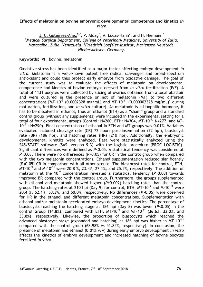
34thAnnual Meeting A.E.T.E. – Nantes, France, 7th – 8th September 2018 76
Effects of melatonin on bovine embryonic developmental competence and kinetics in vitro
J. C. Gutiérrez-Añez1,2, P. Aldag2, A. Lucas-Hahn2, and H. Niemann2
1Medical Surgical Department, College of Veterinary Medicine, University of Zulia,
Maracaibo, Zulia, Venezuela, 2Friedrich-Loeffler-Institut, Mariensee-Neustadt, Niedersachsen, Germany.
Keywords: IVF, bovine, melatonin Oxidative stress has been identified as a major factor affecting embryo development in vitro. Melatonin is a well-known potent free radical scavenger and broad-spectrum antioxidant and could thus protect early embryos from oxidative damage. The goal of the current study was to evaluate the effects of melatonin on developmental competence and kinetics of bovine embryos derived from in vitro fertilization (IVF). A total of 1131 oocytes were collected by slicing of ovaries obtained from a local abattoir and were cultured in the presence or not of melatonin (MT) to two different concentrations [MT-10-9 (0.0002328 mg/mL) and MT-10-11 (0.000002328 mg/mL)] during maturation, fertilization, and in vitro culture). As melatonin is a lipophilic hormone, it has to be dissolved in ethanol, thus an ethanol (ETH) as a “sham” group and a standard control group (without any supplements) were included in the experimental setting for a total of four experimental groups (Control: N=260, ETH: N=304, MT-10-9: N=277, and MT-10-11: N=290). Final concentration of ethanol in ETH and MT groups was 0.01%. Variables
evaluated included cleavage rate (CR) 72 hours post-insemination (72 hpi), blastocyst rate (BR) (186 hpi), and hatching rates (HR) (210 hpi). Additionally, the embryonic developmental kinetics were analyzed. Data were statistically analyzed using the SAS/STAT® software (SAS. version 9.3) with the logistic procedure (PROC LOGISTIC). Significant differences were defined as P<0.05. A statistical tendency was considered at P=0.08. There were no differences (P>0.05) for CR in the control group when compared with the two melatonin concentrations. Ethanol supplementation reduced significantly (P<0.05) CR in comparison with all other groups. The blastocyst rates for control, ETH, MT-10-9 and M-10-11 were 20.8 %, 23.4%, 27.1%, and 25.5%, respectively. The addition of melatonin at the 10-9 concentration revealed a statistical tendency (P=0.08) towards improved BR compared with the control group. Furthermore, the groups supplemented with ethanol and melatonin showed higher (P=0.002) hatching rates than the control group. The hatching rates at 210 hpi (Day 9) for control, ETH, MT-10-9 and M-10-11 were 20.4 %, 52.1%, 53.3%, and 50.0%, respectively. No differences (P>0.05) were observed for HR in the ethanol and different melatonin concentrations. Supplementation with ethanol and/or melatonin accelerated embryo development kinetics. The percentage of blastocysts reaching the hatching stage at 186 hpi (Day 8) was lower (P<0.05) in the
control Group (14.8%), compared with ETH, MT-10-9 and MT-10-11 (36.6%, 32.0%, and 33.8%), respectively. Likewise, the proportion of blastocysts which reached the advanced blastocyst stage (expanded and hatching) at 186 hpi was higher in MT-10-11 compared with the control group (68.98% vs 51.85%, respectively). In conclusion, the presence of melatonin and ethanol (0.01% v/v) during early embryo development in vitro affects the kinetics of embryo development and increased hatching of bovine oocytes fertilized in vitro.

34thAnnual Meeting A.E.T.E. – Nantes, France, 7th – 8th September 2018 77
Detection of cell-free DNA in embryo culture medium: its potential application as a noninvasive method for sex determination in cattle
Tsige Hailay1, Samuel Gebremedhn1, Franca Rings1, Dessiie Salilew-Wondim1, Dawit
Tesfaye1, Schellander Karl1, and Michael Hoelker1,2 1Institute of Animal Science, Animal Breeding and Husbandry group, University of Bonn,
Bonn, north rhine-westphalia, Germany, 2Research Station Frankenforst, Faculty of Agriculture, University of Bonn, Bonn, north rhine-westphalia, Germany.
Keywords: cell-free DNA, culture media, blastocyst The application of noninvasive sex determination methods of embryos is believed to be crucial in assisted reproduction procedures. The aim of this study was to detect cell-free genomic DNA (gDNA) in the embryo surrounding spent media for sex determination of individually cultured bovine embryos. For this, bovine ovaries were collected from slaughter house. Immature oocytes aspirated from follicles of 3-5 mm in diameter, were matured and fertilized in vitro. Embryos were cultured individually starting from eight-cell stage until day 7 blastocyst in 10 µl drops of culture media under oil (SOFaa-medium). To verify the effect of different media on the release of cell-free DNA, individual embryos were cultured either in SOFaa supplemented with 5% exosome-free serum (SOF-EXO) media (n=15) or SOFaa supplemented with 0.1% hyaluronic acid (SOF+HA) media (n=9). Parthenogenetically activated oocytes and the corresponding embryos were cultured in SOF-EXO medium (n=6) to be used as controls. Individual
blastocysts and the corresponding spent media were collected at day 7. Cell-free DNA was isolated from each spent media using QIAamp®Circulating Nucleic Acid kit followed by whole genome amplification using REPLI-g single cell kit (Qiagen, Hilden, Germany). In parallel, extraction of DNA from individual blastocyst was performed using blastocyst Lysis buffer. Multiplex PCR amplification was done to detect sex related fragments in DNA samples from both individual blastocysts and amplified cell-free DNA recovered from corresponding spent media. For this, two different primers; bovine Y-chromosome specific primer and bovine autosomal centromere-specific were used for sex specific PCR amplification. DNA of female and male animal tissues was used as control. The results of the present study revealed that, cell-free DNA was detected in 53.3% of embryos cultured in SOF-EXO spent media. Of these, the sex of 87.5% of individually cultured blastocysts was accurately determined using the cell free DNA isolated from the corresponding spent media. Similarly, cell-free DNA was detected in 55.6 % when embryos were cultured individually in SOF+HA spent media. Among these, 60% of the sex determinations were in accordance between spent media analysis and determination using the blastocyst itself. Moreover, cell-free DNA was detected in 66.7% of culture media drops when harbouring individual parthenogenetic embryos. Noteworthy, sex
determination using cell-free DNA in parthenogenetic embryos was achieved with an accuracy of 100%. Lack of any Y-chromosome specific DNA in these samples could therefore demonstrate absence of genetic pollution. Inaccurate sex determination in some samples could be due to lower amount of cell-free DNA. In conclusion, cell-free DNA released from embryos to their surrounding culture medium can potentially be used as a noninvasive sex determination method prior to embryo transfer in cattle. Further studies should be conducted to improve efficiencies with respect to DNA isolation and amplification in spent media.

34thAnnual Meeting A.E.T.E. – Nantes, France, 7th – 8th September 2018 78
The influence of flunixin meglumine (FM), hCG or a combination of hCG and FM on the conception results in embryo recipient heifers including the passage time
through the cervix and the presence of a large follicle
M. Herudzinska1, B.M. Jaskowski2, J. Czeladko3, J. Kulus1, E. Wedrowska4, M. Gehrke1,
J.M. Jaskowski1, and K.P. Brüssow1 1Centre for Veterinary Sciences at the Nicolaus Copernicus University in Torun, Torun,
Kuyavian-Pomeranian, Poland, 2Department of Reproduction and Clinic of Farm Animals, Faculty of Veterinary Medicine, Wroclaw University of Environmental and Life
Sciences, Wroclaw, Lower Silesia, Poland, 3Private Vet. Praxis, Tykocin, Podlaskie, Poland, 4Department of Applied Informatics and Mathematics in Economy, Nicolaus
Copernicus University in Torun, Torun, Kuyavian-Pomeranian, Poland. Keywords: flunixin meglumine, hCG, recipients The application of hCG before embryo transfer (ET) causes luteinization of a dominant follicle (Rizos, 2012). Non-steroid anti-inflammatory drugs (NSAIDs) lower the pro-inflammatory action of prostaglandins during cervical manipulation (Scenna, 2005). Some authors, however, do not confirm this effect (Nogueira, 2004, Torres, 2013). It is possible that the administration of hCG increases the likelihood of pregnancy in embryo recipients. It should be higher in cows with both CL and the large ovarian follicle. The probability of pregnancy should also be higher in recipients receiving flunixin meglumine (FM) - especially with the prolonged passage time through the cervix (PT). Consequently
the highest pregnancy rate should occur in recipients treated with a combination of FM and hCG. The aim of the study was to assess the probability of pregnancy in cows treated with FM, hCG or a combination of FM and hCG in relation to the PT and the presence of a large ovarian follicle. The conception results of 952 recipients of embryos collected by in vivo delivery (IVD) were included in the analysis. Recipients (CL>15 mm) were selected on day 7 after heat on the base of the ultrasound examination. Each follicle with a diameter of > 5 mm was considered as a large. Follicle of this size is palpable during rectal examination. All embryos were transferred by one experienced veterinarian. The FM was administered intramuscularly (IM) in an amount of 500 mg (Flunimeg 50 ml Fluniksyna –Scanvet Poland Sp. z o.o.), while hCG - IM in an amount of 1500 IU. hCG (Chorulon, Intervet International) was administered 15-2 min. before ET. In order to evaluate the factors determining cows’ fertilization the logit model was estimated with the use of maximum likelihood estimation method and STATA software. The average time of passage through the cervix to the embryo deposition site was 70.1s and it had no significant impact on the probability of conception (p<0.09). The average conception rate was 60.7%. In particular groups the conception rate was 61.3%, 63.4%,
58.5% and 58.3%, respectively, in FM, hCG, FM/hCG and control groups. There was no significant effect of FM administration (p<0.35), hCG (p<0.32) or the combination of both FM/hCG on the conception results in embryo recipients. The presence of a large follicle did not affect the conception rate (p<0.23). The different PT did not impact the results of the hormonal application (odds ratio p<0.08). The presence of a large follicle did not significantly change the treatment effects of used hormonal solutions (p<0.45). Administration of hCG, FM and a combination of both FM and hCG did not affect the probability of pregnancy of embryo recipients. Likewise, the results of conception were not improved by administering hCG to the recipients with a large ovarian follicle. FM application in females with prolonged passage time through the cervix does not affect the probability of pregnancy.

34thAnnual Meeting A.E.T.E. – Nantes, France, 7th – 8th September 2018 79
The influence of dominant follicles and corpora lutea location on the conception rate in embryo recipient heifers
B.M. Jaskowski1, J. Czeladko2, J. Kulus3, J. Sobolewski3, M. Gehrke3, J.M. Jaskowski3, M.
Rogoziewicz3, and K.P. Brüssow3 1Department of Reproduction and Clinic of Farm Animals, Faculty of Veterinary
Medicine, Wroclaw University of Environmental and Life Sciences, Wroclaw, Lower Silesia, Poland, 2Private Vet. Praxis, Tykocin, Podlaskie, Poland, 3Centre for Veterinary Sciences at the Nicolaus Copernicus University in Torun, Torun, Kuyavian-Pomeranian,
Poland. Keywords: Dominant follicle, embryo transfer, dairy cattle Development of the first follicular wave dominant follicle on the ovary ipsilateral to the corpus luteum is associated with a decreased conception rate in dairy artificially inseminated cows (Miura, 2015). In embryo recipients that phenomenon has not been studied yet. Meanwhile, the position of the dominant follicle (DF) could be an additional criterion for the pre-transfer selection of embryo recipients. The aim of the study was to determine the effect of the DF placed ipsi- or contralateral to the ovary with CL on the conception rate. 967 recipients were examined in the study. Heats were synchronized with the use of prostaglandin (2xPG14). On the day of the transfer, the ultrasound examination of the ovaries was performed. Recipients with CL > 15 mm were qualified for the transfer. Additionally, the DF position and diameter were defined. Each follicle
with a diameter > 5 mm was described as dominant. 682 fresh and 287 frozen embryos were used in the study. Pregnancy was examined palpatively 2 months after embryo transfer. Statistical analysis was done with STAPA. A dominant follicle was observed in 928 (95.8%) recipients. The diameter of DFs was between 5 to 22 mm, with the average 10.1 mm. In 443 (47.7%) recipients, the DF was contralateral to the ovary with the CL, in 485 (52.3%) the DF was ipsilateral to the CL. In 39 (4.1%) recipients there was no dominant follicle. The percentage of pregnant recipients, in which embryos were introduced into the uteri horn on the same side as both CL and DF was 60,7% and it was lower (p>0.05) than in recipients with embryos introduced into the uteri horn on the side of the ovary with the CL, and contralateral to the ovary with the DF (61.04%). If the CL was observed on the left ovary and the DF on the right one, 61.6% of recipients were pregnant, while if the CL was observed on the right ovary and the DF contralateral to it, the percentage of pregnant recipients was 59.5. If there was no DF on the ovaries, the conception rate was 55.5% (p>0.05). Introducing embryos to the left horn (with the CL on the left ovary, no DF) resulted in 42.8% of pregnant recipients, while introducing them to the right horn (with the CL on the right ovary, no DF) ended with 59.1% of pregnancies. To conclude, the presence of
the dominant follicle on the ovary ipsi- or contralateral to the ovary with the CL had no significant effect on the pregnancy probability. It seems that the DF location on the ovary in relation to the CL is an insufficient additional criterion for the selection of embryo recipients.

34thAnnual Meeting A.E.T.E. – Nantes, France, 7th – 8th September 2018 80
Reproductive fluids added to embryo culture vs. standard culture in cow: first results on pregnancy rates
J.S. Lopes1,2, M. Hamdi3, E. Alcázar-Triviño4, B. Rodríguez-Alonso3, A. Canha-Gouveia1,2,
E. París-Oller1,2, C. Soriano-Úbeda1,2, S. Cánovas1,2, D. Rizos3, and P. Coy1,2 1Department of Physiology, Faculty of Veterinary Medicine, University of Murcia,
Murcia, Spain, 2Institute for Biomedical Research of Murcia, IMIB-Arrixaca, Murcia, Spain, 3Department of Animal Reproduction, INIA, Madrid, Spain, 4El Barranquillo S.L.,
Torre Pacheco, Murcia, Spain. Keywords: Reproductive fluids, IVP, ET Reproductive fluids (RF), though being part of the natural environment of embryo development, are not yet included in current IVC media. It has been shown in bovine that inclusion of RF in embryo IVC produces blastocysts with higher quality (Hamdi, Rep Fert Dev. RD17286, 2017). In porcine, embryos produced with RF had gene expression and DNA methylation patterns closer to in vivo grown embryos (Cánovas, eLife. 6:e23670, 2017). However, it is still unknown if the transfer of these embryos to recipients can give rise to pregnancies, thus justifying the conduction of this experiment. IVM, IVF and IVC details were already described elsewhere (Hamdi, Rep Fert Dev. RD17286, 2017). In IVC two different culture groups were created, according to the supplementation: RF group (SOF-RF) - 1,25%(v/v) NaturARTs BOF-EL (Embryocloud, Spain) from day 1 to 4 and 1,25%(v/v) BUF-ML from day 4 to 8 - and BSA group (SOF-BSA)
- 3 mg mL-1 BSA from day 1 to 8. Vitrification and warming were performed using commercial media (Kitazato-Dibimed, Spain) with an open-system Cryotop, following manufacturer’s instructions. Vitrification took place on IVC day 7/8 with embryos on stage 6-7 of development. Warming of embryos was performed less than 4h before transfers, loaded in straws and kept at 38,5ºC. Embryo transfers (ET) were made non-surgically to Holstein multiparous recipients either on day 6 or 7 after oestrus detection. Synchronization was made using Double-Ovsynch protocol. A total of 64 ET (n=36, SOF-RF; and n=28, SOF-BSA) were made in a 6 months period, from November 2017 until March 2018. Pregnancies were detected by ultrasound 30 days post-ET. Data were analysed by t-test independent samples with p<0,05 resulting in significant differences (data are means ± SEM). Pregnancy rate (P%) per group did not have a significant difference when comparing recipients of the same day. However, when comparing P% by recipient day there were significant differences: recipients on day 6 had 10,00±5,57 P% (n=30), while recipients on day 7 had 35,29±8,32 P% (n=34). Specific values for SOF-RF were 11,08±10,1 and 36,8±9,6; and for SOF-BSA were 7,77±11,6 and 33,3±10,8, respectively. ET’s are routinely made to recipients on day 6 to 8 post-oestrus detection. Our data are consistent with other reports for P% of IVP embryos for day 7 recipients,
but not for day 6. Additionally, the source of the oocytes is not OPU but rather slaughtered cows, which could decrease our blastocyst development and viability, despite it was maintained for day 7 recipients. In conclusion, the presence of RF in embryo IVC gave rise to pregnancies at a similar level than a control group. Day 6 recipients showed an adverse effect on pregnancy rates, regardless of the group. Further data (calves phenotypes mainly) are necessary to evaluate if the improvements reported at the blastocyst stage by including RF in bovine IVP are also evident after birth. Supported by European Union, Horizon 2020 Marie Sklodowska-Curie Action REPBIOTECH675526; AGL2015-66341-R MINECO-FEDER; 20040/GERM/16 Fundación Seneca.

34thAnnual Meeting A.E.T.E. – Nantes, France, 7th – 8th September 2018 81
Transcervical embryo recovery in Lacaune ewes superovulated with different doses of FSH
J.M.G. Souza-Fabjan1, L.M. Figueira1,2, N.G. Alves2, R.I.T.P. Batista1,3, L.C. Souza4, A.A.
Arrais5, C.F. Silva6, M.C.C. Morais1, and J.F. Fonseca7 1Universidade Federal Fluminense, Niteroi, RJ, Brazil, 2Universidade Federal de Lavras,
Lavras, MG, Brazil, 3Universidade Federal dos Vales do Jequitinhonha e Mucuri, Diamantina, MG, Brazil, 4Cabanha Val di Fiemme, Soledade de Minas, MG, Brazil,
5Universidade Federal Rural do Rio de Janeiro, Seropédica, MG, Brazil, 6Autonomous veterinary doctor, Miracema, MG, Brazil, 7Embrapa Caprinos e Ovinos, Coronel Pacheco,
MG, Brazil. Keywords: Cervix, dairy sheep, uterine flushing This study assessed the effect of different FSH dosages for superovulation and the feasibility of transcervical embryo recovery in Lacaune ewes. Ewes (n=25) received 60 mg medroxiprogesterone acetate sponge (Progespon®, Syntex, Buenos Aires, Argentina) for nine days, 37.5 µg d-cloprostenol i.m. (Prolise®, Tecnopec, São Paulo, Brazil) 24 h before sponge removal and 50 µg gonadorelin (GnRH analogue, Gestran®, Tecnopec, São Paulo, Brazil) 24 h after sponge removal. Superovulatory treatments consisted of 100 mg (G100, n=13) or 200 mg (G200, n=12) of porcine FSH (Folltropin®-V; Bioniche Animal Health, Belleville, Canada), given i.m. (twice daily) for three consecutive days, in decreasing doses (25, 25, 15, 15, 10 and 10%), starting at 60 h before sponge removal.
Ewes were checked for estrus twice daily and were naturally mated by fertile rams (4:1 ratio) while in estrus. Transrectal ovarian ultrasonography was performed at the 5th day after estrus, to count the number of corpora lutea (CL) with Doppler mode ultrasound (Mindray M5VET®, Shenzen, China – 8.0 MHz). All ewes received 37.5 µg d-cloprostenol (Prolise®, Tecnopec, São Paulo, Brazil) and 1 mg estradiol benzoate (Sincrodiol®, OuroFino, Cravinhos, Brazil) i.m. 16 h before uterine flushing and 50 IU oxytocin (Ocitocina forte UCB®, São Paulo, Brazil) i.v. 20 min before uterine flushing. Embryo collection was performed at days 5 or 6 after estrus by transcervical technique (Fonseca et al., Theriogenology, 86:144-151, 2016) in all ewes that showed estrus and had more than 2 CL (n=17). Qualitative data were analyzed by Fisher exact test. Quantitative data were analyzed by generalized linear models, using SAS® software (v 9.3, SAS Institute, Cary, USA). The percentage of ewes that showed estrus and the percentage of responding donors (> 2 CL) did not differ (P>0.05) between treatments: 77% (10/13) and 62% (8/13) for G100 and 100% (12/12) and 83% (10/12) G200, respectively. The number of CL was higher (P<0.05) for G200 (10.5±1.5) than G100 (4.2±1.5). Overall, cervical transposition and uterine flushing was possible in 100% (17/17) of ewes. The total time procedure was 32.3±0.1 min for G100 and 27.7±0.1 min for G200 (P>0.05).The number of
recovered structures and viable embryos per ewe collected was higher (P<0.05) for G200 (7.5 ±0.1 and 6.2 ± 0.1) than G100 (0.4 ± 0.6 and 0.4 ± 0.6), respectively.The dose of 200 mg of FSH promoted greater superovulatory response and recovery of viable embryos by transcervical technique. Probably, the poor ovulatory response with 100 mg of FSH was also affected by the formation of luteinized unovulated follicles. The protocol for cervical relaxation was efficient to allow the transcervical embryo recovery of Lacaune ewes. Financial support: EMBRAPA (Project 02.08.02.005.00.04) and Fapemig (Project CVZ-PPM 00201-17). Authors also acknowledge Vicente Marin Munhoz of Cabanha Val di Fiemme.

34thAnnual Meeting A.E.T.E. – Nantes, France, 7th – 8th September 2018 82
Porcine follicular fluid as chemoattractant improves sperm attraction and in vitro fertilization
Luis Alberto Vieira1,2, Andrea Aguilera3, Alessia Diana1, and Carmen Matas1,2
1Department of Physiology, International Excellence Campus for Higher Education and
Research (Campus Mare Nostrum), University of Murcia, Murcia 30100, Spain, Murcia, Spain, 2Institute for Biomedical Research of Murcia IMIB-Arrixaca, Murcia, Spain, Murcia, Spain, 3Faculty of Natural Sciences (FCEN), National University of Cuyo,
Mendoza, Argentina, Mendoza, Argentina. Keywords: Porcine spermatozoa, follicular fluid, chemotaxis Under physiologic conditions, different biofluids (follicular fluid (FF), oviductal fluid (OF), and secretion of cumulus-oocyte complex (COCs)) take part in the spermatozoa attraction previous to fertilization. Despite the progesterone (P4) being part of the composition of these biofluids, it’s also considered as the main chemoattractant (Blengini et al., Asian Journal of Andrology, 13, 769-773, 2011). However, there are other components not defined in these media that could attract spermatozoa more efficiently. Thus, the aim of this study was to study the ability of biofluids for sperm attraction and the effect on in vitro fertilization (IVF) parameters. Periovulatory OF, FF and conditioned medium (CM) were collected previously described by Soriano et al., 2017. In the present study, a chemotaxis system was designed using a Petri cell culture dishes (35 x 10 mm) with four wells separated by 3 mm (GN627170, Sigma). Six wells
were filled with fresh spermatozoa (20x106/mL) from fertile boars (N=6) that were capacitated for 45 min in 180 µL of the capacitation media (TALP), previously equilibrated for 3h at 38.5ºC and 5% CO2. The opposite wells (six) were filled with TALP (control group) and TALP supplemented with the different chemoattractants: FF, OF, CM, P4, and mixture of all chemoattractans (experimental groups). Afterwards, 3-4 mm long capillaries bridges were placed between the wells containing capacitated spermatozoa and the opposite ones for 20 min. After that, the capillaries were removed and 22 (per replicate) denuded in vitro maturated oocytes were deposited with spermatozoa previously adjusted to 22x103 in each group (chemoattractans and control groups). The experimental groups were: 1) TALP (control), 2) FF (1%), 3) OF (1%), 4) CM (2%) 5) P4 (28.3 pM), and 6) FF, OF, CM, and P4: Σ. After 18 h the oocytes were fixed and IVF parameters were evaluated in each experimental groups. All the data were analysed by ANOVA followed by Tukey post hoc to compare means and standard error (p<0.05). A total of six replicates were carried out. Follicular fluid alone (FF) showed the highest values for ZP binding, penetration rate, spermatozoa/oocyte and pronuclear formation (p<0.05). This preliminary study suggests that FF is the most important chemoattractant for porcine spermatozoa in in-vitro conditions. However, further
experiment will be performed on the embryo development and quality. Supported by Fundación Séneca, Saavedra Fajardo (20020/SF/16). MINECO-FEDER (AGL 2015-66341-R).

34thAnnual Meeting A.E.T.E. – Nantes, France, 7th – 8th September 2018 83
Retrospective survey on equine embryo transfer activities in France in 2014 and 2015
E.Autard de Bragard; J.F. Bruyas
Department of Clinical Sciences, National College of Veterinary Medicine, Food Sciences
and Engineering, Nantes - France Keywords: The objectives of embryo transfer in mares are (1) produce several foals from the same mare during a single breeding season, eventually bred by different stallions; (2) allow donor mares to pursue their sport career while recipient mares carry their foals; and (3) obtain foals from mares unable to carry a pregnancy to term due to genital lesions or diseases. The aim of our study was to analyse data from equine embryo transfer centres in France during the 2014 and 2015 breeding seasons. Our goal was to have an overview of Equine Embryo Transfer activity in France and to compare methods used by centres and their results to evaluate which parameters influence success rates of embryo recovery and transfer. A survey was sent to 62 French centres and included questions about general activities and methods, and specific information of the 2014 and 2015 breeding seasons including donor mares, embryo recoveries performed and every embryo recovered and transferred. Only 18% (11/62) of centres responded to our study. Data from 208 donor
mares (19%) of the 1108 donor mares recorded in France during those years, 669 embryo collections and 305 embryos were analysed. This survey shows that centres used similar methods, but some specific points were different in the collection procedure (flush medium, number of flushes/collection (4 to 6), use of oxytocin), transfer procedure (sedation, NSAID), and in the tools to synchronize ovulations between recipient and donor mares. On average, 3.3 ± 3.8 embryo collections were performed per year from each donor mare (range 1 to 10), with a recovery rate of 41% (277/669) but significantly higher in 2014 (46.9%) than in 2015 (36.3%). This embryo recovery rate was significantly higher in mares under 14 years old, and in cycles with ovulation induction. Embryo collections were performed at 7 (5.5%), 8 (81.5%) or 9 (13%) days after ovulation, without any statistical difference in the recovery rate. Pregnancy rate after embryo transfer was 75% (229/305). Age of embryo, delay of ovulations between recipient and donor (+1 to -4 days), and physiological status of recipient (maiden or not) had no significant effect on the pregnancy rate. As 19% of French donor mares have followed by the same proportion of French transfer centres, one could make a projection on the total embryo transfer activity in France. It
could be estimated that about 1800 embryo collections are performed per year, producing between 900 and 950 embryos. This projection would indicate that the annual report of equine embryo transfer presented by AETE is largely under evaluating the current embryo transfer activity. For example, 606 embryo collections and 365 transfers were reported in 2014 in France by the AETE, while in our study, 322 embryo collections were performed and 169 embryos transferred from only 19% of registered French donor mares. Only donor mares and successful embryo transfers (pregnancy at Day 14) are officially recorded in France; by reporting this number of recorded pregnancies to our estimated number of transfers, the estimated success rate (70-80%) is comparable to that really observed (75%) in the survey.

34thAnnual Meeting A.E.T.E. – Nantes, France, 7th – 8th September 2018 84

34thAnnual Meeting A.E.T.E. – Nantes, France, 7th – 8th September 2018 85
Folliculogenesis, Oogenesis, and Superovulation

34thAnnual Meeting A.E.T.E. – Nantes, France, 7th – 8th September 2018 86
Retrospective analysis of superstimulation with Folltropin®-V in Wagyu versus other beef breeds
C.B. Steinhauser1, C.R. Looney1, J.F. Hasler2, P. Renaud3
1OvaGenix, College Station, Texas, USA, 2Vetoquinol USA, Fort Worth, Texas, USA, 3Vetoquinol N-A Inc., Lavaltrie, Quebec, Canada.
Key words: Wagyu, Folltropin®-V, superstimulation The demand for Wagyu cattle, which originate from the Kobe region of Japan, is growing in many countries. Nevertheless, Wagyu remains an exotic breed in many places and questions persist in relation to the most efficacious superstimulation protocols. The data in this retrospective study were collected from 2012-2018 at the OvaGenix® (Texas) facilities. A total of 792 donors were collected while under direct care of OvaGenix® and were of the following Bos taurus and Bos indicus breeds: Angus (AN; n=120), Beefmaster (BM; n=139), Brangus (BN; n=207), Charolais (CH; n=76), Hereford (HH; n=45), Wagyu (KB; n=99), and Red Brangus (RB; n=106). The superstimulation protocol was the same for all donors and began with insertion of an Eazi-Breed™ CIDR® (Zoetis, Parsippany, USA) on Day 0. An injection (i.m., 100 mcg) of GnRH (Cystorelin®, Merial, Duluth, USA) was administered on Day 2, followed by 7 AM/PM injections (i.m.) of decreasing doses of pFSH (Folltropin®-V, Vetoquinol, Fort Worth, USA) on Days 4-7 and prostaglandin F2α (i.m., 500 mcg, Estrumate, Merck Animal Health, Madison, USA) in the AM of Days 6-7. After CIDR® retrieval on Day 7, GnRH was injected in the AM of Day 8 and donors were
artificially inseminated with frozen/thawed semen the PM of Day 8 and AM of Day 9. Embryos were collected on Day 14 (Day 6 from GnRH). Data from the embryo collections was analyzed with an ANOVA and Tukey’s test to compare breed effect with p<0.05 considered significant. The data indicate that KB donors produce numbers of total ova (number of ova produced by superstimulation) and viable embryos that are comparable to other breeds using the above defined superstimulation protocol. Indeed, KB donors produced more total ova than AN (16.6±1.1 versus 11.4±0.7) and more viable embryos per collection than AN and CH (9.1±0.7, 4.1±0.4, 5.6±0.6, respectively). The BM, BN, RB, and HH donors produced similar numbers of total ova and viable embryos when compared to KB donors. Wagyu donors were also similar to other breeds for the percentage of donors with no ovarian response (5.0%) and donors with no viable embryos (9.0%). It is acknowledged that virtually every other beef and dairy breed responds well to Folltropin®-V and these data indicate that KB donors can also be effectively superstimulated with Folltropin®-V using a similar protocol to other beef breeds. Moreover, these data are similar to mean viable embryo numbers that are audited and published yearly by various national and international embryo transfer associations.

34thAnnual Meeting A.E.T.E. – Nantes, France, 7th – 8th September 2018 87
Physiology of Reproduction in Male and Semen
Technology
34thAnnual Meeting A.E.T.E. – Nantes, France, 7th – 8th September 2018 88
ADAM protein expression in avian sperm and female genital epithelial cells: relation to sperm storage
L. Cordeiro1, C. Riou1,2, and N.Gérard1
1PRC, INRA, UMR INRA85, UMR CNRS 7247, Université de Tours, IFCE, 37380 Nouzilly,
France, 2ALLICE, Station de Phénotypage, 37380 Nouzilly, France. Keywords: ADAM protein, Sperm storage tubes, chicken The cell-cell interaction can be mediated by the interface of a protein-protein. "ADAM is a transmembrane protein that contains a disintegrin and metalloprotease domain and, therefore, potentially has cell adhesion and protease activities" (Primakoff & Myles, Trends Genet., 2000, 16 (2): 83 -7). Avian sperm storage tubes (SST) are uterine-vaginal junction (UVJ) epithelial structures that can store spermatozoa (spz) through an interaction mechanism that is not yet clearly understood. This study was devoted to investigating the presence or absence of ADAM proteins in chicken SST as a model of sperm-epithelial cell adhesion and interaction. For in vitro tests, 3 hens per experiment (2 triplicate experiments, n = 18 hens) were euthanized and the internal epithelium UVJ was collected, digested (1 μg / ml collagenase) and the SST fragments were isolated by Percoll gradient column. SST (100/well), sperm (25.104 sperm/well) cultures and co-culture SST + spz (25.104 sperm / 100 SST) were grown in DMEM 199 medium supplemented with 10% BFS and gentamycin at 37 ° C, 5% CO2 atmosphere at 3 times: 0, 2 and 24h. For in vivo tests, 3 other hens were inseminated with 200 x 106 spz (pool of 6
roosters) and euthanized 24 hours later, while 3 other non-inseminated were used as control. In all experiments, antibody anti-ADAM-1-CT rabbit peptide sequence was used: CSSPGSGGSVDSGP (C-terminal part, close to the transmembrane metalloprotease domain), as described by Fàbrega et al. (Reproductive Biology and Endocrinology, 2011, 9:96). UVJ were isolated and prepared for immunohistochemistry (IHC). The slides were incubated overnight at 4°C with the primary antibody and incubated for 30 min with the secondary antibody ImmPRESSTM HRP Anti-Rabbit/Mouse IgG (Vector) and revealed with peroxidase. Protein concentration was determined individually in the samples, and the pools were prepared with equal protein concentration of each sample. Ten μg of protein in Laemmli buffer (5v:1v) were loaded on SDS-10% PAGE, and then transferred to nitrocellulose filters. The membranes were incubated with anti-ADAM-1 antibody overnight in blocking solution (5% non-fat dry milk in TBST [10 mM Tris, 150 mM NaCl, 0.1% Tween-20, pH 7.4]). The ADAM peptide exhibited sequence homology with Gallus gallus ADAM-12 and ADAM-33 (www.clustal.org/ multiple alignment program). ADAM was detected as a single 90 kDa band in SST samples and 3 bands (90, 45 and 40 KDa) in spz samples. Signals decreased in SST + spz co-culture in the cells and the respective supernatant, but a 90 kDa band was observed in the supernatant after 24h co-culture.
Anti-ADAM IHC UVJ slides showed positive staining for spermatozoa located in the SST lumen and in small vesicles on apical SST cells. We can conclude that ADAM proteins are present in SST epithelial cells and can be released in the presence of spermatozoa, probably modulated by cell-cell interaction. Supported by Université François Rabelais de Tours and Région Centre, France.

34thAnnual Meeting A.E.T.E. – Nantes, France, 7th – 8th September 2018 89
Analysis of zona pellucida binding properties of boar sperm subpopulations separated by carbohydrate affinity
L. González-Brusi, R. López-Úbeda, J.G. Hamzé, P. Cots, J. Ballesta, M. Jiménez-
Movilla, M.J. Izquierdo-Rico, and M. Avilés
Department of Cell Biology and Histology, Faculty of Medicine, University of Murcia, IMIB-Arrixaca, Campus Mare Nostrum, Murcia, Spain.
Keywords: carbohydrate affinity, flow cytometry sorting, ZP binding assay In mammals, sperm-sugar interaction is involved in relevant biological processes such as the sperm binding to the zona pellucida (ZP) and the formation of the sperm oviductal reservoir. The aims of this study are the identification of sperm subpopulations based on their affinity for carbohydrates, the isolation of these sperm subpopulations and the characterization of their ZP binding properties. Ovalbumin (OVA) which has high-mannose type glycans and the Lewisa epitope (Lea), consisting of Galβ1-3[Fucα1-4]GlcNAcβ, were tested. Heterospermic sperm samples from boars of proven fertility were coincubated with OVA-Alexa594 or Lea-FITC for 30 minutes in PBS with 1mM sodium pyruvate. Subpopulations separation was done in a Sony SH800Z flow cytometer with excitation lasers at 488 nm and 561 nm, sorting 3500-4000 cells/second. Sperm were collected in tubes containing 100µl of TEST- 2% egg yolk buffer to minimize oxidative damage. Sperm resuspended in capacitating TALP medium at a concentration of 105
sperm/ml were incubated with 50 intact isolated ZP (n = 3). Isolated ZP were obtained
form porcine ovaries collected at a local slaughterhouse. Cumulus-oocyte complexes were taken from antral follicles and treated with hyaluronidase. ZP were isolated with very thin pipettes in PBS containing protease inhibitors. After 2 h of incubation at 37ºC and 5% CO2, samples were washed twice to detach weakly bound spermatozoa, fixed in PBS with glutaraldehyde 0.5% for 30 min and stained with Hoechst 33342. Spermatozoa bound to each ZP were evaluated with an Olympus IX70 fluorescence microscope. A Student’s test was used to compare between each pair of samples and differences were considered significant when p<0.01. Percentages of spermatozoa from each subpopulation; Lea-, Lea+, OVA- and OVA+ were 77%, 18%, 82% and 14% respectively. A 4-5% of spermatozoa between each positive and negative subpopulation were discarded. The number of sperm bound to the ZP of the Lea-, Lea+, OVA- and OVA+ sperm subpopulations were 51.8±22.5, 18.9±8.8, 58.1±32.3, 17.5±6.8. Two different controls were perfomed. A total of 113.1±32.4 spz/ZP and 38.2±14.1 spz/ were observed for untreated ejaculated spermatozoa and sorted sperm not labelled. In both Lea and OVA, ZP binding was significantly higher in the negative sperm subpopulations compared to the positive ones. Lea+ subpopulation showed a high percentage of non viable spermatozoa (40±10.6%, n = 4), as assessed with propidium iodide, which correlates to
less sperm bound to the ZP, while Lea- viability was over 90%. Our results showed the existence of different sperm subpopulations with a different affinity for carbohydrates. These sperm subpopulations had dissimilar affinity for the ZP and could have a different fertilization ability. The biological significance of these sperm subpopulations will be analyzed in in vitro fertilization assays and binding to the oviductal epithelial cells. Supported by MINECO-FEDER (AGL2015-70159-P).

34thAnnual Meeting A.E.T.E. – Nantes, France, 7th – 8th September 2018 90
Sperm motility in thawed bull semen is increased by a short incubation before
analysis
I.B. Lima-Verde, T. Ntallaris, and J.M. Morrell
Swedish University of Agricultural Sciences, Uppsala, Sweden. Keywords: incubation time, cryopreservation of semen, sperm motility Cryopreservation of semen is a widely used technique, mainly to provide samples with certified origin and quality for use in artificial insemination (AI) in dairy cattle. After thawing, spermatozoa may require a period to recover their proper motility, which is a factor directly related with the semen quality. Our aim was to determine the effects of a short incubation on total and progressive motility after thawing bull semen. The samples were collected from 12 bulls at a commercial bull station (Viking Genetics, Skara, Sweden) and were extended in OptiXcell® (IMV Technologies, L´Aigle, France) to provide a sperm concentration of 69×106 spermatozoa/mL. All samples were frozen in 0.25 mL plastic straws and stored in liquid nitrogen. The straws were thawed at 37°C for 12 s and the semen content was split into four tubes and incubated at 38°C for 0, 5, 10 or 15 min. Sperm motility assessment was performed by computer assisted sperm analysis (CASA – SpermVision, Minitub GmbH, Tiefenbach, Germany), connected to a microscope (Olympus, Tokyo, Japan) with a heated stage (38°C). Aliquots of 5µL from the thawed sperm samples were placed on a warm glass slide with a coverslip. At least
1000 spermatozoa were analyzed in a total of eight fields using the software program (SpermVision) with settings adjusted for bull spermatozoa. Statistical analysis was performed with SAS (version 9.3), using the proc mixed procedure for linear mixed models. Scheffe's adjustment was used for multiple-post ANOVA comparisons. Results are presented as LSMeans ± standard error of means (SEM). An increased incubation time was associated with increased total motility (0 min 44.7±4.45%; 5 min 47.0±4.45%; 10 min 49.0±4.45; 15 min 56.9±4.45) and progressive motility (0 min 41.5±4.34%; 5 min 43.8±4.34%; 10 min 45.9±4.34; 15 min 53.5±4.34). Motilities were significantly greater after 15 min incubation in OptiXcell® than after 0 min (p<0.05). In conclusion, total and progressive motilities may be increased by incubation before analysis, depending on the extender used. This effect should be remembered when comparing sperm handling treatments, or to avoid introducing bias when multiple samples are to be analysed. I Lima-Verde received a fellowship from Brazilian Council of Research (CNPq-Brazil, 204411/2014-9). The project was funded by a project grant from the Swedish Farmers´ Foundation (SLF; H13300339) awarded to JM Morrell.

34thAnnual Meeting A.E.T.E. – Nantes, France, 7th – 8th September 2018 91
Effect of cholescytokinin (CCK) protein on the motility of porcine sperm.
R. López-Úbeda1, FA. García-Vázquez2, M. Balastegui-Alarcón1, P. Cots1, M. Avilés1, and
MJ. Izquierdo-Rico1 1Department of Cell Biology and Histology, Faculty of Medicine, University of Murcia
and IMIB-Arrixaca, Murcia, Spain, 2Department of Physiology, Veterinary School, Campus Mare Nostrum, University of Murcia, and IMIB-Arrixaca, Murcia, Spain.
Keywords: spermatozoa, cholecystokinin, motion parameters The expression of cholecystokinin (CCK) gene has been demonstrated in the testis of several different species and CCK-like peptides have been found in spermatocytes and spermatids of the mouse, rat, and monkey (Persson, PNAS; 86: 6166, 1989). In a recent study, Zhou et al suggested its possible involvement in the regulation of protein tyrosine phosphorylation by modulation the uptake of HCO3- and demonstrated the presence of CCK receptors on sperm surface (Zhou, Reproduction; 150:257, 2015). In addition, our group has detected CCK in the porcine oviduct using microarray technology, real-time RT-PCR and immunohistochemical analyses (Acuña, Reprod Fertil Dev; 29: 2387, 2017). Therefore, the aim of this work was to analyse the effect of CCK on sperm motility. Spermatozoa from fertile boars (15x106/ml) were exposed to different CCK (Bachem, Bubendorf, Switzerland) concentrations according to the bibliography (25 μM and 50 μM) in a capacitating medium (TALP) for 1h at 38.5ºC. TALP medium had a bicarbonate
concentration of 5 mM and the incubator was adjusted to 1% CO2 according to the Henderson-Hasselbalch equation. Moreover, a control group was incubated under the same conditions without CCK protein. Motion parameters (total motility (%) and progressive motility (%)) were determined using a CASA system (ISAS®, Proiser, Valencia, Spain) after of incubation period. For statistical analysis the mean measurements of CASA for each male (n=5) were entered into the ANOVA model and compared by post hoc test (Tukey). Differences were considered statistically significant at p<0.05. The results showed that incubation with CCK increased the percentage of total sperm motility (Control: 53.8±2.8%, 25 μM: 59.4±2.1% and 50 μM: 65.4±4.5%) with statistically significant differences between the three experimental groups. Moreover, an increase in the percentage of progressive sperm motility was observed (Control: 34.4±3.9%, 25μM: 38.4±4.8% and 50 μM: 43.2±5.0%) showing significant differences between the control and 50 μM CCK. In both cases the effect of the protein is dose-dependent. No statistical differences were found between the study groups for the rest of motion parameters. In conclusion, these data could suggest a possible implication of CCK protein in the improvement of the sperm motility in the oviduct before fertilization. More analyses are necessary for clarify the molecular mechanism of this protein.
Supported by MINECO-FEDER (AGL2015-70159-P) and Fundación Séneca (19357/PI/14).

34thAnnual Meeting A.E.T.E. – Nantes, France, 7th – 8th September 2018 92
Porcine periovulatory oviductal fluid decreases the occurrence of Protein Kinase A (PKA) substrates trough sAC/cAMP/PKA during mouse sperm capacitation
C. Matás1,2, M.G. Gervasi3, C. Soriano-Úbeda1, E. París-Oller1, and P.E. Visconti3
1Department of Physiology, Veterinary Faculty. University of Murcia., Murcia, Spain, 2Institute for Biomedical Research of Murcia (IMIB), Murcia, Spain, 3Department of
Veterinary and Animals Sciences, University of Massachusetts., Amherts, Massachusetts, USA.
Keywords: Oviductal fluid, spermatozoa, phosphorylation Several studies have identified important factors involved in the regulation of sperm capacitation, a physiological process necessary to achieve fertilization competence. These events are regulated through activation of the cyclic AMP (cAMP)/protein kinase A (PKA) pathway. On the other hand, porcine oviductal fluids (POF) decreases PKA activity of porcine sperm during capacitation; however, the molecular mechanism of this regulation remains unknown. The aim of this study was to evaluate if porcine periovulatory oviductal fluid (POF) regulates PKA activity during in vitro sperm capacitation in mice. POF was obtained from porcine oviducts (n=40) close to ovulation and it was frozen at -80ºC before use. Sperm samples were obtained from mice cauda epididymides and incubated for 1 hour in: i) non-capacitation media, ii) capacitation media, iii) capacitation media supplemented with 1% POF, iv) capacitation media supplemented
with 1% POF and 100 µM of 3-isobutyl-1-methylxanthine (IBMX), which inhibit adenosine 3',5'-cyclic monophosphate phosphodiesterase (cAMP PDE), v) capacitation media supplemented with 1% POF, and 1 mM dibutyryl-cAMP (db-cAMP), that mimics the action of endogenous c-AMP and vi) capacitation media supplemented with 1% POF, 100 µM of IBMX and 1 mM db-cAMP. The protein phosphorylation pattern on PKA substrates was evaluated by Western blotting (WB). Proteins were separated by electrophoresis on 8% SDS-polyacrylamide gels and electrotransferred to PVDF membranes. The latter were treated with the following antibodies: rabbit monoclonal antibody anti-phosphorylated protein kinase A substrates (1:10000) followed by goat anti-rabbit IgG-HRP (1:10000). After developing, the relative amount of signal was quantified by optical densitometry using ImageJ software. Data were analyzed by two-way ANOVA and Tukey post-hoc test (p < 0.05). Our results indicate that in the presence of POF, spermatozoa showed a lower PKA substrates phosphorylation pattern than those incubated in capacitation media alone (p < 0.05). The effect of the POF was reversed by the presence of db-cAMP and/or IBMX in the media. Besides, our results indicate that POF decreases the sAC/cAMP/PKA pathway during sperm capacitation and that this is not a specie-specific phenomenon. These
effects were not observed under non-capacitating conditions. We identified one of the mechanisms by which POF regulates sperm capacitation. This finding would help to understand the oviductal physiology during gamete interaction and could explain some cases of idiopathic infertility. Further studies are required to evaluate whether FOP regulates PKA-related molecular pathways during sperm capacitation in other species such humans. MINECO-FEDER (AGL 2015-66341-R) and Programa “Salvador de Madariaga” (Ministerio de Educación Cultura y Deporte).

34thAnnual Meeting A.E.T.E. – Nantes, France, 7th – 8th September 2018 93
Effect of swim-up on the activation of apoptosis in frozen bovine semen: A flow cytometric study
Maria Moreira da Silva, Loide Soares, and F. Moreira da Silva
University of the Azores - Faculty of Agrarian Sciences and environment, 9700 Angra do
Heroismo.
Keywords: Bovine sperm, swim-up, flow cytometry Techniques universally used to prepare bovine spermatozoa for in vitro fertilization (IVF) include the swim-up (SU) method and differential density gradient centrifugation, among others. Although it is clear that not all of them have comparable efficiency in selecting high quality sperm, the use of these techniques can be harmful for sperm quality. The aim of the present study was to detect the sub lethal changes in bull spermatozoa immediately post-thawing (PT) and after SU, namely the levels of apoptotic sperm cells, to assess if the sperm treatment before IVF induces apoptosis in bull spermatozoa. For such purpose, three semen straws per bull from six bulls were thawed by placing them in a water bath (37 °C) for 30 sec. Then, sperm apoptosis level was analyzed immediately PT and after the SU procedure using a flow cytometric method by detecting the phosphatidylserine translocation across the plasma membrane using a fluorescein-labeled Annexin-V and propidium iodide (PI). By using these two dyes, four different subpopulations of sperm were observed: a population of apoptotic sperm, a population of early necrotic sperma, a population of necrotic sperm and a population of
fully viable sperm cells. The most consistent effect observed was a significant increase (P < 0.05) in the fraction of apoptotic sperm (Annexin-V +, PI-) after the SU treatment among the six bulls tested. The highest increase of apoptotic spermatozoa after SU was observed on bull four with an increase from 7.0 % ± 0.9 to 59.2 % ± 0.2, respectively after thawing and after SU. On average, including all bulls, apoptotic values raised from 11.8 % ±3.3 to 16.9 % ± 6.5, respectively before and after SU, proving that the bulls presented different values in the activation of apoptosis during the SU process. In addition, the proportion of necrotic sperm (Annexin V-, PI+) was also significantly different among bulls. In particular, bull four, which had the lowest proportion of necrotic sperm, approximately 2% and 4% for PT and SU, respectively. The percentage of viable sperm (Annexin V-, PI-) was significantly different (P < 0.05) among bulls. Bull 5 had the highest (P < 0.05) proportion of viable sperm in the sperm samples immediately PT. Overall, PT treatment had no significant effect in the number of viable spermatozoa. The present study indicates that the SU technique can have an adverse effect on the spermatozoa membrane stability leading to different degrees of apoptosis in sperm during SU. Apoptotic markers found in ejaculated spermatozoa may represent an important tool for the study of male
infertility, and combine with IVF, may be a valuable laboratory routine technique. Authors are partially supported by the project BEMP-ET, financed by the European Union and the Government of the Azores – Grant "ACORES-01-0145-FEDER-000026

34thAnnual Meeting A.E.T.E. – Nantes, France, 7th – 8th September 2018 94
Progesterone induces sperm release from bovine oviductal epithelial cells by modification of the sperm protein and lipid compositions
M. Ramal Sanchez1,2, G. Tsikis1, M.C. Blache1, V. Labas1,3, X. Druart1, N. Bernabò2, P.
Mermillod1, and M. Saint-Dizier1 1INRA-UMR PRC, INRA 85, CNRS 7247, Université de Tours, IFCE, 37380, Nouzilly, France,
2Università degli Studi di Teramo, 64100, Teramo, Abruzzo, Italy, 3Plate-forme de Chirurgie et d’Imagerie pour la Recherche et l’Enseignement (CIRE), Pôle d’Analyse et d’Imagerie des Biomolécules (PAIB), INRA, CHRU de Tours, Université de Tours, 37380
Nouzilly, France. Keywords: Progesterone, BOEC, sperm After mating or insemination, spermatozoa reach the oviduct where they bind to oviductal epithelial cells (OEC) for hours to days in the so called “functional sperm reservoir” before moving towards the fertilization site. During this storage, the interactions between spermatozoa and OEC are believed to play an important role in sperm selection and capacitation. Recently, after measuring progesterone (P4) concentrations in the post-ovulatory bovine tubal fluid (Lamy et al. Theriogenology 86:1409-1420, 2016) our group evidenced that P4(100 ng/mL) was able to trigger sperm release from bovine OEC (BOEC) in vitro, similar to what occurs in vivo, selecting a population of spermatozoa with a higher fertilizing competence (Lamy et al. Reproduction 154:497-508,2017). The aim of this study was to elucidate the underlying mechanisms of action. Frozen-thawed bovine spermatozoa (4x10⁶/mL), after Percoll density gradient (45/90%), were incubated in vitro with confluent BOEC for 30 min (humidified atmosphere, 5% CO2, 38.8 °C). After collecting unbound spermatozoa (UnS), P4 was added to the sperm-BOEC culture for 1h. Then, released spermatozoa (P4-ReS) were collected. A group of spermatozoa was similarly manipulated without BOEC nor P4 (CTRL) and another was treated with P4 without BOEC (P4-CTRL). Proteomic and lipidomic profiles were assessed on Intact Cells by Matrix-Assisted Laser Desorption/Ionization Time-Of-Flight Mass Spectrometry (ICM-MS) for each group. The presence of Binder of Sperm Proteins (BSP)-1, -3 and -5, the most important proteins involved in sperm-BOEC binding, was studied by Western-Blotting. Fluorescence Recovery After Photobleaching (FRAP) analysis coupled with confocal microscopy were performed on P4-ReS and CTRL groups to evaluate the changes in membrane fluidity, event directly related to capacitation (n≥3). In total, 139 m/z (mass/charge) peaks were found as differential m/z on spermatozoa proteomic profiles by ICM-MS. The number of differential peaks was highest between P4-ReS and CTRL (97), followed by P4-CTRL vs. CTRL (61), and UnS vs. CTRL (33). By contrast, only 37 peaks were found as differential m/z on lipidomic profiles, all of them from P4-ReS vs. CTRL (33) and UnS vs. CTRL (32) comparisons. A 3 to 4-fold decrease of BSP-1, -3 and -5 was seen on P4-ReS compared to CTRL (p<0.05), but not in UnS and P4-CTRL groups. Lastly, FRAP analysis showed a higher membrane fluidity on P4-ReS compared to CTRL (KW, p=0.014). In conclusion, these results show that binding to BOEC and then P4-induced release from BOEC triggered major changes in sperm protein and lipid composition whereas P4 by itself had a moderate effect. The UnS displayed an intermediate level of changes in proteins and lipids, suggesting a “BOEC effect” that may be due to a short time binding-release process. A loss of BSPs at the surface and an increase in membrane fluidity were evidenced on P4-ReS, suggesting a membrane destabilization probably involved in the increase of fertilizing competence of this sub-population. MRS research was supported by MSC ITN REPBIOTECH675526.

34thAnnual Meeting A.E.T.E. – Nantes, France, 7th – 8th September 2018 95
Motility and atomic force microscopy observation of avian spermatozoa incubated in uterine fluid
C. Riou1,2, L. Cordeiro1, A. Chaiyasitdhi3,4, O. Srikimkaew5, S. Suebka5, L. Nadal-
Desbarats6, W. Kundhikanjana5, and N. Gérard1 1PRC, INRA CNRS IFCE Université de Tours, F-37380 Nouzilly, 2ALLICE, Station de Phénotypage, F-37380 Nouzilly, 3Biological Engineering Program, KMUT, Bangkok,
Thailand, 4present address: Institut Curie, UMR168, F-75005 Paris, 5School of Physics, Suranaree University of Technology, Nakhon Ratchasima, Thailand, 6U930, INSERM & PPF
ASB, Université de Tours, F-37044 Tours. Keywords: Spermatozoa, Atomic Force microscopy, Avian species In hens, spermatozoa are stored in the genital tract for long periods, and fertilized eggs are produced for up to 3 weeks after insemination. Storage mechanisms and oviducal environment impact on sperm are largely unknown. The aim of this study was to evaluate the effect of uterine fluid (UF) on semen using two different lines of hens that display a long (21 days, F+ line) or a short (10 days, F- line) period of sperm storage. UF from 3 hens of each line was collected 10h after oviposition and pooled. Fresh ejaculates (pool from 6 roosters) were used in both experiments. For the first one, spermatozoa were incubated in vitro in either PBS alone, PBS containing 25% or 50% UF, or in pure UF from both lines for 5 min, 1, 2.5, 5 and 24h at 4°C (3 replicates). For the second experiment, UF from both lines were depleted from proteins >3kDa (d-UF) using
Vivaspin 500. Spermatozoa were incubated in vitro in PBS, d-UF, and pure UF for 5 min, 1 and 2.5h at 4°C (3 replicates). Sperm motility was assessed using computerized method at 41°C (HTM-IVOS II). Metabolites contained in d-UF from both lines were quantified by NMR and sperm morphology was analyzed using Atomic Force Microscopy (AFM). Wilcoxon test was used to compare sperm motility at the different time point. ANOVA analysis was performed to compare sperm motility between the two chicken lines. Welch t-test was used to compare differential metabolites between the two chicken lines. We observed that pure UF and d-UF improve sperm motility compared to PBS (p<0.05). Nevertheless the effect of d-UF was lower than pure UF. In both experiments, sperm motility was higher after incubation in UF or d-UF from F-line than F+ line (p<0.05). NMR analysis reveals that the concentrations of 5 metabolites were higher in d-UF from F+ than F- line (p<0.05), including alanine, succinate, dimethylamide and N-acetyl groups. Moreover, AFM analysis clearly showed an alteration of head morphology of spermatozoa incubated with d-UF from the F- line. This study clearly demonstrates the major role of UF proteins >3kDa on the sperm motility. Nevertheless, the UF fraction (<3kDa) which mainly contains peptides and metabolites improves sperm motility and leads to ultrastructural modification of spermatozoa. Our
findings demonstrate that the microenvironment and complexity is a key element during sperm storage. Supported by Université François Rabelais de Tours and Région Centre, France.

34thAnnual Meeting A.E.T.E. – Nantes, France, 7th – 8th September 2018 96
Sperm selection by density-gradient centrifugation of Merino ram semen cold-stored up to 48 h improves viability and membrane integrity
D.A. Galarza1,2, M. Ladrón de Guevara1, P. Beltrán-Breña1, D. Rizos1, A. López-
Sebastián1, J. Santiago-Moreno1 1Department of Animal Reproduction, INIA, 28040 Madrid, Spain; 2 Faculty of
Agriculture Sciences, University of Cuenca, EC010220, Cuenca, Ecuador. Keywords: sperm, cold-storage, selection Liquid ram semen stored at 5oC would be more competent than frozen/thawed for sheep crossbreeding programs. The aim was to evaluate the kinetics and membrane integrity of Merino ram semen cold-stored up to 48h at 5ºC before and after density-gradient centrifugation (DGC) selection. Pools of 3 normospermic Merino ram (2-7 years) ejaculates were collected by artificial vagina in fifteen sessions (45 ejaculates), diluted to 200x106 spermatozoa/ml with skim milk-based extender contained 6% egg yolk and
cold-stored up to 48h at 5ºC. Motile spermatozoa were separated by BoviPure DGC
(Galarza et al., 2018, Anim Reprod Sci 192: 261-270) using 250l of fresh (n=30) and
cold-stored semen (24h: n=10 and 48h: n=10). The final pellet of 300l was used to assess semen quality. The kinetic parameters were evaluated by computer-assisted sperm analysis (CASA) while plasma, acrosomal and mitochondrial membrane status was analyzed by PI/FITC PNA/Mitotracker fluorescence. The effects of storage time (fresh, 24 & 48h) and sperm selection process were analysed by univariant ANOVA and
Bonferroni´s test (p<0.05). In terms of sperm storage time, CASA analysis of non-selected semen samples showed a significant decrease after storage for 24 and 48h compared to fresh samples with regard to progressive motility [SPM (%): 52.30±4.1 and
36.9±5.5 vs 71.3±1.6], straight line velocity [VSL (m/sec): VSL 132.2±6.1 and 109.7±6.3 vs 176.7±4.3], linearity [LIN (%): 69.2±3.5 and 59.0±5.0 vs 82.0±1.2], and straightness [STR (%): 75.7±3.3 and 66.0±4.3 vs 86.9±0.9], respectively. However, analysis of DGC-selected semen showed a decrease only at storage for 48h when compared to 24h or fresh samples with regards to SPM (35.6±3.9 vs 56.1±6.91 and 59.3±2.6), VSL (83.5±4.4 vs 105.3±6.5 and 110±2.0) and LIN (63.9±3.4 vs 75.0±3.7 and 80.7±2.4), respectively. A comparison between DGC-selected and non-selected samples showed a significant lower total motility [TM (%): 94.4±0.8 vs 85.4±1.90], VSL (176.7±4.2 vs 110.0±2.0) and wobble [WOB (%): 94.2±0.6 vs 88.5±1.5] only for fresh semen. Fluorescence analysis evidenced a
decrease only in 24h cold-stored non-selected compared with fresh semen with regard to plasma membrane integrity [PMI (%): 64.8±2.9 vs 80.1±1.7], high mitochondrial function [HMF (%): 88.2±1.6 vs 93.9±1.0] and total intact plasma/intact acrosome/high mitochondrial function [IPIAHM (%): 61.8±3.1vs 78.7±2.0]. In contrast, no differences were observed between fresh and cold-stored DGC-selected semen. A comparison between selected and non-selected semen showed a significant increase of PMI (64.8±3.14 to 89.4±2.32), HMF (88.2±1.26 to 96.0±1.26) and IPIAHM (61.8±3.14 to 87.6±2.04) only for 24h. These results suggest that kinetic activity of cold-stored and DGC-selected ram spermatozoa is maintained and the selection process results in improved viability and membrane integrity. Therefore, liquid storage combined with DGC-selection might become a good alternative to fresh or frozen non-selected semen to be used for artificial insemination in sheep crossbreeding programs.

34thAnnual Meeting A.E.T.E. – Nantes, France, 7th – 8th September 2018 97
Embryology, Developmental Biology, and
Physiology of Reproduction

34thAnnual Meeting A.E.T.E. – Nantes, France, 7th – 8th September 2018 98
Oviduct extracellular vesicles: a new strategy to optimize porcine in vitro embryo production
A. S. Alcântara-Neto, P. Mermillod, and C. Almiñana Institut National de la Recherche Agronomique-INRA, Nouzilly, France.
Keywords: Oviduct extracellular vesicles, Embryo development, Porcine IVP Oviduct extracellular vesicles (EVs) have been identified as important components of oviductal fluid (OF) and as key modulators of embryo-maternal communication. In an attempt to optimize porcine in vitro embryo production (IVP) outcomes by mimicking the maternal environment, we evaluated the effect of porcine oviduct EVs supplementation during in vitro culture (IVC) on porcine embryo development rate and
quality. The effect of OF supplementation (10%) during IVC was also tested. Thus, five different treatments were used: Control (without supplementation); OF 0-2 (during the first 2 days of IVC); OF 0-7 (during 7 days of IVC); EVs 0-2 and EVs 0-7. Porcine oviducts were collected at local abattoir and flushed with PBS (Sigma, Saint-Louis, USA). EVs were isolated by ultracentrifugation and stored at -80°C until use (3 EVs/OF pools of 4 oviducts pairs each, from gilts at late follicular stage of estrous cycle). EVs protein concentration were measured (21.8-28.3 mg/mL) and diluted in IVC medium (NCSU-23; 0.2 mg/mL). In vitro matured and fertilized presumptive zygotes were cultured under mineral oil, 5% CO2 and 38,8ºC, as detailed above (6 replicates) (Almiñana C., Theriogenology, 64, 1783–1796, 2005). Cleavage rates were evaluated at day 2 and blastocyst rates at days 5, 6 and 7 after fertilization. Embryo quality was assessed by hatching rates and number of cells/blastocyst at day 7. Data were analyzed by one-way ANOVA or Kruskall-Wallis test and expressed as mean ± SEM. EVs treatments (0-2 and 0-7) had a positive effect on cleavage rates being significantly higher than control (P<0.05). Furthermore, EVs 0-2 treatment increased significantly cleavage rates compared to both OF treatments (0-2 and 0-7) (P<0.05) (Cleavage rates: 44.4±0.8; 46.4±1.5; 45.7±1.5; 52.1±2.5; 51.1±2.4; for Control, OF 0-2, OF 0-7, EVs 0-2 and EVs 0-7). Similarly, EVs 0-2 improved blastocyst rates over time, being significantly different
to both OF treatments at day 7 (P<0.05). Moreover, EVs 0-2 showed a tendency to increase blastocyst rates on day 5, 6 and 7 of IVC compared to EVs 0-7 (P=0.052-0.08) (Blastocyst rates on day 5: 8.9±0.8; 11.7±2.6; 3.8±0.4; 13.4±2.7; 7.6±2.2; on day 6: 16±1.5; 15±1.9; 9±1.3; 23±4.1; 16±3.6 and on day 7: 21±1.3; 19±1.7; 10±1.3; 29±2.2; 20±2.2; for Control, OF 0-2, OF 0-7, EVs 0-2 and EVs 0-7). However, the use of OF 0-7 decreased dramatically the blastocyst rates on day 5, 6 and 7 of embryo development (P< 0.05). There were no differences on hatching rates for any of the treatments tested. However, EVs 0-2 and OF 0-2 treatments increased significantly the number of cells/blastocyst when compared to the same treatments during longer IVC times (38.8±3.8; 44.3±4.2; 32.8±2.2; 43.2±3.1; 32.8±1.4; for Control, OF 0-2, OF 0-7; EVs 0-2 and EVs 0-7). In conclusion, these results show that oviduct EVs supplementation during the first two days of development improves blastocyst yield and quality, suggesting that EVs could be a new strategy to improve porcine IVP. Supported by Ciências sem fronteiras fellowship n° 205593/2014-3 and INRA-CI-PHASE funds.

34thAnnual Meeting A.E.T.E. – Nantes, France, 7th – 8th September 2018 99
Physiological concentrations of steroid hormones during in vitro culture changed lipid composition and improved cryosurvival of bovine embryos
Charles Banliat1, Florine Dubuisson2, Emilie Corbin2, Daniel Tomas1, Daniel Le Bourhis3,
Pascal Salvetti3, Valérie Labas1,2, Pascal Mermillod2, and Marie Saint-Dizier2 1INRA,Plate-forme de Chirurgie et d’Imagerie pour la Recherche et l’Enseignement
(CIRE), Pôle d’Analyse et d’Imagerie des Biomolécules (PAIB), CHRU de Tours, Université de Tours, 37380 Nouzilly, France, 2INRA, UMR INRA 85 Physiologie de la Reproduction et
des Comportements, CNRS7247, Université de Tours, IFCE, 37380 Nouzilly, France, 3Allice, 37380 Nouzilly, France.
Keywords: Embryos, Steroid hormones, Lipids Steroid hormones are highly concentrated in the bovine oviductal fluid in the post-ovulatory period, during fertilization and early embryo development. The objective of this study was to evaluate the effect of progesterone (P4), estradiol (E2) and cortisol (CO) at physiological concentrations on bovine embryo development rates and quality in vitro. Bovine oocytes were collected in a local slaughterhouse, matured and fertilized in vitro. Presumptive zygotes were cultured during 8 days in 500 µl of synthetic oviductal fluid supplemented with 55 ng/mL P4, 120 pg/mL E2, 40 ng/ml CO or their combination (P4/E2/CO) at the same concentrations, i.e. mean concentrations previously measured by mass spectrometry (MS) in post-ovulatory oviductal fluids ipsilateral to the corpus luteum. Control embryos were cultured with vehicle (0.1% ethanol). Cleavage and
blastocyst rates were recorded at Day 2 and Days 6-7-8 post-insemination, respectively. At Days 7-8, blastocysts were evaluated for cell number after staining with Hoechst. Day-7 blastocysts were vitrified and evaluated for post-thawing survival for 72h (Live-Dead staining, confocal microscopy). Subgroups of fresh Day-7 blastocysts were individually evaluated for their lipid content by Intact Cell MALDI-TOF MS (ICM-MS) in positive ion and reflectron modes. The effect of hormonal treatments on embryo rates and cell number/MS data was evaluated by ANOVA and Kruskall Wallis tests, respectively. The effect on cryosurvival rates was evaluated by exact Fisher tests. Exposure to steroids did not affect the proportions of embryos that cleaved on Day 2 (78-81%) and developed to the blastocyst stage on Days 6, 7 and 8 (25-31%, 31-36% and 30-37%, respectively; 6 replicates; n=474 oocytes/group). The mean number of cells per blastocyst did not change between groups (97.8-118.2 cells, n=10-24 embryos/group). However, P4 improved the rate of embryo survival at 24 h post-thawing compared with controls (95% vs. 65%, p=0.04) while E2 improved embryo survival at 72 h post-thawing (55 vs. 20%, p=0.02; n=14-22 embryos/group). By ICM-MS, a total of 323 m/z within the 400-1000 mass range, corresponding mostly to phosphocholines and sphingomyelins, were detected. Among them, 119 masses were differentially abundant between groups
(p<0.01; fold-change >1.5 or <0.67; n=11 embryos from 4 replicates/group). Exposure to P4/E2/CO induced the highest changes in embryo lipid composition, with 51 up-regulated and 30 down-regulated lipid species, followed by P4 (10 up- and 24 down-regulated), CO (6 up- and 27 down-regulated) and E2 (6 up- and 7 down-regulated lipid species). In conclusion, the exposure of bovine embryos to physiological concentrations of steroid hormones did not affect in vitro developmental rates but improved blastocyst quality through increased cryotolerance and altered lipid composition, in particular cell membrane phospholipids. Further studies are required to identify the lipids potentially linked to a better embryo cryosurvival and underlying mechanisms. This project was supported by APIS-GENE

34thAnnual Meeting A.E.T.E. – Nantes, France, 7th – 8th September 2018 100
Lipidomic profiling of the bovine oviductal fluid across the estrous cycle
Charles Banliat1,4, Daniel Tomas3, Ana-Paula Teixeira-Gomes2,3, Svetlana Uzbekova1, Valérie Labas1,3, and Marie Saint-Dizier1
1INRA, UMR INRA 85 Physiologie de la Reproduction et des Comportements, CNRS7247,
Université de Tours, IFCE, 37380 Nouzilly, France, 2INRA, UMR INRA 1282 Infectiologie, Santé Publique, Université de Tours, 37380 Nouzilly, France, 3INRA, Plate-forme de
Chirurgie et d’Imagerie pour la Recherche et l’Enseignement (CIRE), Pôle d’Analyse et d’Imagerie des Biomolécules (PAIB), CHRU de Tours, Université de Tours, 37380
Nouzilly, France, 4Evolution XY, 35538 Noyal Sur Vilaine, France. Keywords: Bovine Oviductal Fluid, Tubal, Lipids Sperm capacitation, fertilization and early embryo development take place within the oviduct during the periovulatory period. Phospholipids (PL) are known to be taken up by the spermatozoa, influencing sperm capacitation. PL are also crucial for the embryo membrane fluidity, permeability and post-cryoconservation survival. However, data on the nature of PL present in the oviductal fluid (OF) and their regulation across the estrous cycle are scarce. Matrix-assisted laser desorption/ionization time-of-flight mass spectrometry (MALDI-TOF MS) was used to monitor the abundance of phospholipids in the bovine OF according to the stage of the estrous cycle and the side relative to ovulation. Bovine oviducts were collected at a local slaughterhouse as previously described (Lamy et al, Theriogenology 86:1409, 2016) and classified into 4 stages
according to the ovarian and corpus luteum morphologies (n=19 cows/stage): post-ovulatory (Post-ov), mid luteal (Mid-lut), late luteal (Late-lut) and pre-ovulatory (Pre-ov) stages of the estrous cycle. Follicular fluid was also collected from the Pre-ov follicles: animals with intra-follicular progesterone >160 ng/ml (cystic follicles) were excluded. Oviductal fluids were collected from contra- and ipsilateral oviducts by squeezing and stored in liquid N before analysis. Lipid spectral profiles of individual OF (0.5 µL, 5 technical replicates) were acquired in the m/z range of 400-1100 in positive reflector mode using an UltraFleXtreme MALDI-TOF MS (Bruker). Differential analysis between stages and sides were performed by Kruskal-Wallis (followed by Bonferroni post-test for pairwise comparisons) and Wilcoxon tests, respectively, on normalized intensities using the R software. Peaks were considered to be differentially abundant between 2 stages or sides when the p-value was <0.05[MSD1] and the ratio of normalized intensities >1.5 or <0.67. Principal component analyses (PCA) and hierarchical clustering (HC) were performed on most differential peaks (p<0.01) using R. Lipids profiles were obtained for all OF samples: a total of 209 molecular species were characterized, including phosphatidylcholines, phosphatidylethanolamines and sphingomyelines. No significant differences were identified between ipsi- and contralateral OF at a given stage.
However, in ipsilateral OF, 57 differential peaks were identified between stages, of which 52, 37 and 22 for Pre-ov vs. Mid-lut, Pre-ov vs. Late-lut and Pre-ov vs Post-ov comparisons, respectively. The PCA and HC clearly discriminated the Pre-ov stage from the three others. In conclusion, a global lipidomic approach by MALDI-TOF profiling was applied to the OF for the first time. By this way, we demonstrated that PL in the bovine OF are highly regulated across the estrous cycle, in particular between Pre-ov and the lutal phase. The identification of the different PL species is ongoing. This work will provide new candidates potentially able to regulate sperm physiology and early embryo development. Supported by Evolution XY

34thAnnual Meeting A.E.T.E. – Nantes, France, 7th – 8th September 2018 101
Short- and long-term effects of progesterone and prolactin during the second phase of IVM on metaphase-II chromosomes in bovine oocytes
I.Y. Lebedeva, G.N. Singina, E.N. Shedova, O.S. Mityashova, A.V. Lopukhov, and V.V. Konnova
L.K. Ernst Federal Science Center for Animal Husbandry, Podolsk, Russia.
Keywords: progesterone, prolactin, M-II chromosomes Progesterone (P4) production by cumulus cells rises with oocyte maturation. We have previously shown that exogenous P4 and prolactin (PRL) exert similar inhibitory effects on abnormal changes of M-II chromosomes in bovine oocytes during the second step of two-step IVM (Lebedeva et al., Reprod Domest Anim, 52(S3):53, 2017). The goal of this work was to examine the role of the endogenous P4-related pathway on destructive modifications of M-II chromosomes and implementation of the PRL effect. Bovine cumulus-enclosed oocytes (CEOs) were matured for 12 h in control medium (CM; TCM + 10% fetal calf serum) containing 10 μg/ml FSH and 10 μg/ml LH at 38.5°C and 5% CO2. The CEOs were further matured for 12 h in one of three IVM media: (1) CM, (2) CM containing 10 μM trilostane (TS, inhibitor of 3β-hydroxysteroid dehydrogenase), and (3) CM containing 1 μM mifepristone (MFP, antagonist of the genomic P4 receptor). The following additives to these three media were applied: no additives (Control), 50 ng/ml bovine PRL (Research Center for Endocrinology, Moscow, Russia), or 50 ng/ml P4. A part of CEOs matured for 12 h in all three media was cultured for additional 24 h in CM to test long-term effects during aging. At the end of culture, the state of oocyte
chromosomes was evaluated by the Tarkowski’s method. The content of P4 in culture media was determined by ELISA. Percentage data (n=4-5, 81-106 oocytes per treatment) were arcsine transformed and analyzed by ANOVA. Except for PRL, all chemicals were purchased from Sigma-Aldrich (St. Louis, MO, USA). After 24 h maturation, the rate of M-II oocytes was similar in all groups (80.1-92.6%). In the medium 1, both P4 and PRL reduced (P<0.05) the frequency of M-II chromosome abnormalities (decondensation, adherence, clumping) from 28.4±2.0% (Control) to 16.8±2.7 and 15.2±1.7 %, respectively. In the media 2 and 3, the rates of M-II oocytes with abnormal chromosomes increased in all groups (at least P<0.05); however, these rates were 1.5 times lower in the PRL-treated groups than in the respective control groups. After aging of control groups, a higher incidence of chromosome abnormalities (P<0.01) was observed in CEOs exposed to TS (79.0±2.0%) than MFP (67.3±1.0%) or in untreated CEOs (66.6±0.9%). The addition of P4 to IVM media 1 and 2 led to a 1.2-fold reduction (P<0.01) in the rate of aged oocytes with abnormal chromosomes, while PRL exerted a similar long-term action only in the medium 2. Meanwhile, TS decreased (P<0.001) the content of P4 in IVM media both in the absence of exogenous P4 (27 to 28 times) and in its presence (4 times), whereas PRL and MFP had no effect. This content remained low in aging media
for CEOs matured with TS, although it rose 5 to 11 times in other groups. Thus, during the second phase of IVM, endogenous P4 can exert short- and long-term inhibitory effects on abnormal changes of M-II chromosomes, acting via genomic receptors, but it is not involved in the effect of PRL. The study was supported by the Russian Science Foundation (project 16-16-10069).

34thAnnual Meeting A.E.T.E. – Nantes, France, 7th – 8th September 2018 102
The effect of short-term cytoskeletal inhibitor treatment on embryo metabolism and viability
B. Muller and R.G. Sturmey
Hull York Medical School, Hull, United Kingdom.
Keywords: metabolism, cytoskeleton, nuclear transfer technologies The short-term use of chemical inhibitors of the cytoskeleton in oocytes is necessary for a range of micromanipulation events, including mitochondrial replacement therapy (MRT) and somatic cell nuclear transfer (SCNT). These advanced methods have been shown to be effective; however the full safety of such techniques has yet to be demonstrated comprehensively. To begin to address this knowledge gap, we have measured the impact of short-term cytoskeletal inhibitor treatment on embryo development and metabolism. Abattoir-derived bovine oocytes were matured and fertilized under standard conditions. At the early pronuclear (ePN) stage, after 9 hours co-incubation with motile sperm, presumptive zygotes were exposed to one of cytochalasin B, latrunculin A, or nocodazole for 15 minutes. Mitochondrial response to treatment was assessed in real-time, and metabolic activity was measured in resultant embryos at cleavage and blastocyst stage. At these stages, treated embryos were transferred into 4μl individual culture droplets for 24 hours. Spent media was analysed using enzyme-linked fluorometric assays to measure glucose, lactate and pyruvate depletion/appearance and HPLC to measure the
turnover of 18 key amino acids. In total, 36 embryos were assessed per group (three treatment groups and one control group) across 3 independent replicates at each developmental stage (acute, cleavage and blastocyst). Significance was tested to the level of p<0.05 using one-way ANOVA with Tukey’s post-hoc. The presence of cytoskeletal inhibitors at the ePN stage did not have an immediate impact on mitochondrial activity using a real-time assessment of response. Moreover, embryo development rate to cleavage (78.6±6.4, 72.3±4.8 and 70.1±6.9 compared to 73.9±6.9 in Cyt B, Lat A, Nocod and control respectively) and blastocyst stages (37.7±3.7, 30.5±2.8 and 29.8±2.0 compared to 33.3±3.0) was unchanged. Oxygen, glucose and pyruvate consumption were not significantly altered at either cleavage or blastocyst stage, however showed higher variance in embryos derived from zygotes exposed to cytoskeletal inhibitors. Altered turnover of arginine, glutamine, lysine, threonine and tyrosine was observed in at least one treatment group at later stages of development, and changes in trend in overall amino acid turnover were noted. These data indicate that short-term cytoskeletal treatment does not induce an immediate metabolic response and does not cause gross changes to mitochondrial function in pre-implantation stage embryos. A 15 minute exposure at the ePN stage
does, however, induce subtle alterations in amino acid metabolism at both cleavage and blastocyst stages, indicating a legacy-effect on treated embryos. These data are critical to consider as MRT moves into the clinic and is also of relevance to SCNT. Overall, these findings support the need for further research to understand the impact of using cytoskeletal inhibitors during early embryonic development.

34thAnnual Meeting A.E.T.E. – Nantes, France, 7th – 8th September 2018 103
The effect of chilling on the viability of in vitro produced bovine embyos
M.E. Mutikainen, T.M. Hamama, M. Weldenegodguad, and J.M.H. Peippo Natural Resources Institute Finland (Luke), Jokioinen, Finland.
Keywords: cattle, blastocyst, elongation Conventional freezing of in vitro produced bovine embryos has usually significant negative impact on subsequent embryo viability. Vitrification, on the other hand, does not allow direct embryo transfers. Since long distance shipment of fresh in vitro produced embryos is also not recommended without incubator-like shipment conditions, the aim of the present study was to evaluate the possibility to transport in vitro produced bovine embryos overnight as chilled aiming at direct embryo transfers on farms. Unless stated otherwise, all the chemicals were purchased from Sigma-Aldrich (St. Louis, MO, USA). Slaughterhouse-derived oocytes were matured in TCM199 with glutamax-I (Gibco™; Invitrogen Corporation, Paisley, UK) supplemented with 0.25 mM Na-pyruvate, 100 IU/ml penicillin, 100 μg/ml streptomycin, 2 ng/ml FSH (Puregon, Organon, Oss, Netherlands), 1 μg/ml β-estradiol (E-2257) and 10% heat inactivated FBS (Gibco™, New Zealand) at 38.5°C in maximal humidity in 5% CO2 in air. Zygotes were cultured in G1/G2 media (Vitrolife, Göteborg, Sweden) supplemented with FAFBSA (4 mg/ml) and L-carnitine (1.5 mM) at 38.5°C in maximal humidity in 5% O2, 5% CO2 and 90% N2. Transferable day-7 embryos were packed in straws in Hepes-buffered TCM199
supplemented with FAFBSA (1 mg/ml). After 24-h storage at 5°C embryos were either cultured for 72 h in vitro (until hatching) or transferred (10 embryos/recipient) into recipient heifers (until elongation). Following chilling, the in vitro hatching rates were lower for the morulae 18.2% (n=22) than for the blastocyst stage embryos 68.5% (n=89) (p<0.05, Fisher exact test). Four day-7 recipient heifers were subjected to three separate embryo transfers each (a total of 120 blastocysts). Following the 12 non-surgical embryo collections on day 14 intact elongated embryos were recovered in seven collections (58.3%) yielding on average 4 embryos per collection. The average length of the elongated conceptuses was 9.95 mm (range 0.5-60 mm). In conclusion, using the presented conditions, in vitro produced bovine blastocysts are able to start elongation phase in recipient heifers after 24-h chilling period. Business Finland (4294/31/2017) for financial support
34thAnnual Meeting A.E.T.E. – Nantes, France, 7th – 8th September 2018 104
Interferon tau exerts concentration dependent actions on bovine neutrophil gene dynamics favoring implantation
Manjari Pasumarti1, Ajay K Dang2, and Iqbal Hyder3
1Krishi Vigyan Kendra, Dr.YSR Hortculture Universty, Periyavaram, Andhra Pradesh,
India, 2ICAR-National Dariy Research Institute, Karnal, Haryana, India, 3NTR College of Veterinary Science, Gannavaram, Andhra Pradesh, India.
Keywords: Interferon tau, Neutrophils, Implantation It is a proven fact that Interferon tau (IFNT) signals pregnancy recognition in ruminants. Apart from local actions on endometrium, it can reach systemic circulation and exert effects on various immune cells including neutrophils, thereby leading to immunomodulation via specific expression of interferon-stimulated genes (Meyerholz et al., Reprod Domest Anim 51(1):175-7, 2016). Though some studies have been performed to analyze the in vivo dynamics of neutrophils during bovine peri-implantation period (Shirasuna et al., Reproduction 150:217-225, 2015), there is no data available on exclusive effects of IFNT on neutrophils which is the basis of our study. The genes selected for the study were interferon-stimulated genes like Interferon-stimulated gene 15 (ISG15), 2'-5'-oligoadenylate synthetase 1 (OAS1), Interferon-induced GTP-binding protein MX1 and MX2, interferon-gamma-inducible protein 16 (IFI16), Interferon Induced Protein 44 (IFI44) along with other genes like Platelet-endothelial-cell adhesion molecule-1 (PECAM-1), L-selectin, Integrin alpha M and Progesterone-inducible molecule
(PIBF) that determine many of the effector functions of neutrophils like motility, trafficking, phagocytosis etc. Healthy Sahiwal heifers (n=10) aged 1.5-2 years were used for blood collection by jugular venipuncture [10 ml]. Neutrophils were isolated by established protocol [Manjari et al., Vet Immunol Immunopathol 173:44–49, 2016]. Cells were incubated with recombinant bovine Interferon-tau (MyBioSource Inc., San Diego, California, USA) in three different concentrations [1, 5 or 10 ng/ml] for 3 hrs at 37°C. At the end of the treatment, cells were retrieved from the wells for RNA isolation and gene expression study using real time PCR using specific primers for selected genes [Manjari et al., Vet Immunol Immunopathol 173:44–49, 2016; Biol Rhythms Res 49(2),329-333, 2018] and of CD31. The relative expression ratio of the target gene was calculated as per existing method (Livak and Thomas, Methods 25:402–408, 2001). All the data were analyzed by repeated measures one-way ANOVA for within group analysis followed by Fischer’s multiple comparison test using SAS software, version 9.1 (SAS Institute inc., CARY, NC, USA). It was observed in the study that at lower concentrations of IFNT, ISG15, IFI16, PIBF and L-selectin were significantly up regulated, whereas at higher concentrations the same were significantly down regulated. Irrespective of concentration, MX genes, IFI44, and OAS1 were significantly up regulated and CD31,
CD11b were significantly down regulated. The results show that at lower concentrations of IFNT, the neutrophil activity with respect to chemoattraction is stimulated whereas at higher concentrations the same is reduced. Hence, it can be concluded that IFNT exerts concentration dependent actions on neutrophil gene expression dynamics finely modulating its activity enabling the cells to exert temporal variation in their destined functions ultimately leading to successful implantation. This work was supported by the DBT, Government of India.

34thAnnual Meeting A.E.T.E. – Nantes, France, 7th – 8th September 2018 105
Analyzing the effects of bovine Interferon tau and female sex steroids on neutrophil pro and anti inflammatory triggers to understand implantation
Manjari Pasumarti1 and Iqbal Hyder1
1Krishi Vigyan Kendra, Dr.YSR Hortculture Universty, Periyavaram, Andhra Pradesh,
India, 2NTR College of Veterinary Science, Gannavaram, Andhra Pradesh, India. Keywords: Implantation, Inflammation, Neutrophils Bovine Interferon tau, a hormone of maternal recognition of pregnancy exerts both paracrine and endocrine effects on reproductive organs and also proven to have an effect on neutrophils (Shirasuna et al., Reproduction 150:217-225, 2015) which is partly understood till date. Though neutrophils are detrimental to the foreign invading antigen, they can also play a role in favoring implantation. Two gens that encode proteins important in the balance of inflammatory responses are IL-8 receptor (CXCR1) and Glucocorticoid receptor alpha (GRα) transcription factor. IL-8 binds CXCR1 and activates NF-κB associated proinflammatory pathways implantation whereas GRα can repress the activity of NF-κB thereby reducing inflammation. The differential roles of Interferon-tau [IFNT], Progesterone [P4] and Estradiol in triggering pro- and anti-inflammatory responses during peri-implantation period were assessed in cows. An ex vivo study was done by isolating neutrophils from the animals post artificial insemination (AI) at defined intervals (day 0, 4, 8, 12, 14, 16, 18, 21, 24, 30, 40) and analyzing the expression changes of GRα and CXCR1 genes. Isolation of neutrophils from the blood was performed
as per the established protocol [Manjari et al., Vet Immunol Immunopathol 173:44–49, 2016]. An in vitro study was also done isolating neutrophils from prepubertal heifers and subjecting them to supplementation of IFNT (MyBioSource Inc., USA) [1, 5, 10 ng/ml], Progesterone and 17β-estradiol (Sigma Chemical Co., USA) at [10, 50, 100, 500, 1000 ng/ml], and [10, 50, 100, 500, 1000 ng/ml] respectively. Post supplementation, RNA was isolated from cells using Trizol method and was reverse transcribed to cDNA which was used for qPCR via LightCycler® 480 Instrument [Roche, Switzerland] using the SYBR Green kit [Thermo Scientific, USA] and specific primers for GRα, CXCR1, GAPDH, beta actin. Average values of the two housekeeping genes were used as reference for normalization of target gene for relative quantification. The relative expression ratio of the target gene was calculated as per established method (Livak and Thomas, Methods 25:402–408, 2001). Repeated measures one-way ANOVA was used for analysis of Data for within group analysis followed by Fischer’s multiple comparison test using SAS software 9.1 (SAS Institute inc., USA). We observed that from day 14 post AI, there is a significant up-regulation of neutrophil GRα and CXCR1 in pregnant and non-pregnant cows respectively. The in vitro study showed that IFNT significantly up-regulates GRα whereas P4 significantly up-regulates
CXCR1. Whereas estradiol significantly downregulates GRα but did not show any significant effects of CXCR1. The total leukocyte count and neutrophil count were also significantly higher in non-pregnant cows. We conclude that IFNT efficiently mediates neutrophil immunosuppression during peri-implantation period in cows by interacting with Interleukin 8 receptor and can also exert ligand independent actions on GRα. The work is funded by DBT, Govt.of India

34thAnnual Meeting A.E.T.E. – Nantes, France, 7th – 8th September 2018 106
Role of RNA isoform expression in sex determination in mice
B. Planells1,2, I. Gómez-Redondo1, E. Pericuesta1, P. Lonergan2 and A. Gutiérrrez-Adán1 1Departamento de Reproducción Animal, Instituto Nacional de Investigación y
Tecnología Agraria y Alimentaria, Madrid, Spain, 2School of Agriculture and Food
Science University College Dublin, Dublin, Ireland.
Keywords: sex determination, splicing The majority of eukaryotic genes produce multiple transcriptional isoforms from the same locus. Thus, mRNA isoforms are molecules of different exon composition and length, which may code for different forms of the corresponding protein. They may be produced from different transcriptional starting sites, terminated at different polyadenylation sites, or as a consequence of alternative splicing (AS). As isoform changes may be masked by gene-level measurements, estimation of isoform expression provides a better resolution than gene expression to evaluate dynamic developmental processes. The genes that determine gonadal sex determination (GSD)in mice are known, but knowledge of the molecular pathways specifying GSD is still incomplete, in part because of these AS mechanisms. To identify differentially expressed isoforms (DEI), differentially expressed genes (DEG) and AS changes during GSD in mice, we performed a transcriptional analysis of RNA isoforms, genes and AS of XX and XY gonads during GSD at embryonic day 11 (E11) and early sex differentiation at day 12 (E12) using RNAseq. Two pairs of genital ridges (dissected from 2 different fetuses) were pooled per
sample. Three samples were collected per sex. RNAseq libraries were prepared from male and female samples (n=3 per sex) at E11 and E12. DEG was evaluated with DESeq and edgeR packages, and genes were considered differentially expressed when both tests returned a statistically significant result (cutoff: P<0.01). Quantification of transcript abundance was performed with Salmon, and DEI analysis was carried out with edgeR package (cutoff: P<0.01). As events were evaluated with vast-tools software. Gene analysis identified 729 and 1691 DEGs between males and females at E11 and E12, respectively. Hundreds of these genes are related with GSD and early sex differentiation and could be good candidate genes for sex reversal; also, many of them appeared to be grouped in clusters on several chromosomes. Interestingly, increased expression at E11 in males was significantly enriched in RNA splicing and mRNA processing gene ontology (GO) terms. Isoforms analysis identified 705 and 1348 DEIs between males and females at E11 and E12, respectively. We found 14 genes at E11 and 19 genes at E12 with different isoforms expressed in males and females. Many DEI did not shown differences in the DEG analysis. In addition to the isoforms, 1167 differentially AS events were observed between females and males at E11. At E11 there was an enrichment in intron retention (IR) in females, and at E12 there was enrichment in IR and exon skipping in
females. Eighty-five genes exhibited expression of different AS events in both males and females at E11, and 184 at E12. Some of these AS genes are transcription factors that could play an important role in GSD, like Jarid2a Jumonji family member essential for AS sex determination in reptiles. Our data indicate that RNA isoforms expression and splicing regulatory mechanisms constitute a common feature among sex determination in distant phyla, including mammals. Funded by the European Union (675526-REP-BIOTECH–H2020-MSCA-ITN -2015)

34thAnnual Meeting A.E.T.E. – Nantes, France, 7th – 8th September 2018 107
The effect of vitamin c on the developmental competences and quality of pig blastocysts obtained after in vitro fertilization
I. Rajska, K. Poniedzialek-Kempny, K. Sobol, and B. Gajda
Department of Reproductive Biotechnology and Cryoconservation. National Research
Institute of Animal Production, Balice/Krakow, Poland. Keywords: pig, embryo, vitamin c Vitamin C is one of the antioxidants used in in vitro culture media for oocytes and embryos of animal species. It has been shown, that vitamin C has antioxidant properties and reduces the level of reactive oxygen species (ROS), which cause damage to structures in oocytes and embryos. The aim of the study was to determine the effect of vitamin C on developmental competences and the quality of pig embryos obtained after IVF and IVC. IVF embryos were obtained from in vitro-matured and in vitro fertilized oocytes. The putative zygotes were cultured in the NCSU-23 medium with the addition of 20 µg/mL of vitamin C (exp. group) or without it (control), at 39oC and 5% CO2, 5% O2 and 90% N2 in the air up to the blastocyst stage. The IVF derived blastocysts from the exp. (n=11) and control (n=7) groups were subjected to TUNEL assay according to the manufacture protocol (TUNEL reagent In Situ Cell Detection Kit, Roche Diagnostic, Germany). The analysis was carried out under an epifluorescence microscope using filters: 520 nm (TUNEL) and 358-461 nm (DAPI). Statistical analysis was performed using the t-test and chi-square test. It was observed that the rate of embryos cleavage was
slightly higher in the control group than in the exp. group (22.0% and 17.5%, respectively, no statistical differences). Simultaneously, the percentage of blastocysts was significantly higher in the exp. group than in the control group (51.3% and 16.7%, respectively P<0.01). The largest percentage of blastocysts was obtained on day 6 of IVC supplementing with vitamin C (54.5%), while in the control group on day 7 (57.1%). It was observed also that the mean number of cell nuclei was slightly higher (41.4±7.7) and the mean number of apoptotic nuclei was slightly lower (1.3±1.2) in embryos cultured in the presence of vitamin C compared to embryos from the control group (37.0±6.7 and 1.9±1.8, respectively), but no differences were observed. The TUNEL index was 3.1% for the exp. group and 5.0% for the control group. The study showed that in vitro culture in the presence of vitamin C may improve production of pig blastocysts and has possibly no significant effect on their quality. It should be noted, these are preliminary results and it is planned to continue research on a larger number of pig embryos. Supported by Fund of Own Research of IZ PIB no. 04-10-02-21 (2017-2018) and BIOSTRATEG no. 09-7.13.5

34thAnnual Meeting A.E.T.E. – Nantes, France, 7th – 8th September 2018 108
Sperm storage in hen’s reproductive tract: metabolic composition of the uterine fluid after artificial insemination
C. Riou1,2, L. Cordeiro1, L. Nadal-Desbarats3, and N. Gérard1
1PRC, INRA CNRS IFCE Université de Tours, Nouzilly, France, 2ALLICE, Station de
Phénotypage, Nouzilly, France, 3U930, INSERM & PPF ASB, Université de Tours, Tours, France.
Keywords: sperm storage, uterine fluid metabolite, 1H-NMR Avian uterine fluid (UF) has been demonstrated to prolong sperm survival, maintain the fertility potential of the fowl sperm, and impact the filling rate of sperm storage tubules from utero-vaginal junction (UVJ) (Ahammad et al., Journal of Poultry Science, 50, 74-82, 2013). Two lines of hens previously selected on hatched chicks were used, one exhibiting a long (F+, 14-16 days) and the other one a short (F-, 4-6 days) duration of fertile period (Beaumont et al., British Poultry Science, 33, 649-661, 1992), as a major consequence of a good or a poor sperm storage ability, respectively (Brillard et al., Journal of Reproduction and Fertility, 114, 111-117, 1998). Therefore, UF analysis from F+ and F- hens represents a good approach to study molecules involved in UVJ sperm storage process. In this study, the comparison of UF metabolites from F+ (n=5-7) and F- (n=7-9) hens was investigated and quantified by high resolution proton nuclear magnetic resonance (1H-NMR). Moreover, the effect of sperm storage on UF metabolic composition was investigated 24 hours, 1 week, 2 weeks or 3 weeks after artificial insemination (n=5-
9/ times). 1H-NMR analysis was done on a Bruker DRX-500 spectrometer (Bruker SADIS, Wissembourg, France). Topspin 2.1 software and AMIX software package were used to process 1H-NMR spectra prior to assign them using our in-house database and online databases, including HMDB (http://www.hmdb.ca) and the Chenomx NMR Suite 8.1 evualuation edition (Chenomx Inc, Edmonton, Canada). To identify discriminant metabolite between the two chicken lines and between the time-condition after insemination, Orthogonal Projections Latent Structures Discriminant Analysis (OPLS-DA) were performed using the SIMCA 13 software. Equality of means was tested using Welch’s t-test for each discriminant metabolite (p<0.05). To identify the most significantly affected metabolic pathways, the discriminant metabolite signature of each chicken line was analyzed by metabolite set enrichment analysis, implemented in Metaboanalyst 3.0 (htpp://www.metaboanalyst.ca). Eleven discriminant metabolites between the two lines were identified by OPLS-DA in the UF. There was an over-representation of inositol and galactose metabolism pathways in the UF of F- line (p<0.01). On the other hand, metabolisms of steroid (p<0.001), tryptophan (p<0.01), arginine and proline (p<0.05), as well as mitochondrial electon transport chain (p<0.05), were over-represented in the UF of F+ line. While no metabolites were discriminant in
F+ UF between before and after insemination, OPLS-DA revealed that ten metabolites were discriminant in F- UF. Among them, fumarate (p<0.05) and myo-inositol (p<0.05) metabolites were more concentrated after insemination than before, whatever the duration after insemination. In contrast, dimethylamine was less concentrated after insemination than before (p<0.05). Our results indicate that metabolic composition of uterine fluid is associated with avian sperm storage duration in female genital tract and is related to sperm storage capacity. Supported by Universite Francois Rabelais de Tours and Region Centre, France.

34thAnnual Meeting A.E.T.E. – Nantes, France, 7th – 8th September 2018 109
Local embryo effect on the transcriptomic response of the oviductal epithelial cells results from in vivo and in vitro approaches
B. Rodríguez-Alonso1,2, M. Hamdi2, J.M. Sánchez1, A. Gutierrez-Adan2, P. Lonergan1, and
D. Rizos2 1University College Dublin, Dublin, Ireland, 2Instituto Nacional de Investigación y
Tecnología Agraria y Alimentaria (INIA), Madrid, Spain. Keywords: Bovine, oviduct-embryo interaction Based on previous data, the presence of a single 8-cell embryo does not alter the transcriptome of the cells of the oviduct, although this apparent lack of response might be due to a local effect at the precise position of the embryo which is missed if the whole oviduct is studied. Thus, we aimed to study the local embryo effect on the transcriptomic response of the epithelial cells of the oviduct in vivo and in vitro. For the in vivo experiment, 15 cross-bred beef heifers were synchronized, artificially inseminated and slaughtered on Day 2.5 after estrus. The oviducts from each animal were isolated, trimmed free of tissue and divided between ampulla and isthmus. The ipsilateral isthmus was then divided into smaller sections (2 cm). Each section was sequentially flushed until the embryo was located (n=4; three at 2-cell stage and one at 8-cell stage), opened and scraped longitudinally to obtain the epithelial cells. Cells were snap-frozen in liquid nitrogen for gene expression analysis. The in vitro approach consisted of the co-culture of fifty in vitro produced embryos (2- to 4-cells) on a bovine
oviductal epithelial cells (BOEC) monolayer. BOEC from the ampulla and isthmus of ipsilateral oviducts collected during the early luteal phase were mechanically harvested and separately cultured with TCM-199+10% FCS in 5% CO2 in air at 38.5°C for 7 days until confluence. In vitro 2- to 4-cell embryos were produced in parallel. A day before co-culture, BOEC medium was replaced with SOF+5% FCS. The groups were: Ampullary BOEC co-cultured with (A+) and without (A -) embryos; isthmic BOEC with (I+) and without (I -) embryos. After 24h of co-culture, BOEC were recovered from each group and snap frozen for gene expression analysis (5 replicates). Ten transcripts previously reported to be differentially expressed between the isthmus of pregnant and cyclic heifers (Maillo et al. Biol Reprod. 2015. 92: 144) were analysed in BOEC recovered from both experiments: STK32A, SLC26A3, KERA, QRFPR, MCTP1, SOD3, PRELP, VAT1L, SOCS3, CCL20. Data were analysed using one-way ANOVA and t-test. The results from in vivo samples revealed that 6 out of 10 transcripts (STK32A, SLC26A3, QRFPR, MCTP1, SOCS3 and CCL20) were different between the segment where the embryo was collected and other locations within the ipsilateral oviduct which suggested the presence of an embryo site-specific signal. Comparison between the ipsilateral embryo site with the contralateral site revealed only one transcript different (VAT1L). Regarding the in vitro BOEC co-culture, 3
out of 10 genes (SLC26A3, KERA and QRFPR) were not expressed. For the remainder of the genes analysed, no differences were detected. In conclusion, under our experimental conditions, in vivo the embryo elicits site-specific signals in the oviduct, while in vitro evidence for these signals were not observed neither by the presence of the embryo, nor by the spatial differences of the bovine oviduct. EU, Horizon 2020 Marie Sklodowska-Curie Action, REPBIOTECH 675526; Spanish MINECO AGL2015-70140-R & AGL2015-66145-R

34thAnnual Meeting A.E.T.E. – Nantes, France, 7th – 8th September 2018 110
Investigating the impact of hyperglycaemia on bovine oviduct epithelial cell physiology and secretions in vitro
K.K. Roussi and R.G. Sturmey
Hull York Medical School, Hull, United Kingdom.
Keywords: oviduct, hyperglycaemia, oviduct fluid The oviduct, or Fallopian tube, provides the environment for gamete activation, fertilisation and the early stages of embryo development. However, little is known of the composition of the oviduct fluid in health and disease. The aim of this study was to investigate the impact of a hyperglycaemic challenge on the physiology of oviduct epithelial cells and the composition of fluid that they create in vitro. We have used the bovine material as a model for determining the impact of metabolic diseases on human fertility. Primary bovine oviduct epithelial cells were cultured in DMEM-F12, at 39oC, 5%CO2 for 6 days. Cell identity was confirmed using confocal, optical and TEM microscopy. The cells were grown to confluence on a semi-permeable membrane. Barrier integrity was confirmed by measuring TransEpithelial Electrical Resistance (TEER) and fluorescein transport assays. Once confluence was achieved, the apical medium was discarded and cells were cultured in an air:liquid interface. Once confluence was confirmed, physiological (7.3mM) and hyperglycaemic (mild:8.5mM, severe:11mM) concentrations of glucose were added together with or without 20ng/ml of insulin to the basal compartment for either 24h (Experiment 1; Acute) or 7 days (Experiment 2;
Chronic). The nutrient composition of apical secretions was analysed by enzyme linked assays and high-performance liquid chromatography. RNA was extracted from the cells for quantitative real-time PCR analysis of key genes related to oviduct physiology (OVGP1, ERa) and glucose transport (INSR, SLC2A1, SLC2A3). Insulin-only and glucose-free controls were included. Data were analysed using Kruskal-Wallis test with Dunn’s post-hoc or two-way ANOVA where appropriate. Experiment 1 showed that an acute hyperglycaemic challenge in the basolateral compartment did not change the luminal concentrations of glucose, pyruvate or lactate, or the amino acid content of the cell secretions. By contrast, in experiment 2, 7-day basolateral exposure to hyperglycaemia in the absence of insulin reduced the volumes of oviduct-derived fluid (8.5mM+insulin and 11mM+insulin vs all treatments without insulin, p<0.05), increased luminal concentrations of glucose (7.3mM+insulin and 8.5mM+insulin vs 11mM-insulin, p<0.05) and modified the secretion of alanine, glycine, glutamine and arginine. Gene expression was not significantly modified in any of the genes tested, associated with oviduct epithelial cell function or glucose transport. Using an in vitro oviduct model, we have shown that exposure to hyperglycaemia in the presence or absence of insulin enriched oviduct secretions for glucose and modified the amino acid composition. Furthermore,
insulin reduced fluid flow across the oviduct monolayer. These data suggest that the periconceptual environment can be modified in hyperglycaemia; such modifications may affect gamete and embryo physiology.

34thAnnual Meeting A.E.T.E. – Nantes, France, 7th – 8th September 2018 111
Effects of cumulus cells and prolactin on histone acetylation during the prolonged culture of matured bovine oocytes
E.N. Shedova, G.N. Singina, A.V. Lopukhov, and V.V. Konnova
L.K. Ernst Federal Science Center for Animal Husbandry, Podolsk, Russia.
Keywords: oocyte aging, histone acetylation Aging of mammalian mature oocytes attained the metaphase-II stage heavily reduces their quality and developmental capacity. Therefore, the knowledge of physiological factors modulating the speed of oocyte aging is of great importance for successful assisted reproductive technologies. The goal of the present research was to study effects of cumulus cells (CCs) and pituitary hormone, prolactin (PRL), on the dynamics of age-associated epigenetic changes during the prolonged culture of bovine oocytes in vitro. Bovine cumulus-enclosed oocytes (CEOs) were cultured for 20 h in the following maturation medium: TCM 199 containing 10% fetal calf serum, 10 μg mL–1 porcine FSH, and 5 μg mL–1 ovine LH. Unless otherwise stated, all chemicals were purchased from Sigma-Aldrich (St. Louis, MO, USA). After IVM, CEOs were transferred to the aging medium consisting of TCM-199 supplemented with 10% fetal calf serum and cultured for 0, 12 or 24 h, in the absence (Control) or presence of 50 ng mL–1 bovine PRL (Research Center for Endocrinology, Moscow, Russia). A portion of in vitro-matured oocytes were denuded of their CCs and cultured for 12 or 24 h in the control aging medium. At the end of maturation or prolonged culture, levels of acetylation of histone H4 at lysine 12
(acH4K12) and histone H3 at lysine 14 (acH3K14) in M-II oocytes were determined by immunostaining with specific antibodies (polyclonal rabbit anti-histone H4 lysine 12 and monoclonal rabbit anti-histone H3 lysine 14, both from Abcam, Cambridge, MA, USA). The fluorescence signal was evaluated using ZEN 2 Pro software (Carl Zeiss, Oberkochen, Gemany) and assigned to one of four grades (intense, moderate, weak and absent). Also, before analysis oocytes were counterstained with DAPI to visualize chromosomes. The data from 4 replicates (71-88 oocytes per treatment) were analyzed by ANOVA. In the control group of CEOs, a rise in the rate of oocytes with the intense fluorescence signal of acH4K12 occurred by 12 h of aging (64.1 ± 2.5% (12 h) v.52.7 ± 1.6% (0 h); P < 0.01) and persisted up to 24 h (86.5 ± 2.5%; P < 0.001). At the same time level of acH3K14 in aged oocytes increased only between 0 and 12 h of the prolonged culture (from 73.5 ± 1.8 to 92.0 ± 1; P < 0.001) and then this elevation ceased (93.5 ± 1.0 (24 h)). The addition of PRL to the aging medium or removal of CCs decreased levels of acH4K12 and acH3K14 in matured oocytes. In the PRL-treated groups, these effects were found after 24 h-aging, but in the groups of oocytes cultured in the absence of CCs, the rate of oocytes with the intense signal of acH4K12 and acH3K14 was lower than in the control group of CEOs throughout all culture periods (at least P < 0.05). Thus, elevated levels of
H4K12 and H3K14 acetylation in matured oocytes during their prolonged culture suggest that these epigenetic changes may be caused by aging of ova. Furthermore, in bovine CEOs, CCs accelerate acH4K12 and acH3K14, whereas PRL decelerate these actions of CCs. The study was supported by FASO Russia and RFBR (project No. 17-29-08035)

34thAnnual Meeting A.E.T.E. – Nantes, France, 7th – 8th September 2018 112
Apoptosis resistance of bovine cumulus-oocyte complexes is modulated via progesterone-dependent pathways at the terminal step of in vitro maturation
G.N. Singina, I.Y. Lebedeva, E.N. Shedova, E.K. Montvila, A.A. Smekalova, and E.V.
Tsyndrina
L.K. Ernst Federal Science Center for Animal Husbandry, Podolsk, Russia. Keywords: progesterone, prolactin, oocyte apoptosis Cumulus-derived progesterone (P4) is an important pro-survival factor, with its production significantly increasing during bovine oocyte transition from M-I to M-II stage. The aim of the present research was to compare effects of endogenous and exogenous P4 during the terminal step of in vitro maturation on apoptosis resistance of bovine oocytes and cumulus cells. Bovine cumulus-oocyte complexes (COCs) were matured for 12 h in TCM 199 containing 10% fetal calf serum (FCS), 10 μg/ml of porcine FSH, and 10 μg/ml of ovine LH at 38.5°C and 5% CO2. Unless otherwise stated, all chemicals were purchased from Sigma-Aldrich (St. Louis, MO, USA). Then COCs were transferred to and matured for 12 h in the following systems: (1) TCM 199 containing 10% FCS, (2) TCM 199 containing 10% FCS and 10 μM trilostane (TS, inhibitor of 3β-hydroxysteroid dehydrogenase), and (3) TCM 199 containing 10% FCS and 1 μM mifepristone (MFP, antagonist of the genomic P4 receptor). All these systems were supplemented with either 0 (Control) or 50 ng/ml of P4. After 24 h IVM, a portion of COCs was cultured for 24 h in TCM 199 containing 10% FCS to induce aging. Following culture, oocyte apoptosis
was detected using the TUNEL kit (Roche, Indianapolis, USA); nuclei were stained with DAPI. Cumulus expression of pro-apoptotic marker Bax was assessed by immunocytochemical staining using Bax antibodies (Bio-Rad, Hercules, CA, USA). All data (n=5, 83-100 oocytes per treatment) were arcsine transformed and analyzed by ANOVA. After 24 h IVM in System 1, the rate of M-II oocytes with apoptotic signs was lower in the P4-treated group than in Control (9.4±1.1 vs. 17.1±2.5%, P<0.05, respectively). However, P4 did not affect this rate in the presence of TS or MFP. In the control group in Systems 1, the addition of MFP to the IVM medium resulted in a 3.2-fold decrease (P<0.05) in the original apoptosis rate. Both TS and MFP enhanced 1.2 to 1.4 times (P < 0.05) Bax expression in cumulus cells surrounding matured oocytes, whereas P4 did not change it. In System 1 following 24 h aging, maturation of CEOs in the presence of P4 led to a reduction from 52.0 ± 5.1% (Control) to 28.3 ± 2.7% (P4) (P < 0.05) in the rate of apoptotic oocytes. In its turn, MFP diminished 3.2 times (P < 0.001) the apoptosis rate in the control group and did not affect this rate in the P4-treated group. The expression of Bax in aged cumulus cells increased 1.2 to 1.6 times (P<0.05), but was not related to the presence of TS, MFP, or P4 in IVM media. Our findings indicate that, during the terminal step of IVM, exogenous P4 inhibits apoptotic processes in oocytes through non-genomic
receptors only with the availability of cumulus-derived P4, whereas blocking of the genomic receptors, in itself, also leads to a decrease in oocyte apoptosis. At the same time the expression of pro-apoptotic Bax in cumulus cells is enhanced at inhibition of genomic P4 receptors and does not depend on exogenous P4. This research was supported by the Russian Science Foundation (project 16-16-10069)

34thAnnual Meeting A.E.T.E. – Nantes, France, 7th – 8th September 2018 113
Rescue potential of supportive embryo culture conditions on bovine embryos derived from metabolically-compromised oocytes
A. Smits, W.F.A. Marei, J. De Bie, P.E.J. Bols, B. Meulders, and J.L.M.R. Leroy
Gamete Research Centre, University of Antwerp, Wilrijk, Belgium.
Keywords: Metabolic disorders like obesity are linked to subfertility. The upregulated lipolysis leads to elevated saturated (stearic; SA, palmitic; PA) and unsaturated (oleic; OA) fatty acids (FAs), both in serum and in follicular fluid. While their ratio determines the severity of lipotoxicity, exposure to these FAs has a detrimental impact on oocyte quality. Insulin-transferrin-selenium (ITS, as a mitogenic and antioxidative support) is used in in vitro culture medium to maximize blastocyst yields. We hypothesize that supportive culture media (containing e.g. ITS) can minimize cellular stress levels and rescue development and quality of embryos derived from metabolically-compromised oocytes. In this study, bovine oocytes were exposed to different ratios of PA, SA and OA; 1) pathophysiological concentrations: 150, 75 and 200 µM respectively (HI COMBI), 2) only high PA: 150, 28 and 21 µM respectively (HI PA), compared to 3) physiological basal concentrations: 23, 28 and 21 µM respectively as a control (BASAL). Presumptive zygotes were cultured in SOF medium with or without ITS. Cleavage rates were recorded 48h post insemination (p.i.) and blastocyst rates at day 7 (D7) and 8 (D8) p.i. (n=905 oocytes, 3 repeats). D8
blastocysts were evaluated for apoptotic cell indexes (n=227) by caspase-3 immunostaining or snap frozen for mRNA expression of genes involved in ER unfolded protein responses (UPR) (Atf4, Atf6), oxidative stress (SOD2, GPx, CAT) and mitochondrial UPR (HSPE1, HSPD1) (n=356). Categorical and numerical data were analysed using binary logistic regression and ANOVA, respectively, and were Bonferroni-corrected for multiple testing. In the absence of ITS during culture, HI PA exposure during maturation significantly reduced cleavage (64.2% vs. 78.3%) and D7 blastocyst rates (12.4% vs. 24.3%) and tended to reduce D8 blastocyst rates (22.0% vs. 32.5% P=0.098) compared to BASAL. Maturation in HI COMBI had no effect on development. However, surviving blastocysts derived from HI PA- and HI COMBI-treated oocytes showed a significant increase in apoptosis. In the presence of ITS during culture, maturation in HI PA or HI COMBI had no significant effect on developmental competence whereas apoptosis was not alleviated by ITS. Within the HI PA group, ITS supplementation rescued embryo cleavage rate (by 15.1%, P<0.05), proportion of ≥4-cell embryos (by 13.9%, P<0.05) and tended to increase D7 blastocyst rate (by 8.2%, P=0.076) compared to the HI PA-treated group cultured without ITS. In the absence of ITS during culture, HSPD1 expression of D8 blastocysts from PA-treated oocytes was significantly
increased compared to BASAL-treated oocytes (P<0.05), an effect that was normalised by ITS. Within the HI PA-treated group, ITS tended to decrease HSPD1 expression (P=0.069). Other genes were not affected. We conclude that ITS supplementation during embryo culture enhances development and alleviates mitochondrial stress (at mRNA level) of embryos derived from metabolically compromised oocytes (HI PA). However, produced embryos still showed higher apoptosis, indicating inferior quality.

34thAnnual Meeting A.E.T.E. – Nantes, France, 7th – 8th September 2018 114
Oviductal cells express Stearoyl-CoA desaturase that can protect the embryo against saturated fatty acids
H. Aardema1, P.K. Fontes1, P.L.A.M. Vos1, B.M. Gadella1,2, B.A.J. Roelen1, and H.T.A.
van Tol1 1Department of Farm Animal Health, Faculty of Veterinary Medicine, Utrecht
University, Utrecht, The Netherlands, 2Department of Biochemistry and Cell Biology, Utrecht, The Netherlands.
Keywords: NEFA, oviduct, embryo Metabolic stress conditions, characterized by elevated free fatty acid (NEFA) levels are associated with reduced fertility in mammals. Particulary saturated NEFAs have a negative impact on the developmental competence of the oocyte (Leroy et al., Reproduction; 130: 485-495, 2005), while mono-unsaturated NEFAs, present at relatively high levels in follicular fluid (Aardema et al., Biol Reprod; 88: 164, 2013), are able to protect against lipotoxic events induced by saturated NEFA (Aardema et al., Biol Reprod; 85: 62-69, 2011). Cumulus cells protect the oocyte against lipotoxicity by Stearoyl-CoA desaturase I (SCD-I) activity that converts saturated stearic acid (SA) into mono-unsaturated oleic acid (OA) (Aardema et al., Biol Reprod; 96:982-992, 2017). The oocyte appears to mature in a ‘protected environment’, which may be in contrast to the metabolic condition in the oviduct. The current study investigates the impact of physiologic NEFA levels on embryos and whether oviduct epithelial cells (OECs) express
SCD-I. Cumulus-oocyte-complexes (COCs) originating from 2-8 mm follicles of bovine slaughterhouse ovaries were collected, in vitro matured (day -1) and fertilized (day 0) according to our standard protocol (Aardema et al., Biol Reprod; 85: 62-69, 2011). From day 1 embryos were exposed to CONTROL, or to BASAL NEFA (100 µM OA + 80 µM palmitic acid (PA) + 70 µM SA; the 3 dominating NEFAs in follicular fluid) or HIGH NEFA levels (200 µM OA + 150 µM PA + 100 µM SA) during the complete embryo culture period. Fatty acids (10 mM; NEFA) were complexed to BSA (FA:BSA ratio of 5:1). At day 8 of culture, blastocyst rate was scored. OECs were collected from infundibulum, ampulla and isthmus, at the pre-and post-ovulatory, early-and late-luteal phase of the reproductive cycle for quantitative RT-PCR analysis. RNA was extracted and reverse transcription was performed on total RNA, SCD-I mRNA expression levels were normalized by the geometric mean of reference genes GAPDH and ACTB. Statistical analysis was performed with SPSS 24.0, by general linear model. Exposure to NEFA dring embryo culture resulted in a significantly reduced blastocyst rate versus the control condition at day 8 of culture (19.7±2.7%), for the BASAL NEFA (7.2±4.5%, p=0.021) and HIGH NEFA condition (3.7±3.6%, p=0.020; n=~340 COCs, 3 runs per group). The SCD-I expression in the isthmus region was higher during the pre- versus the post-ovulatory
and early-luteal phase (p<0.05). Interestingly, SCD-I mRNA was expressed in each region of the oviduct during all phases of the reproductive cycle. These data indicate that embryos are very prone to NEFA and are threatened by metabolic stress. In particular since total NEFA levels in oviductal fluid resemble the levels in blood (Jordaens et al., Reprod Biol; 17:281-284, 2017). At this point, the NEFA composition in the oviduct is largely unknown. A major question, which is currently investigated, is whether the OECs that express SCD-I are capable of influencing the NEFA composition in the oviduct in the benefit of the embryo.

34thAnnual Meeting A.E.T.E. – Nantes, France, 7th – 8th September 2018 115
Enhancement of the developmental capacity of metabolically compromised bovine oocytes and embryos by water soluble vitamin E (TROLOX) depends on the timing of
the treatment
J. De Bie, W.F.A. Marei, P.E.J. Bols, and J.L.M.R. Leroy,
Gamete Research Centre, University of Antwerp, Wilrijk, Belgium. Keywords: Maternal metabolic disorders are associated with elevated concentrations of free fatty acids (FFA) in serum, follicular- and oviductal fluid. Previous studies have shown that pathophysiological FFA concentrations, and in particular the saturated palmitic acid (PA), jeopardize bovine oocyte and embryo developmental competence in vitro. Moreover, gene expression and proteomic analysis of FFA exposed bovine oocytes point towards oxidative stress related pathways. As such, antioxidants may be a key factor in improving oocyte and embryo developmental competence. We investigated if the use of TROLOX, a water soluble vitamin E analogue and antioxidant, during IVM or IVC could enhance developmental competence of PA-exposed oocytes and embryos in vitro. Hereto, 1279 bovine oocytes were routinely matured, fertilized and cultured until day 8 in 2 different experiments (6 repeats each). In Exp.1, oocytes were exposed to pathophysiological follicular PA concentrations (150µM), after which the zygotes were cultured under solvent control (ethanol, PA-SC) or TROLOX (100µM, PA-TROLOX) conditions. In Exp.2, oocytes were matured under SC or TROLOX (100µM) conditions,
then exposed to pathophysiological oviductal PA concentrations (230µM) during culture (SC-PA, TROLOX-PA). In each experiment a solvent control was included (SC-SC). Cleavage (48h post insemination, pi), blastocyst rates (D8 pi), the rates of D8 blastocysts/cleaved zygotes and the rates of D8 expanded and hatched blastocysts/total blastocysts were calculated. Developmental competence data were compared using a binary logistic regression model and Bonferroni post-hoc test (IBM SPSS Statistics 24). In Exp.1, cleavage of PA-SC (71%) was not significantly different from SC-SC (79%, P=0.133). D8 blastocyst rates of PA-SC (22%) tended to be lower compared with SC-SC (32%; P=0.064). Compared to PA-SC, we showed that TROLOX during IVC was not able to neutralize the PA insult during IVM (PA-TROLOX, 23%; P>0.100). The rates of total D8 blastocysts/cleaved zygotes and D8 expanded and hatched blastocysts/total blastocysts were not significantly different. In Exp.2, cleavage, D8 blastocyst rates and D8 blastocysts on total cleaved zygotes of SC-PA (59%, 9%, 14%, respectively) were significantly reduced compared with SC-SC (79%, 32%, 39%, respectively; P<0.0001). Cleavage and D8 blastocysts/cleaved zygotes of TROLOX-PA (68% and 24%, respectively) tended to be improved compared with SC-PA (P<0.1). Moreover, the addition of TROLOX during IVM could significantly increase D8 blastocyst rates (17%) of PA-exposed embryos
(P=0.022), but not to control levels (32%). TROLOX during IVM significantly improved blastocyst development into expanded and hatched blastocysts when embryos were exposed to PA (SC-PA, 49% vs. TROLOX-PA, 68%; P=0.025) to levels similar to controls (SC-SC, 63%). In conclusion, the antioxidant TROLOX can protect oocytes from metabolic stress insults after fertilization, but metabolically compromised oocytes cannot be rescued by the addition of TROLOX during embryo culture.

34thAnnual Meeting A.E.T.E. – Nantes, France, 7th – 8th September 2018 116
Elevated non-esterified fatty acid concentrations during in vitro maturation affect the transcriptome profile of day 14 bovine embryos 7 days after transfer
K.L.J. Desmet1, W.F.A. Marei1, C. Richard2, I. Hue2, S. Andries1, P.E.J. Bols1, and
J.L.M.R. Leroy1 1Gamete Research Centre, University of Antwerp, Wilrijk, Belgium, 2UMR BDR, INRA,
ENVA, Université Paris Saclay, Jouy-en-Josas, France. Keywords: NEFA, transcriptome, embryo transfer We showed earlier that exposure to elevated non-esterified fatty acid (NEFA) concentrations during in vitro oocyte maturation (IVM) affects post-hatching development of day (D) 14 bovine embryos (Desmet et al., Anim. Reprod. 14,p947,2017). Lipotoxic conditions during IVM influence DNA methylation in the D7 embryo and may thus affect its transcriptome during later development. Therefore the aim was to analyse the transcriptome profile of D14 embryos to investigate which mechanisms mediate carryover effects of adverse maturation condition on post-hatching development. Bovine oocytes were matured for 24h under 2 conditions: 1) basal physiological NEFA conditions (BAS; 28μM stearic acid (SA), 21μM oleic acid (OA), 23μM palmitic acid (PA)); and 2) high PA concentration (most predominant in follicular fluid during negative energy balance) (HPA; 150μM PA, 28µM SA, 21µM OA). After fertilization, zygotes were cultured in SOF with serum. 8 blastocysts (normal and expanded, equally distributed per
treatment and per replicate) were transferred to healthy non-lactating Holstein Friesian cows at D7 (n=8, 5 replicates). Each cow was used once for each treatment in a cross-over design. After transcervical recovery, D14 concepti (n=45) were dissected into embryonic disc (ED) and extra-embryonic tissue (EXT). ED (n=11BAS/7PA) and EXT (n=13BAS/8PA) were subjected to RNA sequencing (without RNA amplification). Differential expression was established in a DESeq2 model based on Negative Binomial distribution. Samples were divided by sample type for further analysis. A false discovery rate (FDR) of 10% was used as cut-off for differentially expressed genes (DEG) and P-values were Benjamini-Hochberg corrected. Ingenuity Pathway Analysis (IPA) and Gene Set Enrichment Analysis (GSEA) were performed. Recovery rate at D14 was not significantly different between treatments. Within ED and EXT datasets, only 14 and 0 DEGs were detected in HPA embryos compared to BAS embryos, respectively. However, when comparing concepti of similar morphological class (spherical/ovoid/tubular) and sex, higher numbers of DEGs could be detected (e.g. in ED dataset up to 6 times more DEGs). Overall, more DEGs were observed in ED compared to EXT at each morphological stage (except male tubular embryos). IPA and GSEA showed that affected pathways were related to cell growth and adhesion,
metabolism, endoplasmic reticulum stress, mitochondrial respiratory chain complex and epigenetic mechanisms. To conclude, elevated PA exposure during IVM has carryover effects on the transcriptome profile of D14 concepti although only good quality D7 embryos with the same morphology have been transferred. D14 transcriptome patterns were dependant on morphology (elongation stage) and cell type (ED versus EXT) but common pathways affected were related to cellular development, metabolism and epigenetics. This suggests that metabolic stress during oocyte maturation may have long-lasting effects on embryo development that may lead to reduced fertility in high-yielding dairy cows.

34thAnnual Meeting A.E.T.E. – Nantes, France, 7th – 8th September 2018 117
The effects of undernutrition and supplementation with cactus silage in Boujaâd ewes on offspring growth performances
K. EL Khalil1,2, N. Hamidallah2, A. Benmoula1,3, A. Badi1,3, and B. El Amiri1
1INRA-Centre Régional de la Recherche Agronomique de Settat, BP589, Settat, Morocco, 2Laboratoire de biochimie neurosciences, Faculté des Sciences et Techniques, Université
Hassan1, BP 577, 26000, Settat, Morocco, 3cLaboratoire d’agroalimentaire et santé, Faculté des Sciences et Techniques, Université Hassan1, BP 577, 26000, Settat, Morocco. Keywords: Dietary restriction, Cactus silage, physiological stage In Moroccan agriculture, sheep farming is extremely diversified but it is facing many constraints like the extensive mode of its management and the effects of climate change. The later makes food resources getting more scarced, thing that can affect the survival of the embryo and fetus development. In this context the cactus plant is currently gaining an interest in several countries to feed animals as it ensur a food supply in the case of a critical drought situation (Arba et al., Agri.Mar, 2009, 215-222). Thus, the aim of this work was to study the effects of nutrient restriction and total substitution of concentrate by cactus silage in Boujaâd ewes during late pregnancy and early lactation on neonatal offspring performance. The experiments were carried out at INRA-CRRA-Settat- MAROC. Thirty-three Boujaâd ewes at similar ages (3 months of gestation) and with an average weight of 50.83±1.49 kg were used. The experiment started at 3 month ewe’s gestation until 1 month lactation. Ewes were randomly
assigned to three groups, a control group CG (n=11), a dietary restricted group RG (n=11) (deficit ration in energy and proteins) and a group cactus-based diet CBG (n=11). The CG and CBG groups received the same energy amount. The food rations, adopted in this work were randomly selected in a way to simulate the under nutrition in small livestock producers during drought periods in Morocco. The body weight was measured monthly for all ewes. While, the lamb’s body weight was measured at birth and one month later and lamb mortality and the rate of abortion were assessed. Statistical analyzes were performed using the JMP SAS 11.0.0 program and lamb mortality and the rate of abortion data were assessed by X2 analysis of contingency tables. This study showed significant effect (P <0.05) on all parameters. The ewe body weight in late pregnancy and early lactation was higher in CG (53.36±1.25 kg and 49.21±2.09 kg respectively) compared to the RG (49.71±2.08 kg and 46.02±0.98 kg respectively) and CBG (49.18±2.24 kg and 45.92±0.98 kg respectively). The rate of abortion and mortality after birth were higher in RG (27.27% and 36.36% respectively) compared to the CG (7 % and 8.2% respectively) and CBG (4% and 5% respectively). The weight of lambs at birth was significantly affected by the diet adopted, the weight was higher in CBG (3.4±0.51 kg) and CG (3.12±0.34 kg) compared to RG (2.4±0.12 kg). Similarly, weight of lambs one
month later was higher in CBG (18.1±0.91 kg) and lower in the RG (6.98±2.15 kg), while in CG the weight was intermediary (11.16±0.28 kg). It is concluded that ewe undernutrition in late gestation and early lactation affect negatively lamb growth performances and mortality rate. However, the use of cactus silage as a feed for ewes can be used as an alternative to concentrate especially in arid area with limited food resources. We thank the CNRST and the Ministry of high education in Morocco for supporting a part of this work (Project: PPR15/47).

34thAnnual Meeting A.E.T.E. – Nantes, France, 7th – 8th September 2018 118
Piglets obtained by transfer of embryos received after in vitro fertilization of oocytes matured with thymosin: a preliminary study
K. Poniedzialek-Kempny, I. Rajska, L. Gajda, B. Gajda, and Z. Smorag
National Research Institute of Animal Production, Balice/Krakow, Poland.
Keywords: pig, oocyte, thymosin One of the most important issues of in vitro production of pig embryos is the quality of oocytes. Thymosin (TH) is a 28- peptide hormone, released by the Thymus. TH accelerates the healing of dermal burn wounds, injured cells or tissues as well as regenerates skin and stimulates hair regrowth (Tseng et al.,2002, Kim and Kwon, 2014). Until now the regeneration functions were examined only on somatic cells. In our recent study we showed that maturation of pig oocytes in a medium supplemented with thymosin increased the number of matured oocytes with lower morphological quality and improved the quality of in vitro obtained pig blastocysts (Gajda et al., 2015). The purpose of this preliminary study was to investigate the effect of maturation of oocytes with or without thymosin on in vivo survival of pig embryos obtained after in vitro fertilization. Cumulus-oocyte complexes (COCs) were obtained from ovaries collected from slaughtered gilts. COCs were selected based on cytoplasm morphology and cumulus cell layers appearance and cultured for 42–44 h in a modified TCM-199 medium supplemented with 1 mg/ml of synthetic TH (LipoPharm.pl) (experimental group) or without TH (control group). After maturation, oocytes from the experimental and
control groups were assessed and fertilized in vitro. Presumptive zygotes were cultured in the NSCU-23 medium for 72 h at 39o C, in an atmosphere of 5% CO2 and 5% O2 in humidified air. Embryos were transferred surgically, under general anesthesia, into a single oviduct of synchronized 6 month-old recipients gilts. Each of the two gilts from the experimental group and one from the control group received fifty embryos. On days 28-30 and 40, the diagnosis of pregnancy was performed by ultrasonography. The pregnant recipients were monitored until parturition, and the total number of piglets born within individual litters was determined. After transfer of embryos from the experimental group (50 to each gilt) into 2 recipients and 50 from the control group into 1 recipient, both gilts that received embryos obtained after in vitro fertilization of oocytes matured with TH became pregnant and delivered a total of 16 live piglets. Pregnancy was not achieved after transfer of embryos from the control group. In conclusion, in our preliminary study we suggest that the maturation of pig oocytes with thymosin supports the in vivo survival of IVF embryos. This study permitted us to set up the IVP-embryo transfer successfully in our lab allowing the birth of the first piglets obtained after IVP and ET in Poland. Further experiments are ongoing to validate the results of this study.
Supported by Fund of own Research IZ PIB, project no. 07-2.06.7 and BIOSTRATEG2/297267/14/NCBR/2016

34thAnnual Meeting A.E.T.E. – Nantes, France, 7th – 8th September 2018 119
PredOSEgenesis: A two-layer classifier for identifying oogenesis, spermatogenesis and embryogenesis-related proteins
M. Rahimi, A. Mohammadi-Sangcheshmeh, and M.R Bakhtiarizadeh
University of Tehran, Tehran, Iran.
Keywords: SVM, protein, fertility Successful spermatogenesis and oogenesis are the two genetically independent processes preceding embryo development. Therefore, further studies are required to discover more proteins associated with the development of germ cells and embryogenesis in order to shed more light on the processes. Here, we extended our previous study (Rahimi, M., Bakhtiarizadeh, M. R. & Mohammadi-Sangcheshmeh, A. OOgenesis_Pred: A sequence-based method for predicting oogenesis proteins by six different modes of Chou's pseudo amino acid composition. J Theor Biol 414, 128-136, doi:10.1016/j.jtbi.2016.11.028 (2017)) and offered a new algorithm to predict not only the proteins are involved in oogenesis, but also those implementing spermatogenesis and embryogenesis processes. First we extracted 345, 641, and 831 proteins through searching the UniProtKB database with gene ontology terms for “oogenesis”, “spermatogenesis” and “embryogenesis”, respectively. Then we developed a new method based on the support vector machine (SVM) and informative protein physicochemical properties (1920 different features including amino acid composition, autocorrelation, Quasi sequence-order, two types of pseudo-amino acid
compositions and etc.) for predicting new fertility-related proteins and their classes (oogenesis, spermatogenesis and embryogenesis). Moreover, we employed a feature selection approach by using 10 different feature weighting methods on general datasets (combination of oogenesis, spermatogenesis and embryogenesis) to identify the more important protein features for fertility/non-fertility-related proteins. Our model achieved 80.79%, 80.54% and 79.74% prediction accuracy by five-fold cross validation test and 82.03%, 80.15% and 77.16% prediction accuracy using the independent test for datasets with 50% identity for Oogenesis, spermatogenesis and embryogenesis respectively. Furthermore, our results of feature selection revealed that Isoleucine and Serine frequency, Dipeptide Composition, Quasi sequence-order, composition, distribution and conjoint triad are important features for fertility-related proteins prediction. Interestingly, we found that the role importance of serine and isoleucine in fertility-related proteins and related biological process are highlighted. Isoleucine is believed to be related to male fertility through its synthetic and metabolic activities. For instance, mutation of encoding gene of ubiquitin-specific protease 26 (responsible for a valine to isoleucine change) has been reported to cause male infertility and adversely affect the testicular function. Cytochrome P4501A1 participates
in isoleucine–valine exchange; mutation of its heme-binding region is also associated with infertile men. The importance of isoleucine in sex-determining region Y (SRY) protein has been highlighted, specifically the orientation of isoleucine side chain in DNA minor groove. The key role of the glycine and serine-rich sequences in oogenesis and folliculogenesis processes is highlighted by explaining their relationship to bone morphogenetic protein (BMP) family. Finally, we developed a two-layer classifier software, named as “PrESOgenesis”.

34thAnnual Meeting A.E.T.E. – Nantes, France, 7th – 8th September 2018 120
Characterization of sperm-oviduct extracellular vesicles interactions at various stages of the bovine estrous cycle and their effects on sperm physiology
J. Saulnier1,2, X. Druart1, K. Reynaud1, M. Saint-Dizier1, and C. Almiñana Brines1
1UMR PRC, INRA 85, CNRS 7247, Université de Tours, IFCE, 37380 Nouzilly, France, 2Department of Reproductive Biology—CECOS, Normandie University, UNIROUEN, Rouen
University Hospital, 76000 Rouen, France. Keywords: Extracellular vesicles, sperm-oviductal interactions Extracellular vesicles (EV), recognized as an important mechanism of cell-to-cell communication, have been identified in the oviductal fluid (OF) and have been proposed as key modulators of sperm-oviduct interactions. Hence, the aims of this study were: 1) to characterize sperm-oviduct EV interactions across the estrous cycle and 2) to identify the effects of these interactions on sperm viability and motility. Oviducts with their attached ovaries were collected from bovine reproductive tracts at a local slaughterhouse and classified into 3 stages of the cycle: Pre-Ovulatory (PE), Post-Ovulatory (PO) and Luteal Phase (LP). After OF collection, EV were isolated by ultracentrifugation and resuspended in 50 µL PBS (pool 300 µl from 2-3 cows). A pool of frozen-thawed washed spermatozoa (spz) from 2 bulls (Holstein and Normand; 85% viability and 60% motility after Percoll) was used. To analyze sperm-EV interactions and survival, EV were labeled with a green fluorescent dye (PKH67) and co-incubated (at 3.3% (v/v)) with spz for 2, 8 or 16 h at 37ºC, 5% CO2in IVF media (Tyrode medium), then
stained with Hoechst 33342 and Ethidium Homodimer (to visualize dead spz) and evaluated by confocal microscopy (~900 spz counted). Sperm motility was assessed after 2, 8 or 24 h of co-incubation with and without (Control) EV (at 1 or 10% (v/v)) by CASA. Each experiment was done in triplicate. The effects of the stage on interactions and of interactions on sperm viability were analyzed by chi-squared statistical test. The effect of interactions on sperm motility were analyzed by Kruskal-Wallis test. The proportion of spz interacting with EV (EV-spz) increased over time, ranging from 1-2% of EV-spz after 2h of co-incubation to 4-10% after 8h and 62-80% after 16 h. Overall, a significantly higher proportion of spz were bound to LP-EV compared to PE- and PO-EV (8h: 9% vs 5 and 4% and 16h: 76% vs 66 and 63%, respectively; p<0.01). Besides, five different patterns of sperm-EV interactions were observed with a marked effect of the stage of the cycle. After 16h of co-incubation, PE- and PO-EV were mainly detected over the sperm head in comparison to LP-EV (70 and 54% vs 29% of EV-spz, respectively; p<0.01) whereas LP-EV were distributed over the head and intermediate piece compared to PE- and PO-EV (58% vs 41 and 28% of EV-spz, respectively; p<0.01). LP-, PE and PO-EV had a detrimental effect on sperm viability compared to controls at 8h (48- 52% vs 61%) and 16h (27-28% vs 35%; p<0.01). Furthermore, PE-EV (10%) decreased significantly the % of
motile & progressive spz after 2h incubation (10.3% vs 31,1% and 4.0% vs 19.3%; p<0.05), and PO-EV (10%) the % of progressive spz after 8h incubation (2.6% vs 5.4%; p<0.05). In conclusion, we showed that oviductal EV interacted with cryopreserved bovine spz in a stage-specific manner. Long co-incubation times with EV impaired sperm viability and motility. These results pave the way for further research on sperm-EV interactions in the maternal tract. Supported by INRA-CI-PHASE grant

34thAnnual Meeting A.E.T.E. – Nantes, France, 7th – 8th September 2018 121
Health status in the Latvian native breed of Latvian Brown dairy cows that are intended for multiple ovulation and embryo transfer
I. Sematovica, V. Antane, and E. Eihvalde
Latvia University of Life Sciences and Technologies, Faculty of Veterinary Medicine,
Jelgava, Latvia. Keywords: extinct cow breed, preservation The Latvian Brown (LB) dairy cows are native cattle breed endemic in Latvia. It is possible to save LB cow breed by using multiple ovulation (MO) and embryo transfer (ET) due to the ERAF project No.1.1.1.1/16/A/025, BioReproLV. In Latvia MOET in cows has been restarted after 35 years of interruption. Many of the LB gene fond (GF) animals are at advanced age, they live in small farms without a calculated feeding ration, and many of these small private farm owners do not have appropriate education relevant to the cow physiology beside their own experience. The aim of the present study was to find out the health status in genetically valuable LB GF cows intended for MOET. Clinical examination, blood morphological (leucocytes, erythrocytes, haemoglobin, haematocrit, mean corpuscular volume, mean corpuscular haemoglobin, platelets) and biochemical analysis (alanine aminotransferase (ALAT), aspartate aminotransferase (ASAT), gamma glutamyltransferase (GGT), alkaline phosphatase (AP), urea, creatinine (CREA), total protein (TP), albumin (Alb), sodium (Na), potassium (K), calcium (Ca), phosphorus (P), chloride (Cl), cholesterol (CHOL), triglycerides (TG), as well as glucose (Glc) and ß-
hydroxybutyric acid (BHB) by using express test FreeStyle Optimum Neo H) were performed in 12 animals. Data are expressed as the mean ± SD and one sample t-test and independent samples t-test were performed for statistical analysis considering the significance level of P=0.05. The donor cows were 5.3±2.66 years old (max. 9). Body score condition =2.0 points was in 3 cows (25%) and =3.5 was in 4 cows (33%). No signs of illness were established by general clinical and reproductive tract examinations in 9 cows (75%), but 3 cows (25%) were rejected because of ovarian cysts, pyometra and subclinical ketosis. Blood morphological parameters were in the reference ranges in all cows. ALAT (40.3±6.60 u/L) was increased in 4 (33%), GGT (39.5±6.72 u/L) was increased in 7 (58.3%) and BHB (1.7±0.25 mmol/L) was increased in 3 cows (25%), (P=0.05). Urea (1.6±0.86 mmol/L) was decreased (P=0.05) in 4 cows (33%). These results indicate impaired liver health and functionality due to inaccuracies in nutrition. Other biological parameters as ASAT (84.4±17.86 u/L), AP (60.0±83.16 u/L), CREA (72.9±27.81 µmol/L), TP(73.6±6.39 g/L), Alb (32.5±5.27 g/L), Na (142.3±2.3 mmol/L), K (5.1±0.48 mmol/L), Ca (2.5±0.63 mmol/L), P (2.1±0.33 mmol/L), Cl (98.7±1.92 mmol/L), CHOL (4.5±1.69 mmol/L), TG (0.2±0.06 mmol/L), Glc (2.9±0.72) were in the reference ranges in all cows. MO using pregnant mare serum gonadotropin and double artificial insemination
were carried out for 7 cows till now and only 2 embryos were obtained. In conclusion, because of the inapropriate LB GF health status, it is necessary to conduct blood morphological and biochemical analyses in all cows intended for MOET. The express-tests (Glc, BHB) are the first signals of inaccuracies regarding cow metabolism, and blood biochemical analyses have proved it.

34thAnnual Meeting A.E.T.E. – Nantes, France, 7th – 8th September 2018 122

34thAnnual Meeting A.E.T.E. – Nantes, France, 7th – 8th September 2018 123
Cloning, Transgenesis, and Stem Cells

34thAnnual Meeting A.E.T.E. – Nantes, France, 7th – 8th September 2018 124
Adipose mesenchymal stem cells: a new tool to restore interesting genotypes by cloning in the rabbit
N. Daniel1, G. Morin2, C. Richard1,3, and V. Duranthon1
1UMR BDR, INRA, ENVA, Université Paris Saclay, 78350, Jouy-en-Josas, France, 2UCEA,
UE 1298, INRA, Université Paris Saclay, 78350, Jouy-en-Josas, France, 3MIMA2 platform, INRA, Université Paris Saclay, 78350, Jouy-en-Josas, France.
Keywords: nuclear transfer, multipotent, adipose Our objective was to investigate somatic cell nuclear transfer (SCNT) as a tool for restoration of particular genotypes (genome edited mainly) in the New-Zealand rabbit. SCNT efficiency is founded on the capacity of donor cells to be reprogrammed to a totipotent state. Consequently, the less differentiated donor cells are, the more easily they could be reprogrammed by a recipient ooplasm. In rabbit, the lack of functional embryonic stem cells is thus a problem. In Ali/Bas rabbit, V. Zakhartchenko et al. (Biol Reprod.84p229. 2011) opened interesting perspectives with the use of bone marrow multipotent cells as donor cells for SCNT. Thus, multipotent mesenchymal stem cells (MSC) could be attractive for our purpose. From this prerequisite but looking for multipotent cells accessible in the least invasive way for the donor rabbit, we tested the ability of adipose-derived mesenchymal stem cells (ASC) to give birth to cloned animals. ASC were easily recovered from abdominal fat under anaesthesia. For this preliminary study, we used 2 different batches of commercial ASC (RBXMD-01001/Cyagen
Biosciences, Neu-Isenburg, Germany) chosen for their multipotent state and strong capacity to expand maintaining this state. We used cumulus cells (CC) as “control” of development potential since they have been used widely for SCNT and most rabbit live clones were produced from freshly prepared CC. Nuclear transfer and embryo transfer were performed as described by N. Daniel et al. (Methods Mol Biol.1222 p15. 2015 and Cold Spring Harb Protoc. 2010). The pregnancies were followed by ultrasound monitoring as described by P. Chavatte-Palmer et al. (Theriogenology.69 p859. 2008). In vitro and in vivo embryo developments were compared by Chi-2 or non-parametric Fisher’s exact test and differences were considered significant at P<0.05. We first compared 2 ASC lines to make sure that the individual characteristics of each do not influence the developmental competence of SCNT embryos. No significant differences were observed for cleavage, blastocyst, implantation and pregnancy rates, nor for development to term. We then compared ASC versus (vs) CC. ASC showed higher in vitro development rates: 88% (492/559) vs 73.5% (180/245) and 46.1% (65/141) vs 32.2% (79/245) for cleavage and blastocyst rates respectively. At mid-gestation, pregnancy rates were not significantly different: 40.1% (9/22) vs 50% (4/8). Term pregnancies were obtained for 1 and 3 recipient females respectively. One clone was born from ASC and 5 from CC.
Embryo competence to develop to term was thus significantly lower for ASC 0.4% (1/247) vs 3.6% (5/138). Large Offspring Syndrome was observed for 1 ASC and 2CC clones. Further studies are thus necessary to decrease LOS incidence in rabbit cloning, but our study showed that ASC, which are easily available for multiple cloning sessions, are compatible with full term pregnancy after SCNT. This study was supported by CRB-Anim (ANR-11-INBS-0003) and Revive (ANR-10-LABX-73)

34thAnnual Meeting A.E.T.E. – Nantes, France, 7th – 8th September 2018 125
Detection of adult stem cell marker leucine-rich repeat-containing G-protein-coupled receptor 5 (LGR5) transcripts in bovine oviduct epithelial cells
P.K. Fontes1,2, H.H.WHenning3, H.T.A van Tol1, P.L.A.M Vos1, T.A.E. Stout1,3, and B.M.
Gadella1,4 1Department of Farm Animal Health, Faculty of Veterinary Medicine, Utrecht
University, Utrecht, The Netherlands, 2Department of Pharmacology, Institute of Biosciences, São Paulo State University, Botucatu, São Paulo, Brazil, 3Department of Equine Sciences, Faculty of Veterinary Medicine, Utrecht University, Utrecht, The Netherlands, 4Department of Biochemistry and Cell Biology, Faculty of Veterinary
Medicine, Utrecht University, Utrecht, The Netherlands. Keywords: mRNA abundance, oviduct, stem cell Oviduct epithelial cells (OEC) are cultured in vitro to study their function with the aim of improving conditions for in vitro embryo production. However, primary culture of OEC is hampered by their limited lifespan and low passage capacity. This study aimed to determine whether the OEC population includes adult stem cells of the type identified in other tissues. Stem cells have the capacity to both self-renew and differentiate into cell types of ecto-, endo or mesodermal lineages. The leucine-rich repeat-containing G-protein-coupled receptor 5 (LGR5) is considered an adult stem cell marker in the epithelia of many tissues. In a first step to identify adult stem cells in oviduct epithelium, we investigated the presence of LGR5 in OEC. Oviducts from cows obtained
from a commercial slaughterhouse were dissected post-mortem and classified into four estrous cycle stages based on ovarian morphology: immediate post-ovulatory, mid-luteal, late-luteal, and pre-ovulatory (n = 3/stage). The OEC from each oviductal segment (infundibulum, ampulla, and isthmus) from ipsilateral and contralateral (relative to the CL or pre-ovulatory follicle) oviducts were collected separately using a mechanical technique for OEC collection. Briefly, the OEC from infundibulum and ampulla were scraped with a thin blade, and the sections of isthmus were gently squeezed with forceps. Relative LGR5 level was analyzed by RT-qPCR, and normalized using the geometric mean of the two best reference genes (GAPDH encoding glyceraldehyde dehydrogenase and RLP15 encoding ribosomal protein L15). Student’s t-test was used to evaluate differences in LGR5 levels between ipsilateral and contralateral oviducts and ANOVA with post-hoc Tukey-Kramer test to assess differences in OEC LGR5 levels between the three oviductal segments and four estrous cycle stages (JMP software; SAS Institute Cary, NC). No differences in LGR5 levels were observed between ipsi- and contralateral oviducts (P>0.05). However, when compared to ampulla, LGR5 was detected 156.4 ± 121.3 times more in the isthmus, while 0.51 ± 0.22 lower in the infundibulum (P<0.05). LGR5 levels were estrous cycle stage independent (P>0.05)
with the exception of the isthmus in which 2.75 ± 1.15 and 1.73 ± 0.85 times higher LGR5 levels were detected during the pre-ovulatory stage when compared to the mid- and late-luteal stages, respectively (P<0.05). In short, LGR5 mRNA was detected in all oviductal segments throughout the estrous cycle with highest levels in pre-ovulatory isthmus. The detection of LGR5 transcripts suggests the presence of adult stem cells among bovine OEC. Experiments are ongoing to confirm the presence and location of LGR5 protein and to isolate LGR5 positive cells for future improvement of OEC cultures. Supported by São Paulo Research Foundation (FAPESP, # 2017/13481-8).

34thAnnual Meeting A.E.T.E. – Nantes, France, 7th – 8th September 2018 126
Early microinjection of bovine zygotes reduces mosaicism rates following CRISPR-mediated genome edition
I. Lamas-Toranzo, F. Cornudella-Ardiaca, J. Cobos-Figueroa, O. Ousinde, and P.
Bermejo-Álvarez
INIA, Madrid, Madrid, Spain. Keywords: CRISPR, Mosaicism, IVF Direct gene ablation (knock-out, KO) can be achieved by CRISPR microinjection at the zygote stage. KO generation mediated by CRISPR is based on the generation of insertion-deletions (indels) at the coding region of a gene that can disrupt its open reading frame leading to a truncated protein. Gene disruption occurs only when the indel is not multiple of three, therefore not all randomly generated indels produce a KO. Direct KO generation requires all alleles (indels) harbored by an individual to be frame-disrupting. In this sense, a reduction in the number of indels generated in a given individual increases the chances of direct KO generation. Ideally, CRISPR should generate 2 indels, one on each parental chromosome, however, if the edition occurs after DNA replication, mosaicism (i.e. the presence of more than two indels on the same embryo) arises. The objective of this study has been to develop a protocol based on early microinjection of bovine oocytes to reduce mosaicism rate following CRISPR edition. A preliminary study was conducted to evaluate the shortest gamete co-incubation time compatible with normal developmental rates. Fertilization rates were dramatically reduced at 8 or 9 h
co-incubation times, but 10 h yielded similar cleavage rates than the conventional 20 h in vitro fertilization (83.8±5.0 vs. 90.9±5.5 for 10 or 20 h groups, respectively, ANOVA (p>0.05)). Next, we evaluated DNA replication timing by 5-Ethynyl-2'-deoxyuridine (EdU) incorporation from 10 to 20 hours post-insemination (hpi), observing that most zygotes began their S-phase well before 20 hpi (zygotes in S-phase (%): 39.0±5.61; 78.0 ±7.54; 92.5±4.13 77.5±4.48; 56.1±6.41; 37.5±5.09, at 10, 12, 14, 16, 18 and 10 hpi respectively; mean±s.e.m., 3 replicates). Finally, we performed CRISPR microinjection (300 ng/µl Cas9 mRNA and 100 ng/µl sgRNA) at 10 or 20 hpi, targeting an intronic region where indels do not exert any detrimental effect on development. Genome edition rates of blastocysts derived from microinjected zygotes was similar in both microinjected groups (85.7 % -6/7- for 20 hpi vs. 92.3% -12/13- for 10 hpi). However, clonal sequencing of the edited embryos (10 clones sequenced per embryo) revealed a significantly lower incidence of mosaicism in blastocysts from 10 hpi group (100 % -6/6- for 20 hpi vs. 33.3% -4/12- for 10 hpi. Fisher’s exact test (p<0.05)). In conclusion, an earlier deliver of CRISPR components reduces mosaicism rates and achieves similar developmental and genome edition rates.
This study is supported by the project AGL2014-58739-R from the Spanish Ministry of Economy and Competitiveness, and 757886-ELONGAN from the European Research Council. ILT is supported by a FPI contract and PBA by a Ramón y Cajal contract.

34thAnnual Meeting A.E.T.E. – Nantes, France, 7th – 8th September 2018 127
The use of a novel microfluidic culture device and predictive metabolic profiling as a means to improve murine embryo developmental competence in vitro
P.J. McKeegan1, F. Colucci2, H.M. Picton1, and V. Pensabene2
1Reproduction and Early Development Research Group, Discovery and Translational
Science Department, Leeds Institute of Cardiovascular and Metabolic Medicine, School of Medicine, University of Leeds, Leeds, UK, 2School of Electronic and Electrical
Engineering, University of Leeds, Leeds, UK. Keywords: Microfluidics, metabolism, embryo quality Successful embryo development in vitro is directly dependent on the provision of an optimal culture environment that supports coordinated embryonic cell division, metabolism, and genetic and epigenetic development. A number of attempts have been made over recent years to use microfluidic devices in IVF as a means to control the culture environment and so improve embryo developmental competence (quality) in vitro. In this study we have designed, engineered and tested a novel microfluidic device for the in vitro production of murine embryos from the 1 cell zygote stage to the blastocyst stage. Soft lithography was used to prepare microfluidic devices in polydimethylsiloxane (PDMS). The microfluidic device consists of a 400 nL circular chamber (radius 750 mm) where 10 embryos can be loaded, kept in static culture for the full period of culture and visualized by optical and fluorescent microscopy. A series of 2 experiments were conducted to evaluate the efficacy of our microfluidic device for
mouse embryo culture. Cryopreserved, IVF-derived, mouse embryos of strain C57BL/6N, provided by MRC Harwell, UK were cultured in KSOM media. Microfluidic culture was used in conjunction with non-invasive analysis of glucose (G), pyruvate (P) and lactate (L) metabolism in spent zygote culture media as a means to improve embryo quality. In both experimental series, data from microfluidic cultures were compared to equivalent end point analyses of control embryos grown in conventional microdrop cultures under oil.Experiment 1: 2 cell embryos were thawed and cultured in groups of 8-10 in microfluidic devices (n=46) or 10µl control (n=32) drops for 3 days at 37oC under 5%CO2/5%O2/N2 balance. Embryos were removed to individual culture drops for 24h for analysis of energy substrate turnover using the method of Guerif et al. (PLOS ONE, 2013) followed by transfer to fibronectin-coated dishes for assessment of attachment and outgrowth according to the method of Hannan et al. (Endocrinology, 152 (12), p4948-4956, 2011). Blastocyst grade, hatching, attachment, outgrowth rates, and pyruvate and glucose consumption were assessed and were similar between device and control groups (P>0.05). However, lactate output was significantly reduced following device culture vs controls (4.1±0.8 vs 1.4±0.3 pmol/embryo/hr, P=<0.0001). GPL metabolism did not predict embryo attachment or outgrowth in either culture environment.
Experiment 2: 1 cell zygotes were cultured individually overnight for analysis of GPL metabolism and assigned to culture groups based on pyruvate consumption, with 1 device and 1 microdrop group per tertile per culture with 10 embryos per group (total n=60 device and n=60 control). Following group culture, individual blastocyst pyruvate consumption was reduced (5.4±2.2 vs 12±1.5 pmol/embryo/hr, P=<0.0001). Pyruvate consumption tertile was unaffected by device culture. Device culture was non-toxic and did not affect embryo development. However, blastocyst pyruvate consumption and lactate output were reduced compared to controls. This may suggest microfluidic culture can be utilised to achieve a controlled, moderate metabolic phenotype, reducing variation between embryo metabolism.

34thAnnual Meeting A.E.T.E. – Nantes, France, 7th – 8th September 2018 128

34thAnnual Meeting A.E.T.E. – Nantes, France, 7th – 8th September 2018 129
Support Biotechnologies: Cryopreservation and
Cryobiology, Diagnosis through Imaging, Molecular Biology, and "OMICS"

34thAnnual Meeting A.E.T.E. – Nantes, France, 7th – 8th September 2018 130
The use of different progestin devices in ovarian stimulation protocol affects gene expression in sheep cumulus-oocyte complexes (COC)
G.M. Bragança1, R.I.T.P. Batista1, C.V. de Souza1, J.D.R. dos Santos1, E.K.N. Arashiro1,
J.F. da Fonseca2, F.Z. Brandão1, and J.M.G. de Souza-Fabjan1 1Universidade Federal Fluminense, Nitéroi, Rio de Janeiro, Brazil, 2Empresa Brasileira
de Pesquisa Agropecuária - Unidade Caprinos e Ovinos, Sobral, Ceará, Brasil. Keywords: oocyte, progestogen Progesterone (P4) and its analogues (progestins) are commonly used in estrus synchronization protocols and to hold the LH surge during ovarian stimulation to allow oocyte recovery in live donor ewes. However, recent evidence suggests that some progestins may have a deleterious effect on embryo quality after long-term use. Thus, the present study aimed to evaluate the effect of two progestin devices used during ovarian stimulation on the COC quality in donor ewes. A total of 30 pluriparous ewes had their estrus and follicular wave synchronized by a short-term protocol (Bragança et al., Reprod., Fertil. Dev., published online, 2018). At 80 h after sponge removal, all ewes received 80 mg of pFSH (Folltropin-V, Bioniche Animal Health, Ontario, Canada) in three applications (50%, 30% and 20%) every 12 h. For stimulation, the ewes were allocated into three groups (n=10 each): MAP, in which ewes received intravaginal sponges containing 60 mg of medroxiprogesterone acetate (Progespon, Zoetis, São Paulo, Brazil); P4, in which a silicone device impregnated with 0.33 mg of natural P4 (CIDR, Eazi-Breed,
Zoetis) was applied; and Control, in which the ewes did not receive any device (only luteal P4). COCs were recovered by laparoscopy and morphologically graded as viable (GI/II, homogeneous ooplasm and at least a complete cumulus cells layer; and GIII, homogeneous ooplasm and/or partially denuded) or poor quality (GIV, heterogeneous ooplasm or degenerated). To infer development competence, viable COCs were stained with brilliant cresyl blue (BCB) and classified as BCB+ (competent) and BCB- (non-competent). Pools of 10 BCB+ COCs/group were used for gene expression analysis by real-time PCR of oocyte competence markers (ZAR1, zygote arrest 1; MATER, maternal antigen that embryo requires; GDF9, growth differentiation factor 9; BMP15, bone morphogenetic protein 15; RELN, reelin; Bcl-2, B-cell lymphoma 2; and BAX, Bcl-2 associated X protein) and steroidogenic pathway-related genes (ERα, estrogen receptor α; LHr, LH receptor; FSHr, FSH receptor; and StAR, steroidogenic acute regulatory protein). An ANOVA was then conducted to compare the variables followed by a Tukey test. No significant difference (P>0.05) was observed for the number of viable COCs per ewe (MAP: 5.7 ± 1.0, P4: 7.7 ± 0.7 and Control: 5.7 ± 1.1) or the percentage of BCB+
(MAP: 61%, P4: 58% and Control: 65%). However, the gene expression profile was affected by the type of progestin used. FSHr, LHr and RELN genes were up-regulated
(P<0.05) in the P4 as compared with the MAP group, while LHr and RELN genes were down-regulated in the MAP as compared with the Control (P<0.05). Finally, FSHr, LHr, Erα, as well as the Bcl-2, ZAR1 and GDF9 were up-regulated in the P4 as compared with the Control group (P<0.05). In conclusion, the progestin device alters the expression of genes related to quality and the steroidogenesis pathway in fully-grown COCs, and the use of a natural P4 device may improve the development competence of COCs. Further studies including IVP are necessary to confirm our findings.

34thAnnual Meeting A.E.T.E. – Nantes, France, 7th – 8th September 2018 131
Analysis of correlation between zona pellucida birefringence using polarized light microscopy and resistance to trypsin digestion in different mammals
P. Cots1, J. Sánchez-Férez2, E. Gómez3, L. Gónzalez-Brusi1, M.J. Izquierdo-Rico1, R.T.
López-Donaire1, J. Ballesta1, and M. Avilés1 1Department of Cell Biology and Histology, University of Murcia, IMIB-Arrixaca CMN,
Murcia, Spain, 2IMFER, Murcia, Spain, 3SERIDA, Gijón, Spain. Keywords: Zona pellucida, Hardening, Birefringence The Zona Pellucida (ZP) is an extracellular matrix of mammalian oocyte and early embryo. It plays physiologically important roles in fertilization including binding of spermatozoa and prevention of polyspermic penetration. Sensitiveness of ZP to protease digestion and resistance to enzymatic solubility is species-specific. Polarized light microscopy allows monitoring the multilaminar ZP structure and identifying three differentiated ZP layers. This study investigated whether retardance and thickness of the inner, middle and outer layers of different species determines resistance to trypsin digestion. Immature oocytes from bitch (n=6), cow (n=10), mouse (n=5), pig (n=16) and rat (n=9) were collected from ovaries and analysed by polarized light microscope (polscope). The retardance and width of each layer of the ZP was analysed and measured by Oosight Meta®software (CRI, Woburn, MA, USA). Oocytes were incubated with trypsin solution (5mg/mL diluted in PBS) at 37ºC (Merck KGaA, Darmstadt, Germany). The time of ZP digestion was recorded and correlated with the birefringence
and thickness outcomes. Analyses between groups were performed through a general lineal model, followed by Bonferroni’s post-hoc test (p<0.05). Pearson’s correlation between species and parameters was analysed. The ZP of dog, mouse and rat showed lower ZP resistance to trypsin digestion (35.32±0.80 min, 71.38±2.16 min, 38.00±1.49 min, respectively) compared to cow (111.09±3.90 min) and pig ZPs (403.14±23.84 min). As hypothesized, the inner, middle and outer layer were observed in bitch, cow, mouse, pig and rat oocytes. In all species the inner layer showed the highest birefringence. Furthermore, birefringence and thickness values differed between different species. Similar to ZP resistance, the relative thickness of inner ZP layer of pig (56.7%) and cow (56.8%) were higher than inner ZP layer relative width of bitch (47.6%), mouse (30.3%) and rat (47.7%). Finally, pig, cow and bitch showed higher birefringence, measured as retardance value, (5.98±0.24nm, 8.83±0.21nm and 6.75±0.30nm, respectively) than mouse (0.97±0.07nm) and rat (1.24±0.10nm) ZPs. No significant correlations between ZP resistance to trypsin digestion and ZP birefringence or thickness were found in any analysed species. In conclusion, differences were observed among the ZP of different species related with
the ZP protein organization using the polscope microscopy. No correlations were observed between trypsin digestion and polscope analysis. However, an increased number of oocytes should be analysed to confirm these preliminary results. Supported by MINECO AGL2015-70159-P and FEDER.

34thAnnual Meeting A.E.T.E. – Nantes, France, 7th – 8th September 2018 132
Comparison of survival rates of two thawing methods for vitrified biopsied bovine in vitro-produced blastocysts
N. González1, J. Scherzer1, M. Reichenbach1, and H. Zerbe2
1Bayern-Genetik GmbH, Landshut, Germany, 2Ludwig-Maximilian University, Munich,
Germany.
Keywords: biopsied embryos, vitrification, survival rate When embryos are genotyped, pregnancy rates after transfer are lower due to the need for biopsy and cryopreservation. The main objective of this study was to assess in vitro survival rates of biopsied vitrified in vitro-produced blastocysts using two different vitrification devices and a one- vs. three-step warming system. Embryos were produced by a standard in vitro protocol and placed in the culture medium (synthetic oviductal fluid (SOF) supplemented with 5% oestrus cow serum (OCS), 40µl/ml basal medium Eagle (BME) and 10 µl/ml non-essential amino acid solution (MEM), at 38.5ºC in 5% CO2 and 5% O2). A total number of 215 of grade 1 blastocysts (IETS codes 6 and 7) were randomly divided into a biopsy group (BG, n=76) and a control group (CG, n=139). Blastocysts were biopsied using a microblade mounted on a micromanipulator. A small portion of the trophoblast, approximately 15%, was cut off. Both groups were then vitrified using the Cryotop ® (Kitazato, Tokio, Japan) or the new VitTrans device (Morató and Mogas, Cryobiology, volume 68, issue 2, pages 288–293, 2014). For vitrification, all blastocysts were exposed to an equilibration medium with
7.5% EG + 7.5% DMSO in holding medium (HM), consisting of TCM-199 with 20% fetal calf serum, moved into a drop with 16.5% EG + 16.5% DMSO + 0.5M sucrose in HM and then placed in a microdroplet on the Cryotop ® (Biopsy, n=39; Control, n=94) or VitTrans device (Biopsy, n=37; Control, n=45). Warming of embryos using the VitTrans device for vitrification occurred in a one-step procedure by incubating them at 38.5ºC in 0.5M sucrose solution for 5 minutes. Embryos vitrified with the Cryotop system were warmed using the conventional three-step procedure by incubating them at 38.5ºC in 1M sucrose solution for 1 min, 0.5M sucrose solution for 3 min and 0M sucrose (HM) for 5 min. After warming, all embryos were placed in the culture medium. Morphology and re-expansion were evaluated 24h post-warming. The embryo survival rate was defined as the ratio of blastocysts that were able to re-expand with regards to the total number of warmed blastocysts. A 67% survival rate was observed in all biopsied embryos compared to controls (71%) (SAS, PROC GLM; p-value=0.5319). The ratio of re-expanded embryos of the 3-step warming system was 73% (biopsy group (69%) and control group (75%), Cryotop, n=133), in comparison with 63% using the 1-step warming system (biopsy (65%) and control group
(62%), VitTrans, n=82); (SAS, PROC GLM; p-value=0.1123). In conclusion, the biopsied embryos showed good in vitro development after vitrification and warming. However, based on our results, additional research is required to increase the re-expansion rate of embryos using the one-step warming procedure. The viability of the embryos needs to be confirmed by successful establishment of pregnancies. This work was sponsored by Bayern-Genetik GmbH. We are thankful for the support of Dr. Huber and his team at Südfleisch Waldkraiburg GmbH, as well as Teresa Mogas at UAB.

34thAnnual Meeting A.E.T.E. – Nantes, France, 7th – 8th September 2018 133
Design of pJuno-beads to study the molecular mechanisms of sperm-oocyte interaction
J.G. Hamze1, R. Romar2, and M. Jiménez-Movilla1
1Departamento de Biología Celular e Histología, Facultad de Medicina, Universidad de
Murcia. IMIB., Murcia, España., 2Departamento de Fisiología, Facultad de Veterinaria, 30071 Campus Espinardo, Murcia, España.
Keywords: Porcine Juno, Conjugated beads, Gamete interaction Juno, an essential protein in mammalian fertilization, was identified as the receptor for Izumo1 on mouse eggs and proven that the interaction between both proteins is conserved within mammals (Bianchi, Nature 508(7497): 483–487, 2014). Moreover, we have developed an in vitro 3D model mimicking oocyte’s properties to study the molecular mechanisms involved in gamete interaction in pigs (Hamze, Animal Reprod 13: 647, 2016; Hamze, Animal Reprod 14 (3): 974, 2017). Thus, the objectives of this study were i) to construct expression plasmids encoding the sequence of porcine Juno (F1STK4), ii) to express this recombinant protein in mammalian cells and, iii) to create a new 3D model consisting of magnetic beads (His Mag Sepharose™ Excel) conjugated with porcine pJuno. cDNA encoding nucleotide sequence (672 bp) of Juno protein in Sus scrofa was kindly donated by Dr. Enrica Bianchi (Cell Surface Signalling Laboratory, Wellcome Sanger Institute, UK). pJuno was PCR-amplified with reverse primer contained a sequence encoding a 6 histidine-tag and cloned into a pcDNA3.1 (+). pJuno-Cherry
(pcDNA3.1 (+)) was designed including mCherry fluorescent protein in-frame with 3’ region of pJuno and tagged with 6-histidines before stop codon (GeneArt, Life Technology). Expression plasmids were verified by DNA sequencing and purified. Both proteins were in vitro expressed in Chinese Hamster Ovary cells (CHO) and, once secreted, identified by SDS-PAGE and western blot. Juno-Cherry expression was monitored by fluorescence microscopy. Then, the secreted recombinant proteins were conjugated with the magnetic beads. Groups of 60-65 beads conjugated to Juno were co-incubated for 1 and 2h with a heterospermic dose of boar spermatozoa in TALP medium at a final concentration of 200.000 spermatozoa/mL. After co-incubation, the beads were washed twice in PBS, fixed and stained with Hoechst 33342. The percentage of beads with at least one sperm bound (BSB) and the mean number of sperm bound per bead (S/B) were scored by fluorescence microscopy. Secreted proteins and their adhesion to the beads was confirmed by western blot with anti-His antibody. Recombinant pJuno showed a molecular weight of ≈30kDa and pJuno-Cherry ≈50kDa. The BSB rate was similar at both incubation times, being 96.1 ± 1.7% at 1h (n= 128) and 92.8 ± 2.5% at 2h (n=111). The S/B was 15.0 ± 1.2 and 16.0 ± 1.4, respectively after 1 and 2h co-incubation. In conclusion, in this study, recombinant pJuno and pJuno-Cherry were in
vitro expressed and identified for the first time. Moreover, the 6 histidine-tag of the recombinant protein allowed its successful conjugation to the magnetic beads generating a new and promising 3D model. Our preliminary results show that boar spermatozoa binds to Juno-beads but further studies are necessary to characterize this sperm-egg interaction and to gain more knowledge on the molecular basis of this interaction in pigs. Supported by MINECO and FEDER (AGL2015-70159-P) and Fundación Seneca-Agencia de Ciencia y Tecnología de la Región de Murcia (18931/JLI/13), “Jóvenes Líderes en Investigación”.

34thAnnual Meeting A.E.T.E. – Nantes, France, 7th – 8th September 2018 134
Effect of vitrification on the functional activity of mitochondria in porcine oocytes during in vitro maturation
T. Kuzmina1, T. Stanislavovich1, and D. Tatarskaya2
1Russian Research Institute of Farm Animal Genetics and Breeding — Branch of the L.K.
Ernst Federal Science Center for Animal Husbandry, St.Petersburg-Pushkin, Russia, 2EmbryLife Center of Reproductive Technologies, St.Petersburg, Russia.
Keywords: oocyte, mitochondria, vitrification Problems in development an effective technology for vitrification of female gametes are determined by the features of oocytes structure, the sensitivity of cellular compartments to the action of ultralow temperatures, the toxicity of cryoprotectants. The aim of the present study was to evaluate the effect of pre-treatment of cumulus-oocyte complexes (COCs) with the fluid from the follicles (FF) d ≤3mm before vitrification on the mitochondrial activity (MA) in native and devitrified oocytes. MA was evaluated in: native oocytes; native oocytes pre-treated with FF (120 min, 37oC); devitrified oocytes; devitrified oocytes pre-treated with FF. Vitrification was performed by equilibration of oocytes in: CPA1:0.7 M dimethylsulphoxide (Me2SO) +0.9 M ethylene glycol (EG), 30 sec; CPA2:1.4 M Me2SO + 1.8 M EG, 30 sec; CPA3:2.8 M Me2SO + 3.6 M EG + 0.65 M trehalose, 20 sec and loading into straws. After thawing COCs washed in 0.25 M,0.19 M and 0.125 M trehalose in TCM-199 and finally in TCM-199. COCs were cultured in maturation medium (NCSU 23) supplemented with 10% FF (d≤3 - 6 mm), 0.1 mg/ml
cysteine, 10 IU/ml eCG and 10 IU/ml hCG at 38.5oC in a humidified atmosphere containing 5% CO2. COCs cultured in maturation medium with pieces of follicle’s wall (600-900 μm in length, Abeydeera L, et al., Biol Reprod. 58:213-218,1998). After 22 h of culture COCs and pieces of wall were washed and transferred into the same maturation medium but without hormonal supplements for next 22 h of culture. MA was measured by fluorescence probe MitoTracker Orange CMTM Ros, intensity of fluorescence expressed in µA/oocyte, chromatin status was evaluated with Hoechst 33342 (H.Torner et al., Reprod Dom Anim 42, 176–183, 2007). Oocytes were examined using confocal laser scanning system Leica TCS SP5. Chemicals were purchased from Sigma-Aldrich (Moscow, Russia). Data were analyzed by Student's t-test. Chromatin status and MA of 467 oocytes were evaluated (in 4 replicates, 27-30 oocytes/group). Before cultivation level of MA in native oocytes was significantly higher than in devitrified oocytes (331 ± 18,9 μA and 321 ± 17,7 μA vs. 105 ± 10, 3 μA and 150 ± 18,9 μA, P<0.01). Pre-treatment oocytes with FF increased level of MA in diplotene, metaphase-I and II devitrified oocytes (105 ± 10,3 μA vs. 150 ± 18,9 μA, 149 ± 12,1 μA vs. 209 ± 9,9 μA and 117 ± 17,1μA vs. 155 ± 11,9 μA, P<0.05). There were no differences in the level of MA at metaphase II stage of native and devitrified oocytes that have pre-treated with FF (158 ±
12,8 μA vs. 155 ± 11,9 μA). Features of the functional activity of mitochondria in native and devitrified oocytes during IVM have been identified. Treatment of porcine oocyte with FF (d≤3 mm) prior to vitrification had a positive effect on the mitochondrial function in devitrified oocytes. The obtained results supplement data concerning the functioning of mitochondria in porcine oocytes at the influence of ultralow temperatures. This study was supported by the FASO, Russia (Project #181180215901329) and RFBR (Project #18-016-00147A).

34thAnnual Meeting A.E.T.E. – Nantes, France, 7th – 8th September 2018 135
Cryodamage of oocytes frozen in antral follicles of bovine ovarian tissue fragments
A.V. Makarevich, L. Olexikova, M. Foldesiova, E. Kubovicova, and J. Pivko Research Institute for Animal Production Nitra, National Agricultural and Food Centre,
Luzianky-near-Nitra, Slovakia.
Keywords: cryopreservation, oocytes, antral follicles Cryopreservation of ovarian tissues is largely developing method. Successful preservation of primordial follicles was documented in many animal species. There are reports that also oocytes in the antral follicles of ovarian fragments can be successfully cryopreserved, despite the fact that the cavity with follicular fluid is a barrier for freezing. The aim of our study was to assess histological and ultrastructural status of oocytes from small antral follicles cryopreserved in the ovarian fragments in order to judge about their developmental competence. Ovarian fragments (n=451; approximate size of 4x4 mm), containing antral follicles (2-4 mm), were isolated from undefined cows at a local abattoir, and frozen by two methods previously used for freezing of ovarian primordial follicles. For solid surface vitrification (SSV), ovarian fragments were exposed to 4% ethylene glycol (EG) in DPBS+10% FBS for 15 min and then rinsed in a vitrification solution composed of 35% EG and 0.4 M trehalose in DPBS+10% FBS. After 5 min equilibration in an ice bath, fragments were placed in a minimum volume of vitrification solution onto the surface of a metal plate pre-cooled by an immersion into a liquid nitrogen (LN). For liquid vitrification (LV), ovarian fragments were equilibrated in a
vitrification medium containing 40% EG, 30% Ficoll 70, 1M sucrose and 4 mg/ml of BSA at room temperature for 5 min. Then the tissues in 1.8 ml cryovials were placed into LN. After thawing the fragments were processed for histology and ultrastructure analyses; part of oocytes were tested for the ability to mature in vitro (IVM). After 24 h of IVM in maturation medium (TCM 199 (Gibco), sodium pyruvate (0.25 mmol.l-1), gentamycine (0.05 mg.ml-1), fetal bovine serum 10% and FSH/LH (1/1 I.U., Pluset) at 38.5 °C and 5 % CO2, the oocytes were fixed in formalin, stained with a DAPI dye and fluorescently evaluated. None of the oocytes from the SSV group were matured in vitro and only 8.4% from the LV group showed the signs of nuclear maturation in contrast to 60.6% in the control (fresh) group. In all frozen ovarian fragments, regardless of vitrification technique, serious damages of oocytes at the light or electron microscopy levels were detected like extensive vacuolization and disintegration of the ooplasm and organelle dislocation. Visible zona pellucida cracks and deformities of oocytes were caused likely by the mechanical action of ice crystals formed in the follicle cavity. The granulosa cell nuclei were largely pyknotic. Germinal vesicles showed disintegrated nuclear envelope. Microvilli of cytoplasmic membrane were disrupted. The damaged zona pellucida acquired layer-like structure, and cells of corona radiata showed extensive damages. In
conclusion, our experiments did not confirm that the oocytes, frozen in small antral follicles from ovarian tissue fragments using SSV or LV, are able to mature in vitro, due to extensive cellular damages revealed by histological and ultrastructural analyses. Grant support: the Agency for the support of Science and Technology, Slovak Republic (APVV-15-0196)
34thAnnual Meeting A.E.T.E. – Nantes, France, 7th – 8th September 2018 136
MitoQ rescues early embryo development of metabolically-compromised bovine oocytes in vitro
W.F.A. Marei, L. Van den Bosch, P.E.J. Bols, and J.L.M.R. Leroy
Gamete Research Centre, University of Antwerp, Wilrijk, Belgium.
Keywords: MitoQ, embryos Maternal metabolic disorders e.g. obesity have been linked with reduced oocyte quality and subfertility. In these cases, upregulated lipolysis increases free fatty acid concentrations, predominantly palmitic acid (PA), in blood and ovarian follicular fluid, causing direct detrimental effects on oocyte quality and IVF results. Using proteomic analyses, we found that high cellular oxidative stress, mitochondrial dysfunction and unfolded protein responses (UPR) are key mechanisms explaining PA-linked lipotoxicity in bovine oocytes. Carryover of cellular stress to subsequent early embryonic stages increases the risk of embryo cell death and explains the reduced blastocyst rates observed in vitro. Mitochondria-targeted therapeutics are increasingly used to treat metabolic diseases, however, their efficiency in rescuing development of metabolically-compromised oocytes has not been examined. In the present study, oocytes matured in media containing high pathophysiological PA concentration (150µM) or solvent (Control) were cultured in the presence or absence of mitoquinone (MitoQ; a mitochondria-targeted antioxidant, 1µM) (from day 1 post-fertilization (D1) until D8, in serum-free FA-free SOF media). Embryo cleavage and fragmentation (at D2) and blastocyst rates (D8)
were recorded. Gene expression patterns of markers of cellular stress were examined in the resultant D8 blastocysts. Numerical data were analyzed by ANOVA, and categorical data by binary logistic regression, followed by Bonferroni correction. At D2, PA had no effect (P>0.1) on embryo cleavage (63.6 vs. 70.2%) and fragmentation (14.2 vs. 12.9%) rates compared to control. However, PA significantly reduced blastocyst rate at D8 (16.9 vs. 25.7%, P<0.05). MitoQ supplementation during culture to PA-derived embryos significantly reduced embryo fragmentation (7.3 vs. 14.2%) and rescued embryo development to the blastocyst stage (25.0 vs. 16.9%) compared with PA-group cultured in the absence of MitoQ. Day 8 blastocysts derived from the PA group had significantly (P<0.05) higher mRNA expression of genes related to oxidative stress (CAT, 5.5±3.1 folds; SOD2, 3.4±0.4; but not GPx, 1.99±1.03); mitochondrial UPR (HSPE1, 4.2±1.6 folds, and HSPD1, 10.6±5.8 folds); and ER stress (ATF4, 2.2±0.29 folds; ATF6, 25.99±12.5 folds; and HSPA5, 10.7±4.7 folds). In contrast, MitoQ supplementation to PA-embryos during culture significantly reduced the expression of the affected genes to control levels (P>0.05). For oocytes matured in control conditions, MitoQ supplementation during culture had no significant effects on development and gene expression patterns (P>0.1). In conclusion, these results provide further evidence that mitochondria play a central
role in the pathogenesis of reduced oocyte developmental capacity under metabolic stress. Mitochondrial-targeted therapy (MitoQ) during early development of embryos derived from metabolically-compromised oocytes may be an efficient tool to reduce cellular stress level and maximize embryo development to the blastocyst stage. Further investigations are in progress to examine the quality of the rescued embryos.

34thAnnual Meeting A.E.T.E. – Nantes, France, 7th – 8th September 2018 137
mtDNA content of bovine cumulus cells derived from oocytes with different developmental competence following individual culture
A. Martínez-Moro1,2, I. Lamas-Toranzo1, and P. Bermejo-Álvarez1
1INIA, Madrid, Madrid, Spain, 2Procreatec, Madrid, Madrid, Spain.
Keywords: Mitochondria, mtDNA, oocyte competence Mitochondria are the most abundant organelles in the mammalian oocyte and early embryo, where they serve relevant metabolic and signal transduction functions. Oocyte´s mitocondrial DNA (mtDNA) content increases during folliculogenesis and it has been positively linked with fertilization outcomes following IVF in humans, bovine and pigs. Cumulus cells are closely associated with the oocyte they nourish and may constitute a valuable biological material to perform non-invasive assays of oocyte´s developmental competence. The objective of this study has been to determine whether oocytes exhibiting a diverse developmental competence following individual in vitro maturation (iIVM), fertilization (iIVF) and culture (iIVC) exhibit differences in the mtDNA content of their cumulus cells. Cumulus-oocyte complexes (COCs) were obtained from slaughterhouse ovaries aspirating 2 to 8 mm follicles. Following conventional morphological COCs selection, iIVM was performed in 10 µl drops of maturation medium (TCM199 supplemented with 10 ng/ml EGF, 10 % serum and gentamicin) covered by mineral oil. Following maturation, cumulus cells were removed by pipetting in a 0.1 % hyaluronidase solution in PBS. Cumulus cells were pelleted by
centrifugation at 1500 g for 5 min and pellet was snap frozen and stored at -80 ºC until analysis. Denuded oocytes were individually fertilized and cultured in 40 and 10 µl drops, respectively, covered under mineral oil. For mtDNA analysis cumulus cells samples were sorted in three groups based on the developmental competence of their corresponding oocyte following iIVF and iVC: 1) uncleaved (n=15), 2) cleaved but not reaching the blastocyst stage (n=15), and 3) developed to blastocyst (n=11). Cumulus cells DNA was extracted by Picopure and mtDNA was quantified by qPCR using specific primers for the mitocondrial gene ND1 and the autosomal gene RN18S1, used to normalize mtDNA copy number to the number of cells present in the sample. Relative mtDNA abundance did not differ between the different groups analyzed (1.08±0.17 vs. 1±0.14 vs. 1.12±1.7 for oocytes resulting in not cleavage, cleavage arrest or blastocysts, respectively: mean ± s.e.m., ANOVA p>0.05), suggesting that cumulus cells mtDNA is not predicitive of oocyte developmental ability under these conditions. This work and AMM have been supported by the project IND2017/BIO-7748 by Madrid Region Government. ILT is supported by a FPI contract and PBA by a Ramón y Cajal Contract from the Spanish Ministry of Economy and Competitiveness.

34thAnnual Meeting A.E.T.E. – Nantes, France, 7th – 8th September 2018 138
Good pregnancy rate of bovine biopsied and vitrified IVP embryos
E. Mullaart, F. Dotinga, H. Flapper, H. Knijn, and R. Schaap CRV, Arnhem, The Netherlands.
Keywords: IVP, biopsy, pregnancy Genomic selection is now routinely used to select the best animals for breeding. Basically the same method can also be used to select the best embryo. Since the process of genomic selection (i.e. genotyping and breeding value estimation) takes a few days, the embryo need to be frozen. Good pregnancy results are already obtained using biopsied flushed embryos after slow freezing with Ethylene Glycol (Ponsart Reprod Fertil Dev., 26, 12-21, 2013). However, when using IVP embryos, pregnancy rates are too low for commercial use (30% compared to 50%). Here we tested if vitrification of IVP embryos after biopsy give pregnancy results that are good enough to use the method in a commercial setting. IVP embryos are made from OPU derived COCs using standard production method of our company CRV (Merton, Reprod Domest Anim. 47, 1037-42, 2012). At day 7 quality 1 and 2 embryos are biopsied using the blade biopsy method. The DNA in the biopsy is pre-amplified and used for genotyping. The remaining embryo is vitrified using a mixture of Ethylene Glycol, DMSO and Ficoll. The CVM Vitrification Block and CVM Fibreplugs™ with sleeves were used as vitrification method. The embryos are placed for 2 minutes in a 7,5% EG/DMSO solution and 40 sec in a 15% EG/DMSO/Ficoll solution. Subsequently, the
embryo is transferred on the nylon hook of the fibreplug in a total volume of 1,5µl and plunged into the LN and stored. Before transfer the embryos are thawed in a 3 step washing method using base medium containing Fetal Calf Serum and a decreasing percentage of sucrose. Embryos (53 quality 1 and 57 quality 2) were transported at a temperature of 25ºC and subsequently transferred to recipient animals on 5 different farms. Pregnancy rates are scored at 3 months after transfer of the embryo by ultrasound scanning. The results indicate that good pregnancy results are obtained from both quality 1 (55%) and quality 2 (44%) embryos. These pregnancy data are not different from those obtained with non-biopsied embryos (results not shown). As expected quality 1 embryos seem to give a higher pregnancy rate compared to quality 1, but numbers are yet too low to obtain a statistical significant difference. We conclude that using vitrification as freezing method for biopsied IVP embryos, good pregnancy rates can be obtained (especially for quality 1) and can be used in a commercial setting.

34thAnnual Meeting A.E.T.E. – Nantes, France, 7th – 8th September 2018 139
Peri-natal basic blood biochemistry and health of calves born from frozen and vitrified IVP cattle embryos
A.V. Murillo, M. Muñoz, D. Martin-Gonzalez, S. Carrocera, and E. Gomez
SERIDA, Centro de Biotecnología Animal, Gijón, Asturias, Spain.
Keywords: Techniques to cryopreserve bovine IVP embryos include vitrification/warming (V/W) and freezing/thawing (F/T). V and F differ in cryoprotectant concentration, cooling rates, and both may differently affect embryo physiology and survival. In this study we analyze whether calves born from V/W and F/T differ in metabolism and health status. Embryos were produced in vitro from slaughterhouse oocytes, fertilized with N=3 bulls (2 Holstein and 1 Asturiana de los Valles –AV-) and cultured in SOFaaci as described (Gómez et al, Reprod Fertil Dev, 29:1932-43; 2017). Subsequently, expanded blastocysts were subjected to V/W (Gómez et al, 2017), or to F/T with an ethylene-glycol based method modified from Sanches et al, 2016 (Theriogenology, 85:1147-51). In experimental herd, N=34 embryos were singly transferred to recipient heifers (Holstein, AV and their crosses), within replicates of N=5-7 embryos in order to obtain age- and sire-matched calves. Gestation was allowed to end without calving induction. At birth, calves suckled colostrum from mothers ad-libitum. Clinical examinations and blood samples were taken 1-4 h after feeding (Day 0), and subsequently at fixed times (10 a.m. on days 15 and 30 of life). Blood was collected in vacuum tubes (lithium heparin) from jugular vein and
directly analyzed in a Vetscan i-STAT One analyzer (Scil Animal Care, Madrid, Spain; CG4+ and Chem-8+ modules). Calves analyzed were N=4 females and N=4 males (V/W); and N=5 males and N=3 females (F/T; with 1 male dead on Day 17). Data were analyzed by ANOVA, and Bonferroni and REGWQ tests for FDR. The following parameters were affected (P<0.05) by treatment (F/T vs V/W): Heart rate (149±5 vs. 136±5 beats/min); pCO2 (39.4±2.1 vs 44.6±2.1 mmHg); packed cell volume (PCV; 22.6±1.2 vs. 26.0±1.2 %PCV) and calculated hemoglobin (7.7±0.4 vs. 8.8±0.4). V/W tended to show higher values than F/T within HCO3, sCO2 and glucose (P<0.10). Day x Treatment interactions (P<0.03) were detected in T, Heart rate, respiratory rate, pCO2, pO2, base excess, HCO3, sCO2, sO2, Na+, glucose and creatinine. Day effects were identified between Day 0 vs. Day 15 and Day 30, whereby base excess, HCO3, sCO2, Na+, creatinine, PCV and hemoglobin decreased; conversely, calf body weight, PO2, sO2 and Ca2+ increased within both F/T and V/W (P values between <0.02 to <.0001). Calf sex affected Cl-, PCV, hemoglobin and urea (P<0.05), while appearance of conjunctival membrane, nasal secretions, K+, lactate and anion gap were not affected. Interestingly, all values were comprised between normalcy intervals described in previous calf studies. However, subtle epigenetic changes may underlie the differences observed between calves born
from F/T and V/W embryos. Comprehensive metabolomics and genomics studies are in course. Acknowledgements: MINECO (AGL2016-78597-R) and FEDER; AM: SENESCYT-Ecuador-II Fase 2013. COST Action 16119, In vitro 3-D total cell guidance and fitness (Cellfit). Principado de Asturias, Plan de Ciencia, Tecnología e Innovación 2013-2017 (GRUPIN 14-114). ASEAVA and ASCOL for semen donation.
34thAnnual Meeting A.E.T.E. – Nantes, France, 7th – 8th September 2018 140
Identification and mathematical prediction of different morphokinetic profiles of in vitro developed bovine embryos
A. P. Reis1, M. Beghiti1,3, S. Messoudi3, B. Marquant-Le Guienne2, L. Laffont1, S. Ruffini1,
E. Canon1, P. Adenot1, N. Le Brusq1, V. Duranthon1, and A. Trubuil3 1UMR BDR, INRA, ENVA, Université Paris Saclay, 78350, Jouy en Josas, France, 2Allice, Recherche et Développement, 78350, Jouy en Josas, France, 3MaIAGE, INRA, 78350,
Jouy en Josas, France. Keywords: embryo, morphokinetics, prediction The current method of embryo classification (IETS, 2013) is based on a static observation of in vivo derived embryos at day 7 post insemination (7 dpi). In vitro produced embryos (PIV) features impair their classification with this method. Morphokinetics is a powerful source of information to improve the comprehension of PIV embryo developmental behaviour. The objective of this study is to develop a methodology to read and predict different in vitro developmental potential of bovine PIV embryos by combinating morphokinetical parameters. Holstein embryos produced from oocytes recovered from slaughterhouse ovaries, in vitro fertilized with the semen of 4 different bulls and cultured for 8 days post insemination (8 dpi). Time lapse pictures were taken every 15 minutes throughout the culture period (672 pictures/embryo; PrimovisionTM). The work was performed in 4 tasks (T): T1) identification of the profiles of in vitro development; T2) identification and standardisation of a reading method for bovine embryos
morphokinetical parameters; T3) mathematical selection of a parsimonious subset of non-correlated parameters and construction of a predictor through the application of a supervised learning approach combining regression and classification (Random Forest) and creation of a mathematical predictor of the embryo development profiles. A total of 172 embryos were observed. T1: 6 morphokinetical profiles were retained: Arrested Embryos (AE: embryos without mitotic activity, showing signs of life); Dead Embryos (DE: embryos with all cells dead); Anarchic Embryos (ANE: embryos with abnormal morphological and/or kinetical development: some of these embryos can result in a blastocyst); Not Hatched Blastocysts (NHB: blastocysts not hatching by 8 dpi); Hatching Blastocysts (HB - blastocysts hatching in vitro from 7.3 dpi to 8 dpi) and Early Hatching Blastocysts (EHB - blastocysts hatching from 6 to 7.2 dpi). T2: a guideline was built to standardise reading of 116 parameters (i.e.: type, timing and duration of cell divisions and embryo cycles, LAG phase, cell degeneration, cytoplasmic particles, fragments, vacuoles,...); T3: a subset of parameters was selected and the mathematical predictor was built. The standardisation of the reading methodology is important to promote scientific exchange and study comparisons on the subject (to our knowledge this work resulted into the first morphokinetics reading guideline for the bovine PIV embryos). In
addition, this initial work highlighted a new concept for the in vitro bovine embryo assessment and further valorisation: it takes into account the very early embryo’s dynamic behaviour to predict its further potential of development. The robustness of the algorythm is satisfactory. The specificity, sensitivity, PPV, NPV of the predictive algorithm range respectively in the intervals [0,944; 0,977], [0,640; 0,962], [0,724; 0,892], [0,931; 0,992] for the different profiles. This predictive method can be useful in the field to select embryos for transfer and for research (groups of embryos sharing potential and morphokinetic similarities).

34thAnnual Meeting A.E.T.E. – Nantes, France, 7th – 8th September 2018 141
Enriched n-3 polyunsaturated fatty acid diet modified oocyte lipid composition and may influence oocyte quality in Prim Holstein dairy cows
S. Uzbekova1, P.Bertevello1, O. Ghazouani1, A.P. Teixeira-Gomes3, A. Seyer2, V.
Maillard1, M. Oseikria1, C. Banliat1, V. Labas3, and S. Elis1 1INRA PRC, Nouzilly, FRANCE, 2Profilomic SA,, Boulogne-Billancourt, FRANCE, 3INRA CIRE
PAIB, Nouzilly, FRANCE. Keywords: n-3 PUFA, oocyte lipids, dairy cows Administration of long chain n-3 polyunsaturated fatty acid (n-3 PUFA) diet to dairy cows may impact oocyte quality (Elis et al, Animal Reproduction Science 164:121, 2016). Addition of docosahexaenoic acid (C22:6 n-3) during IVM led to higher blastocyst rate after IVF (Oseikria et al, Theriogenology 85(9):1625, 2016) and significantly changed oocyte lipid content (Elis et al, J Ovarian Res 10(1):74, 2017). The present objective was to compare lipid content of the oocytes from the dairy cows supplemented with n-3 or n-6 PUFA-enriched diet. Oocyte-cumulus complexes were aspirated by OPU after hormonal ovarian stimulation, from 18 primiparous Holstein dairy cows after 3 or 9 weeks of supplementation with 1% dry matter of either n-3 PUFAs (n=9, micro encapsulated fish oil, OMG750®) or n-6 PUFA (n=9, micro encapsulated soy oil, OMG Soy®) (Kemin). N-3 PUFA level in plasma and follicular fluid was measured after 2, 5 and 7 weeks of supplementation. Immature oocytes from n-3 and n-6 diet groups (60 and 61 oocytes, respectively) were denuded
from CC and analyzed individually using an UltrafleXtreme MALDI-TOF/TOF instrument in positive reflector mode, with DHAP matrix. Lipid spectral profiles (3000 shots per spectra) were acquired for each oocyte. M/z peaks were detected in the range of 160 to 1000 m/z. Values of the normalized peak heights (NPH) were quantified and compared between the two groups by t-test with Benjamini-Hochberg correction. Multivariate Principal Component Analysis (PCA) was performed using differential NPHs. Lipids were identified by high-resolution mass spectrometry LC-MS or by direct infusion combined to top-down MS/MS analyses, and annotated according to Lipid maps database. Concentration of eicosapentaenoic acid (C20:5 n-3) and total n-3 PUFA significantly increased in n-3 group, after 2 weeks of diet in plasma, and after 5 weeks in follicular fluid, as compared to n-6 group. Body weight and milk production did not differ. Lipid profiles of the oocytes showed significant difference between n-3 and n-6 diets (97 up-regulated and 91 down-regulated peaks, p<0.05, fold change >2). PCA allowed clear discrimination of n-3 and n-6 groups. 40 differential peaks were identified (496-827 m/z); among them 12 phosphatidylcholines (PC), 3 phosphatidylethanolamines (PE, C36), 2 sphingomyelins (SM, C35) and lyso-phosphatidylcholine LPC 22:4 were more abundant in n-3 oocytes, whereas 14 PC, PE 30:0, SM 34:1, two LPC (16:0 and 18:0) and two
triglycerides (46:1, 47:1) were more abundant in n-6 group. These variations indicated profound changes in composition of several lipid classes from oocyte membrane and intracellular pool, occurring after only few weeks of n-3 or n-6 PUFA dietary supplement. These cellular lipid changes may influence oocyte capacity to develop better blastocysts after IVF in n-3 supplemented cows (see Elis et al, AETE 2018), and highlight the importance of identifying beneficial oocyte lipid profile to improve embryo biotechnologies issues. Bovomega3 project was funded by Val de Loire Region, France

34thAnnual Meeting A.E.T.E. – Nantes, France, 7th – 8th September 2018 142
Effect of seminal plasma proteins from stallions of proven fertility on frozen epididymal sperm
L.A Vieira1,2, C. Matas1,2, and J. Gadea1,2
1Department of Physiology, International Excellence Campus for Higher Education and
Research (Campus Mare Nostrum), University of Murcia, Murcia 30100, Spain, Murcia, Spain, 2Institute for Biomedical Research of Murcia IMIB-Arrixaca, Murcia, Spain,
Murcia, Spain. Keywords: Seminal plasma, stallion, epididymal sperm The incubation of epididymal sperm with stallion seminal plasma could help to identify the components of the seminal plasma responsible for a beneficial effect or not on cryopreservation process of sperm. Our previous study showed that the preincubation of epididymal sperm with seminal plasma obtained from three different stallions (A, F, O) affected post-thaw sperm parameters differently. While two of them (F, O) improved post-thaw sperm viability and acrosome status of epididymal sperm, the other one (A) reduced these parameters compared to control. The aim was to determine the components of seminal plasma that could be involved in the improvement of the sperm parameters. Seminal samples from these stallions were evaluated using 2D-DIGE and liquid chromatography-mass spectrometry (LC-MS/MS) to discover the proteins involved in protecting spermatozoa during freezing. To our knowledge, this is the first report in the literature that compare the seminal plasma protein composition of several stallions.
Seminal plasma was labeled using the kit AmershamCyDye DIGE Fluors (General Electric Healthcare España S. A., Madrid, Spain, ref. 25-8010-65) for two dimensional-fluorescence difference gel electrophoresis (2D DIGE). In each 2D gel, two samples were compared. Cy3 and Cy5 fluors were used for labeling each sample (A, F and O) separately, whereas Cy2 fluor labeled a mixture of both (A, F and O) and was used as normalization reference. Data were analyzed by one-way ANOVA followed by Dunnett´s multiple comparison tests (P< 0.05). Eight differentially expressed proteins were identified in seminal plasma samples. Four of them showed a higher expression in F and O samples which improved the frozen-thawed epididymal sperm parameters: 1) Cysteine-rich secreted protein 3; 2) Seminal plasma protein A3-like that belongs to the same family of Cysteine-rich secreted protein 3; 3) Thrombospondin type-1 domain-containing protein 7B; and 4) Seminal plasma protein 1 (P< 0.05). On the other hand, the other four proteins showed a higher expression in sample A that reduced the seminal parameters: 5) Polymeric immunoglobulin receptor, 6) Protein serum albumin precursor, 7) Kallikrein-1E2 precursor and 8) Matrix-remodeling-associated protein 5 (P< 0.05). Our results showed that some proteins from F and O seminal plasma were previously related to freezability (Cysteine-rich secreted protein 3 and seminal plasma protein A3-like),
whilst A seminal plasma proteins (Polymeric immunoglobulin receptor, Matrix-remodeling-associated protein 5 and Kallikrein-1E2 precursor) suggest some alteration in the male genital tract. However, further in-depth research is needed to know what proportion of proteins, related to freezing, should be present in seminal plasma to be considered as a good freezability. Supported by Fundación Séneca, Saavedra Fajardo (20020/SF/16). MINECO-FEDER (AGL 2015-66341-R).

34thAnnual Meeting A.E.T.E. – Nantes, France, 7th – 8th September 2018 143
Workshop I: Sanitary and regulations on embryo
transfer

34thAnnual Meeting A.E.T.E. – Nantes, France, 7th – 8th September 2018 144
IETS and HASAC: the genesis of the World Organization for Animal Health (OIE) recommendations for safe trade of embryos
P. Chavatte-Palmer1, M. Thibier2, J. Gard3, and F. Fieni4
1UMR BDR, INRA, ENVA, Université Paris Saclay, Jouy en Josas, France, 2Consultant, Saint Palais- France, 3Department of Clinical Sciences, Auburn University College of
Veterinary Medicine, Auburn, AL, USA, 4Department of Biotechnology and Pathology of Reproduction, Oniris, Nantes- Atlantic National College of Veterinary Medicine, Food
Science and Engineering, Nantes, France. Keywords: The International Society for Embryo Technologies (IETS) was created in 1974. To meet the needs for specific regulation in terms of health safety, IETS created an Import & Export committee, chaired by R Mapletoft, in the early 80’s. A formal relationship between IETS and the World Organization for Animal Health (OIE) was then set up in 1987. With the development of new embryo technologies and the concern about the safety of animals produced with such technologies entering the food chain, the scope of the committee was expanded in 2000 under the initiative of M Thibier so as to include those issues, resulting in the current IETS Health and Safety Advisory Committee (HASAC, https://www.iets.org/comm_hasac.asp). HASAC functions to advise the IETS Board members, IETS members and OIE. HASAC is composed of IETS members from academic, regulatory and industry sectors, under the direction of a chair nominated by the IETS Board of Governors (BoG) (current chair: Julie Gard, succeeding to F Fieni, P Chavatte-Palmer and M Thibier). Its aims are: - to review regularly and extensively the literature in order to evaluate potential risks, based on scientific evidence, with regard to international trade of embryos and human consumption of animal or animal products derived from embryo transfer and related technologies. - to provide guidelines such as codes of practice, recommendations and any information pertinent to the safe movement of embryos and safe introduction into the food chain of animal or animal products derived from reproductive biotechnologies without unduly restricting technological advances and commerce. - to communicate to IETS members all its achievements and upon acceptance by IETS BoG, to all the relevant International Agencies. Three HASAC subcommittees meet annually but work electronically throughout the year: 1. The Research subcommittee maintains and revises literature relevant to the animal health implications of current and emerging technologies. A large database, including an abstract, HASAC interpretation and conclusions on all publications examined, is updated each year. Based on these yearly evaluations, science-based recommendations are then handed for evaluation to the Regulatory subcommittee. 2. The Regulatory subcommittee examines new evidence provided by the scientific subcommittee and discusses whether current recommendations for the management of the risks potentially associated with embryos in terms of pathogen-embryo interactions should be modified. Any proposed modifications to alter text with regards to regulations and/or OIE recommendations are put forward to the BoG and, if approved, subsequently formally proposed to the OIE for incorporation into the Terrestrial Animal health Code. Commission members of OIE most often support the proposed amendments from IETS/HASAC before finally being voted on, and mostly accepted, by the OIE General Assembly. 3. The Manual, Forms and Certificates subcommittee develops, elaborates and publishes a code of practice providing guidelines to practitioners for managing their operations so that pertinent quality assurance standards will be met. Additionally, this committee creates and maintains recording and identification systems of embryos to ensure traceability.

34thAnnual Meeting A.E.T.E. – Nantes, France, 7th – 8th September 2018 145
Viral emergences and consequences for the risk of disease transmission via in vivo
derived embryos
S. Zientara
UPEC, ANSES, INRA, ENVA, UMR 1161 ANSES/INRA/ENVA, Laboratoire de santé animale d’Alfort, Maisons-Alfort, France.
Keywords: Viruses can emerge unexpectedly in different regions of the world. These can induce negative consequences on reproductive performances. This paper presents an update on emerging diseases that may be of primary concern to ruminants and equids and whose impact should therefore be monitored in the context of the European embryo trade. Development of artificial insemination (AI) together with embryo cryopreservation has led to international trade of cattle germplasm for more than 60 years. Although experimental data show that many animal pathogens can be associated with semen and embryos, risk of disease transmission can be substantially reduced or eliminated by applying sanitary protocols recommended by the International Embryo Transfer Society (IETS) and the World Organization of Animal Health (OIE). The basic principle to ensure such a high level of biosecurity for semen relies on the concept of pathogen-free semen collection center. In the case of embryos, practical guidelines have been published in the manual of IETS in order to provide risk management procedures ensuring the safety
of herds using embryo transfer, and embryo washing procedures which are the most effective means of reducing the number of microorganisms associated with germplasm. Although transfer of bovine embryos is much less likely to result in disease transmission than transport of live animal, the sanitary risk associated with bovine embryo transfer remains the subject of scientific investigations and adaptations of national and international legislations (OIE). Concerning transmission risk via ET, the IETS HASAC Committee reviews scientific publications on an annual basis and updates a complete set of more than 400 references, which can be consulted on their website (www.iets.org). All diseases and pathogenic agents have been placed into one of four categories (category 1 to 4) based on the amount of research indicating the likelihood of disease control through the use of embryo transfer. For category 1 diseases, risk of transmission of a given disease from donor to recipient via an embryo is negligible, providing biosecurity measures described for handling embryos, material disinfection, and animal health requirements (semen, donor, and recipients). For category 4, no conclusions are yet possible with regard to the level of transmission risk, or the risk of transmission via embryo transfer might not be negligible even if the embryos are properly handled according to the IETS Manual between collection and transfer. This paper will present
the epidemiological situation in Europe of (re)emerging viral diseases affecting ruminants and belonging to the different categories: Bluetongue (cat 1, 2, 4), Schmallenberg (cat 4), Lumpy Skin Disease (cat 4) and Foot-and-Mouth Disease (cat 1, 3) but also on some diseases affecting the equids: West Nile and Equine infectious anemia.

34thAnnual Meeting A.E.T.E. – Nantes, France, 7th – 8th September 2018 146
The constrains for a registered Embryo Transfer Team concerning bovine embryo importation and exportation
H. Quinton Embryo production Department, EVOLUTION, NOYAL-SUR-VILAINE, FRANCE.
Keywords: The goal of international embryo trading is to allow safe exchanges of genes. That’s why they are only performed between registered Embryo Transfer Teams. Their responsibility is to guarantee health authorities and breeders that the embryos don’t carry contaminant pathogens and that the filiation of the calves born will be certified. This part of the workshop presents the practical, economic and administrative constrains in a French context as an example of the European one. All these constrains increase the cost of the exchanges, above all with no European Union countries. Consequently they limit their development. Practical constrains are not very difficult as they consist by following IETS recommendations. Administrative constrains are inevitable since they are verified during annual controls for the team’s approval. Some other ones could be minimize as, for example, health requirement for export outside European Union. Indeed each country has its own sanitary specifications even for the same disease. Thus, a huge progress will be noticed if all countries could follow the same OIE
recommendations! Meanwhile a specific production for each country must be done. It is then more important to select good donors to maximise the chances and having sufficient number of embryos to limit health tests expenses.

34thAnnual Meeting A.E.T.E. – Nantes, France, 7th – 8th September 2018 147
Consequences of sanitary issues (diseases outbreaks) on bovine semen exportations
from Europe to third countries: history and current situation
O. Gerard* ALLICE, MNE, 149 rue de Bercy, 75012, Paris, France.
Keywords: Today, most of European Members States (MS) involved in bovine genetic or genomic selection are also trading germinal products (GP) all over the world. Exchanging semen and embryos is easier and safer than dealing with live animals and can provide incomes allowing to keep the cost of selection schemes more reasonable and to manage semen stocks. It allows customers to get access to a wider range of sires and provides sanitary guarantees since GP are produced according to high sanitary regulation standards. However, commercial competition with traditional exporting countries such as USA, Canada is very tough. Main criteria taken into account for buying are genetic merit of the sires, biological quality of the products (fertility) and their sanitary safety brought through the respect of EU Directives (88/407, 43/2003), of OIE recommandations and of IETS procedures. Despite the full respect of the regulation, MS had to face various and unexpected emergent diseases during the last 20 years such as Foot and Mouth Disease (FMD) during
2001, Blue Tongue Virus (BTV 1 and BTV8) in 2006/2007 and then again in 2015, Schmallenberg disease (SBV) in 2012 and for France Blue Tongue again (BTV4) in 2017. These different outbreaks were linked to drops in volumes of semen sold from MS to third countries. (Eurostat: http://epp.eurostat.ec.europa.eu/newxtweb/). Surprisingly, this negative impact was rather moderate and short in time even if some third countries took benefit of the situation to stop importations from EU MS. This is still the case of USA, Canada, China and Mercosur who deny the reliability of the RT-PCR test to detect the presence of viral RNA in semen of seropositive bulls. This could be considered as a technical barrier to trade. The positive impact of such outbreaks was to prompt MS to find common solutions to carry on exporting GP: health certificates were renegotiated with most of third countries and adapted, SBV additional declarations were proposed and new diagnostic tests were developped. Cooperation between MS was reinforced and the role of the EU Commission and of OIE was strenghtened. Regarding Blue Tongue, the experience of 2006 was useful to manage and cope with the new outbreaks of 2015 and 2017 in France. Today, new sanitary threats are surrounding EU 28 MS such as Lumpy skin disease in
eastern Europe and Peste des Petits Ruminants in Bulgaria. This means that we have to remain vigilant all the time and to anticipate such negative situations. To do so, MS have to take benefit of their previous experiences and to carry on working in a joint and constructive way.

34thAnnual Meeting A.E.T.E. – Nantes, France, 7th – 8th September 2018 148

34thAnnual Meeting A.E.T.E. – Nantes, France, 7th – 8th September 2018 149
Workshop II: Preservation of in vitro produced
embryos

34thAnnual Meeting A.E.T.E. – Nantes, France, 7th – 8th September 2018 150
Cryopreservation of IVP bovine embryos
B.V. Sanches1, M.M. Seneda2,3, and A.F. Zangirolamo2,3 1Cogent IVF LLC, Hermiston, Oregon, United States, 2Department of Veterinary Clinics -
University of Londrina, Londrina, Parana, Brazil, 3National Institute of Science and
Technology for Dairy Production Chain (INCT–LEITE), Londrina, Parana, Brazil. Keywords: cryopreservation, in vitro-produced embryos, bovine The number of bovine in vitro produced embryos (IVP) has increased year by year. In 2016, a total of 666,215 IVP embryos were produced, exceeding for the first time, the volume of embryos generated in vivo (Perry G. Embryo Technology Newsletter, 35:1-46, 2017). Also, 121,490 frozen embryos and 326,623 fresh embryos were transferred. In the same year, 60,723 frozen embryos were transferred from the OPU-IVP bovine embryo in Brazil, while in the USA this number was 48,533. In this context, the total embryo production is sometimes higher than the number of embryos transferred, so the cryopreservation methods are a good alternative. However, differences have been reported regarding the freezability of Bos taurus vs Bos indicus IVP embryos. Studies have reported quantitatively higher lipid content rates in the indicus embryos rather than taurine counterparts. Despite the IVP advantages, cryopreservation represents a challenge for commercial laboratories and adaptations are necessary for each practice. The cryopreservation technique predominantly used for IVP embryos is vitrification. This ultra-rapid freezing process reduces cell damage caused by the formation of ice crystals
but requires a high concentration of cryoprotectants. Moreover, it is necessary to be trained to perform a morphological evaluation of embryo quality before the loading process. Thereby, the process of thawing and the direct transfer (DT) of embryos to cows make the slow freezing protocol - method previously described for in vivo embryos - more efficient for IVP commercial use. The low concentrations of cryoprotectants are the main advantage of this technique since high concentrations are toxic to embryos. Also, the DT strategy has recently been performed by commercial laboratories, providing good embryo viability after thawing. A study conducted in Brazil with female Girolando donors (1/2 Gir and 1/2 Holstein) (Sanches, B.V. Theriogenology, 85: 1147-1151, 2016), compared pregnancy rates for fresh, vitrified or frozen IVP embryos. The conception rates obtained were 51.35 ± 1.87% (133/259) for the fresh embryos, 35.89 ± 3.87% (84/234) for the vitrified embryos and 40.19 ± 4.65% (125/311) for the frozen directly transferred embryos. These data showed that IVP embryos with sexed semen could be directly transferred with similar conception rates to vitrified embryos. Thus, by facilitating logistics, this technique may become a more practical approach for the transfer of cryopreserved embryos in the field. Due to the promising results, the DT strategy has been applied in large-scale operations, mostly in the US and Brazil. Finally,
shortly, while other companies will start using DT, it is likely to reach the point where most IVP embryos will be frozen, as is the case of semen industry.

34thAnnual Meeting A.E.T.E. – Nantes, France, 7th – 8th September 2018 151
The challenge of vitrifying in vitro-produced porcine embryos
C. Cuello, C.A. Martinez, A. Nohalez, I. Parrilla, M.A. Gil, and E.A. Martinez Dpt. of Animal Medicine and Surgery, University of Murcia, Campus Mare Nostrum and
IMIB-Arrixaca, Murcia, Spain.
Keywords: vitrification, porcine embryo, in vitro-produced Significant progress in pig embryo cryopreservation has been achieved since the development of vitrification as an alternative to slow-freezing, which has resulted ineffective in this species. Currently, high in vitro survival rates and promising reproductive performance after ET can be reached with vitrified in vivo-derived porcine morulae and blastocysts. Although piglets have been obtained from vitrified in vitro-produced (IVP) embryos, their vitrification ability is still far behind that of their in vivo-derived counterparts. The development of successful vitrification procedures for IVP porcine embryos would be relevant not only for the livestock industry, but also for banking genetically modified embryos from swine models of human diseases, which are of great importance for biomedical research. It is well known that some factors affect vitrification outcomes regardless the origin of the embryo (in vivo or in vitro) such as the embryonic stage, the concentration and type of cryoprotectants, the vitrification device or the equilibration temperatures. However, two main aspects make the IVP embryos particularly sensitive to vitrification: A higher lipid content and a much poorer quality compared to in vivo-derived ones. Several studies have focused on the importance of
embryo lipid content and its relation to vitrification tolerance. In this regard, piglets have been obtained after transfer of parthenogenetic and IVM/IVF embryos subjected to delipidation by micromanipulation. However, protocols that involve zona pellucida disruption should be avoided for sanitary reasons. Thus, stimulation of lipolysis with chemicals agents such as forskolin, has been proposed as an interesting and efficient tool to decrease the lipid content that hinder vitrification, while maintaining the zona pellucida integrity. With respect to the quality of embryos, many supplements have been added during in vitro embryo production in order to improve embryo quality and therefore the embryo vitrificability. Among them, those compounds protecting IVP embryos against oxidative stress during in vitro culturing and vitrification-warming procedures, such as L-ascorbic acid, seem to be key to improving vitrification efficiency. Also, the length of the in vitro culture, which is related to the embryonic stage, influences the quality of IVP embryos and, consequently, the vitrification efficiency. Finally, the high incidence of polyspermic penetrations in porcine IVF, which is still an unsolved problem, is a factor that should be taken into account. In this respect, the selection of monospermic embryos would be advisable when vitrification is to be performed. In this workshop we will discuss all these aspects related to the vitrification
of IVP porcine embryos based on our results and also on those from other researchers working in this field. Supported by MINECO-FEDER (AGL2015-69735-R) and Seneca Foundation (19892/GERM/15).

34thAnnual Meeting A.E.T.E. – Nantes, France, 7th – 8th September 2018 152
Effects of (cryo)preservation on the quality of in vitro produced embryos
C. Wrenzycki Clinic for Veterinary Obstetrics, Gynecology and Andrology, Justus-Liebig-University
Giessen.
Keywords: Numerous studies have shown in vitro produced embryos to differ from their in vivo produced counterparts in terms of timing of development and morphology, metabolism, cell number, gene expression patterns and also their chilling sensitivity and their ability to survive cryopreservation. Cryopreservation of gametes and embryos is a crucial step for the widespread application and conservation of animal genetic resources. Differences between cryopreservation methods can be seen for the first time after cryopreservation in re-expansion and hatching rates. Therefore, comparisons between slow cooling and vitrification methodologies have mainly been drawn at a morphological level. However, the effect of cryopreservation on the quality of in vitro produced embryos is also clearly reflected at the molecular level. Over the last two decades it was possible to increase survival rates after thawing/warming by a multiple number of improvements. However, results for post-preservation survival vary immensely. Efforts to improve survival rates were mainly restricted to altering the IVC media or to modifications of the cryopreservation method itself. Additionally, an effect of the age and developmental stage of the embryo has been determined. As vitrification alters fewer transcripts than
conventional cryopreservation, it seems to be the more favourable method for cryopreservation of embryos produced in a SOF-based culture system. Comparing vitrification media supplemented with or without DMSO, it was shown that DMSO-containing media result in an increased number of surviving bovine IVP embryos compared to DMSO-free media. Nevertheless, these embryos show a reduced quality at the molecular level. Due to lower pregnancy rates after transfer of biopsied and cryopreserved embryos especially when they are generated via in vitro production (IVP), an alternative preservation method also needs to be developed. Liquid preservation employing FBS might fulfill the requirements as shown recently. Despite all the refinements, fundamental research is still needed to improve the results mainly with in vitro produced embryos, especially after biopsy, and oocytes. The financial support of the HW Schaumann Foundation and the Förderverein für Bioökonomieforschung e.V. (FBF) is gratefully acknowledged.

34thAnnual Meeting A.E.T.E. – Nantes, France, 7th – 8th September 2018 153
Notes ________________________________________________________________________

34thAnnual Meeting A.E.T.E. – Nantes, France, 7th – 8th September 2018 154
Notes ________________________________________________________________________

34thAnnual Meeting A.E.T.E. – Nantes, France, 7th – 8th September 2018 155
Notes ________________________________________________________________________

34thAnnual Meeting A.E.T.E. – Nantes, France, 7th – 8th September 2018 156
Notes ________________________________________________________________________

34thAnnual Meeting A.E.T.E. – Nantes, France, 7th – 8th September 2018 157
AUTHORS INDEX

A Aardema H. ................................ 114 Adenot P. .................................. 140 Aguilera, Andrea ............................ 82 Alcântara-Neto A.S. ....................... 98 Alcázar-Triviño E. ......................... 80 Aldag P. ..................................... 76 Allai Larbi ................................... 66 Almiñana Brines C. ....................... 120 Almiñana C. ................................. 98 Alsaleh A. ................................... 74 Alves NG. .................................... 81 Andries S. .................................. 116 Antane V. .................................. 121 Arashiro E.K.N. ............................ 130
Arrais A.A. .................................. 81 Avilés M. ............................ 89,91,131
B Badi Abdelmoughit ................... 66,117 Bakhtiarizadeh M.R ....................... 119 Balastegui-Alarcón M. ..................... 91 Ballesta J. .............................. 89,131 Banliat C. .................................. 141 Banliat,Charles......................... 99,100 Batista RITP ............................ 81,130
Beghiti M. .................................. 140 Ben Moula Anass ........................... 66 Benmoula Anass ........................... 117 Bermejo-Álvarez P. ................ 126,137 Bernabò N. .................................. 94 Bertevello P. .............................. 141 Blache M.C. ................................. 94 Bols P.E.J. ................. 113,115,116,136 Bragança G.M. ............................. 130 Brandão F.Z. ............................... 130 Briant E. ..................................... 72 Brüssow K.P. ............................ 78,79 Bruyas J.F. .............................. 74,83
C Cajas Y.N. ................................... 70 Canha-Gouveia A. ......................... 80 Canon E. .................................... 140
Cañón-Beltrán K. ........................... 70 Cánovas S. .................................. 80 Carrocera S. ............................... 139 Chaiyasitdhi A. ............................. 95 Chavatte-Palmer P ........................ 144
Cobos-Figueroa Jorge ..................... 126 Colucci F. .................................. 127 Coquerelle Maxence ....................... 75 Corbin E. .................................... 74 Corbin Emilie ............................... 99 Cordeiro L. ......................... 88,95,108 Cornudella-Ardiaca Ferrán .............. 126 Cots P. .............................. 89,91,131 Coutinho Bartolomeu Claudio ........... 71 Coy P. ........................................ 80 Cuello C. ................................... 151 Czeladko J. ............................. 78,79
D da Fonseca J.F. ........................... 130 Dang Ajay K. ............................... 104 Daniel N. .................................... 124 De Bie J. .............................. 113,115 de Souza CV. ............................... 130 de Souza-Fabjan J.M.G. ......... 74,81.130 Desmarchais A. ............................. 72 Desmet K.L.J. ............................. 116 Desnoes O. .................................. 72 Diana Alessia ............................... 82 Dogru Ismail ................................ 67 Dos Santos Filho Antonio Santana ....... 71 dos Santos J.D.R .......................... 130 Dotinga Femmie ........................... 138
Druart X. ............................... 94,120 Dubuisson Florine .......................... 99 Dupont M. ................................... 72 Duranthon V. ........................ 124,140
E Eihvalde E. ................................. 121 El Amiri Bouchra ...................... 66,117 EL Khalil Kaoutar ..................... 66,117 El Shebli Wasim ............................ 71 Elis S. ................................... 72,141

F Ferré L.B. ................................... 73 Fieni F. ................................. 74,144 Figueira L.M. ............................... 81 Flapper Helga ............................. 138 Foldesiova M. .............................. 135 Fonseca J.F. ................................ 81 Fontes P.K. .......................... 114,125 Freret S. ..................................... 72 Fresno C. ..................................... 73
G Gadea Joaquin ............................ 142 Gadella B.M. ......................... 114,125 Gajda B. .............................. 107.118 Gajda L. .................................... 118 Gamarra Giselle ............................ 75 García-Vázquez F.A. ...................... 91 Gard Julie .................................. 144 Gebremedhn Samuel ...................... 77 Gehrke M. ............................... 78,79 Gérard N. .......................... 88,95,108 Gerard O. .................................. 147 Gergatz Elemer ............................ 66 Gervasi M.G. ................................ 92 Ghazouani O. .............................. 141
Gil M.A. ..................................... 151 Gómez E. .............................. 131,139 Gómez-Redondo Isabel .................. 106 González M.E. ............................... 70 González N. ................................ 132 Gónzalez-Brusi L. ..................... 89,131 Gutierrez-Adan A. ........................ 109 Gutiérrez-Añez J.C. ....................... 76 Gyökér Erzsébet ........................... 66
H
Hailay Tsige ................................. 77 Hamama T-M. ............................. 103 Hamdi M. ............................... 80,109 Hamidallah Naima .................... 66,117 Hamzé J.G. ............................ 89,133 Henning H.H.W. ........................... 125 Herudzinska M. ............................. 78
Hoelker Michael ............................ 77 Hue I. ....................................... 116 Hyder Iqbal .......................... 104,105
I Inocêncio Guido Sebastião ............... 71 Izquierdo-Rico M.J. ............... 89,91,131
J
Jaskowski B.M. ......................... 78,79 Jaskowski J.M. ............................. 78 Jiménez-Movilla M. ....................... 133
K Karl Schellander ............................ 77 Kjelland M.E. ............................... 73 Knijn Hiemke .............................. 138 Köksal Selvi Özge ........................... 67 Konnova V.V. ........................ 101,111
Kubovicova E. ............................. 135 Kulaksiz Recai .............................. 67 Kulus J. .................................. 78,79 Kundhikanjana W. ......................... 95 Kuzmina T. ................................. 134
L Labas V. ................................ 94,141 Labas Valérie .......................... 99,100 Lacaze S. .................................... 75
Ladrón de Guevara M. ................ 70,96 Laffont L. .................................. 140 Lamas-Toranzo I. ................... 126,137 Le Berre L. .................................. 72 Le Bourhis D. .......................... 72, 99 Le Brusq N. ................................ 140 Lebedeva I.Y. ........................ 101,112 Leroy J.L.M.R. ............ 113,115,116,136 Lima-Verde I B. ............................ 90 Lonergan P. .......................... 106,109 Lopes J.S. ................................... 80 López-Donaire R.T. ....................... 131 López-Úbeda R. ........................ 89,91

Lopukhov A.V. ....................... 101,111 Lucas-Hahn A. ............................... 76
M Maillard V. ............................. 72,141 Makarevich A.V. ........................... 135 Marei W.F.A. .............. 113,115,116,136 Marquant Le Guienne Brigitte ...... 75,140 Martinez C.A. .............................. 151 Martinez E.A. .............................. 151 Martínez-Moro Álvaro .................... 137 Martin-Gonzalez D. ....................... 139 Matás C. ..................................... 92 Matas Carmen .............................. 82 McKeegan P.J. ............................ 127
Mermillod P. .................... 74,94,98,99 Messoudi S. ................................ 140 Meulders B. ................................ 113 Millán de la Blanca M.G. ................. 70 Mityashova O.S. ........................... 101 Mohammadi-Sangcheshmeh A. ......... 119 Montvila E.K. .............................. 112 Morais M.C.C. .............................. 81 Moreira da Silva F. ........................ 93 Moreira da Silva Maria .................... 93 Morin G. ..................................... 124 Morrell J.M. ................................. 90 Mouneyres Michell ........................ 75 Mullaart E. ................................. 138 Muller B. ................................... 102 Muñoz M. ................................... 139 Murillo A.V. ................................ 139 Mutikainen M.E. ........................... 103
N Nadal-Desbarats L. ................... 95,108 Nascimento P. .............................. 74 Niemann H. ................................. 76 Nohalez A. ................................. 151 Ntallaris T. .................................. 90
O Olexikova L. ............................... 135 Oliveira de Andrade Rita de kassia ...... 71 Oseikria M. ............................. 72,141
Ousinde Óscar. ............................ 126
P Pancarci Sükrü Metin . .................... 67 París-Oller E. ........................... 80,92 Parrilla I. ................................... 151 Pasumarti Manjari .................. 104,105 Peippo J.M.H. ............................. 103 Pellerin J.L. ................................ 74 Pensabene V. .............................. 127 Pericuesta Eva ............................. 106 Picton H.M. ................................ 127 Pivko J. ..................................... 135 Planells Benjamin ......................... 106 Poniedzialek-Kempny K. ........... 107,118
Q Quinton H. ................................. 146
R
Rahimi M. .................................. 119 Rajska I. .............................. 107,118 Ramal Sanchez M. ......................... 94 Reichenbach M. ........................... 132 Reis A.P. .................................... 140 Reynaud K. ................................. 120 Richard C. ............................ 116,124 Rings Franca ................................ 77 Riou C. .............................. 88,95,108 Rizos D. ......................... 70,80,96,109 Rodríguez-Alonso B. .................. 80,109 Roelen B.A.J. .............................. 114 Rogoziewicz M............................... 79 Romar R. ................................... 133 Ross P.J. ..................................... 73 Roussi K.K. ................................. 110 Ruffini S. ................................... 140
S Saint-Dizier M. .............. 94,97,100,120 Salilew-Wondim Dessiie ................... 77 Salvetti P. .............................. 72, 99 Sanches B.V. ................................ 99

Sánchez J.M. .............................. 150 Sánchez-Férez J. ......................... 109 Saulnier J. ................................. 131 Schaap Rixt ................................. 138 Scherzer J. ................................. 132
Sematovica I. .............................. 121 Seneda M.M. ............................... 150 Seyer A. .................................... 141 Shedova E.N. ................... 101,111,112 Silva C.F. .................................... 81 Singina G.N. ..................... 101,11,112 Smekalova A.A. ........................... 112 Smits A. .................................... 113 Smorag Z. .................................. 118 Soares Loide ................................ 93 Sobol K. .................................... 107 Sobolewski J. ............................... 79 Soriano-Úbeda C. ...................... 80,92 Souza L.C. ................................... 81 Souza-Fabjan J.M.G. ............. 74,81,130 Srikimkaew O. .............................. 95 Stanislavovich T. .......................... 134 Stout T.A.E. ............................... 125
Sturmey R.G. ........................ 102,110 Suebka S. .................................... 95 Szenci Ottó ................................. 66
T Tatarskaya D. ............................. 134 Teixeira-Gomes A.P. ................ 100, 41 Tesfaye Dawit .............................. 77 Thibier Michel ............................. 144 Tomas Daniel .......................... 99,100
Trubuil A. .................................. 140 Tsikis G. ..................................... 94 Tsyndrina E.V. ............................. 112
U Uzbekova S. ..................... 72, 100,141
V Van den Bosch L. ......................... 136 van Tol H.T.A. .......................114, 125 Vieira Luis Alberto ......................... 82
Visconti P.E. ................................ 92 Vos P.L.A.M. ......................... 114,125
W Wedrowska E. .............................. 78 Weldenegodguad M. ...................... 103 Wrenzycki C. .............................. 152
Y
Z Zangirolamo A.F. ......................... 150 Zerbe H. .................................... 132 Zientara S. .................................. 145