32 Animal Origins and the Evolution of Body Plans.
62
32 Animal Origins and the Evolution of Body Plans
-
Upload
cori-black -
Category
Documents
-
view
230 -
download
0
Transcript of 32 Animal Origins and the Evolution of Body Plans.

32Animal Origins and the Evolution of Body Plans

32 Animal Origins and the Evolution of Body Plans
• Animals: Descendants of a Common Ancestor
• Body Plans: Basic Structural Designs
• Sponges: Loosely Organized Animals
• Cnidarians: Two Cell Layers and Blind Guts
• Ctenophores: Complete Guts and Tentacles
• The Evolution of Bilaterally Symmetrical Animals
• Simple Lophotrochozoans
• Lophophorates: An Ancient Body Plan
• Spiralians: Spiral Cleavage and Wormlike Body Plans

32 Animals: Descendants of a Common Ancestor
• Evidence indicates that all animals are descendants of a single ancestral lineage.
• All animals share a set of derived traits:
Similarities in their small-subunit ribosomal RNAs
Similarities in their Hox genes
Special types of cell–cell junctions: tight junctions, desmosomes, and gap junctions
A common set of extracellular matrix molecules, including collagen
32 Animals: Descendants of a Common Ancestor
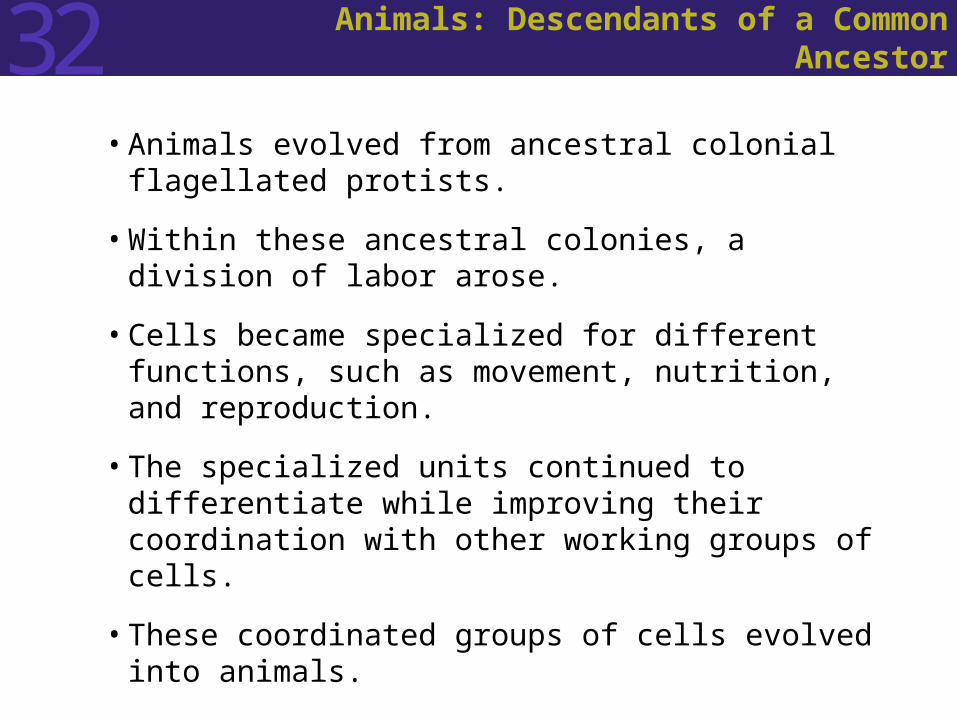
• Animals evolved from ancestral colonial flagellated protists.
• Within these ancestral colonies, a division of labor arose.
• Cells became specialized for different functions, such as movement, nutrition, and reproduction.
• The specialized units continued to differentiate while improving their coordination with other working groups of cells.
• These coordinated groups of cells evolved into animals.
32 Animals: Descendants of a Common Ancestor
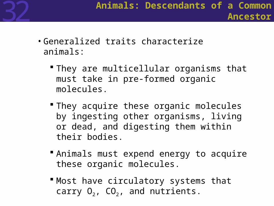
• Generalized traits characterize animals:
They are multicellular organisms that must take in pre-formed organic molecules.
They acquire these organic molecules by ingesting other organisms, living or dead, and digesting them within their bodies.
Animals must expend energy to acquire these organic molecules.
Most have circulatory systems that carry O2, CO2, and nutrients.

32 Animals: Descendants of a Common Ancestor
• Much of the diversity in the animal kingdom evolved as animals acquired the ability to capture and eat many different types of food and to avoid becoming food for other animals.
• The need to move in search of food has favored sensory structures that provide animals with detailed information about their environment.
• Animals expend a considerable amount of energy to maintain relatively constant internal conditions while taking in foods that vary chemically.

32 Animals: Descendants of a Common Ancestor
• Clues to the evolutionary relationships among animals are found in the fossil record, patterns of embryonic development, comparative physiology and morphology, and the structure of molecules such as the small-subunit RNAs and mitochondrial genes.
• The sponges, cnidarians, and ctenophores separated from the other animal lineages early in evolutionary history.
• The remaining animals have been divided into two major lineages: the protostomes and the deuterostomes.

Figure 32.1 A Current Phylogeny of the Animals

32 Animals: Descendants of a Common Ancestor
• Animals form layers of cells during their development from a single-celled zygote to a multicellular adult.
• The embryos of diploblastic animals have two cell layers: an outer ectoderm and an inner endoderm.
• The embryos of triploblastic animals have a third layer, the mesoderm.
• The existence of three cell layers distinguishes the protostomes and deuterostomes from simple animals that diverged earlier.

32 Animals: Descendants of a Common Ancestor
• Protostomes and deuterostomes differ in the fate of the blastopore , the opening of the cavity that forms in the spherical embryo.
• In the protostomes, the mouth arises from the blastopore.
• In the deuterostomes, the blastopore gives rise to the anus.
32 Animals: Descendants of a Common Ancestor
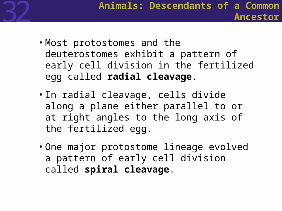
• Most protostomes and the deuterostomes exhibit a pattern of early cell division in the fertilized egg called radial cleavage.
• In radial cleavage, cells divide along a plane either parallel to or at right angles to the long axis of the fertilized egg.
• One major protostome lineage evolved a pattern of early cell division called spiral cleavage.
32 Body Plans: Basic Structural Designs
• The entire structure of an animal, its organ systems, and the integrated functioning of its parts are known as its body plan.
32 Body Plans: Basic Structural Designs
• Overall shape is referred to as symmetry. A symmetrical animal can be divided into similar halves along at least one plane.
• Animals that have no plane of symmetry are said to be asymmetrical.
• In spherical symmetry body parts radiate out from a central point. Spherical symmetry is widespread among the protists.
• An organism with radial symmetry has one main axis around which its body parts are arranged.
• Bilaterally symmetric animals can be divided into mirror images by a single plane.
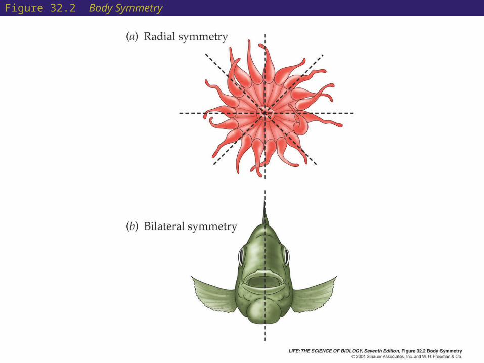
Figure 32.2 Body Symmetry

32 Body Plans: Basic Structural Designs
• Bilateral symmetry is a common characteristic of animals that move freely through their environments.
• Bilateral symmetry is often associated with cephalization: the presence of a head bearing sensory organs and central nervous tissues at the anterior end of the animal.

32 Body Plans: Basic Structural Designs
• Body cavities are fluid-filled spaces that lie between the cell layers of the bodies of many kinds of animals.
• The type of body cavity an animal has influences how it can move.
• Animals can be grouped into three major categories based on the type of body cavity they have: the acoelomates, the pseudocoelomates, and the coelomates.
32 Body Plans: Basic Structural Designs
• Acoelomates lack an enclosed body cavity. The space between the gut and body wall is filled with cells called mesenchyme.
• Pseudocoelomates have a pseudocoel, a liquid filled space in which organs are suspended.
• Coelomates have a coelom that develops within the mesoderm. It is lined with the peritoneum and enclosed on the inside and outside by muscles.
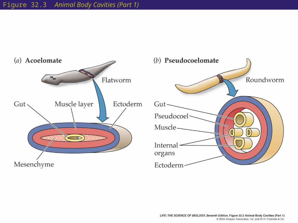
Figure 32.3 Animal Body Cavities (Part 1)

Figure 32.3 Animal Body Cavities (Part 2)
32 Body Plans: Basic Structural Designs
• The fluid-filled body cavities of simple animals function as hydrostatic skeletons.
• When the muscles surrounding fluids contract, the fluids can be moved to other parts of the body, causing these body regions to expand.
• Other forms of skeletons developed in many lineages, including internal skeletons (vertebrate bones), and external skeletons (crab shells, clam shells).
• The form of an animal’s skeleton and body cavities strongly influences the degree to which it can control and change its shape and thus the complexity of the movements it can perform.

32 Sponges: Loosely Organized Animals
• The lineage leading to modern sponges (phylum Porifera) separated from the lineage leading to other animals very early during animal evolution.
• Sponges are sessile—they live attached to the substratum.
• The body plan of sponges is an aggregation of cells built around a water canal system.

Figure 32.4 The Body Plan of a Simple Sponge
32 Sponges: Loosely Organized Animals
• Sponges have a supporting skeleton, either in the form of branching spines called spicules or as an elastic network of fibers.
• Sponges are loosely organized; if a sponge is completely disassociated, its cells can reassemble into a new sponge.
• Sponges depend on water movement through their bodies to obtain food and are often oriented at right angles to current flow so that they may intercept water as it flows past.
• Sponges reproduce both sexually and asexually. In most species, a single individual produces both eggs and sperm. Asexual reproduction is by budding and fragmentation.
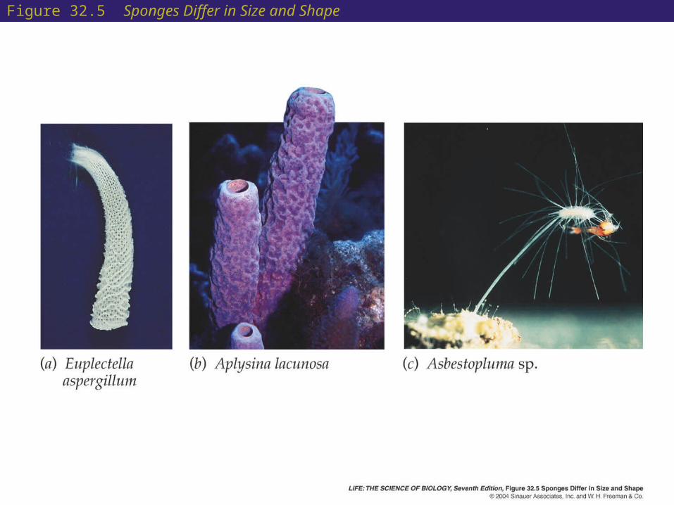
Figure 32.5 Sponges Differ in Size and Shape
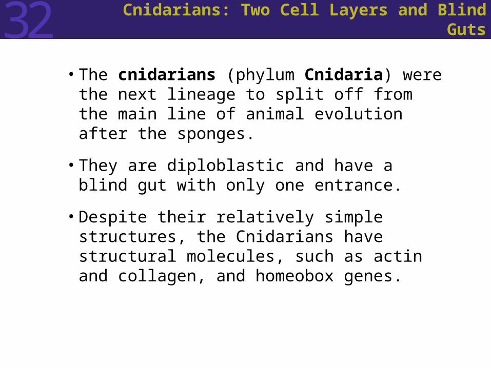
32 Cnidarians: Two Cell Layers and Blind Guts
• The cnidarians (phylum Cnidaria) were the next lineage to split off from the main line of animal evolution after the sponges.
• They are diploblastic and have a blind gut with only one entrance.
• Despite their relatively simple structures, the Cnidarians have structural molecules, such as actin and collagen, and homeobox genes.

32 Cnidarians: Two Cell Layers and Blind Guts
• Cnidarians appeared early in evolutionary history and radiated in the late Precambrian.
• There are about 11,000 species living today.
• The cnidarian body plan combines a low metabolic rate with the ability to capture large prey, allowing cnidarians to survive in environments where prey is scarce.

32 Cnidarians: Two Cell Layers and Blind Guts
• Cnidarians have tentacles with specialized cells called cnidocytes. These cells contain structures called nematocysts that can discharge toxins into their prey.
• The mouth of a cnidarian is connected to a blind sac called the gastrovascular cavity. It functions in digestion, circulation, and gas exchange.
• Cnidarians have epithelial cells with muscle fibers whose contractions allow them to move, as well as nerve nets that integrate body activities.
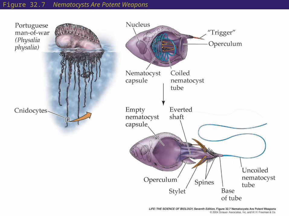
Figure 32.7 Nematocysts Are Potent Weapons

32 Cnidarians: Two Cell Layers and Blind Guts
• The generalized cnidarian life cycle has two stages: The polyp is typically asexual; individual polyps
may reproduce by budding to form colonies. The medusae produce eggs and sperm and
release them into the water.
• A fertilized egg becomes a free-swimming, ciliated larva called a planula that eventually settles to the bottom and transforms into a polyp.

Figure 32.8 A Generalized Cnidarian Life Cycle

32 Cnidarians: Two Cell Layers and Blind Guts
• Corals are also usually sessile and colonial.
• The polyps of corals secrete a matrix of organic molecules upon which calcium carbonate is deposited.
• This matrix forms the eventual skeleton of the coral colony.
• As coral colonies grow, old polyps die and leave their calcareous skeletons behind.
• Living members of the colony form a layer on top of a growing reef of skeletal remains.
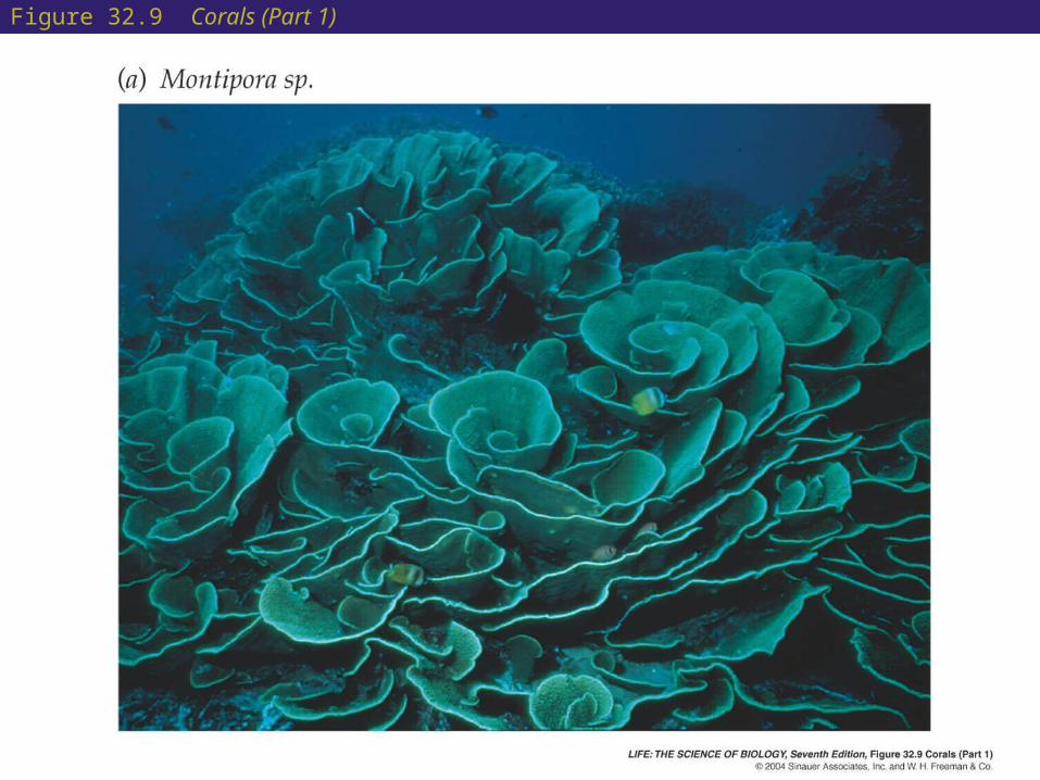
Figure 32.9 Corals (Part 1)
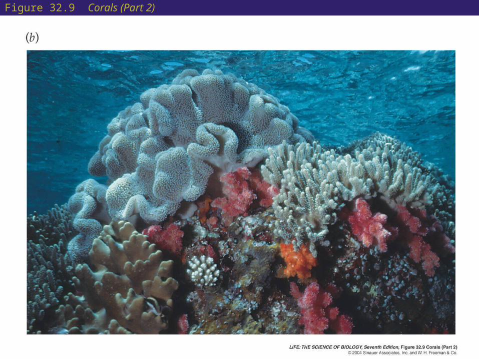
Figure 32.9 Corals (Part 2)

32 The Evolution of Bilaterally Symmetrical Animals
• A common ancestor of all bilaterally symmetrical animals is postulated.
• Zoologists use evidence from genes, development, and the structure of existing animals to infer the form of ancient bilaterians.
• The development of all bilaterally symmetrical animals is controlled by homologous Hox and homeobox genes. It is unlikely that these genes evolved separately in several animal lineages.
• Fossilized tracks from the late Precambrian suggest that early bilaterians had circulatory systems, antagonistic muscles, and a tissue- or fluid-filled body cavity.

Figure 32.13 The Trail of an Early Bilaterian
32 The Evolution of Bilaterally Symmetrical Animals
• The protostomes and the deuterostomes that dominate today’s fauna have been evolving separately since the Cambrian period.
• Members of both lineages are bilaterally symmetrical and have cephalization.
32 The Evolution of Bilaterally Symmetrical Animals
• Shared, derived traits that unite the protostomes include:
A central nervous system consisting of an anterior brain that surrounds the entrance to the digestive tract
A ventral nervous system consisting of paired or fused longitudinal nerve cords
Free-floating larvae with a food-collecting system consisting of compound cilia on multiciliate cells
A blastopore that becomes the mouth
Spiral cleavage (in some species)
32 The Evolution of Bilaterally Symmetrical Animals
• The major shared, derived traits that unite the deuterostomes inlcude:
A dorsal nervous system
Larvae, if present, that have a food-collecting system consisting of cells with a single cilium
A blastopore that becomes the anus
Radial cleavage

32 Simple Lophotrochozoans
• The flatworms (phylum Platyhelminthes) are the simplest of the lophotrochozoans.
• The flatworms are bilaterally symmetrical, unsegmented, acoelomate animals.
• They lack organs for transporting oxygen to internal tissues.
• They have simple organs for excreting metabolic wastes.
• Their flattened form allows each body cell to be near a body surface, a requirement of their body plan.

32 Simple Lophotrochozoans
• The flatworm digestive tract is a mouth opening into a blind sac.
• The sac is often highly branched, increasing the surface area available for the absorption of nutrients.
• Flatworms feed on living or dead animal tissue.
• The motile flatworms move by beating broad bands of cilia.

32 Simple Lophotrochozoans
• The flatworms of the class Turbellaria are probably most similar to ancestral flatworm forms.
• Turbellarians are small, free-living, marine and freshwater animals.
• The head has chemoreceptor organs, simple eyes, and a small brain.
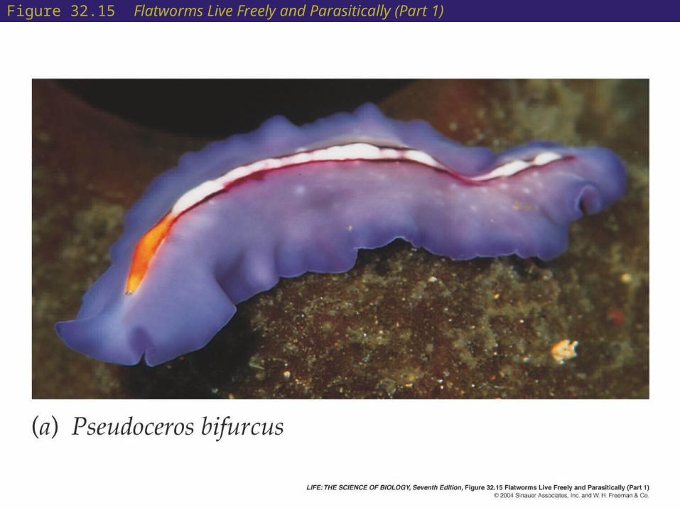
Figure 32.15 Flatworms Live Freely and Parasitically (Part 1)

32 Simple Lophotrochozoans
• Most living flatworms are parasitic, such as the tapeworms (class Cestoda) and the flukes (class Trematoda).
• Parasitic flatworms lack digestive tracts; they absorb digested food from their hosts.
• Some species cause serious diseases, such as schistosomiasis.
• Most parasitic species have complex life cycles involving one or more intermediate hosts and several larval stages.

Figure 32.15 Flatworms Live Freely and Parasitically (Part 2)
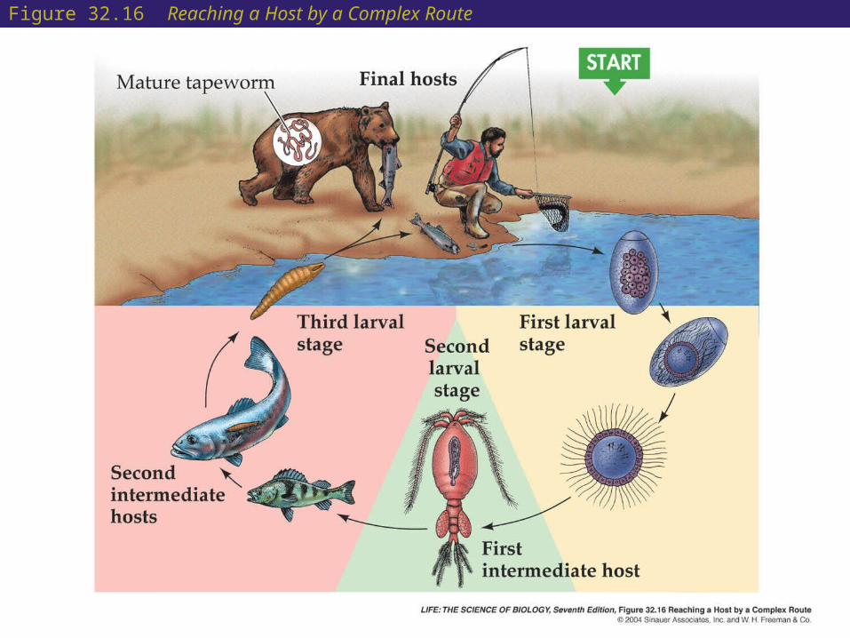
Figure 32.16 Reaching a Host by a Complex Route

32 Lophophorates: An Ancient Body Plan
• The brachiopods (phylum Brachiopoda) are solitary, marine lophophorate animals that superficially resemble bivalve mollusks.
• The shell differs from that of mollusks in that its two halves are dorsal and ventral rather than lateral.
• Brachiopods are either attached to a solid substrate by a short, flexible stalk or embedded in soft sediment.
• Most species release gametes into the water, where they are fertilized.
• More than 26,000 fossil species have been described, but only 350 species survive today.
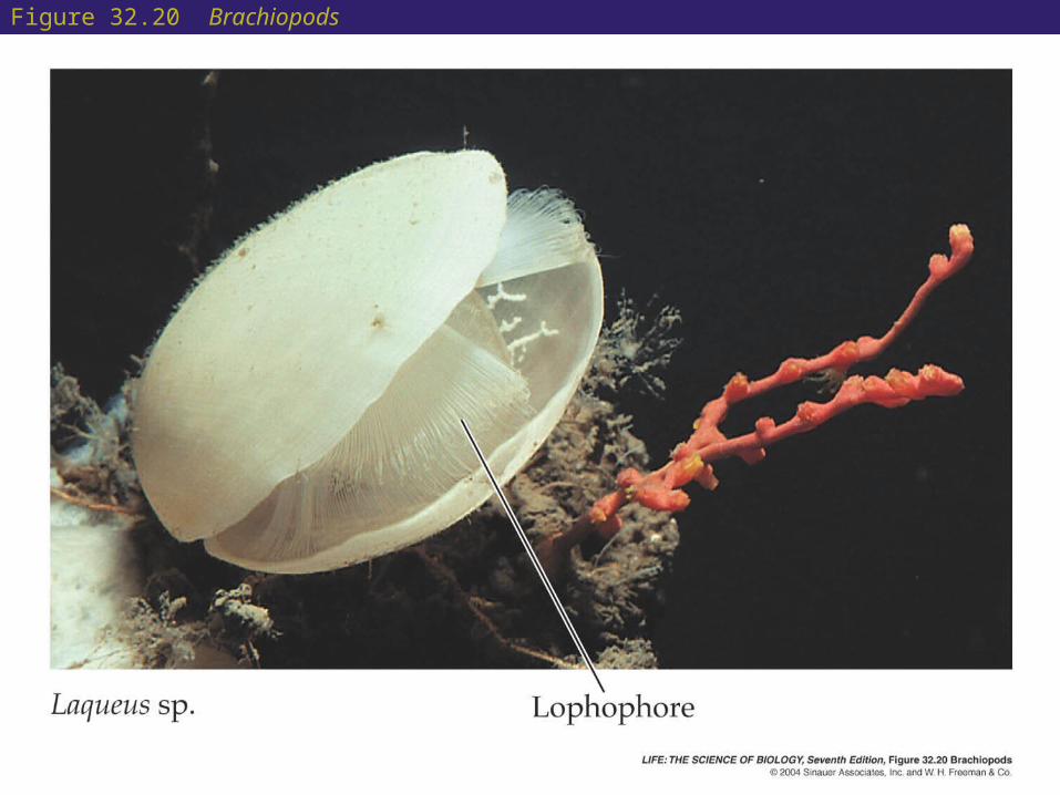
Figure 32.20 Brachiopods

32 Spiralians: Spiral Cleavage and Wormlike Body Plans
• Ribbon worms (phylum Nemertea) are carnivorous spiralians.
• They are similar in structure to the flatworms, but they have a complete digestive tract.
• Small ribbon worms move by beating their cilia; larger ones move by waves of contraction of body muscles.
32 Spiralians: Spiral Cleavage and Wormlike Body Plans
• A body cavity that is segmented allows an animal to alter the shape of its body in complex ways and to control its movements precisely.
• Segmentation evolved several times among spiralians.
• The annelids (phylum Annelida) are a diverse group of segmented worms.
• Annelid species can be found in marine, freshwater, and terrestrial environments.

32 Spiralians: Spiral Cleavage and Wormlike Body Plans
• Nerve cord is found on the ventral side.
• Each segment in an annelid is controlled by a separate nerve center called a segmented ganglion. All the ganglia are connected by nerve cords that coordinate their function.
• The coelom in each segment is isolated from those in other segments.
• Most species lack a rigid, external protective surface.
• The thin body wall serves as a surface for gas exchange and also limits annelids to moist environments, as they lose body water rapidly in dry air.

Figure 32.22 Annelids Have Many Body Segments

32 Spiralians: Spiral Cleavage and Wormlike Body Plans
• The mollusks (phylum Mollusca) range in size from small snails to giant squids that can be more than 18 meters long.
• Mollusks have a unique body plan with three major structural components: foot, mantle ( a hard skeleton structure), and visceral mass that covers the internal organs.
• The molluscan foot is a large, muscular structure that originally was both an organ of locomotion and support for the internal organs.

Figure 32.25 Molluscan Body Plans (Part 1)
32 Spiralians: Spiral Cleavage and Wormlike Body Plans
• The bivalves (class Bivalvia) have a hinged, two-part shell that extends over the sides and top of their body.
• Bivalves are largely sedentary.
• They have greatly reduced heads.
• Feeding is accomplished by bringing water in through an opening called an incurrent siphon and extracting food from the water using their gills.
• Water and gametes exit through another opening, the excurrent siphon.

Figure 32.26 Diversity among the Mollusks (Part 2)

32 Spiralians: Spiral Cleavage and Wormlike Body Plans
• The gastropods (class Gastropoda) are mostly motile, using their large foot to move across a substrate or to burrow through it.
• The gastropods are the most species-rich and widely distributed of the molluscan classes.
• Some gastropods can crawl, whereas others have a modified foot that functions as a swimming organ.
• Gastropods are the only terrestrial mollusks. They have a mantle cavity that is modified into a highly vascularized lung.

Figure 32.25 Molluscan Body Plans (Part 4)
32 Spiralians: Spiral Cleavage and Wormlike Body Plans
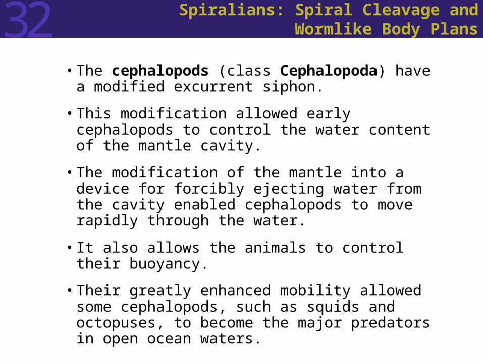
• The cephalopods (class Cephalopoda) have a modified excurrent siphon.
• This modification allowed early cephalopods to control the water content of the mantle cavity.
• The modification of the mantle into a device for forcibly ejecting water from the cavity enabled cephalopods to move rapidly through the water.
• It also allows the animals to control their buoyancy.
• Their greatly enhanced mobility allowed some cephalopods, such as squids and octopuses, to become the major predators in open ocean waters.
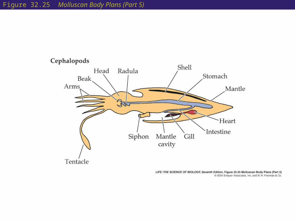
Figure 32.25 Molluscan Body Plans (Part 5)
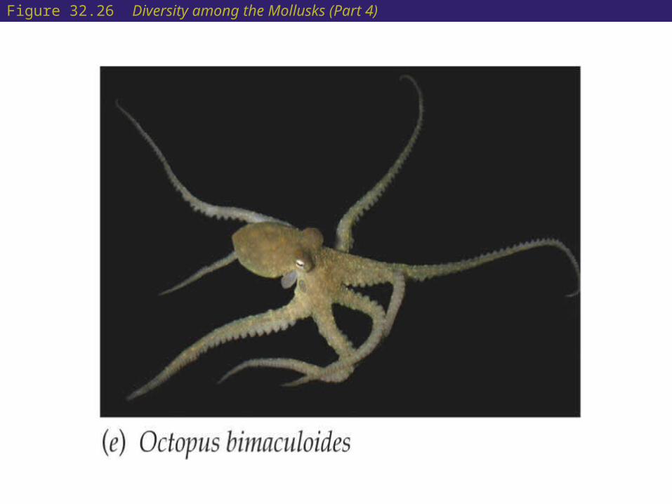
Figure 32.26 Diversity among the Mollusks (Part 4)

32 Spiralians: Spiral Cleavage and Wormlike Body Plans
• Cephalopods include the squids, octopuses, and nautiluses.
• They appeared near the beginning of the Cambrian period about 600 million years ago.
• They were the first large, shelled animals able to move vertically in the ocean.
• Nautiloids are the only cephalopods with external chambered shells that survive today.

Figure 32.26 Diversity among the Mollusks (Part 5)