3 ACCEPTED - Journal of Clinical Microbiology15 None the less, characterization by microscopic...
29
Specific primers for rapid detection of Microsporum audouinii by PCR in clinical 1 samples 2 Roque, H.D. 1 , Vieira, R. 2 , Rato, S 1 , Luz-Martins, M. 1 * 3 1 Laboratório de Micologia, Instituto de Higiene e Medicina Tropical/CREM, Universidade 4 Nova de Lisboa, Portugal. 5 2 Serviço de Dermatologia, Hospital Curry-Cabral, Lisboa, Portugal. 6 7 Specific primers for the identification of M. audouinii 8 9 10 *For correspondence: Dr. Luz Martins, Lab. Micologia, Instituto de Higiene e Medicina 11 Tropical, R. da Junqueira, 1149-008 Lisboa, Portugal. [email protected] 12 ACCEPTED Copyright © 2006, American Society for Microbiology and/or the Listed Authors/Institutions. All Rights Reserved. J. Clin. Microbiol. doi:10.1128/JCM.00759-06 JCM Accepts, published online ahead of print on 27 September 2006 on February 26, 2020 by guest http://jcm.asm.org/ Downloaded from
Transcript of 3 ACCEPTED - Journal of Clinical Microbiology15 None the less, characterization by microscopic...

Specific primers for rapid detection of Microsporum audouinii by PCR in clinical 1
samples 2
Roque, H.D.1, Vieira, R.2, Rato, S 1, Luz-Martins, M.1* 3
1Laboratório de Micologia, Instituto de Higiene e Medicina Tropical/CREM, Universidade 4
Nova de Lisboa, Portugal. 5
2 Serviço de Dermatologia, Hospital Curry-Cabral, Lisboa, Portugal. 6
7
Specific primers for the identification of M. audouinii 8
9
10
*For correspondence: Dr. Luz Martins, Lab. Micologia, Instituto de Higiene e Medicina 11
Tropical, R. da Junqueira, 1149-008 Lisboa, Portugal. [email protected] 12
ACCEPTED
Copyright © 2006, American Society for Microbiology and/or the Listed Authors/Institutions. All Rights Reserved.J. Clin. Microbiol. doi:10.1128/JCM.00759-06 JCM Accepts, published online ahead of print on 27 September 2006
on February 26, 2020 by guest
http://jcm.asm
.org/D
ownloaded from

2
Abstract 1
This report describes application of PCR fingerprinting to identify common species of 2
dermatophytes using the microsatellite primers, M13, (GACA)4 and (GTG)5. The initial 3
PCR analysis rendered a specific DNA fragment for Microsporum audouinii, which was 4
cloned and sequenced. Based on the sequencing data of this fragment, forward 5
(MA_1F) and reverse (MA_1R) primers were designed and verified by PCR to establish 6
their reliability in the diagnosis of M. audouinii. These primers produced a singular PCR-7
band of 431 base pairs specific only to strains and isolates of M. audouinii, based on a 8
global test of 182 strains/isolates belonging to 11 species of dermatophytes. These 9
findings indicate these primers are reliable for diagnostic purposes and we recommend 10
their use in laboratory analysis. 11
ACCEPTED
on February 26, 2020 by guest
http://jcm.asm
.org/D
ownloaded from
3
Introduction 1
Tinea (ringworm) is an infection of keratinized tissues (epidermis, hair, and nails) by a 2
group of specialized fungi, the dermatophytes. Dermatophytes belong to three 3
anamorphic genera, Epidermophyton, Microsporum and Trichophyton, each of which 4
include several recognized species (28). Infections by dermatophytes result in 5
symptoms that vary from mild to severe, depending on the immunological response of 6
the host. Conventional procedures for routine identification of dermatophytes has mainly 7
relied on microscopic examination of colony morphology, colony pigmentation, size and 8
shape of macroconidia or microconidia and colony growth rate under deficient 9
conditions. Misidentification using conventional microbiological methods was one of the 10
main reasons dermatophytes were among the first fungal groups studied using 11
molecular-genetic methods, such as the determination of nDNA, G+C composition and 12
genomic DNA homology (6). These initial molecular approaches chiefly showed that 13
dermatophytes, in general, were fundamentally closely related organisms. 14
None the less, characterization by microscopic examination and in vitro culture of tinea 15
is required for appropriate diagnosis and treatment, as well as for prevention of epidemic 16
radiation of the infection. For example in tinea capitis the identification of the species is 17
important in the establishment of therapy. In general, infections by Trichophyton species 18
need a longer period of treatment than that caused by Microsporum and M. canis which 19
only responds to terbinafine when higher doses are employed. Although in vitro culture 20
is specific and sensitive, it has two significant drawbacks as a useful method for rapid 21
diagnosis. These drawbacks are namely the long incubation period (2 or 3 weeks) 22
required before diagnostic characteristics are developed in culture media and, secondly, 23
the fact that many dermatophyte strains often develop atypical characteristics (different 24
ACCEPTED
on February 26, 2020 by guest
http://jcm.asm
.org/D
ownloaded from
4
colony morphologies, pleomorphism). This latter drawback can result in false negative 1
diagnosis in as high as 15% of cases where there is actual infection (24, 28). Moreover, 2
misdiagnosis may lead to harmful or inadequate treatment. 3
Molecular biological techniques, such as arbitrarily primed PCR (AP-PCR) (16), random 4
amplified polymorphic DNA analysis (RAPD) (21)), restriction analysis of mtDNA (22) 5
and microsatellite-primed PCR (MSP-PCR) (10-12, 25) are considered to be generally 6
adequate for species identification of certain fungi. Such techniques have been used to 7
differentiate 26 species of Candida and up to 17 species of dermatophytes, including 8
intraspecific strains of some species. Nevertheless, species identification based on 9
detection of numerous and complex PCR banding patterns generated by these 10
techniques may be technically allusive and inconclusive. A practical and proven 11
approach to overcome these technical deficiencies is to comparatively analyze individual 12
DNA fragment profiles from the original PCR fingerprint in order to identify species or 13
strain specific amplicons. Once such specific bands are identified, they can be 14
sequenced and specific PCR primers/probes can be designed (9, 17, 18). 15
In the present study, the application of PCR fingerprinting was performed using three 16
sets of primers: M13, (GTG)5, and (GACA)4 for the purpose of species identification and 17
strain-typing of common dermatophytes and related fungi. During analysis of the 18
fingerprints of 11 species of dermatophytes, a specific DNA fragment was detected to be 19
exclusive to only and all strains of Microsporum audouinii examined. Based on the DNA 20
sequence of this amplicon, species-specific PCR primers were designed and tested for 21
rapid and efficient PCR-based detection and to evaluate the diagnostic value of this 22
method. 23
24
ACCEPTED
on February 26, 2020 by guest
http://jcm.asm
.org/D
ownloaded from

5
MATERIAL AND METHODS 1
2
Cultures. During 2002 and 2003 samples were collected from skin, nail and hair from 3
patients suspected of having ringworm as putatively diagnosed in various dermatology 4
clinics in the Lisbon area. A small sample from each of the specimens was suspended in 5
a drop of 20% [w/v] KOH and examined under a microscope. The remaining portion of 6
each specimen was inoculated onto Sabouraud dextrose agar (Difco) and cultured at 7
25ºC for up to 4 weeks. Identification of dermatophyte isolates obtained from these 8
cultures was achieved by conventional microscopic techniques (28). 9
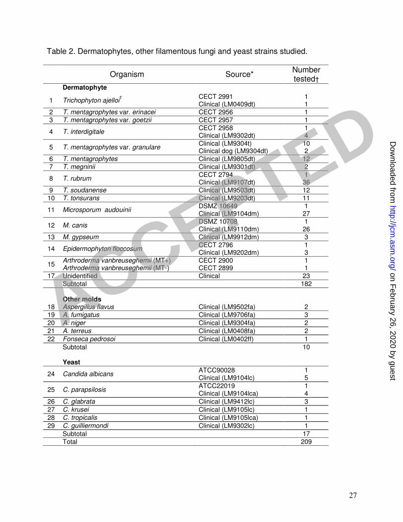
A total of 172 dermatophyte isolates representing three Microsporum species, seven 10
Trichophyton species and Epidermophyton floccosum were included in this study (Table 11
1). Ten reference strains including Microsporum canis, M. audouinii, Trichophyton 12
interdigitale, T. mentagrophytes var. erinacei, T. mentagrophytes var. goetzii, T. ajelloi, 13
T. rubrum, Arthroderma vanbreuseghemii (mating type+), A. vanbreuseghemii (mating 14
type -) and Epidermophyton floccosum (Table 2) were cultured for inclusion as positive 15
controls in PCR tests. 16
At least two clinical isolates of other common filamentous fungi, such as Aspergillus 17
fumigatus, A. niger, A. terreus, A. flavus, Fonsecaea pedrosoi, as well as clinical isolates 18
of Candida albicans (including the strain ATCC 22019), C. krusei, C. parapsilosis 19
(including the strain ATCC 90028), C. glabrata, C. tropicalis, C. guilliermondi and C. 20
neoformans (CBS 132T), were also cultured for PCR tests . 21
Extraction of genomic DNA for PCR. A rapid DNA extraction technique for filamentous 22
fungi (26) was used, with some slight modifications. Briefly, a portion of mycelium from 23
cultures on Sabouraud dextrose broth was added to a 1.5ml tube containing 500µl of 24
ACCEPTED
on February 26, 2020 by guest
http://jcm.asm
.org/D
ownloaded from

6
lysis buffer (50mM Tris l−1 pH 8.0, 50mM EDTA l−1 pH 8.0, 250mM NaCl l−1, 0,3% [w:v] 1
SDS, pH 8.0) and 500mg acid-washed 0.4-0.6mm diameter glass beads (Sigma, St 2
Louis, MO, USA). 3
Mycelia were lysed by continuous vortexing for 15 min on a Labinco BV-L46 vortex (The 4
Netherlands) at the highest intensity setting. Vortexed tubes were incubated at 65ºC for 5
1 hour, then revortexed for additional 10 min. Lysed suspensions were centrifuged at 6
13000g for 15 min at 4°C and supernatants transferred to 1.5ml tubes. Proteinase K 7
(Gibco, BRL, Germany) was added to a final concentration of 500µlml-1. Tubes were 8
incubated at 60ºC for 60 min (13), centrifuged at 13000g for 15 min, the supernatant 9
transferred to new 1.5ml tubes, and diluted 1:750 in 10 mM Tris-HCL (pH 8.0). 10
Human DNA to be used in PCR tests was extracted from blood, using the PuregeneTM 11
DNA Isolation Kit (Gentra systems, Minneapolis, MN, USA), according to the 12
manufacturer's instructions for 300 µl of whole blood. 13
Microsatellite-primed PCR. The core sequence of the phage M13 14
(GAGGGTGGCGGTTCT) was used as a single primer in minisatellite-primed PCR 15
experiments (20) and two synthetic oligonucleotides, (GACA)4 and (GTG)5, were tested 16
with microsatellite-primed PCR experiments (20) (MWG-Biotech AG, Ebersberg, 17
Germany). MSP-PCR was performed in a 25µl reaction mixture containing 10-15ng of 18
genomic DNA (5µl from Tris-HCl dilution), 16mM (NH4)2SO4, 67mM Tris-HCl (pH 9.0), 19
0.01% Tween-20, 2mM MgCl2, 250µM each dNTP, 1U of Taq DNA polymerase (Bioline, 20
London,UK), and 0.8µM of PCR primers as previously described (23). PCR amplification 21
was performed using a Tpersonal Combi PCR system (Biometra, Göettigen, Germany) 22
with the following thermocycling program: one cycle at 95ºC, 5 min; 40 cycles at 93ºC, 23
40s; 50ºC, 60s; and 72ºC, 60s; and a final cycle at 72ºC, 6 min. A tube having no DNA 24
ACCEPTED
on February 26, 2020 by guest
http://jcm.asm
.org/D
ownloaded from

7
was included as template control. After completion of amplification, 10µl of the reaction 1
mixture were added to 3µl of 10x DNA sample buffer containing bromophenol and 2
glycerol. DNA fragments were separated by electrophoresis in 1% (w/v) agarose gels in 3
0.5x TBE buffer at 50 V for 5 hours. The gel was stained in ethidium bromide for 10 min 4
and subsequently examined under UV light. Sizes of amplified DNA fragments were 5
estimated using GeneRuler 100 bp DNA Ladder Plus® (MBI, Fermentas, Germany). 6
DNA cloning, sequencing and primer design. Fragments generated by MSP-PCR 7
were excised from gels and purified using Gel Extraction Jetquick (Genomed, 8
Oeynhausen, Germany). Purified fragments were then cloned into plasmid vectors 9
(pBluescript® II KS [+]) (Stratagene, Integrated Sciences, Melbourne, Australia), 10
according to manufacture’s instructions. Cloned DNA fragments used for sequencing, 11
were amplified by PCR using 50µM of T3 and T7 primers (Invitrogen, California, USA), 12
and purified using the Jetquick/PCR purification kit (Genomed, Oeynhausen, Germany), 13
according to the manufacturer's instructions. Sequencing was performed in an automatic 14
Beckam Coulter 2000 XL sequencer using the T3 and T7 primers. PCR primers specific 15
to the M. audouinii fragment were designed with the aid of the online computer program 16
Primer Design Assistant (PDA) (5). Selected primers were then artificially synthesized 17
(MWG-Biotech AG, Ebersberg, Germany). 18
M. audouinii – specific PCR. PCR using M. audouinii specific primers was performed 19
as described for MSP-PCR but with 0.4µM of each M. audouinii species-specific primer 20
and a thermal cycle program as follows: one cycle at 95ºC, 3 min; 35 cycles at 93ºC, 30 21
s; 62ºC, 30s and 72ºC, 30s. A tube with no template DNA was included as a negative 22
control. The resultant PCR products were examined in the same fashion as for MSP-23
PCR. 24
ACCEPTED
on February 26, 2020 by guest
http://jcm.asm
.org/D
ownloaded from

8
Multiplex-PCR for reliability assessment of identification. M. audouinii specific 1
primers were used in a PCR analysis using the same conditions described above, but 2
with 0.2µM universal primers for fungi TR1 (5’ – GTT TCT AGG ACC GCC GTA – 3’) 3
and TR2 (5’ – CTC AAA CTT CCA TCG ACT – 3’) (2) added to the reaction mixture. The 4
PCR thermocycle program was the same as that used for the M. audouinii species-5
specific PCR. A blank tube having no template served as a negative control. 6
7
8
9
RESULTS 10
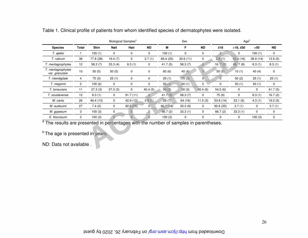
Clinical profile. Clinical data concerning patients from which dermatophytes were 11
identified are summarized in Table 1. T. rubrum was the most prevalent species 12
identified, collected exclusively from skin and nail samples, mainly from male patients 13
above 18 years in age. M. audouinii was the second most prevalent species detected, 14
almost exclusively from hair samples from children at a ratio of 2:1 male to female. The 15
third most prevalent species was M. canis, isolated mainly from skin and hair samples of 16
children at a ratio of 1:2 male to female. T. mentagrophytes was isolated mostly from 17
skin and nails, from all age groups ≤50 years, equally from both genders. T. 18
mentagrophytes var. granulare was identified exclusively from skin and nail samples, 19
mostly in young (≤18), and eldest (>50) patients equally in both genders. T. tonsurans 20
was isolated from skin and nail samples from patients ≤18 years, from both male and 21
female patients. These clinical profiles were similar for those of patients from whom T. 22
soudanense was isolated, except the isolation occurring chiefly from hair. 23
ACCEPTED
on February 26, 2020 by guest
http://jcm.asm
.org/D
ownloaded from

9
Microsatellite primed PCR. Identification of certain species of dermatophytes using the 1
microsatellite primers of M13, (GTG)5, and (GACA)4 has been reported previously, but 2
using different PCR conditions (11, 12). Fig. 1 shows the electrophoretic band patterns 3
obtained with primers M13, (GTG)5 and (GACA)4 under our PCR conditions. All sets of 4
primers generated distinct PCR-band profiles of each dermatophyte, with bands varying 5
in intensities and molecular sizes. The PCR fingerprints showed DNA fragments that 6
ranged from 0.2 to 4.9 kb in length for all species. Fig. 1 also shows that all 11 species 7
studied could be differentiated with the primers sets used, with some exceptions, as 8
follows: primer (GTG)5 failed to distinguish T. rubrum, T. soudanense and T. megninii 9
(Fig. 1-B), whereas T. soudanense and T. megninii where indistinguishable with primer 10
(GACA)4 (Fig. 1-C). On the other hand, T. rubrum and T. soudanense presented similar 11
DNA fragments patterns with primer M13 (Fig. 1-A). None of the primers were able to 12
distinguish the mating types of A. vanbreuseghemii (Fig. 1), whose patterns were quite 13
similar to those of T. mentagrophytes, T. mentagrophytes. var. goetzii, T. interdigitale, 14
and T. mentagrophytes. var. granulare (Fig. 1). All primers distinguished T. 15
mentagrophytes var. erinacei from the other varieties of T. mentagrophytes of this study. 16
Nevertheless, the remaining varieties could not be differentiated by any of the three 17
primers used. 18
Only one isolate of M. audouinii, misidentified by visible morphological methods, 19
generated a similar DNA banding profile to that of T. interdigitale (CECT 2958) for all 20
primers tested (data not show). Of the isolates that could not be identified by 21
conventional methods, four produced similar band patterns to those of M. audouinii with 22
all primers, and were thus categorized as being M. audouinii (Fig. 1-A). The remaining 23
isolates of M. audouinii produced similar patterns with the different primers, with the 24
ACCEPTED
on February 26, 2020 by guest
http://jcm.asm
.org/D
ownloaded from

10
exception of two isolates identified as M. audouinii with primer (GACA)4 and as M. canis 1
with the primers M13 and (GTG)5 (data not shown). 2
Analysis of DNA sequences of unique bands for species-specific identification. 3
The M13 primer produced a clearer distinction between M. canis and M. audouinii than 4
the others primers used. A prominent species-specific fragment, of approximately 0.9 kb, 5
generated from the genomic DNA of M. audouinii (reference strain DSMZ 10649) with 6
the M13 primer (Fig.1-A), was excised from the gel, purified, cloned and sequenced. The 7
sequence product of the cloned amplicon was 919 bp in length. From this sequence, a 8
forward primer (MA1_F: 5’- CGA AGC GAG CCT CTA CGG CAA TCC AAA GCA G -3’) 9
and a reverse primer (MA1_R: 5’- CGA AGC ATC TTG CAG GAC TCC -3’) were 10
designed according to protocols outlined in Materials and Methods. These primers 11
generated an amplicon of 431 bp. The primers were tested under several PCR 12
conditions, and with the genomic DNA of all the strains/isolates used in this study. 13
Identification reliability assessment of "MA1" primers. The utility of the MA1_F and 14
MA1_R primers as a tool for identifying M. audouinii (4) was compared to that of the 15
microsatellite primers M13, (GTG)5, and (GACA)4. All strains used in this study were 16
tested with these primers in a Multiplex PCR reaction with universal primers. As shown 17
in Fig. 2 the expected band of 431 bp, using the MA1 primers, was generated only with 18
strains of M. audouinii, whereas, a band of 600 bp was generated by primers TR1 and 19
TR2 for all fungal species, including M. audouinii. In some cases, the 600 bp fragment 20
was weak or absent for the strain/isolates of M. audouinii. The four isolates that could 21
not be identified using conventional methods, but identified as M. audouinii by MSP-PCR 22
fingerprinting, produced the 431 bp fragment when tested with the MA1 primers. 23
ACCEPTED
on February 26, 2020 by guest
http://jcm.asm
.org/D
ownloaded from

11
In order to assess the identification potential for PCR fingerprinting and the M. audouinii 1
specific PCR, parameters were set to evaluate the reliability and accuracy for 2
identification and diagnostic purposes (Table 3). This evaluation was performed with 3
considering that ordinarily identification is made by morphological features. We found 4
that all the primers tested resulted in values showing a high degree of detection 5
sensitivity and specificity (above 89% and 98%, respectively) (see Discussion). Positive 6
and Negative Predictive Values were also congruent with there being a high 7
identification potential of the PCR techniques developed in this study. 8
9
DISCUSSION 10
Infections of humans by dermatophytes have historically been considered to be of little 11
particular medical importance, in view that such infections were generally mild and not 12
life-threatening. Moreover, many of the more significant etiologic agents were restricted 13
to specific geographic areas and not involved in widespread epidemics. However, 14
demographic changes over the prior two decades have resulted in widespread infections 15
caused by a variety of species of dermatophytes. Nowadays, cutaneous infections 16
caused by dermatophytic fungi occur in every age group, worldwide. The millions of 17
individuals who are infected by these pathogens, contribute to health care costs in the 18
tens of millions of dollars on an annual basis (8), especially for treatment when diagnosis 19
is not quickly established. 20
Over the past few years the number of cases of tinea capitis due to anthropophyllic fungi 21
has significantly increased, particularly in some European city communities (1). In 22
Portugal, recent epidemiological data on dermatophyte infections are not available. In a 23
study of isolates from a clinical laboratory attending to patients from the Lisbon area, T. 24
ACCEPTED
on February 26, 2020 by guest
http://jcm.asm
.org/D
ownloaded from

12
rubrum was the most frequently isolated agent of dermatophytosis (28.5%), with M. 1
audouinii (22.6%) and T. soudanense (14.3%) being the second and third dermatophyte, 2
respectively. Tinea capitis was the most diagnosed dermatophytosis followed by tinea 3
pedis. This pattern in the Portuguese clinic is distinct from the one observed throughout 4
other countries in Western Europe, but similar to that found in some African countries (P. 5
Viegas, A. Pinto, A. Santos, M. Pinheiro, and R. Vieira. Reun. Centro Soc. Port. Dermat. 6
Vener., abst.72, 2003) . In our laboratory, skin related dermatophytes were the most 7
frequently identified, followed by tinea capitis. The most isolated agent was T. rubrum, 8
followed by M. audouinii and M. canis (Table 1). 9
In Portugal, there are recent data suggesting M. audouinii and T. soudanense as the 10
most frequent dermatophytes isolated from tinea capitis. These fungi, prevalent in 11
African countries, are now infecting European children and becoming increasingly 12
responsible for outbreaks in European schools (27). Our records are congruent with 13
these data. Still M. canis is a major tinea capitis etiologic agent. Its widespread and 14
capability of infecting distint tissues account for the high number of hair infections 15
detected (Table 1). 16
In this study, differentiation of species involved in tinea capitis was possible by MSP-17
PCR using three different primers. As previously described, it was possible to 18
discriminate all the species with primers (GACA)4 (11), M13 and (GTG)5 (12). Variability 19
among varieties of T. mentagrophytes was observed for T. mentagrophytes var. erinacei 20
(CECT 2956) for all three primers. Slight differences were observed among the 21
remaining varieties of T. mentagrophytes with these primers, as well. Similarity in 22
fingerprinting patterns between A. vanbreuseghemii (both mating types) and varieties of 23
T. mentagrophytes (with the exception of T. mentagrophytes. var. erinacei) reflects the 24
ACCEPTED
on February 26, 2020 by guest
http://jcm.asm
.org/D
ownloaded from

13
close phylogenetic relation among them (14). The failure of primers (GTG)5 and (GACA)4 1
to discriminate T. soudanense and T. megninii is evidence of their close phylogenetic 2
proximity. In fact, in a previous report (15) T. soudanense was reclassified as T. 3
violaceum and T. megninii was considered an ambiguous taxonomic classification. 4
These three species belong to the T. rubrum complex, as shown by the high degree of 5
similarity in their DNA fragment patterns in our study. 6
Application of MSP-PCR has enabled rapid identification of several dermatophyte 7
species/varieties (10-12). Nevertheless, development of specific primers for different 8
species would improve specificity of PCR based tests and eliminate problems of 9
ambiguities in band pattern analysis. Development of a T. rubrum specific probe (9) and, 10
more recently, a specific M. canis primer set (18) have demonstrated the diagnostic 11
importance of such primer-specific approaches. 12
Difficulty in identifying M. audouinii by macro and microscopic features, especially in 13
distinguishing it from M. canis, and time-consuming laboratory diagnosis led us to design 14
a specific primer for identification of M. audouinii. To ensure that no false negative 15
results were obtained from the PCR reactions using our MA1 primers, a universal 16
primer-set generating an amplicon of 600 bp was added to serve as positive control of 17
the PCR reaction. This mix of primer-sets produced two fragments (of 600 bp and 431 18
bp; Fig. 2) in the strains/isolates of M. audouinii and just one fragment of 600 bp in the 19
non M. audouinii strains/isolates (Fig. 2). 20
The reliability of primers MA1_F and MA1_R as a diagnostic tool showed the MA1 21
primers had high specificity, sensibility, PPV, and PVN rates (all values near 100%) for 22
the strains/isolates tested (Table 3). It is worth mentioning, that two of the false positive 23
tests obtained with primers M13 and (GTG)5, were attributed to two isolates of M. 24
ACCEPTED
on February 26, 2020 by guest
http://jcm.asm
.org/D
ownloaded from
14
audouinii that where identified as M. canis by M13 and (GTG)5 , and as M. audouinii by 1
(GACA)4 and MA1. Considering these findings, we conclude the two isolates are M. 2
audouinii. Also, the one False Negative resulting in all primer tests was attributed to one 3
isolate identified as M. audoinii by morphological features. However, the MSP-PCR 4
primers identified this isolate as T. interdigitale. Moreover, this isolate also did not 5
produce the 431bp band expected for M. audouinii with primers MA1. This shows that 6
primers MA1 had 100% accuracy in the study sample. 7
In conclusion, the results of our study strongly support use of the MA1 M. audouinii-8
specific-primers for the identification of M. audouinii in the diagnosis of tinea capitis. In 9
the future, PCR-based identification, as outlined in this study, can be applied directly to 10
skin and hair samples. This has the advantage relative to conventional culture methods, 11
of making identification in a shorter time-frame (one to four days instead of two to three 12
weeks), and eliminating problems of culture failure and contamination. The PCR 13
approach does rely, however, on efficient extraction of nucleic acids that avoid possible 14
degradation of DNA, which could lead to false negative results. In our laboratory we 15
have already started to use this technique as a diagnostic tool in parallel with the 16
conventional methods. So far, results have been 100% compatible. Moreover, detection 17
of dermatophytes by PCR technique instead of culture will allow a rapid diagnosis of the 18
species that will lead to a better managment of infections caused by these fungi. 19
20
21
ACKNOWLEDGEMENTS 22
We thank the Unit of Microbiology, Biotechnology, and Molecular Biology of the Institute 23
of Applied Science and Technology (ICAT), Portugal for the kindly given reference 24
ACCEPTED
on February 26, 2020 by guest
http://jcm.asm
.org/D
ownloaded from

15
strains of the CECT collection, Inês Costa for has given us some clinical strains, Mário 1
Gadanho for the critical suggestions, to Abdou, Teresa and Sandra for the research 2
assistance. We also thank Dr. Bruce Campbell's kind help in reviewing the manuscript. 3
ACCEPTED
on February 26, 2020 by guest
http://jcm.asm
.org/D
ownloaded from

16
REFERENCES 1
1. Aly, R., R. Hay, A. Del Palacio, and R. Galimberti. 2000. Epidemiology of tinea 2
capitis. Med. Mycol. 38 Suppl 1:183-188. 3
2. Bock, M., M. Maiwald, R. Kappe, P. Nickel, and H. Naher. 1994. Polymerase 4
chain reaction-based detection of dermatophyte DNA with a fungus-specific 5
primer system. Mycoses 37:79-84. 6
3. Brilhante, R., G. Paixao, L. Salvino, M. Diogenes, S. Bandeira, M. Rocha, J. 7
dos Santos, and J. Sidrim. 2000. Epidemiology and ecology of 8
dermatophytoses in the City of Fortaleza: Trichophyton tonsurans as important 9
emerging pathogen of tinea capitis. Rev. Soc. Bras. Med. Trop. 33:417-25. 10
4. Campbel, M. J., and D. Machin. 1990. Medical statistics: a commonsense 11
approach. John Wiley & Sons, Ltd., Chichester, United Kingdom. 12
5. Chen, S. H., C. Y. Lin, C. S. Cho, C. Z. Lo, and C. A. Hsiung. 2003. Primer 13
Design Assistant (PDA): a web-based primer design tool. Nucleic Acids Res. 14
31:3751-3754. 15
6. Davison, F., and D. Mackenzie. 1984. DNA homology studies in the taxonomy of 16
dermatophytes. Sabouraudia 22:117-23. 17
7. Devliotou-Panagliotidou, D., T. Koussidou-Eremondi, G. Chaidemenos, M. 18
Theodoridou, and A. Minas. 2001. Tinea capitis in adults during 1981-95 in 19
northern Greece. Mycoses 44:398-400. 20
8. Drake, L., S. Dinehart, E. Farmer, R. Goltz, G. Graham, M. Hardinsky, C. 21
Lewis, D. Pariser, J. Skouge, S. Webster, D. Whitaker, B. Butler, B. Lowery, 22
B. Elewski, M. Elgart, P. Jacobs, J. Lesher, and R. Scher. 1996. Guidelines of 23
care for superficial mycotic infections of the skin: tinea corporis, tinea cruris, tinea 24
ACCEPTED
on February 26, 2020 by guest
http://jcm.asm
.org/D
ownloaded from

17
faciei, tinea manuum, and tinea pedis. Guidelines/Outcomes Committee. 1
American Academy of Dermatology. J. Am. Acad. Dermatol. 34:282-6. 2
9. El Fari, M., H. Tietz, W. Presber, W. Sterry, and Y. Graser. 1999. Development 3
of an oligonucleotide probe specific for Trichophyton rubrum. Br. J. Dermatol. 4
141:240-245. 5
10. Faggi, E., G. Pini, and E. Campisi. 2002. PCR Fingerprinting for Identification of 6
Common Species of Dermatophytes. J. Clin. Microbiol. 40:4804-4805. 7
11. Faggi, E., G. Pini, E. Campisi, C. Bertellini, E. Difonzo, and F. Mancianti. 8
2001. Application of PCR to Distinguish Common Species of Dermatophytes. J. 9
Clin. Microbiol. 39:3382-3385. 10
12. Graser, Y., M. el Fari, W. Presber, W. Sterry, and H. Tietz. 1998. Identification 11
of common dermatophytes (Trichophyton, Microsporum, Epidermophyton) using 12
polymerase chain reactions. Br. J. Dermatol. 138:576-82. 13
13. Graser, Y., A. Kuijpers, M. El Fari, W. Presber, and G. de Hoog. 2000. 14
Molecular and conventional taxonomy of the Microsporum canis complex. Med. 15
Mycol. 38:143-53. 16
14. Graser, Y., A. Kuijpers, W. Presber, and G. De Hoog. 1999. Molecular 17
taxonomy of Trichophyton mentagrophytes and T. tonsurans. Med. Mycol. 18
37:315-30. 19
15. Graser, Y., A. F. A. Kuijpers, W. Presber, and G. S. de Hoog. 2000. Molecular 20
Taxonomy of the Trichophyton rubrum Complex. J. Clin. Microbiol. 38:3329-3336. 21
16. Liu, D., S. Coloe, J. Pedersen, and R. Baird. 1996. Use of arbitrarily primed 22
polymerase chain reaction to differentiate Trichophyton dermatophytes. FEMS 23
Microbiol. Lett. 136:147-50. 24
ACCEPTED
on February 26, 2020 by guest
http://jcm.asm
.org/D
ownloaded from

18
17. Liu, D., L. Pearce, G. Lilley, S. Coloe, R. Baird, and J. Pedersen. 2002. PCR 1
identification of dermatophyte fungi Trichophyton rubrum, T. soudanense and T. 2
gourvilii. J. Med. Microbiol. 51:117-122. 3
18. Liu, D., L. Pearce, G. Lilley, S. Coloe, R. Baird, and J. Pedersen. 2001. A 4
specific PCR assay for the dermatophyte fungus Microsporum canis. Med. Mycol. 5
39:215-9. 6
19. Martins, M. L. M. 2001. Ph.D. thesis. Universidade Nova de Lisboa, Lisboa. 7
Polimorfismos Fenéticos e Diagnóstico Molecular de Cryptococcus neoformans. 8
20. Meyer, W., T. G. Mitchell, E. Z. Freedman, and R. Vilgalys. 1993. Hybridisation 9
probes for conventional DNA fingerprinting used as single primers in the 10
polymerase chain reaction to distinguish strains of Cryptococcus neoformans. J. 11
Clin. Microbiol. 31:2274-2280. 12
21. Mochizuki, T., N. Sugie, and M. Uehara. 1997. Random amplification of 13
polymorphic DNA is useful for the differentiation of several anthropophilic 14
dermatophytes. Mycoses 40:405-409. 15
22. Mochizuki, T., S. Watanabe, and M. Uehara. 1996. Genetic homogeneity of 16
Trichophyton mentagrophytes var. interdigitale isolated from geographically 17
distant regions. J. Med. Vet. Mycol. 34:139-143. 18
23. Sampaio, J. P., M. Gadanho, S. Santos, F. Duarte, C. Pais, A. Fonseca, and 19
J. W. Fell. 2001. Polyphasic taxonomy of the genus Rhodosporidium: 20
Rhodosporidium kratochvilovae and related anamorphic species. Int. J. Syst. 21
Evol. Microbiol. 51:687-697. 22
24. Simpanya, M. F. 2000. Dermatophytes: Their taxonomy, ecology and 23
pathogenicity. In R. K. S. Kushawaha and J. Guarro (ed.), Biology of 24
ACCEPTED
on February 26, 2020 by guest
http://jcm.asm
.org/D
ownloaded from

19
Dermatophytes and other Keratinophilic Fungi. Revista Iberoamericana de 1
Micologia, Bilbao. 2
25. Thanos, M., G. Schonian, W. Meyer, C. Schweynoch, Y. Graser, T. Mitchell, 3
W. Presber, and H. Tietz. 1996. Rapid identification of Candida species by DNA 4
fingerprinting with PCR. J. Clin. Microbiol. 34:615-621. 5
26. van Burik, J., R. Schreckhise, T. White, R. Bowden, and D. Myerson. 1998. 6
Comparison of six extraction techniques for isolation of DNA from filamentous 7
fungi. Med. Mycol. 36:299-303. 8
27. Weill, F. X., V. Bernier, J. Maleville, V. Amathiuex, F. Claverie, N. Mihalikova, 9
F. Djossou, B. Félix, B. Couprie, and A. Taieb. 1999. Épidémie de teignes du 10
cuir chevelu à Microsporum audouinii var. langeronii dans un groupe scolaire 11
bordelais. J. Mycol. Méd. 9:52-56. 12
28. Weitzman, I., and R. Summerbell. 1995. The dermatophytes. Clin. Microbiol. 13
Rev. 8:240-259. 14
15
16
17
18
19
20
ACCEPTED
on February 26, 2020 by guest
http://jcm.asm
.org/D
ownloaded from

20
Figure 1A 1
1 2 3 54 6 7 8 9 10 11 12 13 14 15 16 17 18
3000
2000
1500
1200
1031900800700600
500400
3000
2000
1500
1031900800700600
500
400
1200
19 20 21 221 2 3 54 6 7 8 9 10 11 12 13 14 15 16 17 18
3000
2000
1500
1200
1031900800700600
500400
3000
2000
1500
1031900800700600
500
400
1200
19 20 21 22
2
ACCEPTED
on February 26, 2020 by guest
http://jcm.asm
.org/D
ownloaded from

21
Figure 1B 1
1 2 3 54 6 7 8 9 10 11 12 13 14 15 16 17 18
3000
2000
1500
1200
1031900800700600
500
400
3000
2000
1500
12001031900800700600500
400
1 2 3 54 6 7 8 9 10 11 12 13 14 15 16 17 18
3000
2000
1500
1200
1031900800700600
500
400
3000
2000
1500
12001031900800700600500
400
2
ACCEPTED
on February 26, 2020 by guest
http://jcm.asm
.org/D
ownloaded from

22
Figure 1C 1
1 2 3 54 6 7 8 9 10 11 12 13 14 15 16 17 18
3000
2000
1500
1200
1031900800700
600
500
400
3000
2000
1500
1200
1031
900800700
600500
400
1 2 3 54 6 7 8 9 10 11 12 13 14 15 16 17 18
3000
2000
1500
1200
1031900800700
600
500
400
3000
2000
1500
1200
1031
900800700
600500
400
2
ACCEPTED
on February 26, 2020 by guest
http://jcm.asm
.org/D
ownloaded from

23
1
Fig. 1 DNA products from dermatophytes amplified by microsatellite primed PCR using 2
primer M13 (A), (GTG)5 (B), and (GACA)4 (C). (A) Lane1 and 22 contains molecular 3
weight marker with molecular weights in bp at the left and right respectively margins: 4
lane 2, DNA products generated from M. audouinii (DSMZ 10649); lane 3 to 4, M. 5
audouinii (LM9104dm); lane 5 to 6, M. audouinii (Unidentified); lane 7, M. canis (DSMZ 6
10708); lane 8, E. floccosum (CECT 2796); lane 9, T. ajelloiT (CECT 2991); lane 10, T. 7
mentagrophytes var. erinaceii (CECT 2956); lane 11, T. mentagrophytes var. goetzii 8
(CECT 2957); lane 12, T. interdigitale (CECT 2958); lane 13, T. mentagrophytes var. 9
granulare (LM9304t); lane 14, T. mentagrophytes (LM9805dt); lane 15, A. 10
vanbreuseghemii (MT+, CECT 2900); lane 16, A. vanbreuseghemii (MT-, CECT 2899); 11
lane 17, T. rubrum (CECT 2794); lane 18, T. tonsurans (LM9203dt); lane 19, T. 12
soudanense (LM9503dt); lane 20, T. megninii (LM9301dt); lane 21, M. gypseum 13
(LM9912dm). The arrow points the specific DNA fragment of M: audouinii. (B and C) 14
Lane1 and 18 contains molecular weight marker with molecular weights in bp at the left 15
and right respectively margins: lane 2, DNA products generated from M. audouinii 16
(DSMZ 10649); lane 3, M. canis (DSMZ 10708); lane 4, E. floccosum (CECT 2796); lane 17
5, T. ajelloiT (CECT 2991); lane 6, T. mentagrophytes var. erinaceii (CECT 2956); lane 18
7, T. mentagrophytes var. goetzii (CECT 2957); lane 8, T. interdigitale (CECT 2958); 19
lane 9, T. mentagrophytes var. granulare (LM9304t); lane 10, T. mentagrophytes 20
(LM9805dt); lane 11, A. vanbreuseghemii (MT+, CECT 2900); lane 12, A. 21
vanbreuseghemii (MT-, CECT 2899); lane 13, T. rubrum (CECT 2794); lane 14, T. 22
tonsurans (LM9203dt); lane 15, T. soudanense (LM9503dt); lane 16, T. megninii 23
(LM9301dt); lane 17, M. gypseum (LM9912dm). 24
ACCEPTED
on February 26, 2020 by guest
http://jcm.asm
.org/D
ownloaded from

24
Figure 2 1
30002000150012001031900800700600500400
1 2 3 4 5 6 7 8 9 10 11 12 13 14 15
2000150012001031900800700600500400
300016 17 18 193000
2000150012001031900800700600500400
1 2 3 4 5 6 7 8 9 10 11 12 13 14 15
2000150012001031900800700600500400
300016 17 18 193000
2000150012001031900800700600500400
1 2 3 4 5 6 7 8 9 10 11 12 13 14 15
2000150012001031900800700600500400
300016 17 18 193000
2000150012001031900800700600500400
1 2 3 4 5 6 7 8 9 10 11 12 13 14 15
2000150012001031900800700600500400
300016 17 18 19
2
30002000150012001031900800700600500
400300
20 21 22 31
2000150012001031900800700600500400300
300030002000150012001031900800700600500
400300
23 24 25 26 27 28 29 30 32 33 34
2000150012001031900800700600500400300
300030002000150012001031900800700600500
400300
20 21 22 31
2000150012001031900800700600500400300
300030002000150012001031900800700600500
400300
23 24 25 26 27 28 29 30 32 33 34
2000150012001031900800700600500400300
300030002000150012001031900800700600500
400300
20 21 22 31
2000150012001031900800700600500400300
300030002000150012001031900800700600500
400300
23 24 25 26 27 28 29 30 32 33 34
2000150012001031900800700600500400300
300030002000150012001031900800700600500
400300
20 21 22 31
2000150012001031900800700600500400300
300030002000150012001031900800700600500
400300
23 24 25 26 27 28 29 30 32 33 34
2000150012001031900800700600500400300
3000
3
Fig. 2 DNA products amplified in PCR using M. audouinii – specific primers MA1_F and 4
MA1_R and universal primers TR1 and TR2. Lanes 1, 19, 20 and 34 contain DNA 5
molecular weight marker 100 bp DNA Ladder Plus® (MBI, Fermentas, Germany) with 6
molecular weights on the left and right in bp; lanes 18 and 33 are negative controls; lane 7
2, DNA products generated from M. audouinii (DSMZ 10649); lane 3, M. canis (DSMZ 8
10708); lane 4, E. floccosum (CECT 2796); lane 5, T. ajelloiT (CECT 2991); lane 6, T. 9
mentagrophytes var. erinaceii (CECT 2956); lane 7, T. mentagrophytes var. goetzii 10
(CECT 2957); lane 8, T. interdigitale (CECT 2958); lane 9, T. mentagrophytes var. 11
granulare (LM9304t); lane 10, T. mentagrophytes (LM9805dt); lane 11, A. 12
vanbreuseghemii (MT+, CECT 2900); lane 12, A. vanbreuseghemii (MT-, CECT 2899); 13
lane 13, T. rubrum (CECT 2794); lane 14, T. tonsurans (LM9203dt); lane 15, T. 14
soudanense (LM9503dt); lane 16, T. megninii (LM9301dt); lane 17, M. gypseum 15
ACCEPTED
on February 26, 2020 by guest
http://jcm.asm
.org/D
ownloaded from

25
(LM9912dm); lane 18, E. floccosum; lane 21 to 28, M. audouinii; lanes 29 to 32 M. 1
audouinii (Unidentified). 2
ACCEPTED
on February 26, 2020 by guest
http://jcm.asm
.org/D
ownloaded from
26
Table 1. Clinical profile of patients from whom identified species of dermatophytes were isolated.
Biological Samplesa Sex Ageb
Species Total
Skin
Nail
Hair
ND M F ND ≤18 >18, ≤50 >50 ND
T. ajelloi 1 100 (1) 0 0 0 100 (1) 0 0 0 0 100 (1) 0
T. rubrum 36 77.8 (28) 19.4 (7) 0 2.7 (1) 69.4 (25) 30.6 (11) 0 2.8 (1) 44.4 (16) 38.9 (14) 13.9 (5)
T. mentagrophytes 12 58.3 (7) 33.3 (4) 8.3 (1) 0 41.7 (5) 58.3 (7) 0 16. 7 (2) 66. 7 (8) 8.3 (1) 8.3 (1)
T. mentagrophytes var. granulare
10 50 (5) 50 (5) 0 0 60 (6) 40 (4) 0 50 (5) 10 (1) 40 (4) 0
T. interdigitale 4 75 (3) 25 (1) 0 0 25 (1) 75 (3) 0 0 50 (2) 25 (1) 25 (1)
T. megninii 2 100 (2) 0 0 0 50 (1) 50 (1) 0 0 50 (1) 50 (1) 0
T. tonsurans 11 27.3 (3) 27.3 (3) 0 45.4 (5) 50 (3) 50 (3) 45.4 (6) 54.5 (6) 0 0 41.7 (5)
T. soudanense 12 8.3 (1) 0 91.7 (11) 0 41.7 (5) 58.3 (7) 0 75 (9) 0 8.3 (1) 16.7 (2)
M. canis 26 46.4 (13) 0 42.9 (12) 3.8 (1) 28 (7) 64 (16) 11.5 (3) 53.8 (14) 23.1 (6) 4.3 (1) 19.2 (5)
M. audouinii 27 7.4 (2) 0 92.6 (25) 0 66.7 (18) 33.3 (9) 0 92.6 (25) 3.7 (1) 0 3.7 (1)
M. gypseum 3 100 (3) 0 0 0 66.7 (2) 33.3 (1) 0 66.7 (2) 33.3 (1) 0 0
E. floccosum 3 100 (3) 0 0 0 100 (3) 0 0 0 0 100 (3) 0 a The results are presented in percentages with the number of samples in parentheses.
b The age is presented in years.
ND: Data not available . ACCEPTED
on February 26, 2020 by guest http://jcm.asm.org/ Downloaded from
27
Table 2. Dermatophytes, other filamentous fungi and yeast strains studied.
Organism Source* Number tested†
Dermatophyte
1 Trichophyton ajelloiT CECT 2991
Clinical (LM0409dt) 1 1
2 T. mentagrophytes var. erinacei CECT 2956 1 3 T. mentagrophytes var. goetzii CECT 2957 1
4 T. interdigitale CECT 2958 Clinical (LM9302dt)
1 4
5 T. mentagrophytes var. granulare Clinical (LM9304t) Clinical dog (LM9304dt)
10 2
6 T. mentagrophytes Clinical (LM9805dt) 12 7 T. megninii Clinical (LM9301dt) 2
8 T. rubrum CECT 2794 Clinical (LM9107dt)
1 36
9 T. soudanense Clinical (LM9503dt) 12 10 T. tonsurans Clinical (LM9203dt) 11
11 Microsporum audouinii DSMZ 10649 Clinical (LM9104dm)
1 27
12 M. canis DSMZ 10708 Clinical (LM9110dm)
1 26
13 M. gypseum Clinical (LM9912dm) 3
14 Epidermophyton floccosum CECT 2796 Clinical (LM9202dm)
1 3
15 Arthroderma vanbreuseghemii (MT+) Arthroderma vanbreuseghemii (MT-)
CECT 2900 CECT 2899
1 1
17 Unidentified Clinical 23 Subtotal 182 Other molds
18 Aspergillus flavus Clinical (LM9502fa) 2 19 A. fumigatus Clinical (LM9706fa) 3 20 A. niger Clinical (LM9304fa) 2 21 A. terreus Clinical (LM0408fa) 2 22 Fonseca pedrosoi Clinical (LM0402ff) 1 Subtotal 10 Yeast
24 Candida albicans ATCC90028 Clinical (LM9104lc)
1 5
25 C. parapsilosis ATCC22019 Clinical (LM9104lca)
1 4
26 C. glabrata Clinical (LM9412lc) 3 27 C. krusei Clinical (LM9105lc) 1 28 C. tropicalis Clinical (LM9105lca) 1 29 C. guilliermondi Clinical (LM9302lc) 1 Subtotal 17 Total 209
ACCEPTED
on February 26, 2020 by guest
http://jcm.asm
.org/D
ownloaded from

28
* CECT, Colleccíon Española de Cepas Tipo, Valência, España; DSMZ, Deutsche Sammlung von Mikroorganismen
und Zellkulturen GmbH, Germany; ATCC, American Type Culture Collection. The numbers in brackets are
the species designation at the Micology Laboratory in the Institute of Hygiene and Tropical Medicine.
ACCEPTED
on February 26, 2020 by guest
http://jcm.asm
.org/D
ownloaded from

29
Table 3: Reliability of PCR identification of M. audouinii compared with the standard method of
identification based on morphological features.
No. Strains (% total) based on PCR detection of M. audouiniib
Primers Parameter
MA1 M13 (GTG)5 (GACA)4
True positive 27 (12.9) 25 (12.0) 25 (12.0) 27 (12.9)
False positive 0 (0.0) 2 (1.0) 3 (1.4) 2 (1.0)
True negative 181 (86.6) 179 (85.6) 178(85.2) 179 (85.6)
False negative 1 (0,5) 3 (1.4) 3 (1.4) 1 (0.5)
Sensitivity 96.4 89.3 89.3 96.4
Specificity 100 98.9 98.3 98.9
PPVa 100 92.6 89.3 93.1
NPVa 99.5 98.4 98.3 99.4
aPPV, positive predictive value; NPV, negative predictive value.
bValues in parentheses refer to percentages of the total of strains/isolates (n = 209)
ACCEPTED
on February 26, 2020 by guest
http://jcm.asm
.org/D
ownloaded from


![SCIENCE CHINA Life Sciences - Springer · tions, such as tinea capitis, tinea corporis, tinea inguinalis, tinea manus, tinea unguium and tinea pedis [1–3]. Unlike](https://static.fdocuments.in/doc/165x107/5d1b54ac88c993283c8ce38a/science-china-life-sciences-springer-tions-such-as-tinea-capitis-tinea-corporis.jpg)