
2sasas-asse~ a safe subject and, although demanding of some ...
32
BULLETIN OF THE NEW YORK ACADEMY OF MEDICINE VO(I.. 40, No. I I NOVEMIBER 1964 SOME CONTRIBUTIONS OF ELECTRON A1ICROSCOPY TO PROBLEM IS IN PATHOLOGY The Twewty-Fourth Mliddleton; Goldsmith JLecture* II. 11l. ZNIMMER.\IAN Chief, Laboratory D)ivision, Mlontefiore Hos.pital Professor of Pathology, Columhia University (Collegre of Plysicians and Surgeons New York, N. Y. HAS frequently been tile custom for a Middleton Gold- snmith lecturer to employ the podium for the hour placed S IT pi at his discretion in reviewing past achievements in the field of pathology. The historical retrospective survey is 2sasas-asse~ a safe subject and, although demanding of some scholar- ship, discretion, tolerance and, perhaps, even wit, it is certain to arouse no great disagreement from the listeners. The recital of glorious vic- tories won by science in the past leaves everyone content and edified, and certainly not disputatious. He who elects to discuss current ad- vances, however, invites criticism if not downright disbelief. The rare lecturer who chooses to develop a thesis in support of new pathways to be followed or new instruments to be employed in the pursuit of * Presented at the meeting of the New York Pathological Society, at The New York Academy of Medicine, February 27, 1964.
Transcript of 2sasas-asse~ a safe subject and, although demanding of some ...

BULLETIN OF
THE NEW YORK ACADEMY
OF MEDICINE
VO(I.. 40, No. I I NOVEMIBER 1964
SOME CONTRIBUTIONS OF ELECTRONA1ICROSCOPY TO PROBLEMIS
IN PATHOLOGY
The Twewty-Fourth Mliddleton; Goldsmith JLecture*
II. 11l. ZNIMMER.\IAN
Chief, Laboratory D)ivision, Mlontefiore Hos.pitalProfessor of Pathology, Columhia University (Collegre of Plysicians and Surgeons
New York, N. Y.
HAS frequently been tile custom for a Middleton Gold-snmith lecturer to employ the podium for the hour placed
S IT pi at his discretion in reviewing past achievements in thefield of pathology. The historical retrospective survey is
2sasas-asse~ a safe subject and, although demanding of some scholar-ship, discretion, tolerance and, perhaps, even wit, it is certain to arouseno great disagreement from the listeners. The recital of glorious vic-tories won by science in the past leaves everyone content and edified,and certainly not disputatious. He who elects to discuss current ad-vances, however, invites criticism if not downright disbelief. The rarelecturer who chooses to develop a thesis in support of new pathwaysto be followed or new instruments to be employed in the pursuit of
* Presented at the meeting of the New York Pathological Society, at The New York Academy ofMedicine, February 27, 1964.

832
~H.M IMRA
Fig. 1. (Top) Photomicrograph of sagittal section of mouse brain showing large gliomasurrounding central cavity which represents site of dissolved methylcholanthrene
pellet. Hematoxylin-eosin stain; x 9.5.
Fig. 2. (Bottom) Incipient glial proliferation at site of implantation of chemical car-cinogen. Pigment of methylcholanthrene can be seen in a number of gliocytes. Hema-
toxylin-eosin stain; x 680.
Bull. N. Y. Acad. Med.
8 3 2 H. NI. ZIMNI ERMAN

ELECTRON MICROSCOPY AND PATHOLOGY
knowledge begs for disinterest and risks being left to talk to himself.In selecting the subject of this particular Goldsmith lecture the
essayist was fully aware of these hazards. It was not expediency whichdictated the selection, but rather the conviction that the electron micro-scope is already a valuable aid in the solutions of problems in pathologywhich can be approached in no other wvay, and that this instrumentwvith its future modifications will continue to be of considerable valuein the field of biology in the forseeable future.
There are pathologists wvho question the usefulness of the instru-ment on the grounds of high initial cost and subsequent maintenance,space requirements in installation and operation, complexity of designnecessitating long hours of training in its use and recourse to factory-trained specialists for repairs, and finally the necessarily complicatedtechniques in processing tissue for examination. To all these disadvan-tages there must be added, of course, the enormous difficulty in inter-pretation of the electron micrographs. Wie simply haven't as yet any-thing like complete knowledge of the normal ultrastructure of tissuesand cells, let alone the ultrastructural modifications in their infinitevarieties which occur as the result of injury to these tissues and cells.
It should be acknowledged at the outset that electron microscopywill certainly not solve all, probably not most, and even perhaps notmany, problems in biology. But for what this instrument is specificallydesigned to do, namely, to reveal the ultrastructure of things, the seri-ous morphologist has no substitute. The electron microscopist, in smallmeasure perhaps, is today on the same threshold where once stoodGalileo as stated by I. Bernard Cohen, professor of the history ofscience at Harvard University, who wrote that the philosophers andmen of science of that era "had no source of information beyond whatthe unaided eye could see; Galileo had a telescope'
In the presentation which follows, an attempt will be made to showto what degree electron microscopy has been instrumental in elucidat-ing pathogenetic factors and morphologic details in several conditionswhich have remained unresolved until the present. These conditionshave engaged the interest of this investigator and his colleagues for anumber of years; their study has also been actively pursued by manyinvestigators in other laboratories. The experimental approach to theirsolution, utilizing light microscopy, reached an impasse which onlynow holds promise of solution.
Vol. 40, No. 11, November 1964
8 3 3

8 3 4 H. AI. ZLIVEIERAIAN
Fig. 3. Electron micrograph of subependymnal zone of lateral ventricle two monthsfollowing implantation of pellet of dibenzanthracene. Filamentous rods in large numblersand variously directed lie within cytoplasm. These particles are present near crystalspaces left by dissolved carcinogen and lined by double membrane. Note two centrioles
(Ce) in upper left field. Stained with lead hydroxide; x 23,000.
Bull. N. Y. Acad. Med.

ELECTRON MICROSCOPY AND PATHOLOGY
CHEMICAL CARCINOGENESIS
For more than twenty years, on and off, my co-workers and I havebeen engaged in a study of brain tumor production with a variety ofchemical carcinogens in a number of different inbred strains of mice.Already in the first report which detailed these experiments2 it wasshown that after a lapse of nearly a year almost 50 per cent of theanimals developed gliomas at the sites of chemical implantation (Figurei). It was, however, also shown that pellets of the carcinogens werefound embedded in the normal cerebral tissues of mice which failed todevelop tumors. Yet the pellets removed from both the positive andnegative tumor groups were equally effective in producing brain tumorslater when implanted in other mice. This experience indicated thatother factors in addition to the carcinogens were important in tumorproduction.
In a later contribution3 it was shown that incipient glial prolifera-tion occurred at the sites of chemical implantation. Light microscopydisclosed the fact that carcinogenic pigment was found within theseproliferating gliocytes (Figure 2), but this investigative tool was power-less to reveal what went on within the pigmented cells or, indeed, howthe pigment came to occupy its intracellular position. The step-by-stepunraveling of these events was left to my associate, Dr. Fusahiro Ikuta,who undertook their electron microscopic study and documented themin conclusive detail. Utilizing dibenzanthracene as the carcinogenicagent, Dr. Ikuta found the spaces of the dissolved crystalline chemicalwithin two months after implantation (Figure 3). At this stage thecrystals were lined by double membranes, indicating their extracellularposition. But already within the cells there were present many fila-mentous rods strewn in different directions.
Four and one-half months after carcinogen implantation, the elec-tron micrographs revealed both filamentous rods and spheres withinthe cellular cytoplasm (Figure 4). The particles had distinctive shapes,size and structure. At a magnification of 33,ooo diameters, empty spacesof carcinogen left behind when the chemical was dissolved out duringembedding of the tissue were found intracellularly. Under higher mag-nification (x Ioo,ooo) the particles showed continuity of their outershells (Figure 5). There was also a suggestion that a continuity existedbetween endoplasmic reticulum and the outer shell of some particles.
Vol. 40, No. 11, November 1964
8 3 5

8 3 6 H. M. ZIMMERMAN
Fig. 4. Filamentous rods and spheres within cellular cytoplasm from specimen removedfrom right parietal lobe into which was implanted a pellet of carcinogen four andone-half months previously. An empty space formerly occupied by carcinogen is seen
near the nucleus. Stained with lead hydroxide; x 33,100.
ELECTRON 'MICROSCOPY AND PATHOLOGY
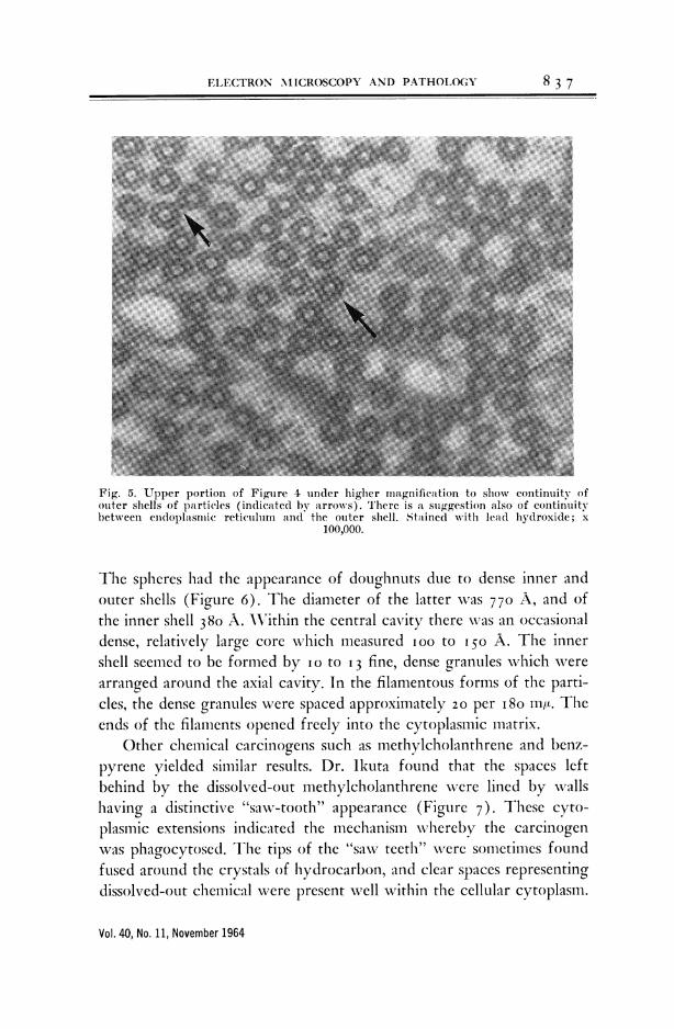
Fig. 5. Upper portion of Figure 4 under higher magnification to show continuity ofouter shells of particles (indicated by arrows). There is at suggestion also of continuitybetween enidoplhsmic reticuluim and the outer shell. Stained with lead hydroxide; x
100,000.
The spheres had the appearance of doughnuts due to dense inner andouter shells (Figure 6). The diameter of the latter was 77o A, and ofthe inner shell 380 A. \Vcithin the central cavity there wvas an occasionaldense, relatively large core which measured i00 to 150 A. The innershell seemed to be formed by io to I3 fine, dense granules which werearranged around the axial cavity. In the filamentous forms of the parti-cles, the dense granules were spaced approximately 20 per i8o nwt. Theends of the filaments opened freely into the cytoplasmic matrix.
Other chemical carcinogens such as methylcholanthrene and benz-pyrene yielded similar results. Dr. Ikuta found that the spaces leftbehind by the dissolved-out methylcholanthrene were lined by wallshaving a distinctive "saxv-tooth" appearance (Figure 7). These cyto-plasmic extensions indicated the mechanism whereby the carcinogenwas phagocytosed. The tips of the "sawv teeth" were sometimes foundfused around the crystals of hydrocarbon, and clear spaces representingdissolved-out chemical were present well within the cellular cytoplasm.
Vol. 40, No. 11, November 1964
8 3 7
83 H. M.ZMMRA
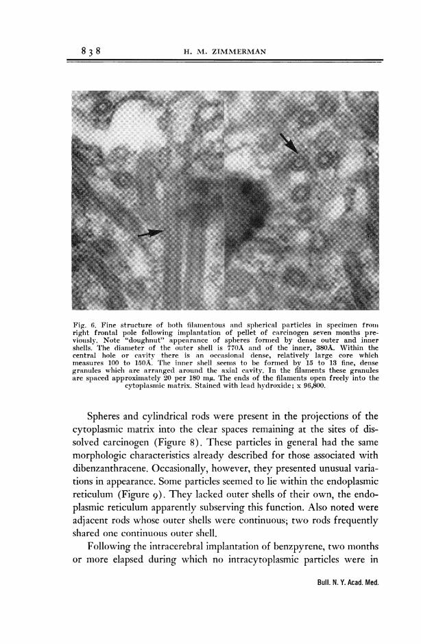
Fig. 6. Fine structure of both filamentous and spherical particles in specimen frolitright frontal pole following implantation of pellet of carcinogen seven months pre-viously. Note "doughnut" appearance of spheres formed by dense outer and innershells. The diameter of the outer shell is 770A and of the inner, 380A. Within thecentral hole or cavity there is an occasional dense, relatively large core whichmeasures 100 to 150A The inner shell seems to be formed by 15 to 13 fine, densegranules which are arranged around the axial cavity. In the filaments these granulesare spaced approximately 20 per 180 mA. The ends of the filaments open freely into the
cytoplasmic matrix. Stained with lead hydroxide; x 96,800.
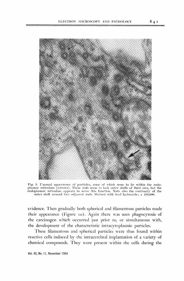
Spheres and cylindrical rods were present in the projections of thecytoplasmic matrix into the clear spaces remaining at the sites of dis-solved carcinogen (Figure 8). These particles in general had the samemorphologic characteristics already described for those associated withdibenzanthracene. Occasionally, however, they presented unusual varia-tions in appearance. Some particles seemed to lie within the endoplasmicreticulum (Figure 9). They lacked outer shells of their own, the endo-plasmic reticulum apparently subserving this function. Also noted wereadjacent rods whose outer shells were continuous; two, rods frequentlyshared one continuous outer shell.
Following the intracerebral implantation of benzpyrene, two monthsor more elapsed during which no intracytoplasmic particles were in
Bull. N. Y. Acad. Med.
8 3 8 H. Al. ZIMMERMAN

ELECTRON \ 1 ICROSCOPY AND) PATHOLOCY 8 3 9
Fig. 7. Electron iuicro-graph showillg empl)ty spaces left by (dissolv e(liout Inethl l-cholanthrene crystals. Note "saw toothl" aippeiannce of walls lining the spaces; tilisillustrates the mechanism of phagocytosis of the carcinogen, as seen by the presence of
clear spaces within the cellular cytoplasm. Stained wvith lead hydroxide; x 90,000.

840 H. M. ZIMMERMAN~~~~~~~~~~~~~~~~~~~~~~~~~~~~~~~~~~~~~~~~~~~~~~~~~~~~~~~~~~~~~~~~~~~~~~~~~~~~~~~~~~~~~~~~~~~~~~~~~~~~~~~~~
Fig. 8. Spheres and cylindrical rods are present in the projections of cytoplalsnmic matrixinto the clear space remaining at the site of dissolved carcinogen. Stained with lead
hydroxide; x 21,500.
Bull. N. Y. Acad. Med.
840 H. Al. ZIMMERMAN
ELECTRON 7A I ICROSCOPY ANI) PATHOLOGY
Fig. 9. 'Unuisual appearrance of pa rticles, sOeIn of which seeni to lie within the enldo-llasmlic reticuluni (a rrows). These rods seem to lack outer shells of their own, but theendoplasinic retictilum appears to serve this function. Note aflso the continuity of the
outer shell around two adjacent ro(Is. Stained with lead hydroxid(e; x 100,000.
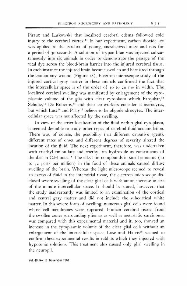
evidence. Then gradually both spherical and filamentous particles madetheir appearance (Figure Io). Again there was seen phagocytosis ofthe carcinogen which occurred just prior to, or simultaneous with,the development of the characteristic intracytoplasmic particles.
These filamentous and spherical particles were thus found withinreactive cells induced by the intracerebral implantation of a variety ofchemical compounds. They were present within the cells during the
Vol. 40, No. 11, November 1964
84 I

84 H.7.ZIMRA
Fig. 10. Numerous spherical and filamentous particles in the cytoplasmic matrix follow-ing the intracerelbral implantation of benzpyrene two months previously. Note theemls)ty Sl)fl(e left by the dissolved carcinogen. Stained with lead hydroxide; x 2,000.
period of the so-called precancerous stage and in most of the animalswith implanted pellets of carcinogen. They were not found within theglial cells which formned gliogenous neoplasms; at a point when theproliferating glial cells became malignant these particles seemed to dis-appear. XVhat their fate is in tunmor production still remains to bedetermined.
Now it is pertinent to inquire into the chemical nature of theseparticles. Blocks of brain tissue from sites of chemical implantationwere fixed in neutral fornmalin and some were then incubated in solu-tions of ribonuclease, others in deoxyribonuclease, trypsin or pepsinfor different lengths of time at 37'C. Following osmium tetroxidefixation, this material was prepared for electron microscopic study.Control specimens which had been incubated for 70 minutes in Veronal
Bull. N. Y. Acad. Med.
8 4 2 H. NI. ZENLIIVIERN/IANr

ELECTRON 'MICROSCOPY AND PATHOLOY 84
Fig. 11. Electron micrograph from specimen obtaiaed three months after dibenzanthra-cene inmIlantcation. The (control block wvas fixed in neutral formalin, then incubated for70 minutes in Veronal acetate buffered wvith mlaignesilumn sull)phate. The nuclear chrollia-tin (N) in the reactive (ell is still electron-dense, an(l (dense spherical p)arti(les (arrow)are also present near ain empty space (P) left by a dissolved carcinogen crystal.Ribosomaes, smaller than the p)articles, are also p)reseiJt aiid dense. Stained( with lead;
x 18,000.
acetate buffered xvith magnesium sulphate still showed the norinalnuclear electron density of the reactive cells (Figure ii). The intra-cytoplasmic spherical particles were still preserved and were found nearthe empty spaces left behind by the dissolved carcinogen crystals.Ribosomes wvere also present. \When the tissue was incubated for 70minutes in a solution of deoxyribonuclease (DNAse) in Veronal acetatebuffered with magnesium sulphate, the nuclear chromatin in the reac-tive cell was nearly completely digested, but the ribosomes and the
Vol. 40, No. 11, November 1964
843

84 .M IMRA
Fig. 12. Nuclear chromatin (N) of the reactive cell is nearly completely digested intissue incubated 70 minutes in a solution of DNAse in Veronal acetate buffer withmagnesium sulphate. Ribosomnes and particles (arrow) of normal density are unaffected.
Stained with lead; x 18,000.
particles were unaffected (Figure 12). Under higher magnification bothribosomes and central cores were found wvell preserved both in longi-tudinal and transverse views of the particles (Figures I3A and B). In-cubating the tissue for 40 minutes in a solution of ribonuclease [RNAse],on the other hand, produced almost complete digestion of both theaxial cores of the particles and the cytoplasmic ribosomes (Figuresi4A and B). Complete disappearance of these structures occurred after
Bull. N. Y. Acad. Med.
844 H. M. ZIAIMERMAN

ELECTRON NIICROSCOPY AND PATHOLOGY
Fig. 1M.A. Fig. 13B
Fig. 13A anmd 1B. The axial caivities of the p'articles, both inl longitudinial and tranisversesection contalin electron-(lelse cores (airrows). The rihosoIIes are also wsell preservelcandi electron-(lense, as aire the inner shells of the p)arti(les. Tissue treated with DNtse.
Stained wdith lead; x 100,000.
6o-minute incubation in RNAse. The inner shells of the filamentousrods also showed advanced digestion. Tissue incubated for 20 minutesin a solution of trypsin did not suffer any digestion of ribosomes, axialcores, or nuclear structures, but there was considerable digestion ofthe structure betxveen the inner and outer shells. Fifteen- to 20-minuteincubation of tissue in a solution of pepsin caused such destruction asto invalidate any interpretation.
From these findings it may be concluded wvith some measure ofsafety that the axial cores of the filamentous rods contain RNA but notDNA. This is also true of the fine granules which form the inner shells
Vol. 40, No. 11, November 1964
845

846 H. M. ZIMMERMAN
Fig. 14A Fig. 14B
Fig. 14A and B. Electron micrograph of tissue incubated for 40 minutes in a solutionof RNAse. Longitudinal and transverse sections of particles reveal an absence of dense
axial cores. Also absent are ribosomes. Stained with lead; x 100,000.
of the particles. Further, trypsin digestion of the structure between theinner and outer shells indicates its protein nature. The chemical com-position of these mysterious particles is at least not incompatible withtheir being viruses. There is, in addition, rather strong morphologicsupport for such a concept. This support comes from Bernhard's studyof tumor viruses4 and from Dalton's study of the Moloney agent inmouse leukemia.5 This agent, which is remarkably similar to our carci-nogen-induced particle, also has a double wall with continuity of theexternal membrane around two adjacent particles. It, too, is a filamen-tous hollow rod with an electron-dense central core. In size, however,it is smaller than our particle.
Much, of course, remains to be done in the further identificationof this chemically induced particle. Its precise role in experimentalglioma production is still to be determined. Why this intracytoplasmic
Bull. N. Y. Acad. Med.
846 H. M. ZIMMERMAN

ELECTRON MICROSCOPY AND PATHOLOGY
Fig. 15. Glioblastoma multiforme involving right medial orbital gyrus. Severe swellingof centrum semiovale with flattening of gyri and herniation of cingulate gyrus. The
swollen hemisphere was firm and relatively dry.
structure first appears after some weeks of exposure of the animal brainto a chemical carcinogen, persisting for some months during the pre-cancerous period, and then mysteriously disappears as the glial cellsproliferate in sufficient numbers to form a tumor, is still unknown. Itis yet to be shown that cell-free suspensions of these particles canthemselves produce gliomas. But considerable progress has been madein the study of cerebral tumorogenesis with the aid of the electronmicroscope after nearly twenty years of frustration.
CEREBRAL SWELLING AND EDEMA
There is yet another problem of considerable importance whichhas occupied the attention of pathologists for over fifty years withouta solution and which is only now being resolved on the ultrastructurallevel. This is the problem of cerebral swelling versus cerebral edema.
Vol. 40, No. 11, November 1964
847

848 H. M. ZIMMERMAN
Fig. 16. Swollen, wet brain of soft consistency whose cut surfaces dripped flu'd.Patient had extracranial malignant lymphoma with anemia and low plasma proteins.
Fig. 17. Perivascular and pericellular fluid accumulation in cerebral cortex of patientwho died in status epilepticus. Luxol-fast blue stain; x 250.
Bull. N. Y. Acad. Med.

ELECTRON MICROSCOPY AND PATHOLOGY
Most pathologists are familiar with the enlargements of cerebral hemi-spheres, affecting mainly the central white matter, which are the seatsof neoplasms, both primary and metastatic; such swellings are oftenvastly out of proportion to the size of the offending tumor (Figure 15).Similar firm and dry swollen brains have been noted in patients whosuccumbed to epileptic and paralytic attacks, and they have been seenin some patients with uremia and various infections. By contrast, thereare brains which are also increased in volume but which are soft andwater-logged, free fluid dripping from the cut surfaces (Figure i6).Such brains are frequently found in fatal cases of acute alcoholic intoxi-cation, in severe head injury, and with such poisoning as carbon mon-oxide, phosphorus and arsenic. They are also found in consequence ofserious disturbances in intracranial hemodynamics, such as may occurin chronic congestive heart disease.
To the firm, dry brain of increased volume, the term cerebral swell-ing has been applied, whereas cerebral edema connotes the enlarged,soft wet brain. Much has been written on the differential features ofthese two states. In general, the German school of pathologists repre-sented by Reichardt6 and Ziilch7 s hold that both macroscopically andpathogenetically these are separate and distinct conditions. Most otherpathologists hold with the view expressed by Greenfield et al.9 who state:"In cases of trauma, tumor or abscess some authors consider that brainswelling and brain oedema can both be present at the same time in thesame brain; and there is a further subdivision of opinion as to whether,in such cases, they can be separated microscopically, and whether cere-bral edema is but a more severe degree of the same process which maygive rise to cerebral swelling."
Conventional microscopic study of these conditions has contributedlittle to their understanding. Often the very process of preparation ofthe tissue for light microscopy, necessitating its dehydration precedingembedding for sectioning, removes what fluid accumulations there areas the basis for either condition. When traces of abnormal fluid accu-mulations survive the drastic artifacts of tissue preparation for histologicexamination, no sharp differences are noted between cerebral edemaand swelling. What is even more important is that little evidence sur-vives to indicate the circumstances surrounding these abnormal intra-cerebral fluid accumulations. In the cortical and central gray matter,evidence of excess fluid accumulation may sometimes be seen in cases
Vol. 40, No. 11, November 1964
849

H. \1. ZL\IMERMAN
Fig. 18. Swollen right cerebral hemisphere of mnouse following cold injury to cortex.Note staining with trypan blue in(licating increasedl perinefab)ility of the blood-brain
barrier.
of both swelling and edema in the form of clear paraneuronal andparaglyocytic spaces (Figure I7). There is also widening of the Vir-chow-Robin spaces. Both neuronal and glial cells appear shrunken andcompressed. \Vhat the condition of the intercellular stroma or neuropilis in regard to its fluid content it is impossible to judge. In the sub-cortical white matter and centrum semiovale, the perivascular spacesare widened and for variable distances around the vessels the myeli-nated fibers are separated and loosened. This may be due to abnormalfluid accumulation, but the appearance somewhat resembles patchydemyelination save for the fact that there is neither a macrophagicresponse nor sudanophilic myelin breakdown.
This wvas the approximate state of our knowledge regarding thisimportant subject up to i959 when my colleagues, Drs. Torack andTerry, and I undertook to study the fine structure of cerebral fluidaccumulation secondary to cold injury to the brain.'0 Recent investiga-tions of the normal central nervous system l)y means of electron micro-scopy had revealed that the cellular elements of the brain apparentlyfitted together so closely that the extracellular space was probably nogreater than two per cent."1-3 It had also been shown by Klatzo,
Bull. N. Y. Acad. Med.
8 5 o H. MX. ZXI.NINIERNIAN
F IFC(,rRON \tICROSCOPY AN)D P}ATHOI.O(Y85
Piraux and Laskowvski that localized cerebral edema followed coldinjury to the cerebral cortex.'4 In our experiment, carbon dioxide icewas applied to the cerebra of young, anesthetized mice and rats fora period of 30 seconds. A solution of trypan blue was injected subcu-taneously into six animals in order to demonstrate the passage of thevital dye across the blood-brain barrier into the injured cerebral tissue.In each instance the injured brain became swollen and herniated throughthe craniotomy wound (Figure i8). Electron microscopic study of theinjured cortical gray matter in these animals confirmed the fact thatthe intercellular space is of the order of i0 to 20 111m in width. Thelocalized cerebral swelling was manifested by enlargement of the cyto-plasmic volume of the glia with clear cytoplasm wvhiclh Farquhar,1"Schultz,'2 De Robertis,15 and their co-workers consider as astrocytes,but which Luse"' and Palay17 believe to be oligodendrocytes. The inter-cellular space was not affected by the swelling.
In view of the strict localization of the fluid within glial cytoplasm,it seemed desirable to study other types of cerebral fluid accumulation.There wvas, of course, the possibility that different causative a.gents,different rates of onset and different degrees of severity altered thelocation of the fluid. The next experiment, therefore, -was undertaikenwith triethyl tin sulfate and triethyl tin hydroxide as constituents ofthe diet in CGH mice.'8 The alkyl tin compounds in small amounts (I 2to 32 parts per million) in the food of these animals caised diffuseswelling of the brain. Whereas the light microscope seemed to revealan excess of fluid in the interstitial tissue, the electron microscope dis-closed severe swelling of the clear glial cells without an increase in sizeof the minute intercellular space. It should be stated, however, thatthe study inadvertently was limited to an examination of the corticaland central gray matter and did not include the subcortical whitematter. In this severe form of swelling, numerous glial cells were foundwhose cell membranes were ruptured. Human cerebral tissue, fromthe swollen zones surrounding glionmas as well as metastatic carcinoma,was compared with this experimental material and it, too, showed anincrease in the cytoplasmic volunme of the clear glial cells without anenlargement of the intercellular space. Luse and Harris'" seemed toconfirm these experimental results in rabbits which they illited vithhypotonic solutions. This treatment also caused only glial swellina inthe neuropil.
Vol. 40, No. 11, November 1964
8 5 I
ZIMMFRMAN
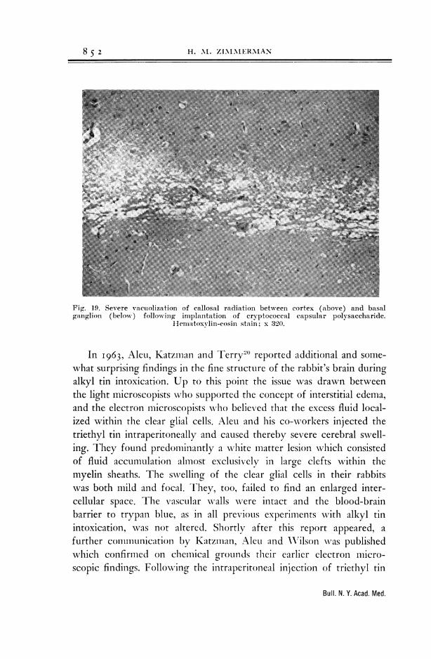
Fig. 19. Severe vacuiolization of callosal radiation between cortex (above) and basalganglion (below) following implantation of cryptococcal capsular polysaccharide.
llematoxvlin-eosin stain; x 320.
In i963, Aleu, Katzman and Terry20 reported additional and some-what surprising findings in the fine structure of the rabbit's brain duringalkyl tin intoxication. Up to this point the issue was drawn betweenthe light microscopists who supported the concept of interstitial edema,and the electron microscopists who believed that the excess fluid local-ized within the clear glial cells. Aleu and his co-workers injected thetriethyl tin intraperitoneally and caused thereby severe cerebral swell-ing. They found predominantly a white matter lesion which consistedof fluid accumulation almost exclusively in large clefts within themyelin sheaths. The swelling of the clear glial cells in their rabbitswas both mild and focal. They, too, failed to find an enlarged inter-cellular space. The vascular walls wvere intact and the blood-brainbarrier to trypan blue, as in all previous experiments with alkyl tinintoxication, was not altered. Shortly after this report appeared, afurther communication by Katzman, AleuL and A\Vilson was publishedwhich confirmed on chemical grounds their earlier electron micro-scopic findings. Following the intraperitoneal injection of triethyl tin
Bull. N. Y. Acad. Med.
8 5 2 H. M. Z11MINIERMAN

LiF(crRQ}N \ MICROSCOPY AND PATHOLOGY
Fig. 20. Iow-poxver electron micrograph to show irregular separation of neural tissueby electron-dense fluid in callosal ra(liation. Stained with uranyl acetate solution; x 4,000.
in rabbits, the water content of the white matter increased by 9i percent compared to the dry weight. The four-hour S3 sulfate space, ameasure of the extraccllular space, remained at the normal level of 2.6per cent. The 24-h1our albumin I131 space also remained at the normallevel of less than 2 per cent. From these and other chemical findingsthe conclusion was reached that the excess fluid accumulation wvas notcxtracellUlar, but was Iocullated w\ithin intranmyelinic spaces.
Evidencc had been tproduced earlier by Evans, Tani and Raimondil
Vol. 40, No. 11, November 1964
8 5 3

85 .M IMRA
Fig. 21. Two adjacent myelin sheaths and surrounding electron-dense, reticulated fluid.There is no limiting membrane to demarcate the fluid, which is in contact with the
outermost myelin lamellae. x 113,000.
that the swelling in the white matter caused by pressure on the brainexerted by an inflated extradural balloon was the result of separationof perivascular glial cells from the basement membrane of the vessels.This was proof that under the conditions of the experiment interstitialfluid accumulation did occur in perivascular regions.
It was apparent from all the previous experiments that the completepicture of intracerebral fluid accumulation was not yet at hand. An-other study was undertaken at this point by my associates, Levine and
Bull. N. Y. Acad. Med.
854 H. MS. ZIMMERMAN
ELECTRON MICROSCOPY AND PATHOLOIC5
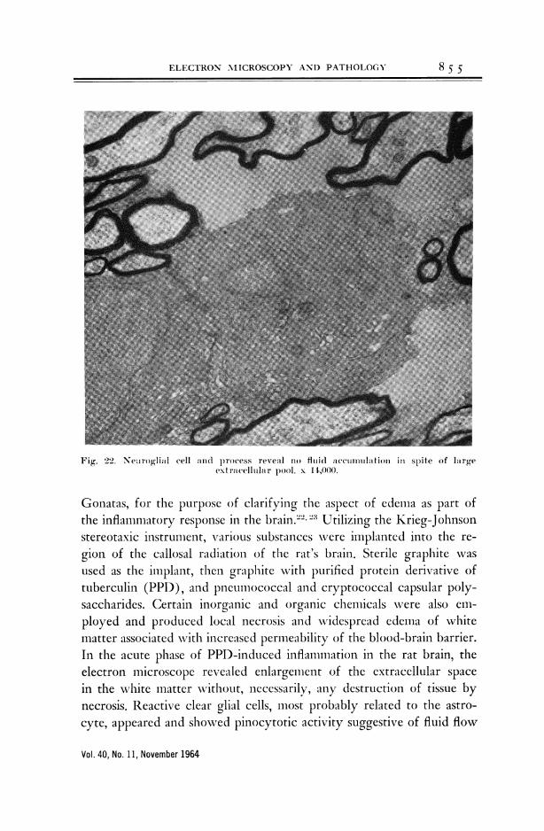
Fig. 22. Nearoglial cell and p)rocess reveal no fluid accumuanlation in spite of largeextracellular pool. x 14,000.
Gonatas, for the purpose of clarifying the aspect of edema as part ofthe inflamnmatory response in the brain."' Utilizing the Krieg-Johnsonstereotaxic instrunment, various substances were implanted into the re-gion of the callosal radiation of the rat's brain. Sterile graphite wasused as the implant, then graphite with purified protein derivative oftuberculin (PPD), and pneumococcal and cryptococcal capsular poly-saccharides. Certain inorganic and organic chemicals wvere also em-ployed and produced local necrosis and widespread edema of whitematter associated with increased permeability of the blood-brain barrier.In the acute phase of PPD-induced inflammation in the rat brain, theelectron microscope revealed enlargement of the extracellular spacein the white matter without, necessarily, any destruction of tissue bynecrosis. Reactive clear glial cells, most probably related to the astro-cyte, appeared and showed pinocytotic activity suggestive of fluid flow
Vol. 40, No. 11, November 1964
8 5 5

856 H. M. ZIMMERMAN~~~~~~~~~~~~~~~~~~~~~~~~~~~~~~~~~~~~~~~~~~~~~~~~~~~~~
Fig. 2.3. Callosal radiaition 18 hours after implantation of p)olysacclinri(le. Astrocvticvascular feet surround basement nmemibraue, sepa rating intercellular fluid front vessel
wadll. x 15,000.
from the extracCIlLlar space into the cell. 71There was also evidence ofphagocytosis of myelin, predominantly by the mnacrophage but also,occasionally, by the reactive clear glial cell. The fluid distribution inPPD-induced edema was studied by Katzman, Gonatas and Levine.'These investigators found that the Geigy blue-stained areas of grayand white matter, which represent the sites of edema and breakdoxnof the blood-brain barrier, showed a greater accumulation of sodiumion and a more rapid uptake of s"04 and albumin I's' than the un-stained areas and the gray and white matter of the brains of controlanimals. In comparing these findings in PPD-induced edema with otherforms of edema, they concluded that the edematous white matter wvascorrelated with the development of a true extracellular space. In ede-
Bull. N. Y. Acad. Med.
8 5 6 H. .N4. ZLNUNIER.AIANN

ELECTRON ]MICROSCOPY AND PATHOLOGY
11iw24\~~~~~~~~~~~~~i4
Fig. 2111
Fig. 24A and B. FIlid acculaeiiiiltion nonitainiivr cryptococcal polysaccharide around asmall vessel in cerebr l tissue nealr the iiipl tnit twelve hours after implantation.
Compaire density of fluid before and ifter staoiniiii wvith uranvli acetate. x 7,000.
matous gray matter, however, the only ultrastructural change observedwas a swelling of the clear glial cells.
More recently Hirano, Levine and I followed the course of spreadin the extracellular space of the cryptococcal polysaccharide duringthe acute stage.2' Following the implantation of the capsular polysac-charide in the anterior end of the callosal radiation of the rat brain,there appeared severe vacuolation of the white matter in light micro-scopic preparations (Figure I9). Under the electron microscope, by
Vol. 40, No. 11, November 1964
8 5 7

8 5 8 H. Al. ZIMMERMAN
Fig. 25. Pools of extracellular fluid adjacent to small vessel near implant site 24 hourspreviously. A. Polysaccharide-containing fluid. B. Plasma-like fluid. Arrow points togap in perivascular ring of astrocytic feet. Extracellular fluid communicates withmaterial in basement membrane of vessel wall. Stained with lead hydroxide; x 22,000.
Insert x 46,000.
Bull. N. Y. Acad. Med.

ELECTRON MICROSCOPY AND PATHOLOGY
contrast, Dr. Hirano found no optically empty spaces except for thelumens of vessels distended by the perfusion of the fixative materials.The cerebral tissue elements were irregularly separated by fluid (Figure20). The myelinated nerve fibers were separated either as groups orindividually by this electron-dense fluid. In all portions of the callosalradiation, and at all the intervals of time covered in this study of theacute phase, most of the fluid was located in the extracellular space(Figure 2 i). It was in contact with the outermost layer of the myeli-nated nerve fibers or with the plasma membrane of the glial or neuronalcell processes. There was no splitting or separation of the myelin lamellaeto form intramyelinic cysts for the harboring of fluid. The neuroglialbodies and processes in the xvhite matter revealed no accumulation offluid within their cytoplasm (Figure 22). Individual cells were oftenwvidely separated from each other by large pools of fluid. Astrocyticfoot processes kept their normal anatomic relationship to the vasculart)asement membrane; separation of the perivascular astrocyte from thebasement membrane of the blood vessel wvas not observed (Figure 23).Preservation of the relationship of endothelial cell, basement membraneand astrocytic foot process was considered to be important becausethe lesion under study had exhibited little alteration of vascular perme-abIility. Extracellular fluid in the cortical neuropil was usually limitedto the junction area between cortex and subcortical white matter; itwas not seen in superficial layers of the cortex. The opposite cerebralhemisphere, including the corpus callosum, was entirely normal.
Certain of these findings were difficult to interpret, the most impor-tant of these being the source of the intercellular fluid in the absenceof an anatomic alteration in the blood vessel wall and its attached glialprocesses. The problem was soon clarified, however, upon furtherstudy.26 Electron micrography revealed that the fluid resulting frompolysaccharide implantation in the corpus callosum was not homogene-ous, but consisted of two types. One, which contained a reticulatedelectron-dense substance, was found to contain the actual implantedpolysaccharide; the other, of less dense appearance, was derived fromthe blood vessels at the site of implantation. At this site, the crypto-coccal polysaccharide could be identified by the presence in the edemafluid of a uniformly distributed reticular substance that stained stronglywith uranyl acetate (Figures 24A and B). The nonreticulated lessdense fluid resembled blood plasma. The two types of fluid differed
Vol. 40, No. 11, November 1964
8 5 9

86o H. M. ZIMMERMAN
Fig. 26. Intercellular fluid near the margin of a cryptococcal polysaccharide implant.Electron-dense fluid (A) is surrounded by less electron-dense fluid (B). The latter is
connected with material in the vascular basement membrane (arrow). x 24,000.
Bull. N. Y. Acad. Med.

ELECTRON MICROSCOPY AND PATHOLOGY 8 6 i
in location as well as in appearance. \Vhen together, the polysaccharide-containing fluid was always in the center and the plasma-like fluid waslocated between the former and the tissue cells. The plasma-like fluidoccurred in the vicinity of blood vessels near the site of implantation.In these regions, separation of perivascular astrocytic foot processesoccurred and plasma-like fluid communicated with the vascular base-ment membrane through gaps between foot processes (Figure 25).Disruption of the astrocytic basement membrane permitted continuitybetween the plasma-like fluid and the basement membrane (Figure 26).These changes were observed only in regions close to the implant andonly during the early stages of the reaction. They were not seen inthe distal portions of the callosal radiation. The differentiation of thetwo types of fluid was clear between 6 and I2 hours after implantation,was less clear after 24 to 35 hours, and was absent after 48 hours. Evi-dently this amount of time wvas necessary for their complete miscibility.
From all the electron microscopic studies on abnormal fluid accu-mulations in the brain wve have learned a number of facts. One is thatdifferent types of injury can produce different responses as regardsfluid localization. In the cortex this is manifested by swelling of theclear glial cells wvith no increase in the intercellular space. Certain inju-ries to white matter are characterized by intramyelinic splitting withfluid accumulation in the cystic spaces thus created. Other injuries resultin vast collections of extracellular fluid sometimes with, but often with-out, simultaneous swelling of the glia. Thus it would appear that thereis an ultramicroscopic difference between cerebral swelling and edema.Confirmation of this is yet to be made in man.
ACKNOWLEDGIMENTS
I wish to acknowledge my indebtedness to my associates, past andpresent, whose work in large measure has made this report possible:Nickos K. Gonatas, Asao Hirano, Fusahiro Ikuta, Seymour Levine,Robert D. Terry, and Richard M. Torack.
REFERENCES
1. Cohen, B. A man who looked to the
stars. N. Y. Times, Feb. 9, 1964.
2. Zimimerman, H. M. and Arnold, II.Experimental brain tumors: tumors
produced with methylicholanthrene, Can-cer Res. 1:919-38, 1941.
3. Zinmmerman, H. M. Nature of glioinas-as revealed by atinmal experimentation,Amer. J. Path. 31:1-29, 1955.
Vol. 40, No. 11, November 1964

86 2 H. M. ZIMMERMAN
4. Bernhard, W. Detection and study oftumor viruses with the electron micro-scope, Cancer Res. 20:712-27, 1960.
5. Dalton, A. J. The Moloney agent. InTumors Induced by Viruses: Ultra-structural Studies. A. J. Dalton and F.Haguenau, eds. New York and London,Academic Press, 1962, pp. 207-18.
6. Reichardt, M. Das Hirnodem. Anhang:Die Hirnschwellung. In Handbuch derspeziellen pathologischen Anatomie undHistologie. Erkrankungen des zentralenNervensystems. Berlin, Gottingen, Hei-delberg, Springer Verl. 1957, pp. 1229-83.
7. Zulch, K. J. Hirnodem und Hirn-schwellung, Virchow Arch. Path. Anat.310:1-58, 1943.
8. Zulch, K. J. Discussion sur le proble'nede l'4d6me cerebrate dans la productiondes troubles mentales, V. Int. Neurol.Cong., Lisbon, 3:42, 1954.
9. Greenfield, J. G. and others, Neuro-pathology. London, E. Arnold, Ltd.1958, p. 103.
10. Torack, R. M., Terry, R. D. and Zim-merman, H. M. Fine structure of cere-bral fluid accumulation. I. Swellingsecondary to cold injury, Amer. J.Path. 35:1135-47, 1959.
11. Farquhar, M. G. and Hartman, J. F.Neuroglial structure and relationshipsas revealed by electron microscopy, J.Neuropath. Exp. Neurol. 16:18-39, 1957.
12. Schultz, R. L., Maynard, E. A. andPease, D. C. Electron microscopy ofneurons and neuroglia of cerebral cor-tex and corpus callosum, Amer. J.Anat. 100:369-407, 1957.
13. Maynard, E. A., Schultz, R. L. andPease, D. C. Electron microscopy ofthe vascular bed of rat cerebral cortex,Amer. J. Anat. 100:409-33, 1957.
14. Klatzo, I., Piraux, A. and Laskowski,E. J. Relationship between edema,blood-brain-barrier and tissue elementsin a local brain injury, J. Neuropath.Exp. Neurol. 17:548-64, 1956.
15. DeRobertis, E., Gerschenfeld, H. M.and Wald, F. Cellular mechanism ofmyelination in the central nervous sys-tem, J. Biophys. Biochem. Cytol. 4:651-56, 1958.
16. Luse, S. A. Electron microscopic ob-servations of the central nervous sys-tem, J. Biophys. Biochem. Cytol. 2:531-42, 1956.
17. Palay, S. L. Electron microscopicalstudy of neuroglia. In Biology ofNeuroglia. W. F. Windle, ed. Spring-field, C. C Thomas, 1958, pp. 24-28.
18. Torack, R. M., Terry, R. D. and Zim-merman, H. M. Fine structure of cere-bral fluid accumulation. II. Swellingproduced by triethyl tin poisoning andits comparison with that in the humanbrain, Amer. J. Path. 36:273-87, 1960.
19. Luse, S. A. and Harris, B. Electronmicroscopy of the brain in experimentaledema, J. Neurosurg. 17:439-46, 1960.
20. Aleu, F., Katzman, R. and Terry, R. D.Fine structure and electrolyte analysesof cerebral edema induced by alkyl tinintoxication, J. Neuropath. Exp. Neurol.22:403-13, 1963.
21. Evans, J. P., Tani, E. and Raimondi,A. J. Electron microscopic study ofbrain swelling. In Trans. Amer. Neurol.Ass. New York, Springer Pub. Co.,1961, pp. 28-29.
22. Levine, S., Zimmerman, H. M., Wenk,E. J. and Gonatas, N. K. Experimentalleukoencephalopathies due to implanta-tion of foreign substances, Amer. J.Path. 42:97-117, 1963.
23. Gonatas, N. K., Zimmerman, H. M. andLevine, S. Ultrastructure of inflamma-tion with edema in the rat brain, Amer.J. Path. 42:455-69, 1963.
24. Katzman, R., Gonatas, N. and Levine,S. Electrolyte and fluid distribution inexperimental inflammatory leukoence-phlalopathy, Arch. Neurol. 10:58-65, 1964.
25. Hirano, A., Zimmerman, H. M. andLevine, S. Fine structure of cerebralfluid accumulation. III. Extracellularspread of cryptococcal polysaccharidesin the acute stage, Amer. J. Path. 45:1-19, 1964.
26. Hirano, A., Zimmerman, H. M. andLevine, S. Fine structure of cerebralfluid accumulation. IV. On the natureand origin of extracellular fluids fol-lowing cryptococcal polysaccharides im-plamntation. 4mer. J. Path. 45(2) :195-207, 1964.
Bull. N. Y. Acad. Med.